

The Consumption of the Fibrous Fraction of Solanum lycocarpum St. Hil. Does Not Preserve the Intestinal Mucosa in TNBS-Induced Rats
 ,
,  , , ,
, , ,  and
and
Abstract
1. Introduction
2. Materials and Methods
2.1. Fruta-Do-Lobo Resistant Starch and Fibrous Fraction Extraction
resistant starch)
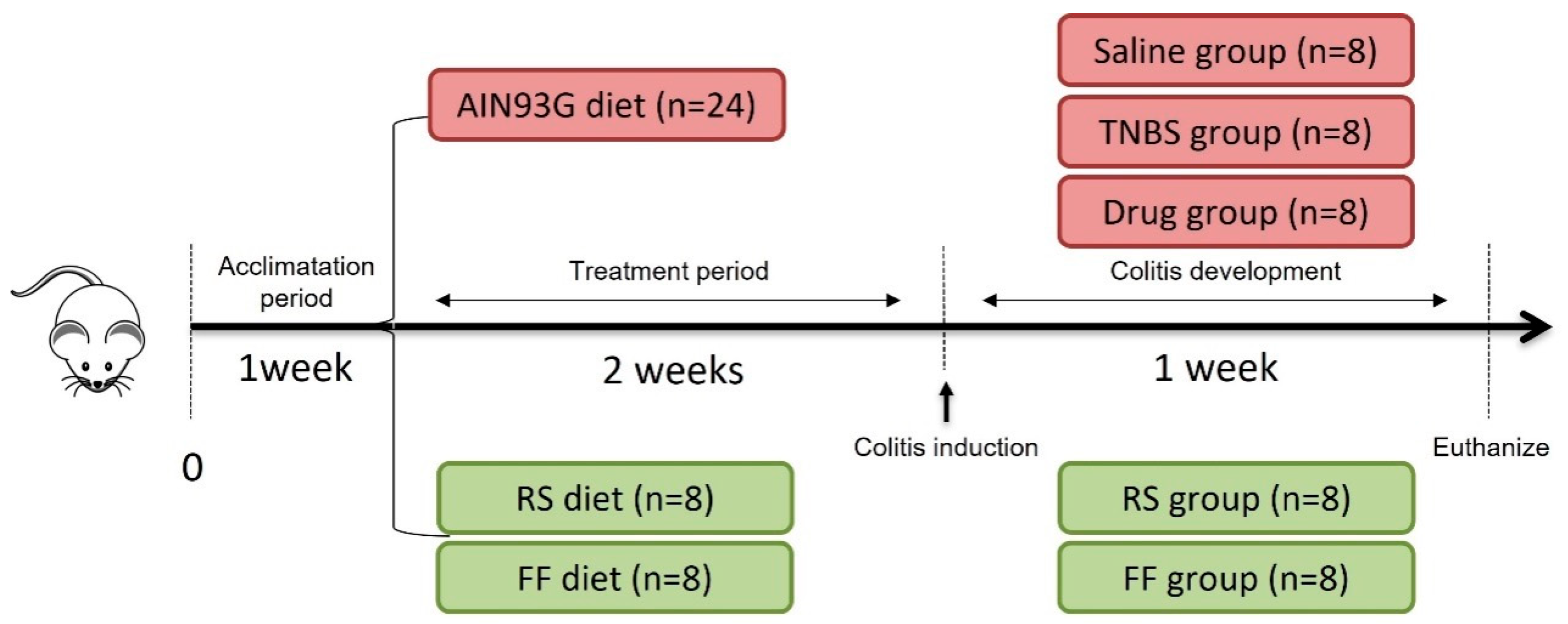
2.2. In Vivo Experimental Protocol
2.3. Tissue Sampling and Analysis
2.4. Fecal Analyses
2.5. Statistical Analyses
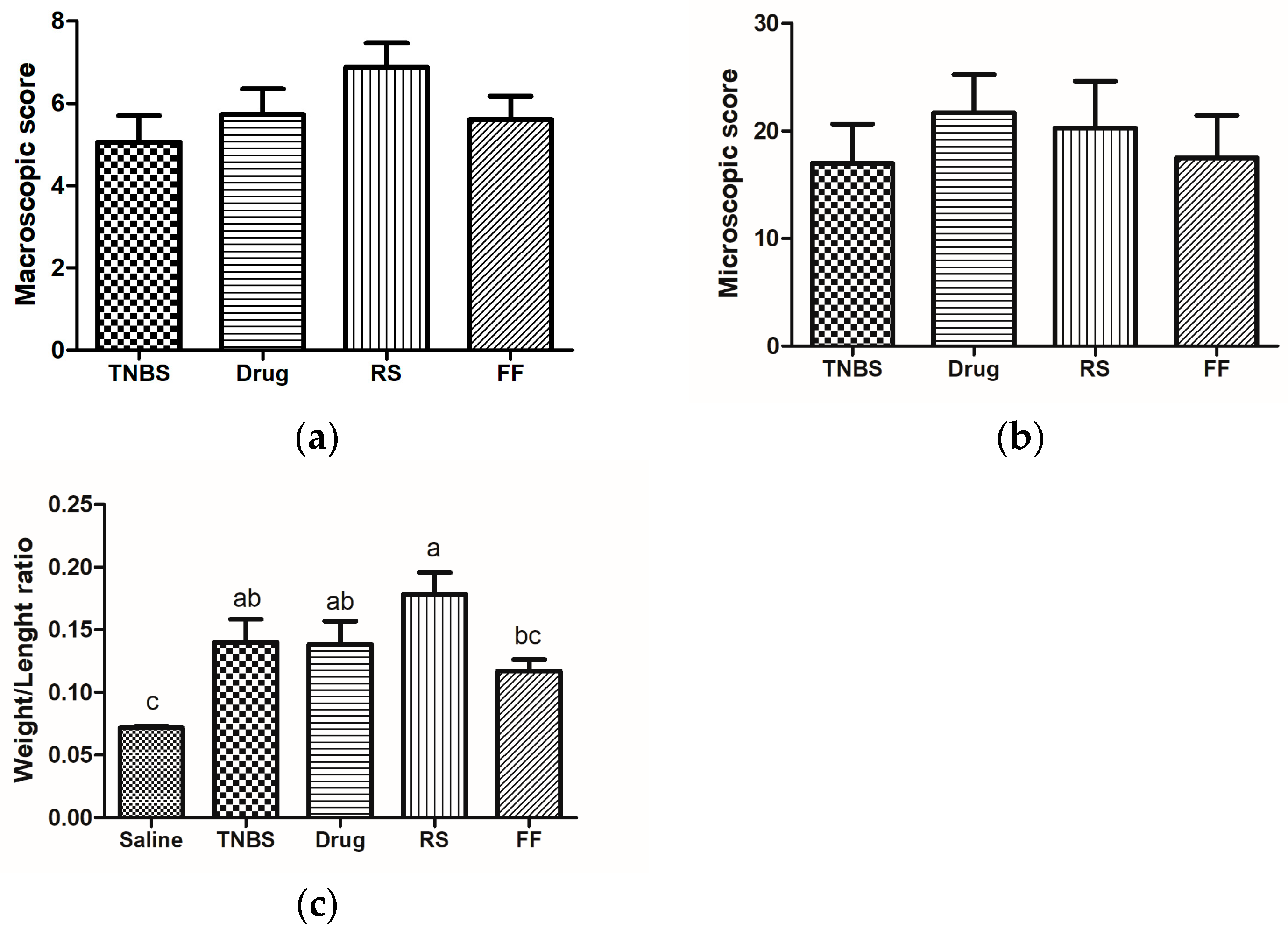
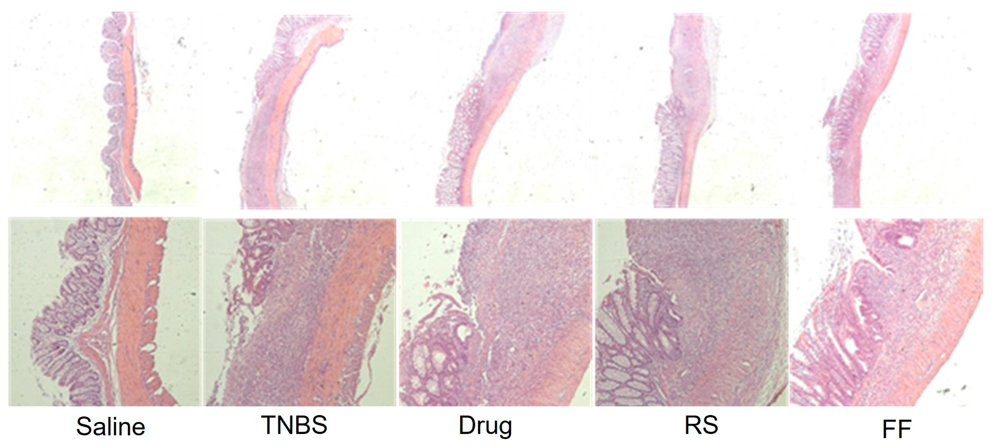
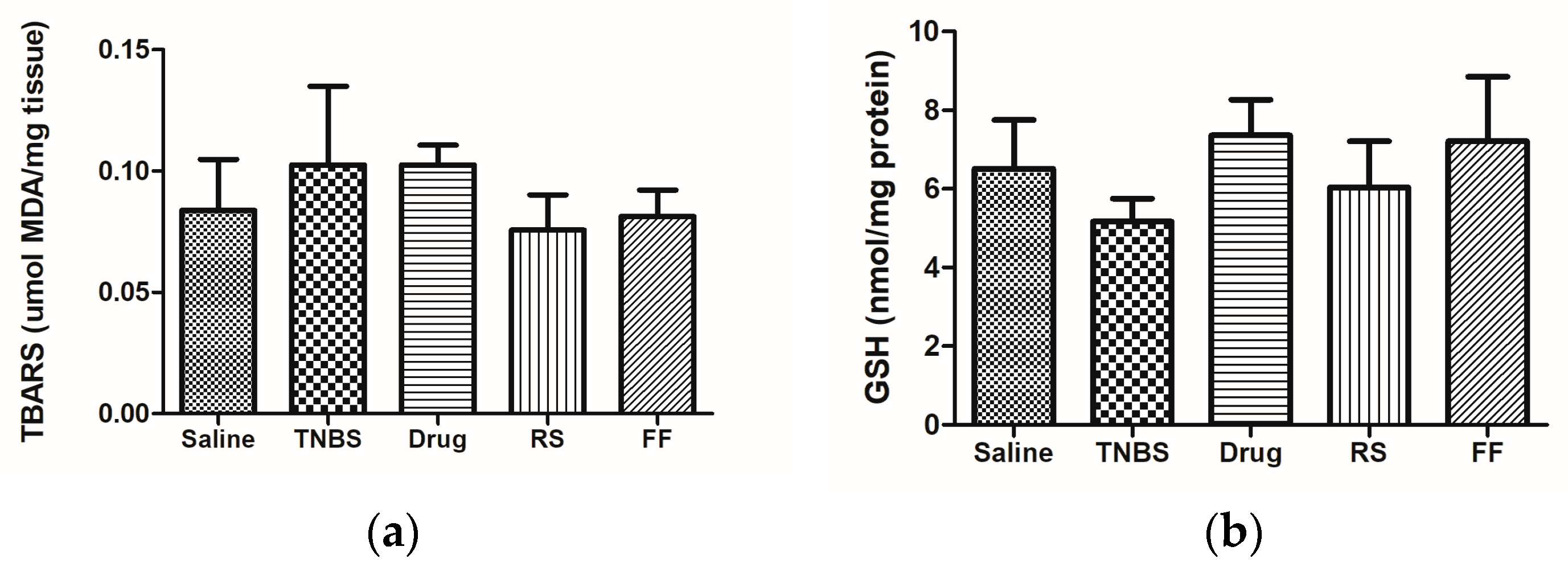
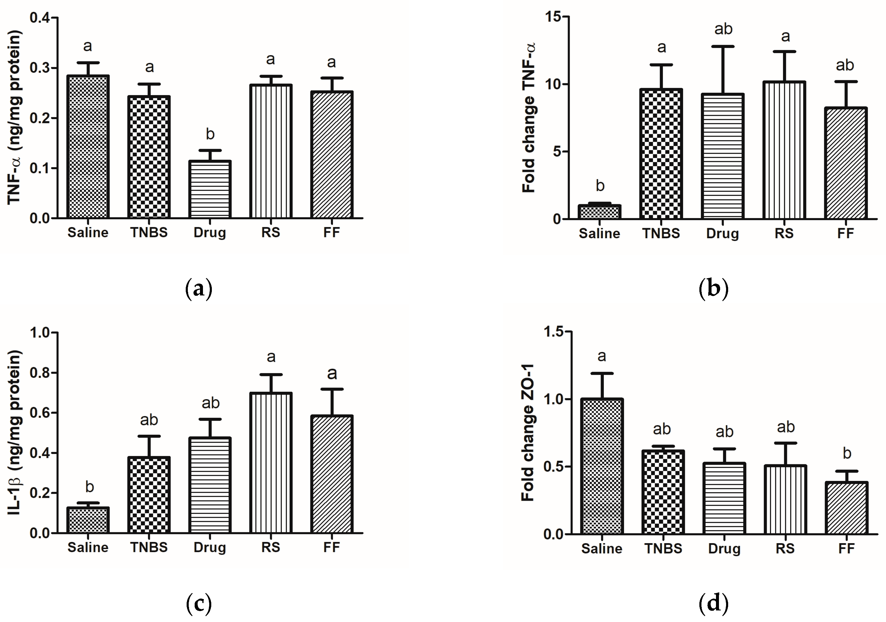
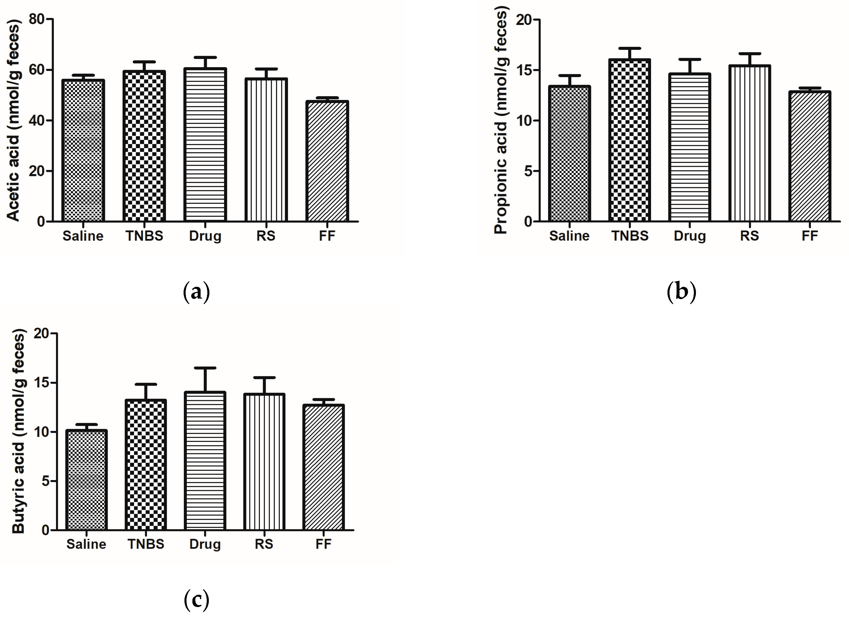
3. Results and Discussion
4. Conclusions
Author Contributions
Funding
Institutional Review Board Statement
Informed Consent Statement
Data Availability Statement
Conflicts of Interest
References
- Fucilini, L.M.P.; Genaro, L.M.; Sousa, D.C.e.; Coy, C.S.R.; Leal, R.F.; Ayrizono, M.d.L.S. Epidemiological profile and clinical characteristics of inflammatory bowel diseases in a brazilian referral center. Arq. Gastroenterol. 2021, 58, 483–490. [Google Scholar] [CrossRef] [PubMed]
- Nascimento, R.P.D.; Machado, A.; Galvez, J.; Cazarin, C.B.B.; Maróstica Junior, M.R. Ulcerative colitis: Gut microbiota, immunopathogenesis and application of natural products in animal models. Life Sci. 2020, 258, 118129. [Google Scholar] [CrossRef] [PubMed]
- Loubet Filho, P.S.; Dias, T.O.; Reis, V.H.d.O.T.; Moya, A.M.T.M.; Santos, E.F.d.; Cazarin, C.B.B. Feed your gut: Functional food to improve the pathophysiology of inflammatory bowel disease. J. Funct. Foods 2022, 93, 105073. [Google Scholar] [CrossRef]
- Dulai, P.S.; Jairath, V.; Khanna, R.; Ma, C.; McCarrier, K.P.; Martin, M.L.; Parker, C.E.; Morris, J.; Feagan, B.G.; Sandborn, W.J. Development of the symptoms and impacts questionnaire for Crohn’s disease and ulcerative colitis. Aliment. Pharmacol. Ther. 2020, 51, 1047–1066. [Google Scholar] [CrossRef]
- Yu, Y.R.; Rodriguez, J.R. Clinical presentation of Crohn’s, ulcerative colitis, and indeterminate colitis: Symptoms, extraintestinal manifestations, and disease phenotypes. Semin. Pediatr. Surg. 2017, 26, 349–355. [Google Scholar] [CrossRef] [PubMed]
- König, J.; Wells, J.; Cani, P.D.; García-Ródenas, C.L.; MacDonald, T.; Mercenier, A.; Whyte, J.; Troost, F.; Brummer, R.J. Human intestinal barrier function in health and disease. Clin. Transl. Gastroenterol. 2016, 7, e196. [Google Scholar] [CrossRef]
- Capaldo, C.T.; Powell, D.N.; Kalman, D. Layered defense: How mucus and tight junctions seal the intestinal barrier. J. Mol. Med. 2017, 95, 927–934. [Google Scholar] [CrossRef]
- Chelakkot, C.; Ghim, J.; Ryu, S.H. Mechanisms regulating intestinal barrier integrity and its pathological implications. Exp. Mol. Med. 2018, 50, 1–9. [Google Scholar] [CrossRef]
- Nishida, A.; Inoue, R.; Inatomi, O.; Bamba, S.; Naito, Y.; Andoh, A. Gut microbiota in the pathogenesis of inflammatory bowel disease. Clin. J. Gastroenterol. 2018, 11, 1–10. [Google Scholar] [CrossRef]
- Vasconcelos, R.S.; Rocha, R.M.; Souza, E.B.; Amaral, V.R.S. Life quality of patients with inflammatory bowel disease: Integrative review. Estima-Braz. J. Enterostomal Ther. 2018, 16, e2118. [Google Scholar] [CrossRef]
- Soliman, G.A. Dietary fiber, atherosclerosis, and cardiovascular disease. Nutrients 2019, 11, 1155. [Google Scholar] [CrossRef]
- Opstelten, J.L.; de Vries, J.H.M.; Wools, A.; Siersema, P.D.; Oldenburg, B.; Witteman, B.J.M. Dietary intake of patients with inflammatory bowel disease: A comparison with individuals from a general population and associations with relapse. Clin. Nutr. 2019, 38, 1892–1898. [Google Scholar] [CrossRef]
- Armet, A.M.; Deehan, E.C.; Thöne, J.V.; Hewko, S.J.; Walter, J. The effect of isolated and synthetic dietary fibers on markers of metabolic diseases in human intervention studies: A systematic review. Adv. Nutr. 2020, 11, 420–438. [Google Scholar] [CrossRef] [PubMed]
- Swann, O.G.; Kilpatrick, M.; Breslin, M.; Oddy, W.H. Dietary fiber and its associations with depression and inflammation. Nutr. Rev. 2020, 78, 394–411. [Google Scholar] [CrossRef] [PubMed]
- Makki, K.; Deehan, E.C.; Walter, J.; Bäckhed, F. The impact of dietary fiber on gut microbiota in host health and disease. Cell Host Microbe 2018, 23, 705–715. [Google Scholar] [CrossRef] [PubMed]
- Prasad, K.N.; Bondy, S.C. WITHDRAWN: Dietary fibers and their Fermented short-chain fatty acids in prevention of human diseases. Mech. Ageing Dev. 2018. [Google Scholar] [CrossRef]
- Silva, J.P.B.; Navegantes-Lima, K.C.; Oliveira, A.L.B.; Rodrigues, D.V.S.; Gaspar, S.L.F.; Monteiro, V.V.S.; Moura, D.P.; Monteiro, M.C. Protective mechanisms of butyrate on inflammatory bowel disease. Curr. Pharm. Des. 2018, 24, 4154–4166. [Google Scholar] [CrossRef]
- Bach Knudsen, K.E.; Lærke, H.N.; Hedemann, M.S.; Nielsen, T.S.; Ingerslev, A.K.; Gundelund Nielsen, D.S.; Theil, P.K.; Purup, S.; Hald, S.; Schioldan, A.G.; et al. Impact of diet-modulated butyrate production on intestinal barrier function and inflammation. Nutrients 2018, 10, 1499. [Google Scholar] [CrossRef]
- Salvi, P.S.; Cowles, R.A. Butyrate and the intestinal epithelium: Modulation of proliferation and inflammation in homeostasis and disease. Cells 2021, 10, 1775. [Google Scholar] [CrossRef]
- Couto, M.R.; Gonçalves, P.; Magro, F.; Martel, F. Microbiota-derived butyrate regulates intestinal inflammation: Focus on inflammatory bowel disease. Pharmacol. Res. 2020, 159, 104947. [Google Scholar] [CrossRef]
- Clerici, M.T.P.S.; Kallmann, C.; Gaspi, F.O.G.; Morgano, M.A.; Martinez-Bustos, F.; Chang, Y.K. Physical, chemical and technological characteristics of Solanum lycocarpum A. St.-HILL (Solanaceae) fruit flour and starch. Food Res. Int. 2011, 44, 2143–2150. [Google Scholar] [CrossRef]
- Farina, F.; Piassi, F.G.; Moysés, M.R.; Bazzolli, D.M.; Bissoli Nde, S. Glycemic and urinary volume responses in diabetic mellitus rats treated with Solanum lycocarpum. Appl. Physiol. Nutr. Metab. 2010, 35, 40–44. [Google Scholar] [CrossRef] [PubMed]
- Perez, A.C.; Franca, V.; Daldegan, V.M., Jr.; Duarte, I.D. Effect of Solanum lycocarpum St. Hill on various haematological parameters in diabetic rats. J. Ethnopharmacol. 2006, 106, 442–444. [Google Scholar] [CrossRef]
- Vieira, G., Jr.; Ferreira, P.M.; Matos, L.G.; Ferreira, E.C.; Rodovalho, W.; Ferri, P.H.; Ferreira, H.D.; Costa, E.A. Anti-inflammatory effect of Solanum lycocarpum fruits. Phytother. Res. 2003, 17, 892–896. [Google Scholar] [CrossRef]
- Pereira, A.P.A.; Angolini, C.F.F.; Paulino, B.N.; Lauretti, L.B.C.; Orlando, E.A.; Silva, J.G.S.; Neri-Numa, I.A.; Souza, J.; Pallone, J.A.L.; Eberlin, M.N.; et al. A comprehensive characterization of Solanum lycocarpum St. Hill and Solanum oocarpum Sendtn: Chemical composition and antioxidant properties. Food Res. Int. 2019, 124, 61–69. [Google Scholar] [CrossRef]
- Han, Y.; Xiao, H. Whole food-based approaches to modulating gut microbiota and associated diseases. Annu. Rev. Food Sci. Technol. 2020, 11, 119–143. [Google Scholar] [CrossRef] [PubMed]
- Manichanh, C.; Borruel, N.; Casellas, F.; Guarner, F. The gut microbiota in IBD. Nat. Rev. Gastroenterol. Hepatol. 2012, 9, 599–608. [Google Scholar] [CrossRef]
- AOAC. Official Method of Analysis: Association of Analytical Chemists, 19th ed.; AOAC: Washington, DC, USA, 2012. [Google Scholar]
- Bligh, E.G.; Dyer, W.J. A rapid method of total lipid extraction and purification. Can. J. Biochem. Physiol. 1959, 37, 911–917. [Google Scholar] [CrossRef]
- Prosky, L.; Asp, N.G.; Schweizer, T.F.; DeVries, J.W.; Furda, I. Determination of insoluble, soluble, and total dietary fiber in foods and food products: Interlaboratory study. J. Assoc. Off. Anal. Chem. 1988, 71, 1017–1023. [Google Scholar] [CrossRef]
- Reeves, P.G. Components of the AIN-93 diets as improvements in the AIN-76A diet. J. Nutr. 1997, 127, 838s–841s. [Google Scholar] [CrossRef]
- Morris, G.P.; Beck, P.L.; Herridge, M.S.; Depew, W.T.; Szewczuk, M.R.; Wallace, J.L. Hapten-induced model of chronic inflammation and ulceration in the rat colon. Gastroenterology 1989, 96, 795–803. [Google Scholar] [CrossRef] [PubMed]
- Bell, C.J.; Gall, D.G.; Wallace, J.L. Disruption of colonic electrolyte transport in experimental colitis. Am. J. Physiol. 1995, 268, G622–G630. [Google Scholar] [CrossRef] [PubMed]
- Krause, P.; Zahner, S.P.; Kim, G.; Shaikh, R.B.; Steinberg, M.W.; Kronenberg, M. The tumor necrosis factor family member TNFSF14 (LIGHT) is required for resolution of intestinal inflammation in mice. Gastroenterology 2014, 146, 1752–1762.e1754. [Google Scholar] [CrossRef] [PubMed]
- Bradford, M.M. A rapid and sensitive method for the quantitation of microgram quantities of protein utilizing the principle of protein-dye binding. Anal. Biochem. 1976, 72, 248–254. [Google Scholar] [CrossRef]
- Ohkawa, H.; Ohishi, N.; Yagi, K. Assay for lipid peroxides in animal tissues by thiobarbituric acid reaction. Anal. Biochem. 1979, 95, 351–358. [Google Scholar] [CrossRef] [PubMed]
- Ellman, G.L. Tissue sulfhydryl groups. Arch. Biochem. Biophys. 1959, 82, 70–77. [Google Scholar] [CrossRef]
- Zhao, G.; Nyman, M.; Jönsson, J.A. Rapid determination of short-chain fatty acids in colonic contents and faeces of humans and rats by acidified water-extraction and direct-injection gas chromatography. Biomed. Chromatogr. 2006, 20, 674–682. [Google Scholar] [CrossRef]
- Cui, J.; Lian, Y.; Zhao, C.; Du, H.; Han, Y.; Gao, W.; Xiao, H.; Zheng, J. Dietary fibers from fruits and vegetables and their health benefits via modulation of gut microbiota. Compr. Rev. Food Sci. Food Saf. 2019, 18, 1514–1532. [Google Scholar] [CrossRef]
- Dreher, M.L. Whole fruits and fruit fiber emerging health effects. Nutrients 2018, 10, 1833. [Google Scholar] [CrossRef]
- DeMartino, P.; Cockburn, D.W. Resistant starch: Impact on the gut microbiome and health. Curr. Opin. Biotechnol. 2020, 61, 66–71. [Google Scholar] [CrossRef]
- McKeown, N.M.; Fahey, G.C., Jr.; Slavin, J.; van der Kamp, J.W. Fibre intake for optimal health: How can healthcare professionals support people to reach dietary recommendations? Br. Med. J. 2022, 378, e054370. [Google Scholar] [CrossRef]
- Passos, M.d.C.F.; Takemoto, M.L.S.; Guedes, L.S. Patters of fiber intake among brazilian adults: Perceptions from an online nationwide survey. Arq. Gastroenterol. 2020, 57, 144–149. [Google Scholar] [CrossRef] [PubMed]
- Bernstein, C.N.; Fried, M.; Krabshuis, J.H.; Cohen, H.; Eliakim, R.; Fedail, S.; Gearry, R.; Goh, K.L.; Hamid, S.; Khan, A.G.; et al. World gastroenterology organization practice guidelines for the diagnosis and management of IBD in 2010. Inflamm. Bowel Dis. 2010, 16, 112–124. [Google Scholar] [CrossRef] [PubMed]
- Veronese, N.; Solmi, M.; Caruso, M.G.; Giannelli, G.; Osella, A.R.; Evangelou, E.; Maggi, S.; Fontana, L.; Stubbs, B.; Tzoulaki, I. Dietary fiber and health outcomes: An umbrella review of systematic reviews and meta-analyses. Am. J. Clin. Nutr. 2018, 107, 436–444. [Google Scholar] [CrossRef] [PubMed]
- Warrilow, A.; Mellor, D.; McKune, A.; Pumpa, K. Dietary fat, fibre, satiation, and satiety-a systematic review of acute studies. Eur. J. Clin. Nutr. 2019, 73, 333–344. [Google Scholar] [CrossRef]
- Carvalho, D.V.; Silva, L.M.A.; Alves Filho, E.G.; Santos, F.A.; Lima, R.P.; Viana, A.; Nunes, P.I.G.; Fonseca, S.; Melo, T.S.; Viana, D.A.; et al. Cashew apple fiber prevents high fat diet-induced obesity in mice: An NMR metabolomic evaluation. Food Funct. 2019, 10, 1671–1683. [Google Scholar] [CrossRef] [PubMed]
- Chang, S.; Cui, X.; Guo, M.; Tian, Y.; Xu, W.; Huang, K.; Zhang, Y. Insoluble dietary fiber from pear pomace can prevent high-fat diet-induced obesity in rats mainly by improving the structure of the gut microbiota. J. Microbiol. Biotechnol. 2017, 27, 856–867. [Google Scholar] [CrossRef]
- Drew, J.E.; Reichardt, N.; Williams, L.M.; Mayer, C.-D.; Walker, A.W.; Farquharson, A.J.; Kastora, S.; Farquharson, F.; Milligan, G.; Morrison, D.J.; et al. Dietary fibers inhibit obesity in mice, but host responses in the cecum and liver appear unrelated to fiber-specific changes in cecal bacterial taxonomic composition. Sci. Rep. 2018, 8, 15566. [Google Scholar] [CrossRef]
- Zheng, Y.; Wang, Q.; Huang, J.; Fang, D.; Zhuang, W.; Luo, X.; Zou, X.; Zheng, B.; Cao, H. Hypoglycemic effect of dietary fibers from bamboo shoot shell: An in vitro and in vivo study. Food Chem. Toxicol. 2019, 127, 120–126. [Google Scholar] [CrossRef]
- Goyal, N.; Rana, A.; Ahlawat, A.; Bijjem, K.R.; Kumar, P. Animal models of inflammatory bowel disease: A review. Inflammopharmacology 2014, 22, 219–233. [Google Scholar] [CrossRef]
- Zhu, L.; Gu, P.; Shen, H. Gallic acid improved inflammation via NF-κB pathway in TNBS-induced ulcerative colitis. Int. Immunopharmacol. 2019, 67, 129–137. [Google Scholar] [CrossRef] [PubMed]
- Fatani, A.J.; Alrojayee, F.S.; Parmar, M.Y.; Abuohashish, H.M.; Ahmed, M.M.; Al-Rejaie, S.S. Myrrh attenuates oxidative and inflammatory processes in acetic acid-induced ulcerative colitis. Exp. Ther. Med. 2016, 12, 730–738. [Google Scholar] [CrossRef] [PubMed]
- Petronilho, F.; Michels, M.; Danielski, L.G.; Goldim, M.P.; Florentino, D.; Vieira, A.; Mendonça, M.G.; Tournier, M.; Piacentini, B.; Giustina, A.D.; et al. Diphenyl diselenide attenuates oxidative stress and inflammatory parameters in ulcerative colitis: A comparison with ebselen. Pathol. Res. Pract. 2016, 212, 755–760. [Google Scholar] [CrossRef] [PubMed]
- da Silva-Maia, J.K.; Batista, Â.G.; Cazarin, C.B.B.; Soares, E.S.; Bogusz Junior, S.; Leal, R.F.; da Cruz-Höfling, M.A.; Maróstica Junior, M.R. Aqueous extract of brazilian berry (Myrciaria jaboticaba) peel improves inflammatory parameters and modulates lactobacillus and bifidobacterium in rats with induced-colitis. Nutrients 2019, 11, 2776. [Google Scholar] [CrossRef] [PubMed]
- Marinov, V.P.; Tzaneva, M.A.; Zhelyazkova-Savova, M.D.; Gancheva, S.; Valcheva-Kuzmanova, S.V. Effects of gallic acid in a rat model of inflammatory bowel disease induced by trinitrobenzenesulfonic acid. Bulg. Chem. Commun. 2019, 51, 22–28. [Google Scholar]
- Bhattacharyya, A.; Chattopadhyay, R.; Mitra, S.; Crowe, S.E. Oxidative stress: An essential factor in the pathogenesis of gastrointestinal mucosal diseases. Physiol. Rev. 2014, 94, 329–354. [Google Scholar] [CrossRef]
- Maurer, L.H.; Cazarin, C.B.B.; Quatrin, A.; Minuzzi, N.M.; Costa, E.L.; Morari, J.; Velloso, L.A.; Leal, R.F.; Rodrigues, E.; Bochi, V.C.; et al. Grape peel powder promotes intestinal barrier homeostasis in acute TNBS-colitis: A major role for dietary fiber and fiber-bound polyphenols. Food Res. Int. 2019, 123, 425–439. [Google Scholar] [CrossRef]
- Antoniou, E.; Margonis, G.A.; Angelou, A.; Pikouli, A.; Argiri, P.; Karavokyros, I.; Papalois, A.; Pikoulis, E. The TNBS-induced colitis animal model: An overview. Ann. Med. Surg. 2016, 11, 9–15. [Google Scholar] [CrossRef]
- Narayanan, S.A.; Metzger, C.E.; Bloomfield, S.A.; Zawieja, D.C. Inflammation-induced lymphatic architecture and bone turnover changes are ameliorated by irisin treatment in chronic inflammatory bowel disease. Faseb J. 2018, 32, 4848–4861. [Google Scholar] [CrossRef]
- Ebrahimi Daryani, N.; Najmi Varzaneh, F.; Hedayat, M.; Taher, M.; Farhadi, E.; Mahmoudi, M.; Nicknam, M.H.; Bashashati, M.; Rezaei, N. Interleukin-23 receptor single nucleotide polymorphisms in ulcerative colitis. A study in Iranian populations. Clin. Res. Hepatol. Gastroenterol. 2014, 38, 360–365. [Google Scholar] [CrossRef]
- Lee, M.J.; Lee, J.K.; Choi, J.W.; Lee, C.S.; Sim, J.H.; Cho, C.H.; Lee, K.H.; Cho, I.H.; Chung, M.H.; Kim, H.R.; et al. Interleukin-6 induces S100A9 expression in colonic epithelial cells through STAT3 activation in experimental ulcerative colitis. PLoS ONE 2012, 7, e38801. [Google Scholar] [CrossRef] [PubMed]
- Morshedzadeh, N.; Rahimlou, M.; Asadzadeh Aghdaei, H.; Shahrokh, S.; Reza Zali, M.; Mirmiran, P. Association between adipokines levels with inflammatory bowel disease (IBD): Systematic reviews. Dig. Dis. Sci. 2017, 62, 3280–3286. [Google Scholar] [CrossRef] [PubMed]
- Mao, L.; Kitani, A.; Strober, W.; Fuss, I.J. The role of NLRP3 and IL-1β in the pathogenesis of inflammatory bowel disease. Front. Immunol. 2018, 9, 2566. [Google Scholar] [CrossRef]
- Montroy, J.; Berjawi, R.; Lalu, M.M.; Podolsky, E.; Peixoto, C.; Sahin, L.; Stintzi, A.; Mack, D.; Fergusson, D.A. The effects of resistant starches on inflammatory bowel disease in preclinical and clinical settings: A systematic review and meta-analysis. BMC Gastroenterol. 2020, 20, 372. [Google Scholar] [CrossRef]
- Shinde, T.; Perera, A.P.; Vemuri, R.; Gondalia, S.V.; Karpe, A.V.; Beale, D.J.; Shastri, S.; Southam, B.; Eri, R.; Stanley, R. Synbiotic supplementation containing whole plant sugar cane fibre and probiotic spores potentiates protective synergistic effects in mouse model of IBD. Nutrients 2019, 11, 818. [Google Scholar] [CrossRef]
- Tian, M.; Li, D.; Ma, C.; Feng, Y.; Hu, X.; Chen, F. Barley leaf insoluble dietary fiber alleviated dextran sulfate sodium-induced mice colitis by modulating gut microbiota. Nutrients 2021, 13, 846. [Google Scholar] [CrossRef] [PubMed]
- Suzuki, T. Regulation of the intestinal barrier by nutrients: The role of tight junctions. Anim. Sci. J. 2020, 91, e13357. [Google Scholar] [CrossRef]
- Ma, N.; Ma, X. Dietary amino acids and the gut-microbiome-immune axis: Physiological metabolism and therapeutic prospects. Compr. Rev. Food Sci. Food Saf. 2019, 18, 221–242. [Google Scholar] [CrossRef]
- Kim, K.N.; Yao, Y.; Ju, S.Y. Short chain fatty acids and fecal microbiota abundance in humans with obesity: A systematic review and meta-analysis. Nutrients 2019, 11, 2512. [Google Scholar] [CrossRef]
- Sun, B.; Hou, L.; Yang, Y. Effects of adding eubiotic lignocellulose on the performance, the gut microbiota, and short-chain fatty acids of layer chickens. Braz. J. Microbiol. 2022, 53, 2205–2213. [Google Scholar] [CrossRef]
- Parada Venegas, D.; De la Fuente, M.K.; Landskron, G.; González, M.J.; Quera, R.; Dijkstra, G.; Harmsen, H.J.M.; Faber, K.N.; Hermoso, M.A. Short chain fatty acids (SCFAs)-mediated gut epithelial and immune regulation and its relevance for inflammatory bowel diseases. Front. Immunol. 2019, 10, 277. [Google Scholar] [CrossRef]
- Almeida-Junior, L.D.; Curimbaba, T.F.S.; Chagas, A.S.; Quaglio, A.E.V.; Di Stasi, L.C. Dietary intervention with green dwarf banana flour (Musa sp. AAA) modulates oxidative stress and colonic SCFAs production in the TNBS model of intestinal inflammation. J. Funct. Foods 2017, 38, 497–504. [Google Scholar] [CrossRef]
- Yusuf, K.; Saha, S.; Umar, S. Health benefits of dietary fiber for the management of inflammatory bowel disease. Biomedicines 2022, 10, 1242. [Google Scholar] [CrossRef] [PubMed]


| Components | RS | FF |
|---|---|---|
| (g/100 g) | ||
| Moisture | 8.66 ± 0.07 | 0.35 ± 0.07 |
| Lipids | 0.24 ± 0.00 | 0.18 ± 0.02 |
| Ash | 0.12 ± 0.02 | 1.69 ± 0.10 |
| Protein | 0.68 ± 0.02 | 3.76 ± 0.06 |
| Total carbohydrates | 60.51 | 46.08 |
| Total fibers | - | 47.94 ± 0.27 |
| Insoluble fibers | - | 47.38 ± 0.01 |
| Soluble fibers | * | 0.56 ± 0.01 |
| Resistant starch | 29.79 ± 0.61 | - |
| Control | RS | FF | |
|---|---|---|---|
| Corn starch (g) | 457.8 | 429.0 | 429.0 |
| Casein (12% protein) (g) | 139.7 | 150.0 | 150.0 |
| Dextrinized corn starch (g) | 132.0 | 142.5 | 142.5 |
| Sucrose (g) | 100.0 | 108.0 | 108.0 |
| Soy oil (g) | 70.0 | 70.0 | 70.0 |
| Cellulose (g) | 50.0 | 50.0 | 50.0 |
| Fruta-do-lobo resistant starch (3.8%) (g) | - | 38.0 | - |
| Fruta-do-lobo fibrous fraction (3%) (g) | - | - | 30.0 |
| Mineral mix (g) | 35.0 | 35.0 | 35.0 |
| Vitamin mix (g) | 10.0 | 10.0 | 10.0 |
| L-cystine (g) | 3.0 | 3.0 | 3.0 |
| Cholinebitartrate (g) | 2.5 | 2.5 | 2.5 |
| Butylhydroquinone (g) | 0.014 | 0.014 | 0.014 |
| Total (g) | 1000 | 1038 | 1030 |
| Proximate Composition | |||
| Moisture (%) | 6.2 ± 0.15 c | 6.7 ± 0.11 b | 7.5 ± 0.08 a |
| Ash (%) | 2.4 ± 0.07 | 2.2 ± 0.10 | 2.3 ± 0.24 |
| Lipids (%) | 6.1 ± 0.57 | 6.4 ± 0.08 | 6.3 ± 0.27 |
| Protein (%) | 12.5 ± 0.39 a | 11.4 ± 0.47 b | 11.3 ± 0.29 b |
| Total carbohydrate (%) | 72.8 | 73.3 | 72.6 |
| Calories (kcal) * | 396.1 | 396.4 | 392.3 |
| Saline | TNBS | Drug | RS | FF | |
|---|---|---|---|---|---|
| Liver (g) | 16.6 ± 1.8 | 14.5 ± 1.93 | 15.6 ± 2.30 | 15.0 ± 1.27 | 14.3 ± 1.08 |
| Spleen (g) | 1.2 ± 0.28 | 1.2 ± 0.20 | 1.2 ± 0.20 | 1.5 ± 0.40 | 1.1 ± 0.14 |
| Cecum (g) | 2.9 ± 0.59 | 3.6 ± 0.36 | 3.3 ± 0.50 | 2.6 ± 0.33 | 4.1 ± 0.46 |
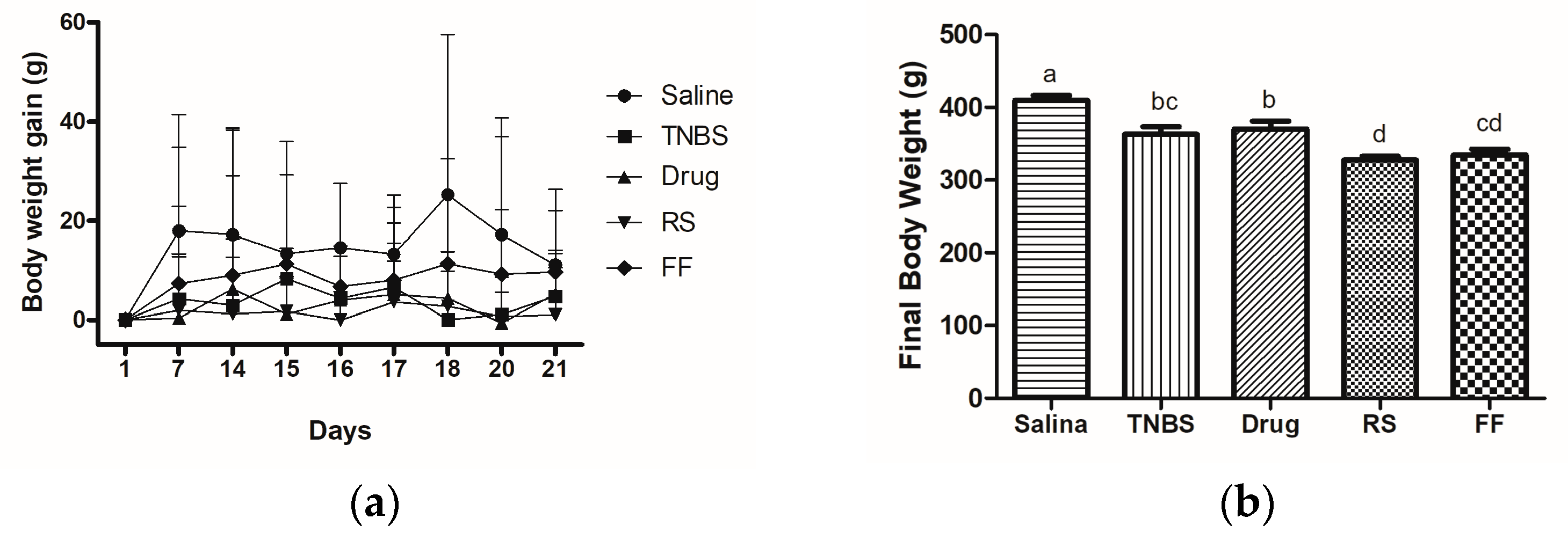
| Body weight gain (g) | 129.9 ± 34.72 a | 72.0 ± 18.19 b | 71.7 ± 25.32 b | 78.0 ± 7.92 b | 81.7 ± 14.49 b |
| Food intake (mg/kg bw) | 25.4 ± 3.4 | 21.0 ± 5.7 | 21.3 ± 5.9 | 17.9 ± 6.4 | 21.3 ± 3.6 |
| Energy intakecal | 104.1 ± 14.0 | 86.0 ± 23.37 | 87.3 ± 24.1 | 73.5 ± 26.1 | 87.5 ± 26.1 |
Disclaimer/Publisher’s Note: The statements, opinions and data contained in all publications are solely those of the individual author(s) and contributor(s) and not of MDPI and/or the editor(s). MDPI and/or the editor(s) disclaim responsibility for any injury to people or property resulting from any ideas, methods, instructions or products referred to in the content. |
© 2024 by the authors. Licensee MDPI, Basel, Switzerland. This article is an open access article distributed under the terms and conditions of the Creative Commons Attribution (CC BY) license (https://creativecommons.org/licenses/by/4.0/).
Share and Cite
Moya, A.M.T.M.; Alexandrino, T.D.; Morari, J.; Reguengo, L.M.; Velloso, L.A.; Leal, R.F.; Junior, S.B.; Pereira, A.P.A.; Pastore, G.M.; Bicas, J.L.; et al. The Consumption of the Fibrous Fraction of Solanum lycocarpum St. Hil. Does Not Preserve the Intestinal Mucosa in TNBS-Induced Rats. Foods 2024, 13, 2949. https://doi.org/10.3390/foods13182949
Moya AMTM, Alexandrino TD, Morari J, Reguengo LM, Velloso LA, Leal RF, Junior SB, Pereira APA, Pastore GM, Bicas JL, et al. The Consumption of the Fibrous Fraction of Solanum lycocarpum St. Hil. Does Not Preserve the Intestinal Mucosa in TNBS-Induced Rats. Foods. 2024; 13(18):2949. https://doi.org/10.3390/foods13182949
Chicago/Turabian StyleMoya, Amanda Maria Tomazini Munhoz, Thaís Dolfini Alexandrino, Joseane Morari, Livia Mateus Reguengo, Licio Augusto Velloso, Raquel Franco Leal, Stanislau Bogusz Junior, Ana Paula Aparecida Pereira, Glaucia Maria Pastore, Juliano Lemos Bicas, and et al. 2024. "The Consumption of the Fibrous Fraction of Solanum lycocarpum St. Hil. Does Not Preserve the Intestinal Mucosa in TNBS-Induced Rats" Foods 13, no. 18: 2949. https://doi.org/10.3390/foods13182949
APA StyleMoya, A. M. T. M., Alexandrino, T. D., Morari, J., Reguengo, L. M., Velloso, L. A., Leal, R. F., Junior, S. B., Pereira, A. P. A., Pastore, G. M., Bicas, J. L., & Cazarin, C. B. B. (2024). The Consumption of the Fibrous Fraction of Solanum lycocarpum St. Hil. Does Not Preserve the Intestinal Mucosa in TNBS-Induced Rats. Foods, 13(18), 2949. https://doi.org/10.3390/foods13182949