Article

Characterization of a Novel Acid-Stable Chitosanase from Lentinula edodes Suitable for Chitooligosaccharide Preparation

by
Yanxin Wang
1,
Yujie Zhao
1,
Jingchen Li
1,
Haobo Zong
1,
Ying Chen
1,
Jinyu Zhou
1,
Xinlian Li
1 and
Xianfeng Ye
2,*
1
College of Agriculture and Biology, Liaocheng University, Liaocheng 252000, China
2
Key Laboratory of Agricultural Environmental Microbiology, Ministry of Agriculture and Rural Affairs, College of Life Sciences, Nanjing Agricultural University, Nanjing 210095, China
*
Author to whom correspondence should be addressed.
Foods 2024, 13(19), 3127; https://doi.org/10.3390/foods13193127
Submission received: 22 August 2024 / Revised: 11 September 2024 / Accepted: 17 September 2024 / Published: 30 September 2024
(This article belongs to the Section Food Biotechnology)

Abstract

:
As high-value chitosan derivatives, chitooligosaccharides (COSs) with biodegradable, biocompatible, nontoxic, antimicrobial, and antioxidant activities have been widely applied in food-related fields. Chitosanases can hydrolyze chitosan to produce COSs. Herein, a chitosanase (LeCho1) from Lentinula edodes was successfully expressed in Escherichia coli and was then purified and characterized. LeCho1 had a low sequence identity with other chitosanases reported from the GH75 family. The recombinant protein showed a molecular mass of 27 kDa on SDS-PAGE. LeCho1 preferentially hydrolyzed chitosan with a high degree of deacetylation (DDA) and exhibited maximal activity (71.88 U/mg) towards 95% DDA chitosan at pH 3.0 and 50 °C. It possessed good stability at pH 2.0–6.0 and temperatures below 45 °C. Its hydrolytic activity was remarkably enhanced by the metal ion Mn2+ at 1 mM, while it was totally inhibited by 1 mM Fe3+ or 10 mM EDTA. Its Km and Vmax values were 0.04 μM and 76.81 μmol·min−1·mg−1, respectively, indicating good substrate affinity. LeCho1 degraded chitosan into COSs with degrees of polymerization (DPs) of 2–5, while it had no action on COSs with DPs of less than 5, revealing its endo-chitosanase activity. This study proved that chitosanase LeCho1 is a promising candidate in the industrial preparation of COSs due to its excellent properties.

1. Introduction

Chitosan, as the N-deacetylated product of chitin, is a linear heteropolysaccharide made up of two monosaccharides, D-glucosamine (GlcN) and N-acetyl-D-glucosamine (GlcNAc), connected through β-1,4-glycosidic bonds [1]. Chitosan has many biological activities, such as non-toxicity, biocompatibility, biodegradability, antimicrobial, antitumor, high blood pressure inhibition, and an immune-enhancing effect [2]; however, its applications have been limited due to its poor solubility at neutral pH and high viscosity. At present, chitosan oligosaccharides (COSs) with degrees of polymerization (DPs) in the range of 2–20 [3], obtained by the depolymerization of chitosan, have received extensive consideration in many sectors due to their exceptional physicochemical properties, including high water solubility and low viscosity [4]. Especially in food-related industries, chitosan and its derivative COSs have been implemented to improve the quality and shelf-life of various food and food products. For example, they have served as potential natural antioxidants to inhibit lipid oxidation in foods rich in polyunsaturated fatty acid and relieve the off-flavor and off-odor of fatty foods [5], and COSs have exhibited higher activity in lowering lipid oxidation due to the higher proportion of –OH groups and NH2 groups after the hydrolysis of chitosan [6]. Furthermore, they were also used as potential antimicrobial agents to suppress microbial spoilage and have been applied in beer brewing and food processing [7,8]; chitosan with lower molecular weight and COSs have been reported to have higher antimicrobial activity due to their unobstructed contact with bacterial cells [9]. Additionally, they possess excellent film-forming properties and have been employed as potential packing materials to provide barriers to aroma, oxygen, oil, and moisture and to enhance the appearance and flavor properties of preserved foods [10].
In general, COSs are prepared by three methods, including the chemical method, which involves acid hydrolysis and oxidative degradation [11] and which results in difficulty in separating and purifying the products, resulting in environmental pollution and the production of harmful by-products. The physical method involves microwave degradation, ultrasonic treatment, and ultraviolet irradiation [12], which require high energy consumption and have a low product yield. The enzymatic degradation method utilizes cellulases or chitosanases [13] and has some difficulties in industrialization due to its high cost in terms of production and harsh conditions in reaction. Compared to the first two methods above, enzymatic conversion to produce COSs has recently drawn widespread attention because of its environmental friendliness, the controllability of the final product, and the efficiency of production. Many enzymes reported are able to hydrolyze chitosan into COSs, such as cellulases, proteases, lipases, chitinases, and chitosanases [14]. Among them, chitosanases are a specific enzyme involved in the hydrolysis of chitosan; thus, exploring a new chitosanase with high activity and high stability under a mild reaction environment is of great significance in the preparation of COSs.
As glycoside hydrolases (GHs), chitosanases can specifically degrade β-1,4-glucoside bonds located in chitosan and produce COSs or glucosamine (GlcN) [15]. Chitosanases have been found in many organisms, such as bacteria, fungi, cyanobacteria, and plants [16]. The enzymes have been classified into three subclasses on the basis of the cleavage positions’ specificity in the substrate chitosan, such as subclass I cleaving GlcN–GlcN and GlcNAc–GlcN linkages, subclass II cutting GlcN–GlcN linkages or GlcN–GlcN linkages, and subclass III cracking GlcN–GlcNAc linkages [17]. Furthermore, these enzymes have also been divided into endo-chitosanases, which could hydrolyze the β-1,4-glycosidic bonds of chitosan and produce a mixture of COSs with various DP values, and exo-chitosanases, which are capable of cutting GlcN residues individually from the chitosan’s non-reducing termini to release a series of monosaccharides. Most of the reported chitosanases belong to the endo-enzymes group. Additionally, based on the similarity of amino acid sequences, chitosanases have been categorized into six glycoside hydrolase (GH) families in the Carbohydrate-Active Enzymes database (CAZy, www.cazy.org, updated in September 2024), including the GH5, GH7, GH8, GH46, GH75, and GH80 families [18], among which, the GH46, GH75 and GH80 families are all made up of chitosanases. Chitosanases from fungi have been reported to be mainly distributed in the GH75 family, while those from bacteria have been found in the GH46 family [19]. In addition, chitosanases with different biological functions have been found in different organisms. For example, chitosan-degrading microorganisms utilize chitosanases to acquire extracellular nutrition [20], fungi from Zygomycetes secrete chitosanases to hydrolyze the structural components of cell walls for morphogenesis [21] and plants employ chitosanases as a defensive weapon against phytopathogens [22]. In addition to this, chitosanases have potential applications in many areas, such as the bioconversion of marine crustacean biomaterials [23], the preparation of fungal protoplasts [21], the biocontrol of plant pathogens [24] and the manufacture of COSs [25]. Presently, various chitosanases from different sources have been reported; however, only a few have been used in industrial production. Therefore, screening for a new chitosanase with excellent properties is still urgently needed.
Previous studies reported that a putative chitosanase from Lentinula edodes (LeCho1) exhibited up-regulated transcription levels post-harvest, and it was proposed that this cell wall degradation-related enzyme was closely related to fruiting body autolysis [26,27]. However, there was no detailed biochemical characterization of this protein. In this study, LeCho1 was heterologously expressed in Escherichia coli with a 6×His tag and purified using Ni-NTA affinity chromatography. The purified protein was then subjected to detailed biochemical characterization, and its hydrolytic pattern was determined. The results of this study illustrate the outstanding characteristics of LeCho1 in the preparation of chitoologosaccharides (COS), laying a crucial foundation for its industrial application.

2. Materials and Methods

2.1. Polysaccharides

Chitosan oligosaccharides (≥95% purity), including chitobiose hydrochloride (ZB-10006), chitotriose hydrochloride (ZB-10007), chitotetraose hydrochloride (ZB-10008), chitopentaose hydrochloride (ZB-10009), and chitohexaose hydrochloride (ZB-10010), were obtained from Zhenzhun Biotechnology Co., Ltd. (Shanghai, China). Chitosan with different degrees of deacetylation (DDA; 70%, 80%, and 95%), chitin, microcrystalline cellulose, and sodium carboxymethylcellulose were procured from Maclin Biochemical Technology Co., Ltd. (Shanghai, China).

2.2. Strains and Plasmids

The genomic DNA of L. edodes cultivation strain H600 was stored in our laboratory and was used as a template to clone the chitosanase gene. E. coli BL21 (DE3) was used to express the recombinant protein. The vector pET29A was employed to construct the recombinant expression plasmid.

2.3. Sequence Analysis

The amino acid sequences of chitosanase LeCho1 were derived from the genome of L. edodes in GenBank (Accession ABH80469.1). Potential signal peptide sequences were predicted using the SignalP-5.0 server (https://services.healthtech.dtu.dk/services/SignalP-5.0/, accessed on 18 February 2022). The theoretical molecular weight and isoelectric point (pI) of LeCho1 were obtained through the Expasy server (https://web.expasy.org/protparam/, accessed on 15 May 2022). Based on amino acid sequences of LeCho1 and previously reported chitosanases from the GH46, GH75, and GH80 families, a phylogenetic tree was constructed in MEGA v.6.06 software with 1000 bootstrap iterations using the neighbor-joining method. All the protein sequences were obtained from NCBI. The sequence identity between LeCho1 and other chitosanases from the GH75 family was determined by amino acid sequence alignment in Clustal W version 2.0. The alignment result was processed using ESPript 3.0 (http://espript.ibcp.fr/ESPript/cgi-bin/ESPript.cgi, accessed on 26 September 2022).

2.4. RNA Extraction and cDNA Synthesis

The mycelia of L. edodes, preserved in our labs, were inoculated onto the potato dextrose agar (PDA) medium and placed in an incubator at 26 °C for 12 d in the dark. The resulting fresh mycelia were collected to extract total RNA using a Spin Column Fungal Total RNA Purification Kit (Sangon, Shanghai, China) according to the manufacturer’s recommendation. The synthesis of cDNA was carried out using a HiScript II Q Select RT SuperMix for qPCR (+gDNA wiper) Kit (Vazyme Biotech Co., Ltd., Nanjing, China) following the technical manual. The resulting cDNA was stored at −80 °C until further use.

2.5. Cloning, Expression and Purification of LeCho1

The nucleotide sequences encoding LeCho1 without the signal peptide were amplified using the forward primer (TAAGAAGGAGATATACATATGGCGCCCATTCCTAGAAATCTTCGGA) and the reverse primer (TAAGAAGGAGATATACATATGGCGCCCATTCCTAGAAATCTTCGG; the cleavage sites are underlined). The product was ligated with the plasmid pET29A digested with Nde I and Xho I using the ClonExpress II/One Step Cloning Kit (Vazyme, Nanjing, China), and transferred into competent cells of E. coli BL21 (DE3). Transformants were screened by colony PCR and confirmed using Sanger sequencing. For protein expression, the cells were grown in LB medium containing kanamycin (50 mg/L) at 37 °C and 220 rpm. When the optical density at 600 nm (OD600) of the culture reached 0.5–0.6, isopropyl-β-D-thiogalactopyranoside (IPTG) with a final concentration of 1 mM was added to induce expression of the target protein. Then, the induced cells were further incubated at 16 °C for 20–24 h, after which they were collected by centrifugation at 12,000× g for 5 min. Following sonication at 4 °C, the recombinant LeCho1 containing a 6×His tag was loaded onto Ni2+-nitrilotriacetic acid (NTA) resin, and the purified fractions were collected and concentrated by ultrafiltration. The purity of the harvested protein was detected using 12% acrylamide sodium dodecyl sulfate-polyacrylamide gel electrophoresis (SDS-PAGE), and the bands were stained with Coomassie G-250. The concentration of the purified protein was determined using the Bradford method with bovine serum albumin (BSA) as the standard [28].

2.6. Hydrolytic Activity Assay

The hydrolytic activity of LeCho1 was measured using the 3,5-dinitrosalicylic acid (DNS) method [29]. The amount of reducing sugar released from the substrates (chitosan, chitin, microcrystalline cellulose, and sodium carboxymethylcellulose) was determined by detecting the absorbance at 540 nm (OD540) of the supernatant from the reaction mixture using GlcN as the standard. The reaction mixture (200 μL), including 2 μg LeCho1 and 0.5% (w/v) substrate in 50 mM sodium acetate buffer (NaAc-HAc, pH 5.0), was incubated at 40 °C for 30 min, after which DNS reagent (200 μL) was added and placed at 100 °C for 10 min. After cooling to room temperature, the supernatant of the above mixture was collected by centrifugation, and its absorbance at OD540 was measured. All reactions were conducted in triplicate. One unit (U) of chitosanase activity was defined as the amount of enzyme liberating 1 μmol of reducing sugar per minute under the above conditions with glucosamine as standard.

2.7. Biochemical Characterization of Recombinant LeCho1

Chitosan (0.5% w/v, 95% DDA) was used as the substrate to examine the enzymatic properties of LeCho1. The optimal pH of LeCho1 was determined by measuring the hydrolytic activity of the enzyme in 50 mM buffers at pH values in the range of 2.0–9.0, including Gly-HCl buffer (pH 2.0–3.0), NaAc-HAc buffer (pH 3.0–6.0), sodium-phosphate buffer (PBS, pH 6.0–7.0), and Tris-HCl buffer (pH 7.0–9.0). To assay the pH stability, purified LeCho1 (2 μg) was incubated in different buffers at 4 °C for 12 h, after which the residual chitosanase activity was detected using chitosan (0.5% w/v, 95% DDA) dissolved in 50 mM Gly-HCl buffer (pH 3.0) at 40 °C for 30 min.
The optimal temperature of LeCho1 was evaluated by examining the hydrolytic activity of the enzyme at various temperatures (20–80 °C) for 30 min in 50 mM Gly-HCl buffer (pH 3.0). To determine the thermal stability, purified LeCho1 in Gly-HCl buffer (pH 3.0) was placed at 20, 30, 40, 50, 60, 70, and 80 °C for 1 h and cooled to room temperature, after which the residual activity was determined at 40 °C and was detected under the standard condition.
The influence of metal ions on chitosanase activity was assessed by incubating purified LeCho1 with EDTA (1 mM and 10 mM) or 1 mM individual metal ions (Na+, K+, Ba2+, Cu2+, Ca2+, Co2+, Fe2+, Mn2+, Mg2+, Ni2+, Zn2+, Fe3+, Al3+, and Cr3+) in Gly-HCl buffer (50 mM, pH 3.0) containing chitosan (0.5% w/v, 95% DDA) at 40 °C for 30 min. The hydrolytic activity of LeCho1 without any additives was defined as 100%.
The kinetic parameters of the protein were determined by measuring the initial reaction velocity at various concentrations of 95% DDA chitosan (0.1%, 0.2%, 0.4%, 0.5%, 0.6%, and 0.8%, w/v). Reactions were performed in Gly-HCl buffer (50 mM, pH 3.0) at 40 °C for 40 min. The Michaelis–Menten constant (Km) and the maximal catalytic rate (Vmax) were determined using Lineweaver–Burk plots.

2.8. Analysis of Hydrolysis Products

The hydrolytic pattern of LeCho1 was determined by analyzing the hydrolysates of substrates, including COSs [(GlN)2 to (GlN)6] and chitosan (0.5% w/v, 95% DDA). Reaction mixtures (200 μL), including 2 μg LeCho1 and 0.5% (w/v) chitosan or 0.1 μM COSs in 50 mM Gly-HCl buffer (pH 3.0), were incubated at 40 °C for different time periods (0, 0.5, 1, 2, 4, 6, and 12 h). After the indicated time, the mixtures were immediately placed at 100 °C for 10 min to stop the reaction, then centrifuged at 12,000× g for 5 min to remove the denatured protein.
The distribution of hydrolysate components was analyzed using thin-layer chromatography (TLC) according to the method described by Wang et al. (2021) [15]. Briefly, the above supernatants were spotted onto the TLC plate (Silica gel 60 F254 aluminum sheet, Merck, Darmstadt, Germany) and developed in a solvent comprising ammonium hydroxide/water/isopropanol (0.3:2.7:7, v/v/v). The dried plate was sprayed with 0.5% ninhydrin reagent (dissolved in ethanol) and heated at 95 °C for 10 min to visualize the products.
In addition, hydrolysates of chitosan after 12 h of enzymolysis were analyzed using matrix-assisted laser desorption ionization-time-of-flight mass spectrometry (MALDI-TOF MS) [30] on a Bruker Autoflex time-of-flight mass spectrometer (Bruker Daltonics, Karlsruhe, Germany). During detection, 2,5-dihydroxybenzamide (DHB) (20 mg/mL DHB dissolved in 30% acetonitrile with 0.1% trifluoroacetic acid) served as the matrix to assist the ionization of hydrolysates. The acceleration voltage was set to 29 kV in reflection mode. All spectra were obtained in positive ion mode. The target mass range was an m/z ratio of 100/2000.

2.9. Statistical Analysis

All experiments were performed independently in triplicate. The values are shown as the means ± standard deviations (SD). To evaluate the variance and significant differences between the different treatments, a single-factor analysis of variance (one-way ANOVA) and Duncan’s test with a confidence level of p < 0.05 were performed using SPSS statistics software version 22.0 (IBM Corporation, Armonk, NY, USA).

3. Results

3.1. Sequence Analysis of LeCho1

The putative chitosanase LeCho1, encoded by an open reading frame consisting of 846 nucleotides, was predicted to be composed of 281 amino acid residues with a 22-aa signal peptide at its N-terminus. The mature chitosanase protein had a predicted molecular mass of 27.3-kDa and a calculated pI of 4.11. A phylogenetic tree including LeCho1 and other chitosanases from the GH46, GH75, and GH80 families revealed that LeCho1 clustered with the chitosanases from the GH75 family and formed an independent branch (Figure 1a). Multiple sequence alignment indicated that LeCho1 exhibited low sequence identity with other members of the GH75 family. For example, it shared approximately 35% sequence identity with the chitosanases from Penicillium chrysogenum (ADG96019.1, 34.75%) and Aspergillus oryzae (BAD08218.2, 34.98%). Moreover, it only shared sequence identities of less than 25% with other chitosanases located in another branch of the GH75 family (Figure 1a). Nevertheless, it contained the four highly conserved amino acid residues (Asp140, Asp142, Asp204, and Glu215, indicated by dots in Figure 1b) typical of the GH75 family; among them, Asp204 and Glu215 (Figure 1b, indicated by blue dots) were predicted to be involved in the catalytic function of the enzyme. These results indicated that LeCho1 was a novel member of the GH75 family.

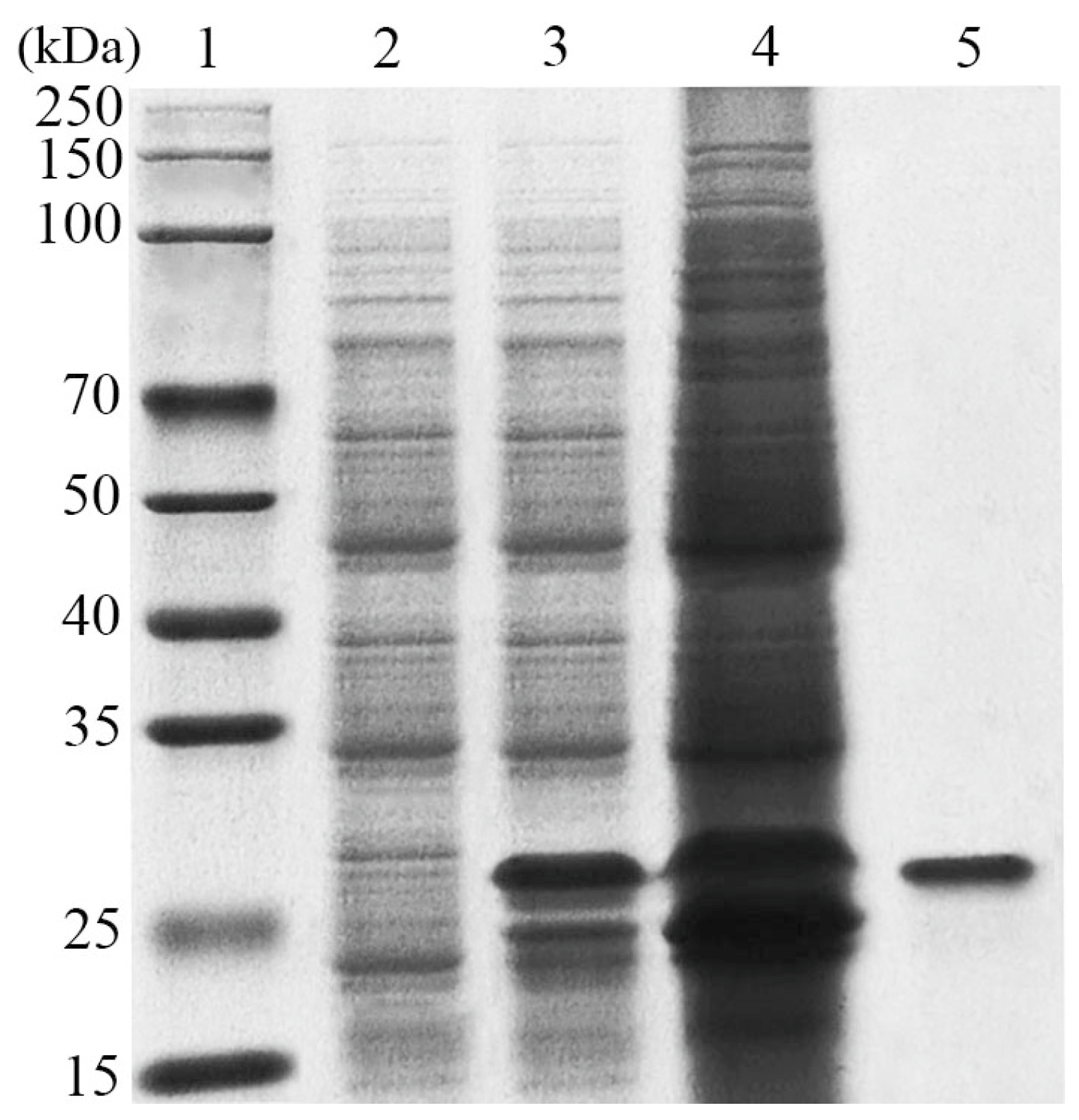
3.2. Expression and Purification of LeCho1

The coding sequence of the chitosanase without the signal peptide was successfully expressed in E. coli BL21 cells containing the recombinant plasmid pET29a-LeCHO1. The recombinant protein with a 6×His-tag, purified using a Ni-NTA affinity column, exhibited a single band on SDS-PAGE corresponding to a molecular weight of approximately 27-kDa (Figure 2), consistent with its calculated molecular mass of 27.3 kDa.

3.3. Substrate Specificity of the Purified Protein

The hydrolytic activity of LeCho1 on different polysaccharides (chitosan, chitin, microcrystalline cellulose, and sodium carboxymethylcellulose) was examined in order to determine its substrate specificity. As shown in Table 1, LeCho1 displayed high hydrolytic activity on chitosan, and its activity was gradually enhanced with the increase of the DDA, with the highest activity of 71.88 U/mg found for 95% deacetylated chitosan. However, it had no activity on chitin or cellulose, indicating the strict substrate specificity of LeCho1 for chitosan.

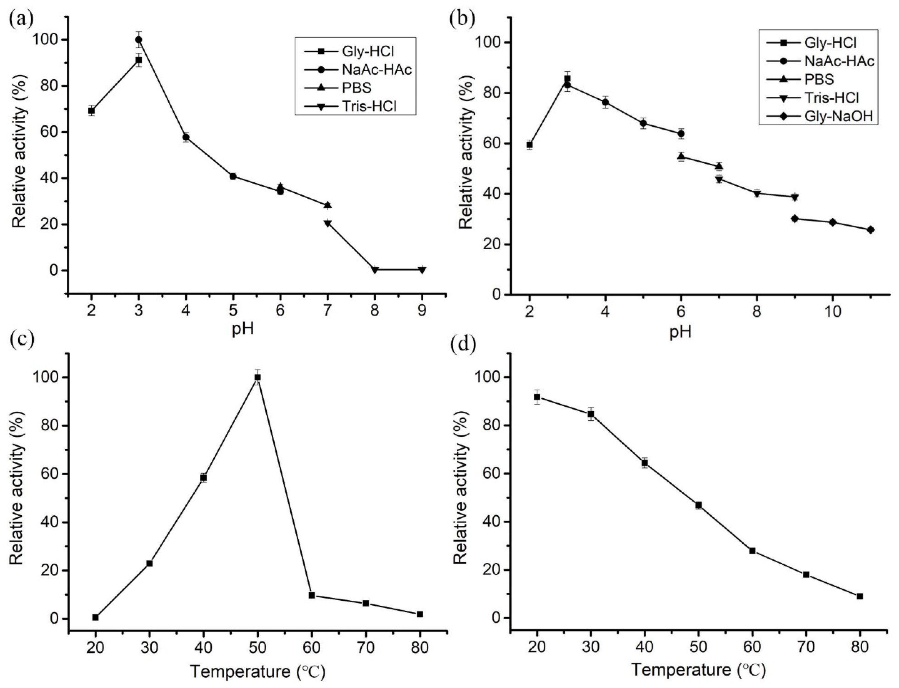
3.4. Enzymological Characterization of LeCho1

During the hydrolysis of chitosan, pH influences not only the activity and stability of the enzyme but also the conformation of the chitosan substrate itself. For example, chitosan has good solubility in an acidic environment while being insoluble under neutral or alkaline conditions. Thus, we investigated the effect of pH on the catalytic activity of LeCho1 with chitosan (0.5% w/v, 95% DDA) as the substrate. LeCho1 displayed the highest chitosanase activity in NaAc-HAc buffer pH 3.0 but also retained more than 40% relative activity at pH values ranging from 2 to 5. However, its activity decreased rapidly at pH above 6.0 with a complete loss of activity at pH 8.0 and 9.0 (Figure 3a), suggesting that LeCho1 is an acid-adapted chitosanase. In addition, the enzyme still maintained more than 50% of the initial activity after incubation at pH 2.0–7.0 for 12 h. By contrast, it retained less than 40% of the initial activity after incubation in alkaline buffers (Figure 3b).
In addition, LeCho1 showed maximal chitosanase activity at 50 °C, with more than 50% relative activity in the temperature range of 40–55 °C. However, its activity dropped sharply above 60 °C (Figure 3c). After incubation at 20–45 °C for 1 h, LeCho1 retained more than 50% of the initial activity, compared to less than 30% at temperatures over 60 °C (Figure 3d), revealing that LeCho1 was a mesophilic enzyme with favorable stability at temperatures below 45 °C.
In addition, the influences of different metal ions on the hydrolytic activity of LeCho1 were also determined. As presented in Table 2, Mn2+ had an obvious stimulatory effect, increasing the enzyme activity by 57%. Additionally, Mg2+ and Ca2+ exhibited a slight promoting effect, with increases of approximately 15%. K+, Ba2+, and Fe2+ had only a slight effect on the hydrolytic activity of LeCho1. By contrast, Na+, Co2+, Ni2+, and Zn2+ had a mild inhibitory effect on the activity of the enzyme. However, Fe3+ exhibited a strong inhibitory effect, resulting in the complete loss of chitosanase activity. As a metal chelating agent, EDTA had an inhibitory effect on the activity of LeCho1, causing a 44% decrease at the concentration of 1 mM and 100% at 10 mM, indicating that LeCho1 is a metalloenzyme.
To determine the kinetic parameters, the effect of the substrate concentration on the hydrolytic activity of LeCho1 was determined using chitosan (95% DDA) in 50 mM Gly-HCl buffer (pH 3.0) at 40 °C. The results showed that the Km and Vmax values of LeCho1 were 0.04 μM and 76.81 μmol·min−1·mg−1, respectively (Figure 4), suggesting a high affinity of the enzyme for the substrate.

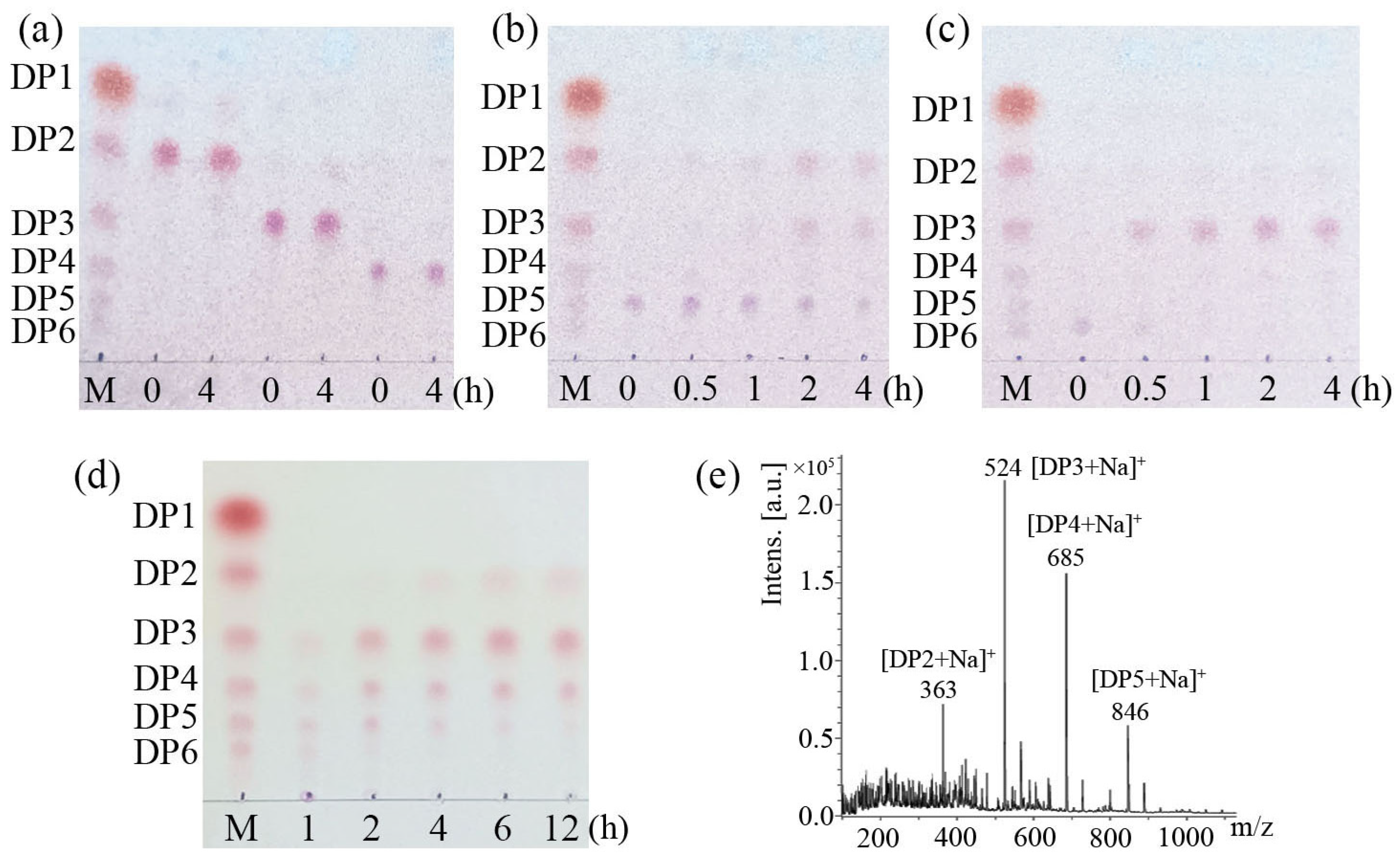
3.5. LeCho1 Is an Endo-Chitosanase

The hydrolytic products of LeCho1 released from chitooligosaccharides, including (GlcN)2, (GlcN)3, (GlcN)4, (GlcN)5, and (GlcN)6, as well as chitosan (0.5% w/v, 95% DDA), were detected using thin layer chromatography (TLC) and mass spectroscopy (MALDI-TOF MS). The TLC analysis showed no hydrolytic products when LeCho1 was incubated with (GlcN)2, (GlcN)3, or (GlcN)4 (Figure 5a), suggesting that the enzyme had no activity on COSs with degrees of polymerization (DP) of less than 5. However, after incubation with LeCho1 for 4 h, most of the (GlcN)5 was hydrolyzed into (GlcN)2 and (GlcN)3 (Figure 5b), while (GlcN)6 was completely degraded into (GlcN)3 (Figure 5c).
Additionally, LeCho1 was also able to effectively hydrolyze chitosan. Time course analysis showed that hydrolysates of chitosan produced COSs with DP 3−6 after 1 h of reaction. After 2 h of reaction, there were large amounts of (GlcN)3, (GlcN)4 and (GlcN)5, with trace amounts of (GlcN)6 in the hydrolysate. As the reaction continued, (GlcN)3 and (GlcN)4 accumulated, while the content of (GlcN)5 and (GlcN)6 gradually decreased. After 12 h of reaction, the hydrolysates were mainly composed of (GlcN)3 and (GlcN)4, a small amount of (GlcN)2, and trace amounts of (GlcN)5 (Figure 5d). In addition, the hydrolysates produced by LeCho1 incubated with chitosan for 12 h were analyzed using MALDI-TOF-MS. Fragments of ions of the products chitobiose ([DP2 + Na]+ at m/z 363), chitotriose ([DP3 + Na]+ at m/z 524), chitotetrose ( [DP4 + Na]+ at m/z 685), and chitopentaose ([DP5 + Na]+ at m/z 846) were present in the mass spectra (Figure 5e). These results indicated that LeCho1 exhibited the endo-type catalytic mode of action in the hydrolysis of chitosan.

4. Discussion

The putative chitosanase LeCho1 was cloned from L. edodes and identified as a novel member of the GH75 family. Phylogenetic analysis revealed that LeCho1 clustered with other chitosanases from the GH75 family, and it also had the conserved catalytic amino acid residues Asp204 and Glu215 that are typical for the GH75 family [31]. However, LeCho1 showed low sequence identity with other chitosanases belonging to the GH75 family, with identity values of less than 40%. In addition, we also found distinct amino acid residues near the active sites, such as Ala200 corresponding to a glycine (Gly) in other chitosanases (Figure 1b, indicated by the black frame) and Met202 corresponding to aromatic amino acids (Tyr, Phe and Trp) of other chitosanases (Figure 1b, indicated by the black frame), which were likely to contribute to its distinct enzymatic properties. To better assess the specific features of LeCho1, we compiled the characteristics of previously reported chitosanases from the GH46, GH75, and GH80 families in Table 3. As can be seen, these enzymes possess molecular weights (Mw) in the range of 20.0–35.0 kDa, optimal temperatures between 40 and 50 °C, and optimal pH values ranging from 5.0 to 7.0 [32,33], as well as the ability to hydrolyze chitosan into COSs. Among them, LeCho1 displayed some similarities with the chitosanases from the GH75 family in terms of molecular weight, optimal temperature, and hydrolytic profile [34,35,36]. However, LeCho1 exhibited significant differences in terms of the optimal pH and influence of metal ions on its catalytic activity. Notably, the optimal pH of LeCho1 was only 3.0, which was significantly more acidic than the optimal pH of other chitosanases. Additionally, the metal ion Mn2+ was found to enhance the hydrolytic activity of most chitosanases [23,37], whereby the activity of LeCho1 was increased by 57%. By contrast, Cu2+ had an inhibitory effect on the activity of most chitosanases [18,38] but no effect on the activity of LeCho1. In contrast, Fe3+ had an obvious inhibitory effect on the activity of LeCho1, which was similar to reports on chitosanases from the GH46 and GH80 families, such as the enzymes from Streptomyces coelicolor A3(2) M145 [39] and Bacillus sp. BY01 [18].
As a promising novel enzyme, LeCho1 has potential applications in the production of bioactive chitooligosaccharides. In contrast to their precursor, chitosan, COSs possess low molecular weight and high solubility in water. They are widely used in many domains, especially in agricultural cereal crop production, which provides the basic guarantee for improving the quality of food raw materials. COSs with desirable DP were reported to act as biostimulants that regulate plant growth and development. Zhang et al. (2016) [40] found that COSs with DP > 3 induced an increase in the contents of soluble protein, soluble sugar, and chlorophyll in wheat seedlings, promoting photosynthesis and plant growth. Moreover, COSs could enhance plant resistance to abiotic stresses such as salinity, drought, heavy metals, and heat due to their ability to scavenge reactive oxygen species. Jia et al. (2019) [41] reported that chitotriose and chitotetraose promoted the health of soybean seedlings at 72 and 42 h of germination, which was attributed to their radical scavenging ability. In addition, COSs were also investigated as a green biocide against phytopathogenic fungi due to the binding of their positively charged amino groups (NH3+) to negatively charged constituents of the fungal cell wall [42], which causes clustering and lysis of fungal mycelia. Wang et al. (2021) [15] discovered that COSs with DP 3–5 were able to inhibit the mycelial growth of Fusarium oxysporum and Magnaporthe oryzae at high concentrations. In this study, LeCho1 was found to effectively cleave chitosan, exhibiting a specific activity of 71.88 U/mg. Moreover, the hydrolysate obtained after 2 h was primarily composed of COSs with DP 3–5 and contained no COSs with a DP of less than 3, giving LeCho1 high competitiveness in the manufacture of desirable COSs. Furthermore, LeCho1 exhibited maximal catalytic activity under acidic conditions (pH 3.0), which is also a major advantage in the preparation of COSs, as low pH is helpful not only in reducing the risk of microbial contamination but also in enhancing the solubility of chitosan and further improving hydrolysis efficiency. Moreover, LeCho1 exhibited a low Michaelis constant, with a Km value of 0.04 μM, indicating that the enzyme has a high affinity for chitosan, which is also beneficial for the preparation of COSs. In addition, the expression of the chitosanase LeCho1 was based on the well-known E. coli BL21 (DE3) expression system, with the advantages of a short fermentation period, high production efficiency, and mature purification process. In short, the efficient expression of LeCho1 and its outstanding enzymatic properties render it a good candidate for the industrial production of chitooligosaccharides.

5. Conclusions

In this study, the chitosanase LeCho1 from L. edodes was identified through bioinformatics analysis, expressed in E. coli, purified, and characterized. LeCho1 was revealed to be a new member of the GH75 family. The purified recombinant enzyme was most effective on 95% deacetylated chitosan, with a specific activity of 71.88 U/mg. LeCho1 was most active at pH 3.0 and 50 °C, with good stability at pH values ranging from 2.0 to 6.0 and temperatures below 40 °C. Its Km and Vmax values were 0.04 μM and 76.81 μmol·min−1·mg−1, respectively, indicating high affinity for the substrate chitosan. LeCho1 exhibited endo-type chitosanase activity, catalyzing the depolymerization of chitosan to yield COSs with DP 2–5. The remarkable properties of LeCho1 indicate its promising application in the production of bioactive COSs.

Author Contributions

Y.W. conceived the experiments and wrote the article. Y.Z. and H.Z. performed the determination of the hydrolysates of chitosan using TLC. J.L. helped to draw the Lineweaver–Burk plot. Y.C. purified the recombinant chitosanase LeCho1. J.Z. and X.L. constructed the recombinant vector. X.Y. provided valuable suggestions about the tests and article. All authors have read and agreed to the published version of the manuscript.

Funding

This work was supported by the Doctoral Research Initiation Fund of Liaocheng University (No 318052291) and the Natural Science Foundation of China (No 32370119).

Institutional Review Board Statement

Not applicable.

Informed Consent Statement

Not applicable.

Data Availability Statement

The original contributions presented in the study are included in the article, further inquiries can be directed to the corresponding author.

Conflicts of Interest

The authors declare no conflict of interest.

References

  1. Mukarram, M.; Ali, J.; Dadkhah-Aghdash, H.; Kurjak, D.; Kačík, F.; Ďurkovič, J. Chitosan-induced biotic stress tolerance and crosstalk with phytohormones, antioxidants, and other signalling molecules. Front. Plant Sci. 2023, 14, 1217822. [Google Scholar] [CrossRef] [PubMed]
  2. Zhou, J.; Wen, B.; Xie, H.; Zhang, C.; Bai, Y.; Cao, H.; Che, Q.; Guo, J.; Su, Z. Advances in the preparation and assessment of the biological activities of chitosan oligosaccharides with different structural characteristics. Food Funct. 2021, 12, 926–951. [Google Scholar] [CrossRef] [PubMed]
  3. Lodhi, G.; Kim, Y.; Hwang, J.; Kim, S.; Jeon, Y.; Je, J.; Ahn, C.; Moon, S.; Jeon, B.; Park, P. Chitooligosaccharide and its derivatives: Preparation and biological applications. BioMed Res. Int. 2014, 2014, 654913. [Google Scholar] [CrossRef] [PubMed]
  4. Benchamas, G.; Huang, G.; Huang, S.; Huang, H. Preparation and biological activities of chitosan oligosaccharides. Trends Food Sci. Tech. 2021, 107, 38–44. [Google Scholar] [CrossRef]
  5. Singh, A.; Benjakul, S.; Huda, N.; Xu, C.; Wu, P. Preparation and characterization of squid pen chitooligosaccharide–epigallocatechin gallate conjugates and their antioxidant and antimicrobial activities. RSC Adv. 2020, 10, 33196–33204. [Google Scholar] [CrossRef] [PubMed]
  6. Singh, A.; Benjakul, S.; Prodpran, T. Chitooligosaccharides from squid pen prepared using different enzymes: Characteristics and the effect on quality of surimi gel during refrigerated storage. Food Prod. Process. Nu. 2019, 1, 5. [Google Scholar] [CrossRef]
  7. Zhao, X.; Yu, Z.; Wang, T.; Guo, X.; Luan, J.; Sun, Y.; Li, X. The use of chitooligosaccharide in beer brewing for protection against beer-spoilage bacteria and its influence on beer performance. Biotechnol. Lett. 2016, 38, 629–635. [Google Scholar] [CrossRef]
  8. Chen, H.; Guo, X.; Zhu, K. The effect of chitosan oligosaccharides on the shelf-life and quality of fresh wet noodles. Carbohyd. Polym. 2023, 309, 120704. [Google Scholar] [CrossRef] [PubMed]
  9. Mittal, A.; Singh, A.; Aluko, R.E.; Benjakul, S. Pacific white shrimp (Litopenaeus vannamei) shell chitosan and the conjugate with epigallocatechin gallate: Antioxidative and antimicrobial activities. J. Food Biochem. 2021, 45, e13569. [Google Scholar] [CrossRef] [PubMed]
  10. Nilsuwan, K.; Guerrero, P.; de la Caba, K.; Benjakul, S.; Prodpran, T. Fish gelatin films laminated with emulsified gelatin film or poly (lactic) acid film: Properties and their use as bags for storage of fried salmon skin. Food Hydrocolloid. 2021, 111, 106199. [Google Scholar] [CrossRef]
  11. Je, J.; Kim, S. Chitooligosaccharides as potential nutraceuticals: Production and bioactivities. Adv. Food Nutr. Res. 2012, 65, 321–336. [Google Scholar] [CrossRef] [PubMed]
  12. Xing, R.; Liu, Y.; Li, K.; Yu, H.; Liu, S.; Yang, Y.; Chen, X.; Li, P. Monomer composition of chitooligosaccharides obtained by different degradation methods and their effects on immunomodulatory activities. Carbohyd. Polym. 2017, 157, 1288–1297. [Google Scholar] [CrossRef]
  13. Hoell, I.A.; Vaaje-Kolstad, G.; Eijsink, V.G. Structure and function of enzymes acting on chitin and chitosan. Biotechnol. Genet. Eng. 2010, 27, 331–366. [Google Scholar] [CrossRef] [PubMed]
  14. Cui, D.; Yang, J.; Lu, B.; Shen, H. Efficient preparation of chitooligosaccharide with a potential chitosanase Csn-SH and its application for fungi disease protection. Front. Microbiol. 2021, 12, 682829. [Google Scholar] [CrossRef] [PubMed]
  15. Wang, Y.; Li, D.; Liu, M.; Xia, C.; Fan, Q.; Li, X.; Lan, Z.; Shi, G.; Dong, W.; Li, Z. Preparation of active chitooligosaccharides with a novel chitosanase AqCoA and their application in fungal disease protection. J. Agric. Food Chem. 2021, 69, 3351–3361. [Google Scholar] [CrossRef]
  16. Thadathil, N.; Velappan, S.P. Recent developments in chitosanase research and its biotechnological applications: A review. Food Chem. 2014, 150, 392–399. [Google Scholar] [CrossRef]
  17. Xu, Y.; Wang, H.; Zhu, B.; Yao, Z.; Jiang, L. Purification and biochemical characterization of a novel chitosanase cloned from the gene of Kitasatospora setae KM-6054 and its application in the production of chitooligosaccharides. World J. Microb. Biot. 2023, 39, 111. [Google Scholar] [CrossRef]
  18. Yang, Y.; Zheng, Z.; Xiao, Y.; Zhang, J.; Zhou, Y.; Li, X.; Li, S.; Yu, H. Cloning and characterization of a cold-adapted chitosanase from marine bacterium Bacillus sp. BY01. Molecules 2019, 24, 3915. [Google Scholar] [CrossRef]
  19. Guo, J.; Wang, Y.; Gao, W.; Wang, X.; Gao, X.; Man, Z.; Cai, Z.; Qing, Q. Gene cloning, functional expression, and characterization of a novel GH46 chitosanase from Streptomyces avermitilis (SaCsn46A). Appl. Biochem. Biotech. 2022, 194, 813–826. [Google Scholar] [CrossRef]
  20. Somashekar, D.; Joseph, R. Chitosanases—Properties and applications: A review. Bioresour. Technol. 1996, 55, 35–45. [Google Scholar] [CrossRef]
  21. Liang, D.C.; Liu, W.G.; Zuo, A.J.; Sun, S.J.; Cheng, N.; Guo, G.; Zhang, J.Y.; De Yao, K. Pre-deliver chitosanase to cells: A novel strategy to improve gene expression by endocellular degradation-induced vector unpacking. Int. J. Pharmaceut. 2006, 314, 63–71. [Google Scholar] [CrossRef] [PubMed]
  22. Hsu, S.; Chung, Y.; Chang, C.; Sung, H. Purification and characterization of two chitosanase isoforms from the sheaths of bamboo shoots. J. Agr. Food Chem. 2012, 60, 649–657. [Google Scholar] [CrossRef]
  23. Doan, C.; Tran, T.; Tran, T.; Nguyen, T.; Nguyen, H.; Tran, T.; Vu, B.; Trinh, T.; Nguyen, A.; Wang, S. Chitosanase production from the liquid fermentation of squid pens waste by Paenibacillus elgii. Polymers 2023, 15, 3724. [Google Scholar] [CrossRef]
  24. Kouzai, Y.; Mochizuki, S.; Saito, A.; Ando, A.; Minami, E.; Nishizawa, Y. Expression of a bacterial chitosanase in rice plants improves disease resistance to the rice blast fungus Magnaporthe oryzae. Plant Cell Rep. 2012, 31, 629–636. [Google Scholar] [CrossRef] [PubMed]
  25. Luo, S.; Qin, Z.; Chen, Q.; Fan, L.; Jiang, L.; Zhao, L. High level production of a Bacillus amlyoliquefaciens chitosanase in Pichia pastoris suitable for chitooligosaccharides preparation. Int. J. Biol. Macromol. 2020, 149, 1034–1041. [Google Scholar] [CrossRef]
  26. Sakamoto, Y.; Nakade, K.; Sato, T. Characterization of the post-harvest changes in gene transcription in the gill of the Lentinula edodes fruiting body. Curr. Genet. 2009, 55, 409–423. [Google Scholar] [CrossRef]
  27. Sakamoto, Y.; Nakade, K.; Sato, S.; Yoshida, K.; Miyazaki, K.; Natsume, S.; Konno, N. Lentinula edodes genome survey and postharvest transcriptome analysis. Appl. Environ. Microb. 2017, 83, e02990-16. [Google Scholar] [CrossRef]
  28. Buger, N. The bradford method for protein quantitation. Methods Vol. Biol. 1994, 32, 9–15. [Google Scholar] [CrossRef]
  29. Wood, I.; Elliston, A.; Ryden, P.; Bancroft, I.; Roberts, I.; Waldron, K. Rapid quantification of reducing sugars in biomass hydrolysates: Improving the speed and precision of the dinitrosalicylic acid assay. Biomass Bioenerg. 2012, 44, 117–121. [Google Scholar] [CrossRef]
  30. Wang, Y.; Li, D.; Li, Z.; Cui, Z.; Ye, X. Functional analysis of a novel endo-β-1,6-glucanase MoGlu16 and its application in detecting cell wall β-1,6-glucan of Magnaporthe oryzae. Front. Microbiol. 2024, 15, 1429065. [Google Scholar] [CrossRef] [PubMed]
  31. Cheng, C.; Chang, C.; Wu, Y.; Li, Y. Exploration of glycosyl hydrolase family 75, a chitosanase from Aspergillus fumigatus. J. Biol. Chem. 2006, 281, 3137–3144. [Google Scholar] [CrossRef] [PubMed]
  32. Park, J.; Shimono, K.; Ochiai, N.; Shigeru, K.; Kurita, M.; Ohta, Y.; Tanaka, K.; Matsuda, H.; Kawamukai, M. Purification, characterization, and gene analysis of a chitosanase (ChoA) from Matsuebacter chitosanotabidus 3001. J. Bacteriol. 1999, 181, 6642–6649. [Google Scholar] [CrossRef] [PubMed]
  33. Yi, J.; Jang, H.; Lee, S.; Lee, K.; Choi, S. Purification and properties of chitosanase from chitinolytic β-Proteobacterium KNU3. J. Microbiol. Biotechn. 2004, 14, 337–343. [Google Scholar]
  34. Zhang, J.; Cao, H.; Li, S.; Zhao, Y.; Wang, W.; Xu, Q.; Du, Y.; Yin, H. Characterization of a new family 75 chitosanase from Aspergillus sp. W-2. Int. J. Biol. Macromol. 2015, 81, 362–369. [Google Scholar] [CrossRef]
  35. Zhou, J.; Liu, X.; Yuan, F.; Deng, B.; Yu, X. Biocatalysis of heterogenously-expressed chitosanase for the preparation of desirable chitosan oligosaccharides applied against phytopathogenic fungi. Acs Sustain. Chem. Eng. 2020, 8, 4781–4791. [Google Scholar] [CrossRef]
  36. Liu, Y.; Li, Y.; Tong, S.; Yuan, M.; Wang, X.; Wang, J.; Fan, Y. Expression of a Beauveria bassiana chitosanase (BbCSN-1) in Pichia pastoris and enzymatic analysis of the recombinant protein. Protein Expres. Purif. 2020, 166, 105519. [Google Scholar] [CrossRef] [PubMed]
  37. Wang, J.; Wang, P.; Zhu, M.; Chen, W.; Yu, S.; Zhong, B. Overexpression and biochemical properties of a GH46 chitosanase from marine Streptomyces hygroscopicus R1 suitable for chitosan oligosaccharides preparation. Front. Microbiol. 2022, 12, 816845. [Google Scholar] [CrossRef]
  38. Peng, N.; Xu, W.; Wang, F.; Hu, J.; Ma, M.; Hu, Y.; Zhao, S.; Liang, Y.; Ge, X. Mitsuaria chitosanase with unrevealed important amino acid residues: Characterization and enhanced production in Pichia pastoris. Appl. Microbiol. Biot. 2013, 97, 171–179. [Google Scholar] [CrossRef] [PubMed]
  39. Liang, J.; He, S.; Sun, J.; Bao, H.; Cui, L. Secretory production and characterization of a highly effective chitosanase from Streptomyces coelicolor A3 (2) M145 in Pichia pastoris. Biotechnol. J. 2024, 19, 2300402. [Google Scholar] [CrossRef] [PubMed]
  40. Zhang, X.; Li, K.; Liu, S.; Xing, R.; Yu, H.; Chen, X.; Li, P. Size effects of chitooligomers on the growth and photosynthetic characteristics of wheat seedlings. Carbohyd. Polym. 2016, 138, 27–33. [Google Scholar] [CrossRef] [PubMed]
  41. Jia, Y.; Ma, Y.; Zou, P.; Cheng, G.; Zhou, J.; Cai, S. Effects of different oligochitosans on isoflavone metabolites, antioxidant activity, and isoflavone biosynthetic genes in soybean (Glycine max) seeds during germination. J. Agric. Food Chem. 2019, 67, 4652–4661. [Google Scholar] [CrossRef] [PubMed]
  42. Kong, M.; Chen, X.; Xing, K.; Park, H. Antimicrobial properties of chitosan and mode of action: A state of the art review. Int. J. Food Microbiol. 2010, 144, 51–63. [Google Scholar] [CrossRef] [PubMed]
Figure 1. Sequence analysis of the chitosanase LeCho1. (a), Phylogenetic analysis of LeCho1. The neighbor-joining phylogenetic tree was constructed using MEGA 6.0 software. Chitosanases from the GH46, GH75, and GH80 families are separated by parentheses on the right. LeCho1 is indicated in black bold font. (b), Sequence alignment of LeCho1 with other chitosanases from the GH75 family. The dots indicate highly conserved amino acid residues typical of the GH75 family, among which blue dots indicate presumptive active site residues. Blue frames indicate conserved sites containing three or more identical amino acid residues in seven proteins. Black frames indicate distinct amino acid residues of LeCho1 compared to other chitosanases from the GH75 family.
Foods 13 03127 g001
Figure 2. SDS-PAGE analysis of the purified recombinant LeCho1 protein. Lane 1, standard molecular mass marker; lane 2, the supernatant of E. coli cells harboring pET29a (empty vector control) after ultrasonication; lane 3, the supernatant of E. coli cells harboring pET29a-LeCHO1 (the recombinant plasmid) after ultrasonication; lane 4, the precipitate of E. coli cells harboring pET29a-LeCHO1 after ultrasonication and centrifugation; lane 5, the purified LeCho1 protein.
Foods 13 03127 g002
Figure 3. Biochemical characterization of purified LeCho1. (a) Optimal pH of LeCho1. (b) pH stability of LeCho1. (c) Optimal temperature of LeCho1. (d) Thermal stability of LeCho1. The data represent the average of independent assays carried out in triplicate. The error bars indicate the standard deviations.
Foods 13 03127 g003
Figure 4. Enzyme kinetics of LeCho1 were measured using different concentrations of 95% deacetylated chitosan in 50 mM Gly-HCl buffer (pH 3.0) at 40 °C. (a) Michaelis–Menten graph. (b) Lineweaver–Burk plot. The data represent the averages of independent experiments performed in triplicate. The error bars indicate the standard deviations.
Foods 13 03127 g004
Figure 5. Analysis of the hydrolytic pattern of LeCho1 with different substrates. M, standard chitooligosaccharides (GlcN, DP1–6). The substrates included chitobiose (a), chitotriose (a), chitotetraose (a), chitopentaose (b), chitohexaose (c), and 95% deacetylated chitosan (d). The hydrolysates were analyzed by TLC. (e) Mass spectrometry analysis of hydrolysates produced by LeCho1 incubated with 95% deacetylated chitosan for 12 h.
Foods 13 03127 g005
Table 1. Substrate specificity of the purified LeCho1 protein.
SubstrateSpecific Activity (U/mg)
Chitosan (DDA 70%)37.18 ± 1.28 c
Chitosan (DDA 80%)50.22 ± 1.73 b
Chitosan (DDA 95%)71.88 ± 2.48 a
Chitin0 d
Microcrystalline cellulose0 d
Sodium carboxymethylcellulose0 d
The data are expressed as the mean ± standard error. The values with different letters were significantly different at p < 0.05, according to Duncan’s test.
Table 2. Effects of metal cations and chelating agent EDTA on LeCho1 activity.
Metal IonsConcentration (mM)Relativity Activity (%)
No addition0100 ± 2.11 c
Na+ (NaCl)195.93 ± 3.09 d
K+ (KCl)1107.89 ± 3.48 c
Ba2+ (BaCl2)1108.46 ± 3.50 c
Cu2+ (CuCl2)198.94 ± 2.72 c
Ca2+ (CaCl2)1110.25 ± 3.56 b
Co2+ (CoCl2)190.32 ± 2.91 e
Fe2+ (FeCl2)1107.32 ± 3.46 c
Mn2+ (MnCl2)1156.63 ± 5.05 a
Mg2+ (MgCl2)1118.71 ± 3.83 b
Ni2+ (NiCl2)194.87 ± 3.06 d
Zn2+ (ZnCl2)195.69 ± 3.09 d
Fe3+ (FeCl3)10.00 ± 0.00 h
Al3+ (AlCl3)185.19 ± 2.75 f
Cd3+ (CdCl3)183.32 ± 2.69 f
EDTA156.17 ± 1.78 g
100.00 ± 0.00 h
The data are shown as the mean ± standard error. The values with different letters were significantly different at p < 0.05, according to Duncan’s test.
Table 3. Characteristics of the chitosanase from different sources.
MicroorganismChitosanaseFamilyMw (kDa)Optimal Temperature (°C)Optimal pHMetal Ions (+)Metal Ions (−)End ProductsReference or Source
Lentinula edodesLeCho1GH7527503.0Mn2+Fe3+DP2–5This study
Aspergillus sp. W-2CsnW2GH7528556.0Mn2+, Ca2+, Mg2+Cu2+, Fe2+, Zn2+, Ni2+, Ge2+DP2–6[34]
Aspergillus fumigatus CJ22-326Csn75GH7523.555–655.0–6.0Mn2+Cu2+, Mg2+DP2–4[35]
Beauveria bassianaBbCSN-1GH7533305.0Mn2+Cu2+, Co2+DP2–3[36]
Streptomyces coelicolor A3(2) M145CsnAGH4626656.0Mn2+, Co2+Fe3+, Al3+, Ni2+DP2–3[39]
Paenibacillus elgii TKU051chitosanaseGH4628556.0Mn2+Cu2+DP2–3[23]
Streptomyces hygroscopicus R1ShCsn46GH4628555.5Mn2+Cu2+, Fe2+, Al3+DP2–6[37]
Bacillus sp. BY01CsnBGH4630355.0Mn2+, Cu2+, Mg2+Fe3+DP2–3[18]
Matsuebacter chitosanotabidus 3001ChoAGH803430–404.0NDAg+DP2–6[32]
β-Proteobacterium KNU3ChoKGH8034706.0Cu2+, Co2+Ca2+, Zn2+, Mg2+ND[33]
Mitsuaria sp. 141ChoAGH8033655.5Fe2+Cu2+, Hg2+DP1–5[38]
Mw: molecular weight. Metal ions (+): the metal ions promoting the activity of chitosanase. Metal ions (−): the metal ions inhibiting the activity of chitosanase. ND: not detected.
Disclaimer/Publisher’s Note: The statements, opinions and data contained in all publications are solely those of the individual author(s) and contributor(s) and not of MDPI and/or the editor(s). MDPI and/or the editor(s) disclaim responsibility for any injury to people or property resulting from any ideas, methods, instructions or products referred to in the content.

