Article

Biodiversity of Aspergillus Species and Their Mycotoxin Production Potential in Dry Meat

by
Toluwase Adeseye Dada
1,2,*,†,
Theodora Ijeoma Ekwomadu
1,
Lubanza Ngoma
1 and
Mulunda Mwanza
1,*
1
Department of Animal Health, Faculty of Natural and Agricultural Sciences, Mafikeng Campus, North-West University, Private Bag X2046, Mmabatho 2735, Mafikeng, South Africa
2
Department of Agricultural Technology, School of Agriculture, Ekiti State Polytechnic, Isan-Ekiti PMB 1101, Nigeria
*
Authors to whom correspondence should be addressed.
This manuscript is part of the Ph.D. Thesis of Toluwase Adeseye Dada as submitted to the Nort-West University (repository.nwu.ac.za).
Foods 2024, 13(20), 3221; https://doi.org/10.3390/foods13203221
Submission received: 24 August 2024 / Revised: 10 September 2024 / Accepted: 8 October 2024 / Published: 10 October 2024
(This article belongs to the Section Food Quality and Safety)

Abstract

:
This study aimed to examine fungi diversity in dried beef meat sold in Ekiti State, characterize the isolated fungi, and determine the aflatoxin-producing ability of the Aspergillus fungi in the samples. Dried beef meat was collected from different markets in Ekiti State and screened for the presence of filamentous fungi using molecular methods. Samples were cultured aseptically on potato dextrose agar (PDA) for fungi isolation, and molecular identification was performed using DNA extraction, Polymerase chain Reaction (PCR), ITS-1/ITS-4 primer pair, and nucleotide sequencing. The results obtained indicated a range of filamentous fungi genera including Aspergillus, Rhizopus, Penicillium, Fusarium, Cladosporium, Alternaria, and other fungi species contaminating the dried meat at (43%), (42%), (3%), (2%), (2%), (1%), and (7%), respectively. High incidences were recorded for Aspergillus flavus, Aspergillus niger, and Aspergillus fumigatus in most of the screened samples. Aspergillus flavus accounted for (24.7%) of all the Aspergillus species isolated with the presence of the gene needed for aflatoxin production. The occurrences of these filamentous fungal species pose a cause for concern, as most of these fungal species are known producers of certain toxic substances. Maximum likelihood phylogenetic analysis showed a high similarity index score, which indicated a good relationship between isolated Aspergillus Species and the closely related strains from GenBank, isolated from different sources and countries. The implication of this study is that consumer health may be at risk through exposure to contaminated dried meat.

1. Introduction

Meat is defined as the flesh of animals used as food, and the main sources of high-quality protein are meat, poultry, fish, eggs, nuts, and legumes and dairy products such as milk, yogurt, and cheese [1,2]. The consumption of meat is increasing worldwide, driven by population growth and rising incomes. It is high in minerals (iron, zinc, and selenium) and vitamins (vitamin D, riboflavin, and B12), as well as all of the essential amino acids [3]. Fresh meat is highly perishable owing to its biological structure; its preservation and freshness can be altered by various factors, including temperature, packaging, endogenous enzymes, moisture, light, and microbes [4].
The traditional practice of keeping meat in dried form is popular among Nigeria’s rural population, especially for people who are in their late fifties and health-conscious consumers, and this is due to its low-fat and high mineral content, also partly owing to the lack of functionality and availability of modern storage facilities required to maintain them [5]. Therefore, processing them into a dry state for preservation is a necessity in order to have them available for consumption. Examples of dried meats include but are not limited to Banda (boiled and sundried meat), Balangu (smoked chunks of meat), and Kilishi (sliced and sundried meat), the most well-known products [5]. Dried meat is used as an appetizer, a light refreshment to entertain guests, and as a delicacy with or without beverages, and it can be stored for six-to-twelve months.
These traditional methods make dried beef susceptible to fungal and other food pathogen contamination along the processing, drying, and storage stages [6]. When traditionally processed, dried meat products are typically stored in wooden baskets of varying sizes covered with clean cement paper bags, clothes, polythene bags, jute bags, trays, and cardboard boxes, whereas others are casually piled on polythene on the floor in unventilated rooms. Other factors that promote the growth of fungus on dried meat include the quality of meat in raw-material form, physical and biochemical parameters, processing techniques, and hygiene conditions [7].
The occurrence and development of fungi on dried meats decreases quality and raises safety concerns, resulting in significant monetary loss. Reference [8] observed that the presence of undesirable fungal growth may lead to undesirable economic loss to the producers by increasing production cost and product losses. It might also represent a potential health hazard to consumers if the fungal contaminants are pathogenic or toxigenic. The meat is then rejected by consumers due to discolouration and low quality of the meat product. Several studies have shown the presence of fungi and mycotoxins in kundi, fish, banda, and other dried products. Some of them are shown to have the potential to produce secondary metabolites known as mycotoxins, which are capable of remaining in the product long after the fungi have died. Mycotoxins can lead to a range of harmful health effects and present a significant risk to both humans and livestock. These effects vary from acute poisoning to more serious long-term conditions, such as immune system deficiencies and cancer [9,10].
The consumption of meat contaminated with Aspergillus species can expose consumers to aflatoxicosis, a disease caused by the ingestion of aflatoxins. These mycotoxins, mainly produced by Aspergillus flavus and Aspergillus parasiticus, are among the most toxic, with Aflatoxin B1 (AFB1) recognized as carcinogenic. Despite increasing global food safety awareness, there are limited data on the biodiversity of Aspergillus species, their mycotoxin-producing potential, and their phylogenetic relationships in dried meat sold in Ekiti State, Nigeria. This study seeks to investigate the diversity of Aspergillus species, their potential to produce mycotoxins, and their phylogenetic relationships in dried meat sold for human consumption in the region. The findings will help in implementing stricter regulations and raising awareness of the health risks associated with these fungi.

2. Materials and Methods

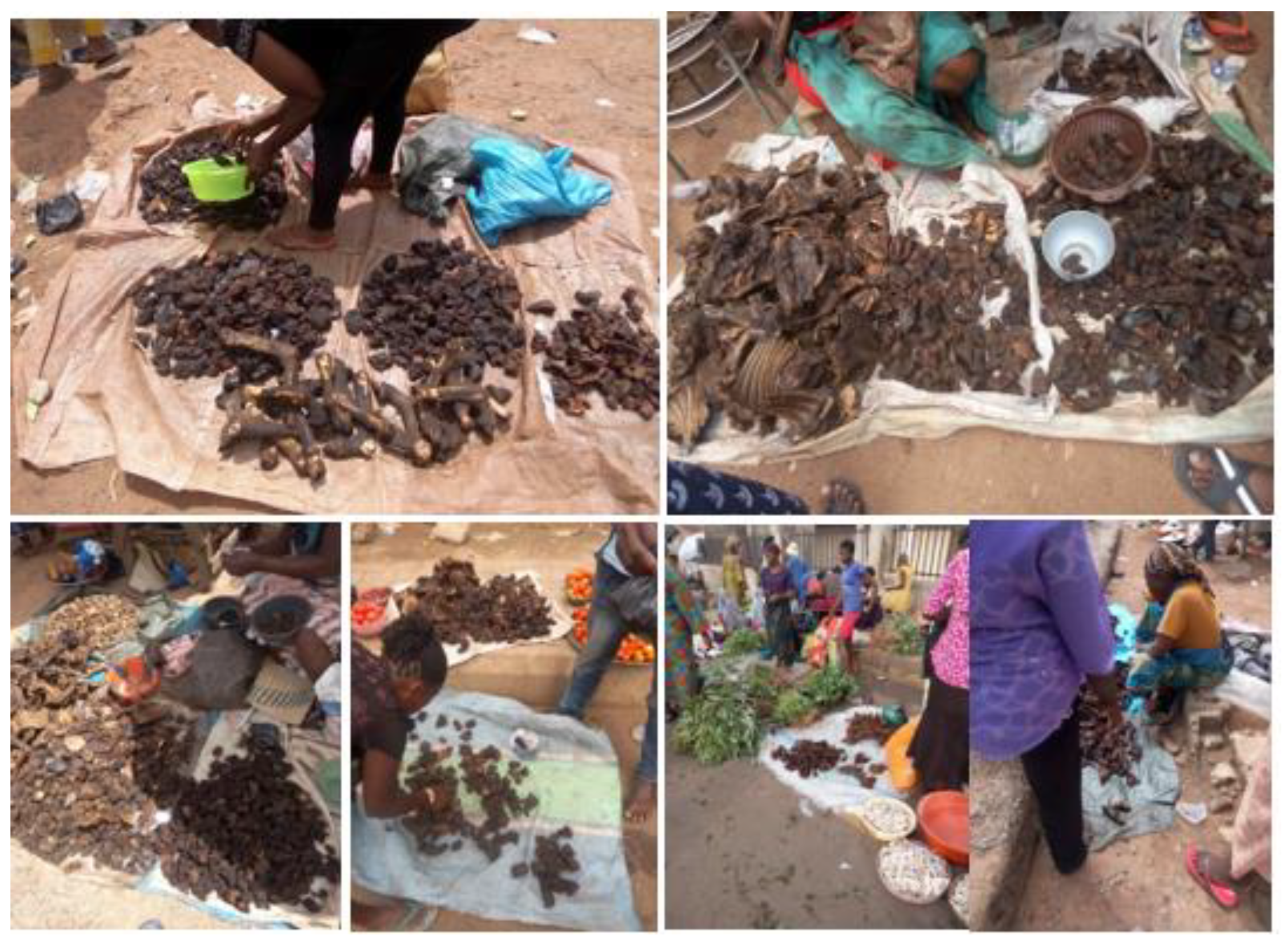
2.1. Study Area and Sampling Locations

This study was carried out at the department of Animal Health, Faculty of Natural Science and Agriculture, North-West University, South Africa, and samples were purchased from ten major local government areas of Ekiti State in Nigeria; these areas are as follows: Ado Ekiti and Igede town represent the central area, Ikole and Omuo town represent the eastern area, Aramoko and Ijero represent the western area, Oye and Otun represent the northern area, and Ilawe and Emure represent the southern area.

2.2. Sampling and Sample Preparation

One hundred and eight (108) samples of dry beef, each weighing about 200 g to 300 g, were obtained in triplicate from different markets in Ekiti State. The sampling design was a result of the availability of dry meat at the different markets. Representative samples were collected in plastic bags and sealed for transportation to the Food, Toxicology Research Laboratory, North-West University, Mafikeng Campus, South Africa, following all necessary protocols. All of the samples collected were meant for human consumption. On arrival at the laboratory, the meat samples were ground using a laboratory mill (IKA M20, Merck, Darmstadt, Germany) and stored at −20 °C for further analysis.

2.3. Mycological Screening of Samples

To determine the fungal population in the samples, the method described by [11], with slight modifications, was used. Potato Dextrose Agar (PDA) and Malt Extract Agar (MEA) were used. Potato Dextrose Agar and Malt Extract Agar were prepared by dissolving PDA (39 g) and MEA (39 g) in 1 litre of distilled water, respectively. Both agars were autoclaved at 121 °C and 15 psi for 15 min and cooled in a 50 °C water bath, and eight (8) millilitres (mL) each of 1% chloramphenicol and 1% streptomycin antibiotic solution were added to the agar before pouring into sterile Petri dishes.

2.4. Fungal Colony Counts

One (1) gram of each sample in triplicate was suspended in 9 mL of sterile Ringer’s solution vortexed for 2 min and serially diluted to make six dilutions (10−6) per sample. Ringer’s solution was prepared by dissolving two Ringer’s tablets in 1 litre of distilled water, which was autoclaved at 121 °C and 15 psi for 15 min. A 0.1 mL portion of the suspension was spread plated on PDA in triplicates and incubated at 28 °C for 48 h, and, subsequently, the colonies in each plate were counted, the average mean value was recorded as the fungal load for the 3 replicates in each sample, and the colony-forming unit (CFU/g) was calculated for each sample.
CFU/g = Number of colonies × reciprocal of the dilution factor
Plating volume (0.1 mL)

2.5. Fungal Isolation from Sample Cultures

One gram of each sample was suspended in 9 mL of sterile Ringer’s solution and vortexed for 2 min. A 0.1 mL aliquot of the suspension was spread plated in triplicate on Potato Dextrose Agar (PDA) and Malt Extract Agar (MEA) and incubated in the dark at 25 °C for 7 days. The individual fungal colony was carefully transferred into sterile solid MEA plates for final purification at 25 °C for 5 days before the fungal DNA was extracted. Identification of the isolated fungal was performed using a molecular method by PCR analysis and subsequently sequenced. The isolation occurrence of each species was calculated using the method described by [12].
Frequency   ( % ) =   N u m b e r   o f   s a m p l e s   c o n t a m i n a t e d   w i t h   a   s p e c i e   o r   g e n u s T o t a l   n u m b e r   o f   s a m p l e s × 100
Relative   Density   ( % ) = N u m b e r   o f   i s o l a t e s   o f   s p e c i e s   o r   g e n u s T o t a l   n u m b e r   o f   i s o l a t e s

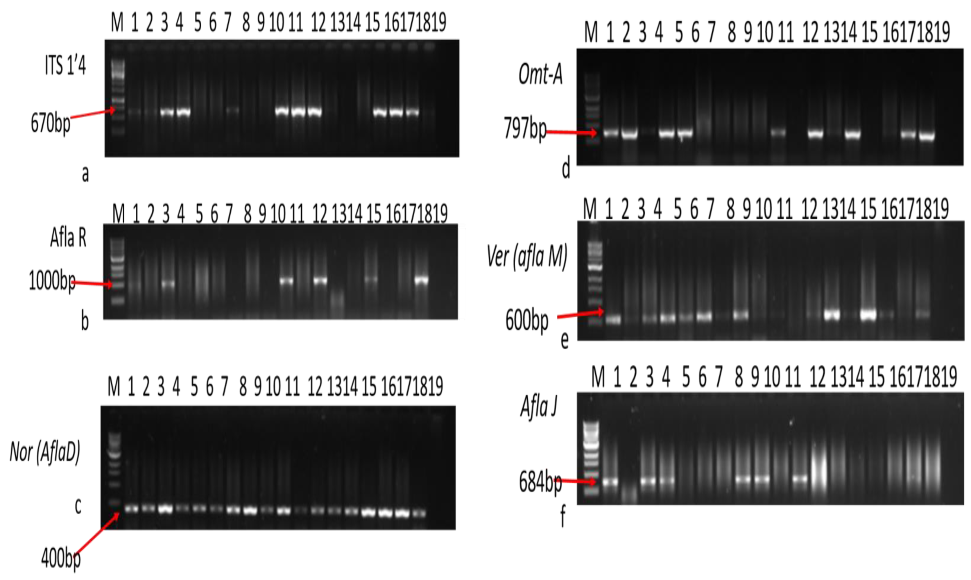
2.6. Molecular Characterization of Fungal Isolates

2.6.1. DNA Extraction

Deoxyribonucleotide (DNA) extraction was undertaken using a fungal/bacterial DNA extraction kit (Zymo Research Corporation, Tustin, CA, USA). Fungal spores from 4- to 5-day-old cultures were taken into the Bashing BeadTM lysis tubes, and 750 μL of DNA lysis solution was added to the tubes. This was followed by lysing of the fungal cells using a disruptor genie bead beater fitted with a 2 mL tube holder assembly (Scientific Industries Inc., New York, NY, USA) and processed at maximum speed for 15 min and centrifuged at 10,000 rpm for 1 min. The supernatant was transferred to a zymo-spin IV spin filter in a collection tube and centrifuged at 10,000 rpm for 1 min. The filtrate was added to 1200 μL of fungal DNA-binding buffer and centrifuged in a Zymo-Spin IIIc column placed in a collection tube at 10,000× g for 1 min. This was followed by washing DNA in the column using a pre-wash buffer of 200 μL and a wash buffer of 500 μL. Finally, after washing, 100 μL of the DNA elution buffer was added to the column and centrifuged at 10,000× g for 30 min to elute the DNA. Then, the eluted DNA was stored at −70 °C for further analysis.

2.6.2. Agarose Gel DNA Electrophoresis

Deoxyribonucleotide acid (DNA) gel electrophoresis was carried out by the preparation of 1% agarose gel 2 g of agarose (Fermentas Life Science, Vilnius, Lithuania) in 98 mL 1× TAE buffer (Fermentas Life Science, Lithuania), which was boiled in a microwave and cooled to approximately 60 °C. This was followed by the addition of 1 mL of ethidium bromide (Sigma-Aldrich, St. Louis, MO, USA) to the agarose solution. The agarose solution was poured into a casting chamber (Bio-Rad Laboratories, Hercules, CA, USA) and allowed to set. The casting chamber containing the solid agarose was further inserted into the electrophoresis tank (Bio-Rad Laboratories, CA, USA) filled with 1× TAE buffer. Fungal DNA (5 μL) was loaded into the wells. The chamber was closed and run at 400 V and 80 mA for 50 min, and DNA was viewed using the ChemDoc gel imaging system (Bio-Rad Laboratories, CA, USA).

2.6.3. PCR Amplification of Extracted Genomic DNA

The amplification of the Internal Transcribed Spacer Region (ITS rDNA) of the fungal isolates from the dry meat was performed using Polymerase Chain Reaction (PCR) with the universal ITS 1 (TCCGTAGGTGAACCTGCGG) and ITS 4 region primers (TCCTCCGCTTATTGATATGC). These primers were obtained from Inqaba Biotechnical Industrial (Pty) Ltd. (Pretoria, South Africa). The PCR amplicons were analyzed through electrophoresis on 1% (w/v) agarose gel to check the anticipated size of the amplicons (670 bp) and viewed by using Chem Doc Image Analyzer [13]. The final PCR solution consisted of 12 μL of master mix, 1 μL each for forward and reverse primers, and 3 μL of DNA, constituted to a final volume of 25 μL with nuclease-free water. Polymerase chain reaction was performed using an Eppendorf 96-well Thermocycler (Eppendorf, Hauppauge, NY, USA), and cycling conditions were set as pre-dwelling at 94 °C for 1 min, 35 cycles of denaturation at 94 °C for 1 min, annealing at 54 °C for 1 sec, extension at 72 °C for 1 min 30 s, post-dwelling at 72 °C for 9 min, and holding at 4 °C until the samples were retrieved.

2.6.4. Sequencing of PCR Products

The purified PCR products were sequenced at Inqaba Biotechnical Industrial (Pty) Ltd., Pretoria, South Africa, using the PRISMTM Ready Reaction Dye Terminator Cycle Sequencing Kit with the dideoxy chain cessation method and electrophoresed with a model ABI PRISM® 3500XL DNA Sequencer (Applied Biosystems, Foster City, CA, USA) by means of following the manufacturer’s instructions.

2.6.5. Sequence Analysis

Finch TV software, version 1.4.0, was employed for the analysis of chromatograms, (sense and antisense) ensuing from sequencing reactions for superior sequence assertion. The subsequent chromatographs were edited using Bio Edit Sequence Alignment Editor according to [14], after which the resultant consensus ITS rDNA sequences were imported into the NCBI database for blasting using the Basic Local Alignment Search Tool (BLAST) for homology in the identification of the likely organisms [15]. The sequences were subsequently deposited in GenBank for the allocation of accession numbers.

2.6.6. Detection of Aflatoxin-Producing Genes of Aspergilla

The DNA of assumed Aspergilla was analyzed for the presence of five important aflatoxin-producing genes (aflR, aflJ, aflM, aflD, and omt-A) in the isolated Aspergillus flavus, which was detected using a previously used and reported set of primers by [16], using primers described in Table 1. The primers were provided by Inqaba Biotechnical Industrial (Pty) Ltd., Pretoria, South Africa, and the PCR was conducted using a PCR Thermal Cycler (Applied Biosystems).

2.6.7. Optimization of Polymerase Chain Reaction

Polymerase Chain Reaction conditions were optimized separately for the target genes. A reaction volume of 25 μL, containing 8.5 μL nuclease-free water, 12.5 μL PCR Master Mix, 1 μL of oligonucleotide forward and reverse primers (10 μm), and 2 μL template DNA mixed in the PCR tubes, were used [21]. The thermal cycle conditions are also shown in Table 1, with varying annealing temperatures ranging from 58 to 75 °C for the five genes. The PCR-amplified products were checked on 1% gel by electrophoresis and visualized under the gel documentation system for electrophoretic bands at the various base pair regions for each gene.

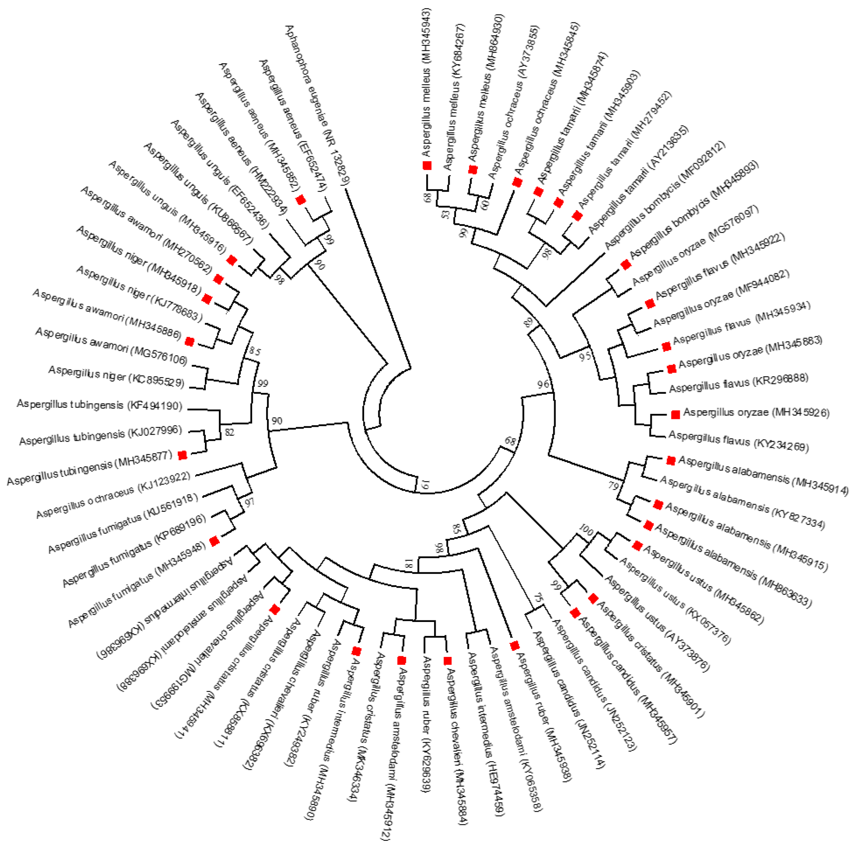
2.7. Phylogenetic Analysis

The evolutionary study was carried out using Molecular Evolutionary Genetics Analysis (MEGA) 5 [22], and the neighbour-joining method was used for evolutionary history, according to [23]. The evolutionary distances were calculated using the parameter method of [24], using the transversional substitutions per site unit of the number.

2.8. Statistical Analyses

Statistical analyses were performed using Microsoft Excel 2013 for Windows 2010. Descriptive statistics were employed to present the data collected from individual species isolated from each dried meat sampled and was considered one isolate. The concentrations were calculated based on the average recovery values acquired for each analyte. This was carried out using IBM SPSS version 25 software, with a statistical significance level of p < 0.05.

3. Results and Discussion

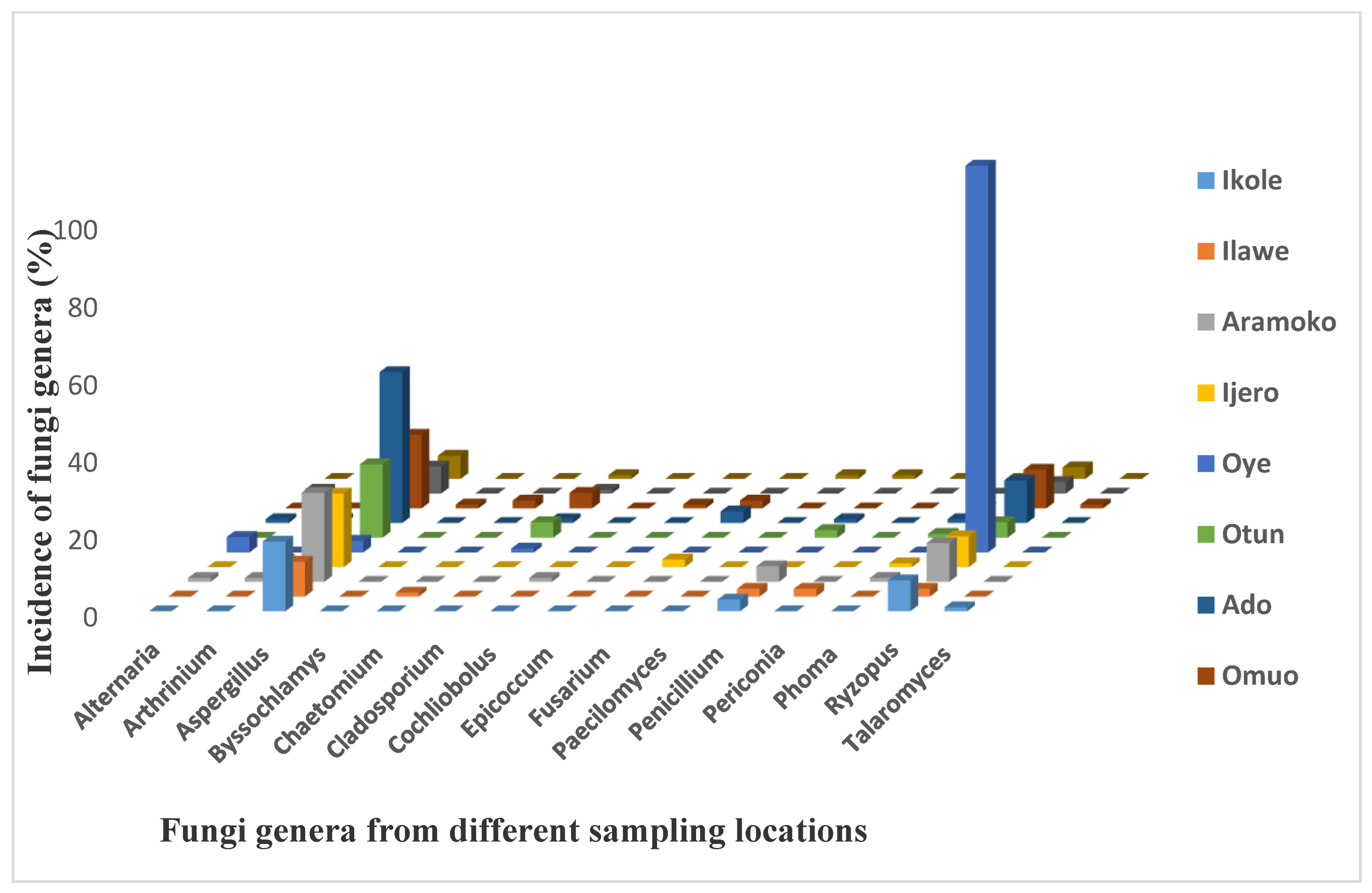
3.1. The Fungal Counts and Distribution of Fungal Isolates in Dry Meat

A total of 118 fungal isolates were isolated through the ITS section amplification and sequencing of the PCR amplicons of the isolates. One hundred percent (100%) of the dry meat analyzed was contaminated with Aspergillus of different species, with fungal counts ranging from 1.0 to 9.5 log10 cfu/g with a mean of 3.1 log10 cfu/g (Table 2). The dried meat sample from Ikole had the highest colony-forming unit range of 2.0 × 10²–1.6 × 10⁴, with an average mean value of 2.6 × 10³, though with 10 different types of fungi species contaminating them. The least colony-forming unit range was recorded in Ilawe meat samples with a range value of 1.0 × 101–4.5 × 10², with an average mean value of 1.3 × 10², and a total number of 11 fungi species contaminating them. The low fungal counts of the dried meat might be due to the low moisture content, which reduced the growth of fungi in the dry meat [25]. A similar report of fungal count (1.8 × 10³–4.6 × 10³ cfu/g) was reported for sun-dried meat samples (Kundi) obtained from major markets in Ibadan, Oyo State, in Nigeria [26].
This result shows that the majority of the fungi species isolated from the different locations are peculiar to the sampling environment and have been previously reported from other food samples, which implies that their occurrence in dried meat samples could be due to cross-contamination, as many of the dried meats are seen displayed close to other agricultural commodities (Figure 1). In addition, the fungi biodiversity could also be due to storage, the selling environment, and possible interactions with buyers. Customers are known to touch dried meats when buying them in order to select the ones that are deemed fit or of good quality (Figure 1). Another likely means of contamination is cross-contamination from the seller, who frequently touches the dried meat either when helping the buyer to select some good meat devoid of visible fungi contamination, when displaying the dried meat for sale, when packing after the end of daily sales, or when giving change to the buyers (Figure 1). Additionally, vehicular and human movements can also contribute to meat contamination as it moves from one end to the other. This movement sometimes raises dust, which eventually perches on or contaminates the dried meat, as they are not covered or protected from the environment where they are sold.
The overall fungal contamination results (Figure 2) showed that all the sampled locations have Aspergillus and Rhizopus fungal genera. These were found to contaminate the dry meat across the locations, whereas Penicillium genera were found in 6 out of the 10 locations investigated. The remaining fungal genera were found randomly in dry meat samples from other locations, with at least four different types occurring in Ikole, Oye, and Igede, respectively, whereas the highest fungal genera with 8 fungi were recorded in Ado and Omuo, followed by Aramoko with 7 fungal genera contamination. The dominance of Aspergillus and Rhizopus species in dry meat samples analyzed corresponds with the high incidence of Aspergillus and Rhizopus species in regions with high temperatures and humidity [27], with regard to the climatic conditions of Ekiti State (Nigeria). The state enjoys a tropical climate with two distinct seasons. These are the rainy season (April–October) and the dry season (November–March). The temperature ranges between 21 °C and 28 °C with high humidity. This finding is in agreement with the known knowledge of Aspergillus species as generally isolated from soil, air, enclosed surroundings, and agricultural products [28,29]. Fusarium and Cladosporium species were also seen to contaminate the bulk of the sampled meats. Penicillium has been reported to infect maize and maize-based produce in West Africa at diverse yield periods [30]. Additional species found to occur with a lesser prevalence comprised A. aeneus, A. alabamensis, A. amstelodami, A. awamori, A. chevalieri, A. melleus, A. ruber, A. tubingensis, A. unguis, Byssochlamys spectabilis, Chaetomium erectum, Cochliobolus sp., Epicoccum sorghinum, Fusarium equiseti, Fusarium incarnatum, Paecilomyces formosus, Penicillium funiculosum, Penicillium mallochii, Periconia macrospinosa, Phoma herbarum, and Talaromyces funiculosus. The higher incidence of Aspergillus, Rhizopus, and Penicillium species in dry meat samples collected across the state could be because Aspergillus, Rhizopus, and Penicillium species are known to be storage fungi that usually contaminate food products under storage [31,32,33]. The occurrence of these fungal species in dry meat is a pointer to possible mycotoxin contamination because the majority of them are known producers of main mycotoxins [34]. For instance, A. flavus and A. niger are known producers of aflatoxins in agricultural produce [35,36].

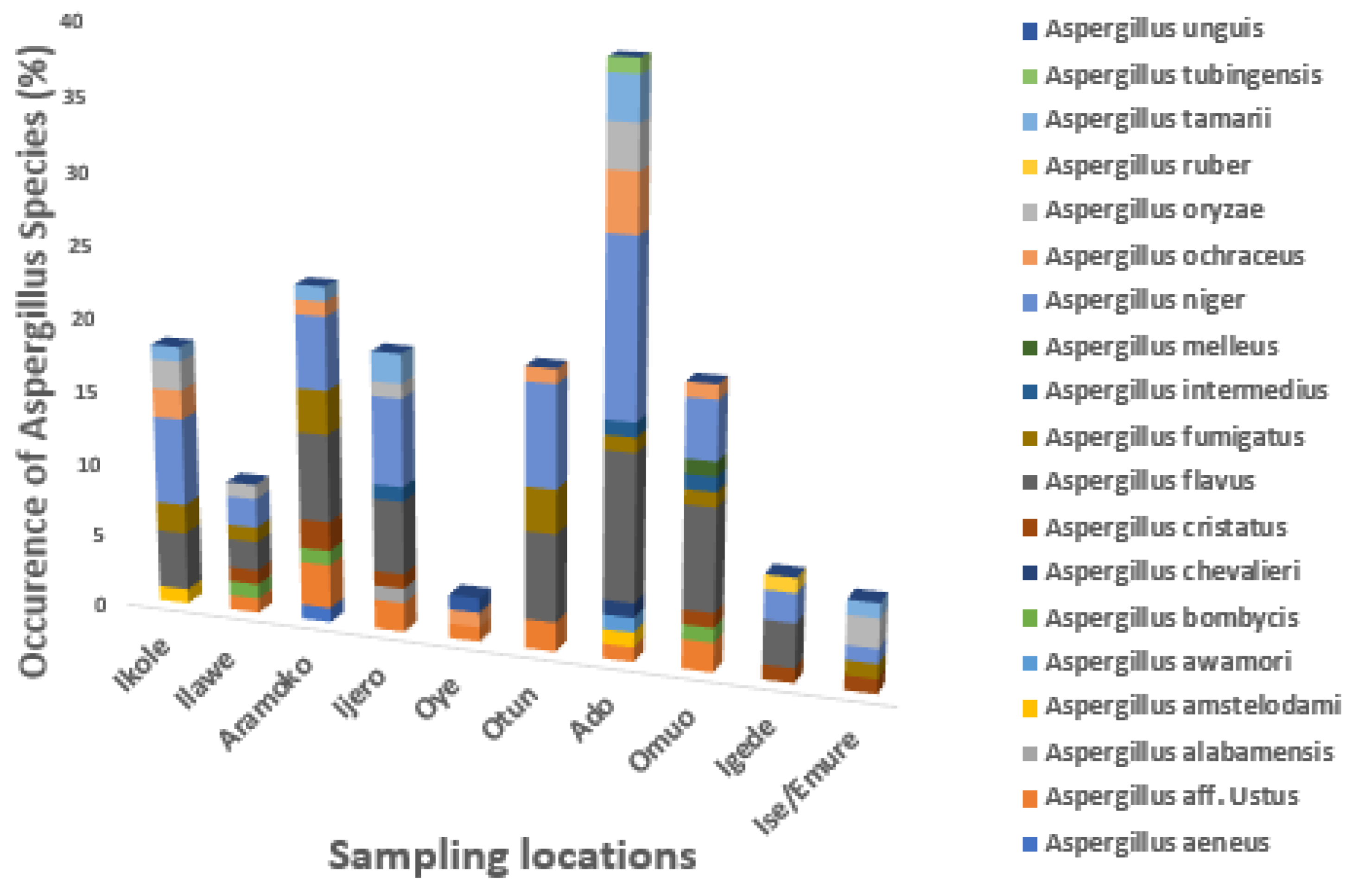
3.2. The Occurrence of Aspergillus Species in Dried Meat across Different Locations

The occurrence of Aspergillus species differs from each location as shown in Figure 3. The findings showed that Aspergillus flavus was not found in Oye and Ise/Emure; Aspergillus oryzae was not found in Aramoko, Oye, Otun, Omuo, and Igede; Aspergillus aeneu was found in Aramoko, and Aspergillus alabamensis was found in Ijero; Aspergillus awamori, Aspergillus chevalieri, and Aspergillus tubingensis were found in Ado; Aspergillus unguis was found only in Oye; and Aspergillus bombycis was found in Ilawe, Aramoko, and Omuo. Aspergillus intermedius was found in Ijero, Ado, and Omuo.
The possession of the aflatoxin gene is shown in Figure 4, where M: 1 kb DNA ladder; lane 1, 3, 4, 7, 10, 11, 12, 16, 17 and 18 shown fungal gene expression while lane 19 is the negative controls. The b gel electrophoretic pattern is shown for PCR products expressing the aflR gene at the 1000 bp region. M: 1 kb DNA ladder; lanes 1, 3, 10, 12, 15, and 18: positive isolates, lane 19: negative control, and lanes 2, 4–9, 11, 13, 14, 16, and 17: negative isolates. The c gel electrophoretic arrangement is shown for PCR products expressing the aflD gene (Nor-1) at the 400 bp region. M: 1 kb DNA ladder; lane 19: negative control, and lanes 1–18: amplification of the nor gene in positive isolates. The d gel electrophoretic pattern for PCR products expressing the omt-A gene at 797 bp is shown. M: 1 kb DNA ladder; lanes 1, 2, 4, 5, 10, 12, 14, 17, and 18: positive isolates, lane 19: negative control; and lanes 3, 6–9, 11, 13, –15, and 16: amplification of omt-A gene in positive isolates. The e gel electrophoretic pattern for PCR products expressing the aflM gene (Ver) at 600 bp is shown. M: 1 kb DNA ladder; lanes 4, 5, 6, 8,13, and 15: amplification not at the anticipated band size; lane 19: negative control; and lanes 1,3,16, and 18: positive isolates. The f Gel electrophoretic pattern for PCR products expressing the aflJ gene at the 684 bp region is shown. M: 1 kb DNA ladder; lanes 1, 3, 4, 8, 9, 11, and 17: positive isolates; lane 19: negative control; and lanes 3, 18, and 24: negative isolates.
This study found that 75% of the isolated Aspergillus flavus strains contained both the aflatoxin biosynthetic regulatory genes, aflR and aflJ, along with three key genes in the aflatoxin biosynthetic pathway: omt-A, aflD (nor-1), and aflM (ver-1) (Table 2). Other isolates showed the presence of one or more of these genes but not the full set. The detection of these genes is concerning, as it indicates the potential for mycotoxin production if environmental conditions become favourable for fungal growth. It has been reported that aflR, omt-A, aflD, and aflM remain the most vital genes from all the 25 reported genes in the aflatoxin biosynthetic group that control aflatoxin production [37]. The aflR gene controls the initiation of the transcript of pathway genes and aflatoxin biosynthesis [38], but its occurrence in the isolates may not automatically result in aflatoxin production since the aflatoxin pathway is controlled by several mechanisms [39,40]. Also, aflJ is likewise an aflatoxin biosynthetic controlling gene identical to the aflR; the dual genes are different transcripts with separate sponsors. It is essential for the expression of other genes in the aflatoxin group and is also involved in the transformation of pathway transitional products to aflatoxins [41]. The aflR protein can bind the promoter area of individual aflatoxin synthesis genes and trigger gene expression [42]. Also, aflatoxin biosynthesis is controlled by many ecological factors, for example, light [43], carbon source, temperature, and pH [44]. Table 3 gives overall information about the sequence of A. flavus that shows gene expression to aflatoxin biosynthetic genes among all the Aspergillus species isolated from the dried beef meat as submitted to GenBank.
The maximum likelihood phylogenetic tree based on the partial ITS rDNA gene sequence, showing the phylogenetic relationships between identified Aspergillus species and the most closely related Aspergillus strains from GenBank, is shown in Figure 5. The numbers at the nodes indicate the levels of bootstrap support based on 1000 resampled data sets. Only values greater than 50% are shown. The scale bar indicates 0.5 nucleotide substitution per site. Aphanophora eugeniea is set as the outgroup. Sequences obtained in this study are denoted with a red circle. This result is divided into six main clades, comprising the following. Clade I: Aspergillus unguis and A. aeneus shared 98% and 99% with the reference of the same species from GenBank. Clade II: Aspergillus fumigatus shared 97%. A. awamori, A. niger (85%), and A. tubingensis shared 82%. Clade III: A. ustus shared 68% with A. ustus in GenBank. A. cristatus shared 68% homology with A. ustus and 99% with A. candidus. A. candidus also shared 68% with the reference strain of A. candidus from GenBank. This similarity index is quite low, except for A. ustus, since it is below the 70% anticipated borderline for the degree of similarity according to [45]. A. ruber shared 98%, with A. amstelodami as the closest relative. The samples of A. cristatus, A. intermedius, A. amstelodami, and A. chevalieri shared 81% with the reference strain from GenBank. Clade IV A. alabamensis shared 79% with the reference strain, while Clade V A. oryzae, A. flavus, and A. bombycis shared 95% with the reference strain. Clade VI A. tamarii shared 98% with the reference strain. A. ochraceus shared 99% with the A. ochraceus and A. melleus reference strains. This high homology percentage shows that they possessed nearly the same nucleotide signature as their relative counterpart from GenBank [46]. This high similarity index expressed by these fungi is beyond the 70% borderline of the expected degree of relatedness [45]. A. melleus from this study shared 60% with A. ochraceus and 68% with A. melleus reference from GenBank. This fungus has a different nucleotide signature, and its low threshold makes it prone to being wiped out [47]. This could be a novel Aspergillus species.

4. Conclusions

Based on the findings of this study, Aspergillus species isolated from dried meat in Ekiti State, Nigeria, showed significant biodiversity and a high potential for mycotoxin production. The presence of key aflatoxin biosynthetic genes in a majority of the isolates highlights the potential health risks associated with consuming contaminated dried meat. Given these findings, there is a need for stricter regulations and increased awareness of the dangers posed by Aspergillus contamination to ensure food safety and protect public health.

Author Contributions

Conceptualization, T.A.D., L.N. and M.M.; Methodology, T.A.D., L.N. and M.M.; Formal Analysis and Investigation, T.A.D., M.M. and L.N.; Writing—Original Draft Preparation, T.A.D.; Writing—Review and Editing, T.A.D., T.I.E. and M.M. All authors have read and agreed to the published version of the manuscript.

Funding

This research was supported by the North-West University Graduate Research Bursary 2016–2018 and the National Research Fund.

Data Availability Statement

The datasets backing the conclusion of this article are included within the article, and the nucleotide sequences presented in this article are accessible through GenBank.

Conflicts of Interest

The authors declare no conflicts of interest.

References

  1. Lawrie, R.A.; Ledward, D. Lawrie’s Meat Science, 7th ed.; Woodhead Publishing: Cambridge, UK, 2014; p. 1. [Google Scholar]
  2. Semba, R.D.; Ramsing, R.; Rahman, N.; Kraemer, K.; Bloem, M.W. Legumes as a sustainable source of protein in human diets. Glob. Food Secur. 2021, 28, 100520. [Google Scholar] [CrossRef]
  3. Millward, D.J.; Garnett, T. Plenary Lecture 3 Food and the planet: Nutritional dilemmas of greenhouse gas emission reductions through reduced intakes of meat and dairy foods: Conference on ‘Over-and undernutrition: Challenges and approaches. Proc. Nutr. Soc. 2010, 69, 103–118. [Google Scholar] [CrossRef]
  4. Zhou, G.H.; Xu, X.L.; Liu, Y. Preservation technologies for fresh meat–A review. Meat Sci. 2010, 86, 119–128. [Google Scholar] [CrossRef]
  5. Egbunike, G.N.; Okubanjo, A.O. Effects of processing upon the quality of Nigerian meat products. Livest. Prod. Sci. 1999, 59, 155–163. [Google Scholar] [CrossRef]
  6. Fonkem, D.N.; Tanya, V.N.; Ebangi, A.L. Effects of season on the microbiological quality of Kilishi, a traditional Cameroonian dried beef product. Tropicultura 2010, 28, 10–15. [Google Scholar]
  7. Wiklund, E.; Malmfors, G.; Finstad, G.L.; Åhman, B.; Skuterud, L.; Adamczewski, J.; Garner, K. Meat quality and meat hygiene. In Reindeer and Caribou, 1st ed.; CRC Press: Boca Raton, FL, USA, 2018; pp. 353–382. [Google Scholar]
  8. Asefa, D.T.; Kure, C.F.; Gjerde, R.O.; Omer, M.K.; Langsrud, S.; Nesbakken, T.; Skaar, I. Fungal growth pattern, sources and factors of mould contamination in a dry-cured meat production facility. Int. J. Food Microbiol. 2010, 140, 131–135. [Google Scholar] [CrossRef]
  9. Ajiboye, E.A.; Alhassan, S.; Adedayo, R.M.; Kolawole, M.O.; Oladosu, O.T. Physicochemical properties and microorganisms isolated from dried meat obtained in Oja-Oba market in Ilorin, Nigeria. Pelagia Res. Libr. 2011, 2, 391–400. [Google Scholar]
  10. Oladejo, D.A.; Adebayo-Tayo, B.C. Moulds, proximate mineral composition and mycotoxin contamination of Banda (“kundi”/ “tinko”) sold in Ibadan, Oyo State, Nigeria. AU J. Technol. 2011, 15, 32–40. [Google Scholar]
  11. Cary, J.W.; Gilbert, M.K.; Lebar, M.D.; Majumdar, R.; Calvo, A.M. Aspergillus flavus secondary metabolites: More than just aflatoxins. Food Saf. 2018, 6, 7–32. [Google Scholar] [CrossRef]
  12. Njobeh, P.B.; Dutton, M.F.; Koch, S.H.; Chuturgoon, A.; Stoev, S.; Seifert, K. Contamination with storage fungi of human food from Cameroon. Int. J. Food Microbiol. 2009, 135, 193–198. [Google Scholar] [CrossRef]
  13. Adetunji, M.C.; Alika, O.P.; Awa, N.P.; Atanda, O.O.; Mwanza, M. Microbiological quality and risk assessment for aflatoxins in groundnuts and roasted cashew nuts meant for human consumption. J. Toxicol. 2018, 2018, 1308748. [Google Scholar] [CrossRef]
  14. Sambrook, J.; Fritsch, E.F.; Maniatis, T. Molecular Cloning: A Laboratory Manual; Cold Spring Harbor Laboratory Press: New York, NY, USA, 1989. [Google Scholar]
  15. Hall, T. Bio Edit; Ibis Therapeutics: Carlsbad, CA, USA, 2004. [Google Scholar]
  16. Altschul, S.F.; Koonin, E.V. Iterated profile searches with PSI-BLAST—A tool for discovery in protein databases. Trends Biochem. Sci. 1998, 23, 444–447. [Google Scholar] [CrossRef]
  17. Rashid, M.; Khalil, S.; Ayub, N.; Ahmed, W.; Khan, A.G. Categorization of Aspergillus flavus and Aspergillus parasiticus isolates of stored wheat grains in to aflatoxigenic and non-aflatoxigenic. Pak. J. Bot 2008, 40, 2177–2192. [Google Scholar]
  18. Gallo, A.; Tandon, M.; Alevizos, I.; Illei, G.G. The majority of microRNAs detectable in serum and saliva is concentrated in exosomes. PLoS ONE 2012, 7, e30679. [Google Scholar] [CrossRef]
  19. Fakruddin, M.D.; Chowdhury, A.; Hossain, M.N.; Ahmed, M.M. Characterization of aflatoxin producing Aspergillus flavus from food and feed samples. Springer Plus 2015, 4, 159. [Google Scholar] [CrossRef]
  20. Abd-Elghany, S.M.; Sallam, K.I. Rapid determination of total aflatoxins and ochratoxins A in meat products by immuno-affinity fluorimetry. Food Chem. 2015, 179, 253–256. [Google Scholar] [CrossRef]
  21. Montso, P.K.; Ateba, C.N. Molecular detection of Clostridium species in beef obtained from retail shops in North West Province, South Africa. J. Food Nutr. Res. 2014, 2, 236–243. [Google Scholar] [CrossRef]
  22. Tamura, K.; Peterson, D.; Peterson, N.; Stecher, G.; Nei, M.; Kumar, S. MEGA5: Molecular evolutionary genetics analysis using maximum likelihood, evolutionary distance, and maximum parsimony methods. Mol. Biol. Evol. 2011, 28, 2731–2739. [Google Scholar] [CrossRef]
  23. Saitou, N.; Nei, M. The neighbor-joining method: A new method for reconstructing phylogenetic trees. Mol. Biol. Evol. 1987, 4, 406–425. [Google Scholar] [CrossRef]
  24. Kimura, M. A simple method for estimating evolutionary rates of base substitutions through comparative studies of nucleotide sequences. J. Mol. Evol. 1980, 16, 111–120. [Google Scholar] [CrossRef]
  25. Ferreira, V.; Barbosa, J.; Vendeiro, S.; Mota, A.; Silva, F.; Monteiro, M.J.; Hogg, T.; Gibbs, P.; Teixeira, P. Chemical and microbiological characterization of alheira: A typical Portuguese fermented sausage with particular reference to factors relating to food safety. Meat Sci. 2006, 73, 570–575. [Google Scholar] [CrossRef] [PubMed]
  26. Adeyeye, S.A. Quality and safety assessment of sun-dried meat product (kundi) from Ibadan, Oyo state, Nigeria. Cogent Food Agric. 2016, 2, 1209074. [Google Scholar] [CrossRef]
  27. Castellari, C.; Quadrelli, A.M.; Laich, F. Surface mycobiota on Argentinean dry fermented sausages. Int. J. Food Microbiol. 2010, 142, 149–155. [Google Scholar] [CrossRef]
  28. Klich, M.A.; Mullaney, E.J.; Daly, C.B.; Cary, J.W. Molecular and physiological aspects of aflatoxin and sterigmatocystin biosynthesis by Aspergillus tamarii and A. ochraceoroseus. Appl. Microbiol. Biotechnol. 2000, 53, 605–609. [Google Scholar] [CrossRef]
  29. Oyetunji, T.O. Mycological evaluation of a ground cocoa-based beverage. Afr. J. Biotechnol. 2006, 5, 2073–2076. [Google Scholar]
  30. Borgemeister, C.; Adda, C.; Sétamou, M.; Hell, K.; Djomamou, B.; Markham, R.H.; Cardwell, K.F. Timing of harvest in maize: Effects on post-harvest losses due to insects and fungi in central Benin, with particular reference to Prostephanus truncatus (Horn) (Coleoptera: Bostrichidae). Agric. Ecosyst. Environ. 1998, 69, 233–242. [Google Scholar] [CrossRef]
  31. Bankole, S.A.; Adebanjo, A. Mycotoxins in food in West Africa: Current situation and possibilities of controlling it. Afr. J. Biotechnol. 2003, 2, 254–263. [Google Scholar]
  32. Makun, H.A.; Dutton, M.F.; Njobeh, P.B.; Phoku, J.Z.; Yah, C.S. Incidence, phylogeny and mycotoxigenic potentials of fungi isolated from rice in Niger State, Nigeria. J. Food Saf. 2011, 31, 334–349. [Google Scholar] [CrossRef]
  33. Pitt, J.I.; Hocking, A.D.; Pitt, J.I.; Hocking, A.D. Primary keys and miscellaneous fungi. In Fungi Food Spoilage; Springer: Boston, MA, USA, 2009; pp. 53–143. [Google Scholar] [CrossRef]
  34. Sweeney, M.J.; Dobson, A.D. Mycotoxin production by Aspergillus, Fusarium and Penicillium species. Int. J. Food Microbiol. 1998, 43, 141–158. [Google Scholar] [CrossRef]
  35. Mulunda, M.; Dzoma, B.; Nyirenda, M.; Bakunzi, F. Mycotoxins occurrence in selected staple food in main markets from Lubumbashi, Democratic Republic of Congo. J. Food Agric. Environ. 2013, 11, 51–54. [Google Scholar]
  36. Reiter, E.V.; Vouk, F.; Böhm, J.; Razzazi-Fazeli, E. Aflatoxins in rice–A limited survey of products marketed in Austria. Food Control 2010, 21, 988–991. [Google Scholar] [CrossRef]
  37. Davari, E.; Mohsenzadeh, M.; Mohammadi, G.; Rezaeian-Doloei, R. Characterization of aflatoxigenic Aspergillus flavus and A. parasiticus strain isolates from animal feedstuffs in northeastern Iran. Iran. J. Vet. Res. 2015, 16, 150. [Google Scholar] [PubMed]
  38. Woloshuk, C.P.; Foutz, K.R.; Brewer, J.F.; Bhatnagar, D.; Cleveland, T.E.; Payne, G. Molecular characterization of aflR, a regulatory locus for aflatoxin biosynthesis. Appl. Environ. Microbiol. 1994, 60, 2408–2414. [Google Scholar] [CrossRef]
  39. Bok, J.W.; Keller, N.P. LaeA, a regulator of secondary metabolism in Aspergillus spp. Eukaryot. Cell 2004, 3, 527–535. [Google Scholar] [CrossRef]
  40. Perrin, R.M.; Fedorova, N.D.; Bok, J.W.; Cramer, R.A., Jr.; Wortman, J.R.; Kim, H.S.; Nierman, W.C.; Keller, N.P. Transcriptional regulation of chemical diversity in Aspergillus fumigatus by LaeA. PLoS Pathog. 2007, 3, e50. [Google Scholar] [CrossRef]
  41. Georgianna, D.R.; Hawkridge, A.M.; Muddiman, D.C.; Payne, G.A. Temperature-dependent regulation of proteins in Aspergillus flavus: Whole organism stable isotope labeling by amino acids. J. Proteome Res. 2008, 7, 2973–2979. [Google Scholar] [CrossRef]
  42. Ehrlich, K.C.; Montalbano, B.G.; Cary, J.W. Binding of the C6-zinc cluster protein, AFLR, to the promoters of aflatoxin pathway biosynthesis genes in Aspergillus parasiticus. Gene 1999, 230, 249–257. [Google Scholar] [CrossRef]
  43. Calvo, A.M.; Wilson, R.A.; Bok, J.W.; Keller, N.P. Relationship between secondary metabolism and fungal development. Microbiol. Mol. Biol. Rev. 2002, 66, 447–459. [Google Scholar] [CrossRef]
  44. Obrian, G.R.; Georgianna, D.R.; Wilkinson, J.R.; Yu, J.; Abbas, H.K.; Bhatnagar, D.; Cleveland, T.E.; Nierman, W.; Payne, G.A. The effect of elevated temperature on gene transcription and aflatoxin biosynthesis. Mycologia 2007, 99, 232–239. [Google Scholar] [CrossRef]
  45. Wayne, L.G.; Brenner, D.J.; Colwell, R.R.; Grimont, P.A.; Kandler, O.; Krichevsky, M.I.; Moore, L.H.; Moore, W.E.; Murray, R.; Stackebrandt, E.S.; et al. Report of the ad hoc committee on reconciliation of approaches to bacterial systematics. Int. J. Syst. Evol. Microbiol. 1987, 37, 463–464. [Google Scholar] [CrossRef]
  46. Aremu, B.R.; Babalola, O.O. Construction of specific primers for rapid detection of South African exportable vegetable macergens. Int. J. Environ. Res. Public Health 2015, 12, 12356–12370. [Google Scholar] [CrossRef] [PubMed]
  47. Konstantinidis, K.T.; Stackebrandt, E. Defining taxonomic ranks. Prokaryotes 2013, 1, 229–254. [Google Scholar]
Figure 1. Open display methods of selling dried meat across the sampling locations in Ekiti State.
Foods 13 03221 g001
Figure 2. Incidence (%) of different fungi genera contaminating dry meat according to various locations.
Foods 13 03221 g002
Figure 3. The occurrence of Aspergillus species in dried meat across different locations.
Foods 13 03221 g003
Figure 4. Gel electrophoretic analysis for PCR products showing A. flavus gene presence at different bp regions.
Foods 13 03221 g004
Figure 5. Maximum likelihood phylogenetic tree for identified Aspergillus species and their most closely related strains from GenBank.
Foods 13 03221 g005
Table 1. Sequences of the nucleotide primers used in this study.
Primer CodeTarget GenePrimer Sequences 5′–3′Product Size (bp)
Nor-FaflD (nor-1)ACCGCTACGCCGGCACTCTCGGCAC400
Nor-RGTTGGCCGCCAGCTTCGACACTCCG
Ver-FaflM (ver-1)GCCGCAGGCCGCGGAGAAAGTGGT600
Ver-RGGGGATATACTCCCGCGACACAGCC
Omt-FaflP (omtA)GTGGACGGACCTAGTCCGACATCAC797
Omt-RGTCGGCGCCACGCACTGGGTTGGGG
AflR-forAflRTATCTCCCCCCGGGCATCTCCCGG1000
AflR-revCCGTCAGACAGCCACTGGACACGG
AflJ-foraflS (aflJ)TGAATCCGTACCCTTTGAGG684
AflJ-revGGAATGGGATGGAGATGAGA
References: [17,18,19,20].
Table 2. Colony-forming unit (Cfu/g) of fungal species recovered from dried meat per location.
LocationCFU g−1 RangeMeanNo of Fungi Isolates
Ikole2.0 × 102–1.6 × 1042.6 × 10310
Ilawe1.0 × 101–4.5 × 1021.3 × 10211
Aramoko1.0 × 102–6.9 × 1031.5 × 10316
Ijero2.0 × 102–3.2 × 1021.5 × 10213
Oye5.0 × 101–4.1 × 1022.9 × 1026
Otun1.0 × 101–1.9 × 1025.4 × 10111
Ado2.0 × 101–2.1 × 1033.2 × 10219
Omuo3.0 × 101–6.4 × 1021.6 × 10216
Igede9.0 × 101–3.2 × 1021.0 × 1027
Ise/Emure1.0 × 101–2.5 × 1029.5 × 1019
Table 3. The potential of isolated A. flavus to produce aflatoxin.
S/NoName of IsolatesNor
afLD
Ver
afLM
Omt
afLP
afLRafLJAPAccession
Number
NCBI %
Similarity
1A. flavus+++++PMH34593699
2A. flavus+++++PMH34587099
3A. flavus+++++PMH345846100
4A. flavus+++++PMH345934100
5A. flavus+++++PMH345922100
6A. flavus+++++PMH34589599
7A. flavus+++++PMH34587999
8A. flavus+++++PMH34595999
9A. flavus+++++PMH34584299
10A. flavus+++++PMH34593299
11A. flavus+++++PMH345881100
12A. flavus+++++PMH345952100
13A. flavus+++++PMH34591399
14A. flavus+++++PMH345910100
15A. flavus+++++PMH34594699
P = potential to produce aflatoxin, + means positive gene expression to the aflatoxin genes, AP = aflatoxin production.
Disclaimer/Publisher’s Note: The statements, opinions and data contained in all publications are solely those of the individual author(s) and contributor(s) and not of MDPI and/or the editor(s). MDPI and/or the editor(s) disclaim responsibility for any injury to people or property resulting from any ideas, methods, instructions or products referred to in the content.

