Communication

Antimicrobial Activity of Grapefruit Seed Extract on Edible Mushrooms Contaminations: Efficacy in Preventing Pseudomonas spp. in Pleurotus eryngii

1
Department of Medical Sciences and Public Health, University of Cagliari, Cittadella Universitaria Monserrato-S.P. Monserrato-Sestu Km 0.700, 09042 Monserrato, CA, Italy
2
Analysis Laboratory, ASL Cagliari, Via Piero della Francesca, 1, 09047 Su Planu, CA, Italy
3
Department of Life and Environmental Sciences, Blocco A, Room 13, University of Cagliari, Cittadella Universitaria Monserrato-S.P. Monserrato-Sestu Km 0.700, 09042 Monserrato, CA, Italy
*
Author to whom correspondence should be addressed.
Foods 2024, 13(8), 1161; https://doi.org/10.3390/foods13081161
Submission received: 23 February 2024 / Revised: 5 April 2024 / Accepted: 9 April 2024 / Published: 11 April 2024

Abstract

:
Pleurotus eryngii is an edible mushroom that suffers significant losses due to fungal contamination and bacteriosis. The Pseudomonadaceae family represents one of the most frequent etiologic agents. Grapefruit seed extract (GSE) is a plant extract that contains different bioactive components, such as naringin, and exhibits a strong antibacterial and antioxidant activity. Over the last decade, GSE use as an alternative to chemical treatments in the food sector has been tested. However, to our knowledge, its application on mushroom crops has never been investigated. This study focuses on evaluating GSE efficacy in preventing P. eryngii yellowing. GSE antibiotic activity, inhibitory and bactericidal concentrations, and antibiofilm activity against several microorganisms were tested with the Kirby–Bauer disk diffusion assay, the broth microdilution susceptibility test, and the Crystal violet assay, respectively. In vitro, the extract exhibited antimicrobial and antibiofilm activity against Staphylococcus aureus 6538 and MRSA (wild type), Escherichia coli ATCC 8739, and Pseudomonas spp. (Pseudomonas aeruginosa 9027, P. fluorescens (wild type)). GSE application in vivo, in pre- and post-sprouting stages, effectively prevented bacterial infections and subsequent degradation in the mushroom crops: none of the P. eryngii treated manifested bacteriosis. Our findings support the use of GSE as an eco-friendly and sustainable alternative to chemical treatments for protecting P. eryngii crops from bacterial contamination, consequently ensuring food safety and preventing financial losses due to spoilage. Furthermore, GSE’s potential health benefits due to its content in naringin and other bioactive components present new possibilities for its use as a nutraceutical in food fortification and supplementation.

Graphical Abstract

1. Introduction

Pleurotus eryngii is a basidiomycete mushroom known in Italy as “cardoncello” [1,2]. It is also known as “royal trumpet” or “royal oyster” and is one of the most valuable edible mushrooms, with several varieties. Typically found in southern Europe, Northern Africa, the Middle East, and Central Asia, it is considered one of the most widely spread species of Pleurotus and has been known since ancient times for its excellent medicinal and nutritional characteristics [3,4,5]. This mushroom possesses a low percentage of calories and has a good concentration of major nutrients, such as protein, peptides, minerals, terpenoids, traces of various elements, fiber, and polysaccharides (P. eryngii polysaccharides—PEPs). These characteristics have aroused particular interest because of the anti-cancer, hepatoprotective, anti-lipidemia, immune system strengthening, and other activities shown in vitro and in animal models [6,7,8,9,10]. For this reason, P. eryngii is classified among functional foods [11,12,13] and used to create “healthy snacks” [14], namely, foods with high nutritional and biological value (rich in fiber and protein, low in salt, sugar, fat, and calories) [11]. Although this edible mushroom has several beneficial properties for our body, some peculiarities, such as a susceptibility to contaminations by mold and bacterial diseases [15], may compromise its quality. Generally, fungus-induced contaminations are attributed to Cladobotryum mycophilum, responsible for “spider’s web disease” [16,17,18], Gliocladium roseum, responsible for “brown spot” [19], and Trichoderma spp., responsible for “green mold” [20]. The bacterial blight culprits belong to several species, such as Pantoea spp. [14], Erwinia beijingensis, Ewingella americana [21,22], Enterobacter amnigenus, and Staphylococcus spp. However, Pseudomonadaceae, such as Pseudomonas tolaasii and P. fluorescens, are the most relevant and responsible for the stem’s yellowing [15,23,24]. It has indeed been noted that the initial colonization of the cap induces a loss in production yield; the size of the fruiting body is affected by the bacterial populations in the pre-harvest stage, consequently jeopardizing the quality of the harvested products [15,25]. The yellowing of P. eryngii is a bacteriosis that manifests with small yellow or light brown spots on the pileum accompanied by water-rich elongated and coalescing areas on the stem. P. eryngii affected by yellowing show a setback in the growth process, turn reddish-brown, and reach the state of rot (Figure 1), which manifests in the final stages with an unpleasant and nauseating odor [23,26]. The occurrence of the infection, as well as its intensity, are influenced by particular environmental conditions, such as high humidity in the growing chambers and hot muggy winds, such as sirocco [15], which are typical of the Mediterranean climate.
Innovative experiments and alternatives to the use of chemicals, potentially polluting substances, are currently being evaluated to prevent such diseases, which, with a drastic decrease in P. eryngii sporophores, cause considerable financial losses to producers. For example, repeated applications of white wine vinegar in fungal cultures at different concentrations have been evaluated; the acetic acid with 3% concentration has, in fact, an antimicrobial activity on P. aeruginosa and other bacteria [23,27]. The scientific community is focused on finding effective molecules propelling biotechnology in an eco-friendly and sustainable direction. Along this line, the present study evaluates grapefruit seed extract’s antimicrobial and antibiofilm activity on several microorganisms in vitro and in vivo, focusing on preventing Pseudomonas spp., in particular P. fluorescens., in Pleurotus eryngii.
Grapefruit seed extract (GSE) is a well-known plant extract with strong antibacterial and antioxidant activity [28,29]. GSE applications span from use in the food sector as a food preservative and infusion into packaging matrices [30,31] to pharmaceutics (e.g., diet supplements, wound healing, glucose and lipid blood level management, etc.) [29]. GSE contains different bioactive components, such as flavonoids, polyphenols, organic acids, and others, that are considered responsible for the antimicrobial and antioxidant activity. Different studies have investigated the mechanism of action of GSE against a wide range of microorganisms, including Pseudomonas spp. GSE antimicrobic activity has been attributed to the disruption of the bacterial membrane and liberation of the cytoplasmatic content [28,32,33,34]. In the literature, the difference in efficacy is reported to depend on the concentration of polyphenols, especially citrus flavonoids, such as naringin [28,35,36]. Naringin (5,7,40-trihydroxyflavanone-7-O-neohexperidoside) is a flavanone glycoside, soluble in water and metabolized by intestinal flora into its aglycone derivative, naringenin [37]. It is a molecule found in several fruits, such as grapes and tomatoes, and especially in citrus fruits, to which it attributes a characteristic bitter taste [38,39,40]. Naringin, being biologically active, expresses several beneficial proprieties in vitro and in vivo, such as anti-cancer and antioxidant activity [41,42,43]. In addition, several models show its role in decreasing the concentration of blood lipids, impacting hypertension, hyperlipidemia, and obesity conditions [39,42,44,45,46]. The possible role of naringin and naringenin as nutraceuticals against several conditions affecting human health is currently under study [47,48,49,50], opening a promising line of research relating not only to food supplements but also to food fortification.
Due to GSE’s functional properties, there has been a growing interest in using GSE in the food sector as an alternative to chemical treatments over the last decade. Several studies have investigated the efficacy and safety of GSE application on foods, whether directly, in combination with coating materials, or incorporated into edible films [29,51]. However, to our knowledge, none of these studies have evaluated the effectiveness of GSE in preventing P. eryngii yellowing.

2. Materials and Methods

From September 2022 to June 2023, specialized technical staff from the Hygiene Laboratory of Cagliari University (accredited according to UNI EN ISO IEC 17025:2017 [52]) carried out several inspections on a mushroom farm growing P. eryngii var. eryngii in the south of Sardinia (Italy). During the inspections, the technical staff evaluated mushroom contamination while sampling P. eryngii specimens (according to UNI EN ISO 7218:2013 [53]); eight basidiomata presenting signs of yellowing and ten without signs of yellowing were collected. Additionally, the producers assisted the technical staff in evaluating the quality of mushrooms based on their appearance, size, color, and texture. The sampled basidiomata were placed in refrigerators at 8 °C, transported to the Hygiene Laboratory of Cagliari University, and analyzed for Pseudomonas aeruginosa. Then, the antimicrobial and antibiofilm activity of the GSE was evaluated on Pseudomonas spp. and other microorganisms. The field experiment took place after in vitro testing, and GSE was atomized on the mushroom growth substrate surface before and after sprouting (see below for a detailed description of each step).

2.1. Grapefruit Seed Extract

The grapefruit seed extract (100 mg; DSLD (Dietary Supplement Label Database): 296039) used for this experiment was a dietary supplement produced by the certified company Solaray, est. 1973 (Park City, UT, USA) and falls under the FDA regulations for production, marketing, and sale. The company policy includes testing at three different stages during the manufacturing process (suppliers: raw materials; factory: at intake and before bottling) and up to six different quality tests of the product, including microbial testing and contaminant testing to guarantee the absence of potentially harmful chemicals and pesticides. The company facility is 455-2 GMP (Good Manufacturing Practices) certified, and its laboratory is ISO 17025:2017 [52] certified. The GSE was formulated with substances from natural origin only. The other ingredients declared by the producer were vegetable glycerin and natural grapefruit flavor, as stated on the label.
Below is a description of the methods we used to analyze the GSE to ensure the absence of synthetic compounds and measure flavonoid compounds: ultra-high performance liquid chromatography–quadrupole time-of-flight mass spectrometry analysis (UHPLC–Qtof-MS) and gas chromatography–mass spectrometry (GC–MS).
UHPLC–Qtof-MS analysis: To 10 μL of extract, 990 μL of methanol was added. The diluted samples were analysed with a 6560 Q-TOF/MS coupled with an Agilent 1290 Infinity II LC system (Agilent Technologies, Palo Alto, CA, USA). An aliquot of 2.0 μL from each sample was injected in a BEH Amide, 1.7 μm, 150 mm × 2.1 mm chromatographic column (Waters Corporation, Milford, MA, USA). The mobile phase consisted of water containing 0.1% formic acid (A) and a mixture of acetonitrile:methanol (9:1) with 0.1% formic acid (B), flowing at a rate of 0.150 mL/min. This phase was applied using the following linear gradient elution, starting with 85% A and 15% B for 0 min, followed by a gradual increase to 21% B over the next 8 min, then an increase to 40% B over the next 4 min, further to 60% B over the next 7 min, and finally to 90% B over the final 2 min. The mass spectrometric analysis was performed with a QToF-MS equipped with an ESI source with Jet Stream technology using the following parameters: drying gas (N2) flow rate, 11.0 L/min; drying gas temperature, 250 °C; nebulizer, 35 psig; sheath gas temperature, 325 °C; sheath gas flow, 10 L/min; capillary, 3500 V; skimmer, 65 V; Oct RF V, 800 V; fragmentor voltage, 100 V. Each sample was analysed in the mass range of m/z 100–1500. During the HPLC–Qtof-MS analysis, the standard compounds (Rutin (Sigma-Aldrich, Milan, Italy, CAS 153-18-4); Naringin (Sigma-Aldrich, Milan, Italy, CAS 10236-47-2; Hesperidin (Sigma-Aldrich, Milan, Italy, CAS 520-26-3; Naringenin (Sigma-Aldrich, Milan, Italy, CAS 67604-48-2)) were co-chromatographed with the samples under the same analytical conditions for identification purposes.
GC–MS analysis: To 10 μL of extract was added 90 μL of BSTFA (N,O-Bis(trimethylsilyl)trifluoroacetamide), and the mixture was placed in an oven for 15 min. After derivatization, each sample was diluted in a 1:2 ratio with hexane. A Trace 1300 gas chromatograph coupled with a TSQ 9000 triple quadrupol mass spectrometer (Thermo Fisher Scientific Inc., Waltham, MA, USA) was used for the sample analysis. The volume injection was of 1 µL in the splitless mode. The injector temperature was set at 200 °C. The gas flow rate was 1 mL/min. The column was a DB5-MS (0.25 μm, 30 m × 0.25 mm) (J&W scientific, Folsom, CA, USA). Initially, the oven temperature was set at 50 °C and held for 10 min. Then, it was increased to 300 at 10 °C/min and held at 300 °C for 10 min. Ions were recorded at 1.6 scan/s in the mass range m/z 50–550. Confirmation of sample components was performed by (a) comparison of their relative retention times and mass fragmentation with those of pure standards and (b) computer matching against NIST, as well as retention indices as calculated according to Kovats for C7–C40 n-alkane standard mixtures in dichloromethane (Sigma-Aldrich, Milan, Italy; product ID: 49452-U, Lot. LRAC3116).
GSE preparation for analysis: The commercial GSE had a density of 0.0033 g/mL (3333.33 μg/mL). GSE was diluted with TBS (tryptic soy broth), and different concentrations were evaluated empirically during the preparation of the laboratory tests. The target concentration identified was 52,000 μg/mL.

2.2. Culture Investigations

The presence and concentration of Pseudomonas aeruginosa were measured (UNI EN ISO 16266:2006 [54]) in the samples (n = 8) presenting symptoms of yellowing disease and in the samples (n = 10) without yellowing. The strains ATCC P. aeruginosa ATCC 9027 and P. fluorescens (wild type) were used as reference microorganisms.

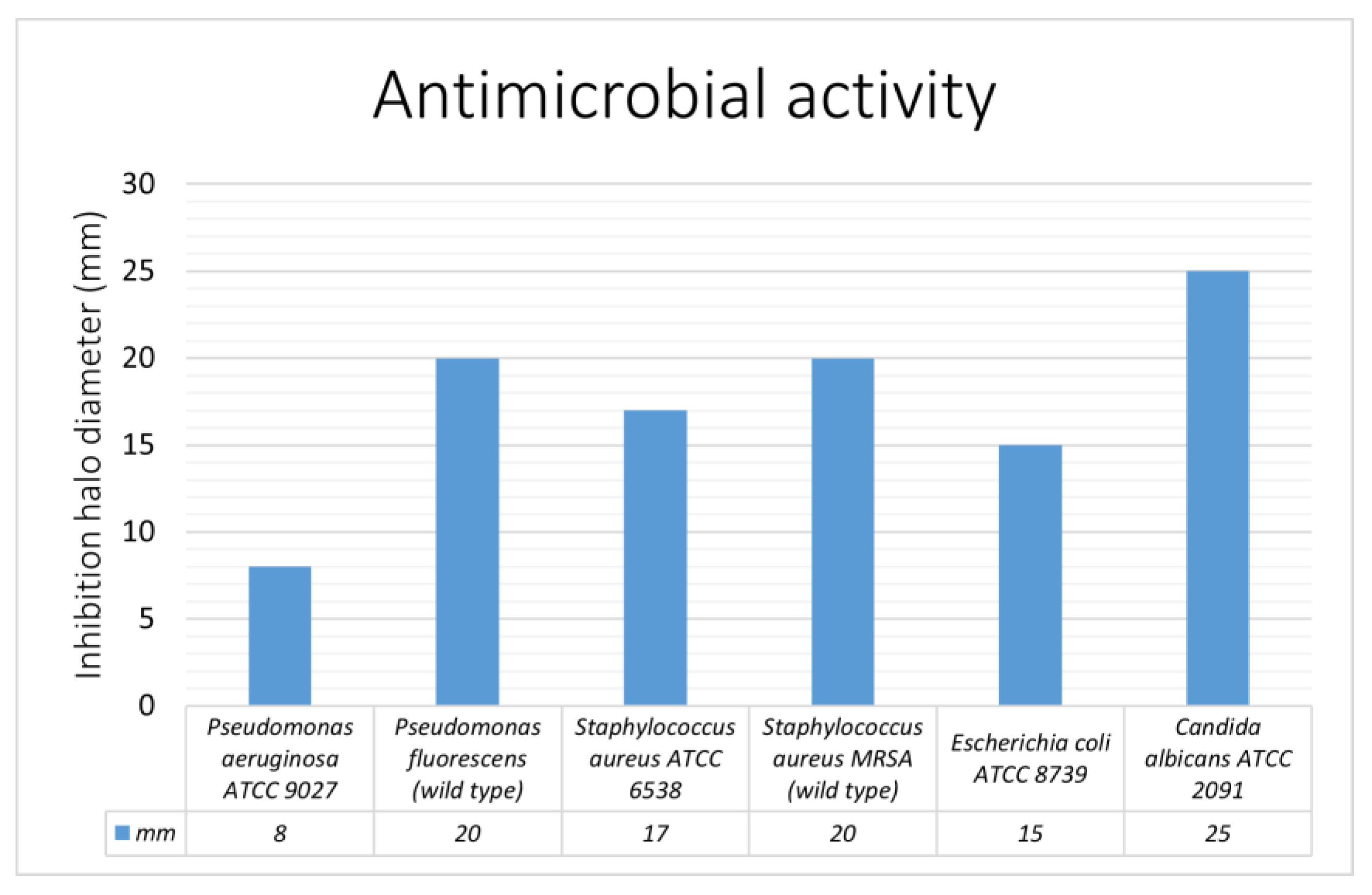
2.3. Antimicrobial Activity—Preliminary Assay

The Kirby–Bauer disk diffusion assay was used as a preliminary assay for evaluating the antimicrobial activity of grapefruit seed extract against several microorganisms: P. aeruginosa ATCC 9027, P. fluorescens (wild type), S. aureus ATCC 6538, S. aureus MRSA wild type, and C. albicans ATCC 2091. In the case of Pseudomonas spp., the microbial suspension was prepared with P. fluorescens and P. aeruginosa previously isolated from the specimens with bacterial disease. Each suspension presented a corresponding concentration of 1 McF (OD600). In addition, Muller–Hinton medium (agar 17.0 g/L, beef infusion solids 2.0 g/L, casein hydrolysate 17.5 g/L, starch 1.5 g/L) was used in standard-diameter Petri dishes (90 mm), with a medium thickness of 4–5 mm. Then, a sterile swab dipped into the suspension was used for surface seeding, repeating this operation four times by rotating the plate 90 degrees each time. After the inoculum absorption, three 6 mm paper discs impregnated with the grapefruit seed extract were positioned on the growth medium. The plates were incubated at 37 °C for 24 h ± 2. Readings were taken the following day by measuring the diameter (mm) of the inhibition halos and obtaining values comparable to standard values per microbial strain, indicating them as sensitive, intermediate, or resistant.

2.4. Inhibitory and Bactericidal Concentration–Broth Microdilution Susceptibility Test

P. aeruginosa ATCC 9027 and P. fluorescens (wild type) inocula were placed in 96-well microplates with a concentration of 100,000 CFU/mL in nutrient broth with the test substance at different concentrations. A set of positive and negative controls was run in triplicate with the samples to ensure reliability. Negative controls consisted of culture broth and GSE to test sterility; positive controls were placed in the microwells with the culture broth and without any treatment to verify inoculum vitality. The same technique was used for S. aureus ATCC 6538, S. aureus MRSA wild type, Escherichia coli 25922, and C. albicans ATCC 2091. The MIC (minimum inhibitory concentration) and MBC (minimum bactericidal concentration) were determined [55]. After 24 h of incubation, the results were read by observing the formation of a pellet; MIC was read (first microwell with no growth), and MBC was determined (first well with no growth after the transfer of a given volume into universal agarized medium).

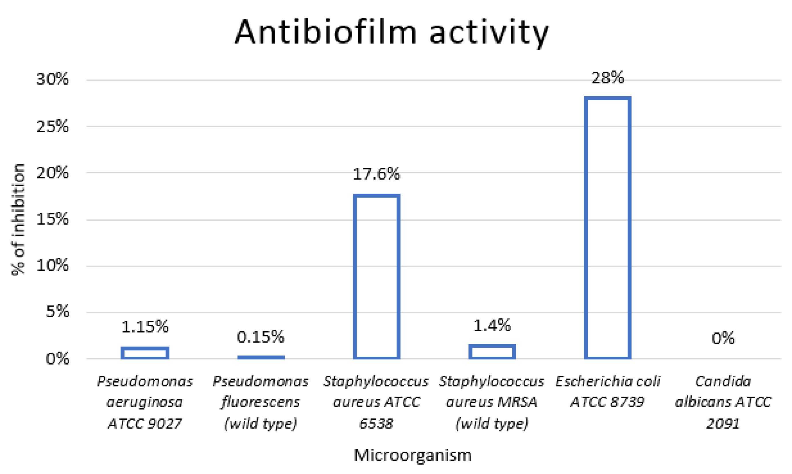
2.5. Antibiofilm Activity

A crystal violet assay [56] was applied to evaluate the antibiofilm activity of grapefruit seed extract on the microorganisms targeted. The microbial strains were revitalized in TSB (tryptic soy broth) for 24 h at 37 °C. Then, 100 µL of the microbial suspension, equal to 0.5 McF with OD600, was transferred to three wells of the microplate with supplementation of 10 µL of 1% glucose solution and incubated at 37 °C for 24 h without agitation. Positive and negative controls and the samples were run in triplicate. For positive controls, 100 μL of microbial suspension was prepared by adding 10 µL of the glucosate solution and 100 µL of 1% DMSO. For negative controls, 100 µL of TBS, 100 µL of grapefruit seed extract at the target concentration, and 10 µL of 1% gluconate solution were added. In the next 24 h of incubation, TBS was withdrawn with a micropipette and replaced with TBS-containing grapefruit seed extract at the target concentration. The controls did not undergo any treatment. After 24 h of incubation at 37 °C, the suspended medium was removed from the treated and positive controls in all microwells, and the formed biofilms were subjected to three washes with 300 μL of 0.01 mol phosphate-buffered saline (PBS, pH = 7.4) to remove weakly bound cells. It was then allowed to dry for one hour inside a thermostat at 37 °C. In the second step, the cells, bound on the surface, were fixed with 200 μL of methanol for 20 min. Excess methanol was removed and allowed to dry for 24 h. Next, staining with 200 μL of 2% Hucker’s Crystal Violet for 15 min was performed. The biofilm thus impregnated was washed three times with 300 μL sterile deionized water to remove the unbound dye and then dried at room temperature for 30 min. Then, 200 μL of the biofilm-bound crystal violet was dissolved in 33% glacial acetic acid, and absorbance was measured at 570 nm (Cary 60 UV–Vis Spectrophotometer). The percentage of biofilm eradication was calculated using the following formula (O.D.: optical density):
%   o f   e r e d i c a t i o n = O . D .   p o s i t i v e   c o n t r o l O . D .   t r e a t e d O . D .   p o s i t i v e   c o n t r o l × 100

2.6. Field Experiment
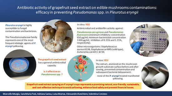
The field experiment was conducted at the mushroom farm from September–October 2023. The external climatic conditions were characterized by temperatures ranging from 19 to 29 °C in September and 16 to 27 °C in October; the average humidity was 67% in September and 72% in October. An optimal microenvironment for the growth of cardoncello was guaranteed by a breathable cloth cover and a wooden support one meter above the ground on which the pre-inoculated mushroom substrate blocks were placed. In addition, to prevent fungal diseases caused by vectors, netting and several traps were placed on the upper arch. The extract was sprayed on mushroom specimens treated (n = 50) at a concentration of 52,000 μg/mL; a control group was sprayed with sterile water (n = 90). The treatment lasted about 20 days for each mushroom block from before the sprouting phase to harvesting. A total of 100 mL of extract diluted in sterile saline solution was sprayed (distance between 20 and 30 cm) on the soil and treated mushrooms (about 5 mL per day diluted in 15 mL of sterile saline solution). The culture was monitored from the early stages of development to the adult stage, recording the number of specimens with bacterial disease both in treated and control groups. The spent substrate from mushroom cultivation was disposed of after the harvesting cycle.

3. Results

3.1. Preliminary Inspections

Among the basidiomata sampled during the inspections on the mushroom farm that took place from September 2022 to June 2023, Pseudomonas aeruginosa was found only in the ones affected by yellowing. The features of the healthy mushrooms defined by the producers during routine screenings, namely, appearance, size, and texture, were displayed to the technical staff and considered the standard for the final evaluation of treated P. eryngii.

3.2. GSE Analysis

The flavonoid compounds measured in the methanolic phase of GSE through UHPLC–Qtof-MS analysis are reported in Table 1.
The polar metabolites detected in the GSE by GC–MS analysis are shown in Table 2. No synthetic compounds were detected. The Kovats indexes calculated for the different compounds are reported in Table 2 in comparison with the Kovats indexes reported in the NIST (National Institute of Standards and Technology) database.
For HPLC–Qtof-MS and GC–MS chromatograms of GSE see Supplementary Figures S1 and S2.

3.3. In Vitro Analysis

The Kirby–Bauer disk diffusion assay demonstrated the antimicrobial activity of GSE against P. aeruginosa ATCC 9027, P. fluorescens wild type, S. aureus ATCC 6538, S. aureus MRSA wild type, E. coli ATCC 8739, and C. albicans ATCC 2091. The inhibition halos for P. aeruginosa ATCC 9027 and P. fluorescens wild type were 8 mm and 22 mm, respectively. The inhibition halo diameters for each of the microorganisms tested are shown in Figure 2.
The MIC and MBC values established through the broth microdilution susceptibility test for P. aeruginosa ATCC 9027, P. fluorescens wild type, S. aureus ATCC 6538, S. aureus MRSA (wild type), and E. coli ATCC 8739 are shown in Table 3.
The percentages of inhibition established through the crystal violet assay for P. aeruginosa ATCC 9027 and P. fluorescens wild type were 1.15% and 0.15%, respectively. The percentages of inhibition for each of the microorganisms tested are shown in Figure 3.
There was no evidence of bacteriostatic and antibiofilm activity on C. albicans ATCC 2091.

3.4. In Vivo Analysis

During the field experiment, none of the P. eryngii treated with the grapefruit seed extract manifested bacteriosis, while three cases were observed in the controls. Field testing of the substance demonstrated protective efficacy in preventing contamination and subsequent bacterial debasement. Furthermore, according to manufacturers, the treated mushrooms were of the same quality as the healthy untreated ones, presenting the same appearance, size, color, and a slightly softer stem.

4. Discussion

The bacteriosis of cardoncello manifests as the appearance of reddish-brown cankers extending from the cap to the stem, inducing a change in the color and organoleptic characteristics of the product [23,57,58]. The Pseudomonadaceae family has been identified as a major culprit in the etiology of such bacterial diseases [58,59], particularly the species aeruginosa, pathogenic to humans, and fluorescens, which may cause acute opportunistic clinical manifestations of bacteremia in individuals with compromised immune systems [60]. Bacterial contamination by Pseudomonadaceae may both directly and indirectly harm the consumer since the presence of lesions on the fungus’ surface promotes contamination by other species of microorganisms [61,62]. Furthermore, a decrease in the number and quality of the fungi grown represents an important risk of economic loss for producers. In this context, our study is the first to evaluate the efficacy of the GSE against P. eryngii bacterial blight and to suggest its possible use for preventive purposes. Nowadays, great efforts are being devoted to finding innovative and natural alternatives to chemicals to prevent alterations in food products and guarantee food safety and maximum productivity. Pure GSE is among the several bioactive compounds originating from natural sources, and it is widely accepted and recognized as safe for direct or indirect use in food. GSE in its pure form is non-toxic and “chemical-free” (marketing term), namely, safe and environmentally friendly, containing natural ingredients only. Therefore, the use of pure GSE on food or food matrices is not expected to harm consumers and the environment. However, some commercial GSEs contain synthetic compounds [63,64], such as benzethonium chloride and benzalkonium chloride, that may derive from the conversion of unstable polyphenols during GSE extraction and purification. These compounds exhibit potent antimicrobial activity and some toxicity at high concentrations [51]. For these reasons, in the present study, we used a GSE free from these synthetic compounds. This approach enabled a reliable evaluation of GSE efficacy in protecting P. eryngii crops from bacterial contamination while considering safety aspects and potential environmental impacts. Therefore, we can state that the antimicrobial activity exhibited by the GSE used in this study is attributable to its natural content in polyphenols, especially flavonoids such as naringin.
The Kirby–Bauer disk diffusion assay demonstrated that the investigated GSE was active against all Gram-positive and Gram-negative bacteria, as well as C. albicans. GSE exhibited the largest zones of inhibition for C. albicans, P. fluorescens, and Staphylococcus MRSA. The crystal violet assay showed that GSE exerted antibiofilm activity on all the microorganisms tested except for C. albicans. In terms of percentage of inhibition, a certain variability was observed, reflecting, in our opinion, the complexity of the biofilm simulated in vitro. These findings are consistent with the literature concerning P. aeruginosa spp., S. aureus MRSA, and E. coli [29,32,65,66,67]. In the literature, the mechanism of GSE antimicrobial activity has been attributed to the disruption of the bacterial membrane and liberation of the cytoplasmatic content [33]. The antibiofilm effect of GSE on S. aureus and E. coli has been attributed to changes in the exopolysaccharide production rate and mobility, as well as changes in hydrophobicity in E. coli only [68].
The field experiment demonstrated that spraying GSE twice a day from before the sprouting phase to harvesting can prevent the growth of P. fluorescens and P. aeruginosa during the cultivation of cardoncello, which is particularly critical under several environmental circumstances. The extract antimicrobial effects were not affected by the 20–30 cm distance required for the application, suggesting that GSE is suitable as a spray. The application of GSE as a measure of prevention of bacterial blight occurrence is worth further investigation not only on P. eryngii but also on other foodstuffs. Furthermore, given the current attention to the nutraceutical use of naringin and other flavonoids, the potential added value of foods supplemented with GSE deserves consideration.

Limitations and Future Directions

Since this work is the first to evaluate the effectiveness of GSE in preventing P. eryngii yellowing, it should be considered a pilot study. It has several limitations, such as the small number of basidiomata we were able to treat due to economic constraints of the producers. We did not conduct a challenge test with Pseudomonas spp. and analyzed the quality of treated mushrooms in terms of appearance, shape, color, and texture only.
The next steps to validate this natural control strategy involve (i) larger scale experimentation with at least three to five biological replicates (including 25–50 mushrooms each), which is also essential to confirm further that the slight changes in the texture due to reiterate nebulization do not affect the final quality of the products; (ii) deep evaluation of treated mushroom quality, including polyphenol oxidase (PPO) activity, sensory analysis, and chemical characterization; and (iii) conducting a challenge test with Pseudomonas spp. Furthermore, in the following stages, we plan to compare GSE with chemical compounds and study its mechanism of action on Pseudomonas spp. to gain a deeper understanding of GSE characteristics and levels of effectiveness. Moreover, considering that naringin’s antioxidant activity is affected by light and high temperatures (>100 °C) [69,70], we plan to investigate the naringin concentration and bioavailability in the final product treated with GSE.

5. Conclusions

P. eryngii yellowing is a disease that can occur in all the basiodiomata development phases, from sprouting to commercial maturation. It can bring huge economic damage to producers due to its rapid spread in P. eryngii cultivations and the current lack of standardized control measures. The present work contributes to the knowledge of the antimicrobial efficacy of natural GSE and provides valuable input to the branch of research aimed at preventing and controlling P. eryngii yellowing. Our findings support the use of GSE to protect P. eryngii crops from bacterial contamination, particularly from Pseudomonas spp., which have often been identified as responsible for the yellowing. Atomizing GSE pre- and post-sprouting represents a promising eco-friendly and sustainable control strategy alternative to chemical treatments to ensure food safety and prevent financial losses due to P. eryngii spoilage. In addition, due to its content in naringin and other bioactive components, GSE opens new horizons regarding its use as a nutraceutical in food fortification and supplementation. Due to the limitations mentioned above, our preliminary findings, although encouraging, require larger and deeper studies to be further validated.

Supplementary Materials

The following supporting information can be downloaded at: https://www.mdpi.com/article/10.3390/foods13081161/s1. Figure S1: HPLC-Qtof/MS chromatogram of GSE; Figure S2: GC-MS chromatogram of GSE.

Author Contributions

Conceptualization, M.M., S.M.P. and V.C.; data curation, M.M., S.M.P. and V.C.; formal analysis, A.S., L.M., C.M., A.B. and V.C.; investigation, M.M., A.S., C.M. and V.C.; methodology, L.M., A.S., C.M., A.B. and V.C.; resources, V.C.; validation, V.C.; writing—original draft, M.M., S.M.P. and V.C.; writing—review and editing, S.M.P., C.M. and V.C. All authors have read and agreed to the published version of the manuscript.

Funding

This research received no external funding.

Institutional Review Board Statement

Not applicable.

Informed Consent Statement

Not applicable.

Data Availability Statement

The original contributions presented in the study are included in the article, further inquiries can be directed to the corresponding author.

Conflicts of Interest

The authors declare no conflicts of interest.

References

  1. Venturella, G.; Palazzolo, E.; Saiano, F.; Gargano, M.L. Notes on a New Productive Strain of King Oyster Mushroom, Pleurotus Eryngii (Higher Basidiomycetes), a Prized Italian Culinary-Medicinal Mushroom. Int. J. Med. Mushrooms 2015, 17, 199–206. [Google Scholar] [CrossRef]
  2. Carlavilla, J.R.; Manjón, J.L. The King Oyster Mushroom Pleurotus Eryngii Behaves as a Necrotrophic Pathogen of Eryngium Campestre. Ital. J. Mycol. 2023, 52, 22–31. [Google Scholar] [CrossRef]
  3. Lv, S.; Zhu, X.; Liu, Z.; Hu, L.; Xu, D.; Chitrakar, B.; Mo, H.; Li, H. Edible Pleurotus Eryngii Papery Food Prepared by Papermaking Process. Foods 2022, 11, 3514. [Google Scholar] [CrossRef] [PubMed]
  4. Teniou, S.; Bensegueni, A.; Hybertson, B.M.; Gao, B.; Bose, S.K.; McCord, J.M.; Chovelon, B.; Bensouici, C.; Boumendjel, A.; Hininger-Favier, I. Biodriven Investigation of the Wild Edible Mushroom Pleurotus Eryngii Revealing Unique Properties as Functional Food. J. Funct. Foods 2022, 89, 104965. [Google Scholar] [CrossRef]
  5. Stajić, M.; Vukojević, J.; Duletić-Lauević, S. Biology of Pleurotus Eryngii and Role in Biotechnological Processes: A Review. Crit. Rev. Biotechnol. 2009, 29, 55–66. [Google Scholar] [CrossRef]
  6. Yu, A.; Ji, Y.; Ma, G.; Xu, J.; Hu, Q. Identification and Preparation of Selenium-Containing Peptides from Selenium-Enriched Pleurotus Eryngii and Their Protective Effect on Lead-Induced Oxidative Damage in NCTC1469 Hepatocytes. J. Sci. Food Agric. 2023, 103, 4522–4534. [Google Scholar] [CrossRef] [PubMed]
  7. Yuan, B.; Ma, N.; Zhao, L.; Zhao, E.; Gao, Z.; Wang, W.; Song, M.; Zhang, G.; Hu, Q.; Xiao, H. In Vitro and in Vivo Inhibitory Effects of a Pleurotus Eryngii Protein on Colon Cancer Cells. Food Funct. 2017, 8, 3553–3562. [Google Scholar] [CrossRef] [PubMed]
  8. Zhang, B.; Li, Y.; Zhang, F.; Linhardt, R.J.; Zeng, G.; Zhang, A. Extraction, Structure and Bioactivities of the Polysaccharides from Pleurotus Eryngii: A Review. Int. J. Biol. Macromol. 2020, 150, 1342–1347. [Google Scholar] [CrossRef] [PubMed]
  9. Chen, J.; Yong, Y.; Xing, M.; Gu, Y.; Zhang, Z.; Zhang, S.; Lu, L. Characterization of Polysaccharides with Marked Inhibitory Effect on Lipid Accumulation in Pleurotus Eryngii. Carbohydr. Polym. 2013, 97, 604–613. [Google Scholar] [CrossRef] [PubMed]
  10. Alam, N.; Yoon, K.N.; Lee, J.S.; Cho, H.J.; Shim, M.J.; Lee, T.S. Dietary Effect of Pleurotus Eryngii on Biochemical Function and Histology in Hypercholesterolemic Rats. Saudi J. Biol. Sci. 2011, 18, 403–409. [Google Scholar] [CrossRef]
  11. Amerikanou, C.; Tagkouli, D.; Tsiaka, T.; Lantzouraki, D.Z.; Karavoltsos, S.; Sakellari, A.; Kleftaki, S.A.; Koutrotsios, G.; Giannou, V.; Zervakis, G.I.; et al. Pleurotus Eryngii Chips—Chemical Characterization and Nutritional Value of an Innovative Healthy Snack. Foods 2023, 12, 353. [Google Scholar] [CrossRef] [PubMed]
  12. Gong, P.; Long, H.; Guo, Y.; Wang, S.; Chen, F.; Chen, X. Isolation, Structural Characterization, and Hypoglycemic Activities In Vitro of Polysaccharides from Pleurotus Eryngii. Molecules 2022, 27, 7140. [Google Scholar] [CrossRef] [PubMed]
  13. Manzi, P.; Marconi, S.; Aguzzi, A.; Pizzoferrato, L. Commercial Mushrooms: Nutritional Quality and Effect of Cooking. Food Chem. 2004, 84, 201–206. [Google Scholar] [CrossRef]
  14. Hess, J.M.; Jonnalagadda, S.S.; Slavin, J.L. What Is a Snack, Why Do We Snack, and How Can We Choose Better Snacks? A Review of the Definitions of Snacking, Motivations to Snack, Contributions to Dietary Intake, and Recommendations for Improvement. Adv. Nutr. 2016, 7, 466–475. [Google Scholar] [CrossRef] [PubMed]
  15. Bruno, G.L.; Rana, G.L.; Sermani, S.; Scarola, L.; Cariddi, C. Control of Bacterial Yellowing of Cardoncello Mushroom Pleurotus Eryngii Using Acetic or Hydrochloric Acid Solutions. Crop Prot. 2013, 50, 24–29. [Google Scholar] [CrossRef]
  16. Gea, F.J.; Carrasco, J.; Suz, L.M.; Navarro, M.J. Characterization and Pathogenicity of Cladobotryum Mycophilum in Spanish Pleurotus Eryngii Mushroom Crops and Its Sensitivity to Fungicides. Eur. J. Plant Pathol. 2017, 147, 129–139. [Google Scholar] [CrossRef]
  17. Kim, M.K.; Seuk, S.W.; Lee, Y.H.; Kim, H.R.; Cho, K.M. Fungicide Sensitivity and Characterization of Cobweb Disease on a Pleurotus Eryngii Mushroom Crop Caused by Cladobotryum Mycophilum. Plant Pathol. J. 2014, 30, 82. [Google Scholar] [CrossRef] [PubMed]
  18. Carrasco, J.; Navarro, M.J.; Santos, M.; Diánez, F.; Gea, F.J. Incidence, Identification and Pathogenicity of Cladobotryum Mycophilum, Causal Agent of Cobweb Disease on Agaricus Bisporus Mushroom Crops in Spain. Ann. Appl. Biol. 2016, 168, 214–224. [Google Scholar] [CrossRef]
  19. Chen, J.T.; Huang, J.W. A Semiselective Medium for Detecting Gliocladium Roseum, the Causal Agent of King Oyster Mushroom Brown Spot. Plant Pathol. Bull. 2004, 13, 107–116. [Google Scholar]
  20. Kredics, L.; Kocsubé, S.; Nagy, L.; Komoń-Zelazowska, M.; Manczinger, L.; Sajben, E.; Nagy, A.; Vágvölgyi, C.; Kubicek, C.P.; Druzhinina, I.S.; et al. Molecular Identification of Trichoderma Species Associated with Pleurotus Ostreatus and Natural Substrates of the Oyster Mushroom. FEMS Microbiol. Lett. 2009, 300, 58–67. [Google Scholar] [CrossRef]
  21. González, A.J.; Gea, F.J.; Navarro, M.J.; Fernández, A.M. Identification and RAPD-Typing of Ewingella Americana on Cultivated Mushrooms in Castilla-La Mancha, Spain. Eur. J. Plant Pathol. 2012, 133, 517–522. [Google Scholar] [CrossRef]
  22. Reyes, J.E.; Venturini, M.E.; Oria, R.; Blanco, D. Prevalence of Ewingella Americana in Retail Fresh Cultivated Mushrooms (Agaricus Bisporus, Lentinula Edodes and Pleurotus Ostreatus) in Zaragoza (Spain). FEMS Microbiol. Ecol. 2004, 47, 291–296. [Google Scholar] [CrossRef] [PubMed]
  23. Bruno, G.L.; De Corato, U.; Rana, G.L.; De Luca, P.; Pipoli, V.; Lops, R.; Scarola, L.; Mannerucci, F.; Piscitelli, L.; Cariddi, C. Suppressiveness of White Vinegar and Steam-Exploded Liquid Waste against the Causal Agents of Pleurotus Eryngii Yellowing. Crop Prot. 2015, 70, 61–69. [Google Scholar] [CrossRef]
  24. Iacobellis, N.S.; Lo Cantore, P. Pseudomonas “Reactans” a New Pathogen of Cultivated Mushrooms. In Pseudomonas Syringae and Related Pathogens; Springer: Dordrecht, The Netherlands, 2003. [Google Scholar]
  25. Soler-Rivas, C.; Jolivet, S.; Arpin, N.; Olivier, J.M.; Wichers, H.J. Biochemical and Physiological Aspects of Brown Blotch Disease of Agaricus Bisporus. FEMS Microbiol. Rev. 1999, 23, 591–614. [Google Scholar] [CrossRef] [PubMed]
  26. Bellettini, M.B.; Bellettini, S.; Fiorda, F.A.; Pedro, A.C.; Bach, F.; Fabela-Morón, M.F.; Hoffmann-Ribani, R. Diseases and Pests Noxious to Pleurotus Spp. Mushroom Crops. Rev. Argent. Microbiol. 2018, 50, 216–226. [Google Scholar] [CrossRef] [PubMed]
  27. Ryssel, H.; Kloeters, O.; Germann, G.; Schäfer, T.; Wiedemann, G.; Oehlbauer, M. The Antimicrobial Effect of Acetic Acid-An Alternative to Common Local Antiseptics? Burns 2009, 35, 695–700. [Google Scholar] [CrossRef] [PubMed]
  28. Cvetnić, Z.; Vladimir-Knežević, S. Antimicrobial Activity of Grapefruit Seed and Pulp Ethanolic Extract. Acta Pharm. 2004, 54, 243–250. [Google Scholar]
  29. Kim, T.; Kim, J.H.; Oh, S.W. Grapefruit Seed Extract as a Natural Food Antimicrobial: A Review. Food Bioprocess Technol. 2021, 14, 626–633. [Google Scholar] [CrossRef]
  30. Yun, D.; Liu, J. Recent Advances on the Development of Food Packaging Films Based on Citrus Processing Wastes: A Review. J. Agric. Food Res. 2022, 9, 100316. [Google Scholar] [CrossRef]
  31. Zayed, A.; Badawy, M.T.; Farag, M.A. Valorization and Extraction Optimization of Citrus Seeds for Food and Functional Food Applications. Food Chem. 2021, 355, 129609. [Google Scholar] [CrossRef]
  32. Reagor, L.; Gusman, J.; McCoy, L.; Carino, E.; Heggers, J.P. The Effectiveness of Processed Grapefruit-Seed Extract as an Antibacterial Agent: I. An in Vitro Agar Assay. J. Altern. Complement. Med. 2002, 8, 325–332. [Google Scholar] [CrossRef]
  33. Heggers, J.P.; Cottingham, J.; Gusman, J.; Reagor, L.; McCoy, L.; Carino, E.; Cox, R.; Zhao, J.G. The Effectiveness of Processed Grapefruit-Seed Extract as an Antibacterial Agent: II. Mechanism of Action and in Vitro Toxicity. J. Altern. Complement. Med. 2002, 8, 333–340. [Google Scholar] [CrossRef] [PubMed]
  34. Barawi, S.; Hamzah, H.; Hamasalih, R.; Mohammed, A.; Abdalrahman, B.; Abdalaziz, S. Antibacterial Mode of Action of Grapefruit Seed Extract against Local Isolates of Beta-Lactamases-Resistant Klebsiella Pneumoniae and Its Potential Application. Int. J. Agric. Biol. 2021, 26, 499–508. [Google Scholar] [CrossRef]
  35. Çiçek Polat, D.; Eryilmaz, M.; Akalin, K.; Coşkun, M. Antimicrobial Activity of Grapefruit Seed. Hacettepe Univ. J. Fac. Pharm. 2018, 38, 1–3. [Google Scholar]
  36. Céliz, G.; Daz, M.; Audisio, M.C. Antibacterial Activity of Naringin Derivatives against Pathogenic Strains. J. Appl. Microbiol. 2011, 111, 731–738. [Google Scholar] [CrossRef]
  37. Zeng, X.; Zheng, Y.; He, Y.; Zhang, J.; Peng, W.; Su, W. Microbial Metabolism of Naringin and the Impact on Antioxidant Capacity. Nutrients 2022, 14, 3765. [Google Scholar] [CrossRef]
  38. Bugianesi, R.; Catasta, G.; Spigno, P.; D’Uva, A.; Maiani, G. Naringenin from Cooked Tomato Paste Is Bioavailable in Men. J. Nutr. 2002, 132, 3349–3352. [Google Scholar] [CrossRef] [PubMed]
  39. Alam, M.A.; Subhan, N.; Rahman, M.M.; Uddin, S.J.; Reza, H.M.; Sarker, S.D. Effect of Citrus Flavonoids, Naringin and Naringenin, on Metabolic Syndrome and Their Mechanisms of Action. Adv. Nutr. 2014, 5, 404–417. [Google Scholar] [CrossRef]
  40. Jourdan, P.S.; McIntosh, C.A.; Mansell, R.L. Naringin Levels in Citrus Tissues: II. Quantitative Distribution of Naringin in Citrus Paradisi MacFad. Plant Physiol. 1985, 77, 903–908. [Google Scholar] [CrossRef] [PubMed]
  41. Chen, R.; Qi, Q.L.; Wang, M.T.; Li, Q.Y. Therapeutic Potential of Naringin: An Overview. Pharm. Biol. 2016, 54, 3203–3210. [Google Scholar] [CrossRef] [PubMed]
  42. Gorinstein, S.; Leontowicz, H.; Leontowicz, M.; Krzeminski, R.; Gralak, M.; Delgado-Licon, E.; Ayala, A.L.M.; Katrich, E.; Trakhtenberg, S. Changes in Plasma Lipid and Antioxidant Activity in Rats as a Result of Naringin and Red Grapefruit Supplementation. J. Agric. Food Chem. 2005, 53, 3223–3228. [Google Scholar] [CrossRef] [PubMed]
  43. Stabrauskiene, J.; Kopustinskiene, D.M.; Lazauskas, R.; Bernatoniene, J. Naringin and Naringenin: Their Mechanisms of Action and the Potential Anticancer Activities. Biomedicines 2022, 10, 1686. [Google Scholar] [CrossRef] [PubMed]
  44. Raja Kumar, S.; Mohd Ramli, E.S.; Abdul Nasir, N.A.; Ismail, N.H.M.; Mohd Fahami, N.A. Preventive Effect of Naringin on Metabolic Syndrome and Its Mechanism of Action: A Systematic Review. Evid. Based Complement. Altern. Med. 2019, 2019, 9752826. [Google Scholar] [CrossRef] [PubMed]
  45. Termkwancharoen, C.; Malakul, W.; Phetrungnapha, A.; Tunsophon, S. Naringin Ameliorates Skeletal Muscle Atrophy and Improves Insulin Resistance in High-Fat-Diet-Induced Insulin Resistance in Obese Rats. Nutrients 2022, 14, 4120. [Google Scholar] [CrossRef]
  46. Mir, I.A.; Tiku, A.B. Chemopreventive and Therapeutic Potential of “Naringenin,” a Flavanone Present in Citrus Fruits. Nutr. Cancer 2015, 67, 27–42. [Google Scholar] [CrossRef] [PubMed]
  47. Massaro, L.; Raguzzini, A.; Aiello, P.; Valencia, D.V. The Potential Role of Naringin and Naringenin as Nutraceuticals Against Metabolic Syndrome. Endocr. Metab. Immune Disord. Drug Targets 2022, 23, 428–445. [Google Scholar] [CrossRef] [PubMed]
  48. Murugesan, N.; Woodard, K.; Ramaraju, R.; Greenway, F.L.; Coulter, A.A.; Rebello, C.J. Naringenin Increases Insulin Sensitivity and Metabolic Rate: A Case Study. J. Med. Food 2020, 23, 343–348. [Google Scholar] [CrossRef] [PubMed]
  49. Nguyen, M.A.; Staubach, P.; Tamai, I.; Langguth, P. High-Dose Short-Term Administration of Naringin Did Not Alter Talinolol Pharmacokinetics in Humans. Eur. J. Pharm. Sci. 2015, 68, 36–42. [Google Scholar] [CrossRef]
  50. Singh, S.; Sharma, A.; Monga, V.; Bhatia, R. Compendium of Naringenin: Potential Sources, Analytical Aspects, Chemistry, Nutraceutical Potentials and Pharmacological Profile. Crit. Rev. Food Sci. Nutr. 2022, 63, 8868–8899. [Google Scholar] [CrossRef] [PubMed]
  51. Roy, S.; Zhang, W.; Biswas, D.; Ramakrishnan, R.; Rhim, J.W. Grapefruit Seed Extract-Added Functional Films and Coating for Active Packaging Applications: A Review. Molecules 2023, 28, 730. [Google Scholar] [CrossRef] [PubMed]
  52. ISO/IEC 17025:2017; General Requirements for the Competence of Testing and Calibration Laboratories. International Standard Organization: Geneva, Switzerland, 2018.
  53. ISO 7218:2007/Amd 1:2013; Microbiology of Food and Animal Feeding Stuffs General Requirements and Guidance for Microbiological Examinations Amendment 1. International Standard Organization: Geneva, Switzerland, 2014.
  54. ISO 16266:2006; Water Quality Detection and Enumeration of Pseudomonas Aeruginosa Method by Membrane Filtration. International Standard Organization: Geneva, Switzerland, 2008.
  55. Angioni, A.; Barra, A.; Cereti, E.; Barile, D.; Coïsson, J.D.; Arlorio, M.; Dessi, S.; Coroneo, V.; Cabras, P. Chemical Composition, Plant Genetic Differences, Antimicrobial and Antifungal Activity Investigation of the Essential Oil of Rosmarinus officinalis L. J. Agric. Food Chem. 2004, 52, 3530–3535. [Google Scholar] [CrossRef] [PubMed]
  56. Stepanović, S.; Vuković, D.; Hola, V.; Di Bonaventura, G.; Djukić, S.; Ćirković, I.; Ruzicka, F. Quantification of Biofilm in Microtiter Plates: Overview of Testing Conditions and Practical Recommendations for Assessment of Biofilm Production by Staphylococci. APMIS 2007, 115, 891–899. [Google Scholar] [CrossRef] [PubMed]
  57. Osdaghi, E.; Martins, S.J.; Ramos-Sepulveda, L.; Vieira, F.R.; Pecchia, J.A.; Beyer, D.M.; Bell, T.H.; Yang, Y.; Hockett, K.L.; Bull, C.T. 100 Years since Tolaas: Bacterial Blotch of Mushrooms in the 21st Century. Plant Dis. 2019, 103, 2714–2732. [Google Scholar] [CrossRef] [PubMed]
  58. Gao, Q.; Liu, Y.; Xie, J.; Zhao, S.; Qin, W.; Song, Q.; Wang, S.; Rong, C. Bacterial Infection Induces Ultrastructural and Transcriptional Changes in the King Oyster Mushroom (Pleurotus Eryngii). Microbiol. Spectr. 2022, 10, e01445-22. [Google Scholar] [CrossRef]
  59. Sajben, E.; Manczinger, L.; Nagy, A.; Kredics, L.; Vágvölgyi, C. Characterization of Pseudomonads Isolated from Decaying Sporocarps of Oyster Mushroom. Microbiol. Res. 2011, 166, 255–267. [Google Scholar] [CrossRef]
  60. Scales, B.S.; Dickson, R.P.; Lipuma, J.J.; Huffnagle, G.B. Microbiology, Genomics, and Clinical Significance of the Pseudomonas Fluorescens Species Complex, an Unappreciated Colonizer of Humans. Clin. Microbiol. Rev. 2014, 27, 927–948. [Google Scholar] [CrossRef] [PubMed]
  61. Lee, H.I.; Jeong, K.S.; Cha, J.S. PCR Assays for Specific and Sensitive Detection of Pseudomonas Tolaasii, the Cause of Brown Blotch Disease of Mushrooms. Lett. Appl. Microbiol. 2002, 35, 276–280. [Google Scholar] [CrossRef] [PubMed]
  62. Venturini, M.E.; Reyes, J.E.; Rivera, C.S.; Oria, R.; Blanco, D. Microbiological Quality and Safety of Fresh Cultivated and Wild Mushrooms Commercialized in Spain. Food Microbiol. 2011, 28, 1492–1498. [Google Scholar] [CrossRef]
  63. Takeoka, G.; Dao, L.; Wong, R.Y.; Lundin, R.; Mahoney, N. Identification of Benzethonium Chloride in Commercial Grapefruit Seed Extracts. J. Agric. Food Chem. 2001, 49, 3316–3320. [Google Scholar] [CrossRef] [PubMed]
  64. Takeoka, G.R.; Dao, L.T.; Wong, R.Y.; Harden, L.A. Identification of Benzalkonium Chloride in Commercial Grapefruit Seed Extracts. J. Agric. Food Chem. 2005, 53, 7630–7636. [Google Scholar] [CrossRef]
  65. Han, H.W.; Kwak, J.H.; Jang, T.S.; Knowles, J.C.; Kim, H.W.; Lee, H.H.; Lee, J.H. Grapefruit Seed Extract as a Natural Derived Antibacterial Substance against Multidrug-Resistant Bacteria. Antibiotics 2021, 10, 85. [Google Scholar] [CrossRef] [PubMed]
  66. Choi, J.S.; Lee, Y.R.; Ha, Y.M.; Seo, H.J.; Kim, Y.H.; Park, S.M.; Sohn, J.H. Antibacterial Effect of Grapefruit Seed Extract (GSE) on Makgeolli-Brewing Microorganisms and Its Application in the Preservation of Fresh Makgeolli. J. Food Sci. 2014, 79, M1159–M1167. [Google Scholar] [CrossRef] [PubMed]
  67. Wang, K.; Lim, P.N.; Tong, S.Y.; Thian, E.S. Development of Grapefruit Seed Extract-Loaded Poly(ε-Caprolactone)/Chitosan Films for Antimicrobial Food Packaging. Food Packag. Shelf Life 2019, 22, 100396. [Google Scholar] [CrossRef]
  68. Song, Y.J.; Yu, H.H.; Kim, Y.J.; Lee, N.K.; Paik, H.D. Anti-Biofilm Activity of Grapefruit Seed Extract against Staphylococcus Aureus and Escherichia Coli. J. Microbiol. Biotechnol. 2019, 29, 1177–1183. [Google Scholar] [CrossRef]
  69. Chaaban, H.; Ioannou, I.; Chebil, L.; Slimane, M.; Gérardin, C.; Paris, C.; Charbonnel, C.; Chekir, L.; Ghoul, M. Effect of Heat Processing on Thermal Stability and Antioxidant Activity of Six Flavonoids. J. Food Process. Preserv. 2017, 41, e13203. [Google Scholar] [CrossRef]
  70. Ioannou, I.; M’hiri, N.; Chaaban, H.; Boudhrioua, N.M.; Ghoul, M. Effect of the Process, Temperature, Light and Oxygen on Naringin Extraction and the Evolution of Its Antioxidant Activity. Int. J. Food Sci. Technol. 2018, 53, 2754–2760. [Google Scholar] [CrossRef]
Figure 1. (a) Pleurotus eryngii without contamination; note the white stem without spots; (b) P. eryngii contaminated after a few days from first signs: appearance of rusty-red spots on the entire stem; (c) P. eryngii with bacteriosis after one week.
Foods 13 01161 g001
Figure 2. Antimicrobial activity-inhibition halo histograms. Diameters of inhibition halos (Kirby–Bauer disk diffusion assay) related to the grapefruit seed extract action are expressed in mm.
Foods 13 01161 g002
Figure 3. Antibiofilm activity—% of inhibition histograms. Percentages obtained with the crystal violet assay.
Foods 13 01161 g003
Table 1. Quantitative analysis and accurate mass for flavonoids determined using UHPLC–Qtof-MS. RT retention time; m/z ratio of mass to charge; Δ (ppm) mass error of an assignment when comparing a theoretical m/z and the experimentally observed m/z.
FlavonoidsRT
(min)
Formulam/z Experimentalm/z TheoreticalΔ (ppm)Major Fragmentaionmg/L
Rutin6.49C27H30O16611.1606611.1607−0.16303.049699.03
Naringin9.75C27H32O14581.1864581.1865−0.17273.075246.57
Hesperidin9.25C28H34O15611.1967611.1970−0.49303.085745.76
Neohesperidin16.11C28H34O15611.1972611.19700.33303.0874168.29
Naringenin19.23C15H12O5273.0758273.07570.36287.09043515.05
Table 2. Percentage composition of polar metabolites detected in the GSE by GC–MS analysis. RT retention time. NA, not available.
RTCompounds%Calculated
Kováts Retention Indexes
Theoretical
Kováts Retention Indexes
18.944Lactic Acid, 2TMS derivative14.6920810551057
21.066Diacetin, TMS3.4895021105NA
22.808Glycerol, 3TMS derivative56.2746612791282
26.651Diacetin, TMS5.5994871105NA
26.9521,2,3-Butanetriol-3TMS10.2754712851286
27.853Butane, 1,2,3-tris(trimethylsiloxy)-TMS0.13145312851285
28.013Monocaproin, 2TMS0.1305218861886
28.613Diglycerol, 4TMS derivative0.39681902NA
30.696Ascorbic acid, 4TMS derivative5.92004819681971
31.7779-Octadecenenitrile0.231352315NA
33.338Citric acid, 4TMS derivative1.75297326182622
34.64Oleamide, TMS derivative1.10565627632765
Table 3. Minimum inhibitory concentration (MIC) and minimum bactericidal concentration (MBC) values established through the broth microdilution susceptibility test for each of the microorganisms tested.
Target MicroorganismsMICMBC
Staphylococcus aureus ATCC 6538162,5 µg/mL650 µg/mL
Staphylococcus aureus MRSA wild type325 µg/mL325 µg/mL
Pseudomonas aeruginosa ATCC 9027
Pseudomonas fluorescens wild type
650 µg/mL1300 µg/mL
Escherichia coli 8739650 µg/mL1300 µg/mL
Candida albicans ATCC 2091--
Disclaimer/Publisher’s Note: The statements, opinions and data contained in all publications are solely those of the individual author(s) and contributor(s) and not of MDPI and/or the editor(s). MDPI and/or the editor(s) disclaim responsibility for any injury to people or property resulting from any ideas, methods, instructions or products referred to in the content.

