
An Overview of the Current Scientific Evidence on the Biological Properties of Abelmoschus esculentus (L.) Moench (Okra)


Abstract
:
1. Introduction
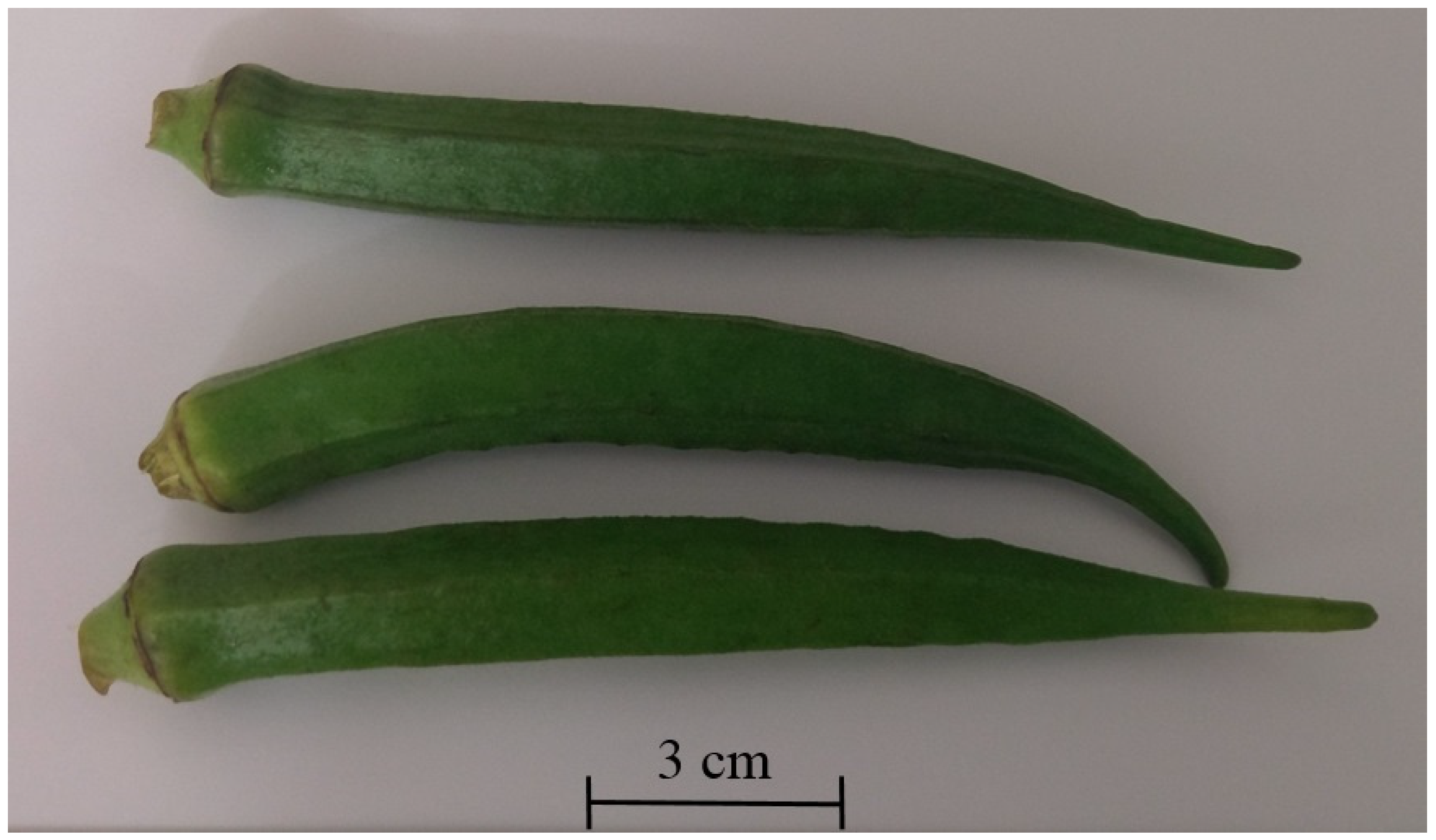
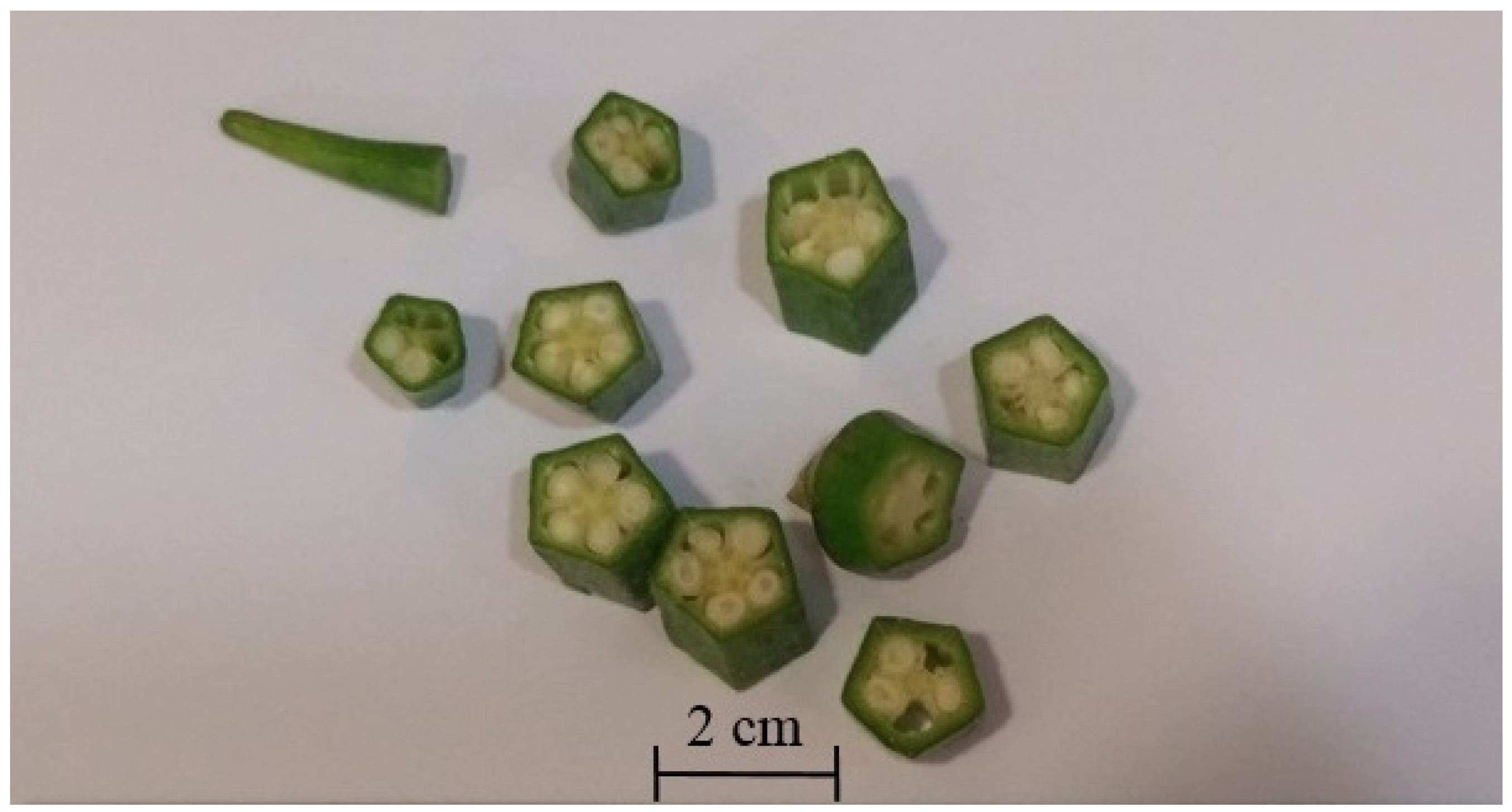
2. Active Ingredients and Nutrition Value in Okra
3. Biological Activities of Okra
3.1. Antidiabetic Effect
3.1.1. Restoration of β-Cell Function
3.1.2. Improvement in Insulin Resistance/Sensitivity via Suppression of PPARs Genes
3.1.3. Enhancement of Antioxidant Enzymes as Well as Scavenging of Free Radicals
3.1.4. Inhibition of Rate of Carbohydrate Digestion and Glucose Absorption
3.1.5. Hypoglycemia and Improving Glucose Tolerance
3.1.6. Prevention of Diabetic Nephropathy
3.2. Antifatigue and Vasoprotective Effect
3.3. Hepatoprotective Activity
3.4. Antihyperlipidemic Activity
3.5. Antitumor Activity
3.6. Neuroprotective Effect
3.7. Skin Protective Effect
3.8. Relief Temporomandibular Joint (TMJ) Inflammatory Hypernociception Through Its Anti-Inflammatory, Antinociceptive, and Analgesic Activity
3.9. Anti-Gastric Ulcer Effect of Okra via Its Gastroprotective Effect and Anti-Adhesive Effect of Helicobacter pylori on the Gastric Epithelial Cells
3.10. Antimicrobial Activity
4. Clinical Evidence of Okra
5. Perspectives
Author Contributions
Funding
Data Availability Statement
Conflicts of Interest
List of Abbreviations
| Abbreviations | Definitions |
| ABCG1 | ATP-binding cassette transporter G1 |
| ABTS | 2,2′-azino-bis(3-ethylbenzothiazoline-6-sulfonic acid |
| AE | Abelmoschus esculentus |
| AIF | Apoptosis-inducing factor |
| ALP | Alkaline phosphatase |
| ALT | Alanine transaminase |
| Akt | Protein kinase B |
| AMP | Adenosine 5′-monophosphate |
| AMPK | Adenosine monophosphate-activated protein kinase |
| AOPP | Advanced oxidation protein products |
| aP2 | Adipocyte protein 2 |
| ApoE | Apolipoprotein E |
| AST | Aspartate transaminase |
| AT-1 | Angiotensin II receptor-1 |
| ATPase | Adenosine 5′-TriPhosphatase |
| Bax | B-cell lymphoma protein 2 associated X |
| Bcl-2 | B-cell lymphoma 2 |
| BLA | Blood lactic acid |
| BMHC-imDCs | Rat bone marrow hematopoietic cells derived immature dendritic cells |
| BrdU | Bromodeoxyuridine |
| CA3 | Cornu Ammonis 3 |
| CAT | Catalase |
| CCl4 | Carbon tetrachloride |
| CD | Cluster of differentiation |
| CK | Creatine kinase |
| CYP7A1 | Cytochrome P450 7A1 |
| DCs cell | Dendritic cells |
| DPP-4 | Dipeptidyl peptidase-4 |
| DPPH | 2,2-Diphenyl-1-picrylhydrazyl |
| EMT | Epithelial-mesenchymal transition |
| FAS | Fatty acid synthase |
| FGF-2 | Fibroblast growth factor-2 |
| FRAP | Ferric reducing ability of plasma |
| FST | Forced swimming test |
| GAG | Glycosaminoglycans |
| Gal-3 | Galectin-3 |
| GGT | Gamma glutamyltransferase |
| GLP-1R | Glucagon like peptide-1 receptor |
| GOT | Glutamate oxaloacetate transaminase |
| GPT | Glutamate pyruvate transaminase |
| GPx | Glutathione peroxidase |
| GR | Glutathione reductase |
| GSH | Glutathione |
| GSH-Px | Glutathione peroxidase |
| GSK-3β | Glycogen synthase kinase-3 beta |
| HbA1c | Glycated hemoglobin |
| HDF | Human dermal fibroblast adult cell |
| HDL | High-density lipoprotein |
| HDLC | High-density lipoprotein-cholesterol |
| HFE | Hemochromatosis protein |
| HG | Hepatic glycogen |
| HO-1 | hemeoxygenase-1 |
| HOMA-IR | Homeostasis model assessment of insulin resistance |
| ICAM-1 | Intercellular adhesion molecule-1 |
| IFN-γ | Interferon gamma |
| IL-6 | Interleukin-6 |
| IBD | Inflammatory bowel disease |
| iNOS | Inducible nitric oxide synthase |
| LDH | Lactate dehydrogenase |
| LDL | Low-density lipoprotein |
| LDL-c | Low-density lipoprotein-cholesterol |
| LOX-1 | Lectin-like oxidized low-density lipoprotein receptor 1 |
| LPL | Lipoprotein lipase |
| LXR | Liver X receptors |
| MAPK | Mitogen-activated protein kinase |
| MCP-1 | Monocyte chemoattractant protein-1 |
| MDA | Malondialdehyde |
| MG | Muscle glycogen |
| MHC | Major histocompatibility complex |
| MIC | Minimum inhibitory concentration |
| MPO | Myeloperoxidase |
| mRNA | Messenger ribonucleic acid |
| mTOR | Mammalian target of rapamycin |
| NAFLD | Non-alcoholic fatty liver disease |
| NF-κB | Nuclear transcription factor-κB |
| NLRP3 | Nucleotide-binding domain and leucine-rich repeat containing family Pyrin domain containing 3 |
| NMDA | N-methyl-D-aspartate |
| NO | Nitric oxide |
| Non-HDLC | Non-high-density lipoprotein-cholesterol |
| NR | NMDA-receptor |
| Nrf2 | Nuclear factor E2-related factor-2 |
| OA | Oleic acid |
| Ox-LDL | Oxidized low-density lipoprotein |
| PCNA | Proliferating cell nuclear antigen |
| PI3K | Phosphoinositide 3-kinase |
| PMRS | Plasma membrane redox system |
| PPAR | Peroxisome proliferator-activated receptor |
| PTP1B | Protein tyrosine phosphatase 1B |
| RG-I | Rhamnogalacturonan-I |
| SDH | Succinate dehydrogenase |
| SOD | Superoxide dismutase |
| SREBP1c | Sterol regulatory element-binding protein 1c |
| SUN | Serum urea nitrogen |
| TBARS | Thiobarbituric acid reactive substances |
| TC | Total cholesterol |
| TG | Triglyceride |
| TGF-β1 | Transforming growth factor β1 |
| TH1 | Type 1 T helper |
| TMJ | Temporomandibular joint |
| TNF-α | Tumor necrosis factor alpha |
| TLR4 | Toll-like receptor 4 |
| TUNEL | Terminal deoxynucleotidyl transferase dUTP nick end labeling |
| UCP2 | Uncoupling protein 2 |
| UV-B | Ultraviolet B radiation |
| VLDL | Very-low-density lipoprotein |
References
- Biswas, T.; Townsend, N.; Huda, M.M.; Maravilla, J.; Begum, T.; Pervin, S.; Ghosh, A.; Mahumud, R.A.; Islam, S.; Anwar, N.; et al. Prevalence of multiple non-communicable diseases risk factors among adolescents in 140 countries: A population-based study. eClinicalMedicine 2022, 52, 101591. [Google Scholar] [CrossRef]
- Kapsak, W.R.M.S.R.D.; Rahavi, E.B.R.D.; Childs, N.M.P.; White, C. Functional Foods: Consumer Attitudes, Perceptions, and Behaviors in a Growing Market. J. Am. Diet. Assoc. 2011, 111, 804–810. [Google Scholar] [CrossRef]
- Rashidinejad, A. The road ahead for functional foods: Promising opportunities amidst industry challenges. Future Postharvest Food 2024, 1, 266–273. [Google Scholar] [CrossRef]
- Fuloria, S.; Mehta, J.; Chandel, A.; Sekar, M.; Rani, N.N.I.M.; Begum, M.Y.; Subramaniyan, V.; Chidambaram, K.; Thangavelu, L.; Nordin, R.; et al. A Comprehensive Review on the Therapeutic Potential of Curcuma longa Linn. in Relation to its Major Active Constituent Curcumin. Front. Pharmacol. 2022, 13, 820806. [Google Scholar] [CrossRef]
- Pareek, A.; Pant, M.; Gupta, M.M.; Kashania, P.; Ratan, Y.; Jain, V.; Pareek, A.; Chuturgoon, A.A. Moringa oleifera: An Updated Comprehensive Review of Its Pharmacological Activities, Ethnomedicinal, Phytopharmaceutical Formulation, Clinical, Phytochemical, and Toxicological Aspects. Int. J. Mol. Sci. 2023, 24, 2098. [Google Scholar] [CrossRef]
- Dantas, T.L.; Alonso Buriti, F.C.; Florentino, E.R. Okra (Abelmoschus esculentus L.) as a Potential Functional Food Source of Mucilage and Bioactive Compounds with Technological Applications and Health Benefits. Plants 2021, 10, 1683. [Google Scholar] [CrossRef]
- Council, N.R. Lost Crops of Africa: Volume II: Vegetables, 1st ed.; National Academies Press: Washington, DC, USA, 2006. [Google Scholar]
- Iwu, M.M. Handbook of African Medicinal Plants, 2nd ed.; CRC Press: Boca Raton, FL, USA; Taylor & Francis Group: Abingdon, UK, 2014. [Google Scholar]
- Ezuruike, U.F.; Prieto, J.M. The use of plants in the traditional management of diabetes in Nigeria: Pharmacological and toxicological considerations. J. Ethnopharmacol. 2014, 155, 857–924. [Google Scholar] [PubMed]
- Lim, T.K. Edible Medicinal And Non Medicinal Plants: Volume 3, Fruits, 1st ed.; Springer: Dordrecht, The Netherlands, 2012. [Google Scholar]
- Esakkimuthu, S.; Mutheeswaran, S.; Arvinth, S.; Paulraj, M.G.; Pandikumar, P.; Ignacimuthu, S. Quantitative ethnomedicinal survey of medicinal plants given for cardiometabolic diseases by the non-institutionally trained siddha practitioners of Tiruvallur district, Tamil Nadu, India. J. Ethnopharmacol. 2016, 186, 329–342. [Google Scholar] [CrossRef] [PubMed]
- Sivasankari, B.; Anandharaj, M.; Gunasekaran, P. An ethnobotanical study of indigenous knowledge on medicinal plants used by the village peoples of Thoppampatti, Dindigul district, Tamilnadu, India. J. Ethnopharmacol. 2014, 153, 408–423. [Google Scholar] [CrossRef]
- Khare, C.P.; Khare, C.P. Indian Medicinal Plants: An Illustrated Dictionary, 2007 ed.; Springer: New York, NY, USA, 2007. [Google Scholar]
- Upadhyay, B.; Parveen; Dhaker, A.K.; Kumar, A. Ethnomedicinal and ethnopharmaco-statistical studies of Eastern Rajasthan, India. J. Ethnopharmacol. 2010, 129, 64–86. [Google Scholar] [CrossRef] [PubMed]
- Warrier, P.K.; Nambiar, V.P.K.; Ramankutty, C. Indian Medicinal Plants: A Compendium of 500 Species; Sangam Books Limited: London, UK, 1993. [Google Scholar]
- Abo, K.A.; Fred-Jaiyesimi, A.A.; Jaiyesimi, A.E.A. Ethnobotanical studies of medicinal plants used in the management of diabetes mellitus in South Western Nigeria. J. Ethnopharmacol. 2008, 115, 67–71. [Google Scholar] [CrossRef]
- Moret, E.S.; Voeks, R.; Rashford, J. Trans-Atlantic Diaspora Ethnobotany: Legacies of West African and Iberian Mediterranean Migration in Central Cuba; Springer: New York, NY, USA, 2013; pp. 217–245. [Google Scholar]
- Odugbemi, T. Outlines and Pictures of Medicinal Plants from Nigeria; University of Lagos Press: Tolu Odugbemi, Nigeria, 2008; 283p. [Google Scholar]
- Quattrocchi, U. CRC World Dictionary of Medicinal and Poisonous Plants: Common Names, Scientific Names, Eponyms, Synonyms, and Etymology (5 Volume Set), 1st ed.; Taylor & Francis Group: Milton, MA, USA, 2012. [Google Scholar]
- Muhammad, I.; Matazu, I.K.; Yaradua, I.A.; Yau, S.; Nasir, A.; Bilbis, S.L.; Abbas, Y.A. Development of Okra-Based Antidiabetic Nutraceutical Formulation from Abelmoschus esculentus (L.) Moench (Ex-maradi Variety). Trop. J. Nat. Prod. Res. (TJNPR) 2018, 2, 80–86. [Google Scholar] [CrossRef]
- Fernández-Ríos, A.; Laso, J.; Hoehn, D.; Amo-Setién, F.J.; Abajas-Bustillo, R.; Ortego, C.; Fullana-i-Palmer, P.; Bala, A.; Batlle-Bayer, L.; Balcells, M.; et al. A critical review of superfoods from a holistic nutritional and environmental approach. J. Clean. Prod. 2022, 379, 134491. [Google Scholar] [CrossRef]
- Elkhalifa, A.E.O.; Alshammari, E.; Adnan, M.; Alcantara, J.C.; Awadelkareem, A.M.; Eltoum, N.E.; Mehmood, K.; Panda, B.P.; Ashraf, S.A. Okra (Abelmoschus Esculentus) as a Potential Dietary Medicine with Nutraceutical Importance for Sustainable Health Applications. Molecules 2021, 26, 696. [Google Scholar] [CrossRef]
- Das, S.; Nandi, G.; Ghosh, L. Okra and its various applications in drug delivery, food technology, health care and pharmacological aspects-a review. J. Pharm. Sci. Res. 2019, 11, 2139–2147. [Google Scholar]
- de Carvalho, C.C.C.R.; Cruz, P.A.; da Fonseca, M.M.R.; Xavier-Filho, L. Antibacterial Properties of the Extract of Abelmoschus esculentus. Biotechnol. Bioprocess Eng. 2011, 16, 971–977. [Google Scholar] [CrossRef]
- Fan, S.; Zhang, Y.; Sun, Q.; Yu, L.; Li, M.; Zheng, B.; Wu, X.; Yang, B.; Li, Y.; Huang, C. Extract of okra lowers blood glucose and serum lipids in high-fat diet-induced obese C57BL/6 mice. J. Nutr. Biochem. 2014, 25, 702–709. [Google Scholar] [CrossRef] [PubMed]
- Hu, L.; Yu, W.; Li, Y.; Prasad, K.N.; Tang, Z.; Carvalho, J.C.T. Antioxidant Activity of Extract and Its Major Constituents from Okra Seed on Rat Hepatocytes Injured by Carbon Tetrachloride. BioMed Res. Int. 2014, 2014, 341291. [Google Scholar] [CrossRef]
- Liao, H.; Liu, H.; Yuan, K. A new flavonol glycoside from the Abelmoschus esculentus Linn. Pharmacogn. Mag. 2012, 8, 12–15. [Google Scholar]
- Liu, J.; Zhao, Y.; Wu, Q.; John, A.; Jiang, Y.; Yang, J.; Liu, H.; Yang, B. Structure characterisation of polysaccharides in vegetable “okra” and evaluation of hypoglycemic activity. Food Chem. 2018, 242, 211–216. [Google Scholar] [CrossRef] [PubMed]
- Monte, L.G.; Santi-Gadelha, T.; Reis, L.B.; Braganhol, E.; Prietsch, R.F.; Dellagostin, O.A.; e Lacerda, R.R.; Gadelha, C.A.A.; Conceição, F.R.; Pinto, L.S. Lectin of Abelmoschus esculentus (okra) promotes selective antitumor effects in human breast cancer cells. Biotechnol. Lett. 2014, 36, 461–469. [Google Scholar] [CrossRef] [PubMed]
- Tongjaroenbuangam, W.; Ruksee, N.; Chantiratikul, P.; Pakdeenarong, N.; Kongbuntad, W.; Govitrapong, P. Neuroprotective effects of quercetin, rutin and okra (Abelmoschus esculentus Linn.) in dexamethasone-treated mice. Neurochem. Int. 2011, 59, 677–685. [Google Scholar] [CrossRef] [PubMed]
- Xia, F.; Zhong, Y.; Li, M.; Chang, Q.; Liao, Y.; Liu, X.; Pan, R. Antioxidant and Anti-Fatigue Constituents of Okra. Nutrients 2015, 7, 8846–8858. [Google Scholar] [CrossRef] [PubMed]
- Shen, D.-D.; Li, X.; Qin, Y.-L.; Li, M.-T.; Han, Q.-H.; Zhou, J.; Lin, S.; Zhao, L.; Zhang, Q.; Qin, W.; et al. Physicochemical properties, phenolic profiles, antioxidant capacities, and inhibitory effects on digestive enzymes of okra (Abelmoschus esculentus) fruit at different maturation stages. J. Food Sci. Technol. 2019, 56, 1275–1286. [Google Scholar] [CrossRef] [PubMed]
- Lin, Y.; Liu, H.-L.; Fang, J.; Yu, C.-H.; Xiong, Y.-K.; Yuan, K. Anti-fatigue and vasoprotective effects of quercetin-3-O-gentiobiose on oxidative stress and vascular endothelial dysfunction induced by endurance swimming in rats. Food Chem. Toxicol. 2014, 68, 290–296. [Google Scholar] [CrossRef]
- Chaemsawang, W.; Prasongchean, W.; Papadopoulos, K.I.; Ritthidej, G.; Sukrong, S.; Wattanaarsakit, P. The Effect of Okra (Abelmoschus esculentus (L.) Moench) Seed Extract on Human Cancer Cell Lines Delivered in Its Native Form and Loaded in Polymeric Micelles. Int. J. Biomater. 2019, 2019, 9404383. [Google Scholar] [CrossRef] [PubMed]
- Ping, M.H. Hyperin Controls the Development and Therapy of Gastric Cancer via Regulating Wnt/β-Catenin Signaling. Cancer Manag. Res. 2020, 12, 11773–11782. [Google Scholar] [CrossRef] [PubMed]
- Yang, J.; Chen, X.; Rao, S.; Li, Y.; Zang, Y.; Zhu, B. Identification and Quantification of Flavonoids in Okra (Abelmoschus esculentus L. Moench) and Antiproliferative Activity In Vitro of Four Main Components Identified. Metabolites 2022, 12, 483. [Google Scholar] [CrossRef]
- Khomsug, P.; Thongjaroe, W.; Pakdeenaro, N.; Suttajit, M.; Chantirati, P. Antioxidative Activities and Phenolic Content of Extracts from Okra (Abelmoschus esculentus L.). Res. J. Biol. Sci. 2010, 5, 310–313. [Google Scholar] [CrossRef]
- Lu, Y.; Demleitner, M.F.; Song, L.; Rychlik, M.; Huang, D. Oligomeric proanthocyanidins are the active compounds in Abelmoschus esculentus Moench for its α-amylase and α-glucosidase inhibition activity. J. Funct. Foods 2016, 20, 463–471. [Google Scholar] [CrossRef]
- Pan, L.-C.; Sun, Y.-Y.; Zhang, X.-L.; Zhu, Z.-Y.; Liu, C.-Y.; Sun, H.-Q.; Geng, X.-Q.; Jiang, W.; Wang, J.-H. Structure, antioxidant property and protection on PC12 of a polysaccharide isolated and screened from Abelmoschus esculentus L. Moench (okra). Nat. Prod. Res. 2021, 36, 1441–1447. [Google Scholar] [CrossRef] [PubMed]
- Zhang, T.; Xiang, J.; Zheng, G.; Yan, R.; Min, X. Preliminary characterization and anti-hyperglycemic activity of a pectic polysaccharide from okra (Abelmoschus esculentus (L.) Moench). J. Funct. Foods 2018, 41, 19–24. [Google Scholar] [CrossRef]
- Vayssade, M.; Sengkhamparn, N.; Verhoef, R.; Delaigue, C.; Goundiam, O.; Vigneron, P.; Voragen, A.G.J.; Schols, H.A.; Nagel, M.-D. Antiproliferative and proapoptotic actions of okra pectin on B16F10 melanoma cells. Phytother. Res. 2010, 24, 982–989. [Google Scholar] [CrossRef] [PubMed]
- Li, Y.; Deng, Y.; Li, Z.; Liu, Z.; Piao, M.; Cui, X. Composition, physicochemical properties, and anti-fatigue activity of water-soluble okra (Abelmoschus esculentus) stem pectins. Int. J. Biol. Macromol. 2020, 165, 2630–2639. [Google Scholar] [CrossRef]
- Liao, Z.; Li, Y.; Liao, L.; Shi, Q.; Kong, Y.; Hu, J.; Cai, Y. Structural characterization and anti-lipotoxicity effects of a pectin from okra (Abelmoschus esculentus (L.) Moench). Int. J. Biol. Macromol. 2023, 238, 124111. [Google Scholar] [CrossRef] [PubMed]
- Wang, K.; Li, M.; Wen, X.; Chen, X.; He, Z.; Ni, Y. Optimization of ultrasound-assisted extraction of okra (Abelmoschus esculentus (L.) Moench) polysaccharides based on response surface methodology and antioxidant activity. Int. J. Biol. Macromol. 2018, 114, 1056–1063. [Google Scholar] [CrossRef]
- Xiong, B.; Zhang, W.; Wu, Z.; Liu, R.; Yang, C.; Hui, A.; Huang, X.; Xian, Z. Preparation, characterization, antioxidant and anti-inflammatory activities of acid-soluble pectin from okra (Abelmoschus esculentus L.). Int. J. Biol. Macromol. 2021, 181, 824–834. [Google Scholar] [CrossRef] [PubMed]
- Zheng, W.; Zhao, T.; Xiangyang, W.U.; Feng, W.; Wang, W.; Ye, Z.O.U.; Zheng, D.; Takase, M.; Qian, L.I.; Huiyu, W.U.; et al. Purification, characterization and immunomodulating activity of a polysaccharide from flowers of Abelmoschus esculentus. Carbohydr. Polym. 2014, 106, 335–342. [Google Scholar] [CrossRef] [PubMed]
- Liu, Y.; Ye, Y.; Hu, X.; Wang, J. Structural characterization and anti-inflammatory activity of a polysaccharide from the lignified okra. Carbohydr. Polym. 2021, 265, 118081. [Google Scholar] [CrossRef]
- Lengsfeld, C.; Titgemeyer, F.; Faller, G.; Hensel, A. Glycosylated Compounds from Okra Inhibit Adhesion of Helicobacter pylori to Human Gastric Mucosa. J. Agric. Food Chem. 2004, 52, 1495–1503. [Google Scholar] [CrossRef] [PubMed]
- Fan, S.; Guo, L.; Zhang, Y.; Sun, Q.; Yang, B.; Huang, C. Okra polysaccharide improves metabolic disorders in high-fat diet-induced obese C57BL/6 mice. Mol. Nutr. Food Res. 2013, 57, 2075–2078. [Google Scholar] [CrossRef] [PubMed]
- Thöle, C.; Brandt, S.; Ahmed, N.; Hensel, A. Acetylated Rhamnogalacturonans from Immature Fruits of Abelmoschus esculentus Inhibit the Adhesion of Helicobacter pylori to Human Gastric Cells by Interaction with Outer Membrane Proteins. Molecules 2015, 20, 16770–16787. [Google Scholar] [CrossRef]
- Ijarotimi, O.S.; Akinola-Ige, A.O.; Oluwajuyitan, T.D. Okra seeds proteins: Amino acid profile, free radical scavenging activities and inhibition of diabetes and hypertensive converting enzymes indices. Meas. Food 2023, 11, 100101. [Google Scholar] [CrossRef]
- de Sousa Ferreira Soares, G.; Assreuy, A.M.S.; de Almeida Gadelha, C.A.; de Morais Gomes, V.; Delatorre, P.; da Conceição Simões, R.; Cavada, B.S.; Leite, J.F.; Nagano, C.S.; Pinto, N.V.; et al. Purification and Biological Activities of Abelmoschus esculentus Seed Lectin. Protein J. 2012, 31, 674–680. [Google Scholar] [CrossRef] [PubMed]
- Musthafa, S.A.; Muthu, K.; Vijayakumar, S.; George, S.J.; Murali, S.; Govindaraj, J.; Munuswamy-Ramanujam, G. Lectin isolated from Abelmoschus esculentus induces caspase mediated apoptosis in human U87 glioblastoma cell lines and modulates the expression of circadian clock genes. Toxicon 2021, 202, 98–109. [Google Scholar] [CrossRef] [PubMed]
- Khatun, H.; Rahman, M.A.; Biswas, M.; Islam, M.A.U.; Murata, Y.; Pongjanyakul, T. Water-soluble Fraction of Abelmoschus esculentus L Interacts with Glucose and Metformin Hydrochloride and Alters Their Absorption Kinetics after Coadministration in Rats. ISRN Pharm. 2011, 2011, 260537. [Google Scholar] [CrossRef] [PubMed]
- Daliu, P.; Annunziata, G.; Tenore, G.C.; Santini, A. Abscisic acid identification in Okra, Abelmoschus esculentus L. (Moench): Perspective nutraceutical use for the treatment of diabetes. Nat. Prod. Res. 2020, 34, 3–9. [Google Scholar] [CrossRef] [PubMed]
- Guo, G.; Xu, W.; Zhang, H.; Hu, X.; Chen, Y.; He, X.; Huang, K.; Ma, S.; Fu, J. Characteristics and antioxidant activities of seed oil from okra (Abelmoschus esculentus L.). Food Sci. Nutr. 2024, 12, 2393–2407. [Google Scholar] [CrossRef] [PubMed]
- Li, Y.-X.; Yang, Z.-H.; Lin, Y.; Han, W.; Jia, S.-S.; Yuan, K. Antifatigue Effects of Ethanol Extracts and Polysaccharides Isolated from Abelmoschus esculentus. Pharmacogn. Mag. 2016, 12, 219–224. [Google Scholar] [CrossRef]
- Sabitha, V.; Ramachandran, S.; Naveen, K.R.; Panneerselvam, K. Investigation of in vivo antioxidant property of Abelmoschus esculentus (L) moench. fruit seed and peel powders in streptozotocin-induced diabetic rats. J. Ayurveda Integr. Med. 2012, 3, 188–193. [Google Scholar] [PubMed]
- Tomoda, M.; Shimizu, N.; Gonda, R.; Kanari, M.; Yamada, H.; Hikino, H. Anticomplementary and hypoglycemic activity of Okra and Hibiscus mucilages. Carbohydr. Res. 1989, 190, 323–328. [Google Scholar] [CrossRef] [PubMed]
- Huang, C.-N.; Wang, C.-J.; Lin, C.-L.; Lin, H.-T.; Peng, C.-H. The nutraceutical benefits of subfractions of Abelmoschus esculentus in treating type 2 diabetes mellitus. PLoS ONE 2017, 12, e0189065. [Google Scholar] [CrossRef] [PubMed]
- Erfani Majd, N.; Tabandeh, M.R.; Shahriari, A.; Soleimani, Z. Okra (Abelmoscus esculentus) Improved Islets Structure, and Down-Regulated PPARs Gene Expression in Pancreas of High-Fat Diet and Streptozotocin-Induced Diabetic Rats. Cell J. 2018, 20, 31–40. [Google Scholar] [PubMed]
- Nasrollahi, Z.; ShahaniPour, K.; Monajemi, R.; Ahadi, A.M. Abelmoschus esculentus (L.) Moench improved blood glucose, lipid, and down-regulated PPAR-α, PTP1B genes expression in diabetic rats. J. Food Biochem. 2022, 46, e14097. [Google Scholar] [CrossRef]
- Nasrollahi, Z.; ShahaniPour, K.; Monajemi, R.; Ahadi, A.M. Effect of quercetin and Abelmoschus esculentus (L.) Moench on lipids metabolism and blood glucose through AMPK-α in diabetic rats (HFD/STZ). J. Food Biochem. 2022, 46, e14506. [Google Scholar] [CrossRef] [PubMed]
- Mishra, N.; Kumar, D.; Rizvi, S.I. Protective Effect of Abelmoschus esculentus Against Alloxan-induced Diabetes in Wistar Strain Rats. J. Diet. Suppl. 2016, 13, 634–646. [Google Scholar] [CrossRef]
- Tian, Z.-H.; Miao, F.-T.; Zhang, X.; Wang, Q.-H.; Lei, N.; Guo, L.-C. Therapeutic effect of okra extract on gestational diabetes mellitus rats induced by streptozotocin. Asian Pac. J. Trop. Med. 2015, 8, 1010–1013. [Google Scholar] [CrossRef] [PubMed]
- Ben-Chioma, A.E.; Tamuno-Emine, D.G.; Dan, D.B. The Effect of Abelmoschus esculentus in Alloxan- Induced Diabetic Wistar Rat. Int. J. Sci. Res. (IJSR) 2015, 4, 540–543. [Google Scholar]
- Sabitha, V.; Ramachandran, S.; Naveen, K.R.; Panneerselvam, K. Antidiabetic and antihyperlipidemic potential of Abelmoschus esculentus (L.) Moench. in streptozotocin-induced diabetic rats. J. Pharm. Bioallied Sci. 2011, 3, 397–402. [Google Scholar] [PubMed]
- Liao, Z.; Zhang, J.; Liu, B.; Yan, T.; Xu, F.; Xiao, F.; Wu, B.; Bi, K.; Jia, Y. Polysaccharide from Okra (Abelmoschus esculentus (L.) Moench) Improves Antioxidant Capacity via PI3K/AKT Pathways and Nrf2 Translocation in a Type 2 Diabetes Model. Molecules 2019, 24, 1906. [Google Scholar] [CrossRef] [PubMed]
- Peng, C.-H.; Lin, H.-C.; Lin, C.-L.; Wang, C.-J.; Huang, C.-N. Abelmoschus esculentus subfractions improved nephropathy with regulating dipeptidyl peptidase-4 and type 1 glucagon-like peptide receptor in type 2 diabetic rats. J. Food Drug Anal. 2019, 27, 135–144. [Google Scholar] [CrossRef] [PubMed]
- Alblihd, M.A.; Alsharif, K.F.; Hamad, A.A.; Ali, F.A.Z.; Hussein, M.T.; Alhegaili, A.S.; Hassan, M.A.; Al-Amer, O.M.; Albezrah, N.K.A.; Almalki, A.A.; et al. Okra [Abelmoschus esculentus (L.) Moench] improved blood glucose and restored histopathological alterations in splenic tissues in a rat model with streptozotocin-induced type 1 diabetes through CD8+ T cells and NF-kβ expression. Front. Vet. Sci. 2023, 10, 1268968. [Google Scholar] [CrossRef]
- Gao, H.; Zhang, W.; Wang, B.; Hui, A.; Du, B.; Wang, T.; Meng, L.; Bian, H.; Wu, Z. Purification, characterization and anti-fatigue activity of polysaccharide fractions from okra (Abelmoschus esculentus (L.) Moench). Food Funct. 2018, 9, 188–211. [Google Scholar] [CrossRef] [PubMed]
- Saravanan, S.; Pandikumar, P.; Pazhanivel, N.; Paulraj, M.G.; Ignacimuthu, S. Hepatoprotective role of Abelmoschus esculentus (Linn.) Moench., on carbon tetrachloride-induced liver injury. Toxicol. Mech. Methods 2013, 23, 528–536. [Google Scholar] [CrossRef] [PubMed]
- Alqasoumi, S.I. ’Okra’ Hibiscus esculentus L.: A study of its hepatoprotective activity. Saudi Pharm. J. 2012, 20, 135–141. [Google Scholar] [CrossRef] [PubMed]
- Huynh Ngoc, T.; Nguyen Ngoc, Q.; Tran, A.; Vo Phung, N. Hypolipidemic effect of extracts from Abelmoschus esculentus L. (malvaceae) on tyloxapol-induced hyperlipidemia in mice. Mahidol Univ. J. Pharm. Sci. 2008, 35, 42–46. [Google Scholar]
- Wang, H.; Chen, G.; Ren, D.; Yang, S.-T. Hypolipidemic Activity of Okra is Mediated Through Inhibition of Lipogenesis and Upregulation of Cholesterol Degradation. Phytother. Res. 2014, 28, 268–273. [Google Scholar] [CrossRef] [PubMed]
- Chen, H.; Jiao, H.; Cheng, Y.; Xu, K.; Jia, X.; Shi, Q.; Guo, S.; Wang, M.; Du, L.; Wang, F. In Vitro and In Vivo Immunomodulatory Activity of Okra (Abelmoschus esculentus L.) Polysaccharides. J. Med. Food 2016, 19, 253–265. [Google Scholar] [CrossRef] [PubMed]
- Ramarao, N.; Desu, B.S.R.; Gaddam, D.P.; Bonam, S.R.; Doreddula, S.K.; Pandy, V.; Da Rocha, J.B.T. Phytochemical Analysis, Antioxidant, Antistress, and Nootropic Activities of Aqueous and Methanolic Seed Extracts of Ladies Finger (Abelmoschus esculentus L.) in Mice. Sci. World J. 2014, 2014, 519848. [Google Scholar]
- Ebrahimzadeh, M.A.; Nabavi, S.M.; Nabavi, S.F. Antidepressant activity of Hibiscus esculentus L. Eur. Rev. Med. Pharmacol. Sci. 2013, 17, 2609–2612. [Google Scholar] [PubMed]
- Yoldaş, M.A.; Bekdaş, M.; Danış, A.; Çetinkaya, A.; Düzcü, S.E.; Alışık, M.; Kocabey, H.; Türel, İ.; Dinçel, G.K. Protective and therapeutic effects of okra seed in acute nontraumatic brain injury. Int. J. Neurosci. 2023, 1–10. [Google Scholar] [CrossRef]
- Rival, D.; Bonnet, S.; Sohm, B.; Perrier, E. A Hibiscus Abelmoschus seed extract as a protective active ingredient to favour FGF-2 activity in skin. Int. J. Cosmet. Sci. 2009, 31, 419–426. [Google Scholar] [CrossRef]
- Naim, Z.; Billah, M.; Ibrahim, M.; Debnath, D.; Masud Rana, S.; Arefin, P.; Emdadul Hasan Mukul, M. Anti-Inflammatory, Analgesic and Anti-Nociceptive Efficacy of Peel of Abelmoschus esculentus Fruits in Laboratory Animal. Curr. Drug Ther. 2015, 10, 113–121. [Google Scholar] [CrossRef]
- Freitas, R.S.; do Val, D.R.; Fernandes, M.E.F.; Gomes, F.I.F.; de Lacerda, J.T.J.G.; SantiGadelha, T.; de Almeida Gadelha, C.A.; de Paulo Teixeira Pinto, V.; Cristino-Filho, G.; Pereira, K.M.A.; et al. Lectin from Abelmoschus esculentus reduces zymosan-induced temporomandibular joint inflammatory hypernociception in rats via heme oxygenase-1 pathway integrity and tnf-α and il-1β suppression. Int. Immunopharmacol. 2016, 38, 313–323. [Google Scholar] [CrossRef]
- Alves, S.M.; Freitas, R.S.; do Val, D.R.; Vieira, L.V.; de Assis, E.L.; Gomes, F.I.F.; Gadelha, C.A.d.A.; Gadelha, T.S.; de Lacerda, J.T.J.G.; Clemente-Napimoga, J.T.; et al. The efficacy of a lectin from Abelmoschus Esculentus depends on central opioid receptor activation to reduce temporomandibular joint hypernociception in rats. Biomed. Pharmacother. 2018, 101, 478–484. [Google Scholar] [CrossRef] [PubMed]
- Ortaç, D.; Cemek, M.; Karaca, T.; Büyükokuroğlu, M.E.; Özdemir, Z.Ö.; Kocaman, A.T.; Göneş, S. In vivo anti-ulcerogenic effect of okra (Abelmoschus esculentus) on ethanol-induced acute gastric mucosal lesions. Pharm. Biol. 2018, 56, 165–175. [Google Scholar] [CrossRef] [PubMed]
- Yan, T.; Nian, T.; Liao, Z.; Xiao, F.; Wu, B.; Bi, K.; He, B.; Jia, Y. Antidepressant effects of a polysaccharide from okra (Abelmoschus esculentus (L) Moench) by anti-inflammation and rebalancing the gut microbiota. Int. J. Biol. Macromol. 2020, 144, 427–440. [Google Scholar] [CrossRef]
- Huang, C.-N.; Wang, C.-J.; Lee, Y.-J.; Peng, C.-H. Active subfractions of Abelmoschus esculentus substantially prevent free fatty acid-induced β cell apoptosis via inhibiting dipeptidyl peptidase. PLoS ONE 2017, 12, e0180285. [Google Scholar] [CrossRef] [PubMed]
- Liao, H.; Dong, W.; Shi, X.; Liu, H.; Yuan, K. Analysis and comparison of the active components and antioxidant activities of extracts from Abelmoschus esculentus L. Pharmacogn. Mag. 2012, 8, 156–161. [Google Scholar] [PubMed]
- Sabitha, V.; Panneerselvam, K.; Ramachandran, S. In vitro α–glucosidase and α–amylase enzyme inhibitory effects in aqueous extracts of Abelmoscus esculentus (L.) Moench. Asian Pac. J. Trop. Biomed. 2012, 2, S162–S164. [Google Scholar] [CrossRef]
- Khatun, H.; Rahman, M.A.; Biswas, M.; Islam, M.A.U. In vitro study of the effects of viscous soluble dietary fibers of Abelmoschus esculentus L. in lowering intestinal glucose absorption. Bangladesh Pharm. J. 2010, 13, 35–40. [Google Scholar]
- Peng, C.-H.; Chyau, C.-C.; Wang, C.-J.; Lin, H.-T.; Huang, C.-N.; Ker, Y.-B. Abelmoschus esculentus fractions potently inhibited the pathogenic targets associated with diabetic renal epithelial to mesenchymal transition. Food Funct. 2016, 7, 728–740. [Google Scholar] [CrossRef] [PubMed]
- Sheu, S.-C.; Lai, M.-H. Composition analysis and immuno-modulatory effect of okra (Abelmoschus esculentus L.) extract. Food Chem. 2012, 134, 1906–1911. [Google Scholar] [CrossRef]
- Mairuae, N.; Connor, J.R.; Lee, S.Y.; Cheepsunthorn, P.; Tongjaroenbuangam, W. The effects of okra (Abelmoschus esculentus Linn.) on the cellular events associated with Alzheimer’s disease in a stably expressed HFE neuroblastoma SH-SY5Y cell line. Neurosci. Lett. 2015, 603, 6–11. [Google Scholar] [CrossRef] [PubMed]
- Patwardhan, J.; Bhatt, P. Flavonoids Derived from Abelmoschus esculentus Attenuates UV-B Induced Cell Damage in Human Dermal Fibroblasts Through Nrf2-ARE Pathway. Pharmacogn. Mag. 2016, 12, S129–S138. [Google Scholar]
- Messing, J.; Thole, C.; Niehues, M.; Shevtsova, A.; Glocker, E.; Boren, T.; Hensel, A. Antiadhesive Properties of Abelmoschus esculentus (Okra) Immature Fruit Extract against Helicobacter pylori Adhesion. PLoS ONE 2014, 9, e84836. [Google Scholar] [CrossRef]
- Yakoob, J.; Abbas, Z.; Mehmood, M.H.; Tariq, K.; Saleem, S.A.; Awan, S.; Malik, A.; Hamid, S.; Khan, R.; Jafri, W. Helicobacter pylori outer membrane protein Q genotypes and their susceptibility to anti-adhesive phytotherapeutic agents. J. Integr. Med. 2017, 15, 398–406. [Google Scholar] [CrossRef] [PubMed]
- Petropoulos, S.; Fernandes, Â.; Barros, L.; Ciric, A.; Sokovic, M.; Ferreira, I.C.F.R. The chemical composition, nutritional value and antimicrobial properties of Abelmoschus esculentus seeds. Food Funct. 2017, 8, 4733–4743. [Google Scholar] [CrossRef]
- Olorunnipa, T.A.; Igbokwe, C.C.; Lawal, T.O.; Adeniyi, B.A.; Mahady, G.B. Anti-helicobacter pylori activity of Abelmoschus esculentus L. moench (okra): An in vitro study. Clin Microb. 2013, 2, 132. [Google Scholar]
- Asmat, U.; Abad, K.; Ismail, K. Diabetes mellitus and oxidative stress—A concise review. Saudi Pharm. J. 2016, 24, 547–553. [Google Scholar] [CrossRef]
- Evans, J.L.; Goldfine, I.D.; Maddux, B.A.; Grodsky, G.M. Are Oxidative Stress−Activated Signaling Pathways Mediators of Insulin Resistance and β-Cell Dysfunction? Diabetes 2003, 52, 1–8. [Google Scholar] [CrossRef] [PubMed]
- Donath, M.Y.; Ehses, J.A.; Maedler, K.; Schumann, D.M.; Ellingsgaard, H.; Eppler, E.; Reinecke, M. Mechanisms of β-Cell Death in Type 2 Diabetes. Diabetes 2005, 54, S108–S113. [Google Scholar] [CrossRef] [PubMed]
- Gaal, L.F.v.; Mertens, I.L.; Block, C.E.d. Mechanisms linking obesity with cardiovascular disease. Nature 2006, 444, 875–880. [Google Scholar] [CrossRef]
- Esch, T.; Stefano, G.B.; Fricchione, G.L.; Benson, H. The role of stress in neurodegenerative diseases and mental disorders. Neuro-Endocrinol. Lett. 2002, 23, 199–208. [Google Scholar]
- Kim, G.H.; Kim, J.E.; Rhie, S.J.; Yoon, S. The Role of Oxidative Stress in Neurodegenerative Diseases. Exp. Neurobiol. 2015, 24, 325–340. [Google Scholar] [CrossRef] [PubMed]
- Gupta, A.; Shetty, S.; Mutalik, S.; Nandakumar, K.; Mathew, E.M.; Jha, A.; Mishra, B.; Rajpurohit, S.; Ravi, G.; Saha, M.; et al. Treatment of H. pylori infection and gastric ulcer: Need for novel Pharmaceutical formulation. Heliyon 2023, 9, e20406. [Google Scholar] [CrossRef]
- Afsharmanesh, M.R.; Mansourian, A.R.; saghaeian Jazi, M.; Ghaffary, S.; Eshghinia, S.; Behnampour, N.; Jafari, S.M. Okra (Abelmoschus esculentus) Intake Improves Lipid Profile and Liver Transaminases in Pre-diabetic Adults: A Randomized Double-blinded Trial. Jundishapur J. Nat. Pharm. Prod. 2024, 19, e143074. [Google Scholar] [CrossRef]
- Bahreini, N.; Saghafi-Asl, M.; Nikpayam, O.; Safaei, E.; Sadra, V.; Fakhr, L.; Beyrampour-Basmenj, H.; Asgharian, P.; Asghari- Jafarabadi, M. Effects of dried okra extract on lipid profile, renal function and some RAGE-related inflammatory genes expression in patients with diabetic nephropathy: A randomized controlled trial. Complement. Ther. Med. 2024, 81, 103027. [Google Scholar] [CrossRef]
- Khodija, U.; Wiboworini, B.; Kartikasari, L. Comparing the Effect of Steamed and Boiled Okra (Abelmoschus esculentus) on Fasting Blood Glucose among Type 2 Diabetes Mellitus Patients with Hypercholesterolemia. Int. J. Nutr. Sci. 2020, 5, 65–71. [Google Scholar]
- Moradi, A.; Tarrahi, M.-J.; Ghasempour, S.; Shafiepour, M.; Clark, C.C.T.; Safavi, S.-M. The effect of okra (Abelmoschus esculentus) on lipid profiles and glycemic indices in Type 2 diabetic adults: Randomized double blinded trials. Phytother. Res. 2020, 34, 3325–3332. [Google Scholar] [CrossRef] [PubMed]
- Nikpayam, O.; Safaei, E.; Bahreyni, N.; Sadra, V.; Saghafi-Asl, M.; Fakhr, L. The effect of Abelmoschus esculentus L. (Okra) extract supplementation on dietary intake, appetite, anthropometric measures, and body composition in patients with diabetic nephropathy. Health Promot. Perspect. 2022, 12, 169–177. [Google Scholar] [CrossRef] [PubMed]
- Nikpayam, O.; Saghafi-Asl, M.; Safaei, E.; Bahreyni, N.; Sadra, V.; Asgharian, P. The effect of Abelmoschus esculentus L. (Okra) extract supplementation on glycaemic control, inflammation, kidney function and expression of PPAR-α, PPAR-γ, TGF-β and Nrf-2 genes in patients with diabetic nephropathy: A triple-blind, randomised, placebo-controlled trial. Br. J. Nutr. 2024, 131, 648–657. [Google Scholar] [PubMed]
- Saatchi, A.; Aghamohammadzadeh, N.; Beheshtirouy, S.; Javadzadeh, Y.; Afshar, F.H.; Ghaffary, S. Anti-hyperglycemic effect of Abelmoschus culentesus (Okra) on patients with diabetes type 2: A randomized clinical trial. Phytother. Res. 2022, 36, 1644–1651. [Google Scholar] [CrossRef]
- Salarfard, M.; Abedian, Z.; Mazlum, S.R.; Rakhshandeh, H.; Akhlaghi, F. The effect of okra powder on blood glucose levels in women with gestational diabetes mellitus: A non-blinded randomized controlled trial. Nurs. Midwifery Stud. 2023, 12, 62–68. [Google Scholar]
- Tavakolizadeh, M.; Peyrovi, S.; Ghasemi-Moghaddam, H.; Bahadori, A.; Mohkami, Z.; Sotoudeh, M.; Ziaee, M. Clinical efficacy and safety of okra (Abelmoschus esculentus (L.) Moench) in type 2 diabetic patients: A randomized, double-blind, placebo-controlled, clinical trial. Acta Diabetol. 2023, 60, 1685–1695. [Google Scholar] [CrossRef] [PubMed]
- Uebelhack, R.; Bongartz, U.; Seibt, S.; Bothe, G.; Chong, P.W.; De Costa, P.; Wszelaki, N. Double-Blind, Randomized, Three-Armed, Placebo-Controlled, Clinical Investigation to Evaluate the Benefit and Tolerability of Two Dosages of IQP-AE-103 in Reducing Body Weight in Overweight and Moderately Obese Subjects. J. Obes. 2019, 2019, 3412952. [Google Scholar] [PubMed]
- Bahari, H.; Shahraki Jazinaki, M.; Rahnama, I.; Aghakhani, L.; Amini, M.R.; Malekahmadi, M. The cardiometabolic benefits of okra-based treatment in prediabetes and diabetes: A systematic review and meta-analysis of randomized controlled trials. Front. Nutr. 2024, 11, 1454286. [Google Scholar] [CrossRef]
- Peng, L.V.; Cooper, J.; De Costa, P.; Chong, P.W. Microbiota Composition and Diversity in Weight Loss Population After the Intake of IQP-AE-103 in a Double-Blind, Randomized, Placebo-Controlled Study. Front. Nutr. 2022, 9, 790045. [Google Scholar] [CrossRef]
- Kontogiorgos, V.; Margelou, I.; Georgiadis, N.; Ritzoulis, C. Rheological characterization of okra pectins. Food Hydrocoll. 2012, 29, 356–362. [Google Scholar] [CrossRef]
- da Silva, R.A.G.; Stocks, C.J.; Hu, G.; Kline, K.A.; Chen, J. Bosutinib Stimulates Macrophage Survival, Phagocytosis, and Intracellular Killing of Bacteria. ACS Infect. Dis. 2024, 10, 1725–1738. [Google Scholar] [CrossRef]
- Kwok, C.T.-K.; Chow, F.W.-N.; Cheung, K.Y.-C.; Zhang, X.-Y.; Mok, D.K.-W.; Kwan, Y.-W.; Chan, G.H.-H.; Leung, G.P.-H.; Cheung, K.-W.; Lee, S.M.-Y.; et al. Medulla Tetrapanacis water extract alleviates inflammation and infection by regulating macrophage polarization through MAPK signaling pathway. Inflammopharmacology 2024, 32, 393–404. [Google Scholar] [CrossRef] [PubMed]
- Kwok, C.T.-K.; Hu, Y.; Tsoi, B.; Wong, F.; Hau, P.-T.; Tam, E.W.-T.; Mok, D.K.-W.; Kwan, Y.-W.; Leung, G.P.-H.; Lee, S.M.-Y.; et al. Medulla Tetrapanacis water extract ameliorates mastitis by suppressing bacterial internalization and inflammation via MAPKs signaling in vitro and in vivo. Food Front. 2024, 1–16. [Google Scholar] [CrossRef]
- Zhu, M.-Z.; Yang, M.-F.; Song, Y.; Xu, H.-M.; Xu, J.; Yue, N.-N.; Zhang, Y.; Tian, C.-M.; Shi, R.-Y.; Liang, Y.-J.; et al. Exploring the efficacy of herbal medicinal products as oral therapy for inflammatory bowel disease. Biomed. Pharmacother. 2023, 165, 115266. [Google Scholar] [CrossRef]
- Gutiérrez-Cuevas, J.; Santos, A.; Armendariz-Borunda, J. Pathophysiological Molecular Mechanisms of Obesity: A Link between MAFLD and NASH with Cardiovascular Diseases. Int. J. Mol. Sci. 2021, 22, 11629. [Google Scholar] [CrossRef]
- Lee, L.K.; Narang, C.; Rees, C.A.; Thiagarajan, R.R.; Melvin, P.; Ward, V.; Bourgeois, F.T. Reporting and Representation of Participant Race and Ethnicity in National Institutes of Health–Funded Pediatric Clinical Trials. JAMA Netw. Open 2023, 6, e2331316. [Google Scholar] [CrossRef] [PubMed]
- Yang, D.; Lew, H.L.; Mak, Y.Y.; Ou, S.J.L.; Lim, J.A.; Lu, Y.; Seah, C.L.Y.; Tan, M.Q.H.; Huang, D.; Tai, E.S.; et al. Incorporation of okra (Abelmoschus esculentus (L.) Moench) seed powder into fresh rice noodles with tapioca starch improves postprandial glycemia, insulinemia and satiety in healthy human volunteers. J. Funct. Foods 2023, 100, 105382. [Google Scholar] [CrossRef]
- Lv, Y.; Cai, X.; Shi, N.; Gao, H.; Zhang, Z.; Yan, M.; Li, Y. Emulsification performance and stabilization mechanism of okra polysaccharides with different structural properties. Food Hydrocoll. 2024, 153, 109997. [Google Scholar] [CrossRef]
- Olawuyi, I.F.; Park, J.J.; Park, G.D.; Lee, W.Y. Enzymatic Hydrolysis Modifies Emulsifying Properties of Okra Pectin. Foods 2022, 11, 1497. [Google Scholar] [CrossRef] [PubMed]
- Aziz, N.S.; Sofian-Seng, N.-S.; Yusop, S.M.; Kasim, K.F.; Mohd Razali, N.S. Functionality of Okra Gum as a Novel Carbohydrate-based Fat Replacer in Ice Cream. Food Sci. Technol. Res. 2018, 24, 519–530. [Google Scholar] [CrossRef]

| Constituents | Reference |
|---|---|
| Carbohydrates | [22] |
| Protein | [22] |
| Dietary fiber | [22] |
| Starch | [22] |
| Sugar | [22] |
| Fat | [22] |
| Total omega-3 fatty acids | [22] |
| Total omega-6 fatty acids | [22] |
| Calcium | [22] |
| Phosphorus | [22] |
| Magnesium | [22] |
| Copper | [22] |
| Selenium | [22] |
| Manganese | [22] |
| Zinc | [22] |
| Sodium | [22] |
| Iron | [22] |
| β-carotene | [23] |
| Nicotinic Acid | [23] |
| Riboflavin | [23] |
| Thiamine | [23] |
| Vitamin A | [23] |
| Vitamin C | [23] |
| Vitamin K | [23] |
| Vitamin B complex | [23] |
| Compound Name | Class | Biological Activity | Isolated from Part of the Plant | References |
|---|---|---|---|---|
| Quercetin 3-O-glucosyl (1→6) glucoside (QDG) | Flavonoids | Antioxidant, hepatoprotective | Seed | [26] |
| Quercetin-3-O-gentiobiose | Flavonoids | Antioxidant and antifatigue Antidiabetic Vasoprotective | Pod | [31,32,33] |
| Isoquercitrin = quercetin 3-O-glucoside (QG). | Flavonoids | Antioxidant Antifatigue Anticancer Antidiabetic Antihyperlipidemic Hepatoprotective | Pod and seed | [25,26,31,34] |
| Rutin | Flavonoids | Antioxidant Antidiabetic Neuroprotective | Pod | [30,32] |
| Quercetin | Flavonoids | Neuroprotective | Pod | [30] |
| Quercetin-3-gentiobioside | Flavonoids | Antitumor | Pod | [35,36] |
| Quercetin-3-sambubioside | Flavonoids | Antitumor | Pod | [36] |
| Quercetin-3-malonylglucoside | Flavonoids | Antitumor | Pod | [36] |
| Catechin | Flavonoids | Antioxidant | Pod | [37] |
| Epicatechin | Flavonoids | Antioxidant | Pod | [37] |
| Proanthocyanidins: oligomeric (epi)gallocatechin | Flavonoids | Antidiabetic | Seed | [38] |
| Procyanidin B1 | Flavonoids | Antioxidant | Seed | [37] |
| Procyanidin B2 | Flavonoids | Antioxidant | Seed | [37] |
| 5,7,3′,4′-tetrahydroxy flavonol-3-O-[β-D-glucopyranosyl-(1→6)]-β-D-glucopyranoside | Flavonoids | Antioxidant | Pod | [27] |
| 5,7,3′,4′-tetrahydroxy-4″-O-methyl flavonol -3-O-β-D-glucopyranoside | Flavonoids | Antioxidant | Pod | [27] |
| Pectic polysaccharide AeP-P-2 | Polysaccharide | Antioxidant Neuroprotective | Pod | [39] |
| Pectic polysaccharide WOP-2 | Polysaccharide | Antidiabetic | Pod | [40] |
| Pectic rhamnogalacturonan | Polysaccharide | Antitumor | Pod | [41] |
| Water soluble pectin | Polysaccharide | Antifatigue | Stem | [42] |
| Pectin OP-1 | Polysaccharide | Antihyperlipidemic Hepatoprotective | Pod | [43] |
| Water-soluble polysaccharide | Polysaccharide | Antioxidant | Pod | [44] |
| Acid-soluble pectin | Polysaccharide | Antiinflammatory Antioxidant | Pod | [45] |
| Polysaccharide OFPS11 | Polysaccharide | Antiinflammatory | Flower | [46] |
| Polysaccharide AP1-b | Polysaccharide | Antiinflammatory | Pod | [47] |
| Acidic soluble polysaccharide | Polysaccharide | Antimicrobial | Pod | [48] |
| Polysaccharide | Polysaccharide | Antihyperlipidemic Antidiabetic | Pod | [49] |
| Rhamnogalacturonan | Polysaccharide | Antidiabetic Antimicrobial | Pod | [28,50] |
| Protein hydrolysate | Protein | Antioxidant Antidiabetic Antihyperlipidemic | Seed | [51] |
| Lectin | Protein | Antitumor Anti-inflammatory Antinociceptive | Seed Pod | [29,52,53] |
| Soluble dietary fiber | Dietary fiber | Antidiabetic | Pod | [54] |
| Abscisic acid | Plant hormones | Antidiabetic | Pod | [55] |
| Linoleic acid | Fatty acids | Antioxidant | Seed | [56] |
| Oleic acid | Fatty acids | Antioxidant | Seed | [56] |
| Palmitic acid | Fatty acids | Antimicrobial | Pod | [24] |
| Stearic acid | Fatty acids | Antimicrobial | Pod | [24] |
| Type of Therapeutic Effects | Type of Experiments | Testing Subjects | Description of the Effects | References |
|---|---|---|---|---|
| Antidiabetic effect | ||||
| In vivo | SD rats | ↓ Exacerbation of β islets → ↓ HbA1, HOMA-IR, and serum glucose levels. | [60] |
| In vivo | Female Wistar rats | ↓ PPAR-α and –γ mRNA in pancreas → ↑ β-cell in large and small islet in pancreas and ↑ reduced islet’s size, pancreatic disruption, and vacuolization. | [61] | |
| In vivo | Male Wistar rats | ↓ Pancreatic beta cell damage, also contain oxidative factors → repair beta cell and ↑ insulin levels. | [62] | |
| In vivo | Female Wistar rats | ↓ PPAR-α and –γ mRNA in pancreas → ↓ HOMA-IR, fasting blood glucose, and ↑ serum insulin. | [61] |
| In vivo | Female C57BL/6 mice | ↓ PPAR-α and –γ mRNA expression in liver, → ↓ HOMA-IR, blood glucose, fasting blood glucose, and serum insulin. | [25] | |
| In vivo | C57BL/6 mice | ↓ PPAR-α, -γ and –β/δ mRNA expression in adipose tissue → ↓ blood glucose and ↑ insulin sensitivity and glucose tolerance. | [49] | |
| In vivo | Male Wistar rats | ↓ PTP1B and PPAR-α expressions in liver tissues →↓ HOMA-IR, blood glucose, and fasting blood glucose. | [62] | |
| In vivo | Male Wistar rats | ↑ AMPK-α activation, ↓ PEPCK ex-pression → ↑ insulin level → ↑ insulin sensitivity. | [63] | |
| In vivo | Male Wistar albino rats | ↑ SOD, CAT, GPx, and GSH levels and ↓ lipid peroxidation (TBARS) in liver, kidney, and pancreases. ↓ Blood glucose. | [58] |
| In vivo | Male Wistar rats | ↑ Erythrocyte GSH level and FRAP content. ↓ Erythrocyte PMRS activity. ↓ Erythrocyte MDA and plasma AOPP. | [64] | |
| In vivo | Male ICR mice | ↓ Fasting blood glucose and serum MDA. ↑ SOD activity and serum insulin levels. | [40] | |
| In vivo | Female and male SD rats | ↑ SOD, GPx, GSH, and CAT content in liver and pancreas → ↓ fasting blood glucose, HbA1c, fasting insulin, and ↑ hepatic glycogen. | [65] |
| In vivo | Long Evans rats | ↓ Glucose absorption → ↓ blood glucose level. | [54] |
| In vivo | Male Wistar albino rats | ↓ Blood glucose level. | [66] |
| In vivo | Male Wistar albino rats | ↓ Blood glucose level and HbA1c. | [67] | |
| In vivo | Male C57BL/6 mice | ↓ Blood glucose level and glucose tolerance. | [28] | |
| In vivo | Male SPF grade C57BL/6 mice | ↓ Fasting blood glucose level. | [68] | |
| In vivo | Male SD rats | ↓ Urine albumin excretion → improve renal function. ↓ Creatinine clearance rate → ↓ hyperfiltration → improve renal function. ↓ Matrix deposition → ↓ renal fibrosis. ↓ Kidney DPP-4 and ↑ GLP-1R expression. ↓ Serum and kidney TBARS. | [69] |
| In vivo | Male Wistar rats | ↓ Reduction of white pulp, ↑ active red pulp, and ↑ hemosiderin deposition → ↑ effect on restoring the normal immunological function of the spleen. | [70] |
| Antifatigue effect | In vivo | Male Kunming mice | ↑ Weight-loaded swimming endurance time. ↑ HG content. ↓ SUN and BLA content. | [57] |
| In vivo | Male Kunming mice | ↑ SDH, ATP, and ATPase levels and ↓ LDH and CK levels → ↑ swimming time, ↓ SUN and BLA content, and ↑ HG and MG content. | [71] | |
| In vivo | Male ICR mice | FRAP and reducing power as well as ↓ hepatic MDA and ↑ SOD and GSH-Px → ↑ swimming time, ↓ BLA and SUN content, and ↑ HG content. | [31] | |
| In vivo | Male SD rat | ↑ Swimming endurance time. ↓ BLA, SUN, and MDA levels. ↑ HG, MG, SOD, and GSH-Px levels. | [33] | |
| Vasoprotective effect | In vivo | Male SD rat | ↓ Serum MDA level. ↑ SOD and GSH-Px levels → ↓ serum MCP-1, IL-6, and TNF-α levels. ↓ Ox-LDL, LOX-1, and NF-κB p65 expression in aortic tissues. ↓ Ox-LDL, LOX-1, and mRNA expression in aortic tissues → endothelial dysfunction ↓ foam cell in aorta, aorta thickness, and intima–medial thickness. | [33] |
| Hepatoprotective effect | ||||
| In vivo | Male Wistar rats | ↑ Hepatic CAT, SOD, and GSH in rats → ↓ hepatic TG, MDA, and TNF-α, serum AST, ALT, ALP, and total bilirubin content in rats, ↑ serum Albumin in rats, as well as ↓ steatosis, inflammation, and necrosis in rat liver. | [72] |
| In vivo | Wistar albino rats | ↓ Serum GOT, GPT, ALP, and GGT levels. ↓ Serum TC and TG levels. ↓ Hepatic MDA and non-protein sulfhydryls (NP-SH) and total protein (TP). ↓ Liver inflammation. | [73] | |
| Antihyperlipidemia effect | In vivo | Female Wistar rats | ↓ PPAR-α and –γ mRNA in pancreas → ↓ serum TG and TC. | [61] |
| In vivo | Female C57BL/6 mice | ↓ PPAR-α and -γ and aP2 mRNA expression in liver → ↓ TG → ↓ hepatic steatosis. | [25] | |
| In vivo | C57BL/6 mice | ↓ PPAR-α, -γ, -β/δ, and UCP2. mRNA expression in adipose tissue and LXR and its target ABCG1, ApoE, CYP7A1, and LPL mRNA expression in liver → ↓ serum TC, LDL-c, and ↑ HDL-C. ↓ Size of white adipocytes. | [49] | |
| Mice white adipocytes tissue | ||||
| In vivo | SD rats | ↓ TG and FFA. ↑ HDL/LDL ratio and HDL. | [60] | |
| In vivo | Male Wistar albino rats | ↓ TC, TG, LDL, and VLDL. ↑ HDL. | [67] | |
| In vivo | ddY mice | ↓ Serum TC and TG. | [74] | |
| In vivo | Male C57BL/6J mice | ↑ CYP7A1 mRNA expression and ↓ SREBP1c and FAS mRNA expression → ↓ serum TG, TC non-HDL-C, non-HDL-C/HDL-C, and hepatic TG, TC, and ↑ fecal bile acid (bile acid excretion). | [75] | |
| Antitumor activity | ||||
| In vivo | BALB/c inbred mice | ↑ Serum TNF-α, IFN-γ, and ↓ IL-10 levels in mice. ↑ Thymus and spleen index and ↑ splenocyte proliferation in mice. | [76] |
| Neuroprotective effect | In vivo | Adult male Swiss albino mice | ↓ Step-down latency → memory impairment. ↓ Acute restraint stress-induced change in biochemical parameters, e.g., plasma corticosterone, TC, TG, and glucose. ↓ Immobility time. ↑ Time spent and number of entries in open arms of elevated plus arms. | [77] |
| In vivo | Male Swiss albino mice | ↓ Duration of immobility in forced swimming test and tail suspension tests → antidepressant activity. | [78] | |
| In vivo | Male ICR mice | ↓ Escape latency time and ↑ time spent om target quadrant → ↑ learning and ↓ memory impairment. ↑ NR2A/B protein expression. ↑ Average number of BrdU-positive cell per section → ↑ dentate gyrus cell proliferation. ↑ Number of CA3 hippocampal neurons and ↓ morphological damage in the CA3 region. | [30] | |
| In vivo | Male Wistar rat | ↓ Malondialdehyde level and ↓ matrix membrane metalloproteinase-9 level. | [79] | |
| Skin protective effect | In vivo | Normal women | ↑ Skin elasticity, firmness, texture, density and ↓ wrinkle in vivo. | [80] |
| Anti-temporomandibular joint (TMJ) inflammatory hypernociception | ||||
| In vivo | Swiss albino mice | ↓ Carrageenan induced paw edema. | [81] |
| In vivo | Wistar rats | [52] | ||
| In vivo | Male Wistar rats | ↓ TNF-αand IL-1βand ↑ HO-1 expression in TMJ tissue → ↓ TNF-α and IL-1β in TMJ tissue and trigeminal ganglion. ↓ Leukocyte cells, MPO activity, and evans blue extravasation in TMJ synovial lavage. ↓ Inflammatory cell influx (↓ inflammatory cell and edema in synovial membrane. | [82] | |
| In vivo | Male Wistar rats | ↓ Evans blue extravasation. ↓ TNF-α in TMJ tissue, trigeminal ganglion, and subnucleus caudalis. | [83] | |
| In vivo | Swiss albino mice | ↓ Acetic acid induced writhing. | [81] |
| In vivo | Male Swiss albino mice | ↓ Acetic acid induced abdominal writhing. | [52] | |
| In vivo | Swiss albino mice | ↓ Licking activity. | [81] |
| In vivo | Male Wistar rats | ↑ Head withdrawal threshold → ↓ mechanical hypernociception. | [82] | |
| In vivo | Male Wistar rats | Activation of central opioid receptors (δ and κ but not µ) → ↓ nociceptive behavior. | [83] | |
| Anti-gastric ulcer effect | ||||
| In vivo | Male Wistar rats | ↓ Ulcer formation. ↓ Blood MDA and GSH levels. ↑ Serum β—carotene and retinol levels. ↑ PCNA-positive nuclei marker → ↑ cell proliferation in gastric mucosal healing area. ↓ TUNEL positive apoptotic cell. ↓ Gastric damage (↓ edema, hemorrhage, and inflammation scores). | [84] |
| Antidepressive effect | ||||
| In vivo | Male C57BL/6 mice | ↓ Toll-like receptor 4 (TLR4)/NF-κB, ↓ NLRP3 inflammasome, and Akt/PI3K pathways, →↓ inflammation. ↑ Activation of MAPK pathways →↑ anti-inflammatory effect → the bidirectional communication of microbiota-gut-brain axis via regulation of inflammation response. | [85] |
| Type of Therapeutic Effects | Type of Experiments | Testing Subjects | Description of the Effects | References |
|---|---|---|---|---|
| Antidiabetic effect | ||||
| In vitro | RINm5F cell | ↓ % subG1. ↓ Procaspase and caspase 3, DPP-4, AMPK, and Bax expression. ↑ GLP-1R, mTOR, and PI3K expression. ↓ apoptosis. | [86] |
| In vitro | N.A. | Good antioxidant activity in DPPH, ABTS, and FRAP. | [25] |
| In vitro | N.A. | Good antioxidant activity in DPPH and FRAP. | [87] | |
| In vitro | N.A. | High antioxidant activity in DPPH and ABTS. | [37] | |
| In vitro | N.A. | Strong antioxidant activity in DPPH and FRAP. | [27] | |
| In vitro | N.A. | High scavenging activity on superoxide and hydroxyl radical. | [40] | |
| In vitro | N.A. | Good antioxidant activity in DPPH. | [62] | |
| In vitro | α-glucosidase and α-amylase | ↓ Activity of α-glucosidase and α-amylase. | [38,88] |
| In vitro | Diffusion system | ↓ Glucose diffusion. | [89] | |
| In vitro | HK-2 | ↓ Vimentin, AT-1, TGF-β1, and DPP-4 expression. ↑ cadherin expression. | [90] |
| Antifatigue effect | In vitro | N.A. | Good antioxidant activity in DPPH, FRAP, and reducing power. | [31] |
| Hepatoprotective effect | ||||
| In vitro | N.A. HepG2 | High in DPPH, hydroxy radical scavenging activity, and total antioxidant capacity. ↑ GSH in HePG2 and → ↓ ALT, AST, and MDA in HepG2. | [72] |
| In vitro | N.A. | Strong reducing power and DPPH, superoxide, and hydroxyl radical scavenging activity ↓ MDA content. ↓ GPT and GOT activity. ↑ SOD and CAT activity. | [26] | |
| In vitro | BRL-3A | |||
| In vitro | HepG2 cells | ↓ OA-induced lipid accumulation, ROS formation, apoptosis, leakage of transaminases, and inflammatory cytokine secretion →↓ lipotoxicity. ↑ Activation of Adenosine 5′-monophosphate (AMP)-activated protein kinase pathway → ↓ lipotoxicity. | [43] |
| Antihyperlipidemia effect | In vitro | N.A. | High bile acid binding capacity. | [75] |
| In vitro | HepG2 cells | ↓ OA-induced lipid accumulation, ROS formation, apoptosis, leakage of transaminases, and inflammatory cytokine secretion →↓ lipotoxicity. ↑ Activation of Adenosine 5′-monophosphate (AMP)-activated protein kinase pathway → ↓ lipotoxicity. | [43] |
| Antitumor activity | ||||
| In vitro | MCF7 and CCD-1059 sk | ↓ Cell growth % in MCF7 but not CCD-1059 sk. ↑ Caspase-3 and -9 mRNA expression. ↑ p21 mRNA expression and BAX/Bcl-2 expression. ↓ Bcl-2 mRNA expression → ↑ apoptosis in MCF7. ↑ Necrosis in MCF7 depend on interaction with cell surface-expressed carbohydrates. | [29] |
| In vitro | Highly metastatic B16F10 | ↓ Proliferation indices and ↑ % apoptosis cells. ↑ % of cells in G2/M and ↓ % of cells in G1. ↓ Cadherins and α5 integrin expression. ↑ Gal-3 expression. | [41] | |
| In vitro | BMHC-imDCs | ↑ Cell size, polymorphic nuclei, dendritic protrusions → ↑ dendritic cell maturation. ↑ MHC class II and CD80/86 expression on the cell surface. ↓ endocytosis activity. ↑ IL-12, IFN-γ, and ↓ IL-10 level → ↑ TH1 response. | [91] |
| In vitro | HepG2 and RAW 264.7 | ↑ NF-κB p65 expression → ↑ iNOS expression and iNOS and TNF-α mRNA expression. ↑ NO, TNF-α, and IL-1β levels. ↑ Phagocytic activity of macrophage. ↑ Macrophage response → ↓ proliferation of HepG2. | [46] | |
| In vitro | RAW 264.7 | ↑ RAW 264.7 proliferation. ↑ iNOS expression in RAW 264.7 → ↑ NO level. ↑ TNF-α, IFN-γ, and IL-10 levels in RAW 264.7. | [76] | |
| Neuroprotective effect | In vitro | N.A. | Good antioxidant activity in FRAP, DPPH, β-Carotene-Linoleic acid, and good chelating effect on ferrous ions. | [77] |
| In vitro | SH-SY5Y (wild type and H63D HFE forms) | ↓ Protein carbonyl l, H2O2, and intracellular ROS levels in cells. ↓ Tau ps199, 202, and 396, and GSK-3β expression. ↓ Intracellular iron in cells. | [92] | |
| Skin protective effect | In vitro | Fibroblast | ↑ Protection % of FGF-2 placed in physiological conditions and concentration of FGF-2 in cells. ↑ Sulphated GAG synthesis in fibroblast. ↑ Fibroblast cell proliferation. | [80] |
| N.A. | Good antioxidant capacity in DPPH, ABTS, and FRAP. ↓ UV-B radiation induced cytotoxicity, DNA damage (nongenotoxic), as well as loss of cell membrane integrity and apoptosis. ↓ Nrf2 and HO-1 protein and mRNA expression → ↓ intracellular ROS and depletion of SOD, CAT, GPx, and GR. | [93] | ||
| In vitro | HDF | |||
| Anti-gastric ulcer effect | ||||
| In vitro | H. pylori and human gastric mucosa | Interactions of compounds from okra with bacterial surface structure → ↓ adhesion of H. pylori in human gastric mucosa. | [48] |
| In vitro | H. pylori and human gastric epithelia AGS cell | ↓ Bacteria binding to SabA, laminin, lactoferrin, BabA, and HpA binding site → ↓ Adhesion of H. pylori in human gastric epithelia AGS cells. Esterification → ↑ anti-adhesive activity. | [50] | |
| In vitro | H. pylori and human adherent gastric adenocarcinoma epithelia cells | ↓ binding to BabA, SabA, and fibronectin binding adhesin → ↓ adhesion of H. pylori in AGS. | [94] | |
| In vitro | H. pylori | H. pylori strains with HopQ genotype or CagA → ↓ adhesion activities. | [95] | |
| Antimicrobial activity | ||||
| In vitro | Bacillus cereus and Micrococcus flavus Staphylococcus aureus, Listeria monocytogenes, Escherichia coli, Enterobacter cloacaea, Salmonella enteritidis, and S. typhimurium | Bacteriostatic activity of different genotypes of okra were lower than streptomycin but comparable to ampicillin especially Listeria monocytogenes, Salmonella typhimurium, and Salmonella enteritidis. | [96] |
| In vitro | Rhodococcus erythrolis R. opacus, Mycobacterium sp., M. aurum, Staphylococcus aureus, Escherichia coli, Xanthobacter Py2, and Pseudomonas aeruginosa | Low minimum inhibitory concentration against S.aureus, Mycobacterium sp., Mycobacterium aurum, and X. Py2. Large inhibition area on the above-mentioned bacteria strains. ↓ Cell viability of bacterial strains. | [24] | |
| In vitro | H. pylori strains | Had zone of inhibition → susceptible to okra. Moderately high MIC. Showed time dose-dependent bactericidal effect. | [97] | |
| In vitro | Aspergillus fumigatus, A.versicolor, A. ochraceus, A. niger, Cladosporium cladosporioides, Penicillium funiculosum, and P. verrucosum | Different genotypes of okra showed better or comparable fungistatic and fungicidal activity than ketoconazole, while bifonazole was much more effective than them. | [96] |
| Study Design | Subjects | Intervention | Description of the Findings | References |
|---|---|---|---|---|
| Randomized, double-blind, placebo-controlled clinical trial | 94 patients with type II diabetes (aged 40–60) in Iran | Treatment: 1000 mg powdered okra thrice per day for 3 months Placebo: with the same dosage | Improved glycemic control: ↓ hba1c, fasting blood glucose (FBG), HOMA-IR, and insulin levels Improved hyperlipidemia: ↓ TG and TC Alleviated inflammation: ↓ high-sensitivity C-reactive protein (hs-CRP) No reported adverse effects | [113] |
| Randomized double-blinded, single-center, plcebo-controlled clinical trial | 48 patients with type II diabetes (aged 30–75) in Iran | Treatment: 10 g okra powder (equivalent to 100 g fresh okra) blended in 150 g yogurt (twice per day lunch and dinner) for 8 weeks Placebo: yogurt with consumable color | Improved glycemic control: ↓ Fasting plasma glucose (FPG), HOMA-IR, and ↑ Quantitative insulin sensitivity checkindex (QUICKI TC, TG LDL-C, LDL-C/ HDL-C ratio No reported adverse effects | [108] |
| Randomized, non-blinded controlled trial | 60 women with gestational diabetes mellitus (aged 18–35) in Iran | Treatment: 3 g of okra skin and seed powder twice per day for 4 weeks. Control: intervention | Improved glycemic control after 2- and 4-week consumption: ↓ fbg and postprandial blood glucose (ppg) | [112] |
| Clinical trial | 40 patients with type II diabetes and hypercholesterolemia (aged 45–65) in Indonesia | Treatment 1: 40 g boiled okra per day for 2 weeks Treatment 2: 40 g stream okra per day for 2 weeks Control: no intervention | Improved glycemic control (both treatments): ↓ fbg | [107] |
| Randomized, double-blinded, placebo-controlled clinical trial | 70 patients with pre-diabetes (aged 30–55) in Iran | Treatment: 2 capsules of 500 mg okra (composed with okra powder + magnesium stearate in 10 to 1 ratio) thrice per day for 8 weeks Placebo: 2 capsules of 500 mg placebo capsules (composed of carboxymethyl cellulose + magnesium stearate in 10 to 1 ratio) thrice per day for 8 weeks | Improved hyperlipidemia: ↓ TC, LDL-C, and ↑ HDL-C Reduced liver and kidney damage: ↓ ALT, AST, and uric acid No side effect | [105] |
| Randomized, double-blind, placebo-controlled clinical trial | 99 patients with diabetes (aged above 18) receiving oral hypoglycemic medications in Iran | Treatment: 1000 mg powdered okra capsule every 6 h for 8 weeks Placebo: microcrystalline cellulose capsule every 6 h for 8 weeks | Improved glycemic control: ↓ FBG, blood sugar, and hba1c No side effect No significant effect on lipid profile | [111] |
| Randomized, triple-blind, placebo-controlled clinical trial | 55 patients with diabetic nephropathy (aged 40–70) in Iran | Treatment: capsule containing 80 mg dried okra extract per day for 10 weeks Placebo: capsule of carboxymethylcellulose per day for 10 weeks | No significant effect on renal function indices, lipid profile, and inflammation | [106] |
| Randomized, triple-blind, placebo-controlled clinical trial | 55 patients with diabetic nephropathy (aged 40–70) in Iran | Treatment: capsule containing 80 mg dried okra extract per day for 10 weeks Placebo: capsule of carbox-ymethylcellulose per day for 10 weeks | ↓ Energy and carbohydrate intake | [109] |
| Randomized, triple-blind, placebo-controlled clinical trial | 55 patients with diabetic nephropathy (aged 40–70) in Iran | Treatment: capsule containing 80 mg dried okra extract per day for 10 weeks Placebo: capsule of carbox-ymethylcellulose per day for 10 weeks | Improved glycemic control: ↓FBG, HOMA-IR, and hba1c (in treatment group but not significant between group) No significant effect on renal function, inflammation | [110] |
| Randomized, double-blind, three-armed, placebo-controlled clinical trial | 101 overweight to moderately obese adults (aged 18–65) in Germany | Treatment 1: high dose IQP-AE-103 (330 mg dehydrated okra powder and 85 mg inulin) thrice per day after meal for 12 weeks Treatment 2: low dose IQP-AE-103 (165 mg dehydrated okra powder and 42.5 mg inulin) for 12 weeks Placebo: capsules containing standard excipients for 12 weeks | Improved anthropometric measures ↓ weight loss, BMI, waist circumference, and hip circumference (both dosage of IQP-AE-103) ↓ Body Fat ↓ Feeling of hunger in 66% subjects (high dosage) No side effects reported | [114] |
Disclaimer/Publisher’s Note: The statements, opinions and data contained in all publications are solely those of the individual author(s) and contributor(s) and not of MDPI and/or the editor(s). MDPI and/or the editor(s) disclaim responsibility for any injury to people or property resulting from any ideas, methods, instructions or products referred to in the content. |
© 2025 by the authors. Licensee MDPI, Basel, Switzerland. This article is an open access article distributed under the terms and conditions of the Creative Commons Attribution (CC BY) license (https://creativecommons.org/licenses/by/4.0/).
Share and Cite
Kwok, C.T.-K.; Ng, Y.-F.; Chan, H.-T.L.; Chan, S.-W. An Overview of the Current Scientific Evidence on the Biological Properties of Abelmoschus esculentus (L.) Moench (Okra). Foods 2025, 14, 177. https://doi.org/10.3390/foods14020177
Kwok CT-K, Ng Y-F, Chan H-TL, Chan S-W. An Overview of the Current Scientific Evidence on the Biological Properties of Abelmoschus esculentus (L.) Moench (Okra). Foods. 2025; 14(2):177. https://doi.org/10.3390/foods14020177
Chicago/Turabian StyleKwok, Carsten Tsun-Ka, Yam-Fung Ng, Hei-Tung Lydia Chan, and Shun-Wan Chan. 2025. "An Overview of the Current Scientific Evidence on the Biological Properties of Abelmoschus esculentus (L.) Moench (Okra)" Foods 14, no. 2: 177. https://doi.org/10.3390/foods14020177
APA StyleKwok, C. T.-K., Ng, Y.-F., Chan, H.-T. L., & Chan, S.-W. (2025). An Overview of the Current Scientific Evidence on the Biological Properties of Abelmoschus esculentus (L.) Moench (Okra). Foods, 14(2), 177. https://doi.org/10.3390/foods14020177