Article

Lactobacillus murinus ZNL-13 Modulates Intestinal Barrier Damage and Gut Microbiota in Cyclophosphamide-Induced Immunosuppressed Mice

1
College of Veterinary Medicine, Northeast Agricultural University, Harbin 150030, China
2
Heilongjiang Provincial Key Laboratory of Pathogenic Mechanism for Animal Disease and Comparative Medicine, Harbin 150030, China
3
Key Laboratory of Dairy Science, Ministry of Education, Northeast Agricultural University, Harbin 150030, China
*
Authors to whom correspondence should be addressed.
Foods 2025, 14(8), 1416; https://doi.org/10.3390/foods14081416
Submission received: 24 March 2025 / Revised: 14 April 2025 / Accepted: 17 April 2025 / Published: 19 April 2025
(This article belongs to the Section Nutraceuticals, Functional Foods, and Novel Foods)

Abstract

:
Cyclophosphamide (CTX) is a widely used anticancer drug in clinical practice; however, its administration can lead to gastrointestinal damage and immune suppression. Lactobacillus murinus (L. murinus) has been shown to regulate immune cell activity and protect the gastrointestinal system, showing potential application as a functional food. The objective of this study was to investigate the effects of L. murinus ZNL-13 on CTX-induced intestinal mucosal injury and gut microbiota in mice. The results demonstrated that L. murinus ZNL-13 significantly alleviated weight loss and intestinal pathological damage. Moreover, in CTX-induced intestinal injury mice, L. murinus ZNL-13 enhanced the release of immune factors, suppressed cell apoptosis, and protected the intestinal mucosal barrier. Additionally, it activated the TLR4/NF-κB pathway, thereby promoting immune cell activity. Furthermore, L. murinus ZNL-13 contributed to the restoration of gut microbial homeostasis by increasing the relative abundance of short-chain fatty acid-producing bacteria. Taken together, this investigation highlights the potential of L. murinus ZNL-13 in protecting the intestinal barrier and enhancing immune function while laying the groundwork for its development as a novel probiotic and functional food.

1. Introduction

Cyclophosphamide (CTX) is currently a widely used broad-spectrum anticancer drug in clinical practice that is commonly used to treat leukemia, lung cancer, breast cancer, and other diseases [1]. Cyclophosphamide (CTX) is a DNA-alkylating agent belonging to the nitrogen mustard class. Its anticancer activity is primarily mediated through metabolic conversion to the cytotoxic metabolite phosphoramide mustard, which disrupts DNA and protein synthesis in cells [2]. Long-term use of CTX may lead to more side effects, such as damage to the gastrointestinal mucosal barrier, immunosuppression, and acute cytotoxicity [3,4]. Additionally, CTX has been reported to induce dysbiosis of the intestinal microbiota, increasing the risk of other immune deficiency-related diseases [5]. It also promotes the colonization of harmful bacteria, disrupts intestinal barrier function, and disturbs the intestinal microbiota balance, affecting the quality of life of patients [6,7]. Therefore, regulating the gut microbiota, inflammation, and intestinal barrier may be a novel potential therapeutic strategy for treating intestinal damage. In recent years, numerous functional foods have emerged to mitigate the side effects of chemotherapy in patients, including natural plant polysaccharides, betulinic acid, probiotics, and others [8,9].
The intestinal tract, as the primary organ for digestion and nutrient absorption, also functions as a critical defense barrier [10]. Serving as a multifunctional system, it operates in close coordination with the intestinal epithelium, neuroendocrine system, innate immunity, and adaptive immunity to maintain its normal barrier function [11,12,13]. A healthy intestine can enhance digestive, absorptive, and metabolic efficiency; strengthen resistance against pathogens; and play a significant role in maintaining immune function in the body [14,15]. Simultaneously, the intestines host a diverse array of bacteria, forming a complex and dynamic ecosystem known as the gut microbiota [16]. The tight junction proteins, intestinal mucus layer, and intestinal epithelial cells collectively form the physical barrier of the intestine [17]. They protect the intestinal structure by maintaining the permeability and integrity of the intestinal mucosa, preventing intestinal bacteria, toxins, and other substances from entering other tissues [18,19]. Intestinal immunity comprises various immune cells such as T cells, B cells, innate lymphoid cells (ILCs), and macrophages [20]. Together, they regulate the production of immune cell factors and immunoglobulins, constituting a crucial defense mechanism of the body’s immune system [21,22]. Therefore, maintaining intestinal health is of paramount importance.
With their beneficial effects on host health, probiotics present a promising strategy for reducing chemotherapy-related side effects [23]. Probiotics can increase the expression of intestinal tight junctions to protect the intestinal integrity of epithelial cells in mice. In addition, they promote immune cell activity and alleviate inflammation [24]. At the same time, their massive colonization continuously consumes oxygen, creating an anaerobic environment, which deprives pathogenic bacteria of oxygen and reduces their ability to reproduce, thereby regulating the gut flora environment [25]. Previous studies have demonstrated that Lactobacillus plantarum has been shown to enhance intestinal barrier function and alleviate tissue damage [26]. In contrast, Lactobacillus murinus can increase the proportion of M2 macrophages, modulate immune cell activity, promote IL-10 release, improve immune function, and alleviate intestinal damage [27]. Furthermore, extracellular vesicles derived from Lactobacillus murinus can enhance immune cell activity, boost intestinal immune function, and repair the intestinal barrier in mice [28]. Additionally, feeding Lactobacillus murinus HF12 to neonates promotes the colonization of beneficial bacteria, reduces the abundance of pathogenic microbiota, and protects the intestine, thereby lowering the incidence of necrotizing enterocolitis [29]. Moreover, Lactobacillus murinus effectively alleviates Candida albicans intestinal infection in mice, activates AhR to regulate the Th17/Treg imbalance, reduces intestinal mucosal damage, and protects the intestinal barrier [30,31]. Toll-like receptors are key receptors for recognizing Gram-positive bacteria [27,32]. Currently, Lactobacillus murinus has been shown to possess immunomodulatory properties, alleviate intestinal mucosal injury, and regulate microbial homeostasis. However, its potential protective effects against CTX-induced intestinal damage remain unclear. Therefore, this study aimed to investigate the protective mechanisms of L. murinus ZNL-13 in a mouse model of CTX-induced immunosuppression and intestinal injury. This provides a foundational basis for its application as a novel dietary food additive to counteract immune dysfunction during tumor therapy and to serve as a daily immune modulator.

2. Materials and Methods

2.1. Bacterial Strains

L. murinus ZNL-13 was isolated and purified from the feces of healthy police dogs at the Heilongjiang Province Police Dog Base. The probiotic isolation method involved direct sampling [33]. L. murinus ZNL-13 was cultured in sterile MRS broth at 37 °C for 14 h to obtain a bacterial suspension.

2.2. Experimental Animals and Sample Collection

A total of 50 4-week-old Kunming mice were purchased from Liaoning Changsheng Biotechnology Co., Ltd. (Shenyang, Liaoning, China). All mice were housed under controlled conditions of temperature (23 °C) and humidity (60%), with a 12 h light–dark cycle and ad libitum access to food and water. After one week of acclimatization, the experiments were conducted. All animal studies (including the mice euthanasia procedure) were performed in compliance with the regulations and guidelines of the Northeast Agricultural University animal care and conducted according to the AAALAC and the IACUC guidelines. All experiments were approved by the Animal Welfare Committee of Northeast Agricultural University (NEAUEC2023 03 127).
All mice were randomly divided into 5 groups: control group (C group), cyclophosphamide model group (CTX group), low-dose (L group), medium-dose (M group), and high-dose (H group) L. murinus ZNL-13 treatment groups. From days 1 to 21, mice in the control group and model group were orally gavaged with 200 μL of physiological saline daily. Mice in the low-, medium-, and high-dose groups were orally administered with fresh L. murinus ZNL-13 suspension at concentrations of 1 × 10⁷, 1 × 10⁸, and 1 × 10⁹ CFU/mL, respectively. Immunosuppressive model was induced by CTX according to previous study. On day 19, except for the control group, all groups were intraperitoneally injected with CTX at a dose of 80 mg/kg for 3 consecutive days [34]. Control group was administered intraperitoneally with the same volume of physiological saline. After 24 h from the last administration, all animals were fasted for 12 h, and then Isoflurane inhalation anesthesia was used to collect blood from the retro-orbital venous plexus, followed by cervical dislocation to euthanize the mice. Colon, spleen, and feces were then collected (Figure 1a). Fresh blood samples were collected in additive-free blood collection tubes, while feces and fresh tissue samples were placed in sterile cryogenic tubes and rapidly frozen in liquid nitrogen before transfer to −80 °C.

2.3. Body Weight and Immune Organ Weight Analysis

The weight of the mice was recorded. After euthanasia, the weight of the spleen was measured. The spleen and thymus index were calculated according to the following formula: spleen and thymus index  =  spleen and thymus weight (mg)/body weight (g) [35].

2.4. Blood Cell and Serological Analysis

Blood was collected using EDTA-K2 anticoagulation tubes, and the amounts of white blood cells (WBC), lymphocytes (Lymph), and neutrophils (Neut) were assessed using a fully automated hematology analyzer (Mairui, Shenzhen, China).
According to the manufacturer’s protocol use, an assay kit was used to detect the levels of IgG, MDA, GSH, SOD, DAO, D-LAC, IL-1ß, IL-6, TNF-α, IL-2, and IL-10 in the serum and intestinal tissues (Jingmei Biological Technology, Yancheng, China). In addition, a high-speed homogenizer was used to homogenize a mixture of intestinal tissue and sterile saline. The supernatant was collected after centrifugation at 10,000× g for 10 min at 4 °C.

2.5. Histological Analysis

The colon samples were treated with a 4% paraformaldehyde solution to fix them and then embedded in paraffin. Next, the samples were sectioned into 4 μm slices and stained using hematoxylin and eosin. Finally, the samples were viewed under light microscopy (BX-FM; Olympus Corp., Tokyo, Japan) at a magnification of 200×.

2.6. mRNA Detection in the Intestine

Total RNA from mouse intestinal tissue was extracted using the Total RNA Extraction Kit (Aidlab, Beijing, China). The extracted RNA was subjected to genomic DNA removal and reverse transcription reaction using a reagent kit (Takara, Dalian, China). For qPCR amplification, TB Green® Premix Ex Taq™ II FAST qPCR was used with the addition of TB Green Premix Ex Taq II FAST qPCR (2×), ddH2O, and forward and reverse primers for amplification (Table 1). The fluorescence intensity was monitored throughout the PCR process using the Light Cycler®480 from Roche (Roche, Germany), and cycle threshold (Ct) values were obtained. The gene relative abundance of mRNA was calculated with the 2−ΔΔCt method, and β-actin was an internal reference gene.

2.7. Western Blot Analysis

The total protein from intestinal tissue was extracted using the traditional protein extraction method with RIPA lysis buffer. The total protein concentration and purity of the samples were determined using a BCA reagent kit. Protein content analysis was performed using SDS-PAGE and Western blotting with NC membranes. After electrophoresis, the membranes were blocked with 5% skim milk powder at 4 °C overnight and then incubated with primary antibodies: β-actin (1:5000, Bioss, Beijing, China), p65 (1:3000, Proteintech, Wuhan, China), TLR4 (1:4000, Proteintech), MYD88 (1:1500, Wanlei, Shenyang, China), Claudin-1 (1:1500, Wanlei, China), Occludin (1:1000, Wanlei, China), ZO-1 (1:1000, Wanlei, China), Bcl-2 (1:1000, Affinity, Liyang, China), Bax (1:4000, Proteintech), caspase-3 (1:1000, Wanlei, China), caspase-8 (1:1000, Wanlei, China), caspase-9 (1:1000, Wanlei, China), and FAS (1:1000, Wanlei, China). After incubation, the membranes were washed three times with TBST and then incubated with secondary antibodies at room temperature for 1.5 h, followed by washing. Protein signals were detected using an ultra-sensitive ECL reagent kit, and images were captured and analyzed using ImageJ (1.53c).

2.8. 16S rRNA Intestinal Content Microbiota Analysis

Microbial community total DNA was extracted according to the instructions of the E.Z.N.A.® DNA kit (Omega Bio-tek, Norcross, GA, USA). PCR amplification of the V3–V4 region of the 16S rRNA gene was performed using upstream primers 338F (5′-ACTCCTACGGGAGGCAGCAG-3′) and 806R (5′-GGACTACHVGGGTWTCTAAT-3′). Purified amplicons were pooled in equimolar amounts and paired-end sequenced on an Illumina PE300 platform (Illumina, San Diego, CA, USA) according to the standard protocols by Majorbio Bio-Pharm Technology Co., Ltd. (Shanghai, China). Subsequently, UPARSE v7.1 software was utilized to cluster operational taxonomic units (OTUs) from the quality-controlled assembled sequences at a 97% similarity threshold and to remove chimeras. The community composition of each sample was then determined at different taxonomic levels. Based on sequenced reads and OTU levels, we analyzed the microbial diversity in mice feces.

2.9. Statistical Analysis

GraphPad Prism 9.5 (GraphPad Software, Inc., San Diego, CA, USA) was used to analyze the data. The overall significant difference was evaluated by single-factor analysis of variance (ANOVA) and Tukey multiple comparisons. A p value < 0.05 was considered to be statistically significant.

2.10. ARRIVE Guidelines

All the research methods contained in the manuscript were carried out in accordance with the requirements of ARRIVE.

3. Result

3.1. L. murinus ZNL-13 Enhances the Growth Performance and Production of Immune Cells in CTX Mice

In the initial stage of model establishment, the five groups of mice showed similar trends in body weight growth. After intraperitoneal injection of CTX, mice in the CTX group showed rapid weight loss and significantly reduced spleen index. Additionally, compared with the C group, the CTX group exhibited messy, dull fur, depressive behavior, and decreased food intake, indicating successful modeling (Figure 1b,c). In contrast, the body weight of the L, M, and H intervention groups was significantly higher than that of the CTX group, and they all had a mitigating effect on the decrease in spleen index. This suggests that L. murinus ZNL-13 exerts a regulatory effect on CTX-induced immune organ atrophy, thereby enhancing immune function in mice.
The results of the blood routine analysis showed that after administration of CTX, the WBC, Lymph, and neutrophil counts in the CTX group showed a marked decreasing trend (p < 0.05). The L, M, and H intervention groups exhibited varying degrees of mitigation. Notably, the mitigation effects of the M and H groups were significantly better than those of the CTX group in terms of WBC and Lymph (p < 0.05) (Figure 1d–f).

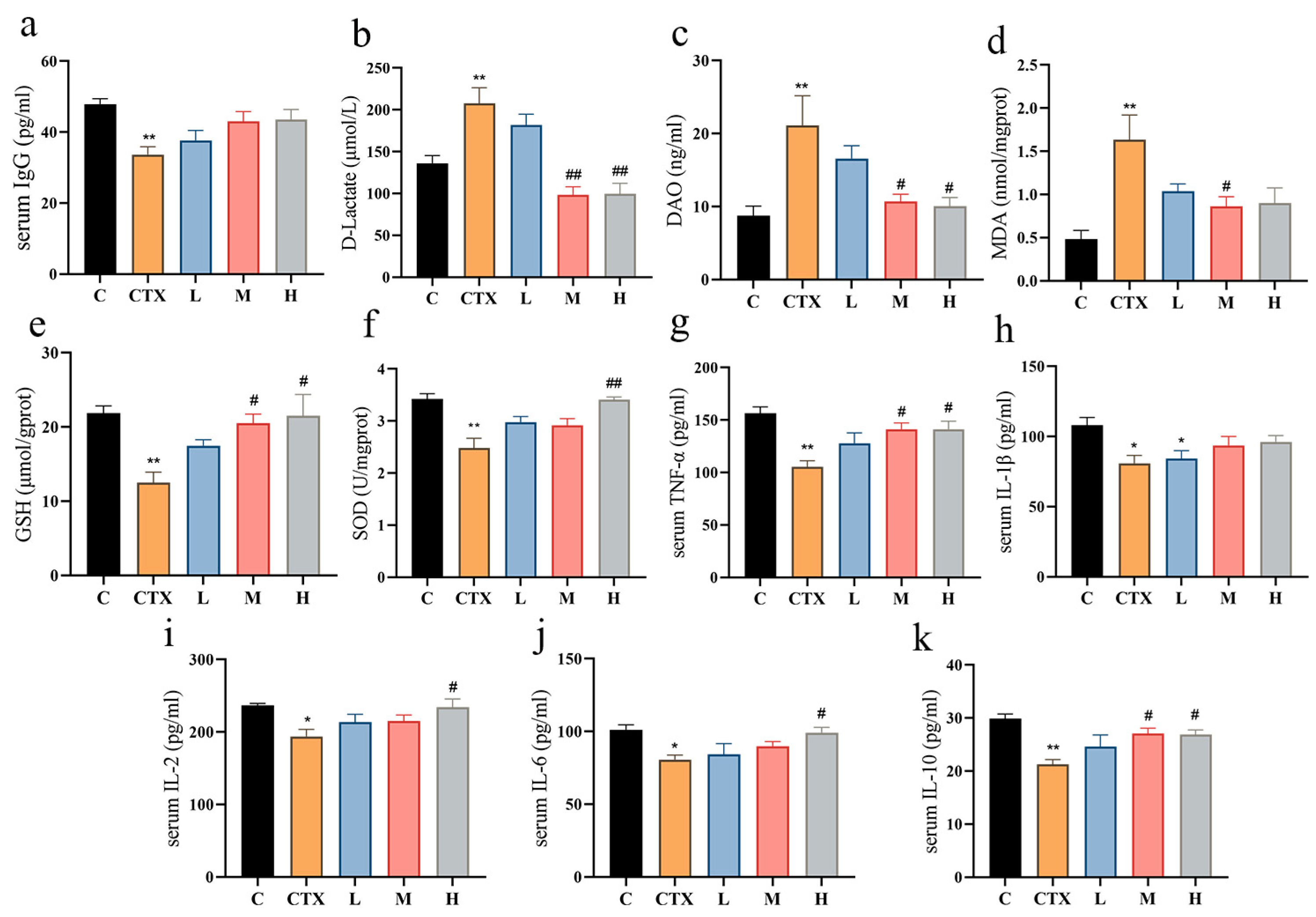
3.2. L. murinus ZNL-13 Alleviates CTX-Induced Oxidative Stress, Immune-Related Cytokines, and Enhances Intestinal Function

After further research, the influence of L. murinus ZNL-13 on serum IgG, intestinal functional markers, oxidative factors, and immune-related cytokines was investigated. The results indicated that compared with the C group, serum immunoglobulin IgG was significantly decreased in the CTX group, while there was an improvement in the L, M, and H groups (Figure 2a). The intestinal functional markers DAO and D-LAC were significantly increased in the CTX group. Pretreatment with L. murinus ZNL-13 significantly attenuated the CTX-induced elevations in serum DAO and D-LAC levels, with marked improvements observed in the M and H groups (p < 0.05), thereby improving intestinal function (Figure 2b,c). Meanwhile, pretreatment with L. murinus ZNL-13 effectively alleviated serum lipid peroxidation (Figure 2d) and enhanced antioxidant capacity (Figure 2e,f). Additionally, compared with the C group, immune-related cytokines in the intestine were significantly decreased in the CTX group. The L. murinus ZNL-13 intervention group mice exhibited some degree of alleviation. For TNF-α, IL-2, IL-6, and IL-10, the H group showed a significant increasing trend in the reduction in cytokines induced by CTX (p < 0.05) (Figure 2g–k).

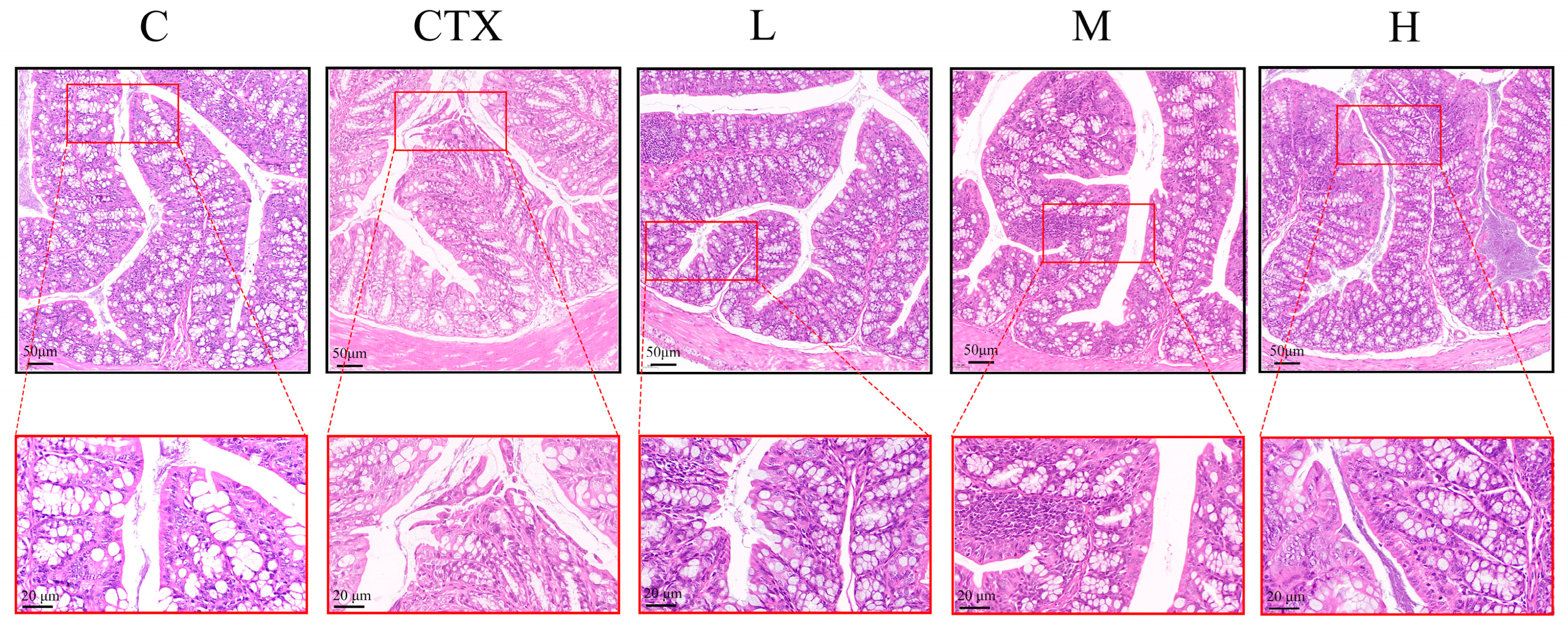
3.3. L. murinus ZNL-13 Alleviates the Damage to Colonic Tissues Induced by CTX

Colonic histopathological examination with H&E staining revealed that L. murinus ZNL-13 has a certain alleviating effect on intestinal damage. In the C group, the villi were intact, and the epithelial absorptive cells were tightly arranged. In the CTX group, the villi were dissolved, goblet cells were ruptured, edges were incomplete, and epithelial absorptive cells were disordered, with infiltration of inflammatory cells and an increase in the thickness of the crypt base. The groups pretreated with L. murinus ZNL-13 exhibited varying degrees of histological improvement characterized by intact goblet cells and well-organized glands arranged in a fence-like structure. Additionally, infiltration of inflammatory cells was observed in the lamina propria of the colon in the CTX group, which was alleviated by treatment with L. murinus ZNL-13 (Figure 3).
The subsequent Western blot analysis and Q-PCR were performed to further investigate the levels of intestinal barrier-related genes (Figure 4a–g). As shown in Figure 4, compared with the blank group, the induction of CTX significantly decreased the protein levels of ZO-1, Occludin, and Claudin-1 (all p <  0.05). Pretreatment with L. murinus ZNL-13 mitigated this trend, with the H group exhibiting a significant alleviation across all three indicators (p < 0.05). Similarly, the RNA results followed the same pattern. In summary, these results suggest that the prophylactic use of L. murinus ZNL-13 can enhance the expression of the intestinal mucin protein MUC2 and tight-junction-related genes and proteins, thereby reducing intestinal permeability and strengthening intestinal barrier function. Notably, the improvement observed in the H group was more pronounced than in the L and M groups.

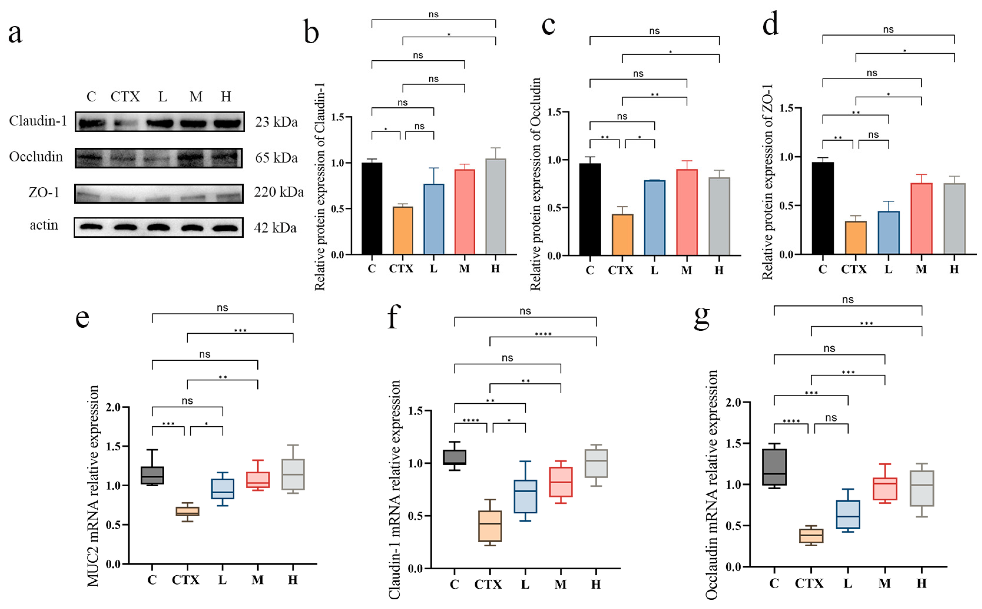
3.4. Effects of L. murinus ZNL-13 on Key Proteins in Colon of CTX Mice

Western blot analysis indicated that the expression levels of TLR4, MYD88, and p65 proteins in the CTX group were significantly decreased compared with the control group (p < 0.05) (Figure 5a–d). The protein levels in the L. murinus ZNL-13 intervention group mice showed an upward trend, with the M and H groups displaying a significant increase compared with the CTX group (p < 0.05). In addition, we utilized Q-PCR (Figure 5e–h) and ELISA assay kits (Figure 5i–m) to measure the levels of intestinal immune-related factors. The results indicated that CTX significantly reduced the levels of IL-1β, IL-2, IL-6, IL-10, and TNF-α (p < 0.01). The expression levels of the groups pretreated with L. murinus ZNL-13 were all increased, with the M and H groups significantly higher than the CTX group (p < 0.05 and p < 0.01, respectively).

3.5. L. murinus ZNL-13 Effectively Attenuates CTX-Induced Apoptosis in Intestinal Tissue Cells

CTX can induce both intrinsic and extrinsic apoptosis. To validate the mechanism by which L. murinus ZNL-13 mitigates apoptosis, further investigation into apoptosis-related genes and proteins was conducted (Figure 6). At the mRNA expression level, apoptosis-related genes including Bax, FAS, FADD, caspase-3, caspase-8, and caspase-9 show an upregulation trend in the CTX group. Conversely, the trend is opposite in the L, M, and H groups, with significant differences observed in the M and H groups (p < 0.05 and p < 0.01). Additionally, Bcl-2 is downregulated in the CTX group but significantly alleviated in the M and H groups (p < 0.01). Further Western blot analysis confirmed these results, with significant upregulation of apoptosis-related genes in the CTX group and significant alleviation in the M and H groups. These results indicate that pretreatment with L. murinus ZNL-13 can significantly alleviate the trend of intestinal cell apoptosis induced by CTX.

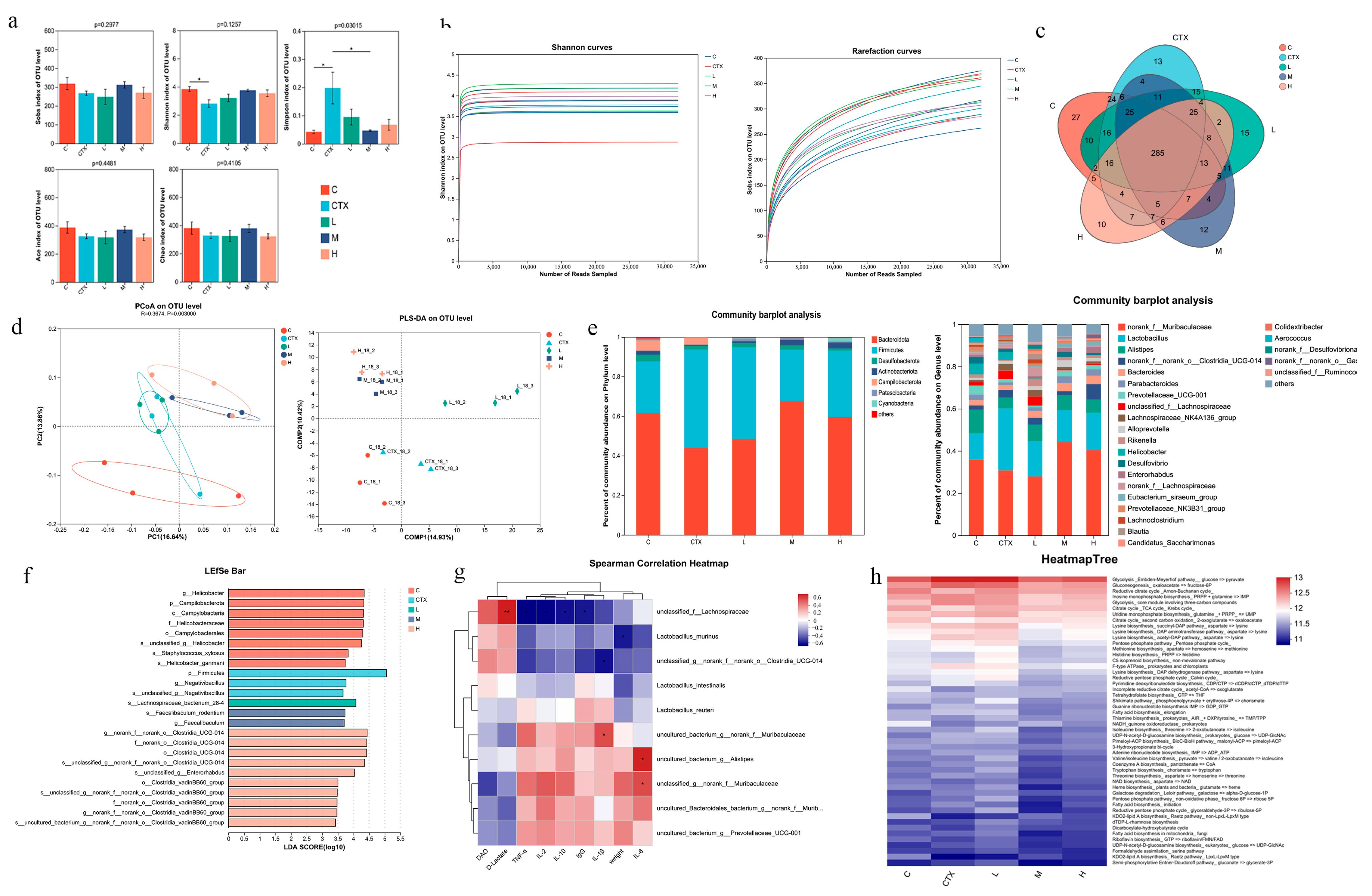
3.6. The Effect of L. murinus ZNL-13 on the Intestinal Microbiota

Using 16S rRNA high-throughput sequencing, we explored the effects of CTX and L. murinus ZNL-13 on the intestinal microbiota. Microbial diversity analysis showed the indexes of Simpson in the L. murinus ZNL-13 group were lower than that in the CTX group; thus, the indexes of Sobs, Chao1, Shannon, and Ace in the L. murinus ZNL-13 group were higher than those in the CTX group, which suggested that the intervention of L. murinus ZNL-13 increased the richness and diversity of the gut microbiota (Figure 7a). The Sobs and Shannon dilution curves tended to flatten (Figure 7b). The Venn diagram reflected the unique and common OTUs in the microbial composition of the five groups, with the number of shared OTUs reaching 47.19% (Figure 7c). Principal Coordinate Analysis (PCOA) and Partial Least Squares–Discriminant Analysis (PLS-DA) were used for beta diversity analysis of the community samples in each group. The results both showed significant differences between the C group and the CTX group, with the M and H groups being closer to each other and distant from the CTX group (Figure 7d). These results indicate that CTX alters the microbial community structure of the C group, and the microbial composition structures of the M and H groups are similar and significantly different from the CTX group.
To further evaluate the specific changes in gut microbiota, we investigated the differences in microbial community composition at the phylum and genus levels (Figure 7e). Bacteroidota and Firmicutes were the predominant phyla in all groups. After CTX administration, there was an increase in Firmicutes abundance and a decrease in Bacteroidota at the phylum level. In contrast, the L. murinus ZNL-13 pretreated group exhibited the opposite trend compared with the CTX group, with an increase in Bacteroidota and Actinobacteriota and a decrease in Campilobacterota, while the abundance of Cyanobacteria increased in the H group. At the genus level, the relative abundance of norank_f__Muribaculaceae, Lachnospiraceae_NK4A136_group, Alistipe, Parabacteroides, Bacteroides, and Prevotellaceae_UCG-001 in the CTX group showed a decreasing trend, while the abundance of Lactobacillus and unclassified_f__Lachnospiraceae significantly increased. L. murinus ZNL-13 pretreatment reversed this trend. The representative species of intestinal microbiota in each group was determined by LEfSe analysis in which the significant differences in bacterial abundance were determined by the non-parametric factorial Kruskal–Wallis sum-rank test and linear discriminant analysis (LDA) (Figure 7f). Based on LEfSe analysis, in the CTX group, the predominant microbial taxa were from the phylum Firmicutes and g__Negativibacillus. L. murinus ZNL-13 pretreatment altered the dominant microbial composition in the intestinal contents of the CTX-treated mice. Spearman correlation analysis was applied to assess the relationships between the intestinal microbial communities in each group and immune parameters. The top ten species ranked by correlation, D-Lactate and unclassified_f_Lachnospiraceae, intestinal tissue IL-1β and uncultured_bacterium_g_norank_f_Muribaculaceae, intestinal tissue IL-6 and uncultured_bacterium_g_Alistipes, unclassified_g_norank_f_Muribaculaceae showed a significant positive correlation horizontally (p < 0.05 and p < 0.01) (Figure 7g). PICRUSt was used for predicting changes in intergroup microbial taxonomic composition and functional annotation (Figure 7h). The heatmap illustrates enrichment of abundance in the Glycolysis and Gluconeogenesis metabolic pathways in the CTX group. In contrast, the dominant functions in the H group are the Reductive Citrate cycle and the Citrate cycle. These results further reveal that the intervention of L. murinus ZNL-13 could increase the immunity of mice by increasing the abundance of probiotics.

4. Discussion

Studies have indicated that CTX can suppress cellular and humoral immune responses. It is considered a primary choice for anticancer treatments, autoimmune diseases, and immunosuppressive regimens for organ transplantation. However, the intestinal tract epithelial barrier disruption and immune dysfunction caused by CTX can lead to opportunistic and pathogenic microbial infections [35,36,37]. The spleen index is a primary indicator for assessing immune function in the human body [6]. After intraperitoneal injection of CTX, the mice exhibited rapid weight loss, noticeable reduction in spleen index, disheveled fur, depressive behavior, and decreased food intake, suggesting successful modeling. Previous studies have shown that long-term consumption of Lactobacillus significantly improves immune organ indices, which aligns with the results of our study [38]. Additionally, CTX induces the generation of reactive oxygen species (ROS), leading to oxidative stress, lipid peroxidation, and cellular damage [39]. Our results indicate that prophylactic administration of L. murinus ZNL-13 significantly alleviates oxidative stress in the intestines and effectively mitigates oxidative damage.
Reports suggest that CTX inhibits the synthesis of tight junction proteins, increases intestinal permeability, and disrupts the intestinal mucosal barrier [8,40]. The disruption of the intestinal mucosal barrier function is a key factor in inducing intestinal damage [41]. In this study, hematoxylin and eosin (H&E) staining results revealed that the colonic wall in the CTX group exhibited thinning, inflammatory cell infiltration, villus breakage, and severe damage to goblet cells. When intestinal mucosal cells undergo necrosis and shed into the lumen and bloodstream, circulating DAO and D-Lactate levels increase. However, L. murinus ZNL-13 significantly alleviated this condition. Dysfunction of tight junction proteins significantly alters cellular permeability, allowing the invasion of harmful substances such as bacterial toxins, which serves as a key pathogenic mechanism in many intestinal diseases [42]. In this study, L. murinus ZNL-13 significantly increased the expression of Claudin-1, Occludin, ZO-1, and MUC2. Therefore, we speculate that prophylactic consumption of L. murinus ZNL-13 holds the potential to strengthen the intestinal barrier and repair intestinal mucosal injury by activating the expression of the intestinal tight junction protein.
Research indicates that approximately 70–80% of the body’s immune cells are aggregated within intestinal tissues [43]. Cytokines are primarily produced by macrophages and lymphocytes, both of which play important roles in immune regulation and inflammatory responses [33,44,45,46,47]. The levels of cytokines TNF-α, IL-1β, IL-2, IL-6, IL-10, and IgG in peripheral circulation and colonic tissues were significantly downregulated following CTX administration, highlighting its profound immunosuppressive effects. L. murinus has been reported to enhance T-cell activity, promote macrophage polarization, and stimulate the production of immune cytokines [28,48,49]. In this study, L. murinus ZNL-13 significantly improved this outcome. Consistent with previous findings, Lactobacillus plantarum KLDS1.0318 can significantly restore cytokine levels, such as IL-6 and TNF-α, reduced by CTX [50]. Multiple studies indicate that the NF-κB signaling pathway plays a crucial role in the induction and secretion of cytokine expression [34]. Additionally, impaired MyD88/p65 signaling in the intestinal epithelium is associated with the reduction in tight junction proteins such as Claudin-1 and Occludin as well as the downregulation of MUC2 expression [51,52,53]. L. casei NCU011054 significantly upregulated the mRNA levels of TLR2, TLR4, TLR6, p65, NF-κB, T-bet, and GATA-3; restored the number of CD4⁺ T cells; and alleviated the inhibitory effects of CTX [54]. The results showed that the NF-κB signaling pathway was significantly suppressed by CTX, while the expression of proteins related to this pathway was markedly upregulated in the L. murinus ZNL-13 pretreatment group. Therefore, we speculate that the mechanism by which L. murinus ZNL-13 regulates cytokine expression involves the activation of the TLR4 receptor, modulation of downstream NF-κB pathway expression, and ultimately, the stimulation of immune cytokine secretion and transcription factor expression, thereby restoring immune balance in CTX-induced intestinal injury in mice. However, previous studies have indicated that probiotics can also alleviate intestinal inflammation by modulating additional signaling pathways, such as MAPK, Nrf2, and PI3K/AKT [55,56,57]. Therefore, TLR4 may not be the sole receptor involved in this process. Further investigations are warranted to explore other molecular mechanisms beyond the TLR4/NF-κB axis that may contribute to L. murinus ZNL-13-mediated immune regulation.
CTX inhibits cellular DNA replication and differentiation; however, it also exhibits cytotoxic effects, accelerating the apoptosis of rapidly proliferating cells in the body [58,59,60]. Studies have shown that acetaminophen can promote the translocation of the pro-apoptotic factor Bax to the mitochondria and facilitate the maturation of caspase-3, ultimately leading to tissue cell damage [61]. Additionally, the inhibition of the MAPK and NF-κB signaling pathways can also trigger apoptosis [62,63]. Lactobacillus plantarum Lp2 effectively regulates the MAPK signaling pathway and downregulates the expression of downstream pro-apoptotic proteins, including caspase-3 and Bax, thereby attenuating CTX-induced hepatocyte apoptosis [55]. This finding indirectly suggests that the TLR4/NF-κB pathway may not be the sole mechanism through which Lactobacillus species, including L. murinus, exert their protective effects. Consistent with previous studies, CTX induces apoptosis, while L. murinus ZNL-13 upregulates Bcl-2 and downregulates the mRNA and protein expressions of Bax, Fas, caspase-3, caspase-8, and caspase-9; preserves mitochondrial membrane integrity; and blocks apoptotic signal transduction mediated by death receptor–ligand interactions, thereby mitigating excessive apoptosis in intestinal cells. These findings suggest that L. murinus ZNL-13 alleviates CTX-induced apoptosis and intestinal injury by suppressing apoptotic pathway activation.
The gut microbiota is a complex system influenced by factors such as the environment and diet, which can impact the composition of microbial communities [64,65]. CTX-induced immune suppression and intestinal damage disrupt the gut microbiota, the organism experiences secondary declines in immune function, which induces increased inflammation in the intestine, leading to a vicious cycle [66,67,68]. CTX has been shown to increase the Simpson index and reduce microbial diversity [69]. Similarly, our study found that prophylactic administration of L. murinus ZNL-13 reduced the Simpson index, which may have a beneficial impact. Both PCA and Partial Least Squares–Discriminant Analysis (PLS-DA) results indicate significant differences in microbial structures between the C group and the CTX group. L. murinus ZNL-13 altered the intestinal microbiota structure of mice. At the phylum level, the relative abundance of Firmicutes increased, while Bacteroidetes, Desulfobacterota, and Actinobacteriota decreased in the CTX group. Bacteroidetes can ferment indigestible polysaccharides to provide an energy source for the host [70]. Prophylactic administration of L. murinus ZNL-13 led to an increase in Bacteroidetes, which may enhance the digestion and absorption of polysaccharides in the intestine. Campylobacterota are pathogens, including members such as Helicobacter and Desulfovibrio, whose abundance increase can exacerbate inflammatory diseases [71]. A significant enrichment of Campylobacterota in the C and CTX groups was observed in this study, while the L. murinus ZNL-13 groups showed minimal presence of such microorganisms. This supports their role in promoting intestinal health, consistent with previous reports [35]. This evidence suggests that L. murinus ZNL-13 can reduce the activity of such microorganisms, helping the host organism restore gut homeostasis and create a healthy intestinal environment.
At the genus level, Helicobacter, which are highly relevant in gut microbial dysbiosis and disease, appeared in both the C and CTX groups, while they were scarcely observed in the L. murinus ZNL-13 groups. SCFAs provide the necessary nutrients and energy for intestinal epithelial cells, preserving the integrity and functionality of the intestinal mucosal barrier, which can block the transport of toxins between the intestine and bloodstream, inhibiting inflammation [72,73]. Muribaculaceae, Bacteroides, and Alloprevotella have been shown to promote the production of short-chain fatty acids (SCFAs). And these bacteria were prominently present in the L. murinus ZNL-13 group [74]. Alistipes and Prevotellaceae_UCG-001 can produce polysaccharide-digesting enzymes that promote the production of SCFAs [75,76,77,78,79,80]. The increased relative abundance of Alistipes and Prevotellaceae_UCG-001 in the M and H groups suggests that supplementing with L. murinus ZNL-13 promotes aiding in the restoration of gut health and enhancing food conversion and absorption capacity after CTX administration. This also helps confirm that the L. murinus ZNL-13 group can alleviate the rapid weight loss induced by CTX.
Additionally, differences in microbiota among groups and representative species, as well as dominant microbial communities, can be identified through LDA score histograms. Negativibacillus, a signature species identified in the CTX group, has been confirmed to be significantly increased in the gut microbiota of patients with ulcerative colitis [81]. However, the signature species identified in the L. murinus ZNL-13 intervention groups have all been reported to exert regulatory effects that contribute to host health. Faecalibaculum rodentium plays a beneficial role in regulating intestinal immunity and inflammation, and its independent administration has been shown to counteract the progression of colitis [82]. Furthermore, closely related genera within the Faecalibaculum group have been demonstrated to influence bile acid metabolism through dehydroxylation processes [83]. An increased abundance of Clostridia UCG-014 has been associated with enhanced production of short-chain fatty acids (SCFAs) [84,85]. Notably, the Clostridia_vadinBB60_group has been identified as a known producer of acetate and propionate [86,87,88]. Acetic acid can enhance the production of reactive oxygen species and phagocytic activity, induce apoptosis, and modulate the recruitment of immune cells such as neutrophils [89]. Butyrate salts have been shown to enhance the activity of natural killer (NK) cells and have the function of regulating the activity of immune cells and altering the production of inflammatory cytokines [90,91]. In addition, Clostridium has been reported to promote the conversion of primary bile acids into secondary bile acids via the activity of 7α-dehydroxylase. Secondary bile acids have been shown to activate intestinal macrophages and Treg cells through TGR5, thereby enhancing immune responses against pathogens [92,93]. Clostridium also facilitates the metabolism of tryptophan into indolepropionic acid, a compound known for its antioxidant properties. Certain tryptophan metabolites have been shown to activate B cells, further contributing to immune modulation. [94,95]. Moreover, Clostridium species can enhance arginine metabolism, resulting in the production of polyamines such as putrescine and spermidine, which are essential for intestinal barrier repair. Notably, spermidine has been shown to enhance immune function by promoting mitochondrial metabolism in natural killer (NK) cells [96,97]. Therefore, we hypothesize that L. murinus ZNL-13 supplementation may improve immune function by modulating the gut microbiota, increasing the production of beneficial metabolites, enhancing intestinal barrier integrity, and promoting immune cell activity. A recent study found that Faecalibaculum rodentium exerts anti-tumor effects in a mouse model of colorectal cancer by producing short-chain fatty acids [98]. This also provides insight into the potential for L. murinus ZNL-13 to co-suppress tumor cell proliferation with CTX on the basis of alleviating CTX-induced damage. In summary, prophylactic administration of L. murinus ZNL-13 promotes the colonization of beneficial bacteria and restores the imbalance in the intestinal microbiota induced by CTX in mice, which is crucial for maintaining the stability of the host microbiota. It also activates immune cell activity and enhances immune function, suggesting the potential beneficial effects of L. murinus ZNL-13 on the intestinal microenvironment of animals or humans in the future.
In summary, prophylactic administration of L. murinus ZNL-13 effectively mitigated intestinal damage in CTX-treated mice to varying degrees. It significantly promoted the secretion of intestinal immune factors such as IL-1β and IL-2, activated the NF-κB pathway to stimulate immune cell activity, and restored immune function. Moreover, it downregulated apoptosis-related genes such as BAX and caspase-3 and alleviated intestinal oxidative damage, contributing to the repair of intestinal injury. Meanwhile, L. murinus ZNL-13 helped regulate gut microbiota homeostasis, increased the abundance of beneficial bacteria, and promoted the restoration of overall health. These findings provide a theoretical basis for the development of L. murinus ZNL-13 as a dietary nutritional supplement.

5. Conclusions

This study investigated the potential protective mechanisms of L. murinus ZNL-13 against CTX-induced immunosuppression and intestinal injury in mice and further revealed the possible role of the gut microbiota in alleviating intestinal damage and activating immune function. These findings support the potential application of L. murinus ZNL-13 as a novel functional food additive for humans or other animals.

Author Contributions

Y.D. performed the study. L.Z. (Luyao Zhang) and D.Q. drafted and submitted the manuscript. R.Y. assisted in accomplishing the experiment. H.J., H.W. and L.Z. (Luyao Zhou) carried out the statistical analysis. J.Z. and N.Z. reviewed the manuscript. All authors approved the submitted version. All authors have read and agreed to the published version of the manuscript.

Funding

This research was funded by the National Key R&D Program of China (2023YFD1801100) and Heilongjiang Universities Collaborative Innovation Project (LJGXCG2022-042).

Institutional Review Board Statement

Not applicable.

Informed Consent Statement

Not applicable.

Data Availability Statement

The original contributions presented in this study are included in the article. Further inquiries can be directed to the corresponding authors. Sequence data in this study were uploaded to the NCBI SRA database, and the SRA accession number is PRJNA1123850 (https://www.ncbi.nlm.nih.gov/bioproject/PRJNA1123850 (accessed on 14 January 2025).

Conflicts of Interest

The authors declare no conflicts of interest.

References

  1. Emadi, A.; Jones, R.J.; Brodsky, R.A. Cyclophosphamide and cancer: Golden anniversary. Nat. Rev. Clin. Oncol. 2009, 6, 638–647. [Google Scholar] [CrossRef]
  2. Ahmed, A.R.; Hombal, S.M. Cyclophosphamide (Cytoxan). A review on relevant pharmacology and clinical uses. J. Am. Acad. Dermatol. 1984, 11, 1115–1126. [Google Scholar] [CrossRef]
  3. Hu, N.; Qu, Y.; Liu, T.Y.; Zhou, Y.; Liu, C.; Wang, J.H.; Yang, B.F.; Li, C.L. Immunomodulatory effects and mechanisms of Tiepishihu Xiyangshen granules on cyclophosphamide induced immuno-suppression via TLR4/MAPKs and PI3K/AKT/FOXO3a signal pathways. J. Ethnopharmacol. 2023, 307, 116192. [Google Scholar] [CrossRef] [PubMed]
  4. Brock, N.; Wilmanns, H. Effect of a cyclic nitrogen mustard-phosphamidester on experimentally induced tumors in rats; chemotherapeutic effect and pharmacological properties of B 518 ASTA. Dtsch. Med. Wochenschr. 1958, 83, 453–458. [Google Scholar] [CrossRef]
  5. Xie, H.; Fang, J.; Farag, M.A.; Li, Z.; Sun, P.; Shao, P. Dendrobium officinale leaf polysaccharides regulation of immune response and gut microbiota composition in cyclophosphamide-treated mice. Food Chem. X 2022, 13, 100235. [Google Scholar] [CrossRef] [PubMed]
  6. Ding, Y.; Yan, Y.; Chen, D.; Ran, L.; Mi, J.; Lu, L.; Jing, B.; Li, X.; Zeng, X.; Cao, Y. Modulating effects of polysaccharides from the fruits of Lycium barbarum on the immune response and gut microbiota in cyclophosphamide-treated mice. Food Funct. 2019, 10, 3671–3683. [Google Scholar] [CrossRef]
  7. Zhou, R.; He, D.; Xie, J.; Zhou, Q.; Zeng, H.; Li, H.; Huang, L. The Synergistic Effects of Polysaccharides and Ginsenosides from American Ginseng (Panax quinquefolius L.) Ameliorating Cyclophosphamide-Induced Intestinal Immune Disorders and Gut Barrier Dysfunctions Based on Microbiome-Metabolomics Analysis. Front. Immunol. 2021, 12, 665901. [Google Scholar] [CrossRef] [PubMed]
  8. Ou, Z.; Zhu, L.; Huang, C.; Ma, C.; Kong, L.; Lin, X.; Gao, X.; Huang, L.; Wen, L.; Liang, Z.; et al. Betulinic acid attenuates cyclophosphamide-induced intestinal mucosa injury by inhibiting the NF-κB/MAPK signalling pathways and activating the Nrf2 signalling pathway. Ecotoxicol. Environ. Saf. 2021, 225, 112746. [Google Scholar] [CrossRef]
  9. Zhao, F.; Ma, T.; Zhang, X.; Zhao, Q.; Zhu, K.; Cao, J.; Liu, Z.; Shen, X.; Li, C. Holothuria leucospilota Polysaccharides Improve Immunity and the Gut Microbiota in Cyclophosphamide-Treated Immunosuppressed Mice. Mol. Nutr. Food Res. 2023, 67, e2200317. [Google Scholar] [CrossRef]
  10. Peterson, L.W.; Artis, D. Intestinal epithelial cells: Regulators of barrier function and immune homeostasis. Nat. Rev. Immunol. 2014, 14, 141–153. [Google Scholar] [CrossRef]
  11. Keely, S.J.; Barrett, K.E. Intestinal secretory mechanisms and diarrhea. Am. J. Physiol. Gastrointest. Liver Physiol. 2022, 322, G405–G420. [Google Scholar] [CrossRef] [PubMed]
  12. Poller, W.C.; Downey, J.; Mooslechner, A.A.; Khan, N.; Li, L.; Chan, C.T.; McAlpine, C.S.; Xu, C.; Kahles, F.; He, S.; et al. Brain motor and fear circuits regulate leukocytes during acute stress. Nature 2022, 607, 578–584. [Google Scholar] [CrossRef] [PubMed]
  13. Yoo, B.B.; Mazmanian, S.K. The Enteric Network: Interactions between the Immune and Nervous Systems of the Gut. Immunity 2017, 46, 910–926. [Google Scholar] [CrossRef]
  14. Miyauchi, E.; Shimokawa, C.; Steimle, A.; Desai, M.S.; Ohno, H. The impact of the gut microbiome on extra-intestinal autoimmune diseases. Nat. Rev. Immunol. 2023, 23, 9–23. [Google Scholar] [CrossRef]
  15. Spencer, J.; Bemark, M. Human intestinal B cells in inflammatory diseases. Nat. Rev. Gastroenterol. Hepatol. 2023, 20, 254–265. [Google Scholar] [CrossRef]
  16. Hu, H.; Sun, W.; Zhang, L.; Zhang, Y.; Kuang, T.; Qu, D.; Lian, S.; Hu, S.; Cheng, M.; Xu, Y.; et al. Carboxymethylated Abrus cantoniensis polysaccharide prevents CTX-induced immunosuppression and intestinal damage by regulating intestinal flora and butyric acid content. Int. J. Biol. Macromol. 2024, 261, 129590. [Google Scholar] [CrossRef]
  17. Bhunia, A.K.; Al-Sadi, R. Editorial: Intestinal epithelial barrier disruption by enteric pathogens. Front. Cell Infect. Microbiol. 2023, 13, 1134753. [Google Scholar] [CrossRef]
  18. Zhang, X.; Akhtar, M.; Chen, Y.; Ma, Z.; Liang, Y.; Shi, D.; Cheng, R.; Cui, L.; Hu, Y.; Nafady, A.A.; et al. Chicken jejunal microbiota improves growth performance by mitigating intestinal inflammation. Microbiome 2022, 10, 107. [Google Scholar] [CrossRef]
  19. Zhang, Y.; Mahmood, T.; Tang, Z.; Wu, Y.; Yuan, J. Effects of naturally oxidized corn oil on inflammatory reaction and intestinal health of broilers. Poult. Sci. 2022, 101, 101541. [Google Scholar] [CrossRef]
  20. Chen, V.L.; Kasper, D.L. Interactions between the intestinal microbiota and innate lymphoid cells. Gut Microbes 2014, 5, 129–140. [Google Scholar] [CrossRef]
  21. Tay, M.Z.; Poh, C.M.; Rénia, L.; MacAry, P.A.; Ng, L.F.P. The trinity of COVID-19: Immunity, inflammation and intervention. Nat. Rev. Immunol. 2020, 20, 363–374. [Google Scholar] [CrossRef]
  22. Wang, X.; Wang, Z.; Wu, H.; Jia, W.; Teng, L.; Song, J.; Yang, X.; Wang, D. Sarcodon imbricatus polysaccharides protect against cyclophosphamide-induced immunosuppression via regulating Nrf2-mediated oxidative stress. Int. J. Biol. Macromol. 2018, 120, 736–744. [Google Scholar] [CrossRef]
  23. Sarin, S.K.; Pande, A.; Schnabl, B. Microbiome as a therapeutic target in alcohol-related liver disease. J. Hepatol. 2019, 70, 260–272. [Google Scholar] [CrossRef] [PubMed]
  24. Ohland, C.L.; Macnaughton, W.K. Probiotic bacteria and intestinal epithelial barrier function. Am. J. Physiol. Gastrointest. Liver Physiol. 2010, 298, G807–G819. [Google Scholar] [CrossRef]
  25. Xie, J.H.; Fan, S.T.; Nie, S.P.; Yu, Q.; Xiong, T.; Gong, D.; Xie, M.Y. Lactobacillus plantarum NCU116 attenuates cyclophosphamide-induced intestinal mucosal injury, metabolism and intestinal microbiota disorders in mice. Food Funct. 2016, 7, 1584–1592. [Google Scholar] [CrossRef] [PubMed]
  26. Zeng, Z.; Huang, Z.; Yue, W.; Nawaz, S.; Chen, X.; Liu, J. Lactobacillus plantarum modulate gut microbiota and intestinal immunity in cyclophosphamide-treated mice model. Biomed. Pharmacother. 2023, 169, 115812. [Google Scholar] [CrossRef] [PubMed]
  27. Hu, J.; Deng, F.; Zhao, B.; Lin, Z.; Sun, Q.; Yang, X.; Wu, M.; Qiu, S.; Chen, Y.; Yan, Z.; et al. Lactobacillus murinus alleviate intestinal ischemia/reperfusion injury through promoting the release of interleukin-10 from M2 macrophages via Toll-like receptor 2 signaling. Microbiome 2022, 10, 38. [Google Scholar] [CrossRef]
  28. Fan, J.; Zhang, Y.; Zuo, M.; Ding, S.; Li, J.; Feng, S.; Xiao, Y.; Tao, S. Novel mechanism by which extracellular vesicles derived from Lactobacillus murinus alleviates deoxynivalenol-induced intestinal barrier disruption. Environ. Int. 2024, 185, 108525. [Google Scholar] [CrossRef]
  29. Obisesan, A.O.; Abiodun, O.O.; Ayeni, F.A. Lactic acid bacteria isolated from women’ breast milk and infants’ faeces have appreciable immunogenic and probiotic potentials against diarrheagenic E. coli strains. BMC Microbiol. 2024, 24, 350. [Google Scholar] [CrossRef]
  30. Chen, H.T.; Li, J.S.; Li, J.; Li, L.; Xu, Z.C.; Zhang, Y.; Wang, R.R. Lactobacillus murinus: A key factor in suppression of enterogenous Candida albicans infections in Compound Agrimony enteritis capsules-treated mice. J. Ethnopharmacol. 2023, 311, 116361. [Google Scholar] [CrossRef]
  31. Zhu, Y.; Tao, X.; Yan, T.; Cao, S.; Jiang, P.; Zhang, Z.; Li, L.; Wu, Q. Lactobacillus murinus alleviated lung inflammation induced by PAHs in mice. Ecotoxicol. Environ. Saf. 2024, 281, 116662. [Google Scholar] [CrossRef] [PubMed]
  32. Ren, C.; Cheng, L.; Sun, Y.; Zhang, Q.; de Haan, B.J.; Zhang, H.; Faas, M.M.; de Vos, P. Lactic acid bacteria secrete toll like receptor 2 stimulating and macrophage immunomodulating bioactive factors. J. Funct. Foods 2020, 66, 103783. [Google Scholar] [CrossRef]
  33. Perelmuter, K.; Fraga, M.; Zunino, P. In vitro activity of potential probiotic Lactobacillus murinus isolated from the dog. J. Appl. Microbiol. 2008, 104, 1718–1725. [Google Scholar] [CrossRef]
  34. Ying, M.; Yu, Q.; Zheng, B.; Wang, H.; Wang, J.; Chen, S.; Nie, S.; Xie, M. Cultured Cordyceps sinensis polysaccharides modulate intestinal mucosal immunity and gut microbiota in cyclophosphamide-treated mice. Carbohydr. Polym. 2020, 235, 115957. [Google Scholar] [CrossRef] [PubMed]
  35. Tian, J.; Zhao, X.; Tang, C.; Wang, X.; Zhang, X.; Xiao, L.; Li, W. Protective effect of Paecilomyces cicadae TJJ11213 exopolysaccharide on intestinal mucosa and regulation of gut microbiota in immunosuppressed mice. Food Res. Int. 2023, 165, 112477. [Google Scholar] [CrossRef]
  36. Gill, N.; Wlodarska, M.; Finlay, B.B. Roadblocks in the gut: Barriers to enteric infection. Cell Microbiol. 2011, 13, 660–669. [Google Scholar] [CrossRef]
  37. Meng, M.; Wang, H.; Li, Z.; Guo, M.; Hou, L. Protective effects of polysaccharides from Cordyceps gunnii mycelia against cyclophosphamide-induced immunosuppression to TLR4/TRAF6/NF-κB signalling in BALB/c mice. Food Funct. 2019, 10, 3262–3271. [Google Scholar] [CrossRef]
  38. Li, S.P.; Zhao, X.J.; Wang, J.Y. Synergy of Astragalus polysaccharides and probiotics (Lactobacillus and Bacillus cereus) on immunity and intestinal microbiota in chicks. Poult. Sci. 2009, 88, 519–525. [Google Scholar] [CrossRef]
  39. Yu, J.; Cong, L.; Wang, C.; Li, H.; Zhang, C.; Guan, X.; Liu, P.; Xie, Y.; Chen, J.; Sun, J. Immunomodulatory effect of Schisandra polysaccharides in cyclophosphamide-induced immunocompromised mice. Exp. Ther. Med. 2018, 15, 4755–4762. [Google Scholar] [CrossRef]
  40. Agúndez, J.A.; Ayuso, P.; Cornejo-García, J.A.; Blanca, M.; Torres, M.J.; Doña, I.; Salas, M.; Blanca-López, N.; Canto, G.; Rondon, C.; et al. The diamine oxidase gene is associated with hypersensitivity response to non-steroidal anti-inflammatory drugs. PLoS ONE 2012, 7, e47571. [Google Scholar] [CrossRef]
  41. Park, C.M.; Song, Y.-S. Luteolin and luteolin-7-O-glucoside inhibit lipopolysaccharide-induced inflammatory responses through modulation of NF-κB/AP-1/PI3K-Akt signaling cascades in RAW 264.7 cells. Nutr. Res. Pract. 2013, 7, 423–429. [Google Scholar] [CrossRef] [PubMed]
  42. Han, Y.; Zhang, Y.; Ouyang, K.; Chen, L.; Zhao, M.; Wang, W. Sulfated Cyclocarya paliurus polysaccharides improve immune function of immunosuppressed mice by modulating intestinal microbiota. Int. J. Biol. Macromol. 2022, 212, 31–42. [Google Scholar] [CrossRef] [PubMed]
  43. Zhang, J.; Zhou, H.C.; He, S.B.; Zhang, X.F.; Ling, Y.H.; Li, X.Y.; Zhang, H.; Hou, D.D. The immunoenhancement effects of sea buckthorn pulp oil in cyclophosphamide-induced immunosuppressed mice. Food Funct. 2021, 12, 7954–7963. [Google Scholar] [CrossRef]
  44. Sun, S.; Xu, X.; Liang, L.; Wang, X.; Bai, X.; Zhu, L.; He, Q.; Liang, H.; Xin, X.; Wang, L.; et al. Lactic Acid-Producing Probiotic Saccharomyces cerevisiae Attenuates Ulcerative Colitis via Suppressing Macrophage Pyroptosis and Modulating Gut Microbiota. Front. Immunol. 2021, 12, 777665. [Google Scholar] [CrossRef]
  45. Awad, W.A.; Ghareeb, K.; Abdel-Raheem, S.; Böhm, J. Effects of dietary inclusion of probiotic and synbiotic on growth performance, organ weights, and intestinal histomorphology of broiler chickens. Poult. Sci. 2009, 88, 49–56. [Google Scholar] [CrossRef]
  46. Emami, N.K.; Dalloul, R.A. Centennial Review: Recent developments in host-pathogen interactions during necrotic enteritis in poultry. Poult. Sci. 2021, 100, 101330. [Google Scholar] [CrossRef] [PubMed]
  47. Sarikaya-Bayram, Ö.; Palmer, J.M.; Keller, N.; Braus, G.H.; Bayram, Ö. One Juliet and four Romeos: VeA and its methyltransferases. Front. Microbiol. 2015, 6, 1. [Google Scholar] [CrossRef]
  48. Bernard-Raichon, L.; Colom, A.; Monard, S.C.; Namouchi, A.; Cescato, M.; Garnier, H.; Leon-Icaza, S.A.; Métais, A.; Dumas, A.; Corral, D.; et al. A Pulmonary Lactobacillus murinus Strain Induces Th17 and RORγt(+) Regulatory T Cells and Reduces Lung Inflammation in Tuberculosis. J. Immunol. 2021, 207, 1857–1870. [Google Scholar] [CrossRef] [PubMed]
  49. Singer, J.R.; Blosser, E.G.; Zindl, C.L.; Silberger, D.J.; Conlan, S.; Laufer, V.A.; DiToro, D.; Deming, C.; Kumar, R.; Morrow, C.D.; et al. Preventing dysbiosis of the neonatal mouse intestinal microbiome protects against late-onset sepsis. Nat. Med. 2019, 25, 1772–1782. [Google Scholar] [CrossRef]
  50. Meng, Y.; Wang, J.; Wang, Z.; Zhang, G.; Liu, L.; Huo, G.; Li, C. Lactobacillus plantarum KLDS1.0318 Ameliorates Impaired Intestinal Immunity and Metabolic Disorders in Cyclophosphamide-Treated Mice. Front. Microbiol. 2019, 10, 731. [Google Scholar] [CrossRef]
  51. Frantz, A.L.; Rogier, E.W.; Weber, C.R.; Shen, L.; Cohen, D.A.; Fenton, L.A.; Bruno, M.E.; Kaetzel, C.S. Targeted deletion of MyD88 in intestinal epithelial cells results in compromised antibacterial immunity associated with downregulation of polymeric immunoglobulin receptor, mucin-2, and antibacterial peptides. Mucosal Immunol. 2012, 5, 501–512. [Google Scholar] [CrossRef]
  52. Johansen, F.E.; Kaetzel, C.S. Regulation of the polymeric immunoglobulin receptor and IgA transport: New advances in environmental factors that stimulate pIgR expression and its role in mucosal immunity. Mucosal Immunol. 2011, 4, 598–602. [Google Scholar] [CrossRef] [PubMed]
  53. Rakoff-Nahoum, S.; Paglino, J.; Eslami-Varzaneh, F.; Edberg, S.; Medzhitov, R. Recognition of commensal microflora by toll-like receptors is required for intestinal homeostasis. Cell 2004, 118, 229–241. [Google Scholar] [CrossRef]
  54. Min, F.; Hu, J.; Huang, T.; Huang, Y.; Nie, S.; Xiong, T.; Xie, M. Effects of Lactobacillus casei NCU011054 on immune response and gut microbiota of cyclophosphamide induced immunosuppression mice. Food Chem. Toxicol. 2023, 174, 113662. [Google Scholar] [CrossRef]
  55. Zhang, N.; Tian, Y.; Wang, Y.; Fan, Y.; Zhang, Y.; Xing, X.; Nan, B.; Ai, Z.; Li, X.; Wang, Y. Ameliorative effect of Lactobacillus plantarum Lp2 against cyclophosphamide-induced liver injury in mice. Food Chem. Toxicol. 2022, 169, 113433. [Google Scholar] [CrossRef] [PubMed]
  56. Li, J.; Feng, S.; Wang, Z.; He, J.; Zhang, Z.; Zou, H.; Wu, Z.; Liu, X.; Wei, H.; Tao, S. Limosilactobacillus mucosae-derived extracellular vesicles modulates macrophage phenotype and orchestrates gut homeostasis in a diarrheal piglet model. NPJ Biofilms Microbiomes 2023, 9, 33. [Google Scholar] [CrossRef]
  57. Tao, S.; Fan, J.; Li, J.; Wu, Z.; Yao, Y.; Wang, Z.; Wu, Y.; Liu, X.; Xiao, Y.; Wei, H. Extracellular vesicles derived from Lactobacillus johnsonii promote gut barrier homeostasis by enhancing M2 macrophage polarization. J. Adv. Res. 2025, 69, 545–563. [Google Scholar] [CrossRef] [PubMed]
  58. Lim, E.J.; Chin, R.; Nachbur, U.; Silke, J.; Jia, Z.; Angus, P.W.; Torresi, J. Effect of Immunosuppressive Agents on Hepatocyte Apoptosis Post-Liver Transplantation. PLoS ONE 2015, 10, e0138522. [Google Scholar] [CrossRef]
  59. Liu, T.; Wu, Y.; Wang, L.; Pang, X.; Zhao, L.; Yuan, H.; Zhang, C. A More Robust Gut Microbiota in Calorie-Restricted Mice Is Associated with Attenuated Intestinal Injury Caused by the Chemotherapy Drug Cyclophosphamide. mBio 2019, 10, e02903-18. [Google Scholar] [CrossRef]
  60. Sherif, I.O. The effect of natural antioxidants in cyclophosphamide-induced hepatotoxicity: Role of Nrf2/HO-1 pathway. Int. Immunopharmacol. 2018, 61, 29–36. [Google Scholar] [CrossRef]
  61. Lv, H.; Xiao, Q.; Zhou, J.; Feng, H.; Liu, G.; Ci, X. Licochalcone A Upregulates Nrf2 Antioxidant Pathway and Thereby Alleviates Acetaminophen-Induced Hepatotoxicity. Front. Pharmacol. 2018, 9, 147. [Google Scholar] [CrossRef] [PubMed]
  62. Wang, C.; Li, S.; Wang, M.W. Evodiamine-induced human melanoma A375-S2 cell death was mediated by PI3K/Akt/caspase and Fas-L/NF-kappaB signaling pathways and augmented by ubiquitin-proteasome inhibition. Toxicol. In Vitro 2010, 24, 898–904. [Google Scholar] [CrossRef]
  63. Lei, Y.; Chan, M.; Liu, H.; Lyu, W.; Chen, L.; Zhong, Y.; Gan, H.; Wang, M.; Qi, M.; Guo, Y.; et al. Evodiamine as the Active Compound of Evodiae fructus to Inhibit Proliferation and Migration of Prostate Cancer through PI3K/AKT/NF-κB Signaling Pathway. Dis. Markers 2022, 2022, 4399334. [Google Scholar] [CrossRef] [PubMed]
  64. Hatton, I.A.; Mazzarisi, O.; Altieri, A.; Smerlak, M. Diversity begets stability: Sublinear growth and competitive coexistence across ecosystems. Science 2024, 383, eadg8488. [Google Scholar] [CrossRef]
  65. Litvak, Y.; Mon, K.K.Z.; Nguyen, H.; Chanthavixay, G.; Liou, M.; Velazquez, E.M.; Kutter, L.; Alcantara, M.A.; Byndloss, M.X.; Tiffany, C.R.; et al. Commensal Enterobacteriaceae Protect against Salmonella Colonization through Oxygen Competition. Cell Host Microbe 2019, 25, 128–139.e125. [Google Scholar] [CrossRef]
  66. Pavel, F.M.; Vesa, C.M.; Gheorghe, G.; Diaconu, C.C.; Stoicescu, M.; Munteanu, M.A.; Babes, E.E.; Tit, D.M.; Toma, M.M.; Bungau, S. Highlighting the Relevance of Gut Microbiota Manipulation in Inflammatory Bowel Disease. Diagnostics 2021, 11, 1090. [Google Scholar] [CrossRef] [PubMed]
  67. Gophna, U.; Sommerfeld, K.; Gophna, S.; Doolittle, W.F.; Veldhuyzen van Zanten, S.J. Differences between tissue-associated intestinal microfloras of patients with Crohn’s disease and ulcerative colitis. J. Clin. Microbiol. 2006, 44, 4136–4141. [Google Scholar] [CrossRef]
  68. Shi, N.; Li, N.; Duan, X.; Niu, H. Interaction between the gut microbiome and mucosal immune system. Mil. Med. Res. 2017, 4, 14. [Google Scholar] [CrossRef]
  69. Chen, S.; Wang, J.; Fang, Q.; Dong, N.; Fang, Q.; Cui, S.W.; Nie, S. A polysaccharide from natural Cordyceps sinensis regulates the intestinal immunity and gut microbiota in mice with cyclophosphamide-induced intestinal injury. Food Funct. 2021, 12, 6271–6282. [Google Scholar] [CrossRef]
  70. Johnson, E.L.; Heaver, S.L.; Walters, W.A.; Ley, R.E. Microbiome and metabolic disease: Revisiting the bacterial phylum Bacteroidetes. J. Mol. Med. 2017, 95, 1–8. [Google Scholar] [CrossRef]
  71. Ma, Y.; Liu, T.; Chen, S.; Shen, H.; Wang, J. Dietary supplementation with L-citrulline improves amino acid composition and broiler performance, and modulates gut microbiota. Front. Microbiol. 2025, 16, 1551012. [Google Scholar] [CrossRef]
  72. Kayama, H.; Okumura, R.; Takeda, K. Interaction Between the Microbiota, Epithelia, and Immune Cells in the Intestine. Annu. Rev. Immunol. 2020, 38, 23–48. [Google Scholar] [CrossRef] [PubMed]
  73. Wang, C.; Xiao, Y.; Yu, L.; Tian, F.; Zhao, J.; Zhang, H.; Chen, W.; Zhai, Q. Protective effects of different Bacteroides vulgatus strains against lipopolysaccharide-induced acute intestinal injury, and their underlying functional genes. J. Adv. Res. 2022, 36, 27–37. [Google Scholar] [CrossRef] [PubMed]
  74. Downes, J.; Dewhirst, F.E.; Tanner, A.C.R.; Wade, W.G. Description of Alloprevotella rava gen. nov., sp. nov., isolated from the human oral cavity, and reclassification of Prevotella tannerae Moore et al. 1994 as Alloprevotella tannerae gen. nov., comb. nov. Int. J. Syst. Evol. Microbiol. 2013, 63, 1214–1218. [Google Scholar] [CrossRef] [PubMed]
  75. Parker, B.J.; Wearsch, P.A.; Veloo, A.C.M.; Rodriguez-Palacios, A. The Genus Alistipes: Gut Bacteria with Emerging Implications to Inflammation, Cancer, and Mental Health. Front. Immunol. 2020, 11, 906. [Google Scholar] [CrossRef]
  76. Sakamoto, M.; Ikeyama, N.; Ogata, Y.; Suda, W.; Iino, T.; Hattori, M.; Ohkuma, M. Alistipes communis sp. nov., Alistipes dispar sp. nov. and Alistipes onderdonkii subsp. vulgaris subsp. nov., isolated from human faeces, and creation of Alistipes onderdonkii subsp. onderdonkii subsp. nov. Int. J. Syst. Evol. Microbiol. 2020, 70, 473–480. [Google Scholar] [CrossRef]
  77. Rau, M.; Rehman, A.; Dittrich, M.; Groen, A.K.; Hermanns, H.M.; Seyfried, F.; Beyersdorf, N.; Dandekar, T.; Rosenstiel, P.; Geier, A. Fecal SCFAs and SCFA-producing bacteria in gut microbiome of human NAFLD as a putative link to systemic T-cell activation and advanced disease. United Eur. Gastroenterol. J. 2018, 6, 1496–1507. [Google Scholar] [CrossRef]
  78. Takeuchi, T.; Kubota, T.; Nakanishi, Y.; Tsugawa, H.; Suda, W.; Kwon, A.T.; Yazaki, J.; Ikeda, K.; Nemoto, S.; Mochizuki, Y.; et al. Gut microbial carbohydrate metabolism contributes to insulin resistance. Nature 2023, 621, 389–395. [Google Scholar] [CrossRef]
  79. Tian, S.; Wang, J.; Wang, J.; Zhu, W. Differential Effects of Early-Life and Postweaning Galacto-oligosaccharide Intervention on Colonic Bacterial Composition and Function in Weaning Piglets. Appl. Environ. Microbiol. 2022, 88, e0131821. [Google Scholar] [CrossRef]
  80. Luise, D.; Le Sciellour, M.; Buchet, A.; Resmond, R.; Clement, C.; Rossignol, M.N.; Jardet, D.; Zemb, O.; Belloc, C.; Merlot, E. The fecal microbiota of piglets during weaning transition and its association with piglet growth across various farm environments. PLoS ONE 2021, 16, e0250655. [Google Scholar] [CrossRef]
  81. Gryaznova, M.V.; Solodskikh, S.A.; Panevina, A.V.; Syromyatnikov, M.Y.; Dvoretskaya, Y.D.; Sviridova, T.N.; Popov, E.S.; Popov, V.N. Study of microbiome changes in patients with ulcerative colitis in the Central European part of Russia. Heliyon 2021, 7, e06432. [Google Scholar] [CrossRef]
  82. Song, Z.; Qiao, Z.; Liu, J.; Han, L.; Chen, X.; Wang, Y. Sea buckthorn berries alleviate ulcerative colitis via regulating gut Faecalibaculum rodentium-mediated butyrate biosynthesis. Phytomedicine 2025, 139, 156490. [Google Scholar] [CrossRef] [PubMed]
  83. Ridlon, J.M.; Wolf, P.G.; Gaskins, H.R. Taurocholic acid metabolism by gut microbes and colon cancer. Gut Microbes 2016, 7, 201–215. [Google Scholar] [CrossRef]
  84. Yu, Z.; Xiaojia, L.; Wei, Z.; Jian, Z.; Aiting, W.; Jing, W.; Lin, Y.; Bangwei, C.; Dan, Y. Baicalin circumvents anti-PD-1 resistance by regulating the gut microbiota metabolite short-chain fatty acids. Pharmacol. Res. 2024, 199, 107033. [Google Scholar] [CrossRef]
  85. Duan, Y.; Guo, F.; Li, C.; Xiang, D.; Gong, M.; Yi, H.; Chen, L.; Yan, L.; Zhang, D.; Dai, L.; et al. Aqueous extract of fermented Eucommia ulmoides leaves alleviates hyperlipidemia by maintaining gut homeostasis and modulating metabolism in high-fat diet fed rats. Phytomedicine 2024, 128, 155291. [Google Scholar] [CrossRef] [PubMed]
  86. Zhang, S.; Lv, H.; Cai, X.; Tang, S.; Zhong, R.; Chen, L.; Zhang, H. Effects of the compound extracts of Caprifoliaceae and Scutellaria baicalensis Georgi on the intestinal microbiota and antioxidant function. Front. Microbiol. 2023, 14, 1289490. [Google Scholar] [CrossRef] [PubMed]
  87. Song, W.; Wang, Y.; Li, G.; Xue, S.; Zhang, G.; Dang, Y.; Wang, H. Modulating the gut microbiota is involved in the effect of low-molecular-weight Glycyrrhiza polysaccharide on immune function. Gut Microbes 2023, 15, 2276814. [Google Scholar] [CrossRef]
  88. Huang, B.; Yin, T.; Fu, S.; Liu, L.; Yang, C.; Zhou, L.; Liu, X.; Zhuang, H.; Cao, Z.; Hua, Z. Inflammation-oriented montmorillonite adjuvant enhanced oral delivery of anti-TNF-α nanobody against inflammatory bowel disease. Proc. Natl. Acad. Sci. USA 2024, 121, e2320482121. [Google Scholar] [CrossRef]
  89. Wang, H.; Ren, P.; Mang, L.; Shen, N.; Chen, J.; Zhang, Y. In vitro fermentation of novel microwave-synthesized non-digestible oligosaccharides and their impact on the composition and metabolites of human gut microbiota. J. Funct. Foods 2019, 55, 156–166. [Google Scholar] [CrossRef]
  90. Lee, K.; Kim, H.J.; Kim, S.A.; Park, S.D.; Shim, J.J.; Lee, J.L. Exopolysaccharide from Lactobacillus plantarum HY7714 Protects against Skin Aging through Skin-Gut Axis Communication. Molecules 2021, 26, 1651. [Google Scholar] [CrossRef]
  91. Macia, L.; Tan, J.; Vieira, A.T.; Leach, K.; Stanley, D.; Luong, S.; Maruya, M.; Ian McKenzie, C.; Hijikata, A.; Wong, C.; et al. Metabolite-sensing receptors GPR43 and GPR109A facilitate dietary fibre-induced gut homeostasis through regulation of the inflammasome. Nat. Commun. 2015, 6, 6734. [Google Scholar] [CrossRef] [PubMed]
  92. Ridlon, J.M.; Ikegawa, S.; Alves, J.M.; Zhou, B.; Kobayashi, A.; Iida, T.; Mitamura, K.; Tanabe, G.; Serrano, M.; De Guzman, A.; et al. Clostridium scindens: A human gut microbe with a high potential to convert glucocorticoids into androgens. J. Lipid Res. 2013, 54, 2437–2449. [Google Scholar] [CrossRef] [PubMed]
  93. Hang, S.; Paik, D.; Yao, L.; Kim, E.; Trinath, J.; Lu, J.; Ha, S.; Nelson, B.N.; Kelly, S.P.; Wu, L.; et al. Bile acid metabolites control T(H)17 and T(reg) cell differentiation. Nature 2019, 576, 143–148. [Google Scholar] [CrossRef]
  94. Dodd, D.; Spitzer, M.H.; Van Treuren, W.; Merrill, B.D.; Hryckowian, A.J.; Higginbottom, S.K.; Le, A.; Cowan, T.M.; Nolan, G.P.; Fischbach, M.A.; et al. A gut bacterial pathway metabolizes aromatic amino acids into nine circulating metabolites. Nature 2017, 551, 648–652. [Google Scholar] [CrossRef]
  95. Dagenais-Lussier, X.; Loucif, H.; Beji, C.; Telittchenko, R.; Routy, J.P.; van Grevenynghe, J. Latest developments in tryptophan metabolism: Understanding its role in B cell immunity. Cytokine Growth Factor. Rev. 2021, 59, 111–117. [Google Scholar] [CrossRef]
  96. Bodogai, M.; O’Connell, J.; Kim, K.; Kim, Y.; Moritoh, K.; Chen, C.; Gusev, F.; Vaughan, K.; Shulzhenko, N.; Mattison, J.A.; et al. Commensal bacteria contribute to insulin resistance in aging by activating innate B1a cells. Sci. Transl. Med. 2018, 10, eaat4271. [Google Scholar] [CrossRef] [PubMed]
  97. Lamas, B.; Vergnaud-Gauduchon, J.; Goncalves-Mendes, N.; Perche, O.; Rossary, A.; Vasson, M.P.; Farges, M.C. Altered functions of natural killer cells in response to L-Arginine availability. Cell Immunol. 2012, 280, 182–190. [Google Scholar] [CrossRef]
  98. Zagato, E.; Pozzi, C.; Bertocchi, A.; Schioppa, T.; Saccheri, F.; Guglietta, S.; Fosso, B.; Melocchi, L.; Nizzoli, G.; Troisi, J.; et al. Endogenous murine microbiota member Faecalibaculum rodentium and its human homologue protect from intestinal tumour growth. Nat. Microbiol. 2020, 5, 511–524. [Google Scholar] [CrossRef]
Figure 1. A schedule of experimental procedures (a); the effects of L. murinus ZNL-13 on the body weight (b); spleen index (c); WBCs (d); Lymphocytes (e); and Neutrophils (f) of immunosuppressed mice. Compared with the C group, * p < 0.05, ** p < 0.01; compared with the CTX group, # p < 0.05.
Foods 14 01416 g001
Figure 2. L. murinus ZNL-13 alleviated CTX-induced immunosuppression and oxidative stress. Immunoglobulin G (IgG) (a); D-Lactate (b); DAO (c); MDA (d); GSH (e); SOD (f); TNF-α (g); IL-1β (h); IL-2 (i); IL-6 (j); and IL-10 (k). Compared with the C group, * p < 0.05, ** p < 0.01; compared with the CTX group, # p < 0.05, ## p < 0.01.
Foods 14 01416 g002
Figure 3. The pathological examination of intestinal tissue histology was conducted through H&E staining. Representative microphotographs were obtained at magnifications of 200× and 400×.
Foods 14 01416 g003
Figure 4. Effects of L. murinus ZNL-13 on the intestinal barrier pathway in the colon of immunosuppressed mice. Detection of Claudin-1, Occludin, and ZO-1 protein expression in colon tissue by Western blot (a); the ratios of the Claudin-1/β-actin (b), Occludin/β-actin (c), and ZO-1/β-actin (d) protein bands for each region were quantified using densitometry and presented in the graph. The relative mRNA expression levels of mucin MUC2 (e) and tight junction protein Claudin-1 (f), Occludin (g), and in the intestinal tissue. ns p > 0.05, * p < 0.05, ** p < 0.01, *** p < 0.001, and **** p < 0.0001.
Foods 14 01416 g004
Figure 5. Effects of L. murinus ZNL-13 on TLR4/MYD88 pathway in colon of immunosuppressed mice. The protein expression of TLR4, NF-κB, and MyD88 in the colon were detected by Western blot (a). The ratios of TLR4/β-actin (b), MyD88/β-actin (c), and NF-κB/β-actin (d) protein bands for each region were quantified using densitometry and presented in the graph. The changes in mRNA levels of IL-1β, IL-2, IL-6, and IL-10 (eh) in colonic tissues, as well as the content of TNF-α, IL-1β, IL-2, IL-6, and IL-10 in colonic tissues, were detected by ELISA (im). ns p >0.05, * p < 0.05, ** p < 0.01, *** p < 0.001, and **** p < 0.0001.
Foods 14 01416 g005
Figure 6. Effects of L. murinus ZNL-13 on the apoptosis pathway in the colon of immunosuppressed mice. The expression levels of mRNA for Bcl-2 (a), Bax (b), caspase-3 (c), caspase-8 (d), caspase-9 (e), FAS (f), and FAdd (g). The protein expression of Bcl-2, Bax, FAS, caspase-3, caspase-8, and caspase-9 in the colon were detected by Western blot (i). The ratios of Fas/β-actin (h), Bcl-2/β-actin (j), Bax/β-actin (k), caspase-3/β-actin (l), caspase-8/β-actin (m), and caspase-9/β-actin (n) protein bands for each region were quantified using densitometry and presented in the graph. ns p > 0.05, * p < 0.05, ** p < 0.01, *** p < 0.001, and **** p < 0.0001.
Foods 14 01416 g006
Figure 7. Effect of L. murinus ZNL-13 on gut microbiota composition Alpha diversity analysis (a); Shannon and sobs Rarefaction curve (b); Venn diagram analysis (c); PCoA and PLS-DA analysis (d); taxonomic composition distribution at the phylum and genus levels of gut microbiota in the C, CTX, L, M and H groups (e); analysis of differences in the microbial taxa shown by LEfSe linear discriminant analysis (LDA) coupled with effect size measurements (f); the Spearman’s correlation between gut microbiota and immune factors (g); microbial function prediction of the five groups by PICRUSt (h). * p < 0.05, ** p < 0.01 indicate the significant associations.
Foods 14 01416 g007
Table 1. Primer sequence.
Target GeneForwardReverse
β-actinGATATCGCTGCGCTGGTCGCATTCCCACCATCACACCCT
Claudin1AAAGCACCGGGCAGATACAGTCATGCCAATGGTGGACACA
OccludinTCCACCTCCTTACAGACCTGAAAGAGTACGCTGGCTGAGAG
MUC2TCCTGACCAAGAGCGAACACACAGCACGACAGTCTTCAGG
IL-1βAATGCCACCTTTTGACAGTGATATCAGGACAGCCCAGGTCAA
IL-2GCGGCATGTTCTGGATTTGACCCTCAGAAAGTCCACCACAGT
IL-6TTCCTCTGGTCTTCTGGAGTTCTGTGACTCCAGCTTATCTCTTG
IL-10CCTGGGTGAGAAGCTGAAGAC CTTGTAGACACCTTGGTCTTGG
TNF-αCCCTCACACTCACAAACCACACAAGGTACAACCCATCGGC
Bcl-2CTTTGAGTTCGGTGGGGTCATGCCAGGAGAAATCAAACAGAGG
BaxCACTAAAGTGCCCGAGCTGAGGAGAGGAGGCCTTCCCAG
FASTGCTTGCTGGCTCACAGTTAAGGAACCCGCCTCCTCAGCTT
FADDGTGTGTGACAATGTGGGGAGGACTCTCCCTTACCCGCTCA
Caspase-3ATGGGAGCAAGTCAGTGGACGTCCACATCCGTACCAGAGC
Caspase-8CGGGAAAAGGGGATGTTGGATCGCTCACTTCTTCTGAGAGC
Caspase-9ATCGAGGATATTCAGCAGGCACCTCGGGTCTCAAGGTCTGT
Disclaimer/Publisher’s Note: The statements, opinions and data contained in all publications are solely those of the individual author(s) and contributor(s) and not of MDPI and/or the editor(s). MDPI and/or the editor(s) disclaim responsibility for any injury to people or property resulting from any ideas, methods, instructions or products referred to in the content.

