Communication

Comparison of Transgenerational Neurotoxicity between Pristine and Amino-Modified Nanoplastics in C. elegans

by
Mingxuan Song
1,
Qinli Ruan
1,* and
Dayong Wang
2,3,*
1
School of Medicine, Nanjing University of Chinese Medicine, Nanjing 210023, China
2
Medical School, Southeast University, Nanjing 210009, China
3
Shenzhen Ruipuxun Academy for Stem Cell & Regenerative Medicine, Shenzhen 518122, China
*
Authors to whom correspondence should be addressed.
Toxics 2024, 12(8), 555; https://doi.org/10.3390/toxics12080555
Submission received: 26 June 2024 / Revised: 26 July 2024 / Accepted: 30 July 2024 / Published: 30 July 2024
(This article belongs to the Section Ecotoxicology)

Abstract

:
Increasing evidence has suggested that nanoplastic pollution has become a global concern. More importantly, transgenerational toxicity can be induced by nanoplastics at predicted environmentally relevant doses (ERDs). Considering that amino modification could increase nanoplastic toxicity, we compared transgenerational neurotoxicity between pristine polystyrene nanoparticle (PS-NP) and amino-modified PS-NP (NH2-PS-NP) in Caenorhabditis elegans. At 0.1–10 μg/L, NH2-PS-NP caused more severe transgenerational toxicity on locomotion and neuronal development. Accompanied with a difference in transgenerational neuronal damage, compared to PS-NP (10 μg/L), NH2-PS-NP (10 μg/L) induced more severe transgenerational activation of mec-4, crt-1, itr-1, and tra-3, which are required for the induction of neurodegeneration. Moreover, NH2-PS-NP (10 μg/L) caused more severe transgenerational inhibition in expressions of mpk-1, jnk-1, dbl-1, and daf-7 than PS-NP (10 μg/L), and RNA interference (RNAi) of these genes conferred susceptibility to the toxicity of PS-NP and NH2-PS-NP on locomotion and neuronal development. NH2-PS-NP (10 μg/L) further caused more severe transgenerational activation of germline ligand genes (ins-3, ins-39, daf-28, lin-44, egl-17, efn-3, and lag-2) than PS-NP (10 μg/L), and RNAi of these ligand genes caused resistance to the toxicity of PS-NP and NH2-PS-NP on locomotion and neuronal development. Our results highlighted more severe exposure risk of amino-modified nanoplastics at ERDs in causing transgenerational neurotoxicity in organisms.

1. Introduction

Together with the increased generation of waste plastics, their ecological risk has also been assessed and received attention [1,2]. This is largely due to the environmental existence of microplastics and nanoplastics caused by release after human use or the fragmentation of waste plastics undergoing degradation [3,4]. Nanoplastics are distributed ubiquitously in the environment encompassing marine and aquatic environments [5,6], and detected in the tissues of organisms and in the food web [7,8]. Accompanied with body accumulation, nanoplastic exposure leads to multiple toxicities in organisms, such as reproductive impairment and damage to organ systems [9,10,11]. The predicted environmentally relevant doses (ERDs) range in ng/L or μg/L for nanoplastics [12,13]. For example, nanoplastics could be detected in sampled Swedish lakes and streams at mean doses of 563 μg/L [14]. Nanoplastics at ERDs could further induce some toxic effects on both plants and animals, such as the induction of oxidative damage and ferroptosis [15,16,17,18]. Moreover, nanoplastics caused transgenerational toxicity in the offspring of exposed organisms, such as rotifers and fish [19,20,21,22].
Caenorhabditis elegans exhibits high sensitivity to environmental pollutants [23,24,25,26]. C. elegans is thus helpful to detect pollutant toxicity at ERDs [27,28,29,30,31]. It can be applied for the toxicological study of both microplastics and nanoplastics in several aspects, such as reproductive toxicology [32,33]. During development, the life cycle of C. elegans is only approximately 4–5 days, and this makes it suitable to assess the transgenerational toxicity of a pollutant [34,35,36]. Pristine and aged polystyrene nanoparticles (PS-NPs) at ERDs resulted in transgenerational damage to the functions of neurons and gonads [37,38]. Transgenerational PS-NP toxicity is regulated by some secreted ligands, including Notch and fibroblast growth factor (FGF) ligands [39,40]. Additionally, epigenetic regulations, such as histone methylation, also control the transgenerational toxicity of PS-NPs [41,42,43].
The nanoplastic toxicity induction was influenced by some determining factors, including sizes, type, and source [44,45]. Besides these, in the parental generation (P0-G), PS-NP toxicity was also influenced by some chemical modifications, including amino modification and epoxy modification [46,47]. For example, amino modification strengthened PS-NP toxicity in P0-G [48]. However, it is not entirely clear why this modification is thought to have this impact. Among the sublethal endpoints used for toxicity assessment, the endpoints reflecting neurotoxicity showed a more sensitive property in nematodes [49]. We assumed that amino-modified PS-NPs at ERDs may induce more severe transgenerational neurotoxicity compared to pristine PS-NPs. Thus, we aimed to compare transgenerational neurotoxicity between pristine and amino-modified PS-NPs. The neurotoxicity of pollutants is reflected by the damage on both the development and function of C. elegans neurons [50]. Locomotion is controlled by the motor neurons in GABAergic neurons [51], and the motor neurons could be damaged by PS-NPs in P0-G [52]. Moreover, some molecular signals (DBL-1, DAF-7, JNK-1, and MPK-1) functioned in neurons to control PS-NP toxicity in P0-G [53,54,55,56]. We further determined the underlying mechanism for possible enhancement in transgenerational PS-NP neurotoxicity by amino modification. The results suggest that exposure to amino-modified nanoplastics carries a more severe risk for causing transgenerational neurotoxicity.

2. Materials and Methods

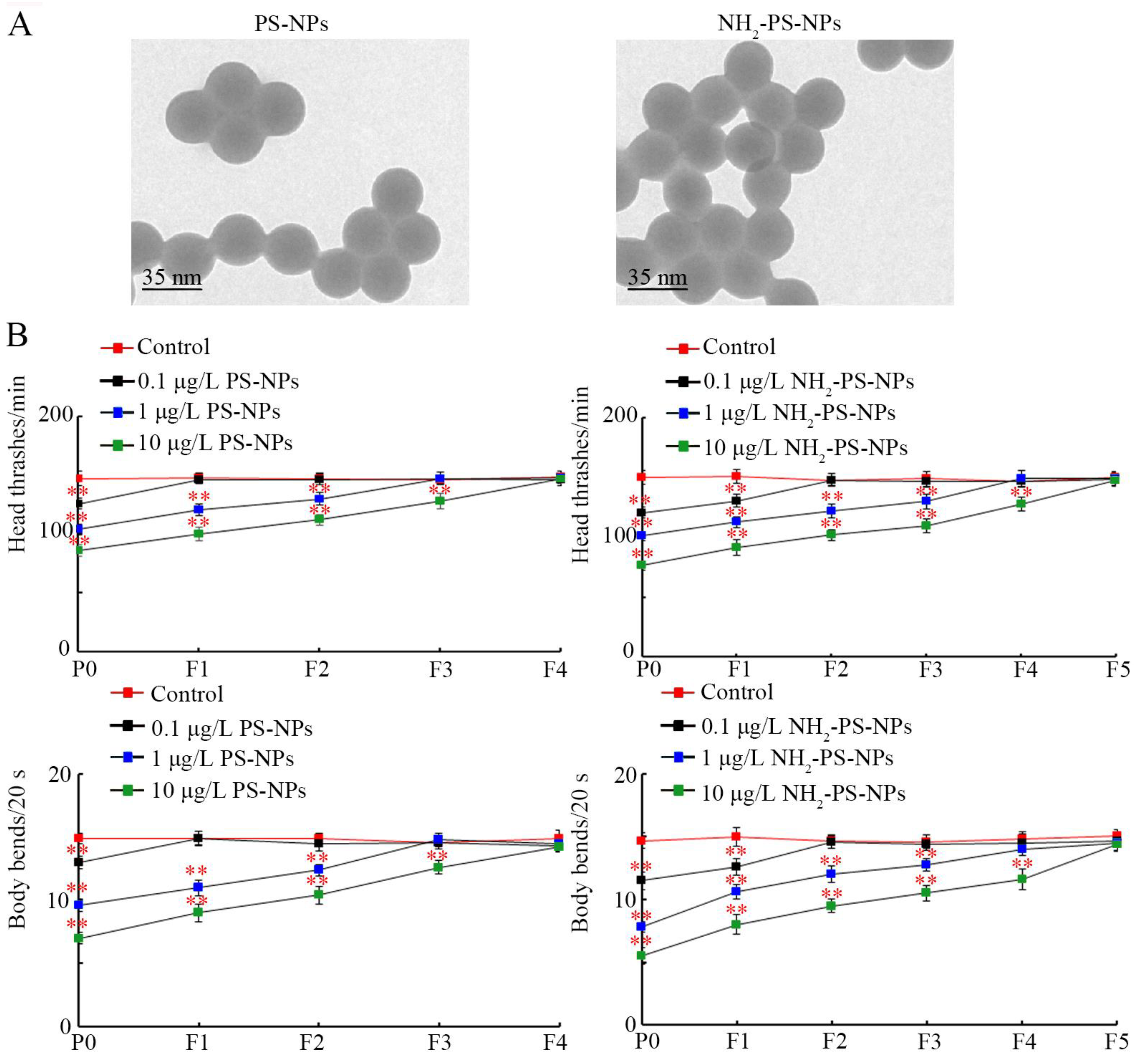
2.1. Nanoplastic Properties

The pristine PS-NPs (35 nm) and amino-modified PS-NPs (NH2-PS-NP, 35 nm) were gifts from Prof. Xianzheng Yuan’s lab [48]. Other reagents were purchased from Sigma-Aldrich (Milwaukee, Germany). The morphology was spherical, and the particle sizes were 34.7 ± 3.6 nm (PS-NPs) and 35.2 ± 3.1 nm (NH2-PS-NPs, 35 nm), respectively, confirmed by transmission electron microscopy (Figure 1A). The zeta potentials of the PS-NPs and NH2-PS-NPs were −19.8 ± 1.79 mV and −25.1 ± 0.87 mV, respectively. The FTIR spectrum and the Raman spectrum of the PS-NPs and NH2-PS-NPs have been described in our previous report [48].

2.2. Animal Maintenance

Wild-type N2 from the Caenorhabditis Genetics Center was grown on nematode growth medium (NGM) plates, and Escherichia coli OP50 was fed as C. elegans food [57]. When adults attained maximum oviposition, they were lysed with a lysis solution (2% HOCl, 0.45 M NaOH). The eggs were placed onto an NGM medium to grow into L1 larvae [58]. C. elegans was cultured in strict accordance with the ARRIVE Guidelines.

2.3. Exposure

Concentrations of PS-NPs (0.1–10 μg/L) were selected [59], which belong to the predicted ERDs of the nanoplastics [12,13,14]. The C. elegans were placed in a solution containing PS-NPs from L1 larvae for 6.5 days, referred to as P0-G. During exposure, the PS-NPs were replaced daily. The eggs of the P0-G were transferred to NGM plates to develop into adulthood, referred to as F1-G. The following generations of offspring (F2-G to Fn-G) were also prepared in the same way.

2.4. Neurotoxicity Assessment

Locomotion reflects the function of the motor neurons [60]. To assess the effect on locomotion, animals were allowed to recover for one minute before assessing their head thrashes and body bends. A head thrash is defined as a change in the direction of head movement [61], and a body bend is defined as a change in direction at mid-body [62]. Fifty animals were analyzed for each treatment.
A transgenic strain of EG1285 with the fused expression of GFP was used to visualize the D-type motor neurons [63]. The extent of neurotoxicity by PS-NPs was reflected by the number of neurons, ventral cord gap, fluorescence intensity, and cell body size of neurons [64]. The cell body size and GFP fluorescence intensity were semi-quantified using Image J software. The number of neurons and the ventral cord gap on the ventral cord were directly counted under a laser confocal microscope. Fifty animals were analyzed for each treatment.

2.5. Gene Expression

The nematodes were added to Trizol reagent to extract their RNA and kept at −80 °C. The cDNA was synthesized next. A quantitative real-time polymerase chain reaction (qRT-PCR) was conducted with the SYBR Green PCR kit (Takara, Kusatsu, Japan). The PCR cycling conditions were an initial denaturation at 95 °C for 5 min, followed by 32 cycles of 94 °C for 30 s, 52 °C for 30 s, and extension at 72 °C for 30 s. tba-1 acted as the reference gene for the normalization of the target genes [65]. Information on the primers is provided in Table S1. Three replicates were performed.

2.6. RNA Interference (RNAi)

RNAi constructs with gene-specific sequences were transforming into E. coli HT115 [66]. The RNAi bacteria were cultured in LA medium overnight, followed by treatment with 100 μg/mL tetracycline and 5 mM isopropyl thiogalactoside for 5 h [67]. RNAi was generated by feeding the L1 larvae with RNAi bacteria. The offspring were exposed to PS-NPs. L4440, an empty vector, acted as the control [68]. The RNAi efficiency was confirmed by qRT-PCR (Figure S1).

2.7. Data Analysis

Data are presented as means ± standard derivation (SD). Statistical analysis was conducted by SPSS v19.0 software (IBM, Armonk, NY, USA). The significant difference among different groups was examined using one-way or two-way analysis of variance (ANOVA) followed by the Tukey post hoc test. The p-value of <0.01 (**) was deemed statistically significant.

3. Results

3.1. Amino Modification Increased Transgenerational Toxicity of PS-NPs on Locomotion

After exposure in P0-G, 0.1 μg/L PS-NPs did not cause toxicity on locomotion in the offspring, whereas 1 μg/L PS-NPs caused a decrease in locomotion from P0-G to F2-G, and 10 μg/L PS-NPs induced a decrease in locomotion from P0-G to F3-G (Figure 1B). Different from these, after P0-G exposure, 0.1 μg/L NH2-PS-NPs could affect locomotion in both P0-G and F1-G; 1 μg/L NH2-PS-NPs altered locomotion from P0-G to F3-G; and 10 μg/L NH2-PS-NPs decreased locomotion from P0-G to F4-G (Figure 1B).

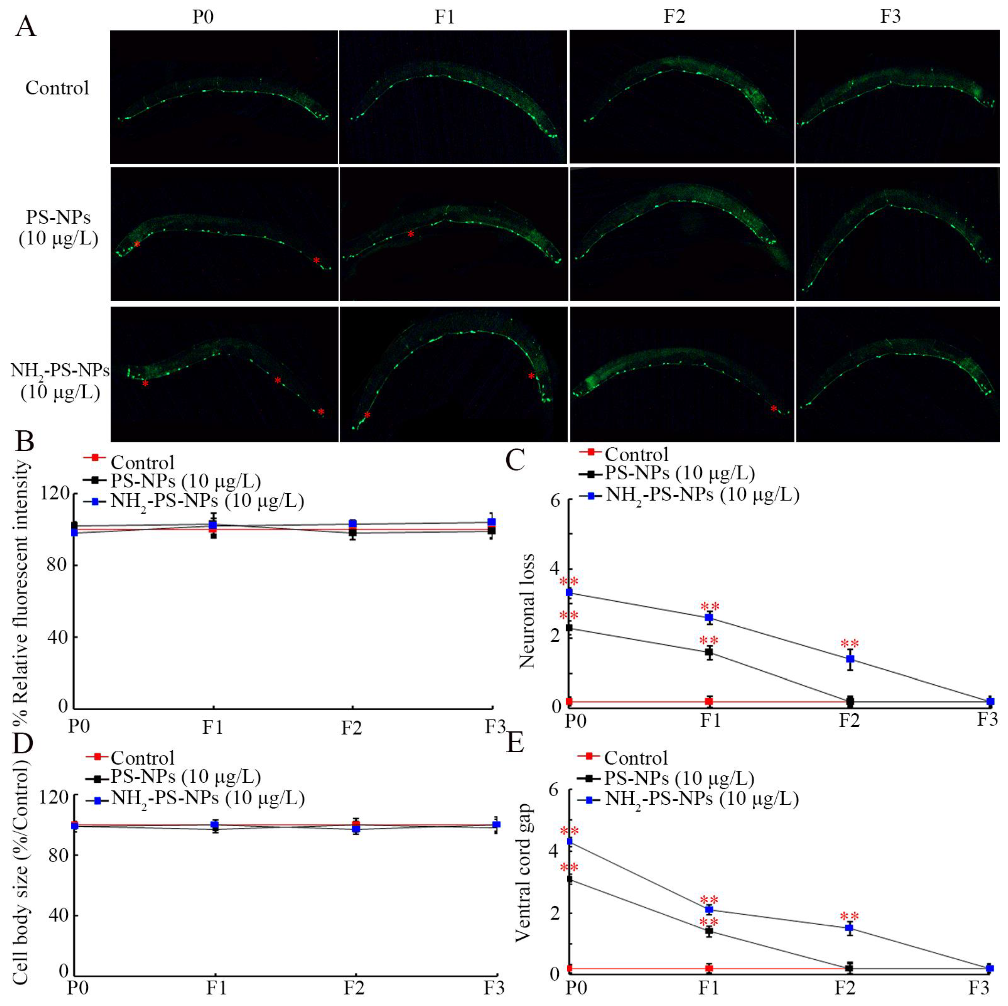
3.2. Amino Modification Increased Transgenerational Toxicity of PS-NPs on Neuronal Development of D-Type Motor Neurons

Motor neurons are located on the ventral nerve cord of the C. elegans GABAergic system. Exposure to 10 μg/L PS-NPs and NH2-PS-NPs all did not affect the fluorescent intensity and size of the cell body of motor neurons in P0-G and in their offspring (Figure 2A–C). However, 10 μg/L PS-NPs resulted in neuronal loss and ventral cord gap in both P0-G and F1-G (Figure 2C,E). Moreover, 10 μg/L NH2-PS-NPs caused neuronal loss and ventral cord gap from P0-G to F2-G (Figure 2C,E).

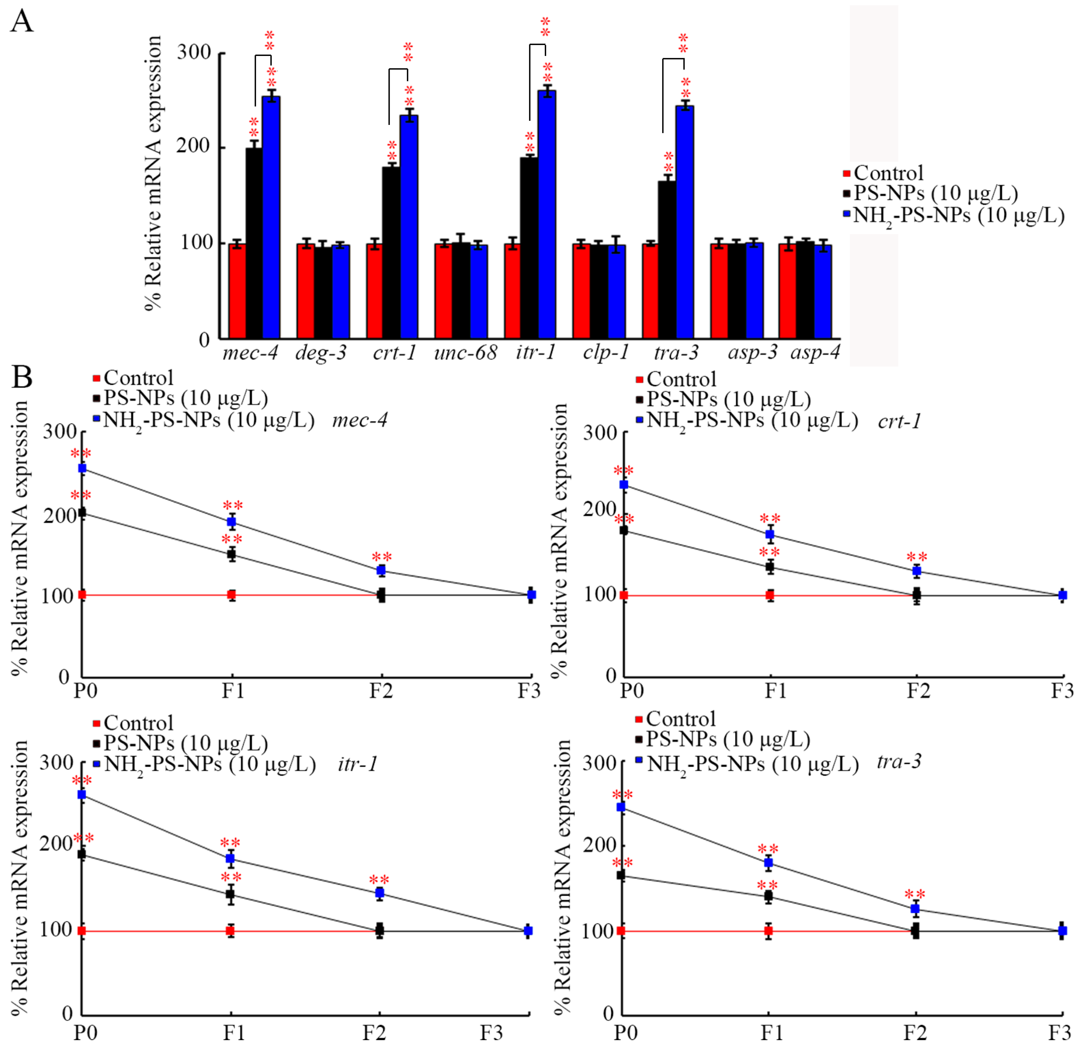
3.3. Amino Modification Strengthened Transgenerational Effect of PS-NPs on Expressions of Genes Governing Neurodegeneration

To determine the molecular basis for the difference between pristine and amino-modified PS-NPs in causing transgenerational toxicity on neuronal development, we compared transgenerational expressions of the genes governing neurodegeneration. Among the examined genes, PS-NPs (10 μg/L) and NH2-PS-NPs (10 μg/L) did not affect the expressions of deg-3, unc-68, clp-1, asp-3, and asp-4 in P0-G (Figure 3A). However, the expressions of mec-4, ctr-1, itr-1, and tra-3 were increased by PS-NPs (10 μg/L) and NH2-PS-NPs (10 μg/L), and NH2-PS-NPs (10 μg/L) caused a more severe increase in the expressions of mec-4, ctr-1, itr-1, and tra-3 compared to those in PS-NP (10 μg/L)-exposed nematodes in P0-G (Figure 3A).
After PS-NP (10 μg/L) exposure, increased expressions of mec-4, ctr-1, itr-1, and tra-3 were detected in F1-G (Figure 3B). Different from this, after NH2-PS-NP exposure, increased expressions of mec-4, ctr-1, itr-1, and tra-3 were observed in F1-G and F2-G (Figure 3B).

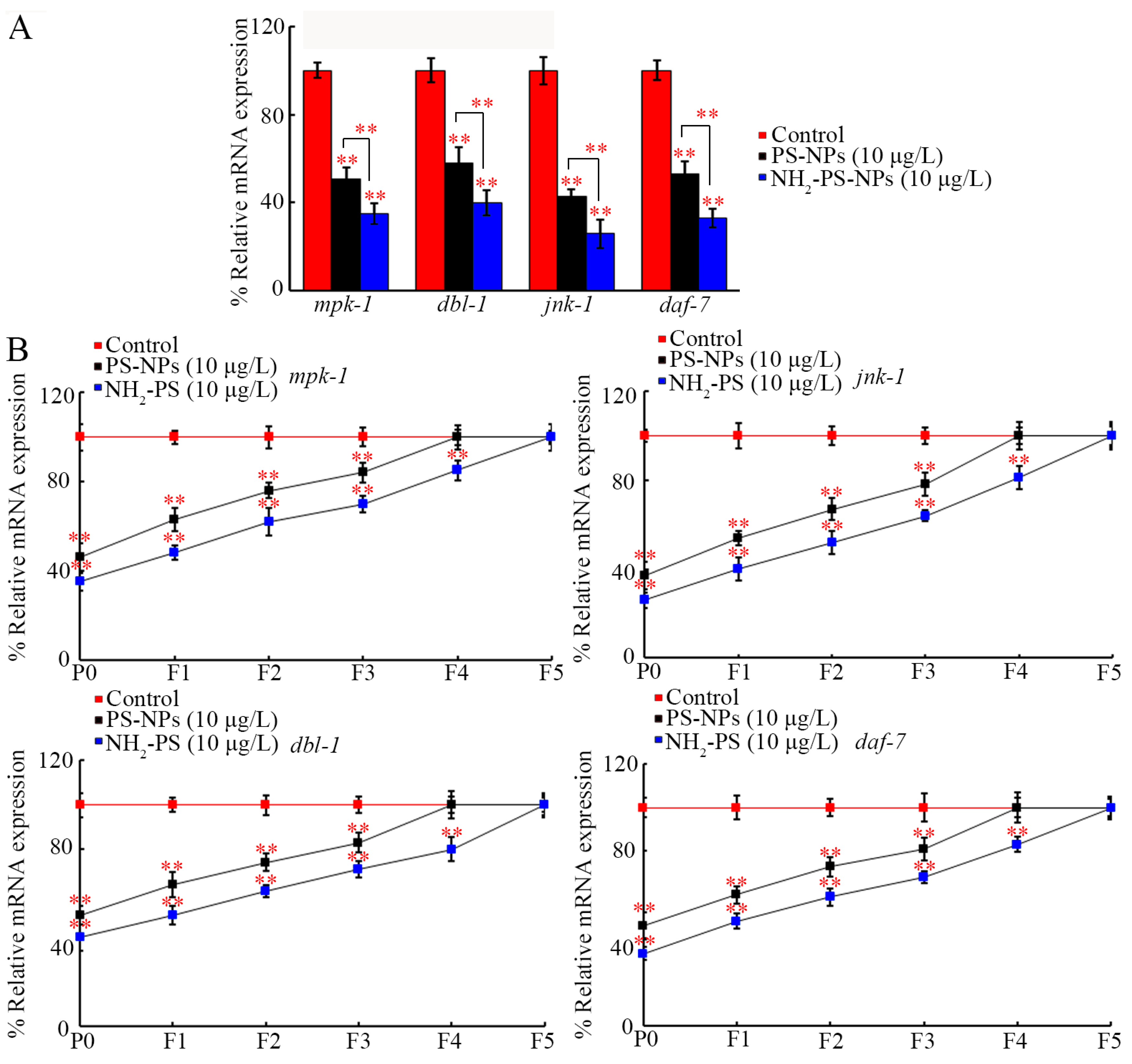
3.4. Amino Modification Strengthened Transgenerational Inhibition of jnk-1, mpk-1, daf-7, and dbl-1 by PS-NP Exposure

Considering the requirement of neuronal JNK-1, MPK-1, DAF-7, and DBL-1 for the toxicity of PS-NPs [53,54,55,56], we also compared the effect between pristine and amino-modified PS-NPs on their expressions. In P0-G, their expressions were decreased by PS-NPs (10 μg/L) and NH2-PS-NPs (10 μg/L), and NH2-PS-NPs (10 μg/L) caused a more severe inhibition in their expressions than PS-NPs (10 μg/L) did (Figure 4A).
After exposure to PS-NPs (10 μg/L) in P0-G, a decrease in these four genes’ expressions was also observed from F1-G to F3-G (Figure 4B). Different from this, after NH2-PS-NP (10 μg/L) exposure in P0-G, a decrease in their expressions could be further found from F1-G to F4-G (Figure 4B).

3.5. RNAi of jnk-1, mpk-1, daf-7, and dbl-1 Increased Neurotoxicity of Both Pristine and Amino-Modified PS-NPs

Under normal conditions, locomotion was not affected by RNAi of jnk-1, mpk-1, daf-7, and dbl-1 (Figure 5A,B). After PS-NP exposure, more severe locomotory inhibition was observed in jnk-1(RNAi), mpk-1(RNAi), daf-7(RNAi), and dbl-1(RNAi) nematodes than that in wild-type nematodes (Figure 5A). Similarly, RNAi of these genes caused a more severe inhibition in locomotion in NH2-PS-NP-exposed nematodes (Figure 5B). Meanwhile, RNAi of these genes resulted in a more severe induction of neurodegeneration reflected by the related endpoints in PS-NP- or NH2-PS-NP-exposed nematodes (Figure S2).

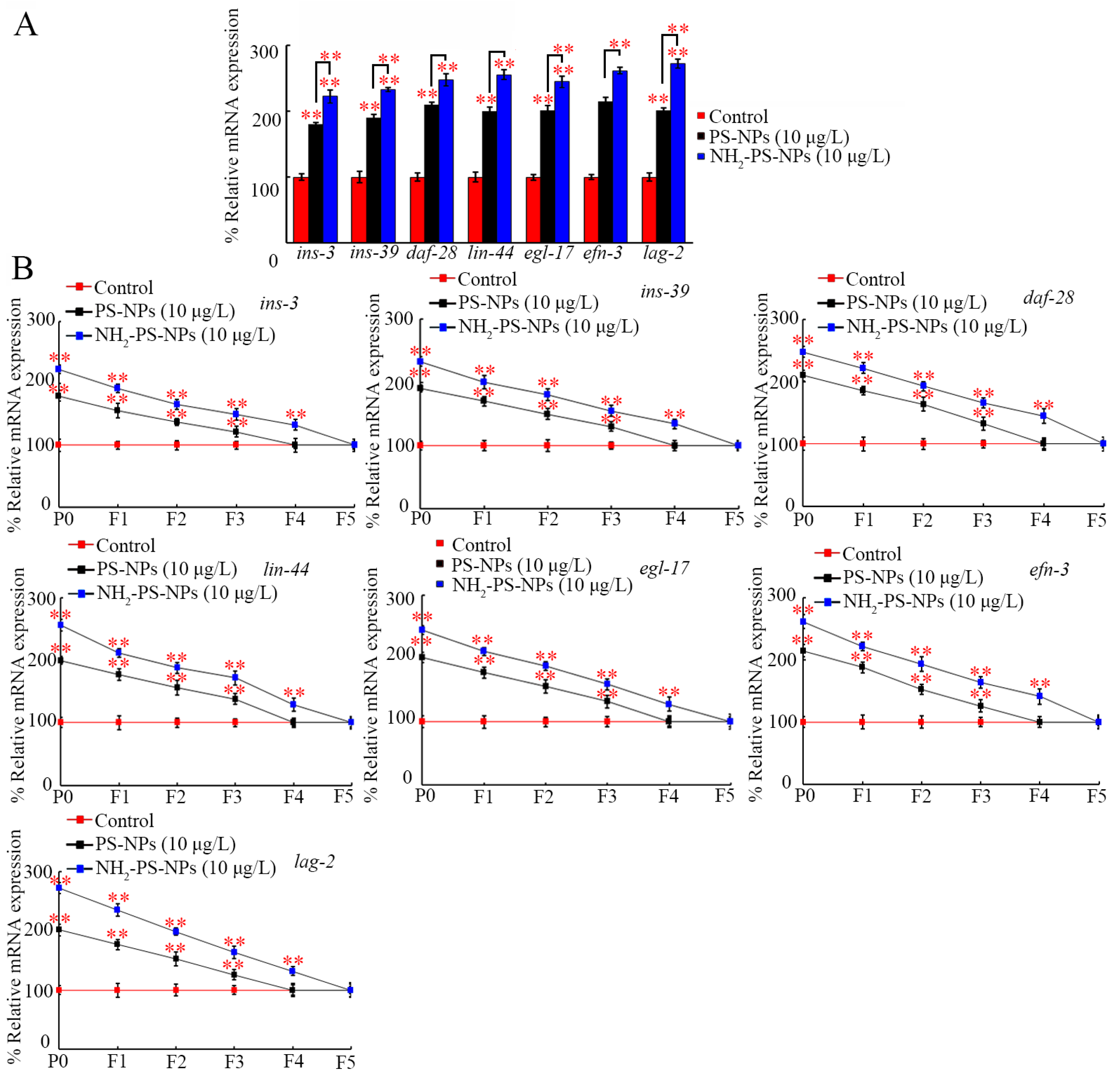
3.6. Amino Modification Strengthened Transgenerational Activation of Germline Ligand Genes by PS-NP Exposure

The germline ligands of insulin, Wnt, FGF, Ephrin, and Notch regulated the transgenerational PS-NP toxicity [39,40,59,69,70]. In P0-G, the germline expressions of ins-3, ins-39, daf-28, lin-44, egl-17, efn-3, and lag-2 were increased by PS-NPs (10 μg/L) and NH2-PS-NPs (10 μg/L), and NH2-PS-NPs (10 μg/L) resulted in a more severe increase in the germline expressions of these ligand genes than PS-NPs (10 μg/L) did (Figure 6A).
After PS-NP (10 μg/L) exposure in P0-G, increased germline expressions of these ligand genes could be further found from F1-G to F3-G (Figure 6B). After exposure to NH2-PS-NPs (10 μg/L) in P0-G, activation of these germline ligand genes was further observed from F1-G to F4-G (Figure 6B).

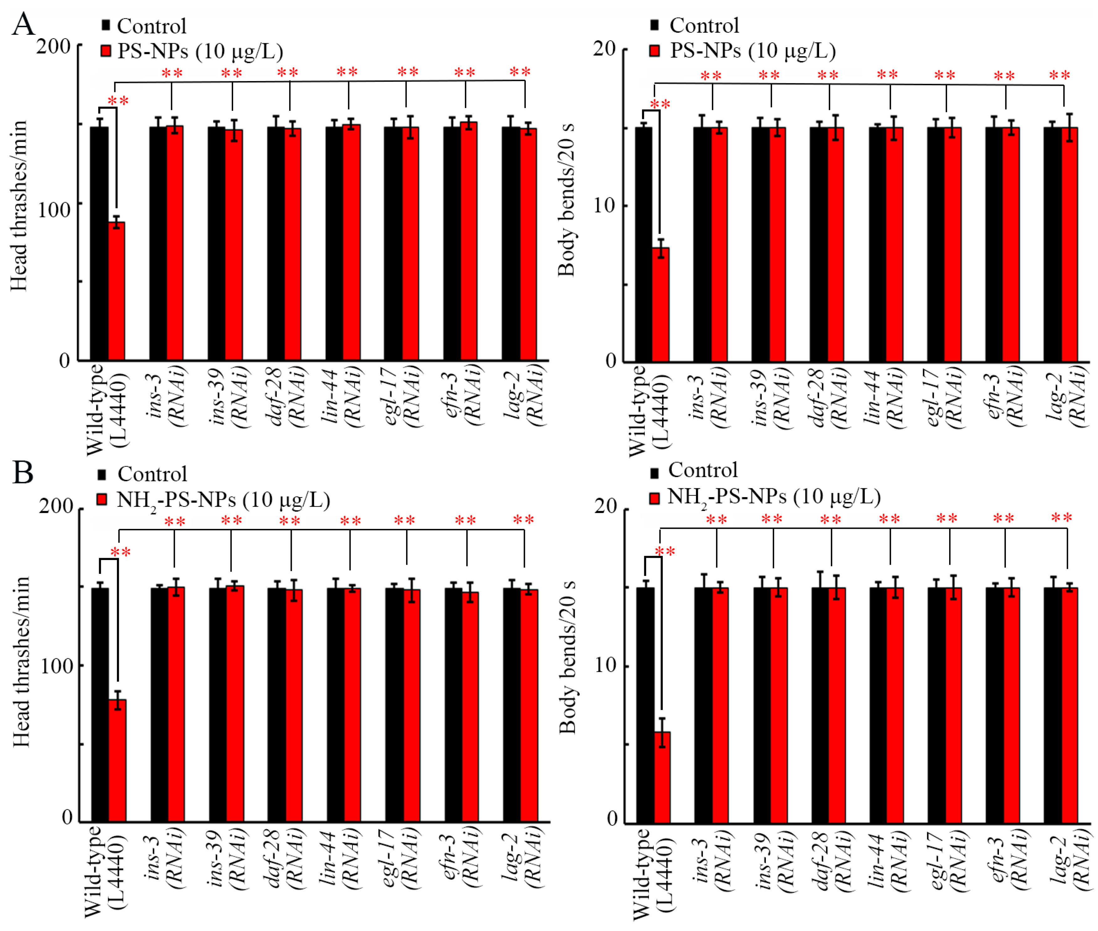
3.7. RNAi of ins-3, ins-39, daf-28, lin-44, egl-17, efn-3, and lag-2 Inhibited Neurotoxicity of Both Pristine and Amino-Modified PS-NPs

The decrease in locomotion caused by PS-NPs and NH2-PS-NPs could be significantly inhibited by RNAi of ins-3, ins-39, daf-28, lin-44, egl-17, efn-3, and lag-2 (Figure 7A,B). Moreover, the neurodegeneration induced by PS-NPs and NH2-PS-NPs was also suppressed by RNAi of ins-3, ins-39, daf-28, lin-44, egl-17, efn-3, and lag-2 (Figure S3).

4. Discussion

It has been well recognized that amino modification could increase the adverse effects of nanoplastics on organisms [47,71]. Amino modification increased both the cytotoxicity and genotoxicity of PS-NPs in A549 cells [71]. Amino modification further enhanced PS-NP toxicity on the reproductive system in male mice [47]. Amino modification could even affect the effect of PS-NPs on microbial communities in sediment [72]. In nematodes, amino modification increased PS-NP reproductive toxicity in P0-G [48]. In the current study, we further observed that amino modification strengthened the transgenerational PS-NP neurotoxicity on both locomotion and neuronal development. With 10 μg/L as the example, PS-NPs caused decreased locomotion behavior from P0-G to F3-G, whereas NH2-PS-NPs resulted in decreased locomotion behavior from P0-G to F4-G (Figure 1B). Meanwhile, compared to the neurodegeneration induced in P0-G and F1-G by PS-NPs (10 μg/L), NH2-PS-NPs (10 μg/L) caused neurodegeneration from P0-G to F2-G (Figure 2). These demonstrated the potential of amino modification in enhancing transgenerational PS-NP toxicity on both locomotion and neuronal development. Compared to the endpoints reflecting neuronal development, the endpoints reflecting locomotion were relatively more sensitive for assessing the transgenerational toxicity of nanoplastics. The reproductive toxicity of PS-NPs was also increased by amino modification, and a 4-day exposure of L1 larvae to NH2-PS-NPs (10 μg/L) induced inhibition in the brood size in both P0-G and F1-G [73]. Hormesis is an adaptative response induced by stresses and pollutants to protect biological systems against damage formation [74,75,76]. What we performed was long-term exposure to both 35 nm PS-NPs and 35 nm NH2-PS-NPs. Nevertheless, we did not observe the hormesis response after exposure to 35 nm PS-NPs and 35 nm NH2-PS-NPs. Different from this, we observed the hormesis response after exposure to 100 nm PS-NPs at similar doses [77,78,79,80].
The transgenerational damage of PS-NPs on the neuronal development of D-type motor neurons by amino modification was partially due to the differential effect on genes governing neurodegeneration. In C. elegans, the activation of DEF-3 and MEC-4, two excitotoxic-like ion channels, triggers neurodegeneration [81]. Both PS-NPs (10 μg/L) and NH2-PS-NPs (10 μg/L) could increase mec-4 expression, and NH2-PS-NPs (10 μg/L) caused a more severe transgenerational increase in mec-4 expression than PS-NPs (10 μg/L) did (Figure 3B). Endoplasmic reticulum (ER)-residential calcium chaperon CTR-1 and inositol 1,4,5-riphosphate receptor ITR-1 act downstream of MEC-4 to control calcium ion release from the ER [82,83]. Following MEC-4 activation, NH2-PS-NPs (10 μg/L) induced a more severe transgenerational increase in ctr-1 and itr-1 expressions than PS-NPs (10 μg/L) did (Figure 3B). Neurodegeneration is directly caused by the activation of the proteases, calpain proteases and aspartyl proteases [84]. Among the genes encoding these proteases, NH2-PS-NPs (10 μg/L) further caused a more severe transgenerational increase in the expression of tra-3 encoding calpain protease than PS-NPs (10 μg/L) did (Figure 3B).
For an enhancement in the transgenerational PS-NP neurotoxicity, we raised two other aspects of the molecular mechanisms. One of them was that amino modification enhanced the transgenerational PS-NP neurotoxicity by causing more severe transgenerational inhibition in MPK-1, JNK-1, DBL-1, and DAF-7. On the one hand, NH2-PS-NPs (10 μg/L) caused a more severe transgenerational decrease in their expressions than PS-NPs (10 μg/L) did (Figure 4B). On the other hand, susceptibility to the toxicity of PS-NPs and NH2-PS-NPs on D-type motor neurons involved in locomotion and development was caused by RNAi of these genes (Figure 5 and Figure S2). In C. elegans, MPK-1, JNK-1, DBL-1, and DAF-7 functioned in neurons to regulate nanoplastic toxicity [53,54,55,56]. Therefore, the transgenerational inhibition of these neuronal signals mediated the toxicity of PS-NPs and NH2-PS-NPs across multiple generations.
Besides the role of these neuronal signals, the transgenerational activation of germline secreted ligands also contributed to the toxicity induction of PS-NPs and NH2-PS-NPs across multiple generations. NH2-PS-NPs (10 μg/L) caused a more severe transgenerational increase in the expressions of germline insulin, Wnt, FGF, Ephrin, and Notch ligand genes than PS-NP (10 μg/L) did (Figure 6B). Moreover, resistance to the neurotoxicity of PS-NPs and NH2-PS-NPs could be induced by RNAi of insulin, Wnt, FGF, Ephrin, and Notch ligand genes (Figure 7 and Figure S3). In C. elegans, germline INS-3, INS-39, DAF-28, LIN-44, EGL-17, EFN-3, and LAG-2 functioned together with their receptors (DAF-2, MIG-1, EGL-15, VAB-1, and GLP-1) to regulate the transgenerational nanoplastic toxicity [39,40,59,69,70]. That is, amino modification could further enhance transgenerational PS-NP neurotoxicity by resulting in a more severe transgenerational activation of germline insulin, Wnt, FGF, Ephrin, and Notch ligand genes.

5. Conclusions

Together, NH2-PS-NPs could cause more severe transgenerational neurotoxicity than PS-NPs at 0.1–10 μg/L in C. elegans. The observation of more severe transgenerational damage on D-type motor neurons by NH2-PS-NPs than PS-NPs was partially due to the more severe transgenerational activation of genes governing neurodegeneration. NH2-PS-NPs also caused more severe transgenerational neurotoxicity than PS-NPs by inducing more severe transgenerational inhibition in the expressions of mpk-1, jnk-1, dbl-1, and daf-7, and transgenerational activation of the expressions of germline ligand genes (ins-3, ins-39, daf-28, lin-44, egl-17, efn-3, and lag-2). Compared with the well-described role of amino modification in increasing the nanoplastic toxicity in P0-G, our results provided an important molecular basis for amino modification to enhance the transgenerational neurotoxicity of PS-NPs. Our data further implied that the exposure risk of amino-modified nanoplastics in causing more severe transgenerational toxicity needs to be carefully paid attention to. In the future, neuroprotective compounds (such as natural extracts or drugs) with the function of attenuating or preventing transgenerational neurotoxicity induced by PS-NPs and NH2-PS-NPs by upregulating MPK-1, JNK-1, DBL-1, and DAF-7 are suggested to be further screened and identified using C. elegans as an animal model.

Supplementary Materials

The following supporting information can be downloaded at https://www.mdpi.com/article/10.3390/toxics12080555/s1: Figure S1: RNAi efficiency of daf-7, jnk-1, dbl-1, and mpk-1; Figure S2: Effect of RNAi of mpk-1, dbl-1, jnk-1, and daf-7 on toxicity of PS-NP and NH2-PS-NP in causing damage on D-type motor neurons on GABAergic system; Figure S3: Effect of RNAi of ins-3, ins-39, daf-28, lin-44, egl-17, efn-3, and lag-2 on toxicity of PS-NP and NH2-PS-NP in causing damage on D-type motor neurons on GABAergic system; Table S1: Primer information for qRT-PCR.

Author Contributions

Investigation and writing the draft, M.S.; supervision and reviewing the draft, Q.R. and D.W. All authors have read and agreed to the published version of the manuscript.

Funding

This study was supported by grants from the Jiangsu Province “The 14th Five-Year Plan” Key Discipline-Public Health and Preventive Medicine (035091005007), the Natural Science Foundation of Guangdong Province (2024A1515011115), and the Shenzhen Basic Research Project (JCYJ20220530163605011).

Institutional Review Board Statement

Not applicable.

Informed Consent Statement

Not applicable.

Data Availability Statement

The original data presented in the study are included in the article/Supplementary Material; further inquiries can be directed to the corresponding authors.

Conflicts of Interest

The authors declare that they have no known competing financial interest or personal relationships that could appear to have influenced the work reported in this paper.

References

  1. MacLeod, M.; Arp, H.P.H.; Tekman, M.B.; Jahnke, A. The global threat from plastic pollution. Science 2021, 373, 61–65. [Google Scholar] [CrossRef]
  2. Leistenschneider, D.; Wolinski, A.; Cheng, J.; Ter Halle, A.; Duflos, G.; Huvet, A.; Paul-Pont, I.; Lartaud, F.; Galgani, F.; Lavergne, É.; et al. A critical review on the evaluation of toxicity and ecological risk assessment of plastics in the marine environment. Sci. Total Environ. 2023, 896, 164955. [Google Scholar] [CrossRef]
  3. Andrady, A.L. Weathering and fragmentation of plastic debris in the ocean environment. Mar. Pollut. Bull. 2022, 180, 113761. [Google Scholar] [CrossRef] [PubMed]
  4. Colwell, J.; Pratt, S.; Lant, P.; Laycock, B. Hazardous state lifetimes of biodegradable plastics in natural environments. Sci. Total Environ. 2023, 894, 165025. [Google Scholar] [CrossRef] [PubMed]
  5. Jebashalomi, V.; Charles, P.E.; Rajaram, R.; Sadayan, P. A critical review on nanoplastics and its future perspectives in the marine environment. Environ. Monit. Assess. 2023, 195, 1186. [Google Scholar] [CrossRef]
  6. Kurniawan, T.A.; Haider, A.; Ahmad, H.M.; Mohyuddin, A.; Umer Aslam, H.M.; Nadeem, S.; Javed, M.; Othman, M.H.D.; Goh, H.H.; Chew, K.W. Source, occurrence, distribution, fate, and implications of microplastic pollutants in freshwater on environment: A critical review and way forward. Chemosphere 2023, 325, 138367. [Google Scholar] [CrossRef]
  7. Hua, X.; Wang, D.-Y. Cellular uptake, transport, and organelle response after exposure to microplastics and nanoplastics: Current knowledge and perspectives for environmental and health risks. Rev. Environ. Contam. Toxicol. 2022, 260, 12. [Google Scholar] [CrossRef]
  8. Zhu, Y.; Che, R.; Zong, X.; Wang, J.; Li, J.; Zhang, C.; Wang, F. A comprehensive review on the source, ingestion route, attachment and toxicity of microplastics/nanoplastics in human systems. J. Environ. Manag. 2024, 352, 120039. [Google Scholar] [CrossRef]
  9. Yang, S.; Li, M.; Kong, R.Y.C.; Li, L.; Li, R.; Chen, J.; Lai, K.P. Reproductive toxicity of micro- and nanoplastics. Environ. Int. 2023, 177, 108002. [Google Scholar] [CrossRef] [PubMed]
  10. Ali, N.; Katsouli, J.; Marczylo, E.L.; Gant, T.W.; Wright, S.; Bernardino de la Serna, J. The potential impacts of micro-and-nano plastics on various organ systems in humans. EBioMedicine 2024, 99, 104901. [Google Scholar] [CrossRef]
  11. Tang, M.; Ding, G.; Lu, X.; Huang, Q.; Du, H.; Xiao, G.; Wang, D. Exposure to nanoplastic particles enhances Acinetobacter survival, biofilm formation, and serum resistance. Nanomaterials 2022, 12, 4222. [Google Scholar] [CrossRef]
  12. Lenz, R.; Enders, K.; Nielsen, T.G. Microplastic exposure studies should be environmentally realistic. Proc. Natl. Acad. Sci. USA 2016, 113, E4121–E4122. [Google Scholar] [CrossRef]
  13. Guimarães, A.T.B.; Estrela, F.N.; Rodrigues, A.S.L.; Chagas, T.Q.; Pereira, P.S.; Silva, F.G.; Malafaia, G. Nanopolystyrene particles at environmentally relevant concentrations causes behavioral and biochemical changes in juvenile grass carp (Ctenopharyngodon idella). J. Hazard. Mater. 2021, 403, 123864. [Google Scholar] [CrossRef]
  14. Materic, D.; Peacock, M.; Dean, J.; Futter, M.; Maximov, T.; Moldan, F.; Rockmann, T.; Holzinger, R. Presence of nanoplastics in rural and remote surface waters. Environ. Res. Lett. 2022, 17, e054036. [Google Scholar] [CrossRef]
  15. Xiao, F.; Feng, L.J.; Sun, X.D.; Wang, Y.; Wang, Z.W.; Zhu, F.P.; Yuan, X.Z. Do polystyrene nanoplastics have similar effects on duckweed (Lemna minor L.) at environmentally relevant and observed-effect concentrations? Environ. Sci. Technol. 2022, 56, 4071–4079. [Google Scholar] [CrossRef]
  16. Qiu, W.; Ye, J.; Su, Y.; Zhang, X.; Pang, X.; Liao, J.; Wang, R.; Zhao, C.; Zhang, H.; Hu, L.; et al. Co-exposure to environmentally relevant concentrations of cadmium and polystyrene nanoplastics induced oxidative stress, ferroptosis and excessive mitophagy in mice kidney. Environ. Pollut. 2023, 333, 121947. [Google Scholar] [CrossRef]
  17. Liu, Z.-Y.; Hua, X.; Zhao, Y.; Bian, Q.; Wang, D.-Y. Polyethylene nanoplastics cause reproductive toxicity associated with activation of both estrogenic hormone receptor NHR-14 and DNA damage checkpoints in C. elegans. Sci. Total Environ. 2024, 906, 167471. [Google Scholar] [CrossRef]
  18. Zhuang, Z.-H.; Liu, T.-W.; Liu, Z.-Y.; Wang, D.-Y. Polystyrene nanoparticles strengthen high glucose toxicity associated with alteration in insulin signaling pathway in C. elegans. Ecotoxicol. Environ. Saf. 2024, 272, 116056. [Google Scholar] [CrossRef]
  19. Yeo, I.C.; Shim, K.Y.; Kim, K.; Jeong, C.B. Maternal exposure to nanoplastic induces transgenerational toxicity in the offspring of rotifer Brachionus koreanus. Comp. Biochem. Physiol. C Toxicol. Pharmacol. 2023, 269, 109635. [Google Scholar] [CrossRef]
  20. Yi, J.; Ma, Y.; Ruan, J.; You, S.; Ma, J.; Yu, H.; Zhao, J.; Zhang, K.; Yang, Q.; Jin, L.; et al. The invisible Threat: Assessing the reproductive and transgenerational impacts of micro- and nanoplastics on fish. Environ. Int. 2024, 183, 108432. [Google Scholar] [CrossRef]
  21. Hua, X.; Zhang, L.; Wang, D.-Y. Nanoplastic at environmentally relevant concentrations activates germline mir-240-rab-5 signaling cascade to affect secreted ligands associated with transgenerational toxicity induction in C. elegans. Environ. Sci. Nano 2024. [Google Scholar] [CrossRef]
  22. Liu, Z.-Y.; Wang, Y.-X.; Bian, Q.; Wang, D.-Y. Transgenerational response of germline nuclear hormone receptor genes to nanoplastics at predicted Environmental doses in Caenorhabditis elegans. Toxics 2024, 12, 420. [Google Scholar] [CrossRef]
  23. Wang, D.-Y. Exposure Toxicology in Caenorhabditis elegans; Springer Nature Singapore Pte Ltd.: Singapore, 2020. [Google Scholar]
  24. Li, H.; Zeng, L.; Wang, C.; Shi, C.; Li, Y.; Peng, Y.; Chen, H.; Zhang, J.; Cheng, B.; Chen, C.; et al. Review of the toxicity and potential molecular mechanisms of parental or successive exposure to environmental pollutants in the model organism Caenorhabditis elegans. Environ. Pollut. 2022, 311, 119927. [Google Scholar] [CrossRef]
  25. Wang, Y.-X.; Liang, G.-Y.; Chao, J.; Wang, D.-Y. Comparison of intestinal toxicity in enhancing intestinal permeability and in causing ROS production of six PPD quinones in Caenorhabditis elegans. Sci. Total Environ. 2024, 927, 172306. [Google Scholar] [CrossRef]
  26. Shao, Y.-T.; Hua, X.; Li, Y.-H.; Wang, D.-Y. Comparison of reproductive toxicity between pristine and aged polylactic acid microplastics in Caenorhabditis elegans. J. Hazard. Mater. 2024, 466, 133545. [Google Scholar] [CrossRef]
  27. Wang, D.-Y. Toxicology at Environmentally Relevant Concentrations in Caenorhabditis elegans; Springer Nature: Dordrecht, The Netherlands, 2022. [Google Scholar]
  28. Yang, Y.; Li, M.; Zheng, J.; Zhang, D.; Ding, Y.; Yu, H.Q. Environmentally relevant exposure to tetrabromobisphenol A induces reproductive toxicity via regulating glucose-6-phosphate 1-dehydrogenase and sperm activation in Caenorhabditis elegans. Sci. Total Environ. 2024, 907, 167820. [Google Scholar] [CrossRef]
  29. Hua, X.; Liang, G.-Y.; Chao, J.; Wang, D.-Y. Exposure to 6-PPD quinone causes damage on mitochondrial complex I/II associated with lifespan reduction in Caenorhabditis elegans. J. Hazard. Mater. 2024, 472, 134598. [Google Scholar] [CrossRef]
  30. Liu, Z.-Y.; Bian, Q.; Wang, D.-Y. Exposure to 6-PPD quinone causes ferroptosis activation associated with induction of reproductive toxicity in Caenorhabditis elegans. J. Hazard. Mater. 2024, 471, 134356. [Google Scholar] [CrossRef]
  31. Hua, X.; Wang, D.-Y. Disruption of dopamine metabolism by exposure to 6-PPD quinone in Caenorhabditis elegans. Environ. Pollut. 2023, 337, 122649. [Google Scholar] [CrossRef]
  32. Qiao, R.; Mortimer, M.; Richter, J.; Rani-Borges, B.; Yu, Z.; Heinlaan, M.; Lin, S.; Ivask, A. Hazard of polystyrene micro-and nanospheres to selected aquatic and terrestrial organisms. Sci. Total Environ. 2022, 853, 158560. [Google Scholar] [CrossRef]
  33. Jewett, E.; Arnott, G.; Connolly, L.; Vasudevan, N.; Kevei, E. Microplastics and their impact on reproduction-can we learn from the C. elegans model? Front. Toxicol. 2022, 4, 748912. [Google Scholar] [CrossRef]
  34. Zhao, Y.; Chen, J.; Wang, R.; Pu, X.; Wang, D. A review of transgenerational and multigenerational toxicology in the in vivo model animal Caenorhabditis elegans. J. Appl. Toxicol. 2023, 43, 122–145. [Google Scholar] [CrossRef]
  35. Li, J.; Dai, L.; Feng, Y.; Cao, Z.; Ding, Y.; Xu, H.; Xu, A.; Du, H. Multigenerational effects and mutagenicity of three flame retardants on germ cells in Caenorhabditis elegans. Ecotoxicol. Environ. Saf. 2024, 269, 115815. [Google Scholar] [CrossRef]
  36. Shao, Y.-T.; Li, Y.-H.; Wang, D.-Y. Polylactic acid microplastics cause transgenerational reproductive toxicity associated with activation of insulin and hedgehog ligands in C. elegans. Sci. Total Environ. 2024, 942, 173746. [Google Scholar] [CrossRef]
  37. Chen, H.; Gu, Y.; Jiang, Y.; Yu, J.; Chen, C.; Shi, C.; Li, H. Photoaged polystyrene nanoplastics result in transgenerational reproductive toxicity associated with the methylation of histone H3K4 and H3K9 in Caenorhabditis elegans. Environ. Sci. Technol. 2023, 57, 19341–19351. [Google Scholar] [CrossRef]
  38. Liu, H.; Wu, Y.; Wang, Z. Long-term exposure to polystyrene nanoparticles at environmentally relevant concentration causes suppression in heme homeostasis signal associated with transgenerational toxicity induction in Caenorhabditis elegans. J. Hazard. Mater. 2023, 459, 132124. [Google Scholar] [CrossRef]
  39. He, W.; Gu, A.; Wang, D. Sulfonate-modified polystyrene nanoparticle at precited environmental concentrations induces transgenerational toxicity associated with increase in germline Notch signal of Caenorhabditis elegans. Toxics 2023, 11, 511. [Google Scholar] [CrossRef]
  40. Hua, X.; Cao, C.; Zhang, L.; Wang, D. Activation of FGF signal in germline mediates transgenerational toxicity of polystyrene nanoparticles at predicted environmental concentrations in Caenorhabditis elegans. J. Hazard. Mater. 2023, 451, 131174. [Google Scholar] [CrossRef]
  41. Yu, C.W.; Luk, T.C.; Liao, V.H. Long-term nanoplastics exposure results in multi and trans-generational reproduction decline associated with germline toxicity and epigenetic regulation in Caenorhabditis elegans. J. Hazard. Mater. 2021, 412, 125173. [Google Scholar] [CrossRef]
  42. Hua, X.; Zhao, Y.; Yuan, Y.-J.; Zhang, L.; Bian, Q.; Wang, D.-Y. Nanoplastics cause transgenerational toxicity through inhibiting germline microRNA mir-38 in C. elegans. J. Hazard. Mater. 2022, 437, 129302. [Google Scholar] [CrossRef]
  43. Zhang, L.; Wang, S.-T.; Zhao, Y.; Bi, K.; Wang, D.-Y. Increase in germline methyltransferases governing methylation of histone H3K9 is associated with transgenerational nanoplastic toxicity in Caenorhabditis elegans. Environ. Sci. Nano 2022, 9, 265–274. [Google Scholar] [CrossRef]
  44. Kögel, T.; Bjorøy, Ø.; Toto, B.; Bienfait, A.M.; Sanden, M. Micro- and nanoplastic toxicity on aquatic life: Determining factors. Sci. Total Environ. 2020, 709, 136050. [Google Scholar] [CrossRef] [PubMed]
  45. Pelegrini, K.; Pereira, T.C.B.; Maraschin, T.G.; Teodoro, L.S.; Basso, N.R.S.; De Galland, G.L.B.; Ligabue, R.A.; Bogo, M.R. Micro- and nanoplastic toxicity: A review on size, type, source, and test-organism implications. Sci. Total Environ. 2023, 878, 162954. [Google Scholar] [CrossRef] [PubMed]
  46. Qu, M.; An, Y.; Jiang, X.; Wu, Q.; Miao, L.; Zhang, X.; Wang, Y. Exposure to epoxy-modified nanoplastics in the range of mug/L causes dysregulated intestinal permeability, reproductive capacity, and mitochondrial homeostasis by affecting antioxidant system in Caenorhabditis elegans. Aquat. Toxicol. 2023, 264, 106710. [Google Scholar] [CrossRef]
  47. Gao, X.; Xu, K.; Du, W.; Wang, S.; Jiang, M.; Wang, Y.; Han, Q.; Chen, M. Comparing the effects and mechanisms of exposure to polystyrene nanoplastics with different functional groups on the male reproductive system. Sci. Total Environ. 2024, 922, 171299. [Google Scholar] [CrossRef] [PubMed]
  48. Qu, M.; Qiu, Y.; Kong, Y.; Wang, D. Amino modification enhances reproductive toxicity of nanopolystyrene on gonad development and reproductive capacity in nematode Caenorhabditis elegans. Environ. Pollut. 2019, 254, 112978. [Google Scholar] [CrossRef] [PubMed]
  49. Zhou, R.; Liu, R.; Li, W.; Wang, Y.; Wan, X.; Song, N.; Yu, Y.; Xu, J.; Bu, Y.; Zhang, A. The use of different sublethal endpoints to monitor atrazine toxicity in nematode Caenorhabditis elegans. Chemosphere 2021, 274, 129845. [Google Scholar] [CrossRef] [PubMed]
  50. Wang, D.-Y. Target Organ Toxicology in Caenorhabditis elegans; Springer Nature: Dordrecht, The Netherlands, 2019. [Google Scholar]
  51. Jin, Y.; Hoskins, R.; Horvitz, H.R. Control of type-D GABAergic neuron differentiation by C. elegans UNC-30 homeodomain protein. Nature 1994, 372, 780–783. [Google Scholar] [CrossRef] [PubMed]
  52. Qu, M.; Kong, Y.; Yuan, Y.; Wang, D. Neuronal damage induced by nanopolystyrene particles in nematode Caenorhabditis elegans. Environ. Sci. Nano 2019, 6, 2591–2601. [Google Scholar] [CrossRef]
  53. Wang, S.-T.; Liu, H.-L.; Zhao, Y.-Y.; Rui, Q.; Wang, D.-Y. Dysregulated mir-354 enhanced the protective response to nanopolystyrene by affecting the activity of TGF-β signaling pathway in nematode Caenorhabditis elegans. NanoImpact 2020, 20, 100256. [Google Scholar] [CrossRef]
  54. Liu, H.-L.; Zhang, R.-J.; Wang, D.-Y. Response of DBL-1/TGF-β signaling-mediated neuron-intestine communication to nanopolystyrene in nematode Caenorhabditis elegans. Sci. Total Environ. 2020, 745, 1141047. [Google Scholar] [CrossRef]
  55. Qu, M.; Li, D.; Zhao, Y.-L.; Yuan, Y.-J.; Wang, D.-Y. Exposure to low-dose nanopolystyrene induces the response of neuronal JNK MAPK signaling pathway in nematode Caenorhabditis elegans. Environ. Sci. Eur. 2020, 32, 58. [Google Scholar] [CrossRef]
  56. Qu, M.; Li, D.; Qiu, Y.-X.; Wang, D.-Y. Neuronal ERK MAPK signaling in response to low-dose nanopolystyrene exposure by suppressing insulin peptide expression in Caenorhabditis elegans. Sci. Total Environ. 2020, 724, 138378. [Google Scholar] [CrossRef] [PubMed]
  57. Brenner, S. The genetics of Caenorhabditis elegans. Genetics 1974, 77, 71–94. [Google Scholar] [CrossRef]
  58. Yuan, C.; Wang, Y.; Zhang, L.; Wang, D. Procatechuic acid and protocatechuic aldehyde increase survival of Caenorhabditis elegans after fungal infection and inhibit fungal virulence. Front. Pharmacol. 2024, 15, 1396733. [Google Scholar] [CrossRef]
  59. Xu, R.; Hua, X.; Rui, Q.; Wang, D. Alteration in Wnt signaling mediates induction of transgenerational toxicity of polystyrene nanoplastics in C. elegans. NanoImpact 2022, 28, 100425. [Google Scholar] [CrossRef] [PubMed]
  60. Hua, X.; Wang, D.-Y. Exposure to 6-PPD quinone at environmentally relevant concentrations inhibits both lifespan and healthspan in C. elegans. Environ. Sci. Technol. 2023, 57, 19295–19303. [Google Scholar] [CrossRef]
  61. Zhang, L.; Wang, Y.-X.; Wang, D.-Y. Paeoniflorin increases the survival of Pseudomonas aeruginosa infected Caenorhabditis elegans at the immunosuppression stage by activating PMK-1, BAR-1, and EGL-1 signals. Arch. Pharm. Res. 2023, 46, 616–628. [Google Scholar] [CrossRef]
  62. Wang, Y.-X.; Wang, D.-Y. Exposure to 6-PPD quinone enhances glycogen accumulation in Caenorhabditis elegans. Environ. Pollut. 2024, 359, 124600. [Google Scholar] [CrossRef]
  63. Wan, X.; Liang, G.-Y.; Wang, D.-Y. Neurotoxicity and accumulation of CPPD quinone at environmentally relevant concentrations in Caenorhabditis elegans. Chemosphere 2024, 361, 142499. [Google Scholar] [CrossRef]
  64. Hua, X.; Wang, D.-Y. Polyethylene nanoparticles at environmentally relevant concentrations enhances neurotoxicity and accumulation of 6-PPD quinone in Caenorhabditis elegans. Sci. Total Environ. 2024, 918, 170760. [Google Scholar] [CrossRef] [PubMed]
  65. Wang, Y.-X.; Hua, X.; Wang, D.-Y. Exposure to 6-PPD quinone enhances lipid accumulation through activating metabolic sensors of SBP-1 and MDT-15 in Caenorhabditis elegans. Environ. Pollut. 2023, 333, 121937. [Google Scholar] [CrossRef] [PubMed]
  66. Shao, Y.; Wang, Y.; Hua, X.; Li, Y.; Wang, D. Polylactic acid microparticles in the range of μg/L reduce reproductive capacity by affecting the gonad development and the germline apoptosis in Caenorhabditis elegans. Chemosphere 2023, 336, 139193. [Google Scholar] [CrossRef] [PubMed]
  67. Liu, T.; Zhuang, Z.; Wang, D. Paeoniflorin mitigates high glucose-induced lifespan reduction by inhibiting insulin signaling in Caenorhabditis elegans. Front. Pharmacol. 2023, 14, 1202379. [Google Scholar] [CrossRef] [PubMed]
  68. Tang, M.; Ding, G.; Li, L.; Xiao, G.; Wang, D. Exposure to polystyrene nanoparticles at predicted environmental concentrations enhances toxic effects of Acinetobacter johnsonii AC15 infection on Caenorhabditis elegans. Ecotoxicol. Environ. Saf. 2023, 262, 115131. [Google Scholar] [CrossRef] [PubMed]
  69. Liu, H.; Zhao, Y.; Hua, X.; Wang, D. Induction of transgenerational toxicity is associated with the activated germline insulin signals in nematodes exposed to nanoplastic at predicted environmental concentrations. Ecotoxicol. Environ. Saf. 2022, 243, 114022. [Google Scholar] [CrossRef]
  70. Zhao, Y.; Hua, X.; Bian, Q.; Wang, D.-Y. Nanoplastic exposure at predicted environmental concentrations induces activation of germline Ephrin signal associated with toxicity formation in the Caenorhabditis elegans offspring. Toxics 2022, 10, 699. [Google Scholar] [CrossRef] [PubMed]
  71. Shi, X.; Wang, X.; Huang, R.; Tang, C.; Hu, C.; Ning, P.; Wang, F. Cytotoxicity and genotoxicity of polystyrene micro- and nanoplastics with different size and surface modification in A549 cells. Int. J. Nanomed. 2022, 17, 4509–4523. [Google Scholar] [CrossRef]
  72. Zhao, J.; Miao, L.; Yao, Y.; Adyel, T.M.; Cheng, H.; Liu, S.; Liu, Y.; Hou, J. Surface modification significantly changed the effects of nano-polystyrene on sediment microbial communities and nitrogen metabolism. J. Hazard. Mater. 2023, 460, 132409. [Google Scholar] [CrossRef]
  73. Sun, L.; Liao, K.; Wang, D. Comparison of transgenerational reproductive toxicity induced by pristine and amino modified nanoplastics in Caenorhabditis elegans. Sci. Total Environ. 2021, 768, 144362. [Google Scholar] [CrossRef]
  74. Erofeeva, E.A. Environmental hormesis in living systems: The role of hormetic trade-offs. Sci. Total Environ. 2023, 901, 166022. [Google Scholar] [CrossRef] [PubMed]
  75. Scuto, M.; Rampulla, F.; Reali, G.M.; Spano, S.M.; Salinaro, A.T.; Calabrese, V. Hormetic nutrition and redox regulation in gut-brain axis disorders. Antioxidants 2024, 13, 484. [Google Scholar] [CrossRef] [PubMed]
  76. Scuto, M.; Ontario, M.L.; Salinaro, A.T.; Caligiuri, I.; Rampulla, F.; Zimbone, V.; Modafferi, S.; Rizzolio, F.; Canzonieri, V.; Calabrese, E.J.; et al. Redox modulation by plant polyphenols targeting vitagenes for chemoprevention and therapy: Relevance to novel anti-cancer interventions and mini-brain organoid technology. Free Radic. Biol. Med. 2022, 179, 59–75. [Google Scholar] [CrossRef] [PubMed]
  77. Liu, H.-L.; Wang, D.-Y. Intestinal mitochondrial unfolded protein response induced by nanoplastic particles in Caenorhabditis elegans. Chemosphere 2021, 267, 128917. [Google Scholar] [CrossRef] [PubMed]
  78. Wang, S.-T.; Zhang, R.-J.; Wang, D.-Y. Induction of protective response to polystyrene nanoparticles associated with methylation regulation in Caenorhabditis elegans. Chemosphere 2021, 271, 129589. [Google Scholar] [CrossRef] [PubMed]
  79. Liu, H.-L.; Tian, L.-J.; Qu, M.; Wang, D.-Y. Acetylation regulation associated with the induction of protective response to polystyrene nanoparticles in Caenorhabditis elegans. J. Hazard. Mater. 2021, 411, 125035. [Google Scholar] [CrossRef]
  80. Yang, Y.-H.; Dong, W.-T.; Wu, Q.-L.; Wang, D.-Y. Induction of protective response associated with expressional alterations in neuronal G protein-coupled receptors in polystyrene nanoparticle exposed Caenorhabditis elegans. Chem. Res. Toxicol. 2021, 34, 1308–1318. [Google Scholar] [CrossRef]
  81. Driscoll, M.; Gerstbrein, B. Dying for a cause: Invertebrate genetics takes on human neurodegeneration. Nat. Rev. Genet. 2003, 4, 181–194. [Google Scholar] [CrossRef] [PubMed]
  82. Mattson, M.P.; LaFerla, F.M.; Chan, S.L.; Leissring, M.A.; Shepel, P.N.; Geiger, J.D. Calcium signaling in the ER: Its role in neuronal plasticity and neurodegenerative disorders. Trends Neurosci. 2000, 23, 222–229. [Google Scholar] [CrossRef]
  83. Xu, K.; Tavernarakis, N.; Driscoll, M. Necrotic cell death in C. elegans requires the function of calreticulin and regulators of Ca2+ release from the endoplasmic reticulum. Neuron 2001, 31, 957–971. [Google Scholar] [CrossRef]
  84. Syntichaki, P.; Xu, K.; Driscoll, M.; Tavernarakis, N. Specific aspartyl and calpain proteases are required for neurodegeneration in C. elegans. Nature 2002, 419, 939–944. [Google Scholar] [CrossRef] [PubMed]
Figure 1. Comparison of transgenerational effect between pristine and amino-modified PS-NPs on locomotion behavior. (A) TEM images of pristine and amino-modified PS-NPs before sonication. (B) Comparison of transgenerational effect between pristine and amino-modified PS-NPs on head thrash and body bend. ** p < 0.01 vs. control.
Toxics 12 00555 g001
Figure 2. Comparison of transgenerational effect between pristine and amino-modified PS-NPs on development of D-type motor neurons. (A) Images of D-type motor neurons. Transgenic strain of EG1285 with the fused expression of GFP was used to visualize D-type motor neurons. The asterisks indicate the position with neuronal loss. (B) Comparison of relative fluorescence intensity. (C) Comparison of neuronal loss. (D) Comparison of cell body size. (E) Comparison of ventral cord gaps. ** p < 0.01 vs. control.
Toxics 12 00555 g002
Figure 3. Comparison of transgenerational effect between pristine and amino-modified PS-NPs on expressions of genes governing neurodegeneration. (A) Comparison of effect between pristine and amino-modified PS-NPs on expressions of genes governing neurodegeneration at P0-G. (B) Comparison of transgenerational effect between pristine and amino-modified PS-NPs on expressions of mec-4, ctr-1, itr-1, and tra-3. ** p < 0.01 vs. control (if not specially indicated).
Toxics 12 00555 g003
Figure 4. Comparison of transgenerational effect between pristine and amino-modified PS-NPs on expressions of mpk-1, dbl-1, jnk-1, and daf-7. (A) Comparison of effect between pristine and amino-modified PS-NPs on expressions of mpk-1, dbl-1, jnk-1, and daf-7 at P0-G. (B) Comparison of transgenerational effect between pristine and amino-modified PS-NPs on expressions of mpk-1, dbl-1, jnk-1, and daf-7. ** p < 0.01 vs. control (if not specially indicated).
Toxics 12 00555 g004
Figure 5. Effect of RNAi of mpk-1, dbl-1, jnk-1, and daf-7 on toxicity of PS-NP (A) and NH2-PS-NP (B) in decreasing locomotion behavior. ** p < 0.01.
Toxics 12 00555 g005
Figure 6. Comparison of transgenerational effect between pristine and amino-modified PS-NPs on expressions of ins-3, ins-39, daf-28, lin-44, egl-17, efn-3, and lag-2. (A) Comparison of effect between pristine and amino-modified PS-NPs on expressions of ins-3, ins-39, daf-28, lin-44, egl-17, efn-3, and lag-2 at P0-G. (B) Comparison of transgenerational effect between pristine and amino-modified PS-NPs on expressions of ins-3, ins-39, daf-28, lin-44, egl-17, efn-3, and lag-2. ** p < 0.01 vs. control (if not specially indicated).
Toxics 12 00555 g006
Figure 7. Effect of RNAi of ins-3, ins-39, daf-28, lin-44, egl-17, efn-3, and lag-2 on toxicity of PS-NPs (A) and NH2-PS-NPs (B) in decreasing locomotion behavior. ** p < 0.01.
Toxics 12 00555 g007
Disclaimer/Publisher’s Note: The statements, opinions and data contained in all publications are solely those of the individual author(s) and contributor(s) and not of MDPI and/or the editor(s). MDPI and/or the editor(s) disclaim responsibility for any injury to people or property resulting from any ideas, methods, instructions or products referred to in the content.

