Article

An Electrochemical Characterisation of Silica–Zirconia Oxide Nanostructured Materials for Fuel Cells

by
Rudzani Sigwadi
1,*,
Touhami Mokrani
2 and
Fulufhelo Nemavhola
3,4
1
Department of Chemical and Materials Engineering, University of South Africa, Private Bag X6, Johannesburg 1710, South Africa
2
Institute for Catalysis and Energy Solutions (ICES), University of South Africa, Private Bag X6, Johannesburg 1710, South Africa
3
College of Graduate Studies, University of South Africa, Private Bag X6, Johannesburg 1710, South Africa
4
Department of Mechanical Engineering, Durban University of Technology, Durban 4001, South Africa
*
Author to whom correspondence should be addressed.
ChemEngineering 2025, 9(2), 26; https://doi.org/10.3390/chemengineering9020026
Submission received: 26 September 2024 / Revised: 12 February 2025 / Accepted: 24 February 2025 / Published: 3 March 2025

Abstract

:
Silica–zirconia nanoparticles were successfully synthesised using the precipitation process. The surface area and shape of the Si-ZrO2 nanoparticles were investigated using BET, X-ray diffraction (XRD), scanning electron microscopy (SEM), and transmission electron microscopy (HRTEM). The HRTEM results demonstrate that silica was successfully integrated into ZrO2 nanoparticles with a mixture of nanorod and nanosphere shapes. The element analysis (EDX) reveals the presence of silica (14.61%) and zirconia (1.18%) nanoparticles, as well as oxygen (83.65) on the surface. The BET results demonstrate a larger surface area of 185 m2/g and pore volume (0.14 cm3/g). The XRD measurements confirmed the transition of amorphous silica into the monoclinic phase of the zirconia nanoparticles. The electrochemical characteristics of the silica–zirconia nanoparticles were tested in a potassium chloride solution. With a large specific surface area and an appropriate pore size distribution, a pair of broad and symmetric redox peaks were centred at −0.15 V and 0.6 V.

1. Introduction

Nanotechnology has become an important field of study due to the unique chemical, biological, electrical, optical, and magnetic properties of nanomaterials [1]. Because the field of nanotechnology has so many uses, such as dental care, biological sensors, medical imaging, assays, diagnostic kits, athletic gear, sunscreens, cosmetics, environmental cleanup, textiles, and gene inactivation, many researchers focus on this field of study [1]. The growth of nanotechnology has enabled researchers to look for new materials and modify already-existing materials to create new procedures. One such approach is the levitation technique, which is a cheap, efficient, reasonably easy, and adaptable method of making materials [2]. Materials of sizes between 1 and 100 nanometres are known as nanoparticles and have special characteristics that distinguish them from bulk materials [3,4]. Their improved surface-to-volume ratio, quantum effects, and altered chemical and physical properties on a nanoscale are the sources of these properties [5,6]. Oxide compounds are not conductive to electricity; however, certain perovskite-structured oxides are electronically conductive, finding application in the cathodes of solid oxide fuel cells and oxygen generation systems. They are insoluble in aqueous solutions and extremely stable, making them useful in ceramic structures and as lightweight structural components in aerospace and electrochemical applications such as fuel cells in which they exhibit ionic conductivity. Zirconia oxide (ZrO2) finds extensive applications in a variety of fields, including biological fields (e.g., biosensors, cancer treatment, and hip replacement), optical coatings, solar cells, fuel cells, dentistry, oxidation selectivity, and catalysis [5,7,8,9,10]. Zirconia is an appealing material for numerous applications due to its exceptional qualities, including its redox, acid–basic, and chemical stability, as well as its mechanical resistance [5,7], high surface-to-volume ratio, and small size [8,9,10]. With a high melting temperature of 2750 °C, which makes it valuable as a refractory material, ZrO2 has a special combination of features that set it apart from all other ceramic materials [11]. Because of the low thermal conductivity of zirconia, it is a suitable choice for thermal protection coatings and insulation materials at high temperatures [12]. Zirconia is characterised by a high hardness and strength and is a durable material for applications that require mechanical stress resistance. ZrO2 can also be used as an additive to enhance the performance of materials, drug delivery, and medical imaging due to its biocompatibility [13]. This property makes it useful in applications related to oxygen sensing and in high-temperature fuel cells [14].
Zirconia is a special high-temperature solid electrolyte because it forms structural defects with oxygen ion vacancies upon doping with specific aliovalent oxides. This significantly increases oxygen ion conductivity [11]. Moreover, zirconia can provide good ionic conductivity and thermochemical stability when doped with different transition metal cations [15] and has shown the possibility of electrochemical measurement using solid-state galvanic cells based on zirconium solid electrolytes [16]. However, ZrO2 has limited applications in industry due to its low acidity, basicity, and porosity, as well as its low BET surface area of 50 m2/g compared to other oxide materials such as SiO2, Al2O3, and TiO2 [17]. Mesoporous silica has a high surface area and well-defined pore structure, ensuring sufficient active sites for catalytic reactions, adsorption processes, and drug delivery applications [18]. The combination of mesoporous silicon and zirconium oxide nanoparticles can achieve an optimal balance between surface area, conductivity, stability, pore size distribution, and mechanical stability, making it very useful for various applications such as electrical equipment, sensors, catalyst supports, and solar cells [7]. The zirconium oxide phase is shown to be more stable when combined with silicon dioxide (SiO2) due to the creation of Si-O-Zr bonds [19]. Furthermore, the benefits of ZrO2/SiO2 binary oxide include the synthesis of Si-OH (silanol) and Zr-OH groups, which operate as sites for increasing the formation tendency of an apatite layer. Silanol groups can connect to biomolecules, resulting in a multifunctional coating for drug delivery, cell targeting, bioimaging, and biosensing [19].

2. Experiment

2.1. Materials

Sodium hydroxide pellets (NaOH) (Sigma-Aldrich (PTY) Ltd., Address: No.4 Aviation Pk 17 Pomona Rd, Pomona AH, Gauteng, 1619, South Africa), ethanol (Sigma-Aldrich), zirconium oxychloride hydrate (ZrOCl2·8H2O) (Sigma-Aldrich), Tetraethyl orthosilicate (TEOS) (Sigma-Aldrich, 98% purity), potassium chloride (KCl) (Sigma-Aldrich), Nafion® solution, D521-Alcohol based 1100 EW at 5 wt.% (Ion Power, New Castle, DE, USA), and silver nitrate (AgNO3) (Sigma-Aldrich).

2.2. Preparation of Si-ZrO2 Nanoparticles

The silica–zirconia oxide (Si-ZrO2) was synthesised by the precipitation method. In total, 0.2 M of ZrOCl2·8H2O and 2 N of NaOH were mixed in a 250 mL glass beaker and stirred with a magnetic stirrer for 2 h [18,19]. A silica precursor, tetraethyl orthosilicate (TEOS), was added to the obtained precipitate. TEOS was added dropwise, and the mixture was set to stir for 5 h. The resultant precipitate was placed in the oven at 150 °C for 24 h [9], then centrifuged and washed with deionised water to remove all traces of chlorine ions (Cl). The sample was then dried at 100 °C for 24 h, calcined at 600 °C for 2 h [18,19], and named Si-ZrO2.

2.3. Characterisation

Brunauer–Emmett–Teller (BET), X-ray diffractometer (XRD), scanning electron microscopy (SEM), high-resolution transmission electron microscopy (HRTEM), thermogravimetric analysis (TGA), and Fourier transform infrared (FTIR) spectroscopy were used to characterise silica–zirconia nanoparticles.

2.4. Electrochemical Measurements

Three electrodes were used for the electrochemical experiments: silver–silver chloride (Ag/AgCl) as the reference electrode, Si-ZrO2 nanoparticles coated on glassy carbon as the working electrode, and Pt wire as the counter electrode. The working electrodes were prepared by ultrasonic mixing of 0.03 g Si-ZrO2 and Nafion® solution (0.5 mL) and absolute ethanol for 30 min. The obtained solution was piped to the working electrode and dried [20]. For electrochemical measurements, 2 M of KCl solution was prepared. To observe the reaction behaviour of Si-ZrO2 nanoparticles, cyclic voltammetry (CV) was conducted at various scanning rates of 100, 50, 30, 20, and 10 mV/s. Electrochemical impedance spectroscopy (EIS) measured the charge transfer properties of the nanoparticles at frequencies from 100 kHz to 0.01 Hz.

3. Results and Discussion

3.1. The High-Resolution Transmission Electron Microscopy

The HRTEM images for Si-ZrO2 nanoparticles are presented in Figure 1. The results obtained for Si-ZrO2 nanoparticles show a mixture of spherical and rod-like shapes as shown in Figure 1a,c, with a nanometre size ranging between 2 and 5 nm. Figure 1a,c reveal that ageing the Si-ZrO2 nanoparticles at a higher temperature resulted in smaller crystal size nanoparticles that are less than 2 nm [19,20,21,22], compared to the 27 nm obtained by Kocjan et al. [23]; this may be due to the synthesis temperature. Figure 1b shows the selected area electron diffraction (SAED) pattern for Si-ZrO2, which clearly displays (1 1 0), (0 1 5), (0 0 6), and (2 2 0) lattice planes for ZrO2 and (1 1 1), (1 1 3), (1 3 3), and (2 2 4) lattice planes for Silica. The lattice planes were detected using the CrysTBox software server 1.10 [24]. The SAED pattern reflects the higher crystallisation for the monoclinic plane [18]. The concentric circles and clear scattering spots in Figure 1 may be due to the sample crystallisation. The HRTEM images and lattice spacing of Si-ZrO2 nanoparticles are shown in Figure 1c. Figure 1c reveals that very crystallised nanoparticles with well-oriented and -defined lattice fringes. The spacing between the lattice fringes was found to be 0.5343 nm, which was confirmed by the XRD monoclinic structure of the (111) plane, indicating that silicon doping can also affect the lattice properties of zirconium nanoparticles. In contrast, Lestari et al. obtained a distance between the fringes of 0.329 nm on zircon nanopowder [25]. The lattice spacing of highly crystalline nanoparticles is consistent with the predicted value of typical monoclinic ZrO2 structures as shown in Figure 1c [26]. The highly crystallised Si-ZrO2 nanoparticles obtained will be a good inorganic nanofiller for the Nafion® membrane and could enhance its performance in fuel cell applications. The EDAX results in Figure 2 demonstrate that the main elemental composition of Si-ZrO2 nanoparticles is silicon (S) at 14.6%, zirconium (Zr) at 1.2%, and oxygen (O) at 83.7%. The EDAX results in Figure 2 (insert) confirmed that the nanoparticles were more than 99.5% pure, and that the purity of the nanoparticles was consistent with the synthesis method.

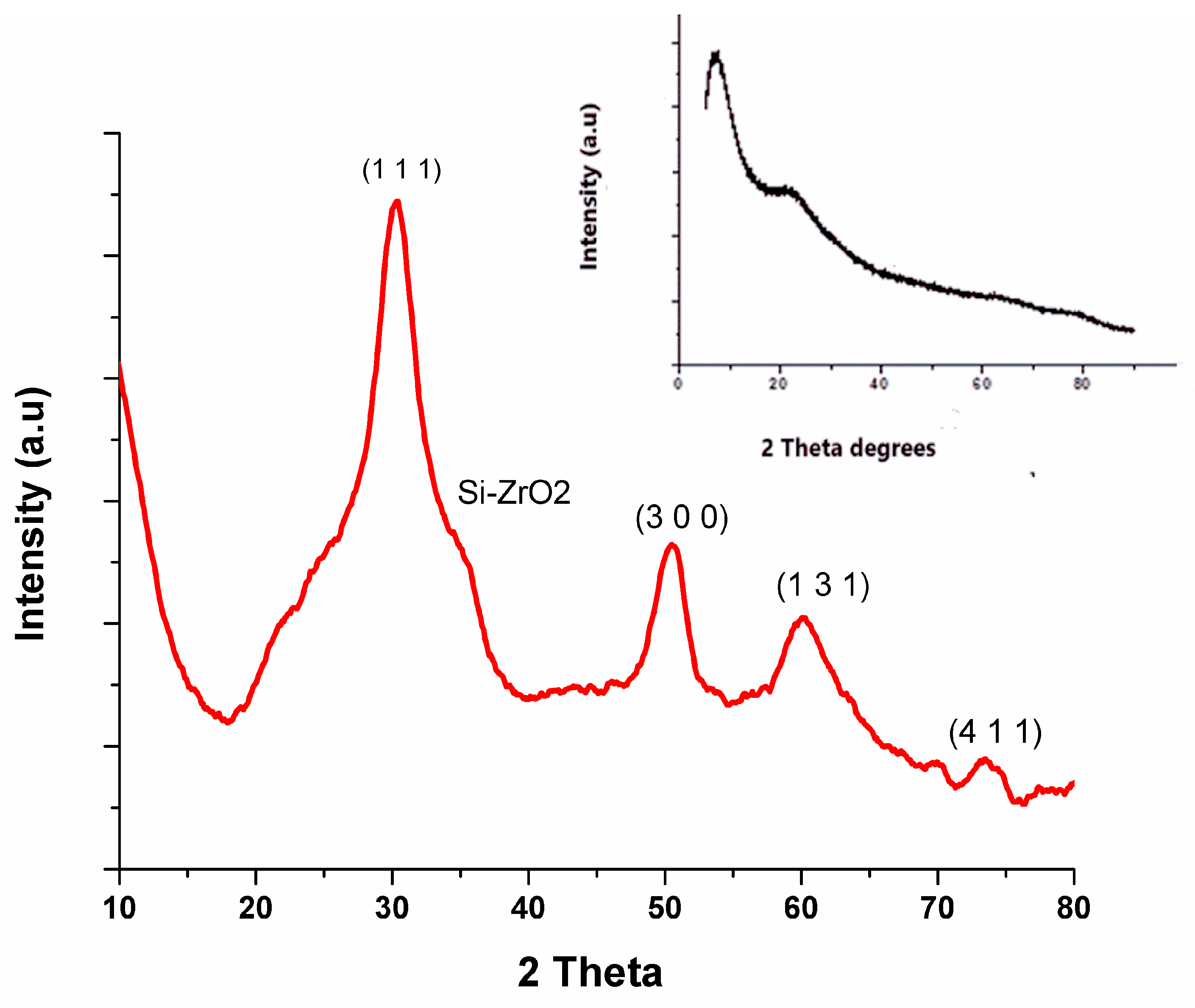
3.2. Structural Analysis

Figure 3 and the insert show the XRD results of Si-ZrO2 and Si nanoparticles. No amorphous silica was found, owing to the crystallisation state of zirconia oxide when calcined over 600 °C, as shown in Figure 3. The insert in Figure 3 shows a broad amorphous peak at 2θ = 20.7–26.2° for the silica nanoparticles; this may be due to their incomplete inner structure and small crystalline size. The diffraction patterns of the Si-ZrO2 nanoparticles changed significantly, with the amorphous phase of silica transforming into a baddeleyite (monoclinic) structure, as seen in Figure 3. Other studies have confirmed that the monoclinic phase is more thermodynamically stable than the tetragonal and cubic phases [20,25,27]. The primary peaks at 2θ were located at 30.4°, 50.6°, 60.1°, and 74.0°, which correspond to the planes (1 1 1), (3 0 0), (1 3 1), and (4 1 1) in Figure 3 [21]. The diffraction pattern exhibits strong and well-defined peaks, indicating a crystalline structure. This shows the successful synthesis of silica–zirconia nanoparticles in one pot, as amorphous silica was transformed into monoclinic zirconia nanoparticles, and that the silica core is well blended with zirconia [27].

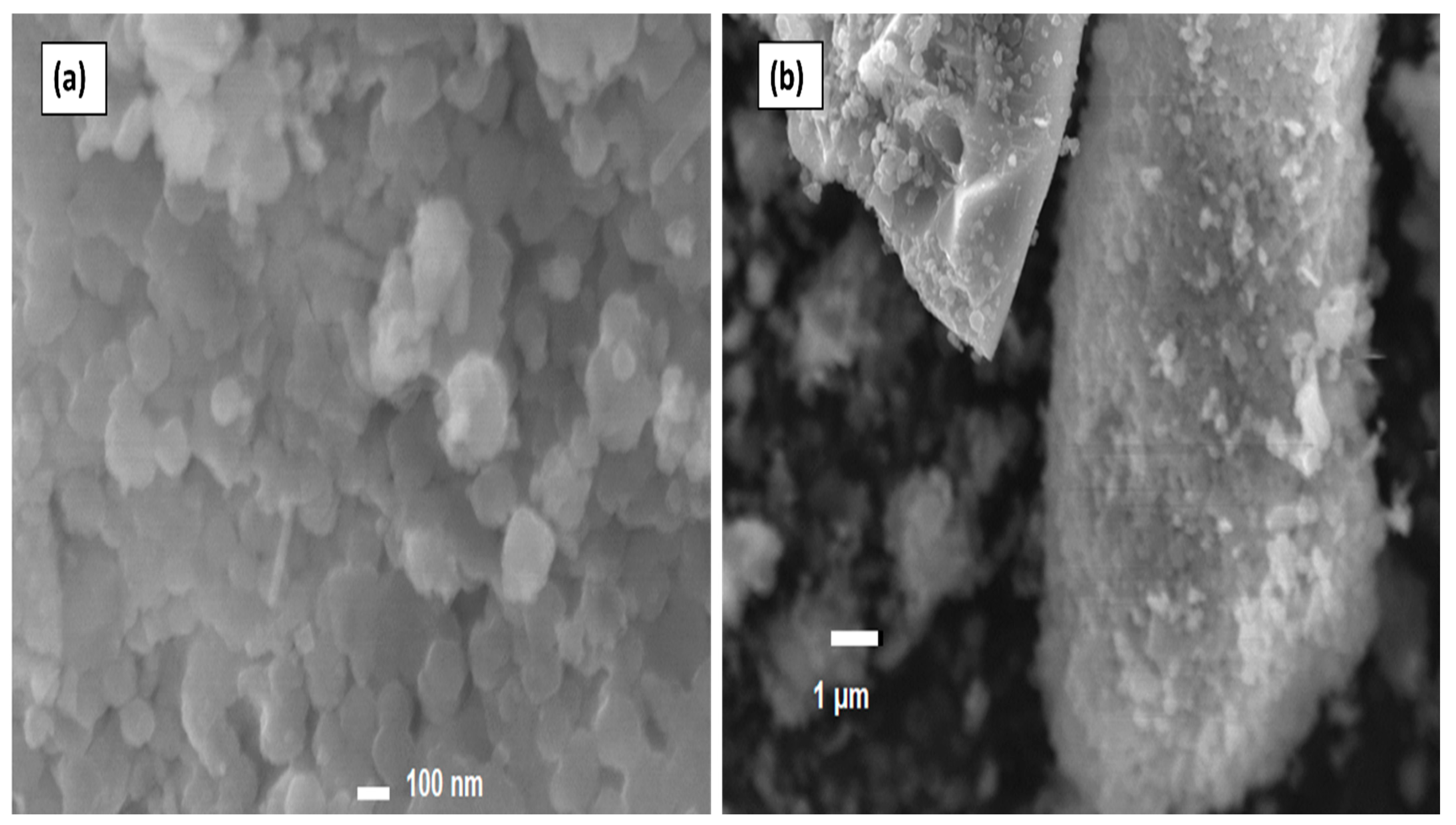
3.3. Scanning Electron Microscopy (SEM) Analysis

Figure 4a,b show the SEM morphology of the Si-ZrO2 nanoparticles in 100 nm and 1 µm scale bars. The Si-ZrO2 nanoparticles show a mixture of rod-like and spherical shapes as shown in Figure 4a., which is confirmed by Lei Xu et al., who obtained a rod-shaped morphology with a diameter ranging between 150 and 200 nm [28].

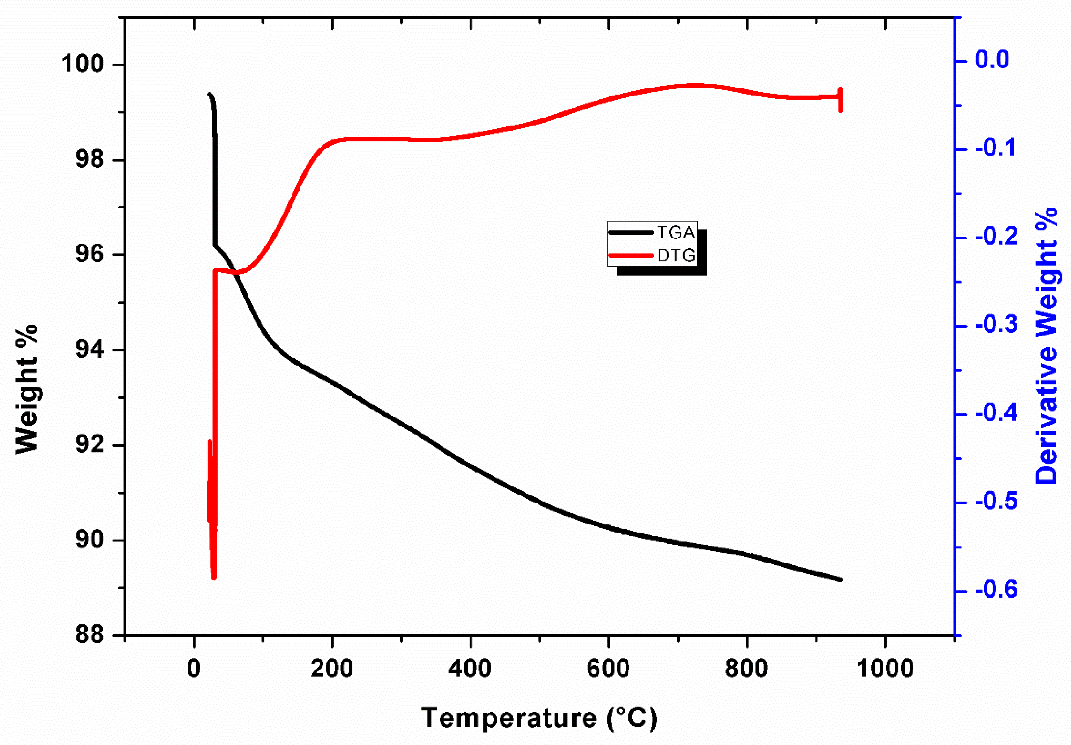
3.4. TGA and Derivative Thermo-Gravimetric (DTG)

Figure 5 shows the TGA and DTG curves of the Si-ZrO2 nanoparticles. As seen in Figure 5, thermal degradation has two weight loss stages. Figure 5 shows an initial weight-loss of 4wt% between 40 °C and 100 °C, which is caused by the evaporation of adsorbed water. The second stage of weight loss occurs between 150 and 900 °C, as a result of unreacted alkoxy groups, organic component degradation, and hydroxyl group removal [20]. The TGA results show that the Si-ZrO2 nanoparticles decompose less and remain stable at temperatures higher than 900 °C with the total weight loss of 12%. The DTG curves in Figure 5 confirm the two weight loss stages observed by TGA, with broad endothermic peaks between 100 °C and 300 °C due to desorbed water [17].

3.5. FTIR Spectrum of ZrO2 Nanoparticles

The FTIR results of the Si-ZrO2 nanoparticles are shown in Figure 6. The bands at 1627 cm−1, 1062 cm−1, and 797 cm−1 are attributed to the symmetric stretching vibration Si-O-Si; this may be due to the addition of TEOS [29,30]. The absorption peaks observed at 1062 cm−1 and 945 cm−1 are assigned to Si–O–Zr bonds generated by silica and the zirconium raw materials [27,31]. The peak at 1452 cm−1 is due to O-H bonding, and the peaks in the region of 1536 cm−1 may be due to the adsorbed moisture. The peak at 2366 cm−1 corresponds to the Zr–OH stretching vibration band, indicating the presence of zirconium nanoparticles. The peaks at 3411 cm−1 are attributed to O-H stretching vibration bands [30,31].

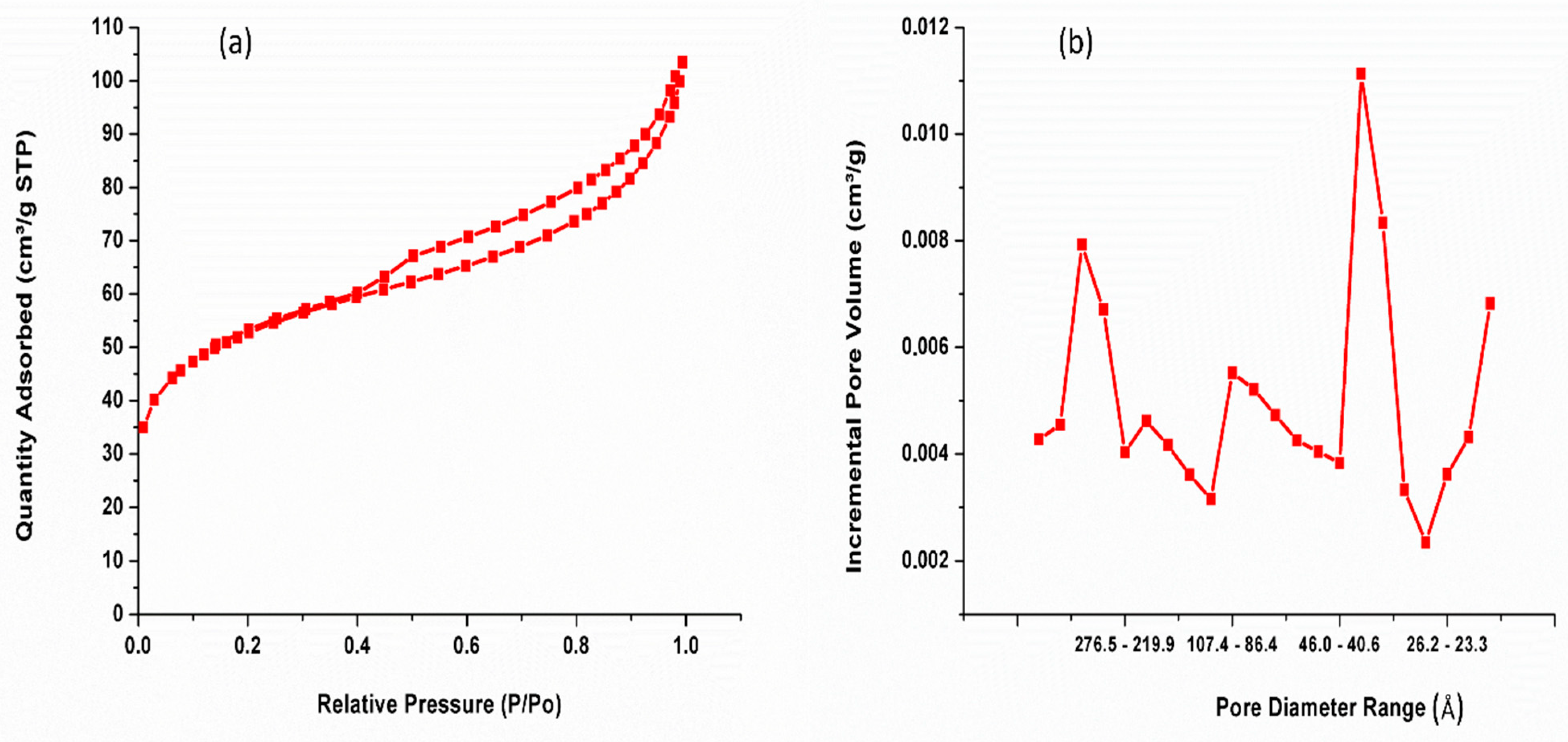
3.6. Nitrogen Adsorption–Desorption

Figure 7 and Table 1 show the N2 adsorption–desorption isotherms and pore size distribution curves and BET-specific surface area of the Si-ZrO2 nanoparticles. Figure 7 shows that the effective diameter of the Si-ZrO2 pores was reduced due to the incorporation of silica into the pores, which also increased the surface area [32]. The BET results in Figure 7a,b and Table 1 show that Si-ZrO2 has a microporous structure with a higher surface area of 185 m2/g. When the samples are aged at a higher temperature of 150 °C, the adsorption–desorption isotherm curves are type IV, with an H3 hysteresis loop, showing that the material has a mesoporous structure [19]. Figure 7b shows that the Si-ZrO2 pore size distribution is narrower, with only one pore size distribution detected, this could be owing to the higher synthesis temperature. The pore distribution data indicate that ageing temperature has an impact on the porosity of zirconia nanoparticles. Si-ZrO2 had a pore volume of 0.14 cm3/g as shown in Table 1. Bumajdad et al. obtained values greater than 144 m2/g and 83 m2/g [33,34]. This is due to the incorporation of tetraethyl orthosilicate into the zirconia solution.

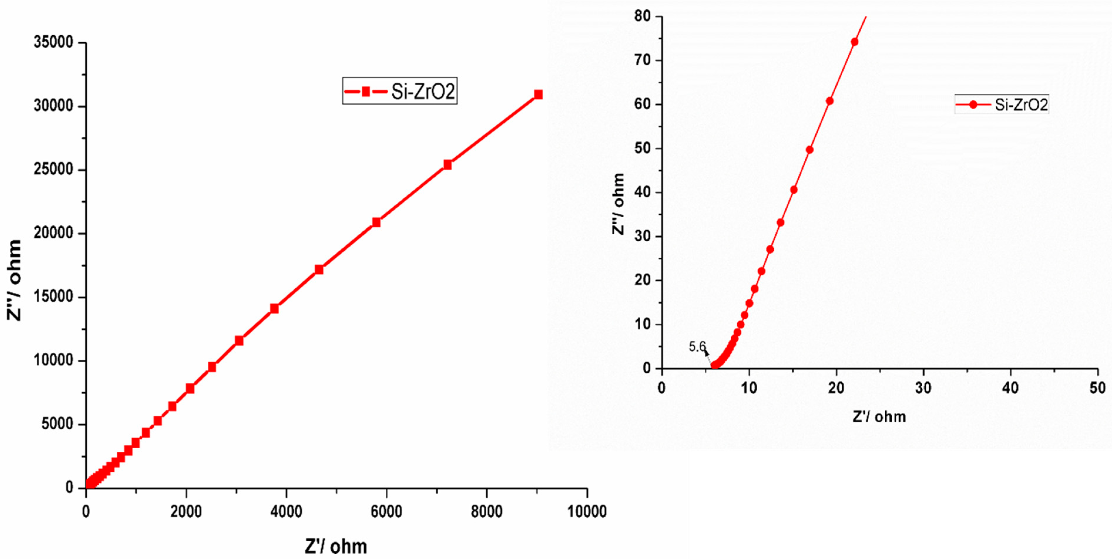
3.7. Electrochemical Results

The cyclic voltammetry of Si-ZrO2 is presented in Figure 8. The Si-ZrO2 nanoparticle electrodes demonstrated characteristic electric double-layer capacitive behaviour with pseudo-rectangular voltametric diagrams in Figure 8. Figure 8 shows that increasing the scan rate from 10 mV s−1 to 100 mV s−1 increases the current due to resistance effects in the pores [20]. The volumetric cycle is higher and faster to complete with an increased electrode capacitance at 50 mV s−1 and 100 mV s−1 than at lower scan rates, demonstrating that the current reactivity of the Si-ZrO2 electrode increases as the scan rate increases, as shown in Figure 8. Furthermore, at an increased scan rate of 100 mV s−1, Si-ZrO2 had a greater area under the voltametric curve and a stronger charge transfer resistance [20], as shown in Figure 8. This may be due to a high BET surface area and wide pores, which contribute to its equivalent performance in electrochemical processes measured with cyclic voltammetry. However, the quasi-rectangular shape, which is indicative of capacitive behaviour, increases dramatically between 50 and 100 mV s–1 which makes it suitable for use in higher charge–discharge devices [35].
The fundamental electrochemical behaviour of Si-ZrO2 in 2 M KCl electrolytes was investigated using electrochemical impendence spectroscopy, as shown in Figure 9. Figure 9 and (insert) show the Nyquist plots for these cells over the frequency range of 100 kHz to 0.01 Hz. Si-ZrO2 has vertical Nyquist plots without a curve zone, as seen in Figure 9 and (insert), due to electron transmission through its porous nanoparticle structure, which generally indicates excellent capacitive behaviour [36]. Figure 9 (insert) shows that, at low frequencies, the curve is virtually vertical, showing optimal capacitive behaviour. The Si-ZrO2 nanoparticles have the same low equivalent series resistance of only 6.25 Ohm, indicating better electrochemical performance in asymmetric devices due to increased electrolyte ion diffusion and mobility during charging and discharging cycles [34]. Furthermore, the huge surface area-to-volume ratio allows for a lot of charge accumulation, which leads to efficient charging. Such properties contribute to increased conductivity, making Si-ZrO2 ideal for use in fuel cell applications.

4. Conclusions

The incorporation of amorphous SiO2 into ZrO2 nanoparticles enhances charge transmission resistance while also providing outstanding capacitance performance, as seen from the electro-chemical studies. Si-ZrO2 nanoparticles exhibit thermal stability under TGA, with a total weight loss of less than 12%. Based on EDX measurements, the major elemental composition of Si-ZrO2 nanoparticles is 14.6% silicon (S), 1.2% zirconium (Zr), and 83.7% oxygen. Both Si and Zr structures are formed in the SAED pattern, which is consistent with the XRD findings. Furthermore, the BET results show that Si-ZrO2 has a higher surface area of 185 m2/g with the adsorption–desorption of type IV isotherm curves. In addition, FTIR reveals Si-O-Zr structures, showing the presence of silica in the zirconia nanoparticles. TEM data show that the composited nanoparticles are crystallised with a particle size of around 5 nm.

Author Contributions

R.S.: methodology, formal analysis, investigation, and original draft writing. F.N.: Formal analysis, investigation, writing (review and editing). T.M.: Methodology: Writing, review, and editing. All authors have read and agreed to the published version of the manuscript.

Funding

This research was funded by the internal funding of the University of South Africa and the National Research Council of South Africa (NRF) Grant number: 150341.

Data Availability Statement

All data are share in this article.

Acknowledgments

The University of South Africa contributed financially. The TEM results (CSIR) UNISA for SEM are also acknowledged.

Conflicts of Interest

The authors declare no conflict of interest.

References

  1. Anjum, S.; Ishaque, S.; Fatima, H.; Farooq, W.; Hano, C.; Abbasi, B.H.; Anjum, I. Emerging Applications of Nanotechnology in Healthcare Systems: Grand Challenges and Perspectives. Pharmaceuticals 2021, 14, 707. [Google Scholar] [CrossRef] [PubMed]
  2. Fu, L.; Wang, B.; Sathyanath, S.K.M.; Chang, J.; Yu, J.; Leifer, K.; Engqvist, H.; Li, Q.; Xia, W. Microstructure of rapidly-quenched ZrO2-SiO2 glass-ceramics fabricated by container-less aerodynamic levitation technology. J. Am. Ceram. Soc. 2023, 106, 2635–2651. [Google Scholar] [CrossRef]
  3. Khan, S.; Mansoor, S.; Rafi, Z.; Kumari, B.; Shoaib, A.; Saeed, M.; Alshehri, S.; Ghoneim, M.M.; Rahamathulla, M.; Hani, U. A review on nanotechnology: Properties, applications, and mechanistic insights of cellular uptake mechanisms. J. Mol. Liq. 2022, 348, 118008. [Google Scholar] [CrossRef]
  4. Hassanzadeh-Tabrizi, S. Precise calculation of crystallite size of nanomaterials: A review. J. Alloys Compd. 2023, 968, 171914. [Google Scholar] [CrossRef]
  5. Lin, H.; Yin, C.; Mo, A. Zirconia Based Dental Biomaterials: Structure, Mechanical Properties, Biocompatibility, Surface Modification, and Applications as Implant. Front. Dent. Med. 2021, 2, 689198. [Google Scholar] [CrossRef]
  6. Haleem, A.; Javaid, M.; Singh, R.P.; Rab, S.; Suman, R. Applications of nanotechnology in medical field: A brief review. Glob. Health J. 2023, 7, 70–77. [Google Scholar] [CrossRef]
  7. Ballem, M.A.; Córdoba, J.M.; Odén, M. Mesoporous silica templated zirconia nanoparticles. J. Nanopart. Res. 2011, 13, 2743–2748. [Google Scholar] [CrossRef]
  8. Raj, S.; Hattori, M.; Ozawa, M. Ag-doped ZrO2 nanoparticles prepared by hydrothermal method for efficient diesel soot oxidation. Mater. Lett. 2019, 234, 205–207. [Google Scholar] [CrossRef]
  9. Hidayat, D.; Syoufian, A.; Utami, M.; Wijaya, K. Synthesis and Application of Na2O/ZrO2 Nanocomposite for Microwave-assisted Transesterification of Castor Oil. ICS Phys. Chem. 2021, 1, 26. [Google Scholar] [CrossRef]
  10. Bansal, P.; Kaur, N.; Prakash, C.; Chaudhary, G.R. ZrO2 nanoparticles: An industrially viable, efficient and recyclable catalyst for synthesis of pharmaceutically significant xanthene derivatives. Vacuum 2018, 157, 9–16. [Google Scholar] [CrossRef]
  11. Sengupta, P.; Bhattacharjee, A.; Maiti, H.S. Zirconia: A Unique Multifunctional Ceramic Material. Trans. Indian Inst. Met. 2019, 72, 1981–1998. [Google Scholar] [CrossRef]
  12. Verdi, C.; Karsai, F.; Liu, P.; Jinnouchi, R.; Kresse, G. Thermal transport and phase transitions of zirconia by on-the-fly machine-learned interatomic potentials. npj Comput. Mater. 2021, 7, 156. [Google Scholar] [CrossRef]
  13. Chitoria, A.K.; Mir, A.; Shah, M. A review of ZrO2 nanoparticles applications and recent advancements. Ceram. Int. 2023, 49, 32343–32358. [Google Scholar] [CrossRef]
  14. Matt, S.B.; Raghavendra, S.; Shivanna, M.; Sidlinganahalli, M.; Siddalingappa, D.M. Electrochemical Detection of Paracetamol by Voltammetry Techniques Using Pure Zirconium Oxide Nanoparticle Based Modified Carbon Paste Electrode. J. Inorg. Organomet. Polym. Mater. 2021, 31, 511–519. [Google Scholar] [CrossRef]
  15. Yilmaz, S.; Cobaner, S.; Yalaz, E.; Horri, B.A. Synthesis and Characterization of Gadolinium-Doped Zirconia as a Potential Electrolyte for Solid Oxide Fuel Cells. Energies 2022, 15, 2826. [Google Scholar] [CrossRef]
  16. Gorbova, E.; Tzorbatzoglou, F.; Molochas, C.; Chloros, D.; Demin, A.; Tsiakaras, P. Fundamentals and Principles of Solid-State Electrochemical Sensors for High Temperature Gas Detection. Catalysts 2021, 12, 1. [Google Scholar] [CrossRef]
  17. Min, H.K.; Kim, Y.W.; Kim, C.; Ibrahim, I.A.; Han, J.W.; Suh, Y.W.; Jung, K.D.; Park, M.B.; Shin, C.H. Phase transformation of ZrO2 by Si incorporation and catalytic activity for isopropyl alcohol dehydration and dehydrogenation. Chem. Eng. J. 2022, 428, 131766. [Google Scholar] [CrossRef]
  18. Ahmadi, F.; Sodagar-Taleghani, A.; Ebrahimnejad, P.; Moghaddam, S.P.H.; Ebrahimnejad, F.; Asare-Addo, K.; Nokhodchi, A. A review on the latest developments of mesoporous silica nanoparticles as a promising platform for diagnosis and treatment of cancer. Int. J. Pharm. 2022, 625, 122099. [Google Scholar] [CrossRef] [PubMed]
  19. Yu, H.; Tong, Z.; Qiao, Y.; Yang, Z.; Yue, S.; Li, X.; Su, D.; Ji, H. High thermal stability of SiO2–ZrO2 aerogels using solvent-thermal aging. J. Solid State Chem. 2020, 291, 121624. [Google Scholar] [CrossRef]
  20. Sigwadi, R.; Mokrani, T.; Dhlamini, M. The synthesis, characterization and electrochemical study of zirconia oxide nanoparticles for fuel cell application. Phys. B Condens. Matter 2020, 581, 411842. [Google Scholar] [CrossRef]
  21. Sigwadi, R.; Dhlamini, M.S.; Mokrani, T.; Nonjola, P. Effect of Synthesis Temperature on Particles Size and Morphology of Zirconium Oxide Nanoparticle. J. Nano Res. 2017, 50, 18–31. [Google Scholar] [CrossRef]
  22. Dong, W.-S.; Lin, F.-Q.; Liu, C.-L.; Li, M.-Y. Synthesis of ZrO2 nanowires by ionic-liquid route. J. Colloid Interface Sci. 2009, 333, 734–740. [Google Scholar] [CrossRef]
  23. Kocjan, A.; Logar, M.; Shen, Z. The agglomeration, coalescence and sliding of nanoparticles, leading to the rapid sintering of zirconia nanoceramics. Sci. Rep. 2017, 7, 2541. [Google Scholar] [CrossRef]
  24. Klinger, M. More features, more tools, more CrysTBox. J. Appl. Crystallogr. 2017, 50, 1226–1234. [Google Scholar] [CrossRef]
  25. Lestari, N.D.; Nurlaila, R.; Muwwaqor, N.F.; Pratapa, S. Synthesis of high-purity zircon, zirconia, and silica nanopowders from local zircon sand. Ceram. Int. 2019, 45, 6639–6647. [Google Scholar]
  26. Mostafa, N.Y.; Zaki, Z.; Mohsen, Q.; Alotaibi, S.; El-moemen, A.A.; Amin, M.A. Carboxylate-assisted synthesis of highly-defected monoclinic zirconia nanoparticles. J. Mol. Struct. 2020, 1214, 128232. [Google Scholar] [CrossRef]
  27. Soontaranon, S.; Limphirat, W.; Pratapa, S. XRD, WAXS, FTIR, and XANES studies of silica-zirconia systems. Ceram. Int. 2019, 45, 15660–15670. [Google Scholar]
  28. Xu, L.; Lei, H.; Ding, Z.; Chen, Y.; Ding, R.; Kim, T. Preparation of the rod-shaped SiO2@C abrasive and effects of its microstructure on the polishing of zirconia ceramics. Powder Technol. 2022, 395, 338–347. [Google Scholar] [CrossRef]
  29. Saravanan, S.; Dubey, R. Synthesis of SiO2 nanoparticles by sol-gel method and their optical and structural properties. Romanian J. Inf. Sci. Technol. 2020, 23, 105–112. [Google Scholar]
  30. Lv, X.; Yuan, L.; Rao, C.; Wu, X.; Qing, X.; Weng, X. Structure and near-infrared spectral properties of mesoporous silica for hyperspectral camouflage materials. Infrared Phys. Technol. 2023, 129, 104558. [Google Scholar] [CrossRef]
  31. Liu, Y.; Guo, Y.; Zhu, Y.; An, D.; Gao, W.; Wang, Z.; Ma, Y.; Wang, Z. A sustainable route for the preparation of activated carbon and silica from rice husk ash. J. Hazard. Mater. 2011, 186, 1314–1319. [Google Scholar] [CrossRef] [PubMed]
  32. Wu, Y.; Xu, Y.; Li, S.; Zhong, L.; Wang, J.; Chen, Y. High-surface-area mesoporous silica-yttria-zirconia ceramic materials prepared by coprecipitation method—The role of silicon. Ceram. Int. 2022, 48, 21951–21960. [Google Scholar] [CrossRef]
  33. Bumajdad, A.; Nazeer, A.A.; Al Sagheer, F.; Nahar, S.; Zaki, M.I. Controlled Synthesis of ZrO2 Nanoparticles with Tailored Size, Morphology and Crystal Phases via Organic/Inorganic Hybrid Films. Sci. Rep. 2018, 8, 3695. [Google Scholar] [CrossRef] [PubMed]
  34. Reddy, C.V.; Babu, B.; Reddy, I.N.; Shim, J. Synthesis and characterization of pure tetragonal ZrO2 nanoparticles with enhanced photocatalytic activity. Ceram. Int. 2018, 44, 6940–6948. [Google Scholar] [CrossRef]
  35. Khalid, S.; Cao, C.; Wang, L.; Zhu, Y. Microwave Assisted Synthesis of Porous NiCo2O4 Microspheres: Application as High Performance Asymmetric and Symmetric Supercapacitors with Large Areal Capacitance. Sci. Rep. 2016, 6, 22699. [Google Scholar] [CrossRef] [PubMed]
  36. Lv, L.-P.; Wu, Z.-S.; Chen, L.; Lu, H.; Zheng, Y.-R.; Weidner, T.; Feng, X.; Landfester, K.; Crespy, D. Precursor-controlled and template-free synthesis of nitrogen-doped carbon nanoparticles for supercapacitors. RSC Adv. 2015, 5, 50063–50069. [Google Scholar] [CrossRef]
Figure 1. (a) HRTEM images, (b) SAED pattern and (c) lattice spacing of Si-ZrO2 nanoparticles.
Chemengineering 09 00026 g001
Figure 2. The EDX spectrum represents the elemental composition of Si-ZrO2 nanoparticles.
Chemengineering 09 00026 g002
Figure 3. XRD pattern of Si-ZrO2. and insert silica nanoparticles.
Chemengineering 09 00026 g003
Figure 4. SEM: (a,b) Si-ZrO2 nanoparticles.
Chemengineering 09 00026 g004
Figure 5. TGA and DTG of Si-ZrO2.
Chemengineering 09 00026 g005
Figure 6. FTIR curves of Si-ZrO2.
Chemengineering 09 00026 g006
Figure 7. Nitrogen adsorption–desorption isotherms (a) and pore size distribution curves (b) for Si-ZrO2 nanoparticles.
Chemengineering 09 00026 g007
Figure 8. CV of Si-ZrO2 in KCl electrodes at scanning rates of 10 mV s−1, 20 mV s−1, 50 mV s−1, and 100 mV s−1.
Chemengineering 09 00026 g008
Figure 9. Nyquist plots of Si-ZrO2 in KCl electrolytes at a range of 10 kHz–1 Hz.
Chemengineering 09 00026 g009
Table 1. BET of ZrO2 nanoparticles: (a) Si-ZrO2 and (b) ZrO2.
Sample IDSurface Area
(m2/g)
Pore Volume
(cm3/g)
Si-ZrO21850.14
ZrO21620.33
Disclaimer/Publisher’s Note: The statements, opinions and data contained in all publications are solely those of the individual author(s) and contributor(s) and not of MDPI and/or the editor(s). MDPI and/or the editor(s) disclaim responsibility for any injury to people or property resulting from any ideas, methods, instructions or products referred to in the content.

