Article

Comparison of a Novel Modality of Erbium-Doped Yttrium Aluminum Garnet Laser-Activated Irrigation and Ultrasonic Irrigation against Mature Enterococcus faecalis Biofilm—An In Vitro Study

by
Gabrijela Kapetanović Petričević
1,
Antonio Perčinić
2,
Ana Budimir
2,
Anja Sesar
3,
Ivica Anić
1 and
Ivona Bago
1,*
1
Department of Endodontics and Restorative Dentistry, School of Dental Medicine, University of Zagreb, 10 000 Zagreb, Croatia
2
Department of Microbiology, University Hospital Center, 10 000 Zagreb, Croatia
3
Department of Endodontics, Oral Patology and Periodontology, Dental Polyclinic Zagreb, 10 000 Zagreb, Croatia
*
Author to whom correspondence should be addressed.
Bioengineering 2024, 11(10), 999; https://doi.org/10.3390/bioengineering11100999
Submission received: 11 September 2024 / Revised: 26 September 2024 / Accepted: 1 October 2024 / Published: 4 October 2024
(This article belongs to the Special Issue Oral Health and Dental Restoration and Regeneration)

Abstract

:
In this in vitro study, we aimed to evaluate and compare the antibacterial efficacy of a novel erbium-doped yttrium aluminum garnet laser modality, shock wave enhanced emission of photoacoustic streaming (SWEEPS), ultrasonically activated irrigation (UAI), and single needle irrigation (SNI) against old bacterial biofilm. A two-week-old Enterococcus faecalis biofilm was cultivated on transversal dentinal discs made from the middle third of the roots of single-rooted, single-canal premolars. Biofilm growth was confirmed using scanning electron microscopy (SEM) and confocal laser scanning microscopy (CLSM). The dentine samples were randomly distributed into three experimental groups and one control group based on the irrigation protocol used: Group 1, SWEEPS; Group 2, UAI; and Group 3, SNI. The root canals were irrigated with a 3% sodium hypochlorite solution. Antibacterial efficacy was evaluated quantitatively through bacterial culture and qualitatively through CLSM and SEM. Both SWEEPS and UAI demonstrated a statistically significant reduction in Enterococcus faecalis colony-forming units (CFUs) (p < 0.001), while SNI did not show a statistically significant reduction (p = 0.553). No significant difference was observed between the efficacy of SWEEPS and UAI (p > 0.05). The SWEEPS and UAI techniques were equally effective in eliminating mature E. faecalis biofilm.

1. Introduction

Mature bacterial biofilm poses a major clinical challenge in root canal treatment because of its resistance to standard antimicrobial protocols and its ability to adapt to root canal environmental changes [1]. Enterococcus faecalis (E. faecalis) is a known, highly resistant bacterium with specific characteristics, such as its capacity to withstand sodium hypochlorite (NaOCl) concentrations as high as 6.5% [2] and pH regulation [3], induce hydroxyapatite reprecipitation in mature biofilms, and form calcified monobiofilms [4]. Clinical studies have shown that it is more frequently associated with secondary and persistent endodontic infections [5,6]. The success rate of root canal retreatment varies between 45% and 83% [7,8], which is significantly lower than that for initial root canal treatments. The eradication of E. faecalis has been of immense interest in the last decade [9]. Disintegration and elimination of bacterial biofilm could be improved by mechanically using different activated irrigation techniques, including ultrasonically activated irrigation (UAI) and novel laser-activated irrigation (LAI) techniques [10,11]. Since E. faecalis can invade dentinal tubules to depths of approximately 500 μm [12], ensuring deeper irrigant penetration and increasing cleaning efficiency within the three-dimensional root canal system is important [12].
UAI energizes the irrigant solution within the canal, creating acoustic streaming, which improves biofilm removal [13]. In contrast, LAI is based on vapor bubble dynamics produced by pulsed lasers, creating specific bubble and cavitation phenomena and acoustic streaming [14]. Previous studies reported that LAI has an effective bactericidal activity and improves smear layer elimination, even from the apical third of the root [15,16]. A novel LAI technique, shock wave enhanced emission photodynamic streaming (SWEEPS), uses a dual-pulse modality, where the second laser pulse is applied just before the collapse of the first laser pulse’s bubble, amplifying the cavitation effect [17,18]. The AutoSWEEPS modality, which uses ultrashort pulses (25 µs) with minimal energy levels (20 mJ), has been reported to be approximately 50% more effective than the standard single-pulse modality in generating pressures within the root canal and significantly enhancing irrigant penetration into the dentinal tubules [19]. Furthermore, recent studies confirm SWEEPS efficacy in the removal of smear layers and debris [11], as well as residual root canal filling material [20,21]. Recent studies confirmed SWEEPS efficacy in E. faecalis biofilm removal when combined with antimicrobial photodynamic therapy (aPDT) [22,23] because it can effectively increase the photosensitizer distribution in the root canal system. However, there is a lack of studies comparing SWEEPS with other activated irrigation techniques. To our best knowledge, only a few recent studies have compared SWEEPS with other irrigation methods for E. faecalis biofilm removal [24]; two studies on multispecies biofilm removal [25,26] reporting similar antimicrobial effects across tested groups.
The aim of this in vitro study was to evaluate and compare the antibacterial efficacy of SWEEPS, UAI, and single needle irrigation (SNI) against a two-week-old E. faecalis monobiofilm. The null hypothesis was that there was no significant difference in the eradication of mature E. faecalis biofilm between the SWEEPS, UAI, and SNI.

2. Materials and Methods

2.1. Selection and Preparation of Dentine Discs

Based on a power analysis using repeated measures analysis of variance (RM-ANOVA), with a significance level (alpha) of 0.05 and 90% test strength, it was necessary to include at least 11 samples per group in the research.
For the purpose of this study, extracted human single-rooted, single-canal premolars were collected. The exclusion criteria included previous root canal treatment and the presence of carious lesions. The presence of a single round root canal was confirmed through a cone beam computed tomography (CBCT) scan (Cranex 3DX; Soredex, Tuusula, Finland; parameters: FOV 595 (5.0 mm) mm; ENDO, 85 µm; 6.3 mA; 90 kV; 8.7 s; i 450.3 mGycm2).
Transversal dentinal discs (1 mm thick, 5 × 5 mm) were made from the middle third of the root using a diamond disk (Isomet 1000, Buehler, IL, USA). Both sides of the samples were polished using three polishing discs with varying degrees of fineness (Presi France, P320, P600, P1200). The samples were then sterilized in hot plasma (Sterrad 100 NX, ASP, Schaffhausen, Switzerland).

2.2. Enterococcus faecalis Biofilm Formation

The E. faecalis ATCC strain (ATCC 29212, AmericanType Culture Collection, Manassas, VA, USA) was used in this study to form a two-week-old bacterial biofilm. The bacterial suspension density was adjusted to 0.5 McFarland, as measured via a densitometer (Densimat, Bio Mérieux, Marcy l’Etoile, France). Sterile dentine discs were placed in 1 mL microtiter wells (Thermo Fisher Scientific, Waltham, MA, USA) containing a Brain Hearth Infusion (BHI) broth (Sigma Aldrich, Burlington, MA, USA). The BHI was replaced every 48 h for 14 days.

2.3. Experimental Irrigation Protocols

A total of 72 dentine samples were prepared and inoculated with bacterial biofilm. In each experimental group, 13 samples (n = 13) were used for quantitative microbiological analysis, while four samples were used for scanning electron microscopy (SEM) and confocal laser microscopy (CFLM) analysis.
In all three experimental groups, each dentine sample was placed into a 1 mL microtiter well (Thermo Fisher Scientific, Massachusetts, USA) containing 500 μL of 3% NaOCl solution. The samples were then subjected to a 90 s irrigation protocol, divided into three cycles of 30 s each use. For each cycle, the dentine sample was transferred to a new well (Thermo Fisher Scientific, Massachusetts, USA) containing 500 μL of fresh 3% NaOCl solution.

2.3.1. Group 1: SWEEPS

The samples were treated using an erbium-doped yttrium aluminum garnet (Er: YAG) laser (LightWalker AT, Fotona, Ljubljana, Slovenia) equipped with a radial laser tip (600 µm, 9 mm, Fotona, Ljubljana, Slovenia). During activation, the radial laser tip was inserted into the well and maintained in a fixed position. The laser parameters were set at Auto SWEEPS mode: pulse energy (20 mJ), pulse frequency (15 Hz), average power (0.60 W), pulse duration (25 µs), and peak power (800 W).

2.3.2. Group 2: Ultrasonically Activated Irrigation

The samples were activated using an endodontic ultrasonic tip (Irri S, 25/25, VDW, Munich, Germany) connected to an ultrasound device (VDW ULTRA; VDW, Munich, Germany), which was set at 20% power as required for the irrigation mode by the manufacturer. During activation, the ultrasonic tip was placed stationary inside the microtiter well.

2.3.3. Group 3: Passive Irrigation (PI)

The samples were passively immersed in microtiter wells filled with 500 μL of 3% NaOCl. As previously explained, after each 30 s cycle, the discs were transferred into another well filled with fresh 3% NaOCl.
Following the irrigation protocol, each sample was immersed in a well containing a sodium thiosulfate solution for 30 s to neutralize NaOCl after the termination of the experimental irrigation protocol. Finally, each sample was immersed in sterile saline.
For each experimental group, seven additional dentine samples were used as controls, receiving no treatment. Five of these control samples served for quantitative analysis to determine the initial number of E. faecalis CFUs, while the other two were for SEM and CLSM analyses to confirm the initial presence of E. faecalis bacterial biofilm.

2.4. Culture Method and Quantification of E. faecalis Colony Forming Units

Quantification of bacteria colony forming units (CFUs) was performed using the culture method, before the irrigation protocols (controls) and after the irrigation protocols. Each dentine sample was placed in a sterile 1 mL tube containing 500 μL of sterile saline and vortexed (V-1 plus, Biosan, Riga, Latvia) for 30 s in touch mode. The prepared suspension was then serially diluted, and 20 µL were added to wells containing 180 μL Mueller–Hinton broth (Sigma-Aldrich, Burlington, MA, USA). Following the serial dilutions, 10 μL of each suspension were plated onto blood agar plates (211037, Becton Dickinson, New York, NY, USA), starting from the highest dilution to the lowest. The samples were incubated for 24 h in a chamber at 37 °C. After 24 h, the colonies were counted, and the specific number of E. faecalis CFU was calculated based on the degree of dilution. The reduction in E. faecalis CFUs following treatment was evaluated by comparing them with the controls.

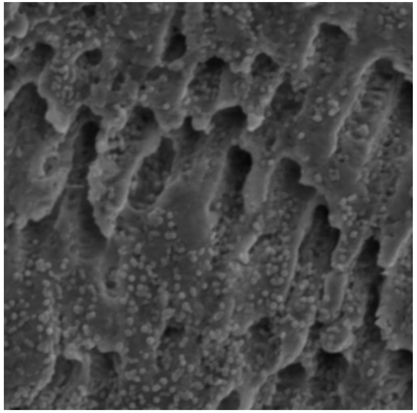
2.5. Scanning Electron Microscopy Analysis

SEM analysis was performed before the irrigation protocols (controls) and after the irrigation protocols. After fixation in formalin, the dentine samples were dehydrated in increasing concentrations of alcohol: 55%, 60%, 75%, 80%, and 95%. Afterward, the samples were exposed to drying at the critical point where water was replaced with CO2 under a pressure of 80 bar. SEM analysis was conducted on a field emission SEM (FE-SEM) model JSM-7000F (Jeol Ltd., Tokyo, Japan). Randomly selected surfaces on the samples were examined, and four micrographs per specimen were captured at magnifications of 5000× and 10,000×, with a 1-micrometer thickness. The analysis was performed by an individual who was blinded to the preceding sample irrigation treatment.

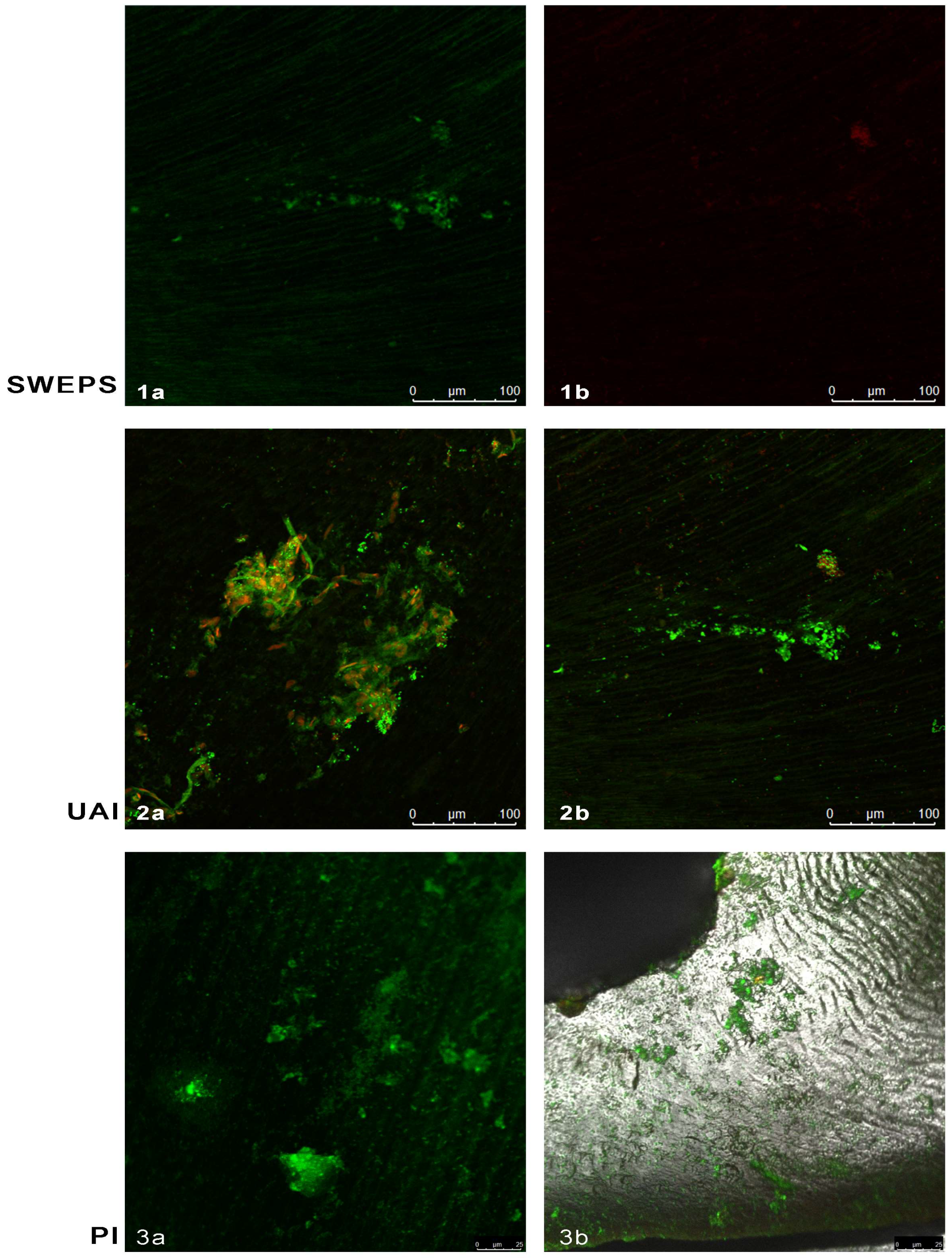
2.6. Confocal Laser Scanning Microscopy Analysis

CLSM analysis was performed before the irrigation protocols (controls) and after the irrigation protocols. The dentine discs were stained with the LIVE/DEAD kit (Invitrogen LIVE/DEAD BacLight Bacterial Viability kit), which contains two dyes, SYTO 9 (green fluorescence) and propidium iodide (red color).
The samples were then covered with 10 µL staining solution consisting of 1 mL 0.9% NaCl, 1 µL SYTO 9, and 1 µL propidium iodide and incubated for 15 min in the dark at 25 °C. After staining, the samples were gently irrigated with saline. The green dye binds to the cell wall of living bacteria, while the red dye binds to the damaged bacteria cell wall. Consequently, the samples were analyzed using a confocal laser microscope (Leica TSC SP8; Leica Microsystems GmbH, Wetzlar, Germany). Randomly chosen surfaces on the sample were taken into consideration, and four micrographs per specimen were taken. The analysis was performed by an individual who was blinded to the preceding sample treatment.
Researchers who performed culture method and CFUs quantification, SEM and CFLM, did not know whether the samples were taken from an experimental group or a control group.

2.7. Statistical Analysis

To distribute results and compare the number of E. faecalis CFUs pre- and post-treatment, the non-parametric Mann–Whitney U test was used because of data inhomogeneity (p < 0.05). A significance level of p = 0.05 was used for all statistical tests. The data analysis was performed using the IBM Statistics 19.0.0.1 software package. (StatSoft, Tulsa, OK, USA).

3. Results

3.1. Results of the Culture Method

Table 1 shows the number of E. faecalis CFUs for each tested group (SWEEPS, UAI, PI) and control samples in each group.
In intragroup analysis, both SWEEPS and UAI showed a statistically significant reduction in E. faecalis CFUs (p < 0.001), while the PI group did not demonstrate a statistically significant reduction (p = 0.553) (Table 1).
Intergroup analysis revealed no statistically significant difference between the SWEEPS and UAI groups (p > 0.05).

3.2. Qualitative Results of Scanning Electron Microscopy and Confocal Laser Scanning Microscopy

Figure 1 (SEM) and Figure 2 (CLSM) illustrate the diffuse growth of E. faecalis biofilm on dentine discs after two weeks of incubation.
Figure 3 shows the SEM of dentine discs following the irrigation protocols under 5000× and 10,000× magnification. In the SWEEPS group (Figure 3(1a,1b)), a small number of planktonic E. faecalis cells were visible with great areas of clean dentine surface. In the UAI group (Figure 3(2a,2b)), fewer E. faecalis colonies were present, mostly clustering into strains and pairs. In contrast, the PI group (Figure 3(3a,3b)) exhibited diffusely spread E. faecalis colonies across the dentine surface, confirming the inadequate efficiency of the PI based on SEM analysis.
Figure 4 shows the CFLM of dentine discs following the irrigation protocols. In the SWEEPS group (Figure 4(1a,1b)), large areas of clean dentine were visible, with few areas with viable E. faecalis colonies (green) (Figure 4(1a)), while several areas contained accumulated dead bacteria (red) (Figure 4(1b)). In the UAI group, an increase in the density of dead E. faecalis colonies (red) was visible, and a lower density of viable colonies (green) compared with the control sample surfaces (Figure 4(2a,2b)). However, colonies were still attached to the sample surface in significant numbers. In the PI group, a large number of viable E. faecalis bacteria clusters (green) were present, with a minimal number of dead bacterial cells (red) (Figure 4(3a,3b)).

4. Discussion

The results of this study revealed that both evaluated activated irrigation techniques, SWEEPS and UAI, were more effective than PI in removing bacterial biofilm from the dentinal surface under in vitro conditions. Furthermore, no significant difference was found between SWEEPS and UAI. Thus, the null hypothesis can be partially rejected. Over the last decade, numerous studies have highlighted LAI as a method of choice for cleaning complex canal anatomies [27], dissolving pulp tissue [28], and removing smear layers and hard tissue debris [29]. Lei et al. [30] demonstrated that LAI increased the efficiency of lower NaOCl concentrations and equalized NaOCl efficiency across different concentrations (0.5–5.25%) in eradicating E. faecalis biofilm. In this study, the NaOCl concentration was consistent across all groups (3%), and the SWEEPS technique showed significantly greater biofilm reduction than PI, as confirmed by SEM and CLSM analyses (Figure 3 and Figure 4). Recently, CLSM has been recommended as a powerful technique for evaluating endodontic disinfection [31].
The results of this study align with those of a recently published study by Robberecht et al. [25], who also found similar efficacy between SWEEPS and sonic irrigation, with needle irrigation being the least effective against biofilm-mimicking hydrogel in the isthmus region of 3D-printed models. Furthermore, in a more recent study by Akdere et al. [24], SWEEPS and UAI exhibited equal antimicrobial efficacy against E. faecalis biofilm in internal resorption cavities. No significant difference was observed between UAI and SWEEPS in this study; however, SWEEPS provided complete elimination of bacteria in three samples. These results in comparison with the study by Akdere et al. [24] suggest that, in a simple canal anatomy, the SWEEPS technique does not provide extra benefit compared to the UAI. More complex root canal anatomy requires advanced irrigation techniques. The results of this study suggest the probable greater potential of the SWEEPS technique; however, more studies on the topic are required. Moreover, SEM and CLSM analyses revealed cleaner dentine disc surfaces after SWEEPS, with few bacteria cells present, compared to UAI, which still contained groups of E. faecalis cells (Figure 3 and Figure 4). In contrast to our findings, De Meyer et al. [26] reported greater efficacy of Er: YAG LAI compared to UAI against dual-species biofilm in a resin root canal model. However, De Meyer et al. used a different biofilm model [26] and employed saline solution as an irrigant, whereas in this study, 3% NaOCl was used. Possibly, the high efficacy of NaOCl could have compensated for the lower efficacy of UAI in this study. Bao et al. [32] evaluated in their study the efficacy of photon-induced photoacoustic streaming (PIPS) and SWEEPS against multispecies biofilms within apical artificial grooves and dentinal tubules, compared to conventional needle irrigation (CNI), UAI, and Eddy. They found SWEEPS, PIPS, and Eddy had similar antibacterial efficacy but greater efficacy than CNI and UAI.
The powerful action of the AutoSWEEPS mode is because of dual pulse modality, meaning the temporal separation between the pulse pairs is continuously swept back and forth, providing an adequate cavitation effect since oscillation times of cavitation bubbles vary considerably depending on pulse energy and cavity diameter [27]. Since oscillation times are shorter in larger cavities [33], leading to more effective bubble dynamics, the large reservoir used (1 mL wells) could also be the reason why, in some samples, biofilm was completely detached. Swimberghe et al. [17] explained the mechanism between biofilm detachment from root canal walls during LAI and the mechanical disruption of fragments coronally. They emphasized that the oscillation of small cavitation bubbles was a determinant in the generation of rapid vertical irrigant movement, causing strong streaming and biofilm detachment.
A few limitations to this study should be emphasized. First, the biofilm in this study was grown on dentinal discs, making it more accessible to the activation technique than the biofilm in complex root canals. Furthermore, the collapse of the bubble, which generates shock waves during SWEEPS, may have occurred easily in the large liquid reservoir used in this study. In confined spaces such as root canals, friction slows down bubble dynamics, leading to shock wave reduction or complete absence [25]. However, Swimberghe et al. [17] in their study used a high-speed camera to capture biofilm detachment and its coronal displacement in the root canal using single-pulse LAI, proving its efficacy in the root canal. They explained that LAI in root canals is based on the generation of smaller cavitation bubbles throughout the entire canal, distant from the primary bubble during its implosion. Moreover, a recent study by Bao et al. [33] confirmed that SWEEPS outperformed needle irrigation, EDDY, UAI, and LAI with photon-induced photoacoustic streaming in treating multispecies biofilms within apical root canals and dentinal tubules. The stationary reservoir of the irrigant could also be the reason of the significantly reduced efficacy of only passive disinfection, in which the discs were only placed in the well filled with NaOCL. Future studies should evaluate the efficacy of SWEEPS compared to standard activation and passive irrigation techniques in more complex root canal systems. Clinical studies with the primary outcome of the SWEEPS in the healing of periapical lesions are also needed.

5. Conclusions

Within the limitations of this study, the SWEEPS and UAI techniques were equally effective in eliminating mature E. faecalis biofilm from dentinal discs and demonstrated superiority over SNI. However, since some samples were completely free of biofilm only in the SWEEPS group, its potential advantage over other techniques warrants further investigation.

Author Contributions

Conceptualization, I.B. and I.A.; methodology, G.K.P., A.B., and A.P.; software, I.A.; validation, I.B., A.B., and I.B.; formal analysis, G.K.P. and A.S.; investigation, G.K.P.; resources, I.B.; data curation, G.K.P.; writing—original draft preparation, G.K.P. and I.B.; writing—review and editing, I.A.; visualization, G.K.P. and A.S.; supervision, I.B.; project administration, I.B.; funding acquisition, I.B. All authors have read and agreed to the published version of the manuscript.

Funding

This research was funded by the research project Croatian Science Foundation “Experimental and clinical evaluation of laser- activated photoacoustic streaming and photoactivated disinfection in endodontic treatment,” grant number: N0 5303.

Institutional Review Board Statement

The study was conducted in accordance with the Declaration of Helsinki, and approved by the Institutional Ethics Committee of the School of Dental Medicine University of Zagreb (:05-PA-30-VI-4/2019, and date of approval: 11 April 2019.).

Informed Consent Statement

Informed consent was obtained from all subjects involved in the study.

Data Availability Statement

Data is contained within the article.

Conflicts of Interest

The authors declare no conflicts of interest.

References

  1. Narayanan, L.L.; Vaishnavi, C. Endodontic microbiology. J. Conserv. Dent. 2010, 13, 233–239. [Google Scholar] [CrossRef] [PubMed]
  2. Del Fabbro, M.; Samaranayake, L.P.; Lolato, A.; Weinstein, T.; Taschieri, S. Analysis of the secondary endodontic lesions focusing on the extraradicular microorganisms: An overview. J. Investig. Clin. Dent. 2014, 5, 245–254. [Google Scholar] [CrossRef] [PubMed]
  3. Evans, M.; Davies, J.K.; Sundqvist, G.; Figdor, D. Mechanisms involved in resistance of Enterococcus faecalis to calcium hydroxide. Int. Endod. J. 2002, 35, 221–228. [Google Scholar] [CrossRef] [PubMed]
  4. Kayaoglu, G.; Ørstavik, D. Virulence factors of Enterococcus faecalis: Relationship to endodontic disease. Crit. Rev. Oral Biol. Med. 2004, 15, 308–320. [Google Scholar] [CrossRef] [PubMed]
  5. Siqueira, J.F.; Rôças, I.N. Polymerase chain reaction-based analysis of microorganisms associated with failed endodontic treatment. Oral Surg. Oral Med. Oral Pathol. Oral Radiol. Endod. 2004, 97, 85–94. [Google Scholar] [CrossRef]
  6. Sedgley, C.; Nagel, A.; Dahlén, G.; Reit, C.; Molander, A. Real-time quantitative polymerase chain reaction and culture analyses of Enterococcus faecalis in root canals. J. Endod. 2006, 32, 173–177. [Google Scholar] [CrossRef]
  7. Torabinejad, M.; Corr, R.; Handysides, R.; Shabahang, S. Outcomes of non-surgical retreatment and endodontic surgery: A systematic review. J. Endod. 2009, 35, 930–937. [Google Scholar] [CrossRef]
  8. Zandi, H.; Petronijevic, N.; Mdala, I.; Kristoffersen, A.K.; Enersen, M.; Rôças, I.N.; Siqueira, J.F., Jr.; Ørstavik, D. Outcome of endodontic retreatment using 2 root canal irrigants and influence of infection on healing as determined by a molecular method: A randomized clinical trial. J. Endod. 2019, 45, 1089–1098.e5. [Google Scholar] [CrossRef]
  9. Haapasalo, M.; Shen, Y.; Wang, Z.; Gao, Y. Irrigation in Endodontics. Br. Dent. J. 2014, 216, 299–303. [Google Scholar] [CrossRef]
  10. Kumar, K.; Teoh, Y.Y.; Walsh, L.J. Root canal cleaning in roots with complex canals using agitated irrigation fluids. Aust. Endod. J. 2023, 49, 56–65. [Google Scholar] [CrossRef]
  11. Yang, Q.; Liu, M.W.; Zhu, L.X.; Peng, B. Micro-CT study on the removal of accumulated hard-tissue debris from the root canal system of mandibular molars when using a novel laser-activated irrigation approach. Int. Endod. J. 2020, 53, 529–538. [Google Scholar] [CrossRef]
  12. Wong, D.T.S.; Cheung, G.S.P. Extension of the bactericidal effect of sodium hypochlorite into the dentinal tubules. J. Endod. 2014, 40, 825–829. [Google Scholar] [CrossRef] [PubMed]
  13. Van Der Sluis, L.W.M.; Vogels, M.P.J.M.; Verhaagen, B.; Macedo, R.; Wesselink, P.R. Study on the influence of refreshment/activation cycles and irrigants on mechanical cleaning efficiency during ultrasonic activation of the irrigant. J. Endod. 2010, 36, 737–740. [Google Scholar] [CrossRef] [PubMed]
  14. DiVito, E.; Peters, O.A.; Olivi, G. Effectiveness of the erbium: YAG laser and new radial and stripped tips in removing the smear layer after root canal instrumentation. Lasers Med. Sci. 2012, 27, 273–280. [Google Scholar] [CrossRef]
  15. Cheng, X.; Xiang, D.; He, W.; Qiu, J.; Han, B.; Yu, Q.; Tian, Y. Bactericidal effect of Er: YAG laser-activated sodium hypochlorite irrigation against biofilms of Enterococcus faecalis isolated from the canal of root-filled teeth with periapical lesions. Photomed. Laser Surg. 2017, 35, 386–392. [Google Scholar] [CrossRef]
  16. Betancourt, P.; Merlos, A.; Sierra, J.M.; Camps-Font, O.; Arnabat-Domínguez, J.; Viñas, M. Effectiveness of low concentration of sodium hypochlorite activated by Er,Cr:YSGG laser against Enterococcus faecalis biofilm. Lasers Med. Sci. 2019, 34, 247–254. [Google Scholar] [CrossRef]
  17. Swimberghe, R.C.D.; Tzourmanas, R.; De Moor, R.J.G.; Braeckmans, K.; Coenye, T.; Meire, M.A. Explaining the working mechanism of laser-activated irrigation and its action on microbial biofilms: A high-speed imaging study. Int. Endod. J. 2022, 55, 1372–1384. [Google Scholar] [CrossRef]
  18. Lukač, N.; Jezeršek, M. Amplification of pressure waves in laser-assisted endodontics with synchronized delivery of Er:YAG laser pulses. Lasers Med. Sci. 2018, 33, 823–833. [Google Scholar] [CrossRef]
  19. Jezeršek, M.; Lukač, N.; Lukač, M.; Tenyi, A.; Olivi, G.; Fidler, A. Measurement of pressures generated in root canal during Er:YAG laser-activated irrigation. Photobiomodul. Photomed. Laser Surg. 2020, 38, 625–631. [Google Scholar] [CrossRef]
  20. Petričević, G.K.; Katić, M.; Anić, I.; Salarić, I.; Vražić, D.; Bago, I. Efficacy of different Er:YAG laser-activated photoacoustic streaming modes compared to passive ultrasonic irrigation in the retreatment of curved root canals. Clin. Oral Investig. 2022, 26, 6773–6781. [Google Scholar] [CrossRef] [PubMed]
  21. Kapetanović Petričević, G.; Katić, M.; Brzović Rajić, V.; Anić, I.; Bago, I. The efficacy of Er:YAG laser-activated shock wave-enhanced emission photoacoustic streaming compared to ultrasonically activated irrigation and needle irrigation in the removal of bioceramic filling remnants from oval root canals-an ex vivo study. Bioengineering 2022, 9, 820. [Google Scholar] [CrossRef] [PubMed] [PubMed Central]
  22. Shahi Ardakani, A.; Afrasiabi, S.; Sarraf, P.; Benedicenti, S.; Solimei, L.; Chiniforush, N. In vitro assessment of SWEEPS and antimicrobial photodynamic therapy alone or in combination for eradicating Enterococcus faecalis biofilm in root canals. Pharmaceutics 2023, 15, 2628. [Google Scholar] [CrossRef] [PubMed]
  23. Ensafi, F.; Fazlyab, M.; Chiniforush, N.; Akhavan, H. Comparative effects of SWEEPS technique and antimicrobial photodynamic therapy by using curcumin and nano-curcumin on Enterococcus faecalis biofilm in root canal treatment. Photodiagn. Photodyn. Ther. 2022, 40, 103130. [Google Scholar] [CrossRef] [PubMed]
  24. Akdere, S.K.; Aydin, Z.U.; Erdönmez, D. Antimicrobial effectiveness of different irrigation activation techniques on teeth with artificial internal root resorption and contaminated with Enterococcus faecalis: A confocal laser scanning,icroscopy analysis. Lasers Med. Sci. 2023, 38, 89. [Google Scholar] [CrossRef] [PubMed]
  25. Robberecht, L.; Delattre, J.; Meire, M. Isthmus morphology influences debridement efficacy of activated irrigation: A laboratory study involving biofilm mimicking hydrogel removal and high-speed imaging. Int. Endod. J. 2023, 56, 118–127. [Google Scholar] [CrossRef]
  26. De Meyer, S.; Meire, M.A.; Coenye, T.; De Moor, R.J. Effect of laser-activated irrigation on biofilms in artificial root canals. Int. Endod. J. 2017, 50, 472–479. [Google Scholar] [CrossRef]
  27. Meire, M.; De Moor, R.J.G. Principle and antimicrobial efficacy of laser-activated irrigation: A narrative review. Int. Endod. J. 2024, 57, 841–860. [Google Scholar] [CrossRef]
  28. Bago, I.; Batelja-Vuletić, L.; Tarle, A.; Sesar, A.; Anić, I. Novel laser activated photoacoustic streaming for removing pulp remnants from round root canals after single file reciprocating instrumentation. Photodiagn. Photodyn. Ther. 2022, 37, 102631. [Google Scholar] [CrossRef]
  29. Vatanpour, M.; Toursavadkouhi, S.; Sajjad, S. Comparison of three irrigation methods: SWEEPS, ultrasonic, and traditional irrigation, in smear layer and debris removal abilities in the root canal, beyond the fractured instrument. Photodiagn. Photodyn. Ther. 2022, 37, 102707. [Google Scholar] [CrossRef]
  30. Lei, L.; Wang, F.; Wang, Y.; Li, Y.; Huang, X. Laser activated irrigation with SWEEPS modality reduces concentration of sodium hypochlorite in root canal irrigation. Photodiagn. Photodyn. Ther. 2022, 39, 102873. [Google Scholar] [CrossRef]
  31. Mattern, R.; Ernst, S.; Böcher, S.; Braun, A.; Wenzler, J.S.; Conrads, G. CLSM-guided imaging for quantifying endodontic disinfection. Antibiotics 2024, 13, 54. [Google Scholar] [CrossRef] [PubMed]
  32. Bao, P.; Liu, H.; Yang, L.; Zhang, L.; Yang, L.; Xiao, N.; Shen, J.; Deng, J.; Shen, Y. In vitro efficacy of Er:YAG laser-activated irrigation versus passive ultrasonic irrigation and sonic-powered irrigation for treating multispecies biofilms in artificial grooves and dentinal tubules: A SEM and CLSM study. BMC Oral Health 2024, 24, 261. [Google Scholar] [CrossRef] [PubMed]
  33. Lukac, N.; Muc, B.; Jezeršek, M.; Lukac, M. Photoacoustic endodontics using the novel SWEEPS Er:YAG laser modality. J. Laser Health Acad. 2017, 2017, 1–7. [Google Scholar]
Figure 1. Scanning electron microscopy of Enterococcus faecalis biofilm on dentine disc after two weeks of incubation.
Bioengineering 11 00999 g001
Figure 2. Confocal laser scanning microscopy of Enterococcus faecalis biofilm on dentine discs after two weeks of incubation.
Bioengineering 11 00999 g002
Figure 3. Scanning electron microscopy of representative dentine discs with Enterococcus faecalis (E. faecalis) biofilm under 5000× (a) and 10,000× (b) magnification after the irrigation protocols: (1a,1b). SWEEPS: visible small number of isolated planktonic cells of E. faecalis; (2a,2b). Ultrasonically activated irrigation (UAI): few E. faeacalis colonies grouping into strains and pairs; (3a,3b). Passive irrigation (PI): dense E. faeacalis colonies diffusely spread all over the sample’s surface.
Bioengineering 11 00999 g003
Figure 4. Confocal laser scanning microscopy of representative dentine discs with Enterococcus faecalis (E. faecalis) biofilm after the irrigation protocols: (1a,1b). SWEEPS: a few areas adhered to living E. faecalis colonies (green) are observed, as well as several areas where dead bacteria accumulated (red) (b) and a large area of clean dentin; (2a,2b). Ultrasonically activated irrigation (UAI): areas with dead colonies (red) of E. faecalis, and significant areas of live bacteria colonies still (green); (3a,3b). Passive irrigation (PI): large areas of live E. faecalis bacteria colonies (green).
Bioengineering 11 00999 g004
Table 1. Number of E. faecalis colony forming units (CFUs) in each tested and control group after the experimental irrigation protocol.
MinimumMaximumPercentile 25MedianPercentile 75
Number of CFUsSWEEPSControl5 × 10123.5 × 10135 × 10121 × 10131.5 × 1013
Experimental group05 × 104000
UAIControl2 × 10131 × 10142.5 × 10137 × 10137.5 × 1013
Experimental group5 × 1052 × 1095 × 1065 × 1065 × 108
PIControl5 × 1072.5 × 10135 × 1085 × 1081 × 1010
Experimental group1 × 1052.5 × 10135 × 1065 × 1085 × 1011
SWEEPS, Shock Wave Enhanced Emission Photoacoustic Streaming; UAI, ultrasonically activated irrigation; PI, passive irrigation.
Disclaimer/Publisher’s Note: The statements, opinions and data contained in all publications are solely those of the individual author(s) and contributor(s) and not of MDPI and/or the editor(s). MDPI and/or the editor(s) disclaim responsibility for any injury to people or property resulting from any ideas, methods, instructions or products referred to in the content.

