Image-Based Monitoring of Thermal Ablation
Abstract
1. Introduction
2. Thermography Monitoring
2.1. MR Thermometry
2.2. Ultrasound Temperature Measurement
2.3. Photoacoustic Temperature Measurement
2.4. CT Thermometry
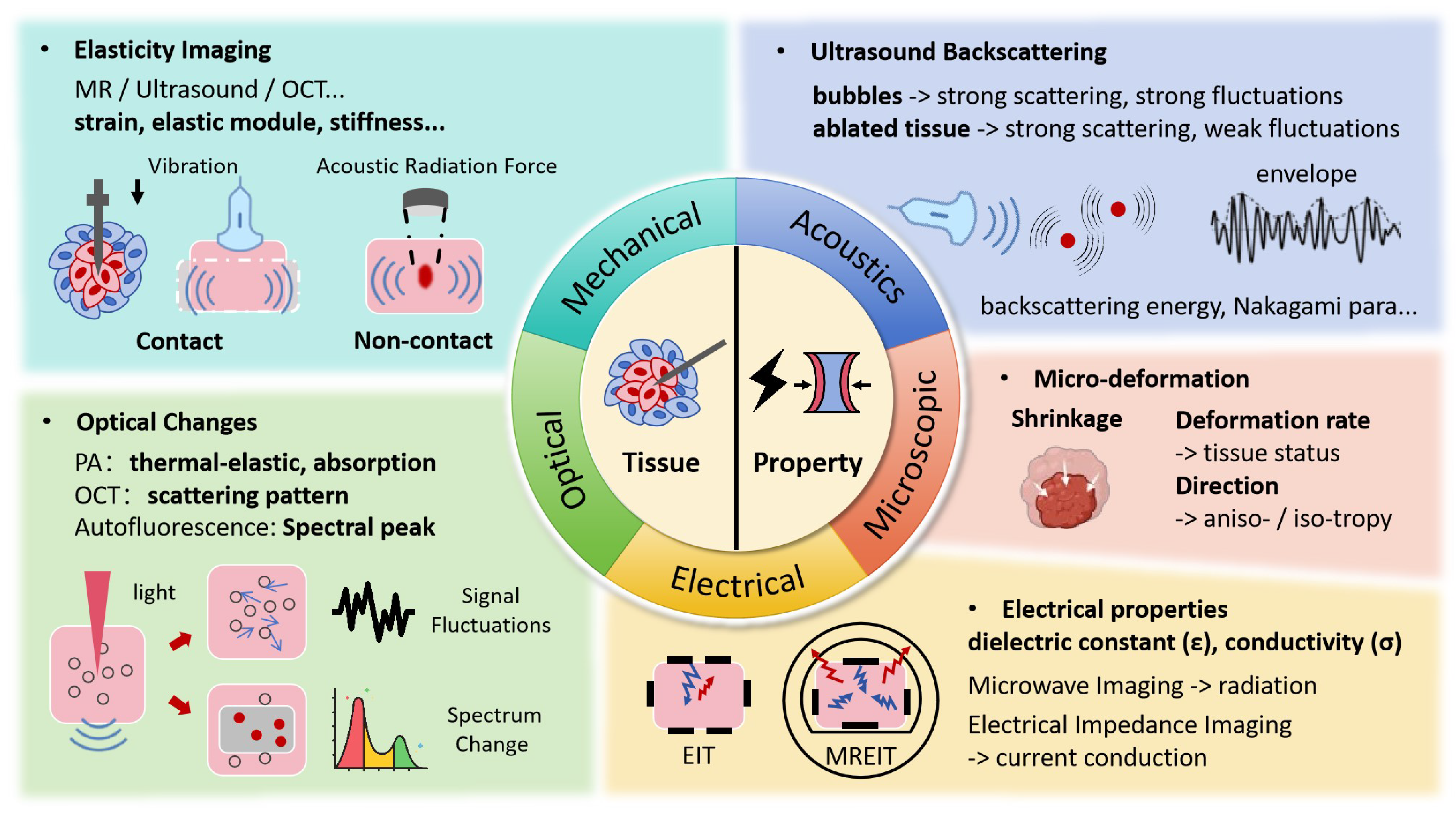
3. Tissue Property Changes Monitoring
3.1. Mechanical Property Changes
3.2. Ultrasonic Backscattering Changes
3.3. Optical Changes
3.4. Electrical Property Changes
4. Discussion
5. Conclusions
Author Contributions
Funding
Acknowledgments
Conflicts of Interest
References
- Kwon, S.; Jung, S.; Baek, S.H. Combination therapy of radiation and hyperthermia, focusing on the synergistic anti-cancer effects and research trends. Antioxidants 2023, 12, 924. [Google Scholar] [CrossRef] [PubMed]
- Geoghegan, R.; Ter Haar, G.; Nightingale, K.; Marks, L.; Natarajan, S. Methods of monitoring thermal ablation of soft tissue tumors—A comprehensive review. Med. Phys. 2022, 49, 769–791. [Google Scholar] [CrossRef] [PubMed]
- Scheidegger, S.; Mingo Barba, S.; Gaipl, U.S. Theoretical evaluation of the impact of hyperthermia in combination with radiation therapy in an artificial immune—Tumor-ecosystem. Cancers 2021, 13, 5764. [Google Scholar] [CrossRef] [PubMed]
- Dai, Q.; Cao, B.; Zhao, S.; Zhang, A. Synergetic thermal therapy for cancer: State-of-the-art and the future. Bioengineering 2022, 9, 474. [Google Scholar] [CrossRef]
- Chu, K.F.; Dupuy, D.E. Thermal ablation of tumours: Biological mechanisms and advances in therapy. Nat. Rev. Cancer 2014, 14, 199–208. [Google Scholar] [CrossRef]
- Tehrani, M.H.; Soltani, M.; Kashkooli, F.M.; Raahemifar, K. Use of microwave ablation for thermal treatment of solid tumors with different shapes and sizes—A computational approach. PLoS ONE 2020, 15, e0233219. [Google Scholar] [CrossRef]
- Desclides, M.; Ozenne, V.; Bour, P.; Faller, T.; Machinet, G.; Pierre, C.; Chemouny, S.; Quesson, B. Real-time automatic temperature regulation during in vivo MRI-guided laser-induced thermotherapy (MR-LITT). Sci. Rep. 2023, 13, 3279. [Google Scholar] [CrossRef]
- Frich, L. Non-invasive thermometry for monitoring hepatic radiofrequency ablation. Minim. Invasive Ther. Allied Technol. 2006, 15, 18–25. [Google Scholar] [CrossRef]
- Ebbini, E.S.; Simon, C.; Liu, D. Real-time ultrasound thermography and thermometry [life sciences]. IEEE Signal Process. Mag. 2018, 35, 166–174. [Google Scholar] [CrossRef]
- Lewis, M.; Staruch, R.; Chopra, R. Thermometry and ablation monitoring with ultrasound. Int. J. Hyperth. 2015, 31, 163–181. [Google Scholar] [CrossRef]
- Blackwell, J.; Kraśny, M.J.; O’Brien, A.; Ashkan, K.; Galligan, J.; Destrade, M.; Colgan, N. Proton resonance frequency shift thermometry: A review of modern clinical practices. J. Magn. Reson. Imaging 2022, 55, 389–403. [Google Scholar] [CrossRef] [PubMed]
- Peters, R.T.; Hinks, R.S.; Henkelman, R.M. Ex vivo tissue-type independence in proton-resonance frequency shift MR thermometry. Magn. Reson. Med. 1998, 40, 454–459. [Google Scholar] [CrossRef] [PubMed]
- Peller, M.; Muacevic, A.; Reinl, H.; Sroka, R.; Abdel-Rahman, S.; Issels, R.; Reiser, M. MRI assisted thermometry for regional hyperthermia and interstitial laser thermotherapy. Der Radiol. 2004, 44, 310–319. [Google Scholar] [CrossRef]
- Feddersen, T.V.; Hernandez-Tamames, J.A.; Franckena, M.; van Rhoon, G.C.; Paulides, M.M. Clinical performance and future potential of magnetic resonance thermometry in hyperthermia. Cancers 2020, 13, 31. [Google Scholar] [CrossRef] [PubMed]
- Ozenne, V.; Bour, P.; Denis de Senneville, B.; Quesson, B. 3D motion strategy for online volumetric thermometry using simultaneous multi-slice EPI at 1.5 T: An evaluation study. Int. J. Hyperth. 2023, 40, 2194595. [Google Scholar] [CrossRef]
- Feddersen, T.V.; Poot, D.H.; Paulides, M.M.; Salim, G.; van Rhoon, G.C.; Hernandez-Tamames, J.A. Multi-echo gradient echo pulse sequences: Which is best for PRFS MR thermometry guided hyperthermia? Int. J. Hyperth. 2023, 40, 2184399. [Google Scholar] [CrossRef]
- Faridi, P.; Shrestha, T.B.; Pyle, M.; Basel, M.T.; Bossmann, S.H.; Prakash, P.; Natarajan, B. Temperature estimation for MR-guided microwave hyperthermia using block-based compressed sensing. In Proceedings of the 2020 42nd Annual International Conference of the IEEE Engineering in Medicine & Biology Society (EMBC), Montreal, QC, Canada, 20–24 July 2020; pp. 5057–5060. [Google Scholar]
- Wong, S.M.; Luo, P.; Keunen, B.; Pichardo, S.; Drake, J.M.; Waspe, A.C. An adaptive targeting algorithm for magnetic resonance-guided high-intensity focused ultrasound controlled hyperthermia. Med. Phys. 2023, 50, 3347–3358. [Google Scholar] [CrossRef]
- Lu, A.; Woodrum, D.; Felmlee, J.; Favazza, C.; Gorny, K. Improved MR-thermometry during hepatic microwave ablation by correcting for intermittent electromagnetic interference artifacts. Phys. Medica 2020, 71, 100–107. [Google Scholar] [CrossRef]
- Straube, W.; Arthur, R.M. Theoretical estimation of the temperature dependence of backscattered ultrasonic power for noninvasive thermometry. Ultrasound Med. Biol. 1994, 20, 915–922. [Google Scholar] [CrossRef]
- Niu, J.; Zhang, H.; Wang, H.; Ta, D.; Liu, Z. Noninvasive temperature estimation in biotissue as a discrete random medium based on backscattered average ultrasonic power. Acta Acust.-Peking 2001, 26, 247–251. [Google Scholar]
- Shaswary, E.; Assi, H.; Yang, C.; Kumaradas, J.; Kolios, M.C.; Peyman, G.; Tavakkoli, J. Noninvasive calibrated tissue temperature estimation using backscattered energy of acoustic harmonics. Ultrasonics 2021, 114, 106406. [Google Scholar] [CrossRef] [PubMed]
- Maraghechi, B.; Kolios, M.C.; Tavakkoli, J. Noninvasive tissue temperature estimation using nonlinear ultrasound harmonics. In Proceedings of the AIP Conference Proceedings, Las Vegas, NV, USA, 2–5 April 2014; AIP Publishing: Melville, NY, USA, 2017; Volume 1821, p. 150009. [Google Scholar]
- Shaswary, E.; Assi, H.; Yang, C.; Kumaradas, J.C.; Kolios, M.C.; Peyman, G.; Tavakkoli, J. Real-time non-invasive control of tissue temperature using high-frequency ultrasonic backscattered energy. In Proceedings of the 2021 IEEE International Ultrasonics Symposium (IUS), Xi’an, China, 11–16 September 2021; pp. 1–4. [Google Scholar]
- Oliveira, L.F.; França, F.M.; Pereira, W.C. A Data-Driven Approach for Estimating Temperature Variations Based on B-mode Ultrasound Images and Changes in Backscattered Energy. Ultrason. Imaging 2024, 46, 3–16. [Google Scholar] [CrossRef] [PubMed]
- Liu, Y.D.; Li, Q.; Zhou, Z.; Yeah, Y.W.; Chang, C.C.; Lee, C.Y.; Tsui, P.H. Adaptive ultrasound temperature imaging for monitoring radiofrequency ablation. PLoS ONE 2017, 12, e0182457. [Google Scholar] [CrossRef] [PubMed]
- Seo, C.; Stephens, D.; Cannata, J.; Dentinger, A.; Lin, F.; Park, S.; Wildes, D.; Thomenius, K.; Chen, P.; Nguyen, T.; et al. Regulating energy delivery during intracardiac radiofrequency ablation using thermal strain imaging. In Proceedings of the 2011 IEEE International Ultrasonics Symposium, Orlando, FL, USA, 18–21 October 2011; pp. 1882–1885. [Google Scholar]
- Souchon, R.; Bouchoux, G.; Maciejko, E.; Lafon, C.; Cathignol, D.; Bertrand, M.; Chapelon, J. Monitoring the formation of thermal lesions with heat-induced echo-strain imaging: A feasibility study. Ultrasound Med. Biol. 2005, 31, 251–259. [Google Scholar] [CrossRef]
- Park, S.; Hwang, J.; Park, J.E.; Ahn, Y.C.; Kang, H.W. Application of ultrasound thermal imaging for monitoring laser ablation in ex vivo cardiac tissue. Lasers Surg. Med. 2020, 52, 218–227. [Google Scholar] [CrossRef]
- Anand, A.; Ramesh, A.; Yeo, S.; Mohammadi, N.; Cetin, M.; Dalecki, D. Deep-learning based insitu ultrasound thermometry for thermal ablation monitoring. J. Acoust. Soc. Am. 2022, 152, A114. [Google Scholar] [CrossRef]
- Teixeira, C.A.; Ruano, M.G.; Ruano, A.E.; Pereira, W.C. A soft-computing methodology for noninvasive time-spatial temperature estimation. IEEE Trans. Biomed. Eng. 2008, 55, 572–580. [Google Scholar] [CrossRef]
- Schule, G.; Huttmann, G.; Framme, C.; Roider, J.; Brinkmann, R. Noninvasive optoacoustic temperature determination at the fundus of the eye during laser irradiation. J. Biomed. Opt. 2004, 9, 173–179. [Google Scholar] [CrossRef]
- Ke, H.; Tai, S.; Wang, L.V. Photoacoustic thermography of tissue. J. Biomed. Opt. 2014, 19, 026003. [Google Scholar] [CrossRef]
- Meng, L.; Deschaume, O.; Larbanoix, L.; Fron, E.; Bartic, C.; Laurent, S.; Van der Auweraer, M.; Glorieux, C. Photoacoustic temperature imaging based on multi-wavelength excitation. Photoacoustics 2019, 13, 33–45. [Google Scholar] [CrossRef]
- Upputuri, P.K.; Das, D.; Maheshwari, M.; Yaowen, Y.; Pramanik, M. Real-time monitoring of temperature using a pulsed laser-diode-based photoacoustic system. Opt. Lett. 2020, 45, 718–721. [Google Scholar] [CrossRef] [PubMed]
- Larina, I.V.; Larin, K.V.; Esenaliev, R.O. Real-time optoacoustic monitoring of temperature in tissues. J. Phys. D Appl. Phys. 2005, 38, 2633. [Google Scholar] [CrossRef]
- Lee, J.; Kubelick, K.P.; Choe, A.; Emelianov, S.Y. Photoacoustic-guided ultrasound thermal imaging without prior knowledge of tissue composition. Photoacoustics 2023, 33, 100554. [Google Scholar] [CrossRef]
- Zhou, Y.; Li, M.; Liu, W.; Sankin, G.; Luo, J.; Zhong, P.; Yao, J. Thermal Memory Based Photoacoustic Imaging of Temperature. Optica 2019, 6, 198–205. [Google Scholar] [CrossRef]
- Brinkmann, R.; Koinzer, S.; Schlott, K.; Ptaszynski, L.; Bever, M.; Baade, A.; Luft, S.; Miura, Y.; Roider, J.; Birngruber, R. Real-time temperature determination during retinal photocoagulation on patients. J. Biomed. Opt. 2012, 17, 061219. [Google Scholar] [CrossRef]
- Ma, Y.; Liu, Y.; Qin, Z.; Shen, Y.; Sun, M. Mild-temperature photothermal treatment method and system based on photoacoustic temperature measurement and control. Biomed. Signal Process. Control 2023, 79, 104056. [Google Scholar] [CrossRef]
- Ma, Y.; Lei, Z.; Gao, Y.; Sun, M. Closed-loop photothermal therapy system based on photoacoustic and ultrasonic dual-mode temperature feedback. In Proceedings of the 2021 IEEE International Ultrasonics Symposium (IUS), Xi’an, China, 11–16 September 2021; pp. 1–4. [Google Scholar]
- Zhang, Y.; Chu, X.; Cao, W.; Wu, S.; Lin, H.; Li, Z. Tissue photothermal effect based on photoacoustic temperature feedback control. In Proceedings of the 5th Optics Young Scientist Summit (OYSS 2022), SPIE, Fuzhou, China, 16–19 September 2022; Volume 12448, pp. 225–232. [Google Scholar]
- Fani, F.; Schena, E.; Saccomandi, P.; Silvestri, S. CT-based thermometry: An overview. Int. J. Hyperth. 2014, 30, 219–227. [Google Scholar] [CrossRef]
- DeStefano, Z.; Abi-Jaoudeh, N.; Li, M.; Wood, B.J.; Summers, R.M.; Yao, J. CT thermometry for cone-beam CT guided ablation. In Proceedings of the Medical Imaging 2016: Image-Guided Procedures, Robotic Interventions, and Modeling, SPIE, San Diego, CA, USA, 28 February–1 March 2016; Volume 9786, pp. 355–364. [Google Scholar]
- Shan, H.; Padole, A.; Homayounieh, F.; Kruger, U.; Khera, R.; Nitiwarangkul, C.; Kalra, M.; Wang, G. Competitive performance of a modularized deep neural network compared to commercial algorithms for low-dose CT image reconstruction. Nat. Mach. Intell. 2018, 1, 269–276. [Google Scholar] [CrossRef]
- Wang, N.; Li, M.; Haverinen, P. Photon-counting computed tomography thermometry via material decomposition and machine learning. Vis. Comput. Ind. Biomed. Art 2023, 6, 2. [Google Scholar] [CrossRef]
- Guo, S.; Wei, S.; Lee, S.; Sheu, M.; Kang, S.; Kang, J.U. Intraoperative Speckle Variance Optical Coherence Tomography for Tissue Temperature Monitoring During Cutaneous Laser Therapy. IEEE J. Transl. Eng. Health Med. 2019, 7, 1–8. [Google Scholar] [CrossRef]
- Lee, C.; Cheon, G.; Kim, D.H.; Kang, J.U. Feasibility study: Protein denaturation and coagulation monitoring with speckle variance optical coherence tomography. J. Biomed. Opt. 2016, 21, 125004. [Google Scholar] [CrossRef] [PubMed]
- Breen, M.S.; Lazebnik, R.S.; Fitzmaurice, M.; Nour, S.G.; Lewin, J.S.; Wilson, D.L. Radiofrequency thermal ablation: Correlation of hyperacute MR lesion images with tissue response. J. Magn. Reson. Imaging 2004, 20, 475–486. [Google Scholar] [CrossRef] [PubMed]
- Bianchi, L.; Cavarzan, F.; Ciampitti, L.; Cremonesi, M.; Grilli, F.; Saccomandi, P. Thermophysical and mechanical properties of biological tissues as a function of temperature: A systematic literature review. Int. J. Hyperth. 2022, 39, 297–340. [Google Scholar] [CrossRef] [PubMed]
- Lepock, J. Cellular effects of hyperthermia: Relevance to the minimum dose for thermal damage. Int. J. Hyperth. 2003, 19, 252–266. [Google Scholar] [CrossRef]
- Eyerly, S.; Vejdani-Jahromi, M.; Dumont, D.; Trahey, G.; Wolf, P. The Evolution of Tissue Stiffness at Radiofrequency Ablation Sites During Lesion Formation and in the Peri-Ablation Period. J. Cardiovasc. Electrophysiol. 2015, 26, 1009–1018. [Google Scholar] [CrossRef]
- Chen, S.S.; Humphrey, J. Heat-induced changes in the mechanics of a collagenous tissue: Pseudoelastic behavior at 37 degrees C. J. Biomech. 1998, 31, 211–216. [Google Scholar] [CrossRef]
- Hofstetter, L.W.; Odéen, H.; Bolster, B.D.; Christensen, D.A.; Payne, A.; Parker, D.L. Magnetic resonance shear wave elastography using transient acoustic radiation force excitations and sinusoidal displacement encoding. Phys. Med. Biol. 2021, 66, 055027. [Google Scholar] [CrossRef]
- Varghese, T.; Techavipoo, U.; Liu, W.; Zagzebski, J.; Chen, Q.; Frank, G.; Lee, F. Elastographic measurement of the area and volume of thermal lesions resulting from radiofrequency ablation: Pathologic correlation. AJR Am. J. Roentgenol. 2003, 181 3, 701–707. [Google Scholar] [CrossRef]
- Pareek, G.; Wilkinson, E.R.; Bharat, S.; Varghese, T.; Laeseke, P.F.; Lee, F.T., Jr.; Warner, T.F.; Zagzebski, J.A.; Nakada, S.Y. Second Prize: Elastographic Measurements of in-Vivo Radiofrequency Ablation Lesions of the Kidney. J. Endourol. 2006, 20, 959–964. [Google Scholar] [CrossRef]
- van Vledder, M.G.; Boctor, E.; Assumpcao, L.; Rivaz, H.; Foroughi, P.; Hager, G.; Hamper, U.; Pawlik, T.; Choti, M. Intra-operative ultrasound elasticity imaging for monitoring of hepatic tumour thermal ablation. HPB 2010, 12, 717–723. [Google Scholar] [CrossRef][Green Version]
- Kang, H.J.; Deshmukh, N.P.; Stolka, P.; Burdette, E.C.; Boctor, E.M. Ultrasound imaging software framework for real-time monitoring of acoustic ablation therapy. In Proceedings of the Medical Imaging 2012: Ultrasonic Imaging, Tomography, and Therapy, SPIE, San Diego, CA, USA, 5–6 February 2012; Volume 8320, pp. 420–425. [Google Scholar]
- Rivaz, H.; Boctor, E.; Foroughi, P.; Zellars, R.; Fichtinger, G.; Hager, G. Ultrasound elastography: A dynamic programming approach. IEEE Trans. Med. Imaging 2008, 27, 1373–1377. [Google Scholar] [CrossRef] [PubMed]
- Mariani, A.; Kwiecinski, W.; Pernot, M.; Balvay, D.; Tanter, M.; Clément, O.; Cuenod, C.; Zinzindohoué, F. Real time shear waves elastography monitoring of thermal ablation: In vivo evaluation in pig livers. J. Surg. Res. 2013, 188, 37–43. [Google Scholar] [CrossRef] [PubMed]
- Boctor, E.; Oliveira, M.D.; Choti, M.; Ghanem, R.; Taylor, R.; Hager, G.; Fichtinger, G. Ultrasound Monitoring of Tissue Ablation Via Deformation Model and Shape Priors. In Medical Image Computing and Computer-Assisted Intervention—Proceedings of the MICCAI 2006: 9th International Conference, Copenhagen, Denmark, 1–6 October 2006; Springer: Berlin/Heidelberg, Germany, 2006; Volume 9, Part 2; pp. 405–412. [Google Scholar]
- Benech, N.; Negreira, C.; Catheline, S. Monitoring local temperature changes in soft tissues by time-reversal elastography. In Proceedings of the 2009 IEEE International Ultrasonics Symposium, Rome, Italy, 20–23 September 2009; pp. 2414–2417. [Google Scholar]
- Li, B.N.; Shen, Z.H.; Chui, C.K.; Numano, T.; Washio, T.; Kobayashi, E. Evaluation of radiofrequency ablation using magnetic resonance elastography. In Proceedings of the World Congress on Medical Physics and Biomedical Engineering, Beijing, China, 26–31 May 2012; Springer: Berlin/Heidelberg, Germany, 2013; pp. 2134–2137. [Google Scholar]
- Chen, J.; Woodrum, D.A.; Glaser, K.J.; Murphy, M.C.; Gorny, K.; Ehman, R. Assessment of in vivo laser ablation using MR elastography with an inertial driver. Magn. Reson. Med. 2014, 72, 59–67. [Google Scholar] [CrossRef] [PubMed][Green Version]
- Choquet, K.; Vappou, J.; Cabras, P.; Ishak, O.; Gangi, A.; Breton, E. Magnetic Resonance Acoustic Radiation Force Imaging (MR-ARFI) for the monitoring of High Intensity Focused Ultrasound (HIFU) ablation in anisotropic tissue. Magn. Reson. Mater. Phys. Biol. Med. 2023, 36, 737–747. [Google Scholar] [CrossRef]
- Corbin, N.; Vappou, J.; Breton, E.; Boehler, Q.; Barbé, L.; Renaud, P.; de Mathelin, M. Interventional MR elastography for MRI-guided percutaneous procedures. Magn. Reson. Med. 2016, 75, 1110–1118. [Google Scholar] [CrossRef]
- Ishak, O.; Breton, E.; Choquet, K.; Josset, A.; Cabras, P.; Vappou, J. Monitoring MR-guided high intensity focused ultrasound therapy using transient supersonic shear wave MR-elastography. Phys. Med. Biol. 2023, 68, 035013. [Google Scholar] [CrossRef]
- Vappou, J.; Bour, P.; Marquet, F.; Ozenne, V.; Quesson, B. MR-ARFI-based method for the quantitative measurement of tissue elasticity: Application for monitoring HIFU therapy. Phys. Med. Biol. 2018, 63, 095018. [Google Scholar] [CrossRef]
- Wu, T.; Felmlee, J.P.; Greenleaf, J.F.; Riederer, S.J.; Ehman, R.L. Assessment of thermal tissue ablation with MR elastography. Magn. Reson. Med. 2001, 45, 80–87. [Google Scholar] [CrossRef]
- Shan, X.; Yang, J.; Xu, P.; Hu, L.; Ge, H. Deep neural networks for magnetic resonance elastography acceleration in thermal-ablation monitoring. Med. Phys. 2022, 49, 1803–1813. [Google Scholar] [CrossRef]
- Yang, F.; Chen, Z.; Xing, D. Single-cell photoacoustic microrheology. IEEE Trans. Med. Imaging 2019, 39, 1791–1800. [Google Scholar] [CrossRef]
- Hosseindokht, Z.; Davoudi, S.; Rahdar, M.; Janahmadi, M.; Kolahdouz, M.; Sasanpour, P. Photoacoustic viscoelasticity assessment of prefrontal cortex and cerebellum in normal and prenatal valproic acid-exposed rats. Photoacoustics 2024, 36, 100590. [Google Scholar] [CrossRef] [PubMed]
- Zaitsev, V.; Matveyev, A.; Matveev, L.; Sovetsky, A.; Baum, O.; Yuzhakov, A.; Omelchenko, A.; Sobol, E. OCT-based strain mapping and compression optical coherence elastography to study and control laser-assisted modification of avascular collagenous tissues. In Proceedings of the BiOS, San Francisco, CA, USA, 1–6 February 2020; Volume 11242, pp. 1124202-1–1124202-11. [Google Scholar]
- Huang, P.C.; Chaney, E.; Iyer, R.R.; Spillman, D.; Odintsov, B.; Sobh, N.; Boppart, S. Interstitial magnetic thermotherapy dosimetry based on shear wave magnetomotive optical coherence elastography. Biomed. Opt. Express 2019, 10, 539–551. [Google Scholar] [CrossRef] [PubMed]
- Su, L.; Tian, W.; Xu, M.; Lin, M.; Zhuang, B.; Huang, T.; Ye, J.; Lv, M.; Xie, X. Performance of Shear Wave Elastography in Delineating the Radiofrequency Ablation Boundary: An in Vivo experiment. Ultrasound Med. Biol. 2019, 45, 1324–1330. [Google Scholar] [CrossRef] [PubMed]
- Sapin-de Brosses, E.; Gennisson, J.L.; Pernot, M.; Fink, M.; Tanter, M. Temperature dependence of the shear modulus of soft tissues assessed by ultrasound. Phys. Med. Biol. 2010, 55, 1701–1718. [Google Scholar] [CrossRef]
- Brosses, E.S.; Pernot, M.; Tanter, M. The link between tissue elasticity and thermal dose in vivo. Phys. Med. Biol. 2011, 56, 7755–7765. [Google Scholar] [CrossRef]
- Kling, A.; Jiang, J. Potential of Determining Thermal Dose for Ablation Therapies Using Ultrasound Elastography: An Ex Vivo Feasibility Study. In Proceedings of the 2018 IEEE International Ultrasonics Symposium (IUS), Kobe, Japan, 22–25 October 2018; pp. 1–4. [Google Scholar] [CrossRef]
- Li, S.; Zhou, Z.; Wu, S.; Wu, W. A Review of Quantitative Ultrasound-Based Approaches to Thermometry and Ablation Zone Identification Over the Past Decade. Ultrason. Imaging 2022, 44, 213–228. [Google Scholar] [CrossRef]
- Yang, K.; Li, Q.; Liu, H.L.; Chen, C.K.; Huang, C.W.; Chen, J.R.; Tsai, Y.W.; Zhou, Z.; Tsui, P.H. Frequency-domain CBE imaging for ultrasound localization of the HIFU focal spot: A feasibility study. Sci. Rep. 2020, 10, 5468. [Google Scholar] [CrossRef]
- Zhang, S.; Shang, S.; Han, Y.; Gu, C.; Wu, S.; Liu, S.; Niu, G.; Bouakaz, A.; Wan, M. Ex Vivo and In Vivo Monitoring and Characterization of Thermal Lesions by High-Intensity Focused Ultrasound and Microwave Ablation Using Ultrasonic Nakagami Imaging. IEEE Trans. Med. Imaging 2018, 37, 1701–1710. [Google Scholar] [CrossRef]
- Huang, S.M.; Liu, H.L.; Li, D.W.; Li, M.L. Ultrasonic Nakagami imaging of high-intensity focused ultrasound-induced thermal lesions in porcine livers: Ex vivo study. Ultrason. Imaging 2018, 40, 310–324. [Google Scholar] [CrossRef]
- Zhang, S.; Han, Y.; Zhu, X.; Shang, S.; Huang, G.; Zhang, L.; Niu, G.; Wang, S.; He, X.; Wan, M. Feasibility of Using Ultrasonic Nakagami Imaging for Monitoring Microwave-Induced Thermal Lesion in Ex Vivo Porcine Liver. Ultrasound Med. Biol. 2017, 43, 482–493. [Google Scholar] [CrossRef]
- Wang, C.Y.; Zhou, Z.; Chang, Y.H.; Ho, M.C.; Lu, C.M.; Wu, C.H.; Tsui, P.H. Ultrasound single-phase CBE imaging for monitoring radiofrequency ablation of the liver tumor: A preliminary clinical validation. Front. Oncol. 2022, 12, 894246. [Google Scholar] [CrossRef] [PubMed]
- Zhou, Z.; Wu, S.; Wang, C.Y.; Ma, H.Y.; Lin, C.C.; Tsui, P.H. Monitoring radiofrequency ablation using real-time ultrasound Nakagami imaging combined with frequency and temporal compounding techniques. PLoS ONE 2015, 10, e0118030. [Google Scholar] [CrossRef] [PubMed]
- Pang, G.A.; Bay, E.; Deán-Ben, X.L.; Razansky, D. Optoacoustic monitoring of real-time lesion formation during radiofrequency catheter ablation. In Proceedings of the Photons Plus Ultrasound: Imaging and Sensing 2015, SPIE, San Francisco, CA, USA, 8–10 February 2015; Volume 9323, pp. 48–54. [Google Scholar]
- Dana, N.; Di Biase, L.; Natale, A.; Emelianov, S.; Bouchard, R. In vitro photoacoustic visualization of myocardial ablation lesions. Heart Rhythm 2014, 11, 150–157. [Google Scholar] [CrossRef] [PubMed]
- Iskander-Rizk, S.; Kruizinga, P.; Beurskens, R.; Springeling, G.; Mastik, F.; de Groot, N.M.; Knops, P.; van der Steen, A.F.; van Soest, G. Real-time photoacoustic assessment of radiofrequency ablation lesion formation in the left atrium. Photoacoustics 2019, 16, 100150. [Google Scholar] [CrossRef]
- Rebling, J.; Landa, F.J.O.; Deán-Ben, X.L.; Razansky, D. A new catheter design for combined radiofrequency ablation and optoacoustic treatment monitoring using copper-coated light-guides. In Proceedings of the Optical Fibers and Sensors for Medical Diagnostics and Treatment Applications XVIII, SPIE, San Francisco, CA, USA, 27–28 January 2018; Volume 10488, pp. 9–14. [Google Scholar]
- Wu, X.; Sanders, J.L.; Dundar, M.M.; Oralkan, Ö. Deep-Learning-Based High-Intensity Focused Ultrasound Lesion Segmentation in Multi-Wavelength Photoacoustic Imaging. Bioengineering 2023, 10, 1060. [Google Scholar] [CrossRef]
- Seevaratnam, S.; Farid, M.; Farhat, G.; Standish, B.A. Analyzing effects of temperature on tissue equivalent phantoms using fiber Bragg gratings and optical coherence tomography. In Proceedings of the Photonics North 2013, SPIE, Ottawa, ON, Canada, 3–5 June 2013; Volume 8915, pp. 15–24. [Google Scholar]
- Seevaratnam, S.; Bains, A.; Farid, M.; Farhat, G.; Kolios, M.; Standish, B.A. Quantifying temperature changes in tissue-mimicking fluid phantoms using optical coherence tomography and envelope statistics. In Proceedings of the Optical Fibers and Sensors for Medical Diagnostics and Treatment Applications XIV, SPIE, San Francisco, CA, USA, 1–2 February 2014; Volume 8938, pp. 134–143. [Google Scholar]
- Lee, S.; Lee, C.; Cheon, G.; Kim, J.; Jo, D.; Lee, J.; Kang, J.U. Ophthalmic laser system integrated with speckle variance optical coherence tomography for real-time temperature monitoring. In Proceedings of the Optical Coherence Tomography and Coherence Domain Optical Methods in Biomedicine XXII, SPIE, San Francisco, CA, USA, 29–31 January 2018; Volume 10483, pp. 39–44. [Google Scholar]
- Lo, W.; Uribe-Patarroyo, N.; Hoebel, K.; Beaudette, K.; Villiger, M.; Nishioka, N.; Vakoc, B.; Bouma, B. Balloon catheter-based radiofrequency ablation monitoring in porcine esophagus using optical coherence tomography. Biomed. Opt. Express 2019, 10, 2067–2089. [Google Scholar] [CrossRef]
- Lo, W.C.; Uribe-Patarroyo, N.; Nam, A.S.; Villiger, M.; Vakoc, B.J.; Bouma, B.E. Laser thermal therapy monitoring using complex differential variance in optical coherence tomography. J. Biophotonics 2017, 10, 84–91. [Google Scholar] [CrossRef]
- Wang, T.; Pfeiffer, T.; Wu, M.; Wieser, W.; Amenta, G.; Draxinger, W.; Van Der Steen, A.F.; Huber, R.; Soest, G.v. Thermo-elastic optical coherence tomography. Opt. Lett. 2017, 42, 3466–3469. [Google Scholar] [CrossRef]
- Müller, H.; Ptaszynski, L.; Schlott, K.; Debbeler, C.; Bever, M.; Koinzer, S.; Birngruber, R.; Brinkmann, R.; Hüttmann, G. Imaging thermal expansion and retinal tissue changes during photocoagulation by high speed OCT. Biomed. Opt. Express 2012, 3, 1025–1046. [Google Scholar] [CrossRef]
- Zhao, X.; Ziv, O.; Mohammadpour, R.; Crosby, B.; Hoyt, W.J.; Jenkins, M.W.; Snyder, C.; Hendon, C.; Laurita, K.R.; Rollins, A.M. Polarization-sensitive optical coherence tomography monitoring of percutaneous radiofrequency ablation in left atrium of living swine. Sci. Rep. 2021, 11, 24330. [Google Scholar] [CrossRef]
- Gil, D.A.; Swift, L.M.; Asfour, H.; Muselimyan, N.; Mercader, M.A.; Sarvazyan, N.A. Autofluorescence hyperspectral imaging of radiofrequency ablation lesions in porcine cardiac tissue. J. Biophotonics 2017, 10, 1008–1017. [Google Scholar] [CrossRef] [PubMed]
- Swift, L.; Asfour, H.; Muselimyan, N.; Larson, C.; Armstrong, K.; Sarvazyan, N. Hyperspectral imaging for label-free in vivo identification of myocardial scars and sites of radiofrequency ablation lesions. Heart Rhythm 2017, 15, 564–575. [Google Scholar] [CrossRef] [PubMed]
- Muselimyan, N.; Swift, L.M.; Asfour, H.; Chahbazian, T.; Mazhari, R.; Mercader, M.A.; Sarvazyan, N.A. Seeing the invisible: Revealing atrial ablation lesions using hyperspectral imaging approach. PLoS ONE 2016, 11, e0167760. [Google Scholar] [CrossRef]
- Muselimyan, N.; Asfour, H.; Sarvazyan, N. Key factors behind autofluorescence changes caused by ablation of cardiac tissue. Sci. Rep. 2020, 10, 15369. [Google Scholar] [CrossRef]
- Esrick, M.A.; Mcrae, D. The effect of hyperthermia-induced tissue conductivity changes on electrical impedance temperature mapping. Phys. Med. Biol. 1994, 39, 133–144. [Google Scholar] [CrossRef]
- Kwon, O.; Chauhan, M.; Kim, H.J.; Jeong, W.C.; Wi, H.; Oh, T.; Woo, E. Fast conductivity imaging in magnetic resonance electrical impedance tomography (MREIT) for RF ablation monitoring. Int. J. Hyperth. 2014, 30, 447–455. [Google Scholar] [CrossRef]
- Bottiglieri, A.; Dunne, E.; Mcdermott, B.; Cavagnaro, M.; Porter, E.; Farina, L. Monitoring Microwave Thermal Ablation using Electrical Impedance Tomography: An experimental feasibility study. In Proceedings of the European Conference on Antennas and Propagation, Copenhagen, Denmark, 15–20 March 2020; pp. 1–5. [Google Scholar]
- Chauhan, M.; Jeong, W.C.; Kim, H.J.; Kwon, O.; Woo, E. Radiofrequency ablation lesion detection using MR-based electrical conductivity imaging: A feasibility study of ex vivo liver experiments. Int. J. Hyperth. 2013, 29, 643–652. [Google Scholar] [CrossRef]
- McEwan, A.; Wi, H.; Nguyen, D.; Jones, P.; Lam, V.; Hawthorne, W.; Barry, M.; Oh, T. Development of electrical impedance tomography of microwave ablation. In Proceedings of the Nanosensors, Biosensors, and Info-Tech Sensors and Systems 2014, SPIE, San Diego, CA, USA, 10–12 March 2014; Volume 9060, pp. 136–142. [Google Scholar]
- Wi, H.; McEwan, A.L.; Lam, V.; Kim, H.J.; Woo, E.J.; Oh, T.I. Real-time conductivity imaging of temperature and tissue property changes during radiofrequency ablation: An ex vivo model using weighted frequency difference. Bioelectromagnetics 2015, 36, 277–286. [Google Scholar] [CrossRef]
- Wang, Y.C.; Chan, T.; Sahakian, A. Real-time estimation of lesion depth and control of radiofrequency ablation within ex vivo animal tissues using a neural network. Int. J. Hyperth. 2018, 34, 1104–1113. [Google Scholar] [CrossRef]
- Caminiti, I.; Ferraioli, F.; Formisano, A.; Martone, R. Adaptive Ablation Treatment Based on Impedance Imaging. IEEE Trans. Magn. 2010, 46, 3329–3332. [Google Scholar] [CrossRef]
- Grasland-Mongrain, P.; Lafon, C. Review on biomedical techniques for imaging electrical impedance. IRBM 2018, 39, 243–250. [Google Scholar] [CrossRef]
- Kok, H.P.; Cressman, E.N.; Ceelen, W.; Brace, C.L.; Ivkov, R.; Grüll, H.; Ter Haar, G.; Wust, P.; Crezee, J. Heating technology for malignant tumors: A review. Int. J. Hyperth. 2020, 37, 711–741. [Google Scholar] [CrossRef] [PubMed]
- Zaltieri, M.; Massaroni, C.; Cauti, F.M.; Schena, E. Techniques for temperature monitoring of myocardial tissue undergoing radiofrequency ablation treatments: An overview. Sensors 2021, 21, 1453. [Google Scholar] [CrossRef]
- Mohammadi, A.M.; Schroeder, J.L. Laser interstitial thermal therapy in treatment of brain tumors–the NeuroBlate System. Expert Rev. Med. Devices 2014, 11, 109–119. [Google Scholar] [CrossRef]
- Wielandts, J.Y.; Almorad, A.; Hilfiker, G.; Gillis, K.; Haddad, M.E.; Vijgen, J.; Berte, B.; Le Polain de Waroux, J.B.; Tavernier, R.; Duytschaever, M.; et al. Biosense Webster’s QDOT Micro™radiofrequency ablation catheter. Future Cardiol. 2021, 17, 817–825. [Google Scholar] [CrossRef]
- De Tommasi, F.; Massaroni, C.; Carnevale, A.; Presti, D.L.; De Vita, E.; Iadicicco, A.; Faiella, E.; Grasso, R.F.; Longo, U.G.; Campopiano, S.; et al. Fiber Bragg Grating Sensors for Temperature Monitoring During Thermal Ablation Procedure: Experimental Assessment of Artefact Caused by Respiratory Movements. IEEE Sens. J. 2021, 21, 13342–13349. [Google Scholar] [CrossRef]
- Salamon, J.; Dieckhoff, J.; Kaul, M.G.; Jung, C.; Adam, G.; Möddel, M.; Knopp, T.; Draack, S.; Ludwig, F.; Ittrich, H. Visualization of spatial and temporal temperature distributions with magnetic particle imaging for liver tumor ablation therapy. Sci. Rep. 2020, 10, 7480. [Google Scholar] [CrossRef]
- Skandalakis, G.P.; Rivera, D.R.; Rizea, C.D.; Bouras, A.; Jesu Raj, J.G.; Bozec, D.; Hadjipanayis, C.G. Hyperthermia treatment advances for brain tumors. Int. J. Hyperth. 2020, 37, 3–19. [Google Scholar] [CrossRef]
- Wang, H.; Zou, J.; Zhao, S.; Zhang, A. Optimization of three-dimensional esophageal tumor ablation by simultaneous functioning of multiple electrodes. Med. Biol. Eng. Comput. 2024. Online ahead of print. [Google Scholar] [CrossRef]
- Jethwa, P.R.; Barrese, J.C.; Gowda, A.; Shetty, A.; Danish, S.F. Magnetic Resonance Thermometry-Guided Laser-Induced Thermal Therapy for Intracranial Neoplasms: Initial Experience. Oper. Neurosurg. 2012, 71, 133–145. [Google Scholar] [CrossRef]
- Primavesi, F.; Swierczynski, S.; Klieser, E.; Kiesslich, T.; Jäger, T.; Urbas, R.; Hutter, J.; Neureiter, D.; Öfner, D.; Stättner, S. Thermographic real-time-monitoring of surgical radiofrequency and microwave ablation in a perfused porcine liver model. Oncol. Lett. 2018, 15, 2913–2920. [Google Scholar] [CrossRef] [PubMed]
- Rossmann, C.; Haemmerich, D. Review of temperature dependence of thermal properties, dielectric properties, and perfusion of biological tissues at hyperthermic and ablation temperatures. Crit. Rev. Biomed. Eng. 2014, 42, 467–492. [Google Scholar] [CrossRef] [PubMed]
- Schröer, S.; Düx, D.; Löning Caballero, J.J.; Glandorf, J.; Gerlach, T.; Horstmann, D.; Belker, O.; Gutt, M.; Wacker, F.; Speck, O.; et al. Reducing electromagnetic interference in MR thermometry: A comparison of setup configurations for MR-guided microwave ablations. Z. Med. Phys. 2024. Online ahead of print. [Google Scholar] [CrossRef] [PubMed]
- Valori, M.; Rebaioli, L.; Marrocco, V.; Modica, F.; Bonelli, F.; Pascazio, G.; Portosi, V.; Prudenzano, F.; Fasano, A.; Lampignano, V.; et al. Manufacturing challenges and technological solutions for microwave ablation (MWA) probe prototyping. Proc. Inst. Mech. Eng. Part B J. Eng. Manuf. 2023, 237, 481–491. [Google Scholar] [CrossRef]
- Lai, C.Y.; Kruse, D.E.; Caskey, C.F.; Stephens, D.N.; Sutcliffe, P.L.; Ferrara, K.W. Noninvasive thermometry assisted by a dual-function ultrasound transducer for mild hyperthermia. IEEE Trans. Ultrason. Ferroelectr. Freq. Control 2010, 57, 2671–2684. [Google Scholar] [CrossRef]
- Ghoshal, G.; Oelze, M.L. Use of quantitative ultrasound to detect temperature variations in biological phantoms due to heating. In Proceedings of the 2009 IEEE International Ultrasonics Symposium, Rome, Italy, 20–23 September 2009; pp. 1780–1783. [Google Scholar] [CrossRef]
- Odéen, H.; Parker, D.L. Magnetic resonance thermometry and its biological applications—Physical principles and practical considerations. Prog. Nucl. Magn. Reson. Spectrosc. 2019, 110, 34–61. [Google Scholar] [CrossRef]
- Byra, M.; Klimonda, Z.; Kruglenko, E.; Gambin, B. Unsupervised deep learning based approach to temperature monitoring in focused ultrasound treatment. Ultrasonics 2022, 122, 106689. [Google Scholar] [CrossRef]
- Wyatt, C.R.; Soher, B.J.; MacFall, J.R. Correction of breathing-induced errors in magnetic resonance thermometry of hyperthermia using multiecho field fitting techniques. Med. Phys. 2010, 37, 6300–6309. [Google Scholar] [CrossRef]
- Liu, K.C.; Chen, T.M. Comparative study of heat transfer and thermal damage assessment models for hyperthermia treatment. J. Therm. Biol. 2021, 98, 102907. [Google Scholar] [CrossRef]
- Breen, M.; Wilson, D.; Saidel, G.; Butts, K.; Chen, L. MRI-guided lased thermal ablation: Model and parameter estimates relating MR thermometry images to cell death. In Proceedings of the IEEE International Symposium on Biomedical Imaging, Arlington, VA, USA, 18 April 2004; Volume 1, pp. 296–299. [Google Scholar]
- Rempp, H.; Clasen, S.; Boss, A.; Claussen, C.; Pereira, P.; Schick, F. Temperature mapping for MR-guided radiofrequency ablation-how accurate can cell necrosis be monitored? In Proceedings of the World Congress on Medical Physics and Biomedical Engineering, Munich, Germany, 7–12 September 2009; Surgery, Nimimal Invasive Interventions, Endoscopy and Image Guided Therapy. Springer: Berlin/Heidelberg, Germany, 2009; Volume 25/6, pp. 24–27. [Google Scholar]
- Breen, M.; Butts, K.; Chen, L.; Saidel, G.; Wilson, D.L. MRI-guided laser thermal ablation: Model to predict cell death from MR thermometry images for real-time therapy monitoring. In Proceedings of the 26th Annual International Conference of the IEEE Engineering in Medicine and Biology Society, San Francisco, CA, USA, 1–5 September 2004; Volume 1, pp. 1028–1031. [Google Scholar]
- Schröer, S.; Alpers, J.; Gutberlet, M.; Brüsch, I.; Rumpel, R.; Wacker, F.; Hensen, B.; Hansen, C. A probabilistic thermal dose model for the estimation of necrosis in MR-guided tumor ablations. Med. Phys. 2024, 51, 239–250. [Google Scholar] [CrossRef]
- Hadadian, Y.; Uliana, J.H.; Carneiro, A.A.; Pavan, T.Z. A novel theranostic platform: Integration of magnetomotive and thermal ultrasound imaging with magnetic hyperthermia. IEEE Trans. Biomed. Eng. 2020, 68, 68–77. [Google Scholar] [CrossRef] [PubMed]
- Oleson, J.R.; Samulski, T.V.; Leopold, K.A.; Clegg, S.T.; Dewhirst, M.W.; Dodge, R.K.; George, S.L. Sensitivity of hyperthermia trial outcomes to temperature and time: Implications for thermal goals of treatment. Int. J. Radiat. Oncol. Biol. Phys. 1993, 25, 289–297. [Google Scholar] [CrossRef] [PubMed]
- Breen, M.; Breen, M.; Butts, K.; Chen, L.; Saidel, G.; Wilson, D. MRI-guided Thermal Ablation Therapy: Model and Parameter Estimates to Predict Cell Death from MR Thermometry Images. Ann. Biomed. Eng. 2007, 35, 1391–1403. [Google Scholar] [CrossRef] [PubMed]
- Yarmolenko, P.S.; Moon, E.J.; Landon, C.; Manzoor, A.; Hochman, D.W.; Viglianti, B.L.; Dewhirst, M.W. Thresholds for thermal damage to normal tissues: An update. Int. J. Hyperth. 2011, 27, 320–343. [Google Scholar] [CrossRef]
- Farina, L.; Weiss, N.; Nissenbaum, Y.; Cavagnaro, M.; Lopresto, V.; Pinto, R.; Tosoratti, N.; Amabile, C.; Cassarino, S.; Goldberg, S. Characterisation of tissue shrinkage during microwave thermal ablation. Int. J. Hyperth. 2014, 30, 419–428. [Google Scholar] [CrossRef]
- Baum, O.I.; Zaitsev, V.Y.; Yuzhakov, A.V.; Sviridov, A.P.; Novikova, M.L.; Matveyev, A.L.; Matveev, L.A.; Sovetsky, A.A.; Sobol, E.N. Interplay of temperature, thermal-stresses and strains in laser-assisted modification of collagenous tissues: Speckle-contrast and OCT-based studies. J. Biophotonics 2020, 13, e201900199. [Google Scholar] [CrossRef]
- Liu, D.; Brace, C. CT imaging during microwave ablation: Analysis of spatial and temporal tissue contraction. Med. Phys. 2014, 41, 113303. [Google Scholar] [CrossRef]
- Lopresto, V.; Strigari, L.; Farina, L.; Minosse, S.; Pinto, R.; D’Alessio, D.; Cassano, B.; Cavagnaro, M. CT-based investigation of the contraction of ex vivo tissue undergoing microwave thermal ablation. Phys. Med. Biol. 2018, 63, 055019. [Google Scholar] [CrossRef]
- Tan, D.; Lim, K.; Wong, Y.; Raziff, H.H.A.; Tan, S.; Sulaiman, N.; Abdullah, B.; Ahmad, H.; Yeong, C. Multivariate Regression Between Hounsfield Unit Shift, Tissue Temperature, and Tissue Contraction: A Feasibility Study of Computed Tomography Thermometry. IEEE Trans. Instrum. Meas. 2021, 70, 1–9. [Google Scholar] [CrossRef]
- Huang, W.; Lu, J.; Tang, R.; Wu, Z.; Wang, Q.; Ding, X.; Wang, Z.; Chen, K. Phase contrast imaging based microbubble monitoring of radiofrequency ablation: An ex vivo study. Front. Oncol. 2020, 10, 1709. [Google Scholar] [CrossRef]
- Wang, S.S.; VanderBrink, B.A.; Regan, J.; Carr, K.; Link, M.S.; Homoud, M.K.; Foote, C.M.; Mark Estes, N.A., III; Wang, P.J. Microwave Radiometric Thermometry and its Potential Applicability to Ablative Therapy. J. Interv. Card. Electrophysiol. 2000, 4, 295–300. [Google Scholar] [CrossRef] [PubMed]
- Zharov, V.P.; Vesnin, S.G.; Suen, J.Y.; Harms, S.E. Photothermal/microwave radiometry for imaging and temperature feedback. In Proceedings of the Biomedical Optoacoustics III, SPIE, San Jose, CA, USA, 20–21 January 2002; Volume 4618, pp. 163–173. [Google Scholar]
- Koruth, J.S.; Dukkipati, S.; Gangireddy, S.; McCarthy, J.; Spencer, D.; Weinberg, A.D.; Miller, M.A.; D’Avila, A.; Reddy, V.Y. Occurrence of steam pops during irrigated RF ablation: Novel insights from microwave radiometry. J. Cardiovasc. Electrophysiol. 2013, 24, 1271–1277. [Google Scholar] [CrossRef] [PubMed]
- Yang, K.; Li, Q.; Liu, H.; Zeng, Q.; Cai, D.; Xu, J.; Zhou, Y.; Tsui, P.H.; Zhou, X. Suppressing HIFU interference in ultrasound images using 1D U-Net-based neural networks. Phys. Med. Biol. 2024, 69, 075006. [Google Scholar] [CrossRef] [PubMed]
- Li, X.J.; Hossain, M.M.; Lee, S.A.; Saharkhiz, N.; Konofagou, E. Harmonic Motion Imaging-Guided Focused Ultrasound Ablation: Comparison of Three Focused Ultrasound Interference Filtering Methods. Ultrasound Med. Biol. 2024, 50, 119–127. [Google Scholar] [CrossRef]
- Grasso, V.; Willumeit-Roemer, R.; Jose, J. Development of an AI-assisted multi-spectral photoacoustic imaging for volumetric molecular tissue composition: A multi-frequency translational approach. In Proceedings of the Photons Plus Ultrasound: Imaging and Sensing 2023, SPIE, San Francisco, CA, USA, 29 January–1 February 2023; Volume 12379, p. 1237902. [Google Scholar]
- Zhang, Y.; Wang, L. Array-based high-intensity focused ultrasound therapy system integrated with real-time ultrasound and photoacoustic imaging. Biomed. Opt. Express 2023, 14, 1137–1145. [Google Scholar] [CrossRef]
- Joseph, F.K.; Manohar, S. Photoacoustic assisted device guidance and thermal lesion imaging for radiofrequency ablation. In Proceedings of the European Conference on Biomedical Optics, Munich, Germany, 23–27 June 2019; Volume 11077, pp. 1107715-1–1107715-3. [Google Scholar]
- Koinzer, S.; Schlott, K.; Portz, L.; Ptaszynski, L.; Baade, A.; Bever, M.; Saeger, M.; Caliebe, A.; Denner, R.; Birngruber, R.; et al. Correlation of temperature rise and optical coherence tomography characteristics in patient retinal photocoagulation. J. Biophotonics 2012, 5, 889–902. [Google Scholar] [CrossRef]
- Lim, H.G.; Kim, H.; Kim, K.; Park, J.; Kim, Y.; Yoo, J.; Heo, D.; Baik, J.; Park, S.M.; Kim, H.H. Thermal ablation and high-resolution imaging using a back-to-back (BTB) dual-mode ultrasonic transducer: In vivo results. Sensors 2021, 21, 1580. [Google Scholar] [CrossRef]
- Ma, Y.; Lei, Z.; Wu, D.; Shen, Y.; Sun, M. A neural network estimation model based light dose control method and system for low-temperature photothermal therapy. Biomed. Signal Process. Control 2023, 85, 104935. [Google Scholar] [CrossRef]


Disclaimer/Publisher’s Note: The statements, opinions and data contained in all publications are solely those of the individual author(s) and contributor(s) and not of MDPI and/or the editor(s). MDPI and/or the editor(s) disclaim responsibility for any injury to people or property resulting from any ideas, methods, instructions or products referred to in the content. |
© 2025 by the authors. Licensee MDPI, Basel, Switzerland. This article is an open access article distributed under the terms and conditions of the Creative Commons Attribution (CC BY) license (https://creativecommons.org/licenses/by/4.0/).
Share and Cite
Wang, X.; Zhao, S.; Zhang, A. Image-Based Monitoring of Thermal Ablation. Bioengineering 2025, 12, 78. https://doi.org/10.3390/bioengineering12010078
Wang X, Zhao S, Zhang A. Image-Based Monitoring of Thermal Ablation. Bioengineering. 2025; 12(1):78. https://doi.org/10.3390/bioengineering12010078
Chicago/Turabian StyleWang, Xinyi, Shiqing Zhao, and Aili Zhang. 2025. "Image-Based Monitoring of Thermal Ablation" Bioengineering 12, no. 1: 78. https://doi.org/10.3390/bioengineering12010078
APA StyleWang, X., Zhao, S., & Zhang, A. (2025). Image-Based Monitoring of Thermal Ablation. Bioengineering, 12(1), 78. https://doi.org/10.3390/bioengineering12010078


