Article

Preparation of Polyphenol-Rich Herbal Beverages from White Willow (Salix alba) Bark with Potential Alzheimer’s Disease Inhibitory Activity In Silico

Institute of Food and Health, School of Agriculture and Food Science, University College Dublin, D04 V1W8 Dublin, Ireland
*
Author to whom correspondence should be addressed.
Beverages 2024, 10(3), 75; https://doi.org/10.3390/beverages10030075
Submission received: 23 July 2024 / Revised: 17 August 2024 / Accepted: 20 August 2024 / Published: 22 August 2024

Abstract

:
White willow (Salix alba) is a medicinal plant traditionally used to treat pain and inflammation. The aims of this study were to produce polyphenol-rich herbal beverages from willow bark with different ethanol content, temperatures, and solvent pH and to explore neuroprotective potentials of willow polyphenols. The phenolic compounds quantified in the willow infusions were salicin, chlorogenic acid, epicatechin, p-salicylic acid, and p-coumaric acid; the former three compounds exhibited promising inhibitory potentials against acetylcholinesterase (AChE) and butyrylcholinesterase (BuChE) in molecular docking studies. Total phenol content and antioxidant activity were maximum when prepared with 50% ethanol-in-water at room temperature. Although aqueous infusions contained fewer total phenols than those extracted with 50% hydroalcoholic solutions, they enhanced the extraction of chlorogenic acid and salicin content, which may possess promising neuroprotective potentials. The addition of citric acids in hot water infusions led to a higher proportion of non-tannins and had a lighter appearance, which may result in less astringent mouthfeel and better consumer acceptance. Overall, the obtained results indicate that willow bark prepared with hot water and/or with addition of citric acids is rich in bioactive compounds with high antioxidant activity and possible neuroprotective activities in silico, which could serve as valuable ingredients for inclusion in functional beverages.

Graphical Abstract

1. Introduction

As the global population is rapidly aging, the morbidity of dementia is increasing [1,2]. Alzheimer’s disease (AD), a chronic neurodegenerative disorder, is responsible for 60 to 70% of cases of dementia worldwide [3]. One hypothesis of AD development is associated with cholinergic neuron damage leading to the deficiency of the neurotransmitter acetylcholine (ACh), which is correlated with cognitive decline and dementia [2]. Acetylcholinesterase (AChE) and butyrylcholinesterase (BuChE) are serine hydrolases that are responsible for hydrolysing ACh and thus work as coregulators of cholinergic neurotransmission [4]. One of the potent treatments for AD is restoring the level of ACh by inhibiting AChE and BuChE [5]. The standard cholinesterase inhibitors, including donepezil and tacrine, have been shown to exhibit competitive inhibition activities in clinical studies [6]. However, the currently used medications for treating AD present some severe side effects, such as hepatotoxicity and SLUGDE syndrome [7]. Current approved medications for AD including cholinesterase inhibitors are symptomatic treatments that can slow down the disease progression, but their effects are limited and individually dependent. Thus, the search for safer natural products with potential protective effects against chronic diseases such as AD has become a research area of interest [8]. The extracts from white willow bark (Salix alba) have been recently reported to possess inhibitory effects against AChE, suggesting potential in the prevention of AD [9,10,11]. The potency of white willow is predominately attributed to its principal compound salicin. However, willow bark also contains a high content of various polyphenols [12]. Thus, its potential in AD prevention may come from the combined contribution of these bioactive compounds.
To recover these desired compounds, the extraction of willow bark is a crucial step. Conventional extraction techniques, including maceration, decoction, infusion, percolation, and Soxhlet extraction, are commonly used. These methods often involve the use of organic solvents, such as acetone and methanol, and application of heat and/or agitation under atmospheric pressure [13]. However, most organic solvents are not suitable for food and pharmaceutical applications; thus, the addition of a purification step is required to remove chemical residues from extracts destined for human consumption. These drawbacks have triggered research into green extraction protocols to overcome the limitations of conventional methods [14]. In this context, using green solvents that are also food-grade would be ideal for food applications. The practice of soaking herbs in ethanol is the traditional method of preparing herbal tinctures and alcoholic elixirs. Alcohol has been used to extract herbal extracts for many centuries because of its efficiency in yielding tinctures rich in bioactive compounds and its ability to preserve the tinctures [15]. Alcohol also has high miscibility with water, which allows the adjustment of the polarity of mixtures for target compounds. However, extracts with a high level of alcohol may not be an ideal ingredient for downstream food production, as the presence of alcohol will interfere with many food components, such as protein. Apart from ethanol, water is the most used solvent in the preparation of medicinal herbal extracts because it is widely available, potable, and low-cost. Water can dissolve a wide range of substances and is an ideal solvent for the extraction of willow polyphenols [16].
Besides the choice of solvent, the efficiency of extraction can also be affected by several parameters, such as extraction temperature and time. High extraction temperature can elevate extraction efficiency due to higher solubility and diffusion rates of phytochemicals into the solvent. A long extraction process can allow thorough penetration of the solvent into the plant cells and cause a more complete extraction. However, an extreme extraction temperature or prolonged extraction process may result in the degradation of targeted compounds or the generation of undesired compounds [16]. The addition of acids to alter the pH of an aqueous solvent can also affect the extraction of polyphenols, as shown in the extraction of catechins from green tea by Vuong et al. [17].
Previous studies on the conventional extraction of polyphenols from white willow bark are limited. [9,10,11] extracted white willow bark using a conventional solid-liquid method with an aqueous solution at 55 °C for 37 min. In another study, willow bark powder was extracted with boiled ethanol by the Soxhlet extraction method for 7 h [18]. However, neither study investigated the impacts of different parameters (e.g., temperature, solvent) on the extraction efficiency of phenolic compounds in willow bark. Gligorić et al. [19] revealed that epicatechin, salicin, and chlorogenic acid showed good binding affinity for AChE through hydrogen bonds and hydrophobic interactions. Thus, these compounds are of great importance for the investigation of how they might be affected by different extraction factors. Additionally, p-salicylic acid and p-coumaric acid, both known for their health benefits [20,21], were detected in willow bark previously, which may also be worth investigation [22]. Therefore, the primary objective of this work was to produce phenolic-rich infusions from white willow bark from different extraction conditions. The secondary aim was to explore the inhibitory potentials of target phenolic compounds in willow bark infusions against the pathological enzymes (AChE and BuChE) associated with Alzheimer’s disease.

2. Materials and Methods

2.1. Plant Material

Dried willow bark (Justingredients, Chepstow, UK) was ground into fine powder (125–355 µm) using a lab mill (Cyclotec 1093, Foss, Hilleroed, Denmark). The willow bark powder was placed in plastic sample containers away from light and stored at ambient temperature.

2.2. Chemicals and Reagents

Citric acid, trisodium citrate, Folin-Ciocalteu (F-C) reagent, sodium carbonate (Na2CO3), gallic acid, cinchonine, formaldehyde, 2,2-diphenyl-1-picrylhydrazyl (DPPH), ascorbic acid, 2, 4, 6-tripyridyl-s-triazine (TPTZ), ferric chloride hexahydrate, formic acid, and the standard salicin, p-salicylic acid, p-coumaric acid, and chlorogenic acid were purchased from Sigma-Aldrich (Wicklow, Ireland). Standard epicatechin was purchased from Extrasynthese (Genay, France). HPLC grade acetonitrile, water, and methanol were obtained from Honeywell (Lennox, Dublin, Ireland). Hydrochloric acid (HCl, 37%) and ethanol (EtOH) were purchased from Fisher (Dublin, Ireland). Deionized water was from Synergy® UV (resistivity > 18.2 M Ω cm−1) (Merck Millipore, Molsheim, France).

2.3. Preparation of Willow Infusions

Willow bark powder (1 g) was placed in a glass bottle that contained 50 mL of solvent and was pre-heated to the extraction temperature. The bottles were then transferred to a shaking water bath (Isotemp SWB27, Thermo Fisher Scientific, Whaltam, MA, USA) at 200 rpm and at the extraction temperature. The resulting willow infusions were removed at the designed time, filtered under vacuum through Whatman No.1 filter paper (Whatman Ltd., Maidstone, UK), and stored at −18 °C in the freezer until further analysis.
In a preliminary study, the effect of extraction time of 15, 20, and 30 min on the recovery of total polyphenol from willow bark was evaluated at room temperature. The results showed no significant difference in the total polyphenol content among extraction time (p > 0.05), and, given the energy- and cost-saving aspects, the extraction time was fixed at 15 min in the following experiments.
Different extraction systems were used as follows: (1) deionized water with addition of EtOH (at ratios of 20%, 30%, 50%, 70%, and 96%, v/v) at 25 °C; (2) deionized water with heating at different temperatures (25, 40, 60, and 80 °C); (3) deionized water with adjusted pH (by addition of 0.1 M citric acid, 0.1 M trisodium citrate, or 20% sodium carbonate) at 25 °C; and (4) hot water (at 80 or 100 °C) with addition of 0.1 M citric acid (to pH 2).

2.4. Determination of the Total Phenolic Content

The total phenolic content (TPC) of the extracts was determined using the method described by Singleton and Rossi [23]. Briefly, 0.05 mL of standard or extract was mixed with 0.5 mL of F—C reagent and 1.5 mL of 20% Na2CO3 solution. The volume was adjusted to 10 mL with deionized water and mixed well. The mixture was allowed to stand for 2 h. A standard curve was prepared using gallic acid (10–400 mg/L). Absorbance was measured at 760 nm using a UV-Vis Spectrophotometer (UV-1240, Shimadzu, Japan). The TPC of the extract was expressed as mg gallic acid equivalents (GAE) per g dried weight (d.w.) of willow powder.

2.5. Separation and Quantification of Phenolic Constituents Content

The total tannin (TT) and non-tannin (NT) contents in willow extracts were separated through cinchonine precipitation following the method reported by Harbourne et al. [24] with minor modifications. Prior to the separation, extracts made with high concentrations of ethanol were diluted with water to lower ethanol levels (<30%), and strongly acidic samples were mixed with Na2CO3 solution to neutralize the pH. The supernatant obtained after centrifugation was used to determine the NT content. The precipitate was redissolved with absolute EtOH and HCl (10%) to measure the TT content. The NT fractions were further separated to simple phenol (SP) and flavonoids using formaldehyde and HCl (10%). All fractions were quantified as mg GAE/g d.w. using the Folin-Ciocalteu procedure.

2.6. Determination of Antioxidant Activity

The in vitro antioxidant activities of willow bark infusions were determined by 2,2-diphenyl-1-picrylhydrazyl (DPPH) radical scavenging activity and ferric reducing antioxidant power (FRAP) assay.

2.6.1. DPPH Assay

The DPPH assay was conducted according to Thaipong et al. [25] with some modifications. In brief, DPPH powder was dissolved in methanol to make the DPPH stock solution (1 mM). The working solution was then prepared by diluting stock solution by a factor of 5. The working solution (2.85 mL) was mixed with 0.15 mL standard (ascorbic acid in a range of 25 to 150 μg/mL) or diluted extract and left in the dark at ambient temperature for 30 min. Absorbances of the samples were read at 515 nm using a UV-Vis Spectrophotometer (UV-1240, Shimadzu, Kyoto, Japan). The antioxidant activities were expressed in mg ascorbic acid equivalent (AAE/g d.w).

2.6.2. FRAP Assay

The FRAP assay was determined based on the method described by [20,21] with minor modifications. Briefly, the FRAP working solutions consisted of 300 mM acetate buffer (pH 3.6), 10 mM TPTZ in 40 mM HCl, and 20 mM ferric chloride solution at a ratio of 10:1:1. The FRAP reagent was heated to 37 °C before use. Willow extracts or standards (150 μL) were mixed with 2850 μL of FRAP working solutions and left in the dark condition for 30 min. Absorbances of the sample were read at 593 nm using a spectrophotometer. The standard curve was prepared with Trolox (25 to 400 μM). Results were expressed in mmol Trolox equivalent (TE/g d.w).

2.7. HPLC Analysis

Prior to analysis, infusions were filtered through a 0.45 μm membrane filter (Fisher Scientific, Dublin, Ireland). HPLC analysis was performed on an Agilent 1200 Series system (Agilent Technologies, Palo Alto, CA, USA) in combination with an Agilent Poroshell SB C18 column (3.0 × 100 mm; 2.7 μm particle size) with a C18 guard column (Poroshell, Agilent, Cork, Ireland). Mobile phases consisted of (A) water and (B) acetonitrile, both with addition of 0.1% formic acid. The gradient used was as follows: 0–22 min, from 7 to 45% B; 22–23 min, from 45% to 7% B; 23–30 min, 7% B. The separations were performed at 30 °C at a flow rate of 0.3 mL/min, and 5 μL of the sample was injected. UV detection was set at 210 nm. Identification and quantification of polyphenol compounds were based on their retention time by comparison with a standard. The representative standards selected for different phenolic groups are salicylates (salicin), phenolic acids (chlorogenic acid, p-coumaric acid, and p-salicylic acid), and flavonoids (epicatechin).

2.8. Color Measurement

The color of willow extracts was determined using a CR-400 Chroma meter (Konica Minolta, Tokyo, Japan). The color values were expressed as L* (lightness/darkness), a* (redness/greenness), and b* (yellowness/blueness). Hue angle is defined as a color wheel with red-purple at an angle of 0°, yellow at 90°, and blue-green at 270°. Chroma is an indication of color saturation. The hue angle (h°) and chroma (C*) were calculated according to McGuire [26].

2.9. Molecular Docking

A molecular docking study was performed to investigate the binding mode between the selective polyphenols in the willow extracts and key enzymes associated with Alzheimer’s disease, viz. acethylcholinesterase (AChE) and butyrylcholinesterase (BuChE) using Autodock Vina 1.1.2 [27]. The three-dimensional (3D) structure of the ligands, viz. salicin (CID 439503), p-salicylic acid (CID 135), p-coumaric acid (CID 637542), chlorogenic acid (CID 1794427), epicatechin (CID 72276), donepezil (CID 3152), and tacrine (CID 1935), were downloaded from the PubChem database (https://pubchem.ncbi.nlm.nih.gov/, accessed on 15 June 2024). The 3D structures of human AChE (PDB ID: 4EY7) and BuChE (PDB ID: 4BDS) were downloaded from RCSB Protein Databank (https://www.rcsb.org/, accessed on 15 June 2024). The selection of PDB structures was based on co-crystallization of known ligands, and their binding sites for each enzyme were used as a reference grid box while performing docking. Prior to the docking, the ligands were prepared by merging non-polar hydrogen atoms and selecting rotatable bonds. The enzymes were prepared by removing water molecules, heteroatoms, and the ligand. AutoDockTools (1.5.6) was used to generate the docking input files. The search grid of AChE was set as center x: −13.988, y: −43.906, and z: 27.108, with dimension size of x: 30, y: 30, and z: 30, and for BuChE was centered at coordinates x: 133.076, y: 116.113, and z: 41.12. In order to increase the accuracy of docking analysis, the exhaustiveness value was changed to 20 and other parameters were set to default. The conformations of docked ligands with the lowest binding energy provided by Vina docking were chosen as the best docking pose and were visualized by PyMol 2.0 (https://pymol.org/2/, accessed on 15 June 2024).

2.10. Statistical Analysis

The results are expressed as mean ± standard deviation. All extractions for each condition were carried out in triplicate and compared by one-way analysis of variance (ANOVA) and Tukey HSD using SPSS 27.0 (IBM SPSS, Chicago, IL, USA), and results with p < 0.05 were considered significantly different. Correlation analysis was carried out using Pearson correlation coefficient.

3. Results and Discussion

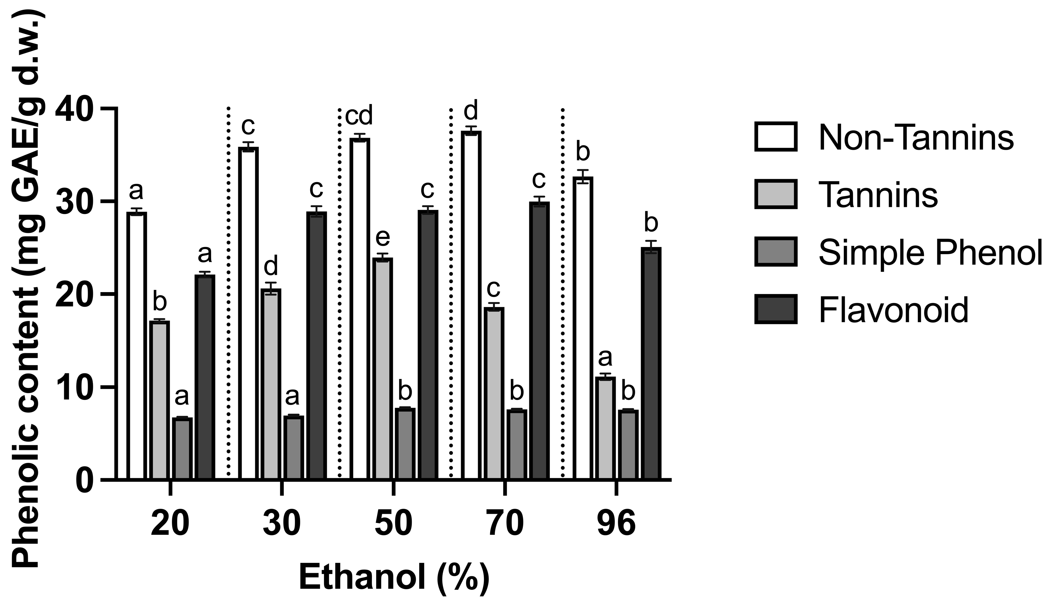
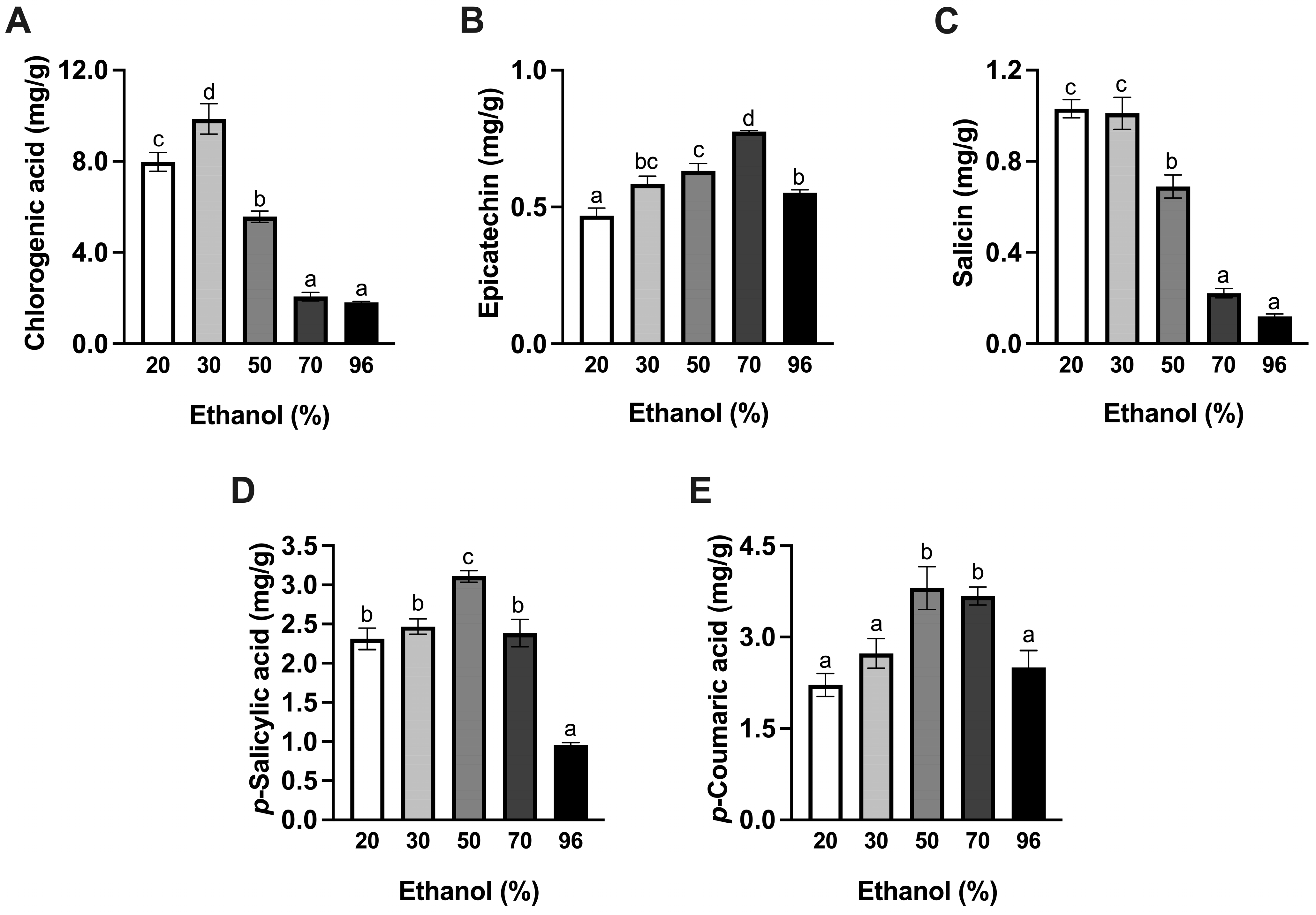
3.1. Effect of Ethanol

As shown in Table 1, ethanol content had a significant impact on the extraction of polyphenols from willow bark (p < 0.05). The TPC increased with an increase in ethanol content from 20% to 50% and reached its maximum at 50% ethanol (61.06 mg GAE/g d.w.). Beyond 50%, the TPC decreased significantly (p < 0.05). The TPC of willow bark in this study was slightly higher than a previous study where the TPC was 22.3 to 53.6 mg GAE/g d.w. when extracting with methanol [10]. This could be due to the differences in extraction solvent, the origin of the plant, environmental and seasonal factors, and harvesting process, which may impact the phenolic content of the willow bark.
Several assays have been commonly used to examine the antioxidant activities of herbal extracts, including DPPH and FRAP [28,29,30]. The DPPH assay determines the ability of an antioxidant to donate an electron to stable the DPPH free radicals. The ability of an antioxidant to reduce ferric to ferrous ions through redox reaction is estimated in the FRAP assay [31]. The change in antioxidant activities of willow infusions determined by DPPH and FRAP assays had similar trends as the TPC (Table 1). The highest DPPH and FRAP values were obtained with 50% ethanol with 103.45 mg AAE/g and 402.49 mmol TE/g, respectively. A high correlation of antioxidant activities with the TPC of willow infusions was observed ( r D P P H / T P = 0.954 ,   r F R A P / T P = 0.977 ,   p < 0.01 ), which may imply that the antioxidant activity is due to the total polyphenol content in the infusions. Similar observations were reported by Das and Eun [32], where the antioxidant activity of green tea extracts was strongly correlated with the content of phenolic compounds. This is possibly because the phenolic compounds act as electron donors to neutralize the reactive oxygen species to form stabilized chemical complexes, thus possessing free radical scavenging activity [33].
Figure 1 shows that the NT, TT, SP, and flavonoid content of willow infusions are influenced by ethanol content. The NT content increased with an increase in ethanol content from 20% to 50% and reached the maximum value with 70% ethanol, but it decreased significantly at 96% ethanol (p < 0.05). The recovery of SP was improved with increasing the content of ethanol to 50% and reached an extraction plateau afterwards. A higher yield of flavonoids was found in ethanolic infusions between 30% and 70% than those with the lowest (20%) or highest (96%) concentration of ethanol. Therefore, an increased flavonoid content could be mainly responsible for the high recovery of NT phenolic compounds with 30–70% ethanol. This finding is in line with a previous study showing that ethanol water mixtures (50%) were more effective than 20% ethanol solutions in the recovery of NT phenols, including flavonoid glucosides and phenolic acids from flaxseeds [34].
Similar to the NT content, an increase in ethanol levels up to 50% showed an increase in the content of TTs in the infusions, while a significant decrease in the content of TTs was found in the infusions with 70% and 96% ethanol. The highest yield of TTs was obtained with 50% ethanol. Similar results were observed by Bosso et al. [35], who found that the extraction efficiency of condensed tannins from grape seeds was increased with an increase of the ethanol concentration in water up to 50%, and it decreased as the ethanol concentration increased from 50% to 100%.
Chlorogenic acid, epicatechin, p-salicylic acid, and p-coumaric acid showed similar evolution as for the NT content (Figure 2). However, the recovery of salicin exhibited a different trend in that it decreased with an increase in ethanol content (Figure 2C). The obtained alcoholic willow infusions contained high levels of chlorogenic acid (1.82 to 9.86 mg/g), p-salicylic acid (0.96–3.11 mg/g), and p-coumaric acid (2.22–3.81 mg/g), whereas epicatechin and salicin content were relatively low, with 0.47–0.78 mg/g and 0.11–1 mg/g, respectively. Our findings are slightly higher than the published data, where chlorogenic acid was 0.04 to 3.8 mg/g [11,19,22,36], p-salicylic acid was 0.31 to 0.48 mg/g [19,22], and p-coumaric acid was 0.14 to 0.17 mg/g [19,22] in willow bark. The content of epicatechin and salicin was in the range of the literature data, where epicatechin was 0.35 to 2.16 mg/g [11,12,19,22,37] and salicin was 0.26 to 1.12 mg/g [10,11,19,22,36,37].
The change in the appearance of willow infusions with various ethanol content can be seen in Figure S1, and the measurable parameters of appearance—lightness, chroma, and hue angle—are shown in Table 1. Ethanol content had a significant impact on the lightness, chroma, and hue angle of the extracts (p < 0.05). The lightness, chroma, and hue angle decreased initially with an increase in ethanol content from 0% to 50%, and the lowest values were found with 50% ethanol, indicating the extracts had a dark and brownish appearance (Figure S1). From 70% and above, an increase in all parameters was observed, which resulted in lighter and more yellow infusions. The change in appearance of willow infusions could be explained by the evolution of tannins present in the infusions. In the past, willow bark has been employed as a natural dye agent due to its high content of tannins [38], which are yellow to light brown in nature and therefore influence the color intensity of the infusions.
Mixtures of ethanol with water have been successfully used in the recovery of polyphenols from other plant materials. Rajha et al. [39] reported that, as ethanol content increased from 0% to 50%, the yield of polyphenols extracted from vine shoots increased significantly and reached its maximum value. Chew et al. [40] also found that the recovery of total phenols and flavonoids from a medicinal plant, Orthosiphon stamineus, increased with an increase in ethanol content up to 40%, but they decreased afterwards. This is probably related to the intermediate polarity of ethanol-in-water solutions (50%), which have a high selectivity for versatile polyphenols present in the plant materials. Water is a good solvent for highly hydroxylated polyphenols such as phenolic acid and their glycosides, whereas ethanol can provide a higher extraction yield of flavonoids and tannins [16]. Moreover, the improvement of polyphenol extraction with the addition of ethanol might be associated with its capability of rupturing the structure of the cell membrane and thus increasing the cell permeability [41].
Overall, the recovery of bioactive compounds from willow bark with the traditional solvent system of ethanol and water was an effective process for obtaining phenolic-rich infusions. The binary mixture of ethanol and water at 50% ratio yielded the highest TPC and antioxidant activity of the infusions. The use of hydroalcoholic solutions containing 30% to 70% ethanol allowed maximum extraction of the majority of phenolic groups (NT, TT, and flavonoids) and phenolic compounds in willow bark (chlorogenic acid, epicatechin, p-salicylic acid, and p-coumaric acid). At maximized conditions for TPC extraction (50% ethanol), the resulting willow infusions contained 5.57 mg/g chlorogenic acid, 0.63 mg/g epicatechin, 0.69 mg/g salicin, 3.11 mg/g p-salicylic acid, and 3.81 mg/g p-coumaric acid. Since tannins are major pigmented compounds found in willow bark, the change in appearance of the extracts may be attributed to the presence of tannins.

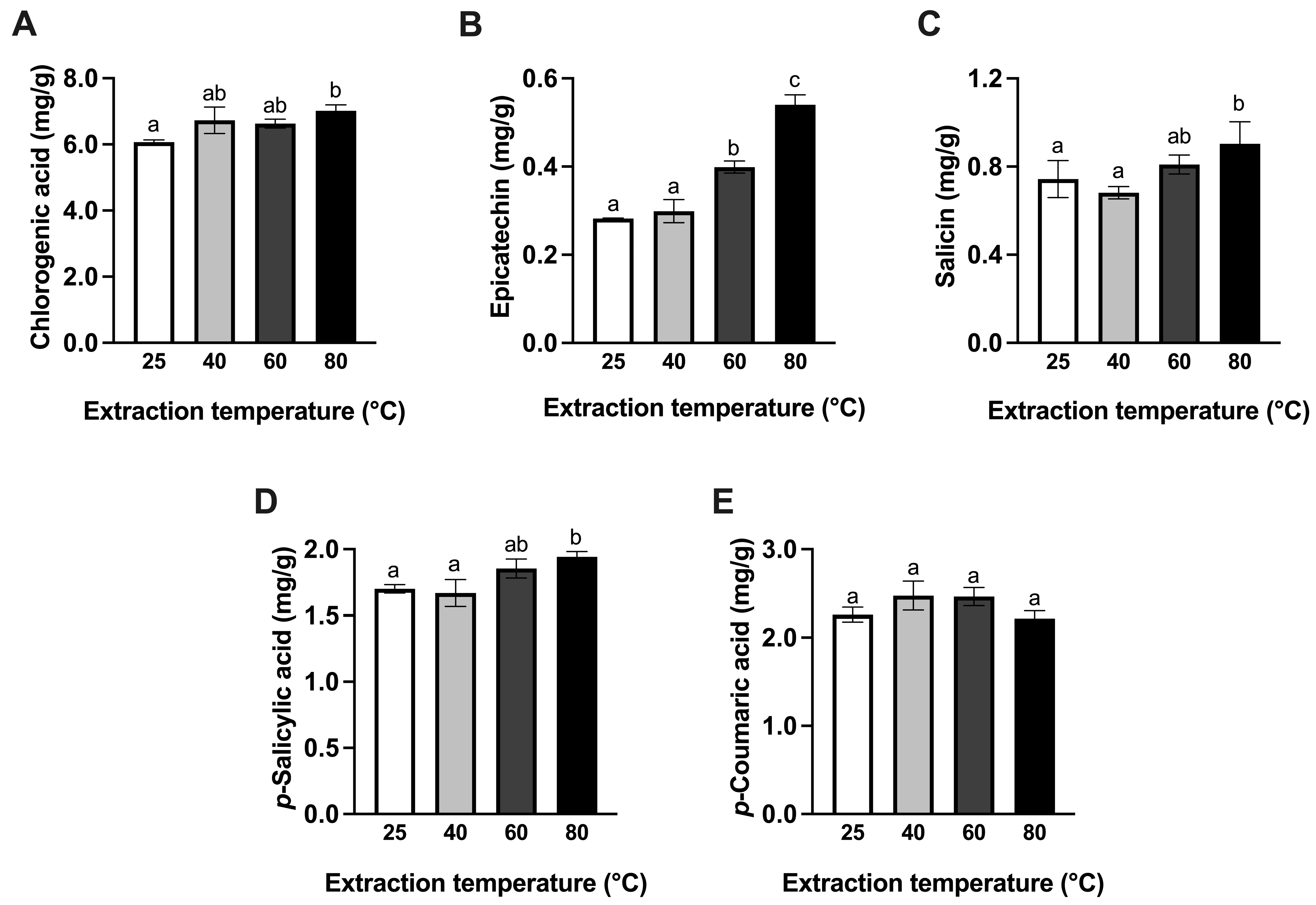
3.2. Effect of Temperature
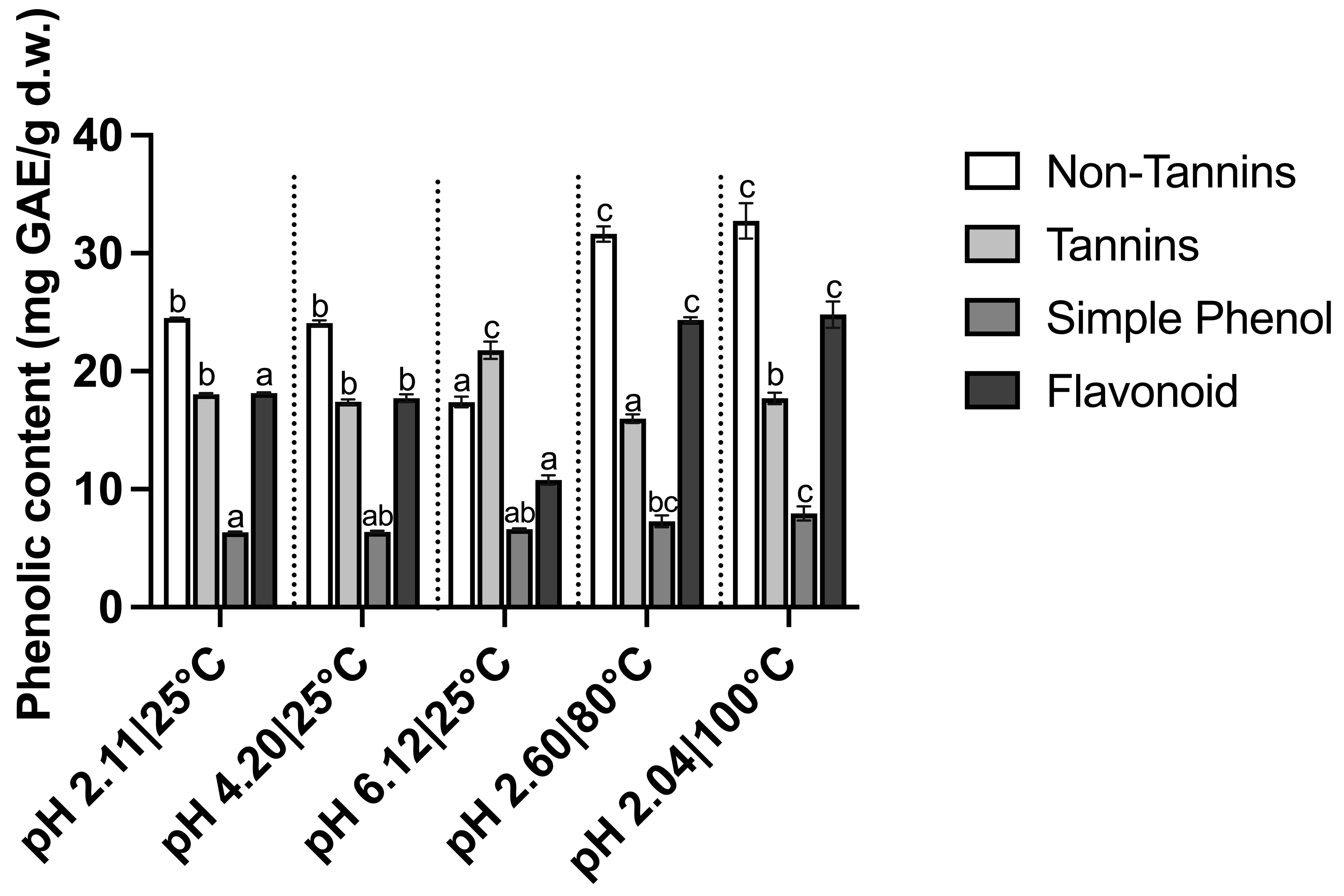
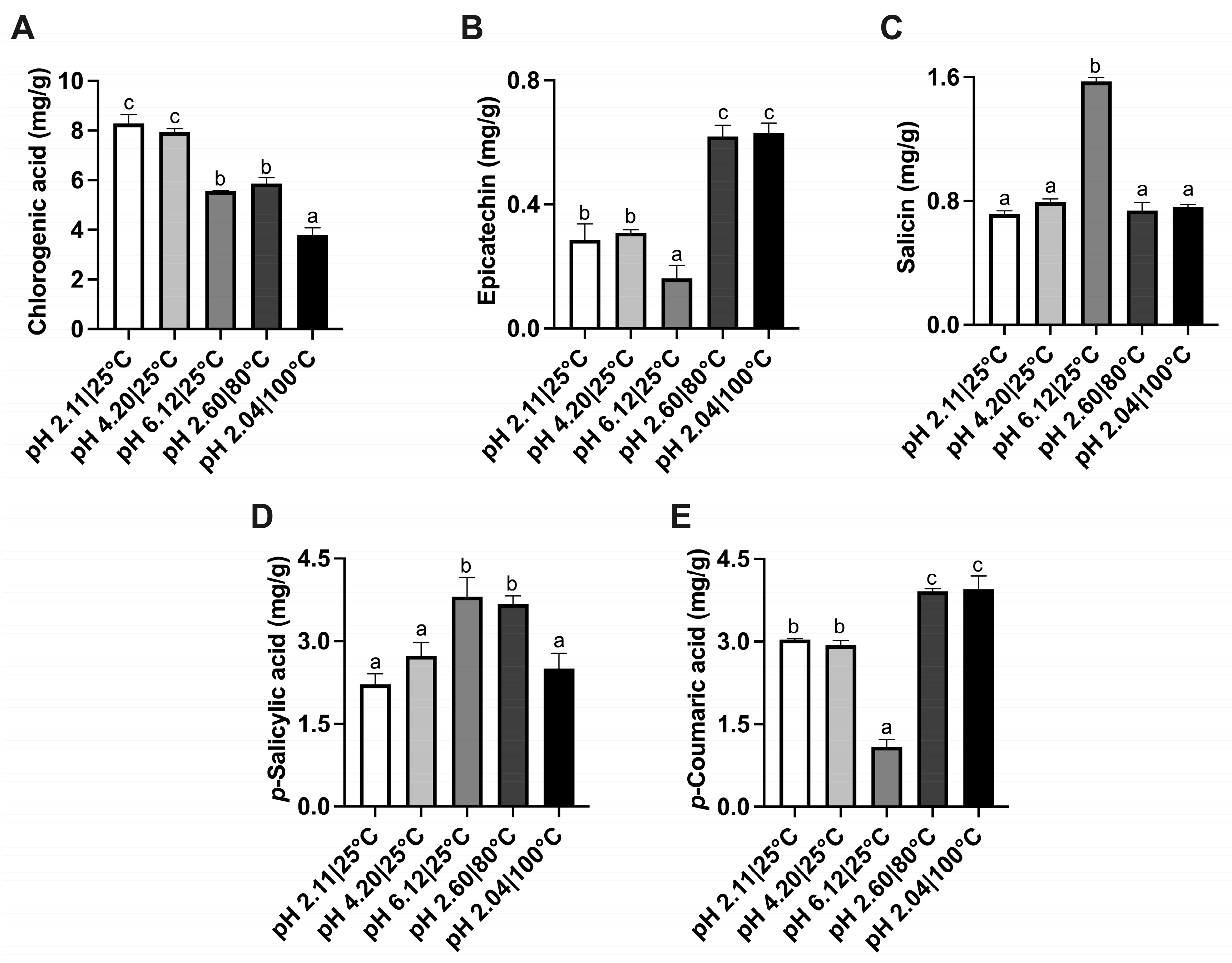
The presence of ethanol in alcoholic willow infusions may limit their application as a food or beverage ingredient. Moreover, it would be advantageous from cost and environmental perspectives to produce herbal infusions by using only water. Therefore, the impact of extraction temperature on the recovery of phenolic compounds from willow bark in an aqueous system was studied.
The TPC of aqueous willow infusions produced at various extraction temperatures is presented in Table 2. Extraction temperatures had a significant effect on the TPC of willow infusions (p < 0.05). An increase in temperature from 25 °C to 80 °C led to a significant increase in TPC, with a maximum value of 46.53 mg GAE/g d.w. at 80 °C (p < 0.05). These results illustrate that heat treatment improves the extraction of phenolic compounds from willow bark. The results obtained are in agreement with Antony and Farid [42], who reviewed the extraction temperature used in conventional techniques and found that the highest total phenol yield from plants was obtained between 60 °C and 80 °C. The extraction of TPC from green tea increased with increasing temperature up to 80 °C [32].
An increase in antioxidant activities was observed in both assays with an increase of extraction temperature from 25 °C to 80 °C (Table 2). This trend was similar to the change in TPC, suggesting that the antioxidant activities were highly associated with phenolic compounds present in the infusions.
Figure 3 shows that the phenolic constituents were significantly influenced by extraction temperature (p < 0.05). NT content increased significantly with an increase in temperature from 25 °C to 40 °C (p < 0.05). A further increase in temperature from 40 °C to 60 °C resulted in a significant decrease in NT (p < 0.05); however, the NT content at 80 °C was not significantly different from the infusion at 40 °C or 60 °C (p  0.05). In contrast to NT, an increase in temperature promotes the release of TT, leading to a higher yield. Kemppainen et al. [43] also found high extraction temperature increased the tannin yield extracted from spruce bark. SP content increased significantly with extraction temperature from 25 °C to 40 °C, and a further increase in temperature did not result in a significant change in SP (p  0.05). There was no significant difference in the flavonoid content between 25 °C, 40 °C, and 80 °C (p  0.05), but a significant decrease was observed at 60 °C in comparison with infusions prepared at 25 °C or 40 °C (p < 0.05).
As shown in Figure 4, the majority of phenolic compounds tended to increase with an increase in extraction temperature up to 80 °C. Chlorogenic acid was significantly lower at 25 °C than at 80 °C (p < 0.05). Epicatechin was not significantly affected by extraction temperature between 25 °C and 40 °C (p  0.05), but it was significantly increased from 40 °C to 80 °C (p < 0.05). There was a significant increase in salicin and p-salicylic acid content between 25 °C and 80 °C (p < 0.05). However, the increase in temperature did not significantly impact the extraction of p-coumaric acid (p  0.05).
The evolution of the color of willow infusions with various extraction temperatures is shown in Figure S2. Extraction temperatures had a significant effect on the lightness, chroma, and hue angle of willow infusions (p < 0.05). An increase in extraction temperature from 25 °C to 80 °C resulted in a darker, redder, and less vivid beverage (decreased L* value, hue angle, and chroma C* value, Table 2). This is in agreement with a previous study that explored the impact of extraction temperature on color change in meadowsweet extracts and found that the extracts became darker and redder when the extraction increased from 60 °C to 100 °C [44].
Water is a widely available environmentally and cost-friendly solvent, and, combined with high temperature, was a good alternative to hydroalcoholic solutions for producing infusions with bioactive compounds. The overall results showed that increasing temperature improved the extraction efficiency of polyphenols from willow bark, and, at 80 °C, yielded the highest TPC and antioxidant activity in willow infusions. Although the individual phenolic components behaved differently when increasing temperatures, generally, they tended to increase with the temperatures. The increase in extraction temperature accelerates the permeability of cell walls and solubility of phenolic compounds, resulting in a higher mass transfer through plant tissues into the solvent [16]. Moreover, the increased yield of TT might be explained by the polymeric structure of tannins such as procyanidins, which require a higher temperature for extraction.
In comparison to the optimal hydroalcoholic extracts (50% ethanol), the hot water infusions produced at 80 °C contained significantly less TPC and antioxidant activity (p < 0.05). Fifty percent ethanol-in-water resulted in a TPC of 61.06 mg GAE/g d.w. compared to that of 46.53 mg GAE/g d.w. in the aqueous infusions at 80 °C. This may suggest the solvent affinity for polyphenols had greater impact than the extraction temperature. However, hot water is preferable for the extraction of some phenolic compounds, including chlorogenic acid (7.02 mg/g) and salicin (0.9 mg/g), which were significantly higher in hot aqueous infusions than in 50% alcoholic infusions (p < 0.05). Also, the epicatechin content in the hot water infusions was 0.54 mg/g, which was not significantly different from its level in 50% ethanol (0.63 mg/g, p  0.05).

3.3. Effect of pH

Manipulation of solvent pH could also affect the extraction of polyphenols from plant materials. Therefore, in line with the concept of green extraction and with a goal of producing potent infusions for beverage applications, food-grade acids and alkali were added to water to extract phenolic compounds from willow bark. Under alkaline conditions (pH 12.1), a large amount of precipitates were formed in the infusions, which may affect their use in the beverage industry. Furthermore, the alkaline pH may induce degradation or dissociation of polyphenols; for example, tea flavanols were degraded at pH 9 or above [45]. Thus, the results of alkaline extraction were excluded in the following discussion.
At room temperature, an increase in the TPC and antioxidant activities was observed by lowering the pH from 6.12 to 2.11 (Table 3). The highest TPC and antioxidant activities were obtained at pH 2.11, with TPC of 41.21 mg GAE/g, DPPH of 61.92 AAE/g, and FRAP of 232 mmol TE/g, respectively. These results are in agreement with Adje et al. [46], who found effective extraction of total phenols, including phenolic acids, flavonols, and anthocyanins, using 1% HCl or 0.01 N sulfuric acid. Rusak et al. [47] also found that the presence of lemon juice in water accelerated the recovery of total phenols from white tea.
The combination of acidic solvent with heat treatment may improve the extraction efficiency through an acidic hydrolysis process. This is because some of the phenolic compounds are attached to the polysaccharides of the cell walls or to the lignin of plant tissues by ester or ether bonds. Acid hydrolysis may induce the cleavage of ether bonds and thus help the release of phenolic compounds in the aqueous solvent [16]. Hence, the synergistic effect of high temperature and acidic solvents on the extraction of polyphenols from willow bark was further investigated. Table 3 shows that increasing the extraction temperature from 25 °C to 80 °C combined with the acidified solvent at pH 2 led to higher TPC and antioxidant activity than at low pH without heating. The highest TPC and antioxidant activity of willow infusions in the pH-modified aqueous solutions was found at 100 °C and pH 2, with TPC of 48.02 mg GAE/g, DPPH of 76.43 AAE/g, and FRAP of 257.11 mmol TE/g, but it was not significantly higher than at 80 °C and pH 2 (p  0.05). Notably, there was no significant increase in the TPC and antioxidant activity observed when comparing the infusions at pH 2.60 and 80 °C (Table 3) with those at 80 °C (Table 2), suggesting that the improved extraction yield could be due to the accelerated temperature rather than the synergistic effect of high temperature and low pH.
The impact of extraction pH and temperature on the phenolic constituents is shown in Figure 5. An increment of NT content was observed with lowering pH and increasing temperature, but there was no significant difference between 80 and 100 °C at pH 2 (p  0.05). Altering the pH did not have a significant impact on the SP content, but it increased slightly with the combination of acidic pH and high temperatures (p < 0.05). The flavonoid content of the infusions was higher at low pH, and it further increased at the high temperature and low pH extraction conditions. These results indicate that the increased flavonoid content could be the reason for a high content of NT at low pH and high temperature. Zimmermann and Gleichenhagen [48] have reported previously that the addition of citric or ascorbic acid and adjusting pH to 3–4.8 improved the extraction of flavanols from tea infusions by 20% at 70 °C. [10,11,19,22,36,37] also found an increased content of total flavonoids in tea extract at 80 °C with water-lemon solutions.
As shown in Figure 5, the TT content was higher in the infusions at pH 6.12 than those of acidic and/or hot aqueous infusions, and the lowest TT content was found in the infusions at pH 2.6 and 80 °C (p < 0.05), which indicates extraction under acidic conditions may affect the recovery of TTs from willow bark. A similar observation was reported by Shahidi and Naczk [49], where the addition of 1% HCl reduced the recovery of tannins from rapeseed. At acidic pHs, tannins tend to form insoluble precipitates due to tannin self-condensation reaction, thus affecting the extraction efficiency [50]. Moreover, hydrolysable tannins are thought to be disassociated and broken into simple phenols at acidic conditions and high extraction temperatures.
The change in the content of individual phenolic compounds was greatly influenced by altering the pH (Figure 6). At 25 °C, the content of chlorogenic acid, epicatechin, and p-coumaric acid was significantly higher in the middle and strong acidic aqueous solutions (pH 2 and 4) than in the neutral solutions (pH 6). Salicin and p-salicylic acid increased significantly with shifting the pH from acidic toward neutral (p < 0.05). The impact of increasing extraction temperature at pH 2 on these phenolic compounds was varied (Figure S3). Chlorogenic acid content decreased significantly with an increase in temperature from 25 °C to 100 °C at pH 2. This may indicate the degradation or isomerization and transformation of chlorogenic acid due to acidic hydrolysis [51]. It is noteworthy that epicatechin content was higher at a lower pH (pH 2.11 & 4.20) than at pH 6.12, and it had a higher concentration at high extraction temperature than at 25 °C and pH 2. This indicates that epicatechin is more resistant to degradation by pH or high temperature, which might be due to its complex multiring aromatic structure. It possesses two aromatic rings that are connected by a six-membered oxygen-containing ring, and their structures are not planar. Thus, it has less susceptibility to structural degradation because of pH alteration or extreme temperatures [52]. There was no significant change in salicin content when increasing temperatures at pH 2. Notably, salicin content decreased significantly at low pH or at combined low pH and high temperature (p < 0.05). Degradation of salicin solution was previously found in the HCl (0.5 M) solution at 90 °C, suggesting that the loss of salicin may be due to acid hydrolysis [53]. At pH 2, p-salicylic acid content increased significantly at 80 °C (p < 0.05), but it decreased at 100 °C. p-coumaric acid demonstrated improved extraction yields with increasing temperature from 25 °C to 80 °C, but a further increase to 100 °C did not enhance its yield.
The color difference of willow bark infusions prepared at various pH levels is presented in Figure S4. Table 3 shows that extraction pH and temperature had a significant effect on the lightness, chroma, and hue angle of the willow infusions (p < 0.05). The lightness of the infusions was 27.34 at pH 2.11, and it gradually decreased to 18.41 at pH 6.12. At pH 2, the lightness decreased with an increase in temperature. The decrease of lightness was concomitant with the decrease in chroma. The decrease of hue angle from 80.7 to 50.95 with increase in pH and temperature indicates the color of the infusions was more red under these conditions.
Overall, the addition of food-grade acids to alter the pH of solvents positively impacted the extraction of polyphenols from willow bark. Moreover, increasing the extraction temperature of acidic aqueous solutions (pH 2) from 25 °C to 80 °C yielded higher levels of TPC and antioxidant activity in the infusions; however, increasing the temperature to 100 °C did not further increase the TPC. The improved recovery of polyphenols using the combination of acidic solutions and heat treatment is likely due to the accelerated extraction temperature rather than a synergistic effect of both factors. Among the pH-adjusted solutions, hot water at pH 2 and 80 °C was selected as the optimal condition since it required less energy than 100 °C and resulted in the infusions with a TPC of 46.70 mg GAE/g d.w., which was similar to those at 80 °C (46.53 mg GAE/g d.w., p  0.05) but significantly lower than 50% hydroalcoholic infusions (61.06 mg GAE/g d.w., p < 0.05). These infusions contained higher levels of NT and flavonoid fractions, which were 31.64 mg GAE/g d.w. and 24.36 mg GAE/g d.w., respectively, compared to 24.1 mg GAE/g d.w. and 17.52 mg GAE/g d.w. in the 80 °C infusions (p < 0.05), but they were lower than those in the 50% hydroalcoholic infusions, which contained 36.86 mg GAE/g d.w. NT and 29.07 mg GAE/g d.w. flavonoids (p < 0.05). Notably, extraction under acidic solutions and high temperatures led to the highest proportion of NT fractions and the lowest proportion of TT fractions among different extraction systems, which may be due to tannin self-condensation at acidic pHs. Thus, the resulting infusions might be more acceptable for consumers because of the astringent mouthfeel and darker appearance associated with tannins.
The high temperature and acidic solutions (at pH 2 and 80 °C) improved the extraction yield of p-salicylic acid (3.67 mg/g) and retained similar levels of chlorogenic acid (5.87 mg/g), epicatechin (0.62 mg/g), salicin (0.74 mg/g), and p-coumaric acid content (3.91 mg/g) when compared to the infusions with 50% ethanol that contained 3.11 mg/g p-salicylic acid, 5.57 mg/g chlorogenic acid, 0.63 mg/g epicatechin, 0.69 mg/g salicin, and 3.81 mg/g p-coumaric acid (p  0.05). Compared to aqueous infusions at 80 °C, acidic willow infusions at 80 °C contained less chlorogenic acid (5.87 mg/g) and salicin (0.74 mg/g, p < 0.05).

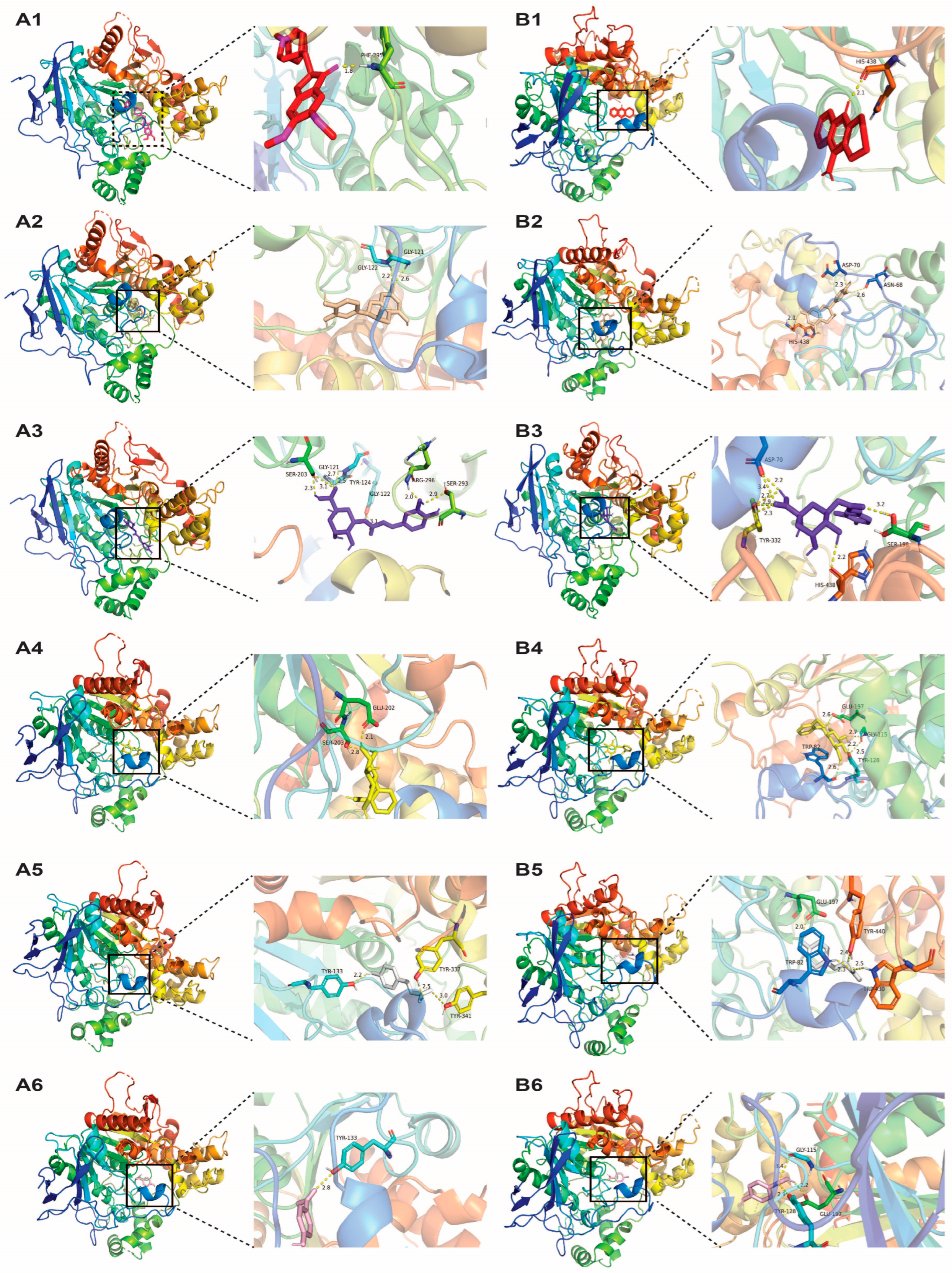
3.4. Neuroprotective Potentials of White Willow Polyphenols

The phenolic compounds in the willow infusions were studied for their binding affinities and interactions with pathological enzymes of AD (AChE and BuChE) via molecular docking. For both enzymes, the active sites take place in a 20 Å deep gorge. The catalytic triad of amino acid residues, including serine, histidine, and glutamate, is laid on the bottom of the gorge, and a peripheral anionic site (PAS) is at the entry to the gorge. Previous studies revealed that ligands can interact with one or both of the active sites and exhibit promising inhibition activities [4,54]. Less docking energy is required for a ligand to bind to the active site of a receptor, better inhibition of the receptor, and more stability of the ligand-receptor complex [55]. Thus, only the lowest binding energy was chosen for each ligand, and the results are shown in Table 4.
The molecular docking results showed that, against AChE, the known inhibitor donepezil was predicted to have the highest binding affinities (−12.3 kcal/mol). Among the willow polyphenols, epicatechin showed the highest binding affinities (−10 kcal/mol), followed by chlorogenic acid (−9.8 kcal/mol), which are comparable to the FDA-approved drug to treat AD. In addition, salicin, p-coumaric acid, and p-salicylic acid also exhibited good affinities toward AChE since their binding energies were negative. The 3D structure of interactions between these ligands and the active sites of AChE and BuChE are shown in Figure 7.
Two hydrogen bond interactions were observed between the phenyl group of epicatechin and amino residues Gly-121 and Gly-122. It also formed strong hydrophobic interaction with prominent amino acid residues (Tyr-124, Trp-286, Tyr-341) located in the PAS and with residue Phe-297, located in the acyl binding pocket of AChE. Moreover, the phenyl ring of epicatechin formed a π - π stacking interaction with Tyr-341, which was the only ligand besides donepezil that showed this interaction. For chlorogenic acid, it showed several hydrogen bonds with AChE, including residues Ser-203 and Ser-293 in the catalytic triad and Tyr-124 in PAS. It hydrophobically interacted with significant residues of PAS, namely Trp-286, Phe-338, and Tyr-341. In addition, a π -cation interaction formed between chlorogenic acid and key residue (His-447) in the catalytic site of AChE. Two hydrogen bond interactions with Glu-202 and Ser-203, a hydrophobic interaction with Phe-338 and Tyr-341, and a π - π stacking interaction with Tyr-337 were observed in the lowest energy conformer of salicin, which is a significant amino acid residue of both PAS and catalytic sites of AChE. p-coumaric acid and p-salicylic acid showed several interactions with the residues only in the PAS. For p-coumaric acid, it showed hydrogen bond interactions with Tyr-133, Tyr-341, and Tyr-337 and a hydrophobic interaction with Trp-86. The phenyl moiety of p-coumaric acid formed a π - π stacking interaction with Trp-86. Interactions of p-salicylic acid with residues Tyr-133, Trp-86, Tyr-337, and Trp-86 were observed. Overall, among the polyphenols, epicatechin, chlorogenic acid, and salicin were found to interact with both active sites of AChE; consequently, they showed better binding affinities than p-coumaric acid and p-salicylic acid.
In the case of BuChE, chlorogenic acid was predicted to have the highest binding affinities (−8.7 kcal/mol) among all the ligands studied, including the standard inhibitor of BuChE tacrine. Epicatechin also showed a comparable binding affinity with BuChE (−8.4 kcal/mol) as tacrine. The maximum binding affinities were −7.4 kcal/mol for salicin, −6.9 kcal/mol for p-coumaric acid, and −6.1 kcal/mol for p-salicylic acid.
Chlorogenic acid showed several hydrogen bond interactions with prominent residues of the catalytic site of BuChE (His-438 and Ser-198). The carboxyl groups on the glucoside moiety of chlorogenic acid formed multiple hydrogen bonds with key residues of PAS of BuChE, namely Asp-70 and Tyr-332. The interaction between chlorogenic acid and Phe-329, an important amino acid residue of the anionic site of BuChE, is hydrophobic. Moreover, Trp-231 formed a π - π stacking interaction with chlorogenic acid. The binding between epicatechin and BuChE was mainly through hydrogen bond interactions, with the residues in the catalytic site and PAS of BuChE, which are His-438, Asn-68, and Asp-70. It also formed a π - π stacking interaction with Trp-82, which is known to promote catalysis of the ligand with the catalytic site of BuChE [4]. The hydroxyl groups on the glucoside of salicin showed several hydrogen bond interactions with amino acid residues Trp-82, Gly-115, Tyr-128, and Glu-197. Salicin also formed a hydrophobic bond interaction with Ala-328 in the PAS of BuChE. p-coumaric acid showed multiple hydrogen bond interactions with Trp-82, Glu-197, Trp-430, and Tyr-440, a hydrophobic interaction with Trp-82, and a π - π stacking interaction with Trp-82. Most of the interactions between p-salicylic acid and amino acid residues were through hydrogen bonding, while Trp-82 formed a π - π stacking interaction.
Taken together, the present molecular docking indicates that willow polyphenols have promising potential in inhibiting both AChE and BuChE in silico through binding at their active sites. Chlorogenic acid, epicatechin, and salicin showed strong binding affinities toward AChE, which is in agreement with a previous molecular docking study conducted by [4,54]. Moreover, these three polyphenols also had good binding affinities for BuChE. These findings provide an insight into the contribution of individual phenolic compounds in inhibiting both AChE and BuChE in silico, which could help the understanding of the neuroprotective effects associated with willow bark. The highest content of chlorogenic acid (8.28 mg/g) was found in the acidic willow infusions without heating (pH 2 and 25 °C, p < 0.05). Aqueous infusions at 80 °C contained higher chlorogenic acid (7.02 mg/g) than those at pH 2 and 80 °C (5.87 mg/g) and 50% ethanol (5.57 mg/g, p < 0.05). Epicatechin was higher in the 50% hydroalcoholic infusions (0.63 mg/g), high temperature and acidic infusions (0.62 mg/g), and high temperature infusions (0.54 mg/g) than in the low pH without heating infusions (0.29 mg/g). Acidic infusions with or without heating (pH 2 and 25 °C or 80 °C) and binary ethanol and water (50:50) infusions contained similar levels of salicin, which were significantly lower than those at high temperature infusions (80 °C, p < 0.05). Hence, willow infusions prepared with hot water at 80 °C contained the highest yields of salicin and epicatechin and substantial quantities of chlorogenic acid, which may possess neuroprotective potential in silico.

4. Conclusions

Polyphenol-rich willow bark beverages were prepared using different extraction protocols. Ethanol-in-water (50%) was found to be an efficient extraction system for maximizing the content of total phenols (61.06 mg GAE/g d.w.) and non-tannin phenols, especially flavonoids. However, non-alcoholic infusions may be more suitable for beverage applications. In aqueous solutions, increasing the extraction temperature improved the recovery of total phenols, with the maximum TPC (46.53 mg GAE/g d.w.) using hot water extraction found at 80 °C, although it was lower than those in 50% hydroalcoholic infusions. In addition to hot water extraction, the addition of food-grade acids to adjust the pH of the solvent has been shown to be another option to improve the extraction efficiency of aqueous solutions. At room temperature, lowering the pH increased the extraction of TPC from willow bark. Moreover, the combination of low pH and hot water extraction further improved the recovery of TPC. Among the pH-adjusted extraction systems, willow infusions prepared at pH 2 and 80 °C contained the maximum yield of TPC (46.7 mg GAE/g d.w.), though this was lower than 50% alcoholic infusions. Notably, these acidic infusions showed the highest ratio of non-tannins to tannins and a lighter appearance. Thus, the obtained infusions may have a less astringent mouthfeel compared to those extracted with hydroalcoholic solutions or high temperature alone, which might be preferrable for consumers. The antioxidant activity of willow extracts was in good correlation with the TPC regardless of extraction system.
In silico studies revealed that some characteristic polyphenols in willow infusions could be docked into the active sites of AChE and BuChE. Epicatechin, chlorogenic acid, and salicin demonstrated good binding affinities toward AChE and BuChE through a number of hydrogen bonds and hydrophobic interactions with prominent amino acid residues of the active sites of both enzymes, supporting their inhibitory potential.
Although willow infusions prepared with alcoholic solutions contained higher TPC than those prepared with aqueous solutions, individual polyphenols behaved differently. Aqueous infusions at 80 °C contained higher levels of chlorogenic acid and salicin than 50% hydroalcoholic infusions or pH 2 and 80 °C infusions. The level of epicatechin was not greatly affected by different preparation protocols (50% ethanol vs. pH 2 and 80 °C vs. 80 °C). Hence, willow infusions produced by hot water extraction at 80 °C may have promising neuroprotective potentials. Additionally, p-coumaric acid content was found to be higher in the infusions prepared with low pH and heating (pH 2 and 80 °C) and 50% ethanol than those at 80 °C alone. Acidic infusions with heating also led to a higher yield of p-salicylic acid than other infusions.
To summarize, hot water extraction and the combination of low pH and heat treatment seem to be feasible preparation methods for non-alcoholic willow infusions for beverage applications. The obtained infusions are enriched with polyphenols and exhibit high antioxidant activities and possible neuroprotective effects in silico. These findings suggest that willow bark infusion could be served as a promising ingredient for functional beverages due to its abundant phenolic compounds and potent biological activity potentials. However, the regulatory status of willow bark for food applications in the European market remains unclear. If a food has not been consumed in the EU to a significant degree before 15 May 1997, it may fall into the novel foods category. Based on the EU novel food catalogue lists, it has not been determined whether willow bark falls into this category; therefore, any food containing willow bark may need to be authorized before being placed on the EU market. Thus, it is important to determine the regulatory status of willow bark for manufacturers who are interested in producing willow beverages for the European market.

Supplementary Materials

The following supporting information can be downloaded at https://www.mdpi.com/article/10.3390/beverages10030075/s1: Figure S1: Appearance of willow extracts with different ethanol concentrations; Figure S2: Appearance of willow extracts produced at different extraction temperatures; Figure S3: Effect of temperature at pH 2 on the phenolic compounds. Figure S4: Appearance of willow extracts at different pH levels and extraction temperatures.

Author Contributions

Conceptualization, L.Z., J.-C.J., and N.H.; writing—original draft preparation, L.Z.; writing—review and editing, L.Z., J.-C.J., and N.H.; supervision, J.-C.J., and N.H. All authors have read and agreed to the published version of the manuscript.

Funding

This research received no external funding.

Data Availability Statement

All data are provided within the manuscript and are available upon request.

Acknowledgments

The authors would like to acknowledge the Ph.D. Scholarship supported by University College Dublin and the China Scholarship Council.

Conflicts of Interest

The authors declare no conflicts of interest.

References

  1. Liao, X.; Huang, Y.; Zhang, Z.; Zhong, S.; Xie, G.; Wang, L.; Xiao, H. Factors associated with health-related quality of life among family caregivers of people with Alzheimer’s disease. Psychogeriatrics 2020, 20, 398–405. [Google Scholar] [CrossRef]
  2. Yang, Z.; Zhou, D.-D.; Huang, S.-Y.; Fang, A.-P.; Li, H.-B.; Zhu, H.-L. Effects and mechanisms of natural products on Alzheimer’s disease. Crit. Rev. Food Sci. Nutr. 2023, 63, 3168–3188. [Google Scholar] [CrossRef]
  3. Leng, F.; Edison, P. Neuroinflammation and microglial activation in Alzheimer disease: Where do we go from here? Nat. Rev. Neurol. 2021, 17, 157–172. [Google Scholar] [CrossRef]
  4. Macdonald, I.R.; Martin, E.; Rosenberry, T.L.; Darvesh, S. Probing the peripheral site of human butyrylcholinesterase. Biochemistry 2012, 51, 7046–7053. [Google Scholar] [CrossRef]
  5. Bohnen, N.I.; Kaufer, D.I.; Hendrickson, R.; Ivanco, L.S.; Lopresti, B.; Davis, J.G.; Constantine, G.; Mathis, C.A.; Moore, R.Y.; DeKosky, S.T. Cognitive correlates of alterations in acetylcholinesterase in Alzheimer’s disease. Neurosci. Lett. 2005, 380, 127–132. [Google Scholar] [CrossRef]
  6. Nordberg, A.; Svensson, A.L. Cholinesterase inhibitors in the treatment of Alzheimer’s disease: A comparison of tolerability and pharmacology. Drug Saf. 1998, 19, 465–480. [Google Scholar] [CrossRef]
  7. Ohbe, H.; Jo, T.; Matsui, H.; Fushimi, K.; Yasunaga, H. Cholinergic Crisis Caused by Cholinesterase Inhibitors: A Retrospective Nationwide Database Study. J. Med. Toxicol. 2018, 14, 237–241. [Google Scholar] [CrossRef]
  8. Deshpande, P.; Gogia, N.; Singh, A. Exploring the efficacy of natural products in alleviating Alzheimer’s disease. Neural Regen. Res. 2019, 14, 1321–1329. [Google Scholar]
  9. Aleman, R.S.; Marcia, J.; Duque-Soto, C.; Lozano-Sánchez, J.; Montero-Fernández, I.; Ruano, J.A.; Hoskin, R.T.; Moncada, M. Effect of Microwave and Ultrasound-Assisted Extraction on the Phytochemical and In Vitro Biological Properties of Willow (Salix alba) Bark Aqueous and Ethanolic Extracts. Plants 2023, 12, 2533. [Google Scholar] [CrossRef]
  10. Zaiter, A.; Becker, L.; Petit, J.; Zimmer, D.; Karam, M.C.; Baudelaire, E.; Scher, J.; Dicko, A. Antioxidant and antiacetylcholinesterase activities of different granulometric classes of Salix alba (L.) bark powders. Powder Technol. 2016, 301, 649–656. [Google Scholar] [CrossRef]
  11. Gligorić, E.; Igić, R.; Srđenović Čonić, B.; Kladar, N.; Teofilović, B.; Grujić, N. Chemical profiling and biological activities of “green” extracts of willow species (Salix L., Salicaceae): Experimental and chemometric approaches. Sustain. Chem. Pharm. 2023, 32, 100981. [Google Scholar] [CrossRef]
  12. Piątczak, E.; Dybowska, M.; Płuciennik, E.; Kośla, K.; Kolniak-Ostek, J.; Kalinowska-Lis, U. Identification and accumulation of phenolic compounds in the leaves and bark of Salix alba (L.) and their biological potential. Biomolecules 2020, 10, 1391. [Google Scholar] [CrossRef]
  13. Maleš, I.; Pedisić, S.; Zorić, Z.; Elez-Garofulić, I.; Repajić, M.; You, L.; Vladimir-Knežević, S.; Butorac, D.; Dragović-Uzelac, V. The medicinal and aromatic plants as ingredients in functional beverage production. J. Funct. Foods 2022, 96, 105210. [Google Scholar] [CrossRef]
  14. Chemat, F.; Vian, M.A.; Cravotto, G. Green Extraction of Natural Products: Concept and Principles. Int. J. Mol. Sci. 2012, 13, 8615–8627. [Google Scholar] [CrossRef]
  15. Van Vuuren, S.F.; Motlhatlego, K.E.; Netshia, V. Traditionally used polyherbals in a southern African therapeutic context. J. Ethnopharmacol. 2022, 288, 114977. [Google Scholar] [CrossRef]
  16. Oreopoulou, A.; Tsimogiannis, D.; Oreopoulou, V. Chapter 15—Extraction of Polyphenols From Aromatic and Medicinal Plants: An Overview of the Methods and the Effect of Extraction Parameters. In Polyphenols in Plants, 2nd ed.; Watson, R.R., Ed.; Academic Press: Cambridge, MA, USA, 2019; pp. 243–259. [Google Scholar]
  17. Vuong, Q.V.; Golding, J.B.; Stathopoulos, C.E.; Nguyen, M.H.; Roach, P.D. Optimizing conditions for the extraction of catechins from green tea using hot water. J. Sep. Sci. 2011, 34, 3099–3106. [Google Scholar] [CrossRef]
  18. Sulaiman, G.M.; Hussien, N.N.; Marzoog, T.R.; Awad, H.A. Phenolic content, antioxidant, antimicrobial and cytotoxic activities of ethanolic extract of Salix alba. Am. J. Biochem. Biotechnol. 2013, 9, 41–46. [Google Scholar] [CrossRef]
  19. Gligorić, E.; Igić, R.; Suvajdžić, L.; Grujić-Letić, N. Species of the Genus Salix L.: Biochemical Screening and Molecular Docking Approach to Potential Acetylcholinesterase Inhibitors. Appl. Sci. 2019, 9, 1842. [Google Scholar] [CrossRef]
  20. Paterson, J.R.; Lawrence, J.R. Salicylic acid: A link between aspirin, diet and the prevention of colorectal cancer. QJM Int. J. Med. 2001, 94, 445–448. [Google Scholar] [CrossRef]
  21. Shen, Y.; Song, X.; Li, L.; Sun, J.; Jaiswal, Y.; Huang, J.; Liu, C.; Yang, W.; Williams, L.; Zhang, H.; et al. Protective effects of p-coumaric acid against oxidant and hyperlipidemia-an in vitro and in vivo evaluation. Biomed. Pharmacother. 2019, 111, 579–587. [Google Scholar] [CrossRef]
  22. Gligorić, E.; Igić, R.; Teofilović, B.; Grujić-Letić, N. Phytochemical Screening of Ultrasonic Extracts of Salix Species and Molecular Docking Study of Salix-Derived Bioactive Compounds Targeting Pro-Inflammatory Cytokines. Int. J. Mol. Sci. 2023, 24, 11848. [Google Scholar] [CrossRef]
  23. Singleton, V.L.; Rossi, J.A. Colorimetry of total phenolics with phosphomolybdic-phosphotungstic acid reagents. Am. J. Enol. Vitic. 1965, 16, 144–158. [Google Scholar] [CrossRef]
  24. Harbourne, N.; Marete, E.; Jacquier, J.C.; O’Riordan, D. Effect of drying methods on the phenolic constituents of meadowsweet (Filipendula ulmaria) and willow (Salix alba). LWT—Food Sci. Technol. 2009, 42, 1468–1473. [Google Scholar] [CrossRef]
  25. Thaipong, K.; Boonprakob, U.; Crosby, K.; Cisneros-Zevallos, L.; Byrne, D.H. Comparison of ABTS, DPPH, FRAP, and ORAC assays for estimating antioxidant activity from guava fruit extracts. J. Food Compos. Anal. 2006, 19, 669–675. [Google Scholar] [CrossRef]
  26. McGuire, R.G. Reporting of Objective Color Measurements. HortScience 1992, 27, 1254–1255. [Google Scholar] [CrossRef]
  27. Trott, O.; Olson, A.J. AutoDock Vina: Improving the speed and accuracy of docking with a new scoring function, efficient optimization, and multithreading. J. Comput. Chem. 2010, 31, 455–461. [Google Scholar] [CrossRef] [PubMed]
  28. Naik, G.; Priyadarsini, K.I.; Mohan, H. Evaluating the antioxidant activity of different plant extracts and herbal formulations. Res. Chem. Intermed. 2005, 31, 145–151. [Google Scholar] [CrossRef]
  29. Duh, P.-D.; Yen, G.-C. Antioxidative activity of three herbal water extracts. Food Chem. 1997, 60, 639–645. [Google Scholar] [CrossRef]
  30. Sukweenadhi, J.; Setiawan, F.; Yunita, O.; Kartini, K.; Avanti, C. Antioxidant activity screening of seven Indonesian herbal extract. Biodiversitas 2020, 21, 2062–2067. [Google Scholar] [CrossRef]
  31. Huang, D.; Ou, B.; Prior, R.L. The chemistry behind antioxidant capacity assays. J. Agric. Food Chem. 2005, 53, 1841–1856. [Google Scholar] [CrossRef]
  32. Das, P.R.; Eun, J.B. A comparative study of ultra-sonication and agitation extraction techniques on bioactive metabolites of green tea extract. Food Chem. 2018, 253, 22–29. [Google Scholar] [CrossRef] [PubMed]
  33. Sroka, Z.; Cisowski, W. Hydrogen peroxide scavenging, antioxidant and anti-radical activity of some phenolic acids. Food Chem. Toxicol. 2003, 41, 753–758. [Google Scholar] [CrossRef] [PubMed]
  34. Boussetta, N.; Soichi, E.; Lanoisellé, J.L.; Vorobiev, E. Valorization of oilseed residues: Extraction of polyphenols from flaxseed hulls by pulsed electric fields. Ind. Crops. Prod. 2014, 52, 347–353. [Google Scholar] [CrossRef]
  35. Bosso, A.; Guaita, M.; Petrozziello, M. Influence of solvents on the composition of condensed tannins in grape pomace seed extracts. Food Chem. 2016, 207, 162–169. [Google Scholar] [CrossRef]
  36. Köhler, A.; Förster, N.; Zander, M.; Ulrichs, C. Inter- and intraspecific diversity of Salix bark phenolic profiles—A resource for the pharmaceutical industry. Fitoterapia 2023, 170, 105660. [Google Scholar] [CrossRef] [PubMed]
  37. Förster, N.; Antoniadou, K.; Zander, M.; Baur, S.; Mittermeier-Kleßinger, V.K.; Dawid, C.; Ulrichs, C.; Mewis, I. Chemoprofiling as Breeding Tool for Pharmaceutical Use of Salix. Front. Plant Sci. 2021, 12, 579820. [Google Scholar] [CrossRef]
  38. Lohtander, T.; Arola, S.; Laaksonen, P. Biomordanting willow bark dye on cellulosic materials. Color. Technol. 2020, 136, 3–14. [Google Scholar] [CrossRef]
  39. Rajha, H.N.; Boussetta, N.; Louka, N.; Maroun, R.G.; Vorobiev, E. A comparative study of physical pretreatments for the extraction of polyphenols and proteins from vine shoots. Food Res. Int. 2014, 65, 462–468. [Google Scholar] [CrossRef]
  40. Chew, K.; Khoo, M.; Ng, S.; Thoo, Y.Y.; Aida, W.W.; Ho, C.W. Effect of ethanol concentration, extraction time and extraction temperature on the recovery of phenolic compounds and antioxidant capacity of Orthosiphon stamineus extracts. Int. Food Res. J. 2011, 18, 1427. [Google Scholar]
  41. Dorta, E.; Lobo, M.G.; Gonzalez, M. Reutilization of mango byproducts: Study of the effect of extraction solvent and temperature on their antioxidant properties. J. Food Sci. 2012, 77, C80–C88. [Google Scholar] [CrossRef]
  42. Antony, A.; Farid, M. Effect of Temperatures on Polyphenols during Extraction. Appl. Sci. 2022, 12, 2107. [Google Scholar] [CrossRef]
  43. Kemppainen, K.; Siika-aho, M.; Pattathil, S.; Giovando, S.; Kruus, K. Spruce bark as an industrial source of condensed tannins and non-cellulosic sugars. Ind. Crops. Prod. 2014, 52, 158–168. [Google Scholar] [CrossRef]
  44. Harbourne, N.; Jacquier, J.C.; O’Riordan, D. Optimisation of the aqueous extraction conditions of phenols from meadowsweet (Filipendula ulmaria L.) for incorporation into beverages. Food Chem. 2009, 116, 722–727. [Google Scholar] [CrossRef]
  45. Liang, Y.; Xu, Y. Effect of pH on cream particle formation and solids extraction yield of black tea. Food Chem. 2001, 74, 155–160. [Google Scholar] [CrossRef]
  46. Adje, F.; Lozano, Y.F.; Lozano, P.; Adima, A.; Chemat, F.; Gaydou, E.M. Optimization of anthocyanin, flavonol and phenolic acid extractions from Delonix regia tree flowers using ultrasound-assisted water extraction. Ind. Crops. Prod. 2010, 32, 439–444. [Google Scholar] [CrossRef]
  47. Rusak, G.; Komes, D.; Likić, S.; Horžić, D.; Kovač, M. Phenolic content and antioxidative capacity of green and white tea extracts depending on extraction conditions and the solvent used. Food Chem. 2008, 110, 852–858. [Google Scholar] [CrossRef]
  48. Zimmermann, B.F.; Gleichenhagen, M. The effect of ascorbic acid, citric acid and low pH on the extraction of green tea: How to get most out of it. Food Chem. 2011, 124, 1543–1548. [Google Scholar] [CrossRef]
  49. Shahidi, F.; Naczk, M. An Overview of the Phenolics of Canola and Rapeseed—Chemical, Sensory and Nutritional Significance. J. Am. Oil Chem. Soc. 1992, 69, 917–924. [Google Scholar] [CrossRef]
  50. Sealy-Fisher, V.J.; Pizzi, A. Increased pine tannins extraction and wood adhesives development by phlobaphenes minimization. Holz Roh Werkst. 1992, 50, 212–220. [Google Scholar] [CrossRef]
  51. Dawidowicz, A.L.; Typek, R. The influence of pH on the thermal stability of 5-O-caffeoylquinic acids in aqueous solutions. Eur. Food Res. Technol. 2011, 233, 223–232. [Google Scholar] [CrossRef]
  52. Friedman, M.; Jürgens, H.S. Effect of pH on the Stability of Plant Phenolic Compounds. J. Agric. Food Chem. 2000, 48, 2101–2110. [Google Scholar] [CrossRef] [PubMed]
  53. Nekhoroshev, S.V.; Nekhorosheva, A.V.; Sabutova, A.B.; Botirov, E.K.; Drenin, A.A.; Slepchenko, G.B.; Gornikov, N.V. Hydrolytic Stability of Aqueous Solutions of Salicin. Pharm. Chem. J. 2020, 54, 857–860. [Google Scholar] [CrossRef]
  54. Cheung, J.; Gary, E.N.; Shiomi, K.; Rosenberry, T.L. Structures of human acetylcholinesterase bound to dihydrotanshinone I and territrem B show peripheral site flexibility. ACS Med. Chem. Lett. 2013, 4, 1091–1096. [Google Scholar] [CrossRef] [PubMed]
  55. Mazumder, M.K.; Choudhury, S. Tea polyphenols as multi-target therapeutics for Alzheimer’s disease: An in silico study. Med. Hypotheses 2019, 125, 94–99. [Google Scholar] [CrossRef]
Figure 1. Effect of ethanol content on the content of phenolic constituents in the willow infusions. Different letters (a–e) for each constituent indicate significant differences (p < 0.05).
Beverages 10 00075 g001
Figure 2. Effect of ethanol content on the content of phenolic compounds: chlorogenic acid (A), epicatechin (B), salicin (C), p-salicylic acid (D), and p-coumaric acid (E) in the willow infusions. Different letters (a–d) indicate significant differences (p < 0.05).
Beverages 10 00075 g002
Figure 3. Effect of extraction temperature on the content of phenolic constituents in the willow infusions. Different letters (a–d) for each constituent indicate significant differences (p < 0.05).
Beverages 10 00075 g003
Figure 4. Effect of extraction temperature on the content of phenolic compounds: chlorogenic acid (A), epicatechin (B), salicin (C), p-salicylic acid (D), and p-coumaric acid (E) in the willow infusions. Different letters (a–c) indicate significant differences (p < 0.05).
Beverages 10 00075 g004
Figure 5. Effect of pH and temperature on the content of phenolic constituents in the willow infusions. Different letters (a–c) for each constituent indicate significant differences (p < 0.05).
Beverages 10 00075 g005
Figure 6. Effect of pH and temperature on the content of phenolic compounds: chlorogenic acid (A), epicatechin (B), salicin (C), p-salicylic acid (D), and p-coumaric acid (E) in the willow infusions. Different letters (a–c) indicate significant differences (p < 0.05).
Beverages 10 00075 g006
Figure 7. The docking poses of different ligands with their receptors: (A) AChE and (B) BuChE in total and detailed view. (A1) represents donepezil (red sticks) docked into the active site of AChE. (B1) represents tacrine (red sticks) docked into the active site of BuChE. (A2) and (B2) epicatechin (wheat sticks) docked in the active sites of AChE and BuChE. (A3) and (B3) represent chlorogenic acid (purple sticks) docked in the active sites of AChE and BuChE. (A4) and (B4) represent salicin (yellow sticks) docked in the active sites of AChE and BuChE. (A5) and (B5) represent p-coumaric acid (white sticks) docked in the active sites of AChE and BuChE. (A6) and (B6) represent p-salicylic acid (light pink sticks) docked in the active sites of AChE and BuChE.
Beverages 10 00075 g007
Table 1. Effect of ethanol content on TPC, antioxidant activity (determined by DPPH and FRAP assays), and color of willow infusions.
Ethanol
(%)
TPC
(mg GAE/g d.w.)
DPPH
(mg AAE/g d.w.)
FRAP
(mmol TE/g d.w.)
Color
Lightness (L*)Chroma
(C*)
Hue Angle (H°)
2044.25 ± 0.50 a75.78 ± 1.07 b284.05 ± 4.76 a18.03 ± 0.09 b16.49 ± 0.17 b52.73 ± 0.21 c
3054.53 ± 0.64 b85.94 ± 0.64 c334.59 ± 0.76 b18.00 ± 0.22 b17.75 ± 0.36 c48.50 ± 1.06 b
5061.06 ± 0.43 c103.45 ± 0.77 d402.49 ± 4.27 c16.76 ± 0.21 a15.29 ± 0.14 a43.89 ± 1.66 a
7054.48 ± 1.26 b85.76 ± 0.20 c341.62 ± 1.79 b19.27 ± 0.19 c19.08 ± 0.22 d53.64 ± 0.64 c
9644.71 ± 0.28 a73.07 ± 0.82 a278.32 ± 1.88 a23.97 ± 0.39 d21.03 ± 0.26 e74.99 ± 0.88 d
* Values in this table are expressed as mean values (n = 3) ± standard deviation. Different superscripts (a–e) in each column indicate significant differences (p < 0.05).
Table 2. Effect of extraction temperature on TPC, antioxidant activity, and color of aqueous willow infusions.
Temperature (°C)TPC
(mg GAE/g d.w.)
DPPH
(mg AAE/g d.w.)
FRAP
(mmol TE/g d.w.)
Color
Lightness (L*)Chroma
(C*)
Hue Angle (H°)
2538.35 ± 0.37 a57.01 ± 0.74 a229.63 ± 1.27 a20.75 ± 0.08 d17.64 ± 0.57 a65.51 ± 0.62 d
4041.99 ± 0.51 b63.00 ± 0.54 b233.97 ± 2.55 a18.50 ± 0.46 c14.80 ± 1.78 a58.19 ± 1.06 c
6044.13 ± 0.52 c67.96 ± 1.55 c247.59 ± 2.93 b16.74 ± 0.20 b14.56 ± 0.04 a50.35 ± 1.29 b
8046.53 ± 0.20 d72.89 ± 0.59 d246.68 ± 1.93 b15.58 ± 0.16 a13.85 ± 0.21 b43.97 ± 0.17 a
Values in this table are expressed as mean values (n = 3) ± standard deviation. Different superscripts (a–d) in each column indicate significant differences (p < 0.05).
Table 3. Effect of pH and temperature on TPC, antioxidant activity, and color of aqueous willow infusions.
pHTemperature (°C)TPC
(mg GAE/g d.w.)
DPPH
(mg AAE/g d.w.)
FRAP
(mmol TE/g d.w.)
Color
Lightness (L*)Chroma (C*)Hue Angle (H°)
2.112541.21 ± 0.18 b61.92 ± 1.83 b232.25 ± 0.86 a27.34 ± 0.19 d23.95 ± 0.26 d80.70 ± 1.05 d
4.202540.26 ± 0.64 b58.18 ± 2.13 b231.68 ± 2.04 a24.40 ± 0.34 c23.80 ± 0.26 d73.05 ± 0.74 c
6.122538.01 ± 0.59 a52.38 ± 2.95 a229.11 ± 1.27 a18.41 ± 0.26 a19.17 ± 0.51 a52.19 ± 0.54 a
2.608046.70 ± 0.58 c72.25 ± 0.75 c245.79 ± 1.57 b24.01 ± 0.34 c21.23 ± 0.72 c59.87 ± 0.26 b
2.0410048.02 ± 1.26 c76.43 ± 1.39 c257.11 ± 1.86 c21.59 ± 0.68 b18.10 ± 0.84 b51.95 ± 1.63 a
Values in this table were expressed as mean values (n = 3) ± standard deviation. Different superscripts (a–d) in each column indicate significant differences (p < 0.05).
Table 4. Summary of molecular docking analysis of different ligands against the key enzymes involved in Alzheimer’s disease, viz. AChE and BuChE.
Receptor (PDB ID)LigandBinding Energy (kcal/mol)Hydrogen BondHydrophobic
Interaction
π Interaction
Amino AcidDistance (Å)
AChE (4EY7)Donepezil−12.3Phe-2951.79Trp-86, Trp-286, Tyr-337, Phe-338, Tyr-341Trp-86, Tyr-341, Trp-286
Epicatechin−10Gly-1212.60Tyr-124, Trp-286, Phe-297, Tyr-341Tyr-341
Gly-1222.19
Chlorogenic acid−9.8Gly-1212.72Trp-286, Phe-338, Tyr-341His-447
Gly-1223.10
Ser-2032.31, 3.12
Ser-2932.93
Arg-2961.99
Tyr-1242.52
Salicin−8.3Glu-2022.09Phe-338, Tyr-341Tyr-337
Ser-2032.80
p-coumaric acid−7.4Tyr-1332.21Trp-86Trp-86
Tyr-3413.01
Tyr-3372.53
p-salicylic acid−6.5Tyr-1332.81Trp-86, Tyr-337Trp-86
BuChE (4BDS)Tacrine−8.5His-4382.11Trp-82, Ala-328, Tyr-332, Trp-430Trp-82
Chlorogenic acid−8.7Asp-702.23, 3.41Phe-329Trp-231
Tyr-3322.71, 2.90, 2.32
His-4382.22
Ser-1983.19
Epicatechin−8.4Asn-682.57-Trp-82
Asp-702.33
His-4382.11
Salicin−7.4Trp-822.58Ala-328-
Gly-1152.49, 2.72
Tyr-1282.19
Glu-1972.61
p-coumaric acid−6.9Trp-822.27Trp-82Trp-82
Glu-1972.01
Trp-4302.50
Tyr-4402.44
p-salicylic acid−6.1Tyr-1282.92-Trp-82
Gly-1153.42
Glu-1972.19
Disclaimer/Publisher’s Note: The statements, opinions and data contained in all publications are solely those of the individual author(s) and contributor(s) and not of MDPI and/or the editor(s). MDPI and/or the editor(s) disclaim responsibility for any injury to people or property resulting from any ideas, methods, instructions or products referred to in the content.

