Article

Development of Low-Caffeine Kombucha Using Lotus Root Tea and an Evaluation of Its Functional Properties

1
Department of Advanced Bioconversionce, Kyungpook National University, Daegu 41566, Republic of Korea
2
School of Food Science and Biotechnology, Kyungpook National University, Daegu 41566, Republic of Korea
3
Department of Food and Nutrition, Daegu University, Gyeongsan 38453, Republic of Korea
4
Research Institute of Tailored Food Technology, Kyungpook National University, Daegu 41566, Republic of Korea
*
Authors to whom correspondence should be addressed.
Beverages 2025, 11(2), 55; https://doi.org/10.3390/beverages11020055
Submission received: 12 March 2025 / Revised: 9 April 2025 / Accepted: 17 April 2025 / Published: 18 April 2025

Abstract

:
Kombucha, traditionally fermented from black or green tea, is well known for its potential health benefits. However, its high caffeine content may limit consumption for certain individuals. Therefore, this study aimed to develop a low-caffeine kombucha using lotus root tea as an alternative to black or green tea. Lotus root was roasted and brewed to prepare the tea base, to which sugar and a SCOBY were added for primary fermentation. Subsequently, Lactobacillus plantarum (1.0 × 10⁹ and 3.0 × 10⁹ CFU/mL) was inoculated to carry out secondary fermentation. The kombucha samples were assessed for their organic acid composition, antioxidant activity, antimicrobial effects, β-glucuronidase inhibition, and protective effects against Salmonella infection in a Caenorhabditis elegans model. The caffeine concentration of lotus root tea kombucha was significantly lower than that of conventional kombucha. L. plantarum fermentation increased the lactic acid concentration and enhanced antimicrobial activity, particularly against Escherichia coli OP50 and Salmonella typhimurium. Additionally, β-glucuronidase inhibition significantly improved, suggesting potential gut health benefits. In C. elegans, kombucha consumption improved survival rates following Salmonella infection, indicating a protective effect. This study demonstrates that fermentation using Lactobacillus plantarum can enhance the bioactivity of lotus root kombucha, highlighting its potential as a low-caffeine functional beverage.

Graphical Abstract

1. Introduction

Kombucha is one of the most popular low-alcohol fermented drinks worldwide and is among the fastest-growing products in the functional beverage market [1,2]. It is traditionally produced by fermenting green or black tea with added sugar using a symbiotic culture of bacteria and yeast (SCOBY) [3,4]. During fermentation, various organic acids and bioactive compounds, including acetic acid, glucuronic acid, and lactic acid, are produced [5]. These compounds help to regulate the gut environment, inhibit the growth of pathogenic bacteria, and protect the intestinal barrier [6]. Additionally, the bacteriocins and microbial metabolites generated during fermentation exhibit antimicrobial activity and anti-inflammatory effects, further supporting gut health [7,8]. As a result, kombucha has gained recognition as a functional fermented beverage that promotes a healthy gut microbiome [3,9]. Beyond its gut-related benefits, kombucha has been reported to offer various health advantages, including anti-inflammatory properties, antioxidant activity, and reductions in cholesterol levels, blood pressure, and cancer cell proliferation. Additionally, it has been associated with improvements in liver function, immune system response, and overall gastrointestinal health [10,11].
Traditional kombucha is fermented using black or green tea as a base, resulting in a relatively high caffeine content [12]. Caffeine is a naturally occurring xanthine alkaloid found in various plants, including coffee, tea, and cocoa [13]. It is widely consumed due to its stimulatory effects on the central nervous system and its ability to enhance energy levels [13]. For the average adult, a daily intake of up to 400 mg is considered safe [14]; however, excessive caffeine consumption has been associated with adverse effects, including adrenal stimulation, increased blood pressure, anxiety, and cardiac arrhythmia [15]. As awareness of the potential health risks associated with caffeine consumption increases, the demand for low-caffeine foods and beverages is growing. These products may serve as more suitable alternatives for caffeine-sensitive individuals, such as pregnant women and children [16,17].
To meet this demand, lotus root (Nelumbo nucifera) has been considered a potential low-caffeine substrate for fermentation. It is a rich source of polyphenols and dietary fiber, serving as a prebiotic that supports gut microbiota balance [18]. Its high insoluble dietary fiber concentration promotes intestinal motility and helps prevent constipation by facilitating bowel movements [19,20]. Recent studies suggest that the bioactive properties of lotus root contribute to gut microbiota homeostasis and improved digestive health [21,22]. Beyond its digestive benefits, lotus root exhibits various biological activities, including antioxidant [23], immunomodulatory [24], anti-obesity [25], hypoglycemic [26], neuropharmacological [27], and memory-enhancing effects [28]. These functional properties are attributed to its phenolic compounds, such as catechol, gallic acid, catechin, epicatechin, gallocatechin, rutin, quercetin, and chlorogenic acid [29]. Additionally, lotus root is rich in flavonoids, vitamin C, and inulin, a prebiotic compound known to support beneficial gut bacteria [30,31,32]. However, its distinct aroma and unique texture limit consumer preference. To enhance its palatability and broaden its applications, it has been processed into various food products, such as lotus root tea, powder, and snacks [33,34]. Given its health-promoting properties, combining lotus root with microbial fermentation presents a promising strategy to further enhance its functional potential. In particular, fermentation using lactic acid bacteria (LAB) can improve both its bioactivity and sensory qualities.
Lactic acid bacteria (LAB) have been widely used in both traditional and modern food fermentation due to their ability to enhance flavor and preserve food. Fermented foods containing LAB are not only valued for their taste, but are also associated with various health benefits [35]. Kombucha provides a favorable environment for the growth of Lactobacillus plantarum due to its fermentation conditions, which support the survival and activity of LAB. The incorporation of LAB in kombucha fermentation has been reported to enhance its functional properties, including the increased production of organic acids, antimicrobial activity, and improved gut microbiota modulation [36,37]. Previous studies have suggested that LAB strains, particularly L. plantarum, enhance the functionality of fermented foods by increasing organic acid production, modulating gut microbiota, and producing bioactive compounds, such as D-saccharic acid 1,4-lactone (SAL), which contributes to detoxification and β-glucuronidase inhibition [38,39,40,41,42]. In particular, LAB stimulates the production of D-saccharic acid 1,4-lactone (SAL), a bioactive compound known for its detoxification effects and β-glucuronidase inhibition, while also promoting the survival of acetic acid bacteria, which play a key role in kombucha fermentation [37,43]. Moreover, L. plantarum displays broad substrate flexibility, genome plasticity, and the efficient production of bioactive compounds—traits that make it more suitable than many other LAB strains for the fermentation of plant-based matrices such as tea and vegetable substrates [44]. Therefore, L. plantarum was selected for this study based on its proven ability to enhance the biofunctional properties of fermented beverages, including kombucha.
Currently available decaffeinated or low-caffeine kombucha products are limited in variety, and often lack the bioactive complexity found in traditional tea-based kombucha. In particular, little research has focused on the use of naturally low-caffeine substrates like lotus root tea as a fermentation base. Furthermore, the enhancement of their functional properties through probiotic co-fermentation, such as with L. plantarum, remains underexplored. To address these gaps, this study aimed to develop a low-caffeine kombucha using lotus root tea and to investigate whether its functional properties could be enhanced through secondary fermentation with L. plantarum. To this end, kombucha samples with and without L. plantarum treatment were compared in terms of organic acid composition, antioxidant capacity, antimicrobial activity, β-glucuronidase inhibition, and protective effects against Salmonella infection using a Caenorhabditis elegans model.

2. Materials and Methods

2.1. Materials

The roasted lotus root used in this study was provided by Daebon Agricultural Corporation (Gyeongsan, Republic of Korea). The symbiotic culture of bacteria and yeast (SCOBY) and SCOBY culture liquid were purchased from Slowoon (Seoul, Republic of Korea), while the sugar was obtained from CJ CheilJedang Co., Ltd. (Incheon, Republic of Korea) through a commercial source.

2.2. Preparation and Fermentation of Lotus Root Tea Kombucha

The processing flow chart for the preparation of lotus root tea kombucha and its Lactobacillus plantarum-fermented variants is presented in Figure 1.

2.2.1. Preparation of Lotus Root Tea Kombucha

To prepare lotus root tea kombucha, 100 g of roasted lotus root was steeped in 1 L of water at a temperature above 85 °C for 15 min. After removing the lotus root, 100 g of sugar was added and completely dissolved. Then, 20 g of SCOBY and 70 mL of SCOBY culture liquid were introduced. The mixture underwent primary fermentation under aerobic and static conditions at 25 °C until the pH reached 3.8. Although the fermentation period varied depending on the condition of the SCOBY, primary fermentation was typically completed within 72 h (3 days).
The fermentation method was developed based on a previously registered patent, with modifications to optimize the final product. Specifically, the fermentation time and temperature were referenced from a domestic patent entitled “Method for manufacturing fermented tea using lotus root and fermented tea produced thereby” (Korean Patent No. 10-2459439) [45].
Following primary fermentation, byproducts were removed, and the liquid was transferred to a pressure-resistant container for secondary fermentation under anaerobic and static conditions for two days. After completion of secondary fermentation, the mixture was centrifuged at 12,000× g for 5 min, and only the supernatant was collected. The resulting kombucha was designated as LK (Lotus root tea kombucha) and used for further fermentation with Lactobacillus plantarum. The kombucha (LK) was prepared in three independent batches.

2.2.2. Additional Fermentation with Lactobacillus plantarum

To introduce Lactobacillus plantarum into the kombucha, the previously prepared LK underwent an additional fermentation step with L. plantarum ATCC14917. L. plantarum was first cultured in MRS broth at 37 °C for 24 h, harvested via centrifugation at 13,000 rpm for 1 min, and washed three times with sterile distilled water. The washed cells were then inoculated in 10 mL of LK at two concentrations: 1.0 × 10⁹ CFU/mL (LK-P1) and 3.0 × 10⁹ CFU/mL (LK-P3). These final concentrations were selected based on their superior β-glucuronidase inhibitory activity observed in preliminary screening experiments (Table S1). Fermentation was carried out under static conditions at 25 °C for six days.

2.3. HPLC Analysis of Caffeine and Organic Acids (SCFAs and Lactic Acid)

The concentrations of caffeine in kombucha were analyzed using high-performance liquid chromatography (HPLC) (Alliance 2996, Waters, Milford, MA, USA) equipped with a photodiode array (PDA) detector. A Kinetex C18 column (5 µm, 100 Å, 250 mm × 4.6 mm; Phenomenex, Torrance, CA, USA) was used with methanol, acetic acid, and water (20:1:79) as the mobile phase. The analysis was conducted at a flow rate of 1.0 mL/min, with the column maintained at 30 °C. Kombucha samples were filtered through a 0.45 µm syringe filter and subsequently analyzed at 280 nm [46].
The organic acid concentration, including acetic acid, propionic acid, butyric acid, and lactic acid in kombucha was analyzed using high-performance liquid chromatography (HPLC) (1260 Infinity, Agilent, Santa Clara, CA, USA) equipped with a refractive index (RI) detector. A Rezex ROA-Organic Acid H+ column (8%, 150 mm × 4.6 mm; Phenomenex) was used, with 0.005 N H2SO4 as the mobile phase. The analysis was conducted at a flow rate of 0.6 mL/min, with the column maintained at 50 °C. Kombucha samples were diluted 10-fold, filtered through a 0.45 µm syringe filter, and subsequently analyzed.

2.4. The Antioxidant Activity

2.4.1. DPPH Radical Scavenging Activity

The antioxidant activity of the samples was evaluated using the 2,2-diphenyl-1-picrylhydrazyl (DPPH) radical scavenging assay [47]. Before analysis, samples were filtered through a 0.22 µm membrane filter. The assay was performed in a 96-well plate, with each well containing 20 µL of the sample and 180 µL of 0.1 mM DPPH solution. A blank control was prepared using ethanol instead of the sample under the same conditions. Ascorbic acid (100 µg/mL) was used as a positive control. The reaction mixtures were incubated in the dark for 30 min to allow sufficient interaction between the samples and DPPH radicals. After incubation, absorbance was measured at 517 nm using a spectrophotometer (SpectraMax iD3, Molecular Devices, San Jose, CA, USA). The DPPH radical scavenging activity was calculated as the percentage of inhibition relative to the blank control using the following equation:
% DPPH radical scavening ability = {(Absblank − Abssample)/Absblank} × 100

2.4.2. ABTS Radical Scavenging Activity

Then, 2,2′-azino-bis(3-ethylbenzothiazoline-6-sulfonic acid)(ABTS) radical cations were generated by mixing 7.0 mM ABTS with 2.6 mM potassium persulfate in distilled water, followed by incubation in the dark at room temperature for 24 h [48]. Before use, the solution was diluted with distilled water to an absorbance of 0.70 ± 0.02 at 732 nm. In a 96-well microplate, 180 µL of the diluted ABTS solution and 20 µL of the sample were added to each well. A control well was prepared by adding 20 µL of distilled water instead of the sample. Ascorbic acid (100 µg/mL) was used as the positive control. The plate was incubated in the dark at room temperature (25 °C) for 30 min. Absorbance was measured at 732 nm using a microplate reader. The ABTS radical scavenging activity was calculated as the percentage of inhibition compared to the control.
% ABTS radical scavening ability = {(Absblank − Abssample)/Absblank} × 100

2.5. Antimicrobial Activity Assay

The antimicrobial activity of kombucha was evaluated against Escherichia coli OP50 and Salmonella typhimurium ATCC 13311 [49]. For preculture, bacterial strains were inoculated into LB and incubated at 37 °C for 20 h. For the main culture, a 200 µL bacterial suspension was mixed with 1800 µL of LB as the control, whereas the test groups contained 200 µL of bacterial suspension, 200 µL of LB, and 1600 µL of kombucha (undiluted or 10-fold diluted) resulting in a final volume of 2 mL. Cultures were incubated statically at 37 °C, and bacterial growth was measured at 0 and 24 h via spectrophotometry at 620 nm. Growth inhibition was determined by comparing the optical density (OD) values of the test samples with those of the control group. The percentage of bacterial growth inhibition (% inhibition) was calculated using the following equation:
% inhibition = {1 − (ODsample 24 h − ODsample 0 h)/(ODcontrol 24 h − ODcontrol 0 h)} × 100

2.6. β-Glucuronidase Inhibition Assay

The β-glucuronidase inhibition assay was performed to evaluate the inhibitory effect of kombucha samples on β-glucuronidase activity [50]. The enzyme solution (10 units/mL) was prepared in 0.1 M phosphate buffer (pH 7.5), and 70 µL was used per reaction. A 30 µL aliquot of filtered sample was added to the enzyme solution and incubated at 37 °C for 15 min. Lotus root tea kombucha samples (LK, LK-P1, and LK-P3) were prepared by diluting them at a 1:100 ratio in distilled water (DW). As a control, 30 µL of distilled water was used instead of the sample. After incubation, 100 µL of p-nitrophenyl-β-glucuronide (0.1 mM, 0.1 M phosphate buffer, pH 7.5) was added as the substrate, and the reaction proceeded at 37 °C. Absorbance was measured at 405 nm at 30 min to quantify the enzymatic activity based on p-nitrophenol release. Saccharide acid 1, 4-lactone (SAL) (IC50 = 0.05 mM) was used as the positive control [51]. The β-glucuronidase inhibition activity (%) was calculated using the following equation:
% inhibition = {(Abscontrol − Abssample)/Abscontrol} × 100

2.7. C. elegans Assays

2.7.1. Maintenance and Strains

Caenorhabditis elegans strains were obtained from the Caenorhabditis Genetics Center (CGC) (Minneapolis, MN, USA). The wild-type strain Bristol N2 was used and maintained on nematode growth medium (NGM) agar plates seeded with Escherichia coli OP50, provided by CGC. The nematodes were maintained at 15 °C under standard laboratory conditions.
For all experiments, synchronized populations of C. elegans at the L4 larval stage were used. Synchronization was achieved using a bleaching solution consisting of 400 µL sodium hypochlorite (NaOCl) and 100 µL of 5 N sodium hydroxide (NaOH). After bleaching, the embryos were incubated in M9 buffer (3 g KH2PO4, 1 M MgSO4, 5 g NaCl, and 6 g Na2HPO4) without food until the L1 larval stage [52]. The synchronized L1 larvae were then transferred to NGM plates seeded with E. coli OP50 and incubated at 20 °C until they reached the L4 stage.

2.7.2. Pharyngeal Pumping Assay

The effects of LK, LK-P1, and LK-P3 on the pharyngeal pumping rate of C. elegans were evaluated [53]. Kombucha medium was prepared by spreading 33 µL of lotus root tea kombucha and 0.2 mL of E. coli OP50 (108 CFU/mL in M9 buffer) onto 60 mm NGM plates. The plates were left at room temperature for 2 h to allow for absorption.
Adult C. elegans feeding on E. coli OP50 were randomly transferred to NGM plates containing E. coli OP50 and kombucha. The pharyngeal pumping activity was recorded using a stereomicroscope (SZMN45TR-ST2, SUNNY KOREA, Jeju, Republic of Korea) at 180× magnification for 1 min. The number of pharyngeal pumps was visually examined and manually counted. Each test was performed twice to ensure reproducibility.

2.7.3. Pathogen Resistance Assays of Salmonella enterica Infection

For infection experiments, Salmonella serovar Typhimurium strain ATCC 13311 was used. The bacteria were cultured in Luria Broth (LB) at 37 °C for 24 h and then resuspended in M9 buffer at a concentration of 108 CFU/mL. A 30 mm NGM plate was seeded with 0.1 mL of the prepared S. typhimurium suspension and allowed to dry before use.
For the pathogen resistance assay, C. elegans were pre-conditioned on kombucha medium at 25 °C for 36 h, where they were fed both E. coli OP50 and kombucha. Subsequently, the worms were transferred to NGM plates seeded only with S. typhimurium to initiate infection. The control group consisted of worms pre-conditioned on E. coli OP50 without kombucha [54].
The pathogen resistance assay was conducted at 30 °C, and the worms were transferred to fresh S. typhimurium-seeded plates every 12 h. The worms were considered dead if they failed to respond to gentle touch. Individuals that died due to internal hatching or vulval rupture were excluded from statistical analysis. All pathogen resistance assays were performed in triplicate, with each plate containing 25 worms.

2.8. Statistical Analysis

All experiments were performed in triplicate, and the results were expressed as mean ± standard deviation (SD). Statistical analysis was conducted using SPSS (Windows version 19.0) software. One-way analysis of variance (ANOVA) followed by Tukey’s post hoc test was used to determine statistical significance, with p < 0.05 considered significant.
The survival curves were analyzed using the Kaplan–Meier method, and the statistical significance between groups was determined using the log-rank test.

3. Results

3.1. Caffeine Concentration of Lotus Root Tea Kombucha

The caffeine concentration of lotus root tea kombucha (LK) and Lactobacillus plantarum-fermented lotus root tea kombucha (LK-P1, LK-P3) is presented in Table 1. The caffeine concentration in LK was 2.75 µg/mL, which was significantly higher than in the L. plantarum-fermented groups (p < 0.05). The caffeine concentrations in LK-P1 and LK-P3 were 2.29 µg/mL and 2.32 µg/mL, respectively, both of which were significantly lower than LK.
Previous studies have investigated caffeine degradation during black tea fermentation, reporting that caffeine levels decreased from 1060 µg/mL on day 0 to 948 µg/mL on day 7 [55]. This indicates that black tea retains significantly higher caffeine levels compared to lotus root tea kombucha fermented for five days. Additionally, an analysis of nine commercially available kombucha products in the United States reported caffeine concentrations ranging from 15.1 µg/mL to 227.4 µg/mL [56]. These findings indicate that the caffeine concentration in lotus root tea kombucha, both with and without L. plantarum fermentation, is considerably lower than in conventional kombucha. During the fermentation process with L. plantarum, the caffeine concentration tended to decrease further; however, no significant difference was observed between LK-P1 and LK-P3.

3.2. Effect of L. plantarum Fermentation on Organic Acids in Lotus Root Tea Kombucha

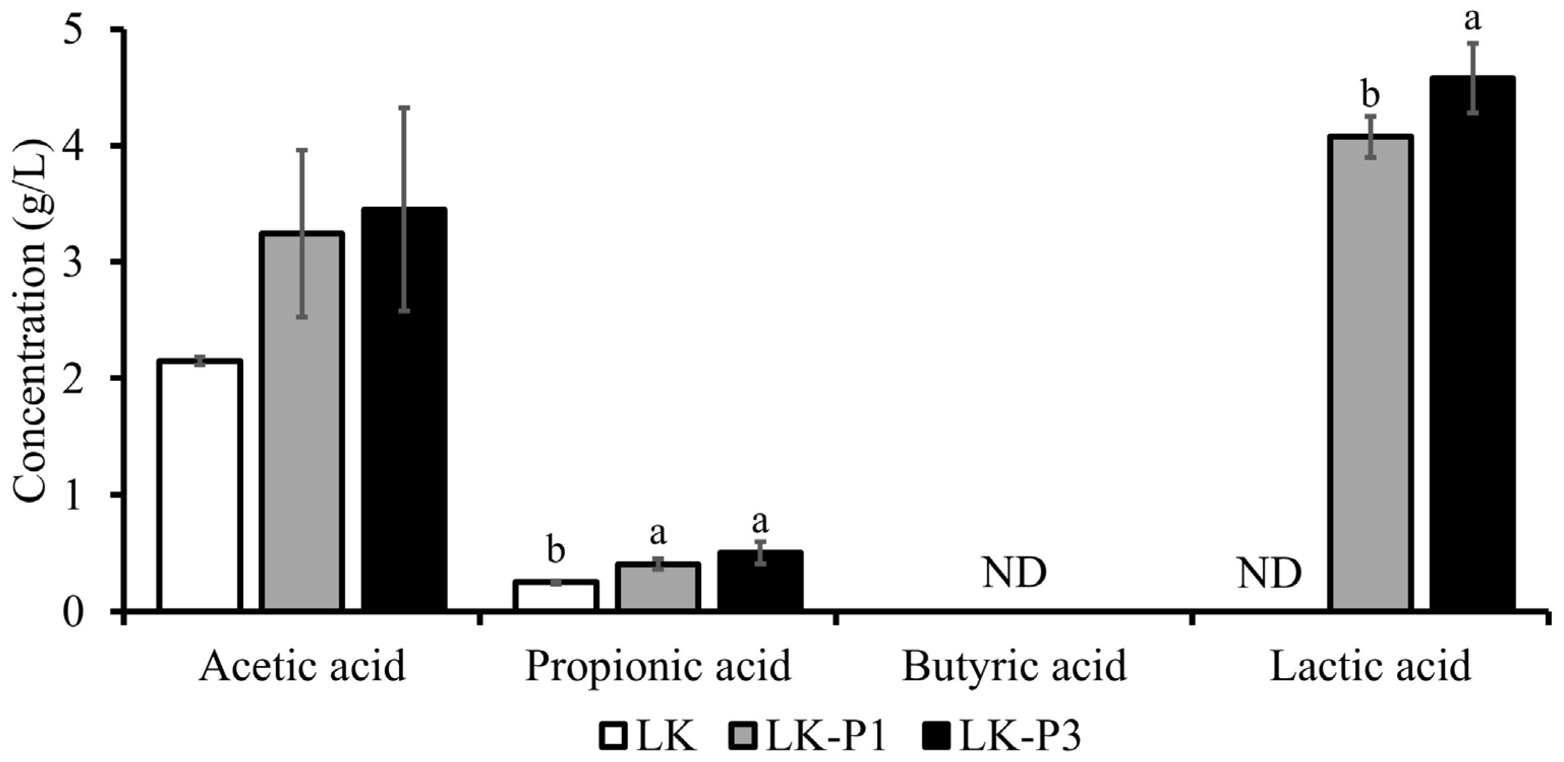
Short-chain fatty acids (SCFAs) are key metabolites produced by gut microbiota, with acetic acid, propionic acid, and butyric acid being the most representative types [57]. The concentration of acetic acid, propionic acid, butyric acid, and lactic acid in lotus root tea kombucha (LK) and L. plantarum-fermented lotus root tea kombucha (LK-P1, LK-P3) are shown in Figure 2.
The acetic acid concentration increased in the L. plantarum-fermented groups compared to LK. Specifically, LK had an acetic acid concentration of 2.15 g/L, whereas LK-P1 and LK-P3 contained 3.24 g/L and 3.45 g/L, respectively. Although this increase was not statistically significant, it suggests that L. plantarum fermentation may enhance acetic acid production.
The concentration of propionic acid also increased following fermentation. LK contained 0.25 g/L of propionic acid, while LK-P1 and LK-P3 had 0.41 g/L and 0.50 g/L, respectively. These differences between LK and both L. plantarum-fermented groups were statistically significant (p < 0.05), while no significant difference was observed between LK-P1 and LK-P3. Butyric acid was not detected (ND) in any of the samples.
The most pronounced change was observed in the lactic acid concentration. While LK contained no detectable lactic acid, its concentration significantly increased in the L. plantarum-fermented groups. LK-P1 exhibited a lactic acid concentration of 4.07 g/L, whereas LK-P3 reached 4.58 g/L. A significant difference (p < 0.05) was observed between LK-P1 and LK-P3, indicating that a higher inoculation level of L. plantarum enhances lactic acid production. A significant decrease in pH was observed in the L. plantarum-fermented groups, consistent with the increased production of organic acids, particularly lactic acid (Table S2).

3.3. Antioxidant Activity of Lotus Root Tea Kombucha

Antioxidant activity plays a crucial role in various physiological functions, including anti-aging, anti-inflammatory effects, and immune enhancement. The antioxidant activity of lotus root tea kombucha (LK) and L. plantarum-fermented lotus root tea kombucha (LK-P1, LK-P3) was evaluated using the DPPH radical scavenging assay (Figure 3A). Ascorbic acid (100 µg/mL) was used as a positive control and exhibited a high antioxidant activity of 90.79%. The antioxidant activity of LK at 1× concentration was 61.37%. Similarly, LK-P1 exhibited a scavenging activity of 61.36%, whereas LK-P3 showed a slightly lower scavenging activity of 53.99%.
Although no statistically significant differences were observed among the samples, LK and LK-P1 exhibited similar antioxidant activities (~61%), while LK-P3 showed a slightly lower scavenging activity (53.99%). Previous studies have reported antioxidant activities ranging from 35.4% to 60.3% for kombucha samples fermented under similar conditions [58], suggesting that lotus root tea kombucha possesses relatively high antioxidant activity.
In addition to DPPH analysis, the ABTS radical scavenging activity of lotus root tea kombucha and L. plantarum-fermented variants was also evaluated (Figure 3B). Ascorbic acid (100 µg/mL), used as the positive control, exhibited a high ABTS radical scavenging activity of 93.05%. The ABTS scavenging activities of LK, LK-P1, and LK-P3 were 92.99%, 93.08%, and 92.89%, respectively, showing uniformly high antioxidant potential across all treatments.
No statistically significant differences were observed among the samples, indicating that the addition of L. plantarum did not significantly influence the ABTS radical scavenging capacity under the tested conditions. These results suggest that lotus root tea kombucha itself possesses strong antioxidant activity, as measured using the ABTS assay.

3.4. Antimicrobial Activity of Lotus Root Tea Kombucha

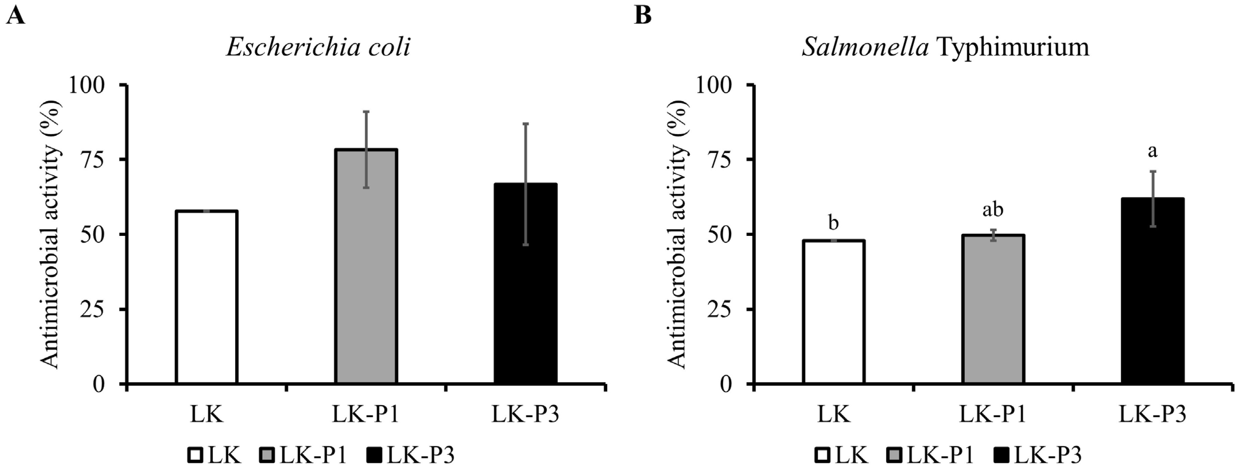
The antimicrobial activity of lotus root tea kombucha (LK) and Lactobacillus plantarum-fermented lotus root tea kombucha (LK-P1, LK-P3) was evaluated against Escherichia coli OP50 and Salmonella typhimurium (Figure 4).
Against E. coli OP50, the inhibition rates were 57.82% for LK, 78.33% for LK-P1, and 66.74% for LK-P3 (Figure 4A). The inhibitory activity of LK-P1 was approximately 1.35 times higher than that of LK, while LK-P3 exhibited a 1.15-fold increase. Although these differences were not statistically significant, L. plantarum-fermented lotus root tea kombucha (LK-P1 and LK-P3) exhibited a trend toward higher inhibitory activity compared to LK.
Similarly, against S. typhimurium, the inhibition rates were 47.88% for LK, 49.55% for LK-P1, and 61.88% for LK-P3 (Figure 4B). The inhibitory activity of LK-P1 was slightly higher than that of LK (1.04-fold increase), whereas LK-P3 exhibited a 1.29-fold increase, which was statistically significant (p < 0.05). Overall, lotus root tea kombucha exhibited approximately 50% antimicrobial activity against E. coli OP50 and S. typhimurium, and this activity was further enhanced by L. plantarum fermentation.

3.5. Inhibitory Effect of Lotus Root Tea Kombucha on β-Glucuronidase Activity

β-Glucuronidase activity is considered an important indicator of gut health, as its excessive levels are often associated with gut dysbiosis, inflammation, and an increased risk of colorectal diseases. Inhibiting β-glucuronidase activity can help maintain a healthier gut microbiota composition, reduce toxin reabsorption, and support overall gastrointestinal health.
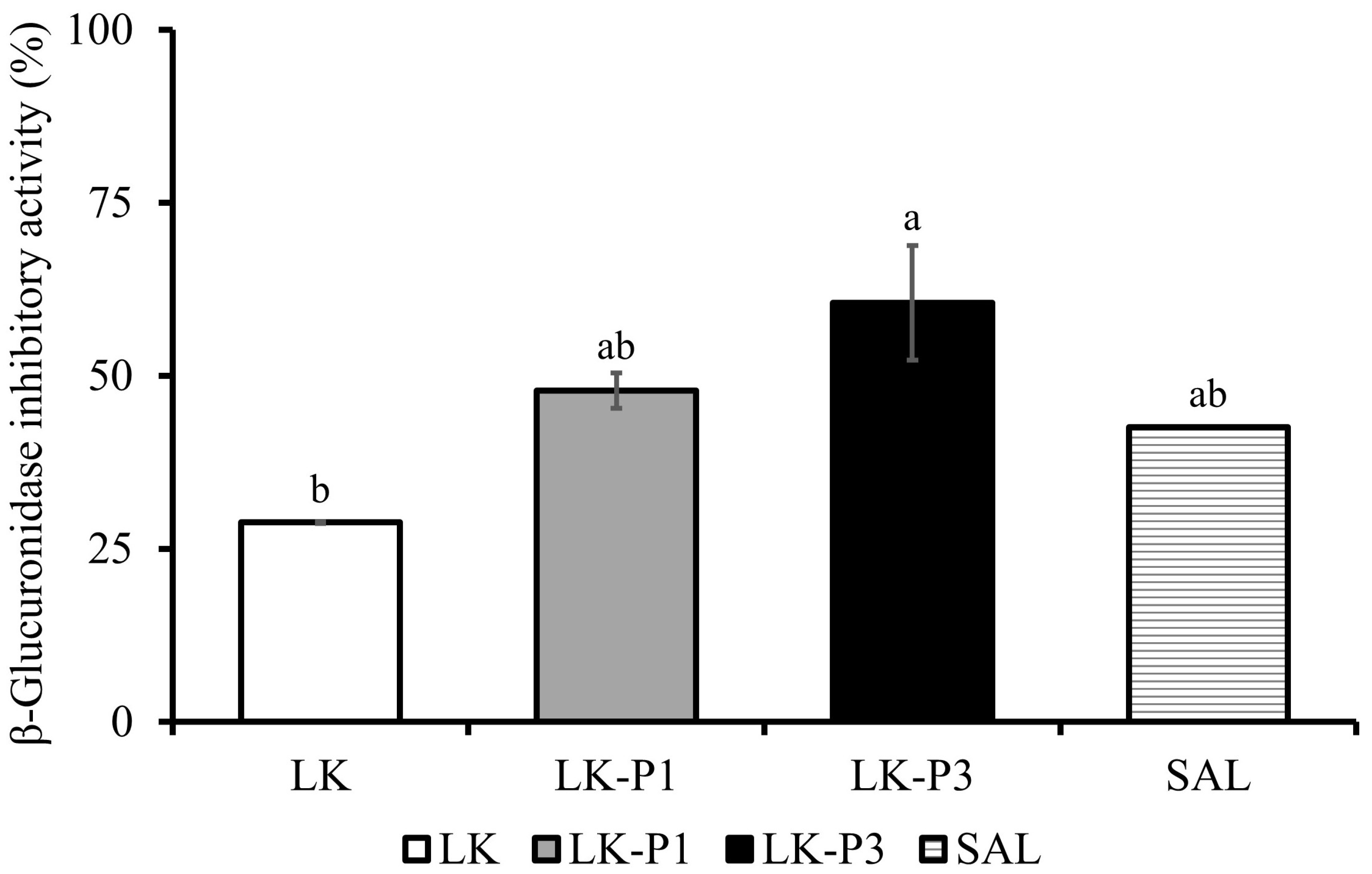
The β-glucuronidase inhibitory activity of lotus root tea kombucha (LK) and Lactobacillus plantarum-fermented lotus root tea kombucha (LK-P1 and LK-P3) was evaluated (Figure 5). The inhibition rates were 28.79% for LK, 47.84% for LK-P1, and 60.51% for LK-P3. Statistical analysis revealed no significant difference between LK and the positive control, Saccharic acid 1,4-lactone (SAL) (IC50 0.05 mM). However, LK-P3 exhibited significantly higher β-glucuronidase inhibition than LK (p < 0.05). No significant difference was observed between LK-P1 and LK-P3, suggesting that β-glucuronidase inhibition reached a plateau at higher L. plantarum inoculation levels. Overall, lotus root tea kombucha exhibited approximately 29% β-glucuronidase inhibitory activity, and this effect was further enhanced by L. plantarum fermentation, reaching up to 60% inhibition.

3.6. Protective Effect of Lotus Root Tea Kombucha Against Salmonella Infection Resistance in C. elegans

The protective effect of lotus root tea kombucha (LK) and Lactobacillus plantarum-fermented kombucha (LK-P1, LK-P3) against Salmonella enterica serovar Typhimurium infection in C. elegans was evaluated by assessing pharyngeal pumping activity and worm survival (Figure 6).
The pharyngeal pumping activity was measured to assess the feeding rate and ingestion ability of C. elegans [59]. The control group exhibited a pharyngeal pumping rate of 227.4 pumps/min, whereas worms exposed to LK showed a significant reduction (146.9 pumps/min) (Figure 6A). In contrast, worms treated with L. plantarum-fermented kombucha exhibited pumping rates of 250.6 pumps/min (LK-P1) and 227.3 pumps/min (LK-P3), showing no significant difference compared to the control group.
The effect of kombucha consumption on survival following S. typhimurium infection was evaluated by monitoring worm viability over time. The control group exhibited the shortest lifespan, with complete mortality observed by 132 h, after which survival monitoring was discontinued (Figure 6). In contrast, worms pre-fed with LK, LK-P1, or LK-P3 demonstrated prolonged survival. Statistical analysis confirmed that survival rates were significantly higher in all kombucha-treated groups compared to the control group (LK: p = 0.007, LK-P1: p = 0.003, and LK-P3: p < 0.001). L. plantarum fermentation further enhanced the protective effect, as lifespan was longer in LK-P1 and LK-P3 compared to LK.
At 12 h post infection, mortality in the control group was 17.6%, whereas LK-, LK-P1-, and LK-P3-treated worms exhibited mortality rates of 5.4%, 8.1%, and 1.4%, respectively (Figure 6C). This trend persisted throughout the experiment, with the control group reaching a peak mortality rate of 40.5% at 24 h. Similarly, LK and LK-P1 groups also exhibited peak mortality at 24 h, but at a lower rate of 29.7%. In contrast, the LK-P3 group exhibited a delayed peak mortality at 36 h, with a rate of 18.9%. These findings suggest that lotus root tea kombucha consumption may delay C. elegans mortality following S. typhimurium infection.

4. Discussion

In this study, lotus root tea kombucha (LK) exhibited inherent antimicrobial activity against Escherichia coli OP50 and Salmonella typhimurium, with enhanced inhibitory effects observed in LK-P1 and LK-P3. This suggests that lotus root tea kombucha itself possesses antimicrobial properties, likely due to the production of organic acids and bioactive metabolites during fermentation. The enhanced inhibition in LK-P1 and LK-P3 correlated with increased acetic acid, propionic acid, and lactic acid levels, with LK-P3 showing the highest lactic acid concentration (approximately 5 g/L). Organic acids such as short-chain fatty acids (SCFAs) and lactic acid exhibit strong antimicrobial activity, playing a crucial role in inhibiting harmful bacteria and improving the intestinal environment [60]. SCFAs also contribute to gut health by strengthening the intestinal mucosa and regulating immune function [61]. This increase in organic acids was accompanied by a significant pH reduction in LK-P1 (3.43) and LK-P3 (3.33) compared to LK (3.74), reinforcing their contribution to pathogen inhibition. These findings suggest that the addition of L. plantarum further amplified the antimicrobial potential of lotus root tea kombucha, likely by enhancing organic acid production and acidification.
In addition to its antimicrobial activity, lotus root tea kombucha exhibited significant β-glucuronidase inhibitory activity, with LK-P1 and LK-P3 showing enhanced effects. β-Glucuronidase is associated with the reactivation of carcinogens and other harmful substances, and its inhibition is linked to a reduced risk of colorectal cancer and intestinal inflammation. While this study did not directly confirm the presence of D-saccharic acid 1,4-lactone (SAL) in lotus root tea kombucha, previous research has identified SAL as a key β-glucuronidase inhibitor in traditional kombucha [62,63,64]. It is plausible that SAL may also be produced during lotus root tea kombucha fermentation, contributing to the observed enzyme-inhibitory activity. Additionally, kombucha has been reported to suppress the growth of β-glucuronidase-producing bacteria (Escherichia coli, Streptococcus spp., Clostridium spp.) [38,65,66,67,68], thereby reducing the overall enzyme levels in the gut. Polyphenols such as catechins, theaflavins, and thearubigins, which are commonly found in kombucha [69,70], may further support β-glucuronidase inhibition by suppressing β-glucuronidase-producing bacteria and protecting intestinal barrier integrity through antimicrobial and antioxidant properties.
Furthermore, the addition of L. plantarum to the kombucha fermentation process enhanced β-glucuronidase inhibition, suggesting a synergistic effect between kombucha-derived bioactive compounds and L. plantarum-mediated fermentation. This bacterium is known for its diverse physiological functions, including antimicrobial [71], anti-inflammatory [72], and enzyme-modulating activities [73]. Previous studies have reported its β-glucuronidase inhibitory effects, and the findings of this study align with these previous results [74]. The precise molecular mechanism underlying β-glucuronidase inhibition in lotus root tea kombucha remain unclear, but the results suggest that multiple factors, including SAL, polyphenols, organic acids, and microbiota modulation, may contribute to its inhibitory activity.
C. elegans has been widely utilized as a model organism to investigate the impact of gut microbiota on host physiology [75,76,77]. The ability to culture C. elegans under germ-free conditions allows for precise experimental control over microbial exposure, making it a valuable model for gut health research [77,78,79]. While previous studies have evaluated the activity of kombucha-derived microorganisms in C. elegans [80], research directly examining the effect of kombucha consumption in this model remains limited. In this study, we assessed the pharyngeal pumping activity to confirm whether kombucha consumption affects C. elegans feeding behavior. Our findings indicated that L. plantarum-fermented kombucha did not negatively impact feeding, as the pharyngeal pumping rate was comparable to that of the control group. This suggests that kombucha ingestion is well tolerated in C. elegans, supporting its suitability as a model for investigating the biological effects of kombucha.
The pathogenicity of S. typhimurium in C. elegans is characterized by its proliferation within the intestine, leading to increased mortality. Infection disrupts pharyngeal peristalsis, induces gut distension due to bacterial overgrowth, and damages the intestinal barrier [81]. In this study, worms pre-exposed to kombucha for 36 h were transferred to S. typhimurium-containing plates, and survival was monitored every 12 h. The results indicated that kombucha significantly extended lifespan by delaying mortality, suggesting a protective effect against S. typhimurium-induced lethality. This protective effect was particularly pronounced in worms exposed to L. plantarum-fermented lotus root tea kombucha, suggesting the potential synergistic effect of kombucha-derived bioactive compounds and L. plantarum fermentation.
Despite a three-fold difference in the L. plantarum inoculum levels, most in vitro functional parameters—including antimicrobial, antioxidant, and β-glucuronidase inhibitory activities—did not show statistically significant differences between LK-P1 and LK-P3. This may be due to a plateau effect, in which functional benefits saturate beyond a certain microbial threshold [82]. However, in the C. elegans infection model, LK-P3 exhibited a more pronounced protective effect, suggesting that higher probiotic concentrations can still exert biologically meaningful outcomes in vivo, even when the in vitro differences are minimal, as supported by previous studies [83].
The protective mechanism of kombucha may be attributed to its organic acid and bioactive compounds. Previous research has shown that SCFAs produced during kombucha fermentation promote the growth of beneficial gut bacteria, protect the intestinal mucosa, and reduce inflammation [84,85]. Additionally, in this study, we directly confirmed the antioxidant activity of lotus root tea kombucha, which may have contributed to mitigating S. typhimurium-induced damage [86]. While our findings provide valuable insights into the potential protective effects of kombucha against S. typhimurium infection, further research is required to elucidate the precise mechanisms underlying its lifespan-extending properties. Future studies should focus on identifying the specific kombucha-derived metabolites responsible for these effects and exploring their interactions with host immune responses and gut microbiota.

5. Conclusions

This study demonstrated that lotus root tea can be used as a low-caffeine base for kombucha production. When further fermented with Lactobacillus plantarum, the resulting kombucha exhibited enhanced functional properties, including increased levels of organic acids, improved antimicrobial activity, and greater inhibition of β-glucuronidase. Moreover, consumption of the fermented kombucha improved survival in Caenorhabditis elegans following Salmonella infection, suggesting protective effects on host health.
These results highlight the potential of lotus root tea kombucha as a functional, non-caffeinated beverage with gut-health-promoting properties. However, further in vivo or clinical studies are required to confirm its health benefits in humans. Future research could also explore its underlying mechanisms of action and assess its sensory acceptability for consumer applications.

Supplementary Materials

The following supporting information can be downloaded at: https://www.mdpi.com/article/10.3390/beverages11020055/s1. Table S1: β-glucuronidase inhibitory activity of lotus root kombucha fermented with varying concentrations of Lactobacillus plantarum. Table S2: pH of lotus root tea kombucha and Lactobacillus-fermented lotus root tea kombucha.

Author Contributions

Investigation, J.S.B. and Y.N.; project administration, S.K.; resources, H.S.K. and E.J.L.; supervision, S.R.K. and M.-R.L.; visualization, J.S.B.; writing—original draft preparation, J.S.B. and S.K.; writing—review and editing, S.R.K. All authors have read and agreed to the published version of the manuscript.

Funding

This research was financially supported by the Ministry of Small and Medium-sized Enterprises (SMEs) and Startups (MSS), Republic of Korea, under the “Regional Specialized Industry Devel-opment Plus Program (R&D, S3401227)” supervised by the Korea Technology and Information Promotion Agency for SMEs.

Institutional Review Board Statement

Not applicable.

Informed Consent Statement

Not applicable.

Data Availability Statement

The original contributions presented in the study are included in the article/Supplementary Materials; further inquiries can be directed to the corresponding authors.

Conflicts of Interest

The authors declare no conflicts of interest.

Abbreviations

The following abbreviations are used in this manuscript:
CFUColony-forming unit
DPPH2,2-Diphenyl-1-Picrylhydrazyl
HPLCHigh-performance liquid chromatography
LABLactic acid bacteria
LKLotus root tea kombucha
LK-P1Lotus root tea kombucha supplemented with 1.0 × 10⁹ CFU/mL of L. plantarum
LK-P3Lotus root tea kombucha supplemented with 3.0 × 10⁹ CFU/mL of L. plantarum
NGMNematode growth medium
ODOptical density
SALD-saccharic acid 1,4-lactone
SCOBYSymbiotic culture of bacteria and yeast
SCFAsShort-chain fatty acids

References

  1. Troitino, C. Kombucha 101: Demystifying the Past, Present and Future of the Fermented Tea Drink. Forbes Website. Available online: https://www.forbes.com/sites/christinatroitino/2017/02/01/kombucha-101-demystifying-the-past-present-and-future-of-the-fermented-tea-drink/ (accessed on 1 February 2017).
  2. Baschali, A.; Tsakalidou, E.; Kyriacou, A.; Karavasiloglou, N.; Matalas, A.-L. Traditional low-alcoholic and non-alcoholic fermented beverages consumed in European countries: A neglected food group. Nutr. Res. Rev. 2017, 30, 1–24. [Google Scholar] [CrossRef] [PubMed]
  3. Kapp, J.M.; Sumner, W. Kombucha: A systematic review of the empirical evidence of human health benefit. Ann. Epidemiol. 2019, 30, 66–70. [Google Scholar] [CrossRef] [PubMed]
  4. Içen, H.; Corbo, M.R.; Sinigaglia, M.; Korkmaz, B.I.O.; Bevilacqua, A. Microbiology and antimicrobial effects of kombucha, a short overview. Food Biosci. 2023, 56, 103270. [Google Scholar] [CrossRef]
  5. Ivanišová, E.; Meňhartová, K.; Terentjeva, M.; Harangozo, Ľ.; Kántor, A.; Kačániová, M. The evaluation of chemical, antioxidant, antimicrobial and sensory properties of kombucha tea beverage. J. Food Sci. Technol. 2020, 57, 1840–1846. [Google Scholar] [CrossRef]
  6. Dittoe, D.K.; Ricke, S.C.; Kiess, A.S. Organic acids and potential for modifying the avian gastrointestinal tract and reducing pathogens and disease. Front. Vet. Sci. 2018, 5, 216. [Google Scholar] [CrossRef]
  7. rDarbandi, A.; Asadi, A.; Mahdizade Ari, M.; Ohadi, E.; Talebi, M.; Halaj Zadeh, M.; Darb Emamie, A.; Ghanavati, R.; Kakanj, M. Bacteriocins: Properties and potential use as antimicrobials. J. Clin. Lab. Anal. 2022, 36, e24093. [Google Scholar] [CrossRef]
  8. Arpaia, N.; Rudensky, A.Y. Microbial metabolites control gut inflammatory responses. Proc. Natl. Acad. Sci. USA 2014, 111, 2058–2059. [Google Scholar] [CrossRef] [PubMed]
  9. Anantachoke, N.; Duangrat, R.; Sutthiphatkul, T.; Ochaikul, D.; Mangmool, S. Kombucha beverages produced from fruits, vegetables, and plants: A review on their pharmacological activities and health benefits. Foods 2023, 12, 1818. [Google Scholar] [CrossRef]
  10. Martínez Leal, J.; Valenzuela Suárez, L.; Jayabalan, R.; Huerta Oros, J.; Escalante-Aburto, A. A review on health benefits of kombucha nutritional compounds and metabolites. CyTA-J. Food 2018, 16, 390–399. [Google Scholar] [CrossRef]
  11. Villarreal-Soto, S.A.; Beaufort, S.; Bouajila, J.; Souchard, J.P.; Taillandier, P. Understanding kombucha tea fermentation: A review. J. Food Sci. 2018, 83, 580–588. [Google Scholar] [CrossRef]
  12. Zhou, D.-D.; Saimaiti, A.; Luo, M.; Huang, S.-Y.; Xiong, R.-G.; Shang, A.; Gan, R.-Y.; Li, H.-B. Fermentation with tea residues enhances antioxidant activities and polyphenol contents in kombucha beverages. Antioxidants 2022, 11, 155. [Google Scholar] [CrossRef] [PubMed]
  13. Crum, H.; LaGory, A. The Big Book of Kombucha: Brewing, Flavoring, and Enjoying the Health Benefits of Fermented Tea; Storey Publishing: North Adams, MA, USA, 2016. [Google Scholar]
  14. Nawrot, P.; Jordan, S.; Eastwood, J.; Rotstein, J.; Hugenholtz, A.; Feeley, M. Effects of caffeine on human health. Food Addit. Contam. 2003, 20, 1–30. [Google Scholar] [CrossRef]
  15. Lovallo, W.R.; Whitsett, T.L.; al’Absi, M.; Sung, B.H.; Vincent, A.S.; Wilson, M.F. Caffeine stimulation of cortisol secretion across the waking hours in relation to caffeine intake levels. Biopsychosoc. Sci. Med. 2005, 67, 734–739. [Google Scholar] [CrossRef] [PubMed]
  16. Parvin, R.; Bhattacharya, S.; Chaudhury, S.S.; Roy, U.; Mukherjee, J.; Gachhui, R. Production, purification and characterization of a novel thermostable caffeine dehydrogenase from Pichia manshurica strain CD1 isolated from kombucha tea. Microbiology 2023, 92, 230–241. [Google Scholar] [CrossRef]
  17. Saimaiti, A.; Zhou, D.-D.; Li, J.; Xiong, R.-G.; Gan, R.-Y.; Huang, S.-Y.; Shang, A.; Zhao, C.-N.; Li, H.-Y.; Li, H.-B. Dietary sources, health benefits, and risks of caffeine. Crit. Rev. Food Sci. Nutr. 2023, 63, 9648–9666. [Google Scholar] [CrossRef] [PubMed]
  18. Showkat, Q.A.; Rather, J.A.; Jabeen, A.; Dar, B.; Makroo, H.A.; Majid, D. Bioactive components, physicochemical and starch characteristics of different parts of lotus (Nelumbo nucifera Gaertn.) plant: A review. Int. J. Food Sci. Technol. 2021, 56, 2205–2214. [Google Scholar] [CrossRef]
  19. Yi, Y.; Huang, X.-Y.; Zhong, Z.-T.; Huang, F.; Li, S.-Y.; Wang, L.-M.; Min, T.; Wang, H.-X. Structural and biological properties of polysaccharides from lotus root. Int. J. Biol. Macromol. 2019, 130, 454–461. [Google Scholar] [CrossRef]
  20. Guan, X.; Feng, Y.; Jiang, Y.; Hu, Y.; Zhang, J.; Li, Z.; Song, C.; Li, F.; Hou, J.; Shen, T. Simulated digestion and in vitro fermentation of a polysaccharide from lotus (Nelumbo nucifera Gaertn.) root residue by the human gut microbiota. Food Res. Int. 2022, 155, 111074. [Google Scholar] [CrossRef]
  21. Zheng, Z.; Gao, W.; Zhu, Z.; Li, S.; Chen, X.; Cravotto, G.; Sui, Y.; Zhou, L. Complexes of Soluble Dietary Fiber and Polyphenols from Lotus Root Regulate High-Fat Diet-Induced Hyperlipidemia in Mice. Antioxidants 2024, 13, 466. [Google Scholar] [CrossRef]
  22. Bangar, S.P.; Dunno, K.; Kumar, M.; Mostafa, H.; Maqsood, S. A comprehensive review on lotus seeds (Nelumbo nucifera Gaertn.): Nutritional composition, health-related bioactive properties, and industrial applications. J. Funct. Foods 2022, 89, 104937. [Google Scholar] [CrossRef]
  23. You, J.S.; Lee, Y.J.; Kim, K.S.; Kim, S.H.; Chang, K.J. Ethanol extract of lotus (Nelumbo nucifera) root exhibits an anti-adipogenic effect in human pre-adipocytes and anti-obesity and anti-oxidant effects in rats fed a high-fat diet. Nutr. Res. 2014, 34, 258–267. [Google Scholar] [CrossRef] [PubMed]
  24. Mukherjee, D.; Khatua, T.N.; Venkatesh, P.; Saha, B.; Mukherjee, P.K. Immunomodulatory potential of rhizome and seed extracts of Nelumbo nucifera Gaertn. J. Ethnopharmacol. 2010, 128, 490–494. [Google Scholar] [CrossRef]
  25. Tsuruta, Y.; Nagao, K.; Shirouchi, B.; Nomura, S.; Tsuge, K.; Koganemaru, K.; Yanagita, T. Effects of lotus root (the edible rhizome of Nelumbo nucifera) on the deveolopment of non-alcoholic fatty liver disease in obese diabetic db/db mice. Biosci. Biotechnol. Biochem. 2012, 76, 462–466. [Google Scholar] [CrossRef] [PubMed]
  26. Mukherjee, P.K.; Saha, K.; Pal, M.; Saha, B. Effect of Nelumbo nucifera rhizome extract on blood sugar level in rats. J. Ethnopharmacol. 1997, 58, 207–213. [Google Scholar] [CrossRef]
  27. Mukherjee, P.K.; Saha, K.; Balasubramanian, R.; Pal, M.; Saha, B. Studies on psychopharmacological effects of Nelumbo nucifera Gaertn. rhizome extract. J. Ethnopharmacol. 1996, 54, 63–67. [Google Scholar] [CrossRef] [PubMed]
  28. Yang, W.M.; Shim, K.J.; Choi, M.J.; Park, S.Y.; Choi, B.-J.; Chang, M.S.; Park, S.K. Novel effects of Nelumbo nucifera rhizome extract on memory and neurogenesis in the dentate gyrus of the rat hippocampus. Neurosci. Lett. 2008, 443, 104–107. [Google Scholar] [CrossRef]
  29. Yi, Y.; Sun, J.; Xie, J.; Min, T.; Wang, L.-M.; Wang, H.-X. Phenolic profiles and antioxidant activity of lotus root varieties. Molecules 2016, 21, 863. [Google Scholar] [CrossRef] [PubMed]
  30. Li, S.; Li, J.; Zhu, Z.; Cheng, S.; He, J.; Lamikanra, O. Soluble dietary fiber and polyphenol complex in lotus root: Preparation, interaction and identification. Food Chem. 2020, 314, 126219. [Google Scholar] [CrossRef]
  31. Tan, S.-J.; Lee, C.-K.; Gan, C.-Y.; Olalere, O.A. Statistical Optimization of Flavonoid and Antioxidant Recovery from Macerated Chinese and Malaysian Lotus Root (Nelumbo nucifera) Using Response Surface Methodology. Molecules 2021, 26, 2014. [Google Scholar] [CrossRef]
  32. Dong, W.; Liu, X.; Yi, Y.; Wang, L.; Hou, W.; Ai, Y.; Wang, H.; Min, T. Evaluation of Pre-Harvest Nutrient Composition and Functional Active Substances in Various Lotus Roots. Foods 2024, 13, 2297. [Google Scholar] [CrossRef]
  33. Saeed, S.M.G.; Tayyaba, S.; Ali, S.A.; Tayyab, S.; Sayeed, S.A.; Ali, R.; Mobin, L.; Naz, S. Evaluation of the potential of Lotus root (Nelumbo nucifera) flour as a fat mimetic in biscuits with improved functional and nutritional properties. CyTA-J. Food 2020, 18, 624–634. [Google Scholar] [CrossRef]
  34. Hwang, E.-S.; Lee, S. Quality characteristics, antioxidant activity, and acrylamide content of lotus root chips prepared using different processing methods. Food Sci. Biotechnol. 2024, 33, 1371–1379. [Google Scholar] [CrossRef]
  35. Dufresne, C.; Farnworth, E. Tea, Kombucha, and health: A review. Food Res. Int. 2000, 33, 409–421. [Google Scholar] [CrossRef]
  36. Masood, M.I.; Qadir, M.I.; Shirazi, J.H.; Khan, I.U. Beneficial effects of lactic acid bacteria on human beings. Crit. Rev. Microbiol. 2011, 37, 91–98. [Google Scholar] [CrossRef]
  37. Yang, Z.; Zhou, F.; Ji, B.; Li, B.; Luo, Y.; Yang, L.; Li, T. Symbiosis between microorganisms from kombucha and kefir: Potential significance to the enhancement of kombucha function. Appl. Biochem. Biotechnol. 2010, 160, 446–455. [Google Scholar] [CrossRef]
  38. Beaud, D.; Tailliez, P.; Anba-Mondoloni, J. Genetic characterization of the β-glucuronidase enzyme from a human intestinal bacterium, Ruminococcus gnavus. Microbiology 2005, 151, 2323–2330. [Google Scholar] [CrossRef] [PubMed]
  39. Ejike, E.N. Probiotic Potentials of Lactobacillus plantarumFMO2, Lactobacillus plantarumZ2 and Lactobacillus pentosusBSR3 Isolated from Fermented Starchy Foods. Curr. Trends Food Sci. 2024, 1. [Google Scholar]
  40. Chen, J.; Mou, L.; Wang, L.; Wu, G.; Dai, X.; Chen, Q.; Zhang, J.; Luo, X.; Xu, F.; Zhang, M. Mixed Bacillus subtilis and Lactiplantibacillus plantarum-fermented feed improves gut microbiota and immunity of Bamei piglet. Front. Microbiol. 2024, 15, 1442373. [Google Scholar] [CrossRef]
  41. Chen, Z.; Liu, Z.; Chen, C.; Fu, Y.; Wang, H.; Zhao, C.; Zhang, J.; Liu, L. The Effect of an Early-Life Lactiplantibacillus plantarum LPJZ-658 Intervention on Performance and Gut Microbiota in Suckling Piglets. Microbiol. Res. 2025, 16, 41. [Google Scholar] [CrossRef]
  42. Biernat, K.A.; Pellock, S.J.; Bhatt, A.P.; Bivins, M.M.; Walton, W.G.; Tran, B.N.T.; Wei, L.; Snider, M.C.; Cesmat, A.P.; Tripathy, A. Structure, function, and inhibition of drug reactivating human gut microbial β-glucuronidases. Sci. Rep. 2019, 9, 825. [Google Scholar] [CrossRef]
  43. Nguyen, N.K.; Dong, N.T.; Le, P.H.; Nguyen, H.T. Evaluation of the glucuronic acid production and other biological activities of fermented sweeten-black tea by kombucha layer and the co-culture with different Lactobacillus sp. strains. Int. J. Mod. Eng. Res. 2014, 4, 12–17. [Google Scholar]
  44. Plessas, S. Advancements in the use of fermented fruit juices by lactic acid bacteria as functional foods: Prospects and challenges of Lactiplantibacillus (Lpb.) plantarum subsp. plantarum application. Fermentation 2021, 8, 6. [Google Scholar] [CrossRef]
  45. 이미령; 김희송; 양동훈; 김익중. Method for Manufacturing Fermented Tea Using Lotus Root and Fermented Tea Produced Thereby. Korean Patent KR1020200061670, 17 October 2022. (In Korean). [CrossRef]
  46. Wanyika, H.; Gatebe, E.; Gitu, L.; Ngumba, E.; Maritim, C. Determination of caffeine content of tea and instant coffee brands found in the Kenyan market. Afr. J. Food Sci. 2010, 4, 353–358. [Google Scholar]
  47. Xie, J.; Schaich, K. Re-evaluation of the 2, 2-diphenyl-1-picrylhydrazyl free radical (DPPH) assay for antioxidant activity. J. Agric. Food Chem. 2014, 62, 4251–4260. [Google Scholar] [CrossRef] [PubMed]
  48. Thanh, N.C.; Chinnathambi, A.; Alahmadi, T.A.; Joshi, D.; Jhanani, G.; Haile, A. An Assessment of Anticancer Activity (MCF-7) and Free Radicals Scavenging Potential of Ferula Asafoetida Synthesized AgNPs through In Vitro Analyses. J. Nanomater. 2022, 2022, 6095005. [Google Scholar] [CrossRef]
  49. Hyrslova, I.; Kana, A.; Nesporova, V.; Mrvikova, I.; Doulgeraki, A.I.; Lampova, B.; Doskocil, I.; Musilova, S.; Kieliszek, M.; Krausova, G. In vitro digestion and characterization of selenized Saccharomyces cerevisiae, Pichia fermentans and probiotic Saccharomyces boulardii. J. Trace Elem. Med. Biol. 2024, 83, 127402. [Google Scholar] [CrossRef]
  50. Medina-Pérez, G.; Peralta-Adauto, L.; Afanador-Barajas, L.; Fernández-Luqueño, F.; Pérez-Soto, E.; Campos-Montiel, R.; Peláez-Acero, A. Inhibition of urease, elastase, and β-glucuronidase enzymatic activity by applying aqueous extracts of opuntia oligacantha CF först acid fruits: In vitro essay under simulated digestive conditions. Appl. Sci. 2021, 11, 7705. [Google Scholar] [CrossRef]
  51. Liew, S.Y.; Sivasothy, Y.; Shaikh, N.N.; Isa, D.M.; Lee, V.S.; Choudhary, M.I.; Awang, K. β-Glucuronidase inhibitors from Malaysian plants. J. Mol. Struct. 2020, 1221, 128743. [Google Scholar] [CrossRef]
  52. Yun, B.; Ryu, S.; Kang, M.; Lee, J.; Yoo, J.; Kim, Y.; Oh, S. Probiotic Lacticaseibacillus rhamnosus GG increased longevity and resistance against foodborne pathogens in Caenorhabditis elegans by regulating microRNA miR-34. Front. Cell. Infect. Microbiol. 2022, 11, 819328. [Google Scholar] [CrossRef]
  53. Trojanowski, N.F.; Raizen, D.M.; Fang-Yen, C. Pharyngeal pumping in Caenorhabditis elegans depends on tonic and phasic signaling from the nervous system. Sci. Rep. 2016, 6, 22940. [Google Scholar] [CrossRef]
  54. Kim, S.; Lee, Y.-R.; Yang, H.; Park, C.-H.; Yun, C.-S.; Jang, B.-C.; Hong, Y.; Park, D.-S. Potential probiotic Lactiplantibacillus plantarum DS1800 extends lifespan and enhances stress resistance in Caenorhabditis elegans model. Front. Physiol. 2024, 15, 1476096. [Google Scholar] [CrossRef] [PubMed]
  55. Chakravorty, S.; Bhattacharya, S.; Chatzinotas, A.; Chakraborty, W.; Bhattacharya, D.; Gachhui, R. Kombucha tea fermentation: Microbial and biochemical dynamics. Int. J. Food Microbiol. 2016, 220, 63–72. [Google Scholar] [CrossRef]
  56. Yang, J.; Lagishetty, V.; Kurnia, P.; Henning, S.M.; Ahdoot, A.I.; Jacobs, J.P. Microbial and chemical profiles of commercial kombucha products. Nutrients 2022, 14, 670. [Google Scholar] [CrossRef] [PubMed]
  57. Akhtar, M.; Chen, Y.; Ma, Z.; Zhang, X.; Shi, D.; Khan, J.A.; Liu, H. Gut microbiota-derived short chain fatty acids are potential mediators in gut inflammation. Anim. Nutr. 2022, 8, 350–360. [Google Scholar] [CrossRef] [PubMed]
  58. Chu, S.-C.; Chen, C. Effects of origins and fermentation time on the antioxidant activities of kombucha. Food Chem. 2006, 98, 502–507. [Google Scholar] [CrossRef]
  59. Han, S.; Hu, F.; Ji, X.; Liu, Y.; Zhang, S.; Wang, Z.; Qiao, K. Polysaccharides from Ziziphus jujuba prolong lifespan and attenuate oxidative stress in Caenorhabditis elegans via DAF-16 and SKN-1. Int. J. Biol. Macromol. 2024, 282, 137482. [Google Scholar] [CrossRef]
  60. Hadinia, N.; Dovom, M.R.E.; Yavarmanesh, M. The effect of fermentation conditions (temperature, salt concentration, and pH) with lactobacillus strains for producing short chain fatty acids. LWT 2022, 165, 113709. [Google Scholar] [CrossRef]
  61. Liu, X.-F.; Shao, J.-H.; Liao, Y.-T.; Wang, L.-N.; Jia, Y.; Dong, P.-J.; Liu, Z.-z.; He, D.-D.; Li, C.; Zhang, X. Regulation of short-chain fatty acids in the immune system. Front. Immunol. 2023, 14, 1186892. [Google Scholar] [CrossRef]
  62. Diez, T.; Cabezas, J.A. Properties of Two Molecular Forms of β-Glucuronidase from the Mollusc Littorina littorea L. Eur. J. Biochem. 1979, 93, 301–311. [Google Scholar] [CrossRef]
  63. KIM, D.; JIN, Y.; JUNG, E.; HAN, M.; Kobashi, K. Purification and characterization of β-glucuronidase from Escherichia coli HGU-3, a human intestinal bacterium. Biol. Pharm. Bull. 1995, 18, 1184–1188. [Google Scholar] [CrossRef]
  64. Yang, Z.W.; Ji, B.P.; Zhou, F.; Li, B.; Luo, Y.; Yang, L.; Li, T. Hypocholesterolaemic and antioxidant effects of kombucha tea in high-cholesterol fed mice. J. Sci. Food Agric. 2009, 89, 150–156. [Google Scholar] [CrossRef]
  65. Sreeramulu, G.; Zhu, Y.; Knol, W. Kombucha fermentation and its antimicrobial activity. J. Agric. Food Chem. 2000, 48, 2589–2594. [Google Scholar] [CrossRef] [PubMed]
  66. Leung, J.W.; Liu, Y.-l.; Leung, P.S.; Chan, R.C.; Inciardi, J.F.; Cheng, A.F. Expression of bacterial β-glucuronidase in human bile: An in vitro study. Gastrointest. Endosc. 2001, 54, 346–350. [Google Scholar] [CrossRef] [PubMed]
  67. Al-Kalifawi, E. Study the antimicrobial effect of kombucha tea on bacteria isolated from diabetic foot ulcer. J. Biotechnol. Res. Cent. (JOBRC) 2014, 8, 27–33. [Google Scholar] [CrossRef]
  68. Jung, Y.; Kim, I.; Mannaa, M.; Kim, J.; Wang, S.; Park, I.; Kim, J.; Seo, Y.-S. Effect of Kombucha on gut-microbiota in mouse having non-alcoholic fatty liver disease. Food Sci. Biotechnol. 2019, 28, 261–267. [Google Scholar] [CrossRef] [PubMed]
  69. Molan, A.-L.; Mahdy, A.S. Iraqi medicinal plants: Total flavonoid contents, free-radical scavenging and bacterial beta-glucuronidase inhibition activities. IOSR J. Dent. Med. Sci. 2014, 13, 72–77. [Google Scholar] [CrossRef]
  70. Gade, A.; Kumar, M.S. Gut microbial metabolites of dietary polyphenols and their potential role in human health and diseases. J. Physiol. Biochem. 2023, 79, 695–718. [Google Scholar] [CrossRef]
  71. Dinev, T.; Beev, G.; Tzanova, M.; Denev, S.; Dermendzhieva, D.; Stoyanova, A. Antimicrobial activity of Lactobacillus plantarum against pathogenic and food spoilage microorganisms: A review. Bulg. J. Vet. Med. 2018, 21, 253–268. [Google Scholar] [CrossRef]
  72. Zare, D.; Aryaee, H.; Mirdamadi, S.; Shirkhan, F. The Benefits and Applications of Lactobacillus plantarum in Food and Health: A Narrative Review. Iran. J. Public Health 2024, 53, 2201. [Google Scholar] [CrossRef]
  73. Ma, H.; Dong, A.; Xu, Y.; Wu, Q.; Lambo, M.T.; Zhang, Y.; Dou, X.; Li, Y. Regulatory effects of high concentrate diet synergistically fermented with cellulase and lactic acid bacteria: In vitro ruminal fermentation, methane production, and rumen microbiome. Anim. Feed Sci. Technol. 2025, 319, 116194. [Google Scholar] [CrossRef]
  74. Arenahalli Ningegowda, M.; Siddalingaiya Gurudutt, P. In vitro fermentation of prebiotics by Lactobacillus plantarum CFR 2194: Selectivity, viability and effect of metabolites on β-glucuronidase activity. World J. Microbiol. Biotechnol. 2012, 28, 901–908. [Google Scholar] [CrossRef]
  75. An, L.; Fu, X.; Chen, J.; Ma, J. Application of Caenorhabditis elegans in lipid metabolism research. Int. J. Mol. Sci. 2023, 24, 1173. [Google Scholar] [CrossRef] [PubMed]
  76. Hashmi, S.; Wang, Y.; Parhar, R.S.; Collison, K.S.; Conca, W.; Al-Mohanna, F.; Gaugler, R. A C. elegans model to study human metabolic regulation. Nutr. Metab. 2013, 10, 31. [Google Scholar] [CrossRef]
  77. Zhang, F.; Berg, M.; Dierking, K.; Félix, M.-A.; Shapira, M.; Samuel, B.S.; Schulenburg, H. Caenorhabditis elegans as a model for microbiome research. Front. Microbiol. 2017, 8, 485. [Google Scholar] [CrossRef] [PubMed]
  78. Cabreiro, F.; Gems, D. Worms need microbes too: Microbiota, health and aging in Caenorhabditis elegans. EMBO Mol. Med. 2013, 5, 1300–1310. [Google Scholar] [CrossRef]
  79. Labrousse, A.; Chauvet, S.; Couillault, C.; Kurz, C.L.; Ewbank, J.J. Caenorhabditis elegans is a model host for Salmonella typhimurium. Curr. Biol. 2000, 10, 1543–1545. [Google Scholar] [CrossRef] [PubMed]
  80. DuMez-Kornegay, R.N.; Baker, L.S.; Morris, A.J.; DeLoach, W.L.; Dowen, R.H. Kombucha Tea-associated microbes remodel host metabolic pathways to suppress lipid accumulation. PLoS Genet. 2024, 20, e1011003. [Google Scholar] [CrossRef]
  81. Aballay, A.; Yorgey, P.; Ausubel, F.M. Salmonella typhimurium proliferates and establishes a persistent infection in the intestine of Caenorhabditis elegans. Curr. Biol. 2000, 10, 1539–1542. [Google Scholar] [CrossRef]
  82. Yang, T.; Fan, X.; Li, D.; Zhao, T.; Wu, D.; Liu, Z.; Long, D.; Li, B.; Huang, X. High Antioxidant capacity of Lacticaseibacillus paracasei TDM-2 and Pediococcus pentosaceus TCM-3 from Qinghai Tibetan plateau and their function towards gut modulation. Foods 2023, 12, 1814. [Google Scholar] [CrossRef]
  83. Frece, J.; Kos, B.; Beganović, J.; Vuković, S.; Šušković, J. In vivo testing of functional properties of three selected probiotic strains. World J. Microbiol. Biotechnol. 2005, 21, 1401–1408. [Google Scholar] [CrossRef]
  84. Faghfuri, E.; Gholizadeh, P. The role of Akkermansia muciniphila in colorectal cancer: A double-edged sword of treatment or disease progression? Biomed. Pharmacother. 2024, 173, 116416. [Google Scholar] [CrossRef] [PubMed]
  85. Feizi, H.; Plotnikov, A.; Rezaee, M.A.; Ganbarov, K.; Kamounah, F.S.; Nikitin, S.; Kadkhoda, H.; Gholizadeh, P.; Pagliano, P.; Kafil, H.S. Postbiotics versus probiotics in early-onset colorectal cancer. Crit. Rev. Food Sci. Nutr. 2024, 64, 3573–3582. [Google Scholar] [CrossRef] [PubMed]
  86. Geronikaki, A.A.; Gavalas, A.M. Antioxidants and inflammatory disease: Synthetic and natural antioxidants with anti-inflammatory activity. Comb. Chem. High Throughput Screen. 2006, 9, 425–442. [Google Scholar] [CrossRef] [PubMed]
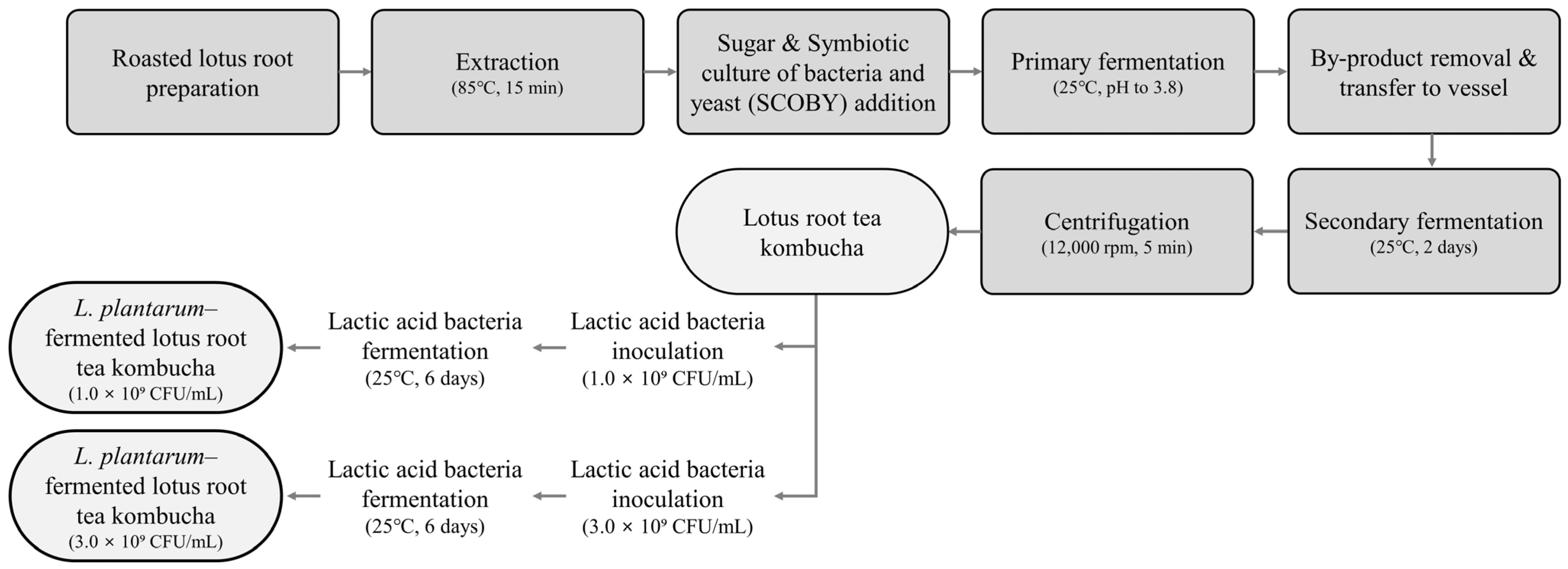
Figure 1. Processing flow chart for the preparation of lotus root tea kombucha (LK) and Lactobacillus plantarum-fermented variants.
Beverages 11 00055 g001
Figure 2. Organic acid profiles of lotus root tea kombucha with and without Lactobacillus plantarum fermentation. LK represents kombucha without L. plantarum, while LK-P1 and LK-P3 represent kombucha supplemented with 1.0 × 10⁹ CFU/mL and 3.0 × 10⁹ CFU/mL of L. plantarum, respectively. The concentrations of acetic acid, propionic acid, butyric acid, and lactic acid were quantified using calibration curves for each compound and expressed as g/L. ND indicates that the compound was not detected. Statistical significance for each organic acid was determined using Tukey’s test (p < 0.05), and different letters indicate significant differences among them. When no significant differences were found, no letters were shown. Error bars represent standard deviations.
Beverages 11 00055 g002
Figure 3. Antioxidant activity of lotus root tea kombucha measured via (A) DPPH and (B) ABTS radical scavenging assays. LK represents kombucha without Lactobacillus plantarum, while LK-P1 and LK-P3 represent kombucha supplemented with 1.0 × 10⁹ CFU/mL and 3.0 × 10⁹ CFU/mL of L. plantarum, respectively. Statistical significance was determined using Tukey’s test (SPSS, n = 3), and different letters indicate significant differences (p < 0.05). When no significant differences were found, no letters were shown. Error bars represent standard deviations.
Beverages 11 00055 g003
Figure 4. Antimicrobial activity of lotus root tea kombucha against Escherichia coli and Salmonella typhimurium. Antimicrobial activity of lotus root tea kombucha (LK) and Lactobacillus-fermented LK against E. coli OP50 (A) and S. typhimurium ATCC 13311 (B). LK represents kombucha without L. plantarum, while LK-P1 and LK-P3 represent kombucha supplemented with 1.0 × 10⁹ CFU/mL and 3.0 × 10⁹ CFU/mL of L. plantarum, respectively. Statistical significance was determined using Tukey’s test (SPSS, n = 3), and different letters indicate significant differences (p < 0.05). When no significant differences were found, no letters were shown. Error bars represent standard deviations.
Beverages 11 00055 g004
Figure 5. β-Glucuronidase inhibitory activity of lotus root tea kombucha. LK represents kombucha without L. plantarum, while LK-P1 and LK-P3 represent kombucha supplemented with 1.0 × 10⁹ CFU/mL and 3.0 × 10⁹ CFU/mL of L. plantarum, respectively. Saccharic acid 1,4-lactone (SAL) (IC50 = 0.05 mM) was used as the positive control. Kombucha samples were diluted to 10 µL/mL before measurement. Statistical significance was determined using Tukey’s test (SPSS, n = 3), and different letters indicate significant differences (p < 0.05). Error bars represent standard deviations.
Beverages 11 00055 g005
Figure 6. Effects of lotus root tea kombucha and Lactobacillus plantarum-fermented kombucha on pharyngeal pumping and survival of C. elegans exposed to Salmonella typhimurium. (A) The effects of kombucha samples on the pharyngeal pumping rate of C. elegans. (B) Survival curve of C. elegans exposed to Salmonella typhimurium after supplementation with kombucha. (C) The effects of kombucha samples on the time-dependent mortality rates of C. elegans following S. typhimurium infection. LK represents kombucha without L. plantarum, while LK-P1 and LK-P3 represent kombucha fermented with L. plantarum at 1.0 × 10⁹ and 3.0 × 10⁹ CFU/mL, respectively. Pharyngeal activity was assessed via manual counting under 180× magnification. Survival analysis was performed using the Kaplan–Meier method and log-rank test (n = 74 worms per group, across three plates). Higher mortality in the heatmap is shown in red; right-censored data indicate worms alive at the end of the experiment. ns, **, and *** indicate levels of statistical significance. ns: not significant; **: p < 0.01; ***: p < 0.001.
Beverages 11 00055 g006
Table 1. Caffeine concentration of lotus root tea kombucha.
LKLK-P1LK-P3
Caffeine
(µg/mL)
2.75 ± 0.02 a2.29 ± 0.09 b2.32 ± 0.10 b
LK: kombucha without L. plantarum, LK-P1: kombucha supplemented with 1.0 × 10⁹ CFU/mL of L. plantarum, and LK-P3: kombucha supplemented with 3.0 × 10⁹ CFU/mL of L. plantarum. Different letters indicate significant differences (p < 0.05) as determined by Tukey’s test (SPSS, n = 3).
Disclaimer/Publisher’s Note: The statements, opinions and data contained in all publications are solely those of the individual author(s) and contributor(s) and not of MDPI and/or the editor(s). MDPI and/or the editor(s) disclaim responsibility for any injury to people or property resulting from any ideas, methods, instructions or products referred to in the content.