Article

Modeling the Thermal Inactivation of Ascospores from Heat-Resistant Molds in Pineapple Juice and Evaluating Disinfection Efficiency of Sodium Hypochlorite and Chlorine Dioxide

by
Thanapoom Maneeboon
1,2,3,
Somsiri Sangchote
4,
Ratchanee Hongprayoon
1,2,5,
Chananya Chuaysrinule
3 and
Warapa Mahakarnchanakul
1,2,6,*
1
Center for Agricultural Biotechnology, Kasetsart University, Kamphaeng Saen Campus, Nakhon Pathom 73140, Thailand
2
Center of Excellence on Agricultural Biotechnology: (AG-BIO/MHESI), Bangkok 10900, Thailand
3
Scientific Equipment and Research Division, Kasetsart University Research and Development Institute (KURDI), Kasetsart University, Bangkok 10900, Thailand
4
Department of Plant Pathology, Faculty of Agriculture, Kasetsart University, Bangkok 10900, Thailand
5
Department of Plant Pathology, Faculty of Agriculture at Kamphaeng Saen, Kasetsart University, Kamphaeng Saen Campus, Nakhon Pathom 73140, Thailand
6
Department of Food Science and Technology, Faculty of Agro-Industry, Kasetsart University, Bangkok 10900, Thailand
*
Author to whom correspondence should be addressed.
Beverages 2023, 9(4), 96; https://doi.org/10.3390/beverages9040096
Submission received: 18 September 2023 / Revised: 28 October 2023 / Accepted: 8 November 2023 / Published: 14 November 2023

Abstract

:
The contamination and spoilage of heat-treated fruit juices by heat-resistant mold ascospores present significant challenges to the food industry. Understanding effective strategies to mitigate this contamination is vital for ensuring the shelf-life and microbial safety of heat-treated fruit juices. This study investigated the thermal resistance of ascospores from different heat-resistant mold species, including Aspergillus laciniosus, A. chevalieri, A. denticulatus, A. siamensis, Hamigera pallida, and Talaromyces macrosporus, isolated from pineapple and sugarcane field soils. Ascospores inactivation kinetics in pineapple juice under heat treatment (75–97 °C) were analyzed using log-linear and Weibull models. Among these species, A. laciniosus displayed the highest heat resistance (δ-value: 104.59 min at 85 °C), while A. siamensis exhibited the lowest (δ-value: 3.39 min at 80 °C). Furthermore, A. laciniosus, the most heat-resistant species, showed notable tolerance to sanitizers. The most effective inactivation was achieved using 1.0% (w/v) sodium hypochlorite for 15 min. Chlorine dioxide, however, was generally ineffective and even activated dormant ascospores in some cases. The combination of hot water (65 °C for 5 min) with sanitizer increased ascospore reduction in most species but did not achieve the 3-log reduction required by the European Standard N13697. This study revealed a correlation between ascospore resistance to heat and chlorine dioxide, offering significant findings for practical inactivation strategies.

1. Introduction

Ascospores of heat-resistant molds play a crucial role in the fruit juice industry, where they pose a significant threat to product quality and safety. Certain heat-resistant mold species, such as Aspergillus (with a neosartorya morph), Paecilomyces (with a byssochlamys morph), Talaromyces, and Hamigera, exhibit resistance to the pasteurization processes employed in fruit juice production [1]. These fungi are classified as those capable of surviving temperatures equal to or exceeding 75 °C for a duration of 30 min or more [2]. Heat-resistant molds have been associated with spoilage incidents affecting various fruit juices and other fruit-based products, including fruit yogurt, ice cream with fruits, fruit juices [3], concentrated apple juice [4], apricot juice [5], and strawberry semi-finished product and sweetened beverages [6]. Additionally, some heat-resistant molds have the potential to produce mycotoxins, such as patulin, byssochlamic acid, verruculogen, and fumitremorgins [7]. This phenomenon results in substantial economic losses for the food industry and poses potential health risks to consumers.
In general, heat-resistant molds are soil-borne, often contaminating plants that grow in direct contact with the ground. Ascospores possess the capacity to contaminate fresh fruits during harvest and persist via various handling stages. The food production and processing environment commonly acts as a significant reservoir of ascospores [8]. Over time, they tend to settle on raw materials, equipment, and utensils.
Thermal inactivation processes, such as pasteurization and sterilization, are primarily used to ensure microbial safety and prevent product spoilage. Traditionally, the log-linear model, rooted in first-order reaction kinetics, has been widely used to describe the thermal inactivation of microorganisms in food. However, recent experimental findings suggest that microbial heat inactivation rarely adheres to first-order kinetics [9,10]. Nonlinear survival models have been proposed as alternatives, capturing microbial inactivation curves that exhibit shoulders, tails, or both. Among these models, the Weibull model has gained popularity due to its versatility, simplicity, and practicality. The scale (δ) and shape (p) parameters of this model depict the level of inactivation and the extent of curvilinearity in the curve, respectively [11].
Preventing the spoilage of heat-treated fruit juices by heat-resistant molds necessitates thorough cleaning and sanitation procedures to reduce or eliminate ascospore contamination on both the produce and the surfaces that come into contact with the fruit’s raw materials. Sodium hypochlorite and chlorine dioxide are chlorine-based sanitizers widely recognized and extensively used for pathogen inactivation via surface decontamination. Sodium hypochlorite is known for its strong oxidizing properties and its effectiveness against a wide range of microorganisms. Chlorine dioxide is a potent disinfectant with broad oxidizing and sanitizing capabilities, requiring fewer chemicals due to its 2.5 times greater oxidizing power compared to chlorine [9]. However, reducing ascospore contamination on fruit surfaces poses a challenge, as molds exhibit greater resistance compared to bacteria.
Previous research has highlighted variations in heat resistance and sanitizer tolerance among heat-resistant mold species [12,13,14]. However, most of these studies have primarily focused on the most common species, such as A. fischeri (previously known as Neosartorya fischeri), Paec. niveus (previously known as Byssochlamys nivea), Paec. variotii (previously known as B. spectabilis), and T. macrosporus [15,16]. Understanding these variations in other heat-resistant mold species is crucial for devising effective strategies involving lethal thermal treatments and sanitizers. Hence, this study examined the thermal inactivation kinetics of heat-resistant mold species, namely A. laciniosus, A. chevalieri, A. denticulatus, A. siamensis, H. pallida, and T. macrosporus, which were isolated from pineapple and sugarcane field soils. Furthermore, the study explored the effectiveness of sodium hypochlorite and chlorine dioxide, along with the combined application of hot water pretreatment and sanitizer, in the inactivation of ascospores.

2. Materials and Methods

2.1. Fungal Strains

A total of six species of heat-resistant mold were employed, comprising three species isolated from pineapple field soils and three species isolated from sugarcane field soil in Thailand. In our previous investigation, all these selected heat-resistant mold species demonstrated high heat resistance as they were able to survive a heat treatment at 75 °C for 30 min in a glucose-buffered solution (12.5 °Brix, pH 3.6), with a reduction of less than 0-log [17]. Initially, they were classified at the genus level based on macroscopic and microscopic characteristics, following the methods described for Aspergillus, Talaromyces [18], and Hamigera [19]. Subsequently, species-level identification was carried out by sequencing the ITS region using universal primers ITS1 (5′TCCGTAGGTGAACCTGCGG3′) and ITS4 (5′TCCTCCGCTTA TTGATATGC3′). The PCR products were sequenced using barcode-tagged sequencing technology by U2Bio Co., Ltd. (Bangkok, Thailand). The resulting sequences were then compared to the sequences of type strains available in the NCBI BLAST nucleotide database and subsequently deposited in the GenBank nucleotide database, with accession numbers as presented in Table 1.

2.2. Production of Ascospores

Firstly, each heat-resistant mold species was cultivated on potato dextrose agar (PDA) (BD Difco, Franklin Lakes, NJ, USA) at a temperature of 30 ± 2 °C for 7 days. To prepare the spore suspension, the surface of the colony was suspended using sterile 0.1% Tween 80 (QReC, Auckland, New Zealand). This suspension was then spread on malt extract agar (MEA) (BD Difco, USA) and incubated at 30 ± 2 °C for 30 days to enhance the heat resistance of ascospores [20]. Subsequently, the ascospores were harvested by adding 10 mL of sterile 0.1% (v/v) Tween 80 to the fungal culture. A sterile glass microscope slide was then gently used to scrape the surface of the colony. The resulting suspension was then centrifuged at 4000× g for 10 min, with the supernatant discarded, and the pellet was washed three times with sterile 0.1% (w/v) peptone (HiMedia, Mumbai, India) to remove any remaining mycelium. To break up ascospore clusters, sterile glass beads were added to the ascospore suspension, which was mixed for 2 min using a vortex mixer. The suspension was then filtered through a sterile cotton layer and centrifuged at 4000× g for 10 min. The filtered ascospore suspensions were subjected to further heat treatment at 65 °C for 15 min in a thermostatic bath (Joanlab, Huzhou, China) to eliminate fungal conidia and vegetative cells. Subsequently, they were stored at −20 °C until further use.
The initial concentration of ascospores was determined by subjecting them to heat at 75 °C for 5 min, followed by serial dilution and the spread plating method on PDA containing 50 mg/L rose bengal (Sigma-Aldrich, St. Louis, MO, USA) and 100 mg/L chloramphenicol (SRL, Mumbai, India). The plates were then incubated at 30 ± 2 °C for 7 days.

2.3. Preparation of Pineapple Juice

Pineapple juice, with an initial pH of 3.6 ± 0.1 and a total soluble solids content of 13.0 ± 0.1 °Brix, was used as the heating medium. Pineapples (Ananas comosus L. Merr.) cv. “Smooth Cayenne” were purchased from a local market in Bangkok, Thailand. The fresh pineapple fruits were washed with tap water, peeled, and then thinly sliced. Subsequently, the samples were homogenized using a stomacher and filtered through a muslin cloth. The initial pH of the pineapple juice was adjusted using either 0.67 M tartaric acid or 0.1 M NaOH prior to autoclaving at 105 °C for 10 min [21].

2.4. Thermal Inactivation Study

Each ascospore suspension was diluted 1:10 with pineapple juice, resulting in a final ascospore concentration of 104–105 ascospores/mL. Subsequently, 3 mL of the diluted ascospore suspension was placed into a sterile polyethylene bag (6.4 × 10.2 cm) and heat sealed with the absence of air using an impulse hand sealer. The bags were then either immersed in a circulating water bath (HYSC Lab, Seoul, Republic of Korea) for heating temperatures ranging from 75 to 90 °C or an oil bath (Biobase OB-2, Shandong, China) for heating temperatures of 95 and 97 °C, both equipped with a thermometer. At various time intervals during the heat treatment, two polyethylene bags were randomly collected and rapidly cooled in ice water (4 °C). The survival of ascospores was determined using a serial dilution method, with 0.1% (w/v) peptone water as the diluent. Duplicate samples were spread on PDA containing 50 mg/L rose bengal and 100 mg/L chloramphenicol. The colonies were subsequently counted after being incubated at 30 ± 2 °C for up to 7 days.

2.5. Mathematical Modeling for Thermal Inactivation

The Geeraerd and Van Impe Inactivation Model Fitting Tool (GInaFiT), a freeware add-in for Microsoft Excel developed by Geeraerd et al. [22], was used to fit both the log-linear and the Weibull models. This software is a valuable tool for evaluating non-log-linear microbial inactivation curves.
For describing the inactivation of ascospores, the Bigelow log-linear model, as proposed by Bigelow and Esty [23], was employed. This model follows first-order kinetics and assumes that ascospores within a population exhibit similar resistance to the applied heat treatment [24]. The log-linear model is expressed as
Log10 (Nt/N0) = −t/D
where Nt and N0 represent the number of ascospores surviving the thermal treatment after exposure for a specific time t (min) and the initial number of ascospores, respectively. The D-value is the heating time (min) required to achieve a 90% reduction in the ascospore population. To determine the Z-values, which represent the increase in temperature required to achieve a 1-log reduction in the D-value, a plot of log D-values against the heating temperatures was generated. The Z-value was determined by taking the negative inverse of the slope of this graph.
Additionally, the Weibull model was employed based on the assumption that different fractions of ascospores within the microbial population possess varying resistance to treatment conditions. The Weibull model in the GInaFiT is a model proposed by Mafart et al. [25]. The model follows a cumulative exponential distribution and is expressed as
Log10 (N) = Log10 (N0) − (t/δ) p
where δ represents the scale parameter denoting the time for the first decimal reduction, and the p-value is the shape parameter that describes the concavity or convexity of the curve. If p-value > 1, the curve exhibits convexity, and if p-value < 1, the curve is concave. The Weibull model utilizes the δ-value instead of the D-value and can be used to calculate the Z*-value (a similar value to the Z-value) [26].
To assess model performance and comparison, the root mean square error (RMSE) and the correlation coefficient (R2) were used. The RMSE measures the average deviation between observed and fitted values, and a smaller RMSE and R2 close to 1 indicate a better fit for the inactivation curve.

2.6. Sanitizer Treatment

Sodium hypochlorite (Cosmos Supply Co., Ltd., Bangkok, Thailand) and chlorine dioxide (Nacotec Co., Ltd., Bangkok, Thailand) solutions were freshly prepared before each experiment. All sanitizer solutions were diluted using distilled water. The sanitizer solutions were tested at three different concentrations: sodium hypochlorite at 0.2, 0.5, and 1.0% (w/v) and chlorine dioxide at 0.5, 5.0, and 10.0 ppm.
To evaluate the in vitro effectiveness of the commercial sanitizers, we followed the European Standard 13697:2001 [27] with some modifications, as reported by Bernardi et al. [28]. On sterile Petri dishes, sterile 304 stainless steel coupons, each with a diameter of 2 cm, were individually placed. A 50 µL ascospore suspension containing 1 × 10⁷ ascospores/mL was pipetted into the center of each coupon. Subsequently, 50 µL of sterile-filtered bovine serum albumin (BSA) (Sigma-Aldrich, USA) solution (30 g/L) was added to simulate the presence of organic matter typically found in the environment. The inoculum was allowed to dry for 2 h under sterile conditions before treatment with sanitizer.
For the hot water pretreatment, 50 µL of ascospore suspension was added to a 1.5 mL microtube containing 1 mL of sterile distilled water, which had been pre-heated to 65 °C in a water bath. The mixture was then incubated in the water bath at the same temperature for 5 min, followed by centrifugation at 4500 rpm for 5 min. The supernatant was subsequently discarded, and the resulting ascospore suspension was pipetted into the center of each sterile coupon and left to dry.
For each sanitizer concentration, 100 µL of the respective sanitizer was added to the coupon containing the fixed ascospores, while 100 µL of sterile distilled water was used as a control. After exposure for 15 min, the coupons were rapidly immersed in 10 mL of nutrient broth (Oxoid, Basingstoke, UK) containing 0.6% (w/v) sodium thiosulfate (QReC, Auckland, New Zealand) and 5 g of sterile glass beads (diameter 2 mm). Sodium thiosulfate served as a neutralizing solution after treatment with chlorine-based sanitizers, and sterile glass beads facilitated the detachment of the inoculum for the recovery of viable ascospores [16]. The mixture was then mixed for 1 min, followed by serial dilution in 0.1% (w/v) peptone water. One mL of the diluted sample was mixed with 20 mL of molten MEA and poured into a sterile Petri dish. The plates were then incubated at 30 ± 2 °C for 5–7 days. All tests were performed in triplicate, and the results were expressed as logarithmic reductions between the positive control and the sanitizer treatment.

2.7. Statistical Analysis

Data were subjected to analysis of variance (ANOVA) followed by Fisher’s least significant difference test with the Minitab version 21 software (Trial Version; Minitab Inc.; State College, PA, USA). Correlation analysis was conducted to explore potential relationships between heat and sanitizer resistances of ascospores. Pearson correlation analysis was used to determine linear relationships between the variables.

3. Results and Discussion

3.1. Inactivation Kinetics of Heat-Resistant Mold Ascospores in Pineapple Juice

The thermal inactivation of ascospores in pineapple juice was conducted at a constant temperature of 75 to 97 °C. To prevent the influence of the varying initial concentration of ascospores (N0) obtained from each species, the data are presented in terms of log (Nt/N0). Figure 1 illustrates the experimental inactivation data and the fitting of the log-linear and the Weibull models of ascospores derived from different species of heat-resistant molds at various temperatures. The observed thermal survival behavior exhibited significant variability among the tested species. It was evident that the inactivation kinetics did not conform to a first-order pattern across all species and temperatures examined.
The survival curves of ascospores from the tested species consisted of three phases: shoulder or tailing phase, log-linear phase, and transition phase between the shoulder and log-linear phase. The width of the shoulder in the survival curve directly indicates the amount of sublethal temperature exposure required to activate dormant ascospores. In general, the shoulder refers to the initial resistance or a delayed period exhibited by ascospores before entering the exponential phase of inactivation [29]. In this study, the inactivation of ascospores of A. laciniosus at 75 °C for 300 min and 80 °C for 240 min resulted in a minimal reduction in ascospore count, suggesting that these heating temperatures were found to be inadequate for achieving a significant inactivation of 90% of the A. laciniosus ascospore population. A similar trend was observed for T. macrosporus ascospores at 75 °C with a heating duration of 300 min. Therefore, the inactivation kinetics for A. laciniosus and T. macrosporus at those temperatures were not estimated.

3.1.1. Log-Linear Model

The fitted model parameters for the log-linear are shown in Table 2. The D-value is a measure of the heat resistance of a given microorganism; a larger D-value indicates microorganisms are harder to inactivate at a specific temperature. As expected, the increase in heating temperature led to a decrease in the D-values. For instance, the observed D-values for A. laciniosus were ranging from 74.93 to 0.89 min for heating temperatures ranging from 85 to 97 °C. The Z-value of this species was estimated at 6.19 °C. The D-values obtained for A. laciniosus in this study were higher than those reported in Pohůnek et al. [30], who reported the D-values of A. laciniosus at 70–95 °C in physiological solution (pH 6.6, 1.2 °Brix), low-sugar strawberry jam (pH 3.5, 57 °Brix), and extra-sugar strawberry jam (pH 3.5, 65 °Brix) were ranging from 55.54 to 12.56, 45.57 to 3.12, and 29.51 to 1.04 min, respectively. However, those strains exhibited higher Z-values in the same samples of 38.7, 21.5, and 17.2 °C, respectively, compared to our study.
T. macrosporus is known to be one of the most heat-resistant fungi [31]. In our study, we determined the D-values of T. macrosporus at temperatures of 80, 85, and 90 °C to be 137.33, 23.85, and 3.96 min, respectively. At higher temperatures ranging from 95 to 97 °C, the D-values ranged from 1.02 to 0.56 min. Previous studies have also reported on the heat resistance of T. macrosporus ascospores [32,33]. Our findings indicate relatively lower D-values compared to those reported by King et al. [34], who reported T. macrosporus isolated from spoiled fruit drink showed the highest D-values of 191 min at 80 °C and 6 min at 90 °C. However, our results show higher D-values than those observed by Evelyn et al. [35], who reported the D-values of ascospores of T. macrosporus in pineapple juice were 63.8, 16.9, and 2.89 min for heating temperatures of 80, 85, and 90 °C, respectively, with a Z-value of 7.9 °C.
In our study, we found that the D-values of A. chevalieri ranged from 69.85 to 0.44 min at heating temperatures ranging from 75 to 90 °C. Previously, during the thermal resistance study in apricot juice, D-values of 34.15 to 3.77 min were reported for A. chevalieri at temperatures ranging from 75 to 83 °C [5]. Indeed, the thermal resistance of ascospores from A. denticulatus, A. siamensis, and H. pallida has not been previously reported in the literature. Therefore, direct comparisons between our study and existing data are not feasible. In our study, we measured the heat resistance of A. denticulatus at temperatures ranging from 75 to 90 °C, resulting in D-values between 31.01 and 2.22 min. Similarly, for A. siamensis, the D-values ranged from 25.29 to 1.64 min at temperatures ranging from 75 to 85 °C. It is worth noting that these two species exhibited lower D-values compared to the other Aspergillus species examined in our study. For H. pallida, the D-values at temperatures of 75, 80, 85, and 90 °C were 31.29, 13.77, 2.15, and 0.46 min, respectively. The heat resistance of H. pallida was found to be lower compared to other Hamigera species. For instance, Kikoku et al. [36] reported D-values of H. striata in blueberry slurry at temperatures of 70, 80, 85, and 90 °C, which were 909, 286, 42.6, and 10.3 min, respectively. Previously, the D-values of H. avellanea were investigated in apple juice, blueberry juice, grape juice, and buffered glucose solution. These values ranged from 11.11 to 66.67 min at 87 °C, from 4.67 to 13.51 min at 90 °C, and from 0.43 to 1.52 min at 95 °C, respectively [6].
The Z-value is defined as the temperature change required to cause a one-log reduction in the D-value. A higher Z-value indicates that ascospores are less sensitive to temperature increases. In the present study, A. denticulatus exhibited the highest Z-value of 12.87 °C, followed by A. siamensis with a Z-value of 8.40 °C. The Z-values obtained in our study are consistent with the reported range of Z-values for heat-resistant mold ascospores, which typically range from 3 to 12.9 °C depending on the species and the composition of the heating medium [13]. In addition, it is worth noting that a high D-value of ascospores does not necessarily imply a high Z-value. Thus, the most heat resistant species was identified based on the highest D-value. Overall, A. laciniosus produced ascospores with the highest heat resistance, followed by T. macrosporus, while A. siamensis produced the least heat-resistant ascospores.

3.1.2. Weibull Model

The Weibull model was employed in this study to analyze the inactivation kinetics parameters, as it was observed that most thermal inactivation curves did not follow the log-linear reduction. In this model, the δ- and p-values correspond to the scale and shape parameters, respectively, of the thermal inactivation curves. Additionally, the δ-values can also be considered as the time of the first decimal reduction or the D-values. The fitted model parameters for the Weibull models can be found in Table 3.
It can be observed that δ-values obtained from all tested species decreased as the heating temperatures increased. This indicates that δ-values followed the same temperature dependence as conventional D-values, with higher values observed at lower heating temperatures. Based on the δ-values, the results indicate that ascospores of A. laciniosus exhibited the highest heat resistance among the tested species. Following this, T. macrosporus displayed relatively high heat resistance, while A. siamensis showed the least heat resistance. Consequently, this model enables the calculation of the Z-value by subsequently plotting the logarithm of the δ-values against the heating temperatures. The Z-value derived from the Weibull model was designated as the Z*-value. Indeed, the lower Z*-value of A. laciniosus and the higher Z*-value of A. denticulatus suggest that these species tend to become more and less sensitive to heat treatment, respectively.
In consideration of the shape parameter (p-value), however, the effect of heating temperature on this parameter is not clearly defined and does not exhibit a regular pattern. The inactivation curves exhibited variations depending on the heating temperature and the heat-resistant mold species, displaying upward concavity (p-value < 1), downward concavity (p-value > 1), and linearity (p-value = 1). In some cases, the p-value is temperature dependent, especially ascospores from the low heat resistance species. For example, the p-values of A. siamensis displayed a negative correlation with temperature but a positive correlation with the scale parameter. Almost all observed p-values were found to be less than 1, which corresponds to a concave upward survival curve, indicating that more ascospores can be killed at this heating temperature and, on the other hand, the remaining ascospores possess the ability to adapt and withstand the applied heat stress condition [37]. These observations are consistent with the findings of Sant’ana et al. [21], who reported a relationship between heating temperature, δ-values, and p-values during a study on the heat resistance of Paec. fulvus in clarified apple juices. In contrast, other previous studies suggested that the p-value is independent of external factors like heating temperature [38]. Our present study found that the p-values of highly resistant, especially A. laciniosus and T. macrosporus, were generally less affected by the heating temperature. In most cases, these p-values were found to be higher than 1, corresponding to a concave downward survival curve. With p-value > 1, it is suggested that the ascospores became more resistant and stronger to thermal treatments, and the remaining ascospores became increasingly susceptible to heat stress.

3.1.3. Comparison of Thermal Inactivation Models

The fitting capacity of the models was assessed using the RMSE and R2 values (Table 4). The RMSE score measures the closeness between the predicted values and the observed values, with lower values indicating a better fit. On the other hand, the R2 value of 1 suggests a strong correlation between the model predictions and the actual experimental data [39]. In this study, both the log-linear model and the Weibull model exhibited similar RMSE values, suggesting that they both adequately fit the inactivation data. However, when considering the R2 values, the Weibull model outperformed the log-linear model in fitting the thermal inactivation of ascospores. The R2 values for the log-linear model ranged from 0.645 to 0.999, while those for the Weibull model ranged from 0.815 to 0.999. As a result, based on the RMSE and R2 values, it can be concluded that the Weibull model is the most suitable and accurate curve-fitting approach for all tested ascospores of heat-resistant mold species. These findings are consistent with previous studies that have demonstrated the strong performance of the Weibull model in describing the non-linear inactivation of ascospores from various heat-resistant molds [10,40].

3.2. Inactivation of Ascospores by Chlorine-Based Sanitizers

Sodium hypochlorite and chlorine dioxide were selected for study as they are commonly used chlorine-based sanitizers in food processing plant disinfection. To simulate the building surface and wash water during commercial fruit processing operation, the inactivation test was conducted on 304 stainless steel surfaces coated with a high organic matter solution (30 g/L BSA). Ascospores were exposed to the sanitizer solution for 15 min. The minimum threshold for fungicidal activity was set at a 3-log reduction in the target fungi.
Figure 2 depicts the impact of different concentrations of sodium hypochlorite and chlorine dioxide on the log reduction in ascospores. Notably, the tested species displayed varying levels of sensitivity to the sanitizers used in this study, indicating a variation in resistance to both sodium hypochlorite and chlorine dioxide. None of the tested concentrations of sodium hypochlorite or hypochlorite were able to achieve the required log reduction in ascospores. Overall, sodium hypochlorite demonstrated the highest efficacy against the tested ascospores.

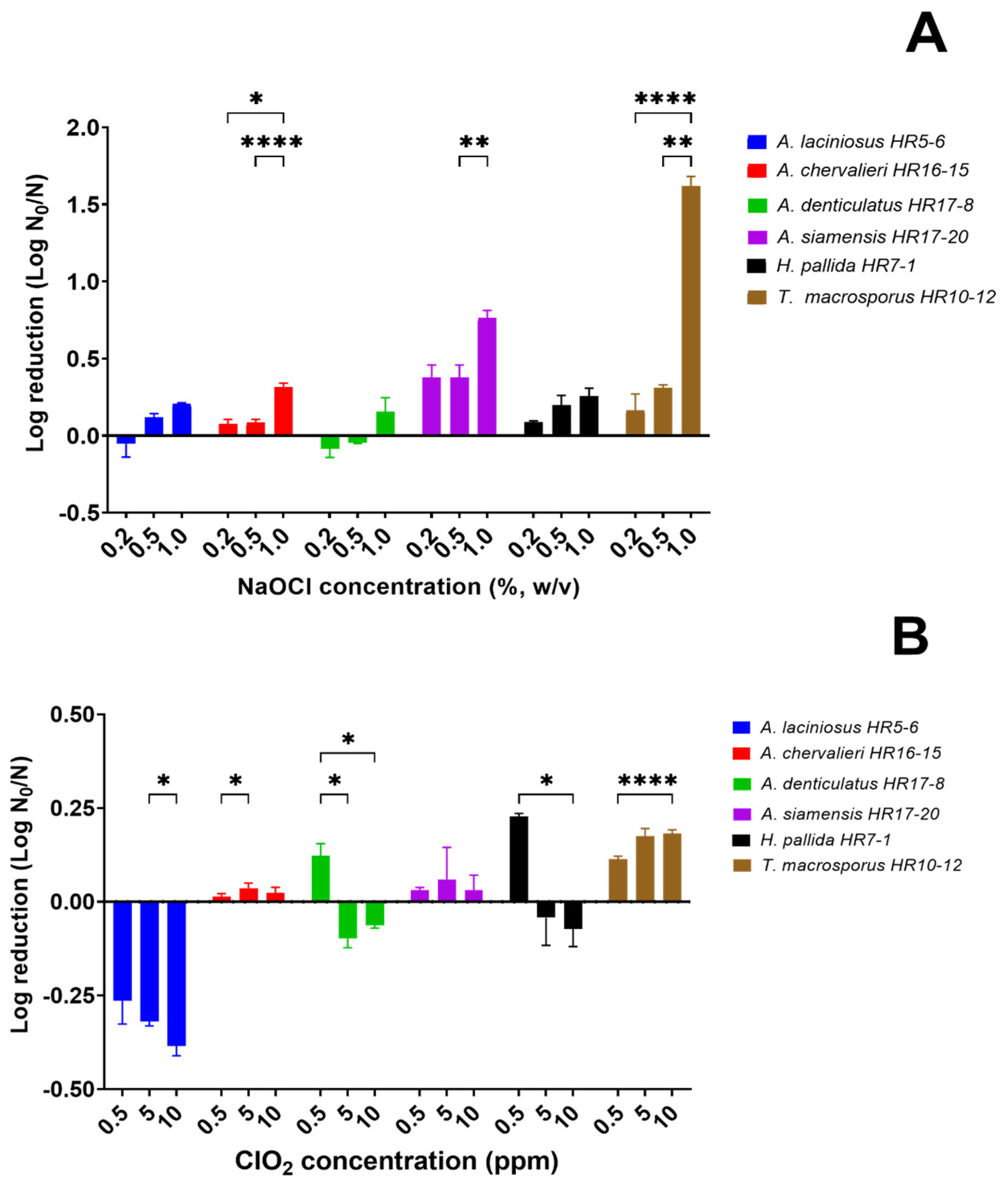
3.2.1. Efficacy of Sodium Hypochlorite

Significant correlations (p < 0.05) were observed between the log reduction values and the increasing concentration of sodium hypochlorite for certain cases, including A. chevalieri, A. siamensis, and T. macrosporus (Figure 2A). This indicates that higher concentrations of sodium hypochlorite led to greater log reduction in ascospores for these species. The highest inactivation of 1.5-log reduction was found in T. macrosporus ascospores at the highest concentration of 1% (w/v), followed by A. siamensis ascospores (0.75-log reduction), whereas the other tested species did not reach 0.5-log reduction. In certain treatments, the activation of ascospores occurred, leading to an increase in the number of ascospores after treatment. As a result, the log reduction values were negative, indicating a failure to reduce the ascospore population. This was observed in cases such as A. laciniosus at a concentration of 0.2% (w/v) and A. denticulatus at concentrations of 0.2% and 0.5% (w/v).
Sodium hypochlorite is a widely used sanitizer in the food industry due to its effectiveness and cost efficiency. Its fungicidal mechanisms rely on the oxidative action of hypochlorous acid (HOCl) and hypochlorite ion (OCl) on cellular components. These compounds penetrate the microbial cell via the cell wall and membrane, where they react with various biomolecules via different mechanisms. This includes inactivating enzymes, disrupting nucleic acids, and causing oxidative damage to cell walls and cellular components. Indeed, the biocidal activity of chlorine compounds can vary depending on the specific chlorine species present [41]. Furthermore, available chlorine inhibits metabolism by oxidizing thiol functions, or amino groups present in the membrane and cytoplasmic enzymes [42]. According to Wan et al. [43], at a lower concentration of sodium hypochlorite (2 mg/L or 0.0002%, w/v), membrane oxidation occurred in A. niger conidia primarily by chlorine, while in Penicillium polonicum conidia, membrane oxidation was induced by both chlorine and ·OH, with ·OH playing a major role.
Sodium hypochlorite is commonly used for the sanitation of equipment and utensils, typically at concentrations ranging from 0.02% to 0.08% (200–800 ppm). For cleaning facilities, higher concentrations of up to 0.12% (1200 ppm) are applied [44]. However, studies have shown that these concentrations are ineffective against ascospores of several heat-resistant mold species [12]. In this study, sodium hypochlorite solution was tested at concentrations recommended by sanitizer manufacturers (0.2%, 0.5%, and 1.0% w/v, corresponding to 2000, 5000, and 10,000 ppm, respectively). Nonetheless, even these concentrations proved to be ineffective against the tested ascospores. Similar results were reported by Stefanello et al. [16], who found that 0.5% (w/v) sodium hypochlorite, the lowest recommended concentration, was ineffective against heat-resistant molds isolated from spoiled thermally processed foods. A higher concentration of sodium hypochlorite solution of 1% (w/v) with a contact time of 15 min was effective against only some strains of Paec. niveus, Paec. fulvus, and A. australensis, resulting in a reduction ranging from 3-log to 3.9-log. Bernardi et al. [28] also investigated the effect of sodium hypochlorite against A. pseudoglaucus (previously known as Eurotium repens) isolated from spoiled bakery products and found that commonly used concentrations ranging from 0.01% to 0.2% (w/v) were ineffective in reducing ascospore counts. Notable efficacy was observed only at a concentration of 1% (w/v), resulting in a significant reduction ranging from 3- to 3.9-log. Therefore, it is recommended to use the highest concentration of sodium hypochlorite recommended by sanitizer manufacturers to control the contamination of heat-resistant mold ascospores. However, high concentrations of sodium hypochlorite can cause corrosion of stainless steel surfaces and may cause irritation [45]. It is important to consider that the presence of residual chlorine in treated water can lead to the creation of potentially hazardous chlorinated by-products, such as trihalomethanes [46].

3.2.2. Efficacy of Chlorine Dioxide

Significant correlations (p < 0.05) were observed between the log reduction values and the increasing concentration of chlorine dioxide for specific species, including A. chevalieri, A. denticulatus, H. pallida, and T. macrosporus (Figure 2B). Nevertheless, chlorine dioxide exhibited the lowest efficacy and demonstrated varying sensitivity among the species tested and the three concentrations used. Overall, H. pallida was found to be more sensitive to chlorine dioxide compared to the other tested species, regardless of the highest reduction of 0.23-log reduction after treatment with the lowest concentration of 0.5 ppm. Furthermore, significant activation of A. laciniosus ascospores was observed across all tested concentrations. Similar activation was also observed in other species, including A. chevalieri and H. pallida.
Chlorine dioxide is also available as an aqueous solution, which is approved from 50 to 200 ppm on food contact surfaces. This sanitizer has a strong oxidation capacity at low concentrations (0.1 mg/L or 0.1 ppm) and in a wide pH range of about 3–8 [47]. In the present study, the chlorine dioxide solution was tested at concentrations recommended by manufacturers for sanitation purposes (0.5, 5, and 10 ppm, equivalent to 0.5, 5, and 10 mg/L, respectively). However, these concentrations proved to be insufficient for inactivating the tested ascospores.
Antimicrobial mechanisms of chlorine dioxide rely on inducing oxidative stress and the subsequent overproduction of reactive oxygen species, which ultimately lead to cell injury [48]. The oxidative stress exerted by chlorine dioxide selectively affects compounds like phenols and thiols, which are essential for the viability and activity of undesirable microorganisms [49]. In a study by Wen et al. [50] on Cladosporium sp., Trichoderma sp., and Penicillium sp., it was observed that exposure to chlorine dioxide led to increased concentrations of extracellular substances and alterations in spore morphology. These changes were attributed to damage to the cell wall and cell membrane of fungal spores, resulting in the leakage of intracellular substances and eventual death of the fungal spores. Previously, Liu et al. [51] reported that chlorine dioxide can inhibit the growth of P. digitatum via a mechanism of action that involves the destruction of membrane integrity in fungal cells. They observed a significant induction of cell apoptosis and noted that the mycelial morphology was severely damaged with an increase in chlorine dioxide concentration.
To date, a limited study has been conducted on the effects of chlorine dioxide on ascospores of heat-resistant molds. Bundgaard-Nielsen and Nielsen [52] reported that the inactivation achieved for ascospores of A. thermomutatus (previously known as N. pseudofischeri), A. pseudoglaucus, and Monascus ruber using 5% (w/v) chlorine dioxide (50,000 ppm) for 10 min resulted in a 3.3-log reduction, >4.5-log reduction, and >4.1-log reduction, respectively. In addition, Dijksterhuis et al. [15] conducted a study in which they treated ascospores of A. fischeri, Pace. Variotii, and T. macrosporus with higher dosages of chlorine dioxide solution ranging from 50 to 1000 ppm. Their findings revealed that ascospores could be effectively inactivated by 100 ppm chlorine dioxide for a minimum of 10 min when the spore density was low (102–103 spores). However, in the case of high spore density (104–105 spores), inactivation required higher concentrations of 200–500 ppm for a duration of 60 min.
Furthermore, it was noted that sodium hypochlorite and chlorine dioxide also exhibited the capacity to stimulate the activation of ascospores in specific species, as evidenced by the negative log reduction. Generally, ascospores of heat-resistant molds tend to remain in a dormant state and cannot initiate growth even under favorable conditions without undergoing an activation treatment. This activation treatment can be achieved via various extreme environmental triggers, such as heat treatment at temperatures of 80–85 °C for several minutes [8], chemicals released from burnt substrates into the soil (e.g., furfural) [53], high pressure within the range of 400 to 800 MPa [54,55], or certain organic acids (e.g., formic acid and propionic acid) and fatty acids (e.g., n-butyric acid, n-valeric acid) [56]. Thus, the breaking of dormancy in ascospores via exposure to extreme conditions enables them to initiate growth and germination, which can subsequently lead to food spoilage. As a result, it is advisable to avoid using these low concentrations of chlorine-based sanitizers, as they could potentially trigger the activation of ascospores in specific heat-resistant mold species.

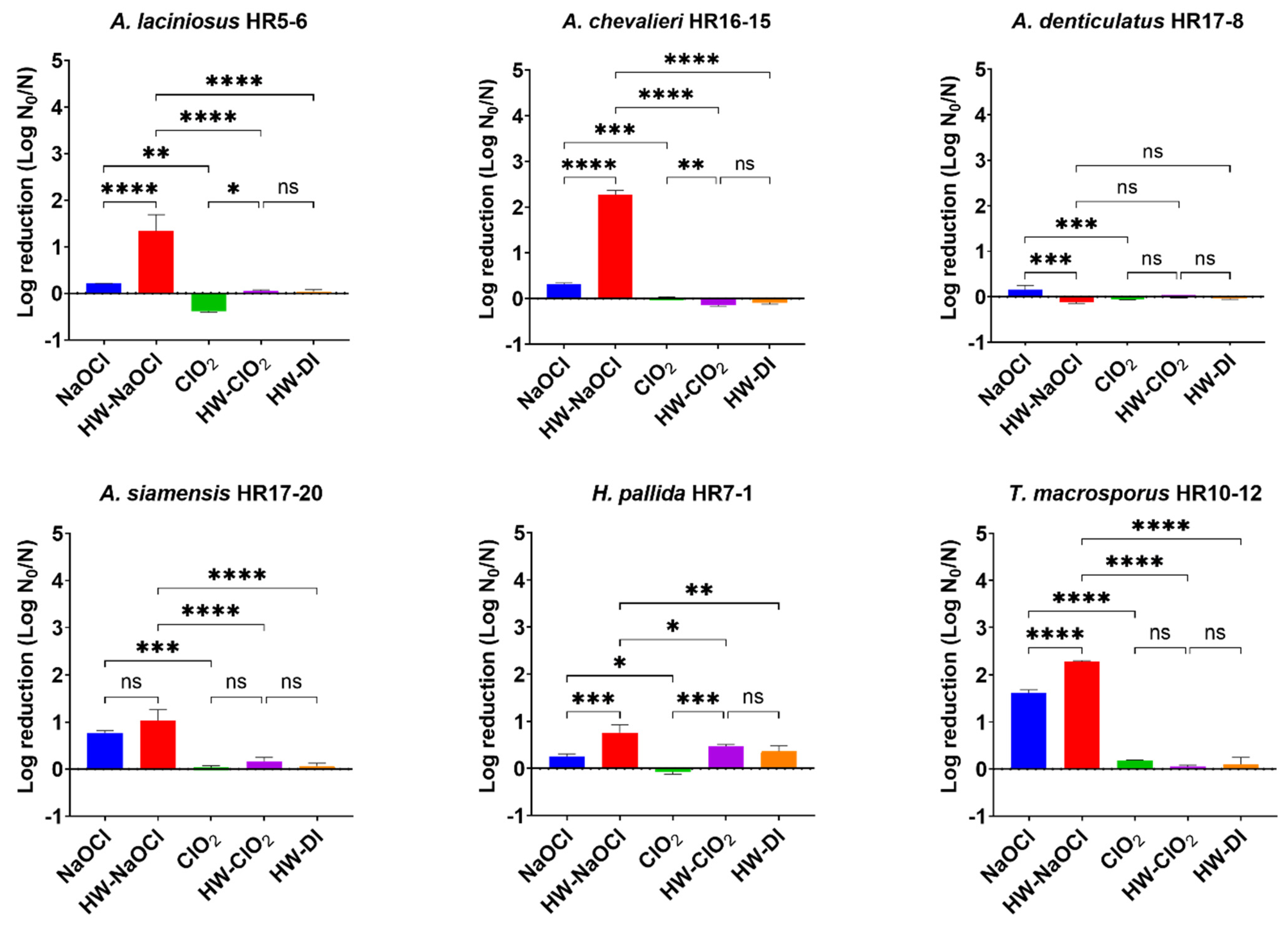
3.2.3. Effect of Hot Water Pretreatment on the Efficacy of Chlorine-Based Sanitizers

To enhance the fungicidal efficacy of chlorine-based sanitizers against heat-resistant mold ascospores, a hot water pretreatment commonly used in the industry was conducted before applying the sanitizer. The ascospores were immersed in hot water at 65 °C for 5 min and then adhered to a stainless steel surface. After drying, the ascospores were exposed to either 1% (w/v) sodium hypochlorite or 10 ppm chlorine dioxide for 15 min.
The impact of the hot water pretreatment followed by the sanitizer on the log reduction in ascospores is shown in Figure 3, in comparison to using sanitizer or hot water alone. It was observed that hot water treatment alone proved capable of inactivating ascospores with a reduction of not more than 0.5-log reduction, depending on the mold species. A combination of hot water followed by sodium hypochlorite resulted in a significantly greater reduction in ascospores compared to sodium hypochlorite alone and hot water alone (p < 0.05), except for A. denticulatus and A. siamensis. This treatment exhibited a significantly higher reduction in A. chevalieri with a 2.27-log reduction and A. laciniosus with a 1.34-log reduction. In contrast, reductions in ascospores resulting from hot water followed by chlorine dioxide did not show a significant difference compared to hot water alone (p > 0.05), but they were significantly higher than those from chlorine dioxide alone (p < 0.05), except for A. denticulatus, A. siamensis, and T. macrosporus. In the case of these three species, no significant difference was observed between these three treatments (p > 0.05). Overall, it is important to highlight that pretreating ascospores with hot water resulted in a significant enhancement in the effectiveness of sodium hypochlorite, achieving a reduction of more than 2-log compared to that of chlorine dioxide (Figure 4). On the other hand, when ascospores were treated with hot water and subsequently exposed to chlorine dioxide, there was no significant reduction in ascospores compared to hot water treatment followed by distilled water instead of sanitizer (as a control).
Indeed, a probable explanation for the observed increase in log reduction in ascospores when using the combination of hot water and sodium hypochlorite could be attributed to heat activation at a sublethal temperature (e.g., 65 °C for 5 min). This heat treatment might trigger or promote the germination of dormant ascospores. Subsequently, the germinated ascospores, which are generally less heat-resistant, become susceptible and are effectively eliminated by sodium hypochlorite in the second step of the treatment process [43]. This dual-step approach seems to be a synergistic strategy, leveraging the effects of both hot water and sodium hypochlorite to achieve a higher reduction in the ascospore population, thus enhancing the overall efficacy of the disinfection process. Similar applications of mild heat or short to moderate times to promote synchronous germination for subsequent inactivation of vegetative cells have been studied in spore-forming enteric pathogens, such as Bacillus cereus [57] and Clostridium difficile [58].
Hot water treatment offers a cost-effective and user-friendly solution, making it a highly practical option. Utilizing hot water pretreatment to clean food contact surfaces before applying a sanitizer can lead to improved sanitization [59]. The sanitizer manufacturer also emphasizes the need to clean and rinse surfaces prior to the application of the sanitizer [60]. Studies have shown that cleaning with hot water alone also shows promise as an alternative hygienization approach to control foodborne pathogens [61]. For instance, most vegetative cells and conidia can be inactivated upon exposure to 60 °C for 5 min [62]. Many previous studies have evaluated combinations of heat treatment with sanitizers to eradicate or inhibit foodborne pathogens. Mertz et al. [63] demonstrated that using quaternary ammonium compounds (5 or 10 ppm), chlorine (10 or 25 ppm), or peracetic acid alone resulted in a 2 to 3-log reduction in Listeria monocytogenes on stainless steel or aluminum surfaces, while moist heat alone led to a 3 to 4-log reduction. However, when moist heat and sanitizer were combined, there was a 5 to 7-log reduction compared to the initial inoculation. Notably, the specific combination of hot water with sanitizer on the survival of heat-resistant mold ascospores has not yet been published.

3.2.4. Correlation Analysis

The results of the Pearson correlation analysis between the thermal resistant characteristics and the tolerance to chlorine-based sanitizers of tested heat-resistant mold species are shown in Table 5. A significant positive correlation was observed between the D- and δ-values (p < 0.01) obtained from the log-linear model and the Weibull model, respectively. This finding suggests that the values representing the scale parameter of the Weibull model can indeed be interpreted as the time required for the first decimal reduction or the D-value. Similarly, the Z*-values derived from the δ-values were significantly positively correlated with the Z-values calculated from the log-linear model (p < 0.05). Additionally, a significant negative correlation was found between the overall log reduction in ascospores when exposed to chlorine dioxide and the D-values (p < 0.05) or δ-values (p < 0.01). On the other hand, the heat resistance of ascospores showed a positive correlation with their tolerance to chlorine dioxide. However, it was noted that there is no available data in the literature concerning the association between heat resistance and tolerance to sanitizers in ascospores. In a recent study involving black yeasts Exophiala phaeomuriformis, E. dermatitidis, and Aureobasidium pullulan, thermal resistance was found to be linked to high-pressure processing tolerance and was inversely associated with halotolerance. However, no significant association was observed between thermal resistance and UV tolerance or sodium hypochlorite tolerance [64].

4. Conclusions

This study highlights the diverse thermal and sanitizer resistance profiles of ascospores from different heat-resistant mold species. The Weibull model proved to be a valuable tool in describing the inactivation parameters of ascospores during heat treatment, with A. laciniosus identified as the most heat-resistant species. Moreover, chlorine-based sanitizers demonstrated varying efficacy against these ascospores, with sodium hypochlorite exhibiting the best results. On the other hand, chlorine dioxide yielded unsatisfactory results for inactivation and even triggered the activation of some species. The combination of hot water and sodium hypochlorite showed promise in enhancing inactivation; however, achieving the required 3-log reduction remained a challenge. It is worth noting that the observed correlation between heat and chlorine dioxide resistance emphasizes the need for tailored disinfection approaches to ensure food safety and quality of thermal process fruit juices. To conclude, this research offers invaluable insights that can inform practices and protocols within the fruit juice manufacturing industry, ultimately contributing to enhanced product safety and consumer satisfaction.

Author Contributions

Conceptualization, W.M., S.S. and R.H.; methodology, T.M. and W.M.; formal analysis, T.M.; investigation, T.M. and C.C.; data curation, T.M.; writing—original draft preparation, T.M.; writing—review and editing, W.M.; visualization, T.M. and C.C.; supervision, W.M., S.S. and R.H.; project administration, W.M. and T.M.; funding acquisition, W.M. All authors have read and agreed to the published version of the manuscript.

Funding

This research was funded by the Center of Excellence on Agricultural Biotechnology, Office of the Permanent Secretary, Ministry of Higher Education, Science, Research and Innovation (AG-BIO/MHESI).

Data Availability Statement

Not applicable.

Acknowledgments

This research was supported by the Center of Excellence on Agricultural Biotechnology, Office of the Permanent Secretary, Ministry of Higher Education, Science, Research and Innovation (AG-BIO/MHESI). The Scientific Equipment and Research Division of the Kasetsart University Research and Development Institute (KURDI), Bangkok, Thailand, provided laboratory facilities.

Conflicts of Interest

The authors declare no conflict of interest.

References

  1. Houbraken, J.; Samson, R.A. Phylogeny of Penicillium and the Segregation of Trichocomaceae into Three Families. Stud. Mycol. 2011, 70, 1–51. [Google Scholar] [CrossRef]
  2. Houbraken, J.; Samson, R.A.; Frisvad, J.C. Byssochlamys: Significance of Heat Resistance and Mycotoxin Production. Adv. Exp. Med. Biol. 2006, 571, 211–224. [Google Scholar]
  3. Aydin, A.; Ulusoy, B.H.; Ergün, Ö. A Survey on Heat-Resistant Moulds in Heat Treated Milk, Milk Products and Fruit Juices. Arch. Leb. 2005, 56, 58–60. [Google Scholar]
  4. de Cássia Martins Salomão, B.; Muller, C.; do Amparo, H.C.; de Aragão, G.M.F. Survey of Molds, Yeast and Alicyclobacillus spp. From a Concentrated Apple Juice Productive Process. Braz. J. Microbiol. 2014, 45, 49–58. [Google Scholar] [CrossRef]
  5. Kocakaya Yildiz, A.; Çoksöyler, N. Heat-Resistance Characteristics of Ascospores of Eurotium chevalieri Isolated from Apricot Juice. Nahrung 2002, 46, 28–30. [Google Scholar] [CrossRef]
  6. Scaramuzza, N.; Berni, E. Heat-Resistance of Hamigera avellanea and Thermoascus crustaceus Isolated from Pasteurized Acid Products. Int. J. Food Microbiol. 2014, 168–169, 63–68. [Google Scholar] [CrossRef]
  7. Samson, R.; Houbraken, J.; Varga, J.; Frisvad, J. Polyphasic Taxonomy of the Heat Resistant Ascomycete Genus Byssochlamys and Its Paecilomyces Anamorphs. Persoonia 2009, 22, 14–27. [Google Scholar] [CrossRef]
  8. Rico-Munoz, E. Heat Resistant Molds in Foods and Beverages: Recent Advances on Assessment and Prevention. Curr. Opin. Food Sci. 2017, 17, 75–83. [Google Scholar] [CrossRef]
  9. Evelyn; Silva, F.V.M. Inactivation of Byssochlamys nivea Ascospores in Strawberry Puree by High Pressure, Power Ultrasound and Thermal Processing. Int. J. Food Microbiol. 2015, 214, 129–136. [Google Scholar] [CrossRef]
  10. Souza, P.B.; Poltronieri, K.F.; Alvarenga, V.O.; Granato, D.; Rodriguez, A.D.; Sant’ana, A.S.; Peña, W.E. Modeling of Byssochamys nivea and Neosartorya fischeri Inactivation in Papaya and Pineapple Juices as a Function of Temperature and Soluble Solids Content. LWT-Food Sci. Technol. 2017, 82, 90–95. [Google Scholar] [CrossRef]
  11. Panagou, E.; Tassou, C.; Manitsa, C.; Mallidis, C. Modelling the Effect of High Pressure on the Inactivation Kinetics of a Pressure-Resistant Strain of Pediococcus damnosus in Phosphate Buffer and Gilt-Head Seabream (Sparus aurata). J. Appl. Microbiol. 2007, 102, 1499–1507. [Google Scholar] [CrossRef]
  12. Copetti, M.V. Sanitizers for Controlling Fungal Spoilage in Some Food Industries. Curr. Opin. Food Sci. 2023, 52, 101072. [Google Scholar] [CrossRef]
  13. Frąc, M.; Jezierska-Tys, S.; Yaguchi, T. Occurrence, detection, and molecular and metabolic characterization of heat-resistant fungi in soils and plants and their risk to human health. In Advances in Agronomy; Sparks, D.L., Ed.; Academic Press: Cambridge, MA, USA, 2015; pp. 161–204. [Google Scholar]
  14. Wyatt, T.T.; Van Leeuwen, M.R.; Golovina, E.A.; Hoekstra, F.A.; Kuenstner, E.J.; Palumbo, E.A.; Snyder, N.L.; Visagie, C.; Verkennis, A.; Hallsworth, J.E.; et al. Functionality and Prevalence of Trehalose-Based Oligosaccharides as Novel Compatible Solutes in Ascospores of Neosartorya fischeri (Aspergillus fischeri) and Other Fungi. Environ. Microbiol. 2015, 17, 395–411. [Google Scholar] [CrossRef]
  15. Dijksterhuis, J.; Meijer, M.; van Doorn, T.; Samson, R.; Rico-Munoz, E. Inactivation of Stress-Resistant Ascospores of Eurotiales by Industrial Sanitizers. Int. J. Food Microbiol. 2018, 285, 27–33. [Google Scholar] [CrossRef]
  16. Stefanello, A.; Magrini, L.N.; Lemos, J.G.; Garcia, M.V.; Bernardi, A.O.; Cichoski, A.J.; Copetti, M.V. Comparison of Electrolized Water And multiple Chemical Sanitizer Action against Heat-Resistant Molds (HRM). Int. J. Food Microbiol. 2020, 335, 108856. [Google Scholar] [CrossRef]
  17. Maneeboon, T.; Sangchote, S.; Hongprayoon, R.; Chuaysrinule, C.; Mahakarnchanakul, W. Occurrence of Heat-Resistant Mold Ascospores in Pineapple and Sugarcane Field Soils in Thailand. Int. J. Microbiol. 2023, 2023, 8347560. [Google Scholar] [CrossRef]
  18. Rico-Munoz, E.; Houbraken, J.; Samson, R.A. Detection and enumeration of heat-resistant molds. In Compendium of Methods for the Microbiological Examination of Foods; Salfinger, Y., Tortorello, M.L., Eds.; American Public Health Association: Washington, DC, USA, 2015; pp. 387–397. [Google Scholar]
  19. Peterson, S.W.; Jurjevic, Z.; Bills, G.F.; Stchigel, A.M.; Guarro, J.; Vega, F.E. Genus Hamigera, Six New Species and Multilocus DNA Sequence Based Phylogeny. Mycologia 2010, 102, 847–864. [Google Scholar] [CrossRef]
  20. Santos, J.L.; Samapundo, S.; Gülay, S.M.; Van Impe, J.; Sant’Ana, A.S.; Devlieghere, F. Inter- and Intra-Species Variability in Heat Resistance and the Effect of Heat Treatment Intensity on Subsequent Growth of Byssochlamys Fulva and Byssochlamys Nivea. Int. J. Food Microbiol. 2018, 279, 80–87. [Google Scholar] [CrossRef]
  21. Sant’Ana, A.; Rosenthal, A.; Massaguer, P.R. Heat Resistance and the Effects of Continuous Pasteurization on the Inactivation of Byssochlamys fulva Ascospores in Clarified Apple Juice. J. Appl. Microbiol. 2009, 107, 197–209. [Google Scholar] [CrossRef]
  22. Geeraerd, A.; Valdramidis, V.; Van Impe, J. Ginafit, a Freeware Tool to Assess Non-Log-Linear Microbial Survivor Curves. Int. J. Food Microbiol. 2005, 102, 95–105. [Google Scholar] [CrossRef]
  23. Bigelow, W.D.; Esty, J.R. The Thermal Death Point in Relation to Time of Typical Thermophilic Organisms. J. Infect. Dis. 1920, 27, 602–617. [Google Scholar] [CrossRef]
  24. Colás-Medà, P.; Nicolau-Lapeña, I.; Viñas, I.; Neggazi, I.; Alegre, I. Bacterial Spore Inactivation in Orange Juice and Orange Peel by Ultraviolet-C Light. Foods 2021, 10, 855. [Google Scholar] [CrossRef] [PubMed]
  25. Mafart, P.; Couvert, O.; Gaillard, S.; Leguérinel, I. On Calculating Sterility in Thermal Preservation Methods: Application of the Weibull Frequency Distribution Model. Int. J. Food Microbiol. 2002, 72, 107–113. [Google Scholar] [CrossRef] [PubMed]
  26. Yan, R.; Pinto, G.; Taylor-Roseman, R.; Cogan, K.; D’Alesandre, G.; Kovac, J. Evaluation of the Thermal Inactivation of a Salmonella Serotype Oranienburg Strain During Cocoa Roasting at Conditions Relevant to the Fine Chocolate Industry. Front. Microbiol. 2021, 12, 576337. [Google Scholar] [CrossRef]
  27. European Union. Bs-En 13697: 2001: Quantitative Non-Porous Surface Test for the Evaluation of Bactericidal and/or Fungicidal Activity of Chemical Disinfectants Used in Food, Industrial, Domestic and Institutional Areas. Test Method and Requirements without Mechanical Action; European Committee for Standardization: Brussels, Belgium, 2001. [Google Scholar]
  28. Bernardi, A.O.; Garcia, M.V.; Copetti, M.V. Food Industry Spoilage Fungi Control through Facility Sanitization. Curr. Opin. Food Sci. 2019, 29, 28–34. [Google Scholar] [CrossRef]
  29. Ruiz, V.; Alonso, R.; Salvador, M.; Condón, S.; Condón-Abanto, S. Impact of Shoulders on the Calculus of Heat Sterilization Treatments with Different Bacterial Spores. Food Microbiol. 2021, 94, 103663. [Google Scholar] [CrossRef]
  30. Pohůnek, V.; Adamcova, M.; Kulišanová, I.; Šístková, I.; Rohlik, B.A.; Ševčík, R. Thermal Inactivation of Aspergillus lacinosus Ascospores as a Function of Temperature and Soluble Solids Content in Fruit Jam. J. Food Nutr. Res. 2019, 58, 275–282. [Google Scholar]
  31. Dijksterhuis, J.; Wyatt, T.; Hanssen, M.; Golovina, E.; Hoekstra, F.; Lugones, L. Abundant Small Protein Icarus inside the Cell Wall of Stress-Resistant Ascospores of Talaromyces Macrosporus Suggests a Novel Mechanism of Constitutive Dormancy. J. Fungi 2021, 7, 216. [Google Scholar] [CrossRef]
  32. Dijksterhuis, J.; van Driel, K.G.; Sanders, M.G.; Molenaar, D.; Houbraken, J.A.; Samson, R.A.; Kets, E.P. Trehalose Degradation and Glucose Efflux Precede Cell Ejection During Germination of Heat-Resistant Ascospores of Talaromyces macrosporus. Arch. Microbiol. 2002, 178, 1–7. [Google Scholar] [CrossRef]
  33. Kikoku, Y. Heat Activation Characteristics of Talaromyces Ascospores. J. Food Sci. 2003, 68, 2331–2335. [Google Scholar] [CrossRef]
  34. King, A.D.; Michener, H.D.; A Ito, K. Control of Byssochlamys and Related Heat-Resistant Fungi in Grape Products. Appl. Microbiol. 1969, 18, 166–173. [Google Scholar] [CrossRef] [PubMed]
  35. Evelyn, E.; Muria, S.R.; Chairul, C.; Fozla, D.; Khoirunnisa, F.K. Thermal Inactivation of Talaromyces flavus Ascospores in Pineapple Juice as Influenced by Temperature, Soluble Solids, and Spore Age. J. Adv. Res. Fluid Mech. Therm. Sci. 2020, 69, 111–119. [Google Scholar] [CrossRef]
  36. Kikoku, Y.; Tagashira, N.; Nakano, H. Heat Resistance of Fungi Isolated from Frozen Blueberries. J. Food Prot. 2008, 71, 2030–2035. [Google Scholar] [CrossRef] [PubMed]
  37. van Boekel, M.A.J.S. On the Use of the Weibull Model to Describe Thermal Inactivation of Microbial Vegetative Cells. Int. J. Food Microbiol. 2002, 74, 139–159. [Google Scholar] [CrossRef] [PubMed]
  38. Couvert, O.; Gaillard, S.; Savy, N.; Mafart, P.; Leguérinel, I. Survival Curves of Heated Bacterial Spores: Effect of Environmental Factors on Weibull Parameters. Int. J. Food Microbiol. 2005, 101, 73–81. [Google Scholar] [CrossRef]
  39. Chuaysrinule, C.; Mahakarnchanakul, W.; Maneeboon, T. Comparative Study on the Effect of Temperature and Water Activity on Aspergillus flavus and Aspergillus carbonarius Isolates Growth and Mycotoxin Production on a Chili Powder Medium. Cogent Food Agric. 2020, 6, 1782097. [Google Scholar] [CrossRef]
  40. Racchi, I.; Scaramuzza, N.; Hidalgo, A.; Cigarini, M.; Berni, E. Sterilization of Food Packaging by Uv-C Irradiation: Is Aspergillus brasiliensis Atcc 16404 the Best Target Microorganism for Industrial Bio-Validations? Int. J. Food Microbiol. 2021, 357, 109383. [Google Scholar] [CrossRef]
  41. Visconti, V.; Coton, E.; Rigalma, K.; Dantigny, P. Effects of Disinfectants on Inactivation of Mold Spores Relevant to the Food Industry: A Review. Fungal Biol. Rev. 2021, 38, 44–66. [Google Scholar] [CrossRef]
  42. Cheshchevik, V.T.; Krylova, N.G.; Cheshchevik, N.G.; Lapshina, E.A.; Semenkova, G.N.; Zavodnik, I.B. Role of Mitochondrial Calcium in Hypochlorite Induced Oxidative Damage of Cells. Biochimie 2021, 184, 104–115. [Google Scholar] [CrossRef]
  43. Wan, Q.; Xia, Y.; Li, Y.; Wu, G.; Wang, J.; Huang, T.; Wen, G. Enhanced Solar Inactivation of Fungal Spores by Addition of Low-Dose Chlorine: Efficiency and Mechanism. Water Res. 2022, 222, 118964. [Google Scholar] [CrossRef]
  44. Bernardi, A.O.; Stefanello, A.; Lemos, J.G.; Garcia, M.V.; Copetti, M.V. Antifungal Activity of Commercial Sanitizers against Strains of Penicillium roqueforti, Penicillium paneum, Hyphopichia burtonii and Aspergillus pseudoglaucus: Bakery Spoilage Fungi. Food Microbiol. 2019, 83, 59–63. [Google Scholar] [CrossRef] [PubMed]
  45. Lin, J.H.H.; Karabucak, B.; Lee, S.M. Effect of Sodium Hypochlorite on Conventional and Heat-Treated Nickel-Titanium Endodontic Rotary Instruments—An In vitro Study. J. Dent. Sci. 2021, 16, 738–743. [Google Scholar]
  46. López-Gálvez, F.; Allende, A.; Truchado, P.; Martínez-Sánchez, A.; Tudela, J.A.; Selma, M.V.; Gil, M.I. Suitability of Aqueous Chlorine Dioxide Versus Sodium Hypochlorite as an Effective Sanitizer for Preserving Quality of Fresh-Cut Lettuce While Avoiding by-Product Formation. Postharvest Biol. Technol. 2010, 55, 53–60. [Google Scholar] [CrossRef]
  47. Ran, Y.; Qingmin, C.; Maorun, F. Chlorine Dioxide Generation Method and Its Action Mechanism for Removing Harmful Substances and Maintaining Quality Attributes of Agricultural Products. Food Bioprocess Technol. 2019, 12, 1110–1122. [Google Scholar] [CrossRef]
  48. Zhu, C.; Chen, Z.; Yu, G. Fungicidal Mechanism of Chlorine Dioxide on Saccharomyces cerevisiae. Ann. Microbiol. 2013, 63, 495–502. [Google Scholar] [CrossRef]
  49. Insignares-Carrione, E.; Gómez, B.B.; Kalcker, A.L. Chlorine Dioxide in COVID-19: Hypothesis About the Possible Mechanism of Molecular Action in SARS-CoV-2. Mol. Genet. Med. 2020, 14, 468. [Google Scholar]
  50. Wen, G.; Xu, X.; Huang, T.; Zhu, H.; Ma, J. Inactivation of Three Genera of Dominant Fungal Spores in Groundwater Using Chlorine Dioxide: Effectiveness, Influencing Factors, and Mechanisms. Water Res. 2017, 125, 132–140. [Google Scholar] [CrossRef]
  51. Liu, X.; Jiao, W.; Du, Y.; Chen, Q.; Su, Z.; Fu, M. Chlorine Dioxide Controls Green Mold Caused by Penicillium digitatum in Citrus Fruits and the Mechanism Involved. J. Agric. Food Chem. 2020, 68, 13897–13905. [Google Scholar] [CrossRef]
  52. Bundgaard-Nielsen, K.; Nielsen, P.V. Fungicidal Effect of 15 Disinfectants against 25 Fungal Contaminants Commonly Found in Bread and Cheese Manufacturing. J. Food Prot. 1996, 59, 268–275. [Google Scholar] [CrossRef]
  53. Pandit, A.; Maheshwari, R. Life-History of Neurospora intermedia in a Sugar Cane Field. J. Biosci. 1996, 21, 57–79. [Google Scholar] [CrossRef]
  54. Dijksterhuis, J.; Teunissen, P. Dormant Ascospores of Talaromyces macrosporus Are Activated to Germinate after Treatment with Ultra High Pressure. J. Appl. Microbiol. 2004, 96, 162–169. [Google Scholar] [CrossRef] [PubMed]
  55. Reyns, K.M.; Veraverbeke, E.A.; Michiels, C.W. Activation and Inactivation of Talaromyces macrosporus Ascospores by High Hydrostatic Pressure. J. Food Prot. 2003, 66, 1035–1042. [Google Scholar] [CrossRef] [PubMed]
  56. Kikoku, Y.; Tagashira, N.; Gabriel, A.A.; Nakano, H. Heat Activation of Neosartorya and Talaromyces Ascospores and Enhancement by Organic Acids. Biocontrol Sci. 2009, 14, 87–95. [Google Scholar] [CrossRef] [PubMed]
  57. Løvdal, I.S.; Hovda, M.B.; Granum, P.E.; Rosnes, J.T. Promoting Bacillus cereus Spore Germination for Subsequent Inactivation by Mild Heat Treatment. J. Food Prot. 2011, 74, 2079–2089. [Google Scholar] [CrossRef]
  58. Rodriguez-Palacios, A.; LeJeune, J.T. Moist-Heat Resistance, Spore Aging, and Superdormancy in Clostridium difficile. Appl. Environ. Microbiol. 2011, 77, 3085–3091. [Google Scholar] [CrossRef]
  59. Holah, J.; Lavaud, A.; Peters, W.; Dye, K.A. Future Techniques for Disinfectant Efficacy Testing. Int. Biodeterior. Biodegrad. 1998, 41, 273–279. [Google Scholar] [CrossRef]
  60. Masuku, S.M.; Babu, D.; Martin, E.M.; O’Bryan, C.A.; Crandall, P.G.; Ricke, S.C. Lethality of Moist Heat and Silver Dihydrogen Citrate Sanitizer Combinations on Listeria spp. Adhered to Components of a Deli Meat Slicer. Food Control 2014, 44, 227–232. [Google Scholar] [CrossRef]
  61. Taormina, P.J.; Dorsa, W.J. Evaluation of Hot-Water and Sanitizer Dip Treatments of Knives Contaminated with Bacteria and Meat Residue. J. Food Prot. 2007, 70, 648–654. [Google Scholar] [CrossRef]
  62. Silva, F.V.M.; Gibbs, P.A.; Nunez, H.; Almonacid, S.; Simpson, R. Thermal Process|Pasteurization. In Encyclopedia of Food Microbiology, 2nd ed.; Batt, C.A., Tortorello, M.L., Eds.; Academic Press: Oxford, UK, 2014; pp. 577–595. [Google Scholar]
  63. Mertz, A.W.; O’Bryan, C.A.; Crandall, P.G.; Ricke, S.C.; Morawicki, R. The Elimination of Listeria monocytogenes Attached to Stainless Steel or Aluminum Using Multiple Hurdles. J. Food Sci. 2015, 80, M1557–M1562. [Google Scholar] [CrossRef]
  64. Cai, S.; Snyder, A.B. Thermoresistance in Black Yeasts Is Associated with Halosensitivity and High Pressure Processing Tolerance but Not with Uv Tolerance or Sanitizer Tolerance. J. Food Prot. 2022, 85, 203–212. [Google Scholar] [CrossRef]
Figure 1. Experimental thermal inactivation curves and fitted values derived from the log-linear model (dotted line) and the Weibull model (solid line) for ascospores of heat-resistant mold species in pineapple juice.
Beverages 09 00096 g001
Figure 2. Log reduction in ascospores from heat-resistant mold species adhered to stainless steel coupons in the presence of BSA (30 g/L) after treatment with sodium hypochlorite (A) and chlorine dioxide (B) for 15 min. The results are expressed as the mean ± SD. ANOVA results with a Tukey post hoc test are indicated with asterisks (* p value < 0.050; ** p value < 0.01; **** p value < 0.0001).
Beverages 09 00096 g002
Figure 3. Effect of a combination of hot water and sanitizer on the log reduction in ascospores from heat-resistant mold species adhered to stainless steel coupons in the presence of BSA (30 g/L). NaOCl: 1% (w/v) sodium hypochlorite for 15 min, HW-NaOCl: a combination of hot water (65 °C, 5 min) and 1% (w/v) sodium hypochlorite for 15 min, ClO2: 10 ppm chlorine dioxide for 15 min, HW-ClO2: a combination of hot water (65 °C, 5 min) and 10 ppm chlorine dioxide for 15 min, and HW-DI: a combination of hot water (65 °C, 5 min) and distilled water for 15 min. The results are expressed as the mean ± SD. ANOVA results with a Tukey post hoc test are indicated with asterisks (ns, not significant; * p value < 0.05; ** p value < 0.01; *** p value < 0.001; **** p value < 0.0001).
Beverages 09 00096 g003
Figure 4. Comparisons of the effect of treatment with sanitizer ((A) sodium hypochlorite, and (B) chlorine dioxide) and a combination of hot water and sanitizer on the overall log reduction in ascospores from heat-resistant mold species. NaOCl: 1% (w/v) sodium hypochlorite for 15 min, HW-NaOCl: a combination of hot water (65 °C, 5 min) and 1% (w/v) sodium hypochlorite for 15 min, ClO2: 10 ppm chlorine dioxide for 15 min, HW-ClO2: a combination of hot water (65 °C, 5 min) and 10 ppm chlorine dioxide for 15 min, and HW-DI: a combination of hot water (65 °C, 5 min) and distilled water for 15 min. ANOVA results with a Tukey post hoc test are indicated with asterisks (ns, not significant; * p value < 0.05; **** p value < 0.0001).
Beverages 09 00096 g004
Table 1. List of heat-resistant mold species used in this study.
SourceSpeciesIsolateGenBank Accession Number
Pineapple field soils, ThailandA. laciniosusHR5-6OP480885
H. pallidaHR7-1OP480891
T. macrosporusHR10-12OP480922
Sugarcane field soils, ThailandA. chevalieriHR16-15OP480965
A. denticulatusHR17-8OP480975
A. siamensisHR17-20OP480981
Table 2. D- and Z-values derived from the log-linear model fits for ascospores of heat resistant mold species in pineapple juice.
SpeciesD-Value (min)Z-Value
(°C)
75 °C80 °C85 °C90 °C95 °C97 °C
A. laciniosusNRNR74.93 ± 3.86 Aa7.78 ± 0.35 Ba1.09 ± 0.09 Ca0.89 ± 0.06 Ca6.19
A. chevalieri69.85 ± 3.17 Aa9.53 ± 0.42 Bbc1.52 ± 0.13 Cd0.44 ± 0.01 Cd- *-6.75
A. denticulatus31.01 ± 3.94 Ab13.56 ± 2.44 Bb4.69 ± 0.69 Cc2.22 ± 0.16 Cc--12.87
A. siamensis25.32 ± 1.04 Ab4.73 ± 0.25 Bc1.64 ± 0.14 Cd---8.40
H. pallida31.29 ± 2.43 Ab13.77 ± 0.67 Bb2.15 ± 0.11 Ccd0.46 ± 0.04 Cd--7.91
T. macrosporusNR137.33 ± 9.21 Aa23.85 ± 2.81 Bb3.98 ± 0.35 Cb1.02 ± 0.08 Ca0.5 6 ± 0.03 Cb7.14
NR = no reduction; * Kinetic parameters were not calculated due to the limited data points within the time interval. Different superscript uppercase letters indicate a statistical difference within rows according to Fisher LSD test (p < 0.05). Different superscript lowercase letters indicate a statistical difference within columns according to Fisher LSD test (p < 0.05).
Table 3. Scale parameter (δ-value), shape parameter (p-value), and Z*-value derived from the Weibull model fits for ascospores of heat-resistant mold species in pineapple juice.
Species75 °C80 °C85 °C90 °C95 °C97 °CZ*-Value (°C)
Scale parameter (δ-value, min)
A. laciniosusNRNR104.59 ± 1.68 Aa7.39 ± 0.56 Ba1.38 ± 0.08 Ca0.81 ± 0.13 Ca5.71
A. chevalieri68.23 ± 1.89 Aa8.65 ± 1.37 Bbc2.57 ± 0.33 Cc0.51 ± 0.01 Ccd--7.02
A. denticulatus24.29 ± 5.57 Ab6.04 ± 3.69 Bc0.79 ± 0.17 Bc0.74 ± 0.16 Bc--9.36
A. siamensis26.02 ± 2.41 Ab3.39 ± 0.35 Bc1.11 ± 0.19 Bc---7.30
H. pallida25.71 ± 4.07 Ab10.57 ± 1.42 Bbc1.70 ± 0.21 Cc0.48 ± 0.10 Ccd--8.38
T. macrosporusNR205.13 ± 10.26 Aa28.70 ± 6.55 Bb5.49 ± 0.17 Cb0.94 ± 0.16 Cb0.76 ± 0.04 Ca6.86
Shape parameter (p-value)
A. laciniosusNRNR1.95 ± 0.12 Aa0.79 ± 0.13 Cc1.65 ± 0.20 Ba0.91 ± 0.13 Cb-
A. chevalieri1.32 ± 0.11 Aa0.93 ± 0.11 BCbc1.52 ± 0.25 Ab1.02 ± 0.33 Bb---
A. denticulatus0.37 ± 0.05 Bc0.33 ± 0.07 Bd0.38 ± 0.03 Bd0.52 ± 0.04 Acd---
A. siamensis1.03 ± 0.09 Aab0.73 ± 0.06 Bc0.63 ± 0.10 Bcd----
H. pallida0.47 ± 0.06 Cc0.77 ± 0.08 Bc0.80 ± 0.08 Bc1.09 ± 0.28 Ab---
T. macrosporusNR1.88 ± 0.32 Aa1.26 ± 0.34 Bb1.92 ± 0.18 Aa0.92 ± 0.12 Cb1.44 ± 0.09 Ba-
NR = no reduction; * Kinetic parameters were not calculated due to the limited data points within the time interval. Different superscript uppercase letters indicate a statistical difference within rows according to Fisher LSD test (p < 0.05). Different superscript lowercase letters indicate a statistical difference within columns according to Fisher LSD test (p < 0.05).
Table 4. Statistical comparison of the log-linear and the Weibull models for the inactivation curves of ascospores from heat-resistant mold species in pineapple juice.
SpeciesRMSER2
75 °C80 °C85 °C90 °C95 °C97 °C75 °C80 °C85 °C90 °C95 °C97 °C
Log-linear model
A. laciniosusNRNR0.0890.0570.2130.197NRNR0.9720.9600.9090.946
A. chevalieri0.0560.2230.2820.090- *-0.9600.9620.9290.992--
A. denticulatus0.1970.3930.4380.328--0.7560.6450.7700.908--
A. siamensis0.1390.1550.197---0.9670.9630.929---
H. pallida0.1380.1700.1620.264--0.9070.9550.9630.929--
T. macrosporusNR0.1220.3830.1850.2590.199NR0.9410.8680.8840.9190.964
Weibull model
A. laciniosusNRNR0.0400.0550.1320.201NRNR0.9940.9630.9650.944
A. chevalieri0.0450.2260.2700.032--0.9750.9610.9330.999--
A. denticulatus0.1280.3080.1220.199--0.9180.8150.9820.975--
A. siamensis0.1420.1020.145---0.9660.9840.962---
H. pallida0.1140.1480.1420.282--0.9400.9660.9720.918--
T. macrosporusNR0.1180.3820.1060.2390.092NR0.9410.8690.9610.9420.992
NR = no reduction; * Kinetic parameters were not calculated due to the limited data points within the time interval.
Table 5. Pearson’s correlation coefficient among the thermal and chlorine-based sanitizer resistances of ascospores of heat-resistant mold species.
Measures123456
1. D-value1
2. δ-value0.988 **1
3. Z-value−0.422−0.3761
4. Z*-value−0.572−0.4730.828 *1
5. Log reduction for NaOCl−0.118−0.238−0.392−0.5671
6. Log reduction for ClO2−0.820 *−0.897 **0.1040.1000.5881
** p value < 0.01; * p value < 0.05.
Disclaimer/Publisher’s Note: The statements, opinions and data contained in all publications are solely those of the individual author(s) and contributor(s) and not of MDPI and/or the editor(s). MDPI and/or the editor(s) disclaim responsibility for any injury to people or property resulting from any ideas, methods, instructions or products referred to in the content.

