
Detection of Porcine Circovirus Type 3 in Serum, Semen, Oral Fluid, and Preputial Fluid Samples of Boars

 ,
,  ,
,
Abstract
:Simple Summary
Abstract
1. Introduction
2. Materials and Methods
2.1. Reagents
2.2. Animal Sources
2.3. Samples
2.3.1. Oral Fluid Collection
2.3.2. Preputial Fluid and Semen Collection
2.3.3. Blood Collection
2.4. DNA Extraction and qPCR
2.5. PCV3 Genome Sequencing and Bioinformatics Analyses
2.6. PCV3-IgG Antibody Detection in Serum
2.7. Statistical Analysis
3. Results
3.1. Detection of PCV3 in Serum, Semen, Oral Fluid, and Preputial Fluid of Boars
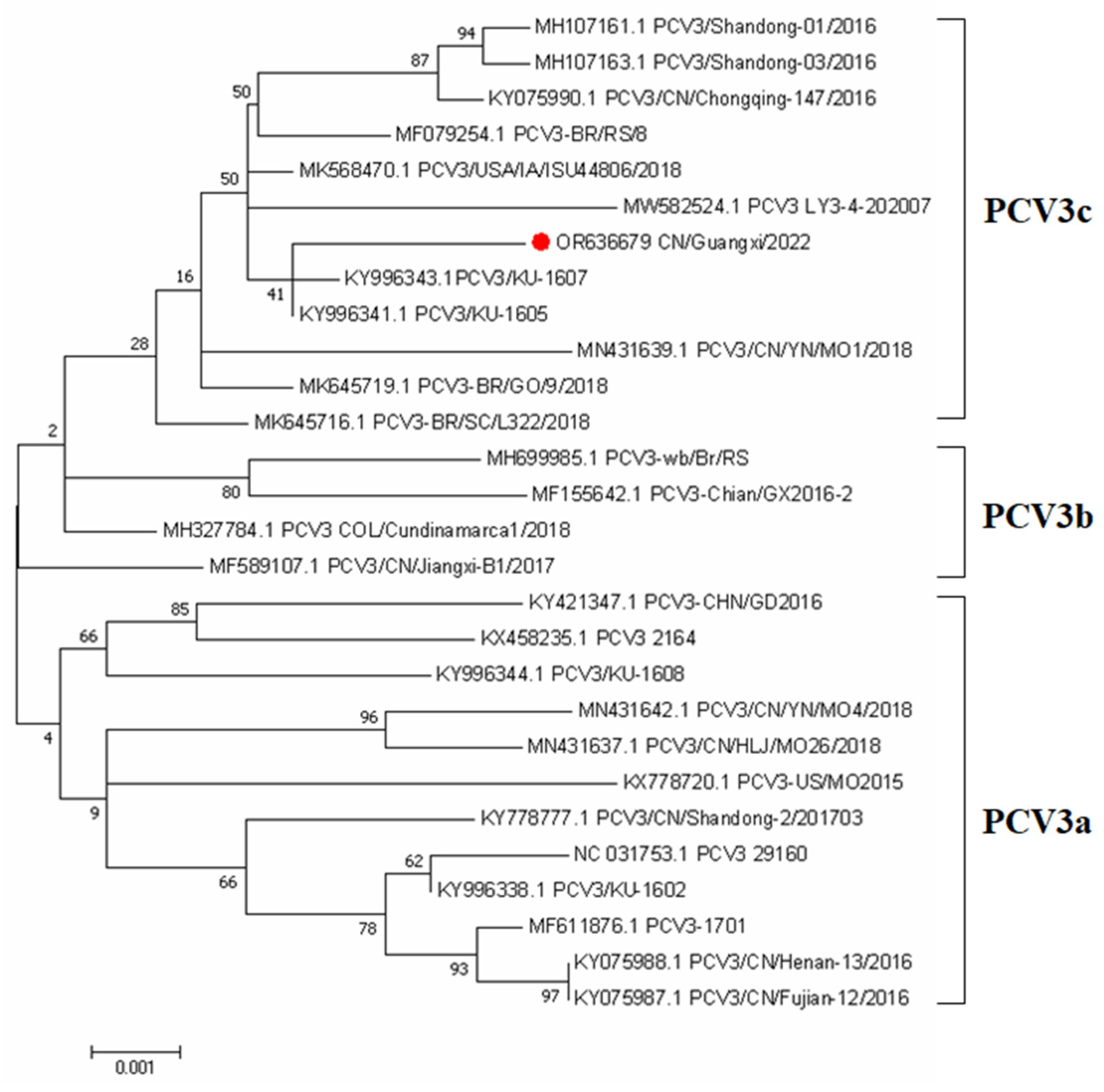
3.2. Phylogenetic Analysis Based on PCV3 Complete Genome Sequences
3.3. Comparison of the Detection of PCV3-IgG in Serum
4. Discussion
5. Conclusions
Author Contributions
Funding
Institutional Review Board Statement
Informed Consent Statement
Data Availability Statement
Conflicts of Interest
References
- Breitbart, M.; Delwart, E.; Rosario, K.; Segales, J.; Varsani, A.; Ictv, R.C. ICTV Virus Taxonomy Profile: Circoviridae. J. Gen. Virol. 2017, 98, 1997–1998. [Google Scholar] [CrossRef]
- Allan, G.M.; Mcneilly, F.; Cassidy, J.P.; Reilly, G.A.; Adair, B.; Ellis, W.A.; Mcnulty, M.S. Pathogenesis of porcine circovirus; experimental infections of colostrum deprived piglets and examination of pig foetal material. Vet. Microbiol. 1995, 44, 49–64. [Google Scholar] [CrossRef]
- Opriessnig, T.; Karuppannan, A.K.; Castro, A.; Xiao, C.T. Porcine circoviruses: Current status, knowledge gaps and challenges. Virus Res. 2020, 286, 198044. [Google Scholar] [CrossRef]
- Tischer, I.; Gelderblom, H.; Vettermann, W.; Koch, M.A. A very small porcine virus with circular single-stranded DNA. Nature 1982, 295, 64–66. [Google Scholar] [CrossRef] [PubMed]
- Rakibuzzaman, A.; Ramamoorthy, S. Comparative immunopathogenesis and biology of recently discovered porcine circoviruses. Transbound. Emerg. Dis. 2021, 68, 2957–2968. [Google Scholar] [CrossRef]
- Alarcon, P.; Rushton, J.; Wieland, B. Cost of post-weaning multi-systemic wasting syndrome and porcine circovirus type-2 subclinical infection in England—An economic disease model. Prev. Vet. Med. 2013, 110, 88–102. [Google Scholar] [CrossRef]
- Alarcon, P.; Rushton, J.; Nathues, H.; Wieland, B. Economic efficiency analysis of different strategies to control post-weaning multi-systemic wasting syndrome and porcine circovirus type 2 subclinical infection in 3-weekly batch system farms. Prev. Vet. Med. 2013, 110, 103–118. [Google Scholar] [CrossRef] [PubMed]
- Saporiti, V.; Franzo, G.; Sibila, M.; Segales, J. Porcine circovirus 3 (PCV-3) as a causal agent of disease in swine and a proposal of PCV-3 associated disease case definition. Transbound. Emerg. Dis. 2021, 68, 2936–2948. [Google Scholar] [CrossRef]
- Jiang, H.; Wang, D.; Wang, J.; Zhu, S.; She, R.; Ren, X.; Tian, J.; Quan, R.; Hou, L.; Li, Z.; et al. Induction of Porcine Dermatitis and Nephropathy Syndrome in Piglets by Infection with Porcine Circovirus Type 3. J. Virol. 2018, 93, e02045-18. [Google Scholar] [CrossRef] [PubMed]
- Fu, X.; Fang, B.; Ma, J.; Liu, Y.; Bu, D.; Zhou, P.; Wang, H.; Jia, K.; Zhang, G. Insights into the epidemic characteristics and evolutionary history of the novel porcine circovirus type 3 in southern China. Transbound. Emerg. Dis. 2018, 65, e296–e303. [Google Scholar] [CrossRef] [PubMed]
- Sukmak, M.; Thanantong, N.; Poolperm, P.; Boonsoongnern, A.; Ratanavanichrojn, N.; Jirawattanapong, P.; Woonwong, Y.; Soda, N.; Kaminsonsakul, T.; Phuttapatimok, S.; et al. The retrospective identification and molecular epidemiology of porcine circovirus type 3 (PCV3) in swine in Thailand from 2006 to 2017. Transbound. Emerg. Dis. 2019, 66, 611–616. [Google Scholar] [CrossRef]
- Jia, Y.; Zhu, Q.; Xu, T.; Chen, X.; Li, H.; Ma, M.; Zhang, Y.; He, Z.; Chen, H. Detection and genetic characteristics of porcine circovirus type 2 and 3 in Henan province of China. Mol. Cell. Probes 2022, 61, 101790. [Google Scholar] [CrossRef] [PubMed]
- Chen, G.H.; Mai, K.J.; Zhou, L.; Wu, R.T.; Tang, X.Y.; Wu, J.L.; He, L.L.; Lan, T.; Xie, Q.M.; Sun, Y.; et al. Detection and genome sequencing of porcine circovirus 3 in neonatal pigs with congenital tremors in South China. Transbound. Emerg. Dis. 2017, 64, 1650–1654. [Google Scholar] [CrossRef] [PubMed]
- Dal Santo, A.C.; Cezario, K.C.; Bennemann, P.E.; Machado, S.A.; Martins, M. Full-genome sequences of porcine circovirus 3 (PCV3) and high prevalence in mummified fetuses from commercial farms in Brazil. Microb. Pathog. 2020, 141, 104027. [Google Scholar] [CrossRef] [PubMed]
- Zhang, H.H.; Hu, W.Q.; Li, J.Y.; Liu, T.N.; Zhou, J.Y.; Opriessnig, T.; Xiao, C.T. Novel circovirus species identified in farmed pigs designated as Porcine circovirus 4, Hunan province, China. Transbound. Emerg. Dis. 2020, 67, 1057–1061. [Google Scholar] [CrossRef]
- Visuthsak, W.; Woonwong, Y.; Thanantong, N.; Poolperm, P.; Boonsoongnern, A.; Ratanavanichrojn, N.; Jirawattanapong, P.; Soda, N.; Kaminsonsakul, T.; Phuttapatimok, S.; et al. PCV3 in Thailand: Molecular epidemiology and relationship with PCV2. Transbound. Emerg. Dis. 2021, 68, 2980–2989. [Google Scholar] [CrossRef]
- Tan, C.Y.; Lin, C.N.; Ooi, P.T. What do we know about porcine circovirus 3 (PCV3) diagnosis so far?: A review. Transbound. Emerg. Dis. 2021, 68, 2915–2935. [Google Scholar] [CrossRef]
- Ku, X.; Chen, F.; Li, P.; Wang, Y.; Yu, X.; Fan, S.; Qian, P.; Wu, M.; He, Q. Identification and genetic characterization of porcine circovirus type 3 in China. Transbound. Emerg. Dis. 2017, 64, 703–708. [Google Scholar] [CrossRef]
- Eddicks, M.; Muller, M.; Fux, R.; Ritzmann, M.; Stadler, J. Detection of porcine circovirus type 3 DNA in serum and semen samples of boars from a German boar stud. Vet. J. 2022, 279, 105784. [Google Scholar] [CrossRef]
- Mora-Diaz, J.; Pineyro, P.; Shen, H.; Schwartz, K.; Vannucci, F.; Li, G.; Arruda, B.; Gimenez-Lirola, L. Isolation of PCV3 from Perinatal and Reproductive Cases of PCV3-Associated Disease and In Vivo Characterization of PCV3 Replication in CD/CD Growing Pigs. Viruses 2020, 12, 219. [Google Scholar] [CrossRef]
- Ha, Z.; Xie, C.Z.; Li, J.F.; Wen, S.B.; Zhang, K.L.; Nan, F.L.; Zhang, H.; Guo, Y.C.; Wang, W.; Lu, H.J.; et al. Molecular detection and genomic characterization of porcine circovirus 3 in pigs from Northeast China. BMC Vet. Res. 2018, 14, 321. [Google Scholar] [CrossRef]
- Temeeyasen, G.; Lierman, S.; Arruda, B.L.; Main, R.; Vannucci, F.; Gimenez-Lirola, L.G.; Pineyro, P.E. Pathogenicity and immune response against porcine circovirus type 3 infection in caesarean-derived, colostrum-deprived pigs. J. Gen. Virol. 2021, 102, 1502. [Google Scholar] [CrossRef]
- Caspari, K.; Henning, H.; Schreiber, F.; Maass, P.; Gossl, R.; Schaller, C.; Waberski, D. Impact of porcine circovirus type 2 (PCV2) vaccination on boar semen quality and quantity using two different vaccines. Theriogenology 2014, 82, 574–579. [Google Scholar] [CrossRef]
- Zhao, X.; Xiang, H.; Bai, X.; Fei, N.; Huang, Y.; Song, X.; Zhang, H.; Zhang, L.; Tong, D. Porcine parvovirus infection activates mitochondria-mediated apoptotic signaling pathway by inducing ROS accumulation. Virol. J. 2016, 13, 26. [Google Scholar] [CrossRef]
- van Rijn, P.A.; Wellenberg, G.J.; Hakze-Van, D.H.R.; Jacobs, L.; Moonen, P.L.; Feitsma, H. Detection of economically important viruses in boar semen by quantitative RealTime PCR technology. J. Virol. Methods 2004, 120, 151–160. [Google Scholar] [CrossRef]
- Choi, C.; Chae, C. Detection of classical swine fever virus in boar semen by reverse transcription-polymerase chain reaction. J. Vet. Diagn. Investig. 2003, 15, 35–41. [Google Scholar] [CrossRef]
- Gatto, I.; Arruda, P.H.; Visek, C.A.; Victoria, J.G.; Patterson, A.R.; Krull, A.C.; Schwartz, K.J.; de Oliveira, L.G.; Arruda, B.L. Detection of atypical porcine pestivirus in semen from commercial boar studs in the United States. Transbound. Emerg. Dis. 2018, 65, e339–e343. [Google Scholar] [CrossRef]
- Goldberg, A.M.; Argenti, L.E.; Faccin, J.E.; Linck, L.; Santi, M.; Bernardi, M.L.; Cardoso, M.R.; Wentz, I.; Bortolozzo, F.P. Risk factors for bacterial contamination during boar semen collection. Res. Vet. Sci. 2013, 95, 362–367. [Google Scholar] [CrossRef] [PubMed]
- Zimmerman, J.J.; Karriker, L.A.; Ramirez, A.; Schwartz, K.J.; Stevenson, G.W. (Eds.) Diseases of Swine, 10th ed.; Blackwell Publishing: Ames, IA, USA, 2019; pp. 1236–1239. ISBN 978-111-935-092-7. [Google Scholar]
- Griffin, J. Methods of sperm DNA extraction for genetic and epigenetic studies. Methods Mol. Biol. 2013, 927, 379–384. [Google Scholar] [CrossRef] [PubMed]
- Jeong, J.; Park, C.; Kang, I.; Park, S.J.; Chae, C. Concurrent vaccination of boars with type 1 and type 2 porcine reproductive and respiratory syndrome virus (PRRSV) reduces seminal shedding of type 1 and type 2 PRRSV. Can. J. Vet. Res. Rev. Can. Rech. Vet. 2017, 81, 108–117. [Google Scholar]
- Wills, R.W.; Zimmerman, J.J.; Yoon, K.J.; Swenson, S.L.; Mcginley, M.J.; Hill, H.T.; Platt, K.B.; Christopher-Hennings, J.; Nelson, E.A. Porcine reproductive and respiratory syndrome virus: A persistent infection. Vet. Microbiol. 1997, 55, 231–240. [Google Scholar] [CrossRef]
- Franzo, G.; Legnardi, M.; Hjulsager, C.K.; Klaumann, F.; Larsen, L.E.; Segales, J.; Drigo, M. Full-genome sequencing of porcine circovirus 3 field strains from Denmark, Italy and Spain demonstrates a high within-Europe genetic heterogeneity. Transbound. Emerg. Dis. 2018, 65, 602–606. [Google Scholar] [CrossRef]
- Qi, S.; Su, M.; Guo, D.; Li, C.; Wei, S.; Feng, L.; Sun, D. Molecular detection and phylogenetic analysis of porcine circovirus type 3 in 21 Provinces of China during 2015–2017. Transbound. Emerg. Dis. 2019, 66, 1004–1015. [Google Scholar] [CrossRef]
- Ge, M.; Ren, J.; Xie, Y.L.; Zhao, D.; Fan, F.C.; Song, X.Q.; Li, M.X.; Xiao, C.T. Prevalence and Genetic Analysis of Porcine Circovirus 3 in China From 2019 to 2020. Front. Vet. Sci. 2021, 8, 773912. [Google Scholar] [CrossRef] [PubMed]
- Deng, J.; Li, X.; Zheng, D.; Wang, Y.; Chen, L.; Song, H.; Wang, T.; Huang, Y.; Pang, W.; Tian, K. Establishment and application of an indirect ELISA for porcine circovirus 3. Arch. Virol. 2018, 163, 479–482. [Google Scholar] [CrossRef] [PubMed]
- Zhang, S.; Wang, D.; Jiang, Y.; Li, Z.; Zou, Y.; Li, M.; Yu, H.; Huang, K.; Yang, Y.; Wang, N. Development and application of a baculovirus-expressed capsid protein-based indirect ELISA for detection of porcine circovirus 3 IgG antibodies. Bmc Vet. Res. 2019, 15, 79. [Google Scholar] [CrossRef] [PubMed]

| Boar No. | qPCR Results (Ct) | ELISA PP (%) | Boar No. | Serum | Semen | Oral Fluid | Preputial Fluid | ELISA PP (%) | |||
|---|---|---|---|---|---|---|---|---|---|---|---|
| Serum | Semen | Oral Fluid | Preputial Fluid | ||||||||
| 1 | 0 | 0 | 34.36 | 33.04 | 52.84 | 15 | 0 | 0 | 37.65 | 31.97 | 142.91 |
| 2 | 0 | 0 | 0 | 37.49 | 88.22 | 16 | 0 | 0 | 0 | 0 | 124.62 |
| 3 | 0 | 0 | 33.03 | 0 | 119.35 | 17 | 0 | 0 | 35.48 | 28.1 | 61.89 |
| 4 | 0 | 0 | 33.68 | 35.86 | 31.96 | 18 | 0 | 0 | 0 | 31.71 | 65.77 |
| 5 | 0 | 0 | 0 | 0 | 100.60 | 19 | 0 | 0 | 36.45 | 0 | 74.27 |
| 6 | 35.5 | 0 | 28.9 | 31.28 | 83.00 | 20 | 0 | 0 | 0 | 0 | 21.89 |
| 7 | 0 | 0 | 35.77 | 0 | 95.61 | 21 | 0 | 0 | 34.11 | 33.38 | 93.49 |
| 8 | 0 | 0 | 30.82 | 30.38 | 150.67 | 22 | 0 | 0 | 33.94 | 0 | 22.73 |
| 9 | 0 | 0 | 0 | 0 | 79.26 | 23 | 0 | 0 | 0 | 0 | 76.30 |
| 10 | 0 | 0 | 38.29 | 0 | 76.86 | 24 | 0 | 0 | 36.74 | 31.5 | 98.00 |
| 11 | 0 | 0 | 33.83 | 30.71 | 80.00 | 25 | 0 | 0 | 36.81 | 0 | 102.00 |
| 12 | 0 | 0 | 33.81 | 0 | 85.08 | 26 | 0 | 0 | 0 | 30.52 | 123.81 |
| 13 | 0 | 0 | 0 | 35.36 | 158.80 | 27 | 0 | 0 | 35.54 | 0 | 88.90 |
| 14 | 0 | 0 | 34.14 | 0 | 141.71 | 28 | 0 | 0 | 0 | 0 | 72.55 |
| Sample Type | Serum | Semen | Oral Fluid | Preputial Fluid |
|---|---|---|---|---|
| Param | 28 | 28 | 28 | 28 |
| Positive number | 1 | 0 | 18 | 13 |
| Positive rate (%) | 3.57 | 0 | 64.28 | 46.4 |
| Type of Positive Sample | A | B | C | D | E |
|---|---|---|---|---|---|
| Number of Boars tested | 28 | 28 | 28 | 28 | 28 |
| Positive number | 0 | 9 | 4 | 8 | 1 |
| Percentage (%) | 0 | 32.1 | 14.3 | 28.6 | 3.6 |
Disclaimer/Publisher’s Note: The statements, opinions and data contained in all publications are solely those of the individual author(s) and contributor(s) and not of MDPI and/or the editor(s). MDPI and/or the editor(s) disclaim responsibility for any injury to people or property resulting from any ideas, methods, instructions or products referred to in the content. |
© 2023 by the authors. Licensee MDPI, Basel, Switzerland. This article is an open access article distributed under the terms and conditions of the Creative Commons Attribution (CC BY) license (https://creativecommons.org/licenses/by/4.0/).
Share and Cite
Qi, S.; He, Q.; Zhang, Z.; Chen, H.; Giménez-Lirola, L.; Yuan, F.; Bei, W. Detection of Porcine Circovirus Type 3 in Serum, Semen, Oral Fluid, and Preputial Fluid Samples of Boars. Vet. Sci. 2023, 10, 689. https://doi.org/10.3390/vetsci10120689
Qi S, He Q, Zhang Z, Chen H, Giménez-Lirola L, Yuan F, Bei W. Detection of Porcine Circovirus Type 3 in Serum, Semen, Oral Fluid, and Preputial Fluid Samples of Boars. Veterinary Sciences. 2023; 10(12):689. https://doi.org/10.3390/vetsci10120689
Chicago/Turabian StyleQi, Song, Qiyun He, Zhewei Zhang, Huanchun Chen, Luis Giménez-Lirola, Fangyan Yuan, and Weicheng Bei. 2023. "Detection of Porcine Circovirus Type 3 in Serum, Semen, Oral Fluid, and Preputial Fluid Samples of Boars" Veterinary Sciences 10, no. 12: 689. https://doi.org/10.3390/vetsci10120689
APA StyleQi, S., He, Q., Zhang, Z., Chen, H., Giménez-Lirola, L., Yuan, F., & Bei, W. (2023). Detection of Porcine Circovirus Type 3 in Serum, Semen, Oral Fluid, and Preputial Fluid Samples of Boars. Veterinary Sciences, 10(12), 689. https://doi.org/10.3390/vetsci10120689