Article

Voluntary Biosurveillance of Streptococcus equi Subsp. equi in Nasal Secretions of 9409 Equids with Upper Airway Infection in the USA

1
William R. Pritchard Veterinary Medical Teaching Hospital, School of Veterinary Medicine, University of California, Davis, CA 95616, USA
2
Department of Obstetrics, Gynecology and Reproduction Biology, Massachusetts General Hospital, Boston, MA 02114, USA
3
Department of Medicine and Epidemiology, School of Veterinary Medicine, University of California, Davis, CA 95616, USA
4
Merck Animal Health, Madison, NJ 07940, USA
*
Author to whom correspondence should be addressed.
Vet. Sci. 2023, 10(2), 78; https://doi.org/10.3390/vetsci10020078
Submission received: 21 December 2022 / Revised: 16 January 2023 / Accepted: 18 January 2023 / Published: 20 January 2023
(This article belongs to the Special Issue Research on Streptococcus in Veterinary Medicine)

Abstract

:

Simple Summary

Strangles caused by Streptococcus equi subsp. equi (S. equi) is considered one of the most prevalent and widely distributed infectious diseases in equids. Large epidemiological studies looking at prevalence factors associated with clinical disease are seldom reported. The present study aimed at describing selected prevalence factors of 9409 equids with acute onset of fever and respiratory signs tested for S. equi by qPCR and to determine the impact of vaccination on S. equi detection. A total of 715 horses (7.6%) tested qPCR-positive for S. equi, of which 226 horses had coinfections with common respiratory viruses (EIV, EHV-1, EHV-4, ERBV). Various prevalence factors were associated with S. equi qPCR-positive status, including season (winter and spring), use (competition and ranch/farm use), and clinical signs (nasal discharge, fever, lethargy, anorexia, and ocular discharge). Vaccination against S. equi was associated with a lower frequency of S. equi qPCR-positive index cases.

Abstract

This study aimed to describe selected epidemiological aspects of horses with acute onset of fever and respiratory signs testing qPCR-positive for S. equi and to determine the effect of vaccination against S. equi on qPCR status. Horses with acute onset of fever and respiratory signs from all regions of the United States were included in a voluntary biosurveillance program from 2008 to 2020 and nasal secretions were tested via qPCR for S. equi and common respiratory viruses. A total of 715/9409 equids (7.6%) tested qPCR-positive for S. equi, with 226 horses showing coinfections with EIV, EHV-1, EHV-4, and ERBV. The median age for the S. equi qPCR-positive horses was 8 ± 4 years and there was significant difference when compared to the median age of the S. equi qPCR-negative horses (6 ± 2 years; p = 0.004). Quarter Horse, Warmblood, and Thoroughbred were the more frequent breed in this horse population, and these breeds were more likely to test qPCR-positive for S. equi compared to other breeds. There was not statistical difference for sex between S. equi qPCR-positive and qPCR-negative horses. Horses used for competition and ranch/farm use were more likely to test qPCR-positive for S. equi (p = 0.006). Horses that tested S. equi qPCR-positive were more likely to display nasal discharge, fever, lethargy, anorexia, and ocular discharge compared to horses that tested S. equi qPCR-negative (p = 0.001). Vaccination against S. equi was associated with a lower frequency of S. equi qPCR-positive status.

1. Introduction

Strangles caused by Streptococcus equi subsp. equi (S. equi) has been reported for centuries and it has been suggested that the spread of this highly contagious disease is a legacy from World War I [1,2]; nonetheless, large populations of horses around the world are still affected by this highly transmissible streptococcal disease, causing an economic burden for their owners. Strangles has been reported as the third-most-common upper respiratory tract infection in the USA after equine herpesvirus-4 (EHV-4) and equine influenza virus (EIV) [3]. The relevance of this disease is confirmed by contemporary research and the publication of two consensus statements by the American College of Veterinary Internal Medicine [4,5].
Streptococcus equi, a well-recognized and highly contagious microorganism with over 230 isolates sequenced worldwide, poses epidemiological challenges since various genotypes relate to outbreaks [6]. Vaccines are commercially available; however, due to the risk of vaccine-related immunological reactions, such as purpura hemorrhagica or abscess formation, horses with high serum titers to S. equi are not routinely vaccinated [7]. Association of strangles with other upper respiratory tract diseases has been poorly described in the literature, as well as association of vaccination status and development of natural disease.
Prevalence of strangles based on qPCR detection has been reported in the USA (6.4%) [3], the United Kingdom (4.6%, North Yorkshire) [8], specific regions in Brazil (2.3%, Rio Grande do Sul) [9], and Colombia (13.5%, Antioquia) [10]. Further, a prevalence of 1.6% based on culture has been reported from Western Canada [11]. Seroprevalence of S. equi by ELISA testing has been reported from various regions in South Africa [12], Israel [13], and Ethiopia [14] with 10.1%, 9.5%, and 8% respectively. There are scarce studies of demographic and prevalence factors associated with strangles. A previous study from the USA described prevalence factors in 49 horses with respiratory signs and qPCR detection of S. equi in nasal secretions [3]. Other studies have focused on clinical markers able to determine the risk of long-term silent carriers following infection [15].
Therefore, the aims of the present study were: (1) to describe the prevalence of S. equi in nasal secretions from 9409 horses with acute onset of fever and respiratory signs enrolled in a voluntary biosurveillance program for respiratory pathogens; (2) to describe demographic and clinical factors in S. equi qPCR-positive horses; and (3) to determine the impact of vaccination status on the detection of S. equi. Authors have hypothesized that the prevalence of S. equi is comparable to that previously reported, as would demographic characteristics, and that vaccinated horses would be less likely to test qPCR-positive for S. equi.

2. Materials and Methods

2.1. Study Population

The study population was composed of horses with acute onset of respiratory signs enrolled in a voluntary surveillance program from March 2008 to December 2020. Two hundred and sixty-one equine veterinary practices were enrolled in the biosurveillance program; the practices were located in various geographical regions (East n = 37, South n = 82, Midwest n = 45, West n = 97). Horses were sampled by equine veterinarians if the equid displayed at least one of the following clinical signs: acute onset of fever (T > 101.5 °F or > 38.5 °C) and/or respiratory signs, including nasal discharge (serous, mucoid, or purulent bilateral nasal discharge) and coughing. Information pertaining to additional clinical signs, such as lethargy, anorexia, ocular discharge, and limb edema, was collected as well. Sample collection was left at the discretion of each of the enrolled equine veterinarians with no case exclusion due to age, sex, or vaccination status.

2.2. Data Collection

The participant veterinarians were asked to fill out a questionnaire with information pertaining to signalment (age, breed, sex), intended use (competition, ranch/farm use, breeding, other use), history of transport, number of affected animals (single, multiple), vaccine history against S. equi, and clinical signs (fever, nasal discharge (serous, mucoid or purulent), cough, lethargy, partial or complete anorexia, ocular discharge (serous, mucoid, or purulent), and limb edema).

2.3. Sample Collection and Analysis

The attending veterinarians performed a physical examination and sampled the horses wearing disposable gloves; a nasal swab was collected using two 6” rayon-tipped swabs (Puritan Products Company LLC, Guilford, ME, USA). Samples were refrigerated and shipped overnight to the laboratory.
DNA purification from nasal swabs was performed on the day of sample arrival to the laboratory using an automated nucleic acid extraction system (QIAcube HT, Qiagen, Valencia, CA, USA) according to the manufacturer’s recommendations. Purified nucleic acids were assayed for the presence of S. equi, equine influenza virus (EIV), equine herpesvirus-1 (EHV-1), EHV-4, and equine rhinitis A and B viruses (ERVs) according to previously validated assays [3]. Further, in order to determine sample quality and efficiency of nucleic acid extraction, all samples were assessed for the presence of the housekeeping gene eGAPDH, as previously described [3].

2.4. Statistical Analysis

Demographic and clinical factors were compared between qPCR S. equi-negative and qPCR S. equi-positive status. Demographic factors included coinfections, breed, use, sex, age (analyzed continuously and categorized into 5-year increments), history of transport, number of affected horses on property, season of submission, and geographic region. Vaccination history (EHV-1/-4, EIV, and S. equi) and the presence of fever, nasal discharge, cough, lethargy, anorexia, ocular discharge, and limb edema were also compared between S. equi qPCR-positive and S. equi qPCR-negative horses. Parametric (chi-square and Student’s t-test) and non-parametric tests (Fisher’s exact and Mann–Whitney U test) were used as appropriate to compare categorical and continuous factors; a p-value < 0.05 was considered statistically significant. All statistical analyses were conducted in StataIC 16.0 (College Station, TX, USA).

3. Results

3.1. Prevalence of S. equi and Coinfections

Out of 9409 horses with acute onset of upper respiratory tract infection, 715 (7.6%) and 8694 horses tested qPCR-positive and qPCR-negative for S. equi, respectively. The most prevalent infection detected in nasal swabs from this horse population was EHV-4 (992 equids (10.5%)), followed by EIV (910 (9.7%)), ERBV (311 (3.3%)), EHV-1 (154 (1.6%)) and ERAV (12 (0.1%)). Coinfections were also detected in the reported horse population (Table 1). There was a statistically significant difference in horses testing qPCR-positive for EHV-4 (p = 0.03), EIV (p < 0.001), and ERBV (p < 0.001) and concurrent qPCR-positive status for S. equi. Horses that tested qPCR-positive against EHV-1 (p = 0.08) and ERAV (p = 0.30) were less likely to test qPCR-positive for S. equi (Table 1). Out of 6123 horses that had single infection, 408 (6.6%) tested qPCR-positive for S. equi in nasal swabs; on the other hand, out of 2409 horses that had multiple infections, 226 (9.4%) tested qPCR-positive for S. equi; there was a statistical difference between these two groups (p < 0.001). In 877 submissions, only one individual respiratory pathogen was tested (additional horses tested once the ethology of an outbreak was identified), out of which 81 (11.3%) horses tested qPCR-positive to S. equi (Table 1).

3.2. Demographics

The main breeds reported were Quarter Horse (44.4%), Warmblood (19.9%), Thoroughbred (13.5%), Arabian (8.6%), Paint Horse (5.7%), Pony/Miniature (4.6%), and draft breeds (3.2%); other breeds totaled 1895 horses. Quarter Horses were more likely to test qPCR-positive for S. equi compared to Warmbloods, Thoroughbreds, ponies, and other breeds (p < 0.001). Thoroughbreds were more likely to test qPCR-positive for S. equi compared to Paint horses, Arabians, draft horses, and ponies (p < 0.001); Warmbloods were more likely to test qPCR-positive for S. equi compared to draft horses and less likely to test qPCR-positive compared to Paint horses, Arabians, ponies, and other breeds (p < 0.001). Ponies were more likely than Arabians and draft horses and less likely than other breeds to test qPCR-positive for S. equi. Finally, Paint horses were less likely than ponies to test qPCR-positive for S. equi (Table 2).
The age of the study horses ranged from 3 months to 32 years. Median age for the S. equi qPCR-negative horses was 6 ± 2.1 years and for the S. equi qPCR-positive horses was 8 ± 4.1 years (p = 0.004); specific data associated with the age of the horses can be found in Table 2. Horses younger than 1 year of age were less likely to test qPCR-positive for S. equi compared to those between 1 and 4 years, 5 and 9 years, 10 and 14 years, and 15 and 19 years of age (p < 0.001). Horses between 1 and 4 years of age were less likely to test qPCR-positive for S. equi compared to horses between 5 and 9 years of age (p = 0.024). Horses between 5 and 9 years of age were more likely to test qPCR-positive for S. equi compared to horses aged 10–14 (p = 0.013) and > 20 years of age (p =0.002; Table 2).
Information on sex was available from 7902 horses with 3305 females and 4597 males (Table 2). Amongst females, 249 (7.5%) tested qPCR-positive for S. equi and 3056 tested qPCR-negative. Amongst males, 366 (7.9%) tested qPCR-positive for S. equi and 4231 tested qPCR-negative. There was no statistical difference for sex between S. equi qPCR-positive and qPCR-negative horses (p < 0.05).
Regarding the use of the horse, there were 3831(40.7%) competition horses, 3458 (36.7%) ranch/farm horses, 442 (4.7%) breeding horses, and 842 (8.9%) horses used for other purposes; the use was not reported for 836 (8.8%) horses (Table 2). Competition and ranch/farm horses were more likely to test qPCR-positive for S. equi (p = 0.006). From the 9409 horses with reported transportation history, 2456 (26%) were transported within 8 weeks. Amongst the horses with recent transportation history, 201 tested qPCR-positive for S. equi in nasal swabs and 2264 tested qPCR-negative. No statistically significant difference was detected (p = 0.14; Table 2).

3.3. Vaccination Status and Clinical Signs

Vaccination against S. equi was reported in 913 horses, from which 89 (9%) tested qPCR-positive for S. equi in nasal swabs (Table 3). Horses vaccinated against S. equi were less likely to test qPCR-positive for S. equi (p = 0.001). Immunization against EHV-1 and -4 and EIV was reported in 3054 and 3032 horses, respectively; there was no statistical difference between vaccination status against EHV-1/-4 and EIV and qPCR test results for S. equi (p > 0.05).
Horses that tested S. equi qPCR-positive in nasal swabs (715) were more likely to have the following clinical signs: nasal discharge (639 horses, 89%), fever (590, 83%), lethargy (539, 75%), anorexia (448, 63%), cough (368, 51%), and ocular discharge (205, 29%) compared to horses that tested S. equi qPCR-negative (p = 0.001; Table 3); limb edema occurred at the same frequency between horses testing S. equi qPCR-positive and qPCR-negative (p > 0.05). No significant differences (p > 0.05) between S. equi qPCR-positive with and without co-infection were observed for clinical signs.

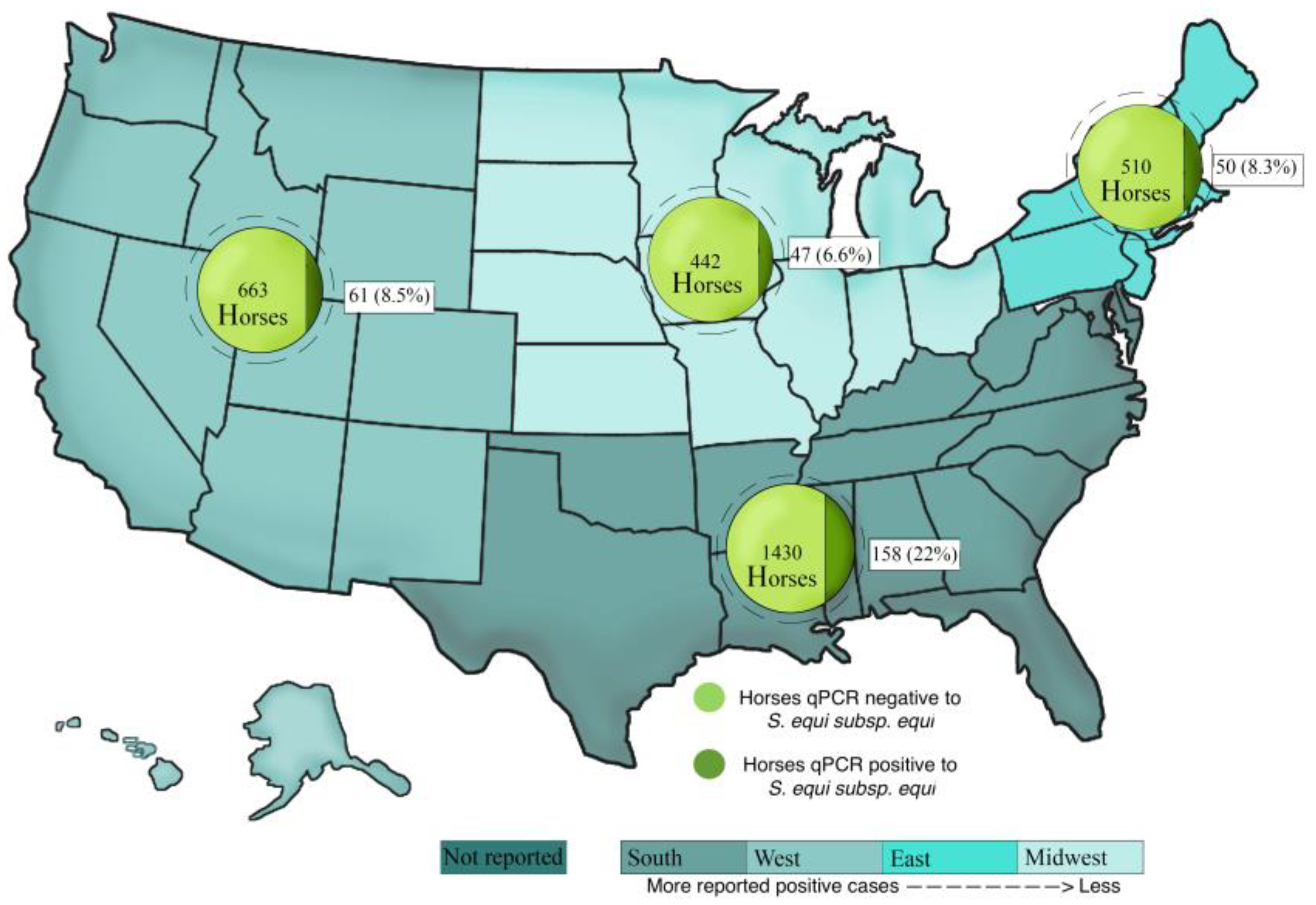
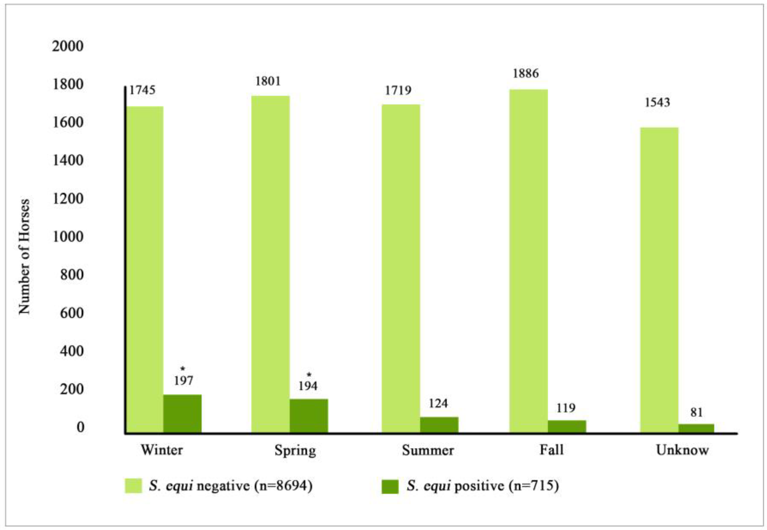
3.4. Location and Seasonality

Horses in this study were grouped by geographic region. Amongst the 3370 horses with reported geographic region, 325 tested qPCR-positive for S. equi. There was no statistically significant difference between regions for horses testing positive or negative for S. equi by qPCR (p = 0.63; Figure 1). The location was unknown in 6039 horses; of those, 390 tested S. equi qPCR-positive.
Horses testing S. equi qPCR-positive were reported during all four seasons. Out of the 705 S. equi qPCR-positive horses, 197 (27.6%) tested positive in winter, 194 (27.1%) tested positive in spring, 124 (17.3%) tested positive in summer, and 119 (16.1%) tested positive in fall (Figure 2). There was a statistical difference between the horses that tested positive in summer/fall compared to those that tested positive in winter/spring (p < 0.001), having more S. equi qPCR-positive horses during winter/spring than during summer/fall. There was not a statistical difference between horses testing qPCR-positive in nasal swabs for S. equi during winter and spring (p = 0.66) or during summer and fall (p = 0.31).

4. Discussion

The present epidemiological study found a high prevalence of S. equi (7.6%) in nasal swabs from clinically ill horses in the USA; coinfections and association with EIV, ERBV, and EHV-4 were present in 31.6% of the S. equi qPCR-positive cases. This study also showed that vaccinated horses were less likely to test qPCR-positive for S. equi. As hypothesized, the prevalence found in this study was similar with the reported prevalence of S. equi from other countries, ranging from 1.66 to 13.5% by microbiological culture [11] or PCR [10]; nonetheless, the techniques for the sampling and detection of S. equi varied amongst the various studies [8,12,13,14]; thus, the results should be compared with caution.
In this study, a high rate of coinfection was detected between S. equi and EIV, and ERBV and EHV-4. Coinfections with various respiratory pathogens are well described and understood in cattle as bovine respiratory disease complex [16]. Viruses damage the respiratory epithelium, impairing both mucociliary clearance and phagocytic activity of macrophages and dendritic cells; hence, one could hypothesize that viral coinfections might predispose bacterial colonization of the respiratory tract. Because the severity of clinical disease between single infection and coinfections was not assessed, the impact of coinfections on disease expression for this horse population cannot be determined at the present time.
Quarter Horse, Warmblood, and Thoroughbred were the more frequent breeds in this horse population, and these breeds were more likely to test qPCR-positive for S. equi compared to other breeds. The present study also documented that competition horses were more likely to test qPCR-positive for S. equi than horses used for other purposes. Because the mentioned breeds are mainly used for competition, it is possible that such horses are at a higher risk of being in contact with clinically or subclinically infected S. equi horses.
Horses that had recent transportation history did not differ from those that did not travel when tested for S. equi. The results are in contrast with a study from Colombia, were transported horses within 3 months of onset of disease were more likely to have detectable S. equi in their guttural pouches [10]. Differences in sampling site (nasal secretions versus guttural pouch lavages) might explain the discrepant results.
Middle-aged horses were more likely to test qPCR-positive in nasal swabs for S. equi than other age groups. Middle-aged horses are more likely to be used for competition and are, therefore, more likely to become exposed to respiratory pathogens. The present age-related observation is in agreement with a retrospective study performed on the East coast of the USA with 108 horses, where the median age was 8 years [17], and with another study performed in Colombia where the median age of S. equi-positive horses was 6 years [10].
Nasal discharge, fever, lethargy, anorexia, cough, and ocular discharge were found associated with horses testing qPCR-positive for S. equi in the present study; those clinical signs are well known to be associated with strangles [18,19].
Vaccination against S. equi has been studied for a long time; however, due to the high genetic variation in these bacteria, standardization of the vaccines and their efficacy has been questioned. Killed and cell extract S. equi vaccines have shown moderate to excellent protection, when comparing vaccinated horses with those that had not been vaccinated [20,21]. Live-attenuated vaccines have been proven safe; however, the efficacy of those vaccines has been questioned [22,23]; moreover, live-attenuated vaccines do not allow for serological differentiation between vaccinated horses and horses with naturally occurring exposure [24]. In the present study, it was found that vaccination against S. equi significantly protected horses against testing S. equi qPCR-positive in nasal secretions. The data suggest a protective effect of S. equi vaccination in the study population. Unfortunately, information pertaining to the type of S. equi vaccine and administration route was unavailable, preventing us from drawing any comparative conclusion on the efficacy of the vaccine type.
Study limitations relate to the voluntary nature of the study and the lack of randomization of enrolled veterinary clinics. While the number of enrolled clinics by geographic region was in line with horse density, it may have impacted the demographic study population, therefore, affecting the various prevalence factors. It is, therefore, important to keep in mind that the data generated from this study are specific to the enrolled horse population. Further, the samples were solely obtained by nasal swabbing and not by guttural pouch lavage; the latter sample is known to be the most sensitive sampling technique [25]. Recent work has shown that a combination of sampling methods can increase the sensitivity and the ability to detect S. equi infection during different stages of infection [15]. Finally, because of the voluntary enrollment of the veterinary clinics, the index cases were not randomly distributed across the United States.

5. Conclusions

The prevalence of S. equi in nasal swabs of horses with respiratory signs from the USA remains high and suggests endemicity; middle-aged horses that compete are more likely to test positive for S. equi in nasal swabs when they present with acute onset of fever and respiratory signs. Finally, vaccination was associated with a lower frequency of S. equi qPCR-positive index cases.

Author Contributions

Conceptualization, N.P.; methodology, N.P. and S.B.; software, K.J.; validation, S.B.; formal analysis, K.J.; investigation, C.J.-M., K.J., S.B., W.V., D.E.C., C.S., B.C., F.B., D.C.B., E.G. and N.P.; resources, N.P., F.B., D.C.B., D.E.C., E.G., B.C., C.S. and W.V.; data curation, K.J.; writing—original draft preparation, C.J.-M. and N.P.; writing—review and editing, C.J.-M., K.J., S.B., W.V., D.E.C., C.S., B.C., F.B., D.C.B., E.G. and N.P.; visualization, C.J.-M. and N.P.; supervision, N.P.; project administration, N.P. and S.B.; funding acquisition, N.P. All authors have read and agreed to the published version of the manuscript.

Funding

This research was funded by Merck Animal Health.

Institutional Review Board Statement

As this study used biological samples collected for diagnostic reasons without any direct actions from the authors’ side influencing the management of each study horse, an institutional animal care and use committee evaluation was not required.

Informed Consent Statement

Informed consent was obtained from all subjects involved in the study.

Data Availability Statement

Data available on request due to privacy restrictions.

Acknowledgments

The authors wish to thank the veterinarians participating in this biosurveillance program for their collaboration in enrolling study horses and collecting nasal secretions.

Conflicts of Interest

C.J.-M., K.J., S.B. and N.P. declare no conflict of interest. W.V., D.E.C., C.S., B.C., F.B., D.C.B. and E.G. work for Merck Animal Health.

References

  1. Paillot, R.; Lopez-Alvarez, M.R.; Newton, J.R.; Waller, A.S. Strangles: A modern clinical view from the 17th century. Equine Vet. J. 2017, 49, 141–145. [Google Scholar] [CrossRef] [PubMed]
  2. Waller, A.S. Strangles: A pathogenic legacy of the war horse. Vet. Rec. 2016, 178, 91–92. [Google Scholar] [CrossRef] [PubMed] [Green Version]
  3. Pusterla, N.; Kass, P.H.; Mapes, S.; Johnson, C.; Barnett, D.C.; Vaala, W.; Gutierrez, C.; McDaniel, R.; Whitehead, B.; Manning, J. Surveillance programme for important equine infectious respiratory pathogens in the USA. Vet. Rec. 2011, 169, 12. [Google Scholar] [CrossRef]
  4. Sweeney, C.R.; Timoney, J.F.; Newton, J.R.; Hines, M.T. Streptococcus equi infections in horses: Guidelines for treatment, control, and prevention of strangles. J. Vet. Intern. Med. 2005, 19, 123–134. [Google Scholar] [CrossRef]
  5. Boyle, A.G.; Timoney, J.F.; Newton, J.R.; Hines, M.T.; Waller, A.S.; Buchanan, B.R. Streptococcus equi infections in horses: Guidelines for treatment, control, and prevention of strangles—Revised consensus statement. J. Vet. Intern. Med. 2018, 32, 633–647. [Google Scholar] [CrossRef] [PubMed] [Green Version]
  6. Morris, E.R.A.; Hillhouse, A.E.; Konganti, K.; Wu, J.; Lawhon, S.D.; Bordin, A.I.; Cohen, N.D. Comparison of whole genome sequences of Streptococcus equi subsp equi from an outbreak in Texas with isolates from within the region, Kentucky, USA, and other countries. Vet. Microbiol. 2020, 243, 108638. [Google Scholar]
  7. Kemp-Symonds, J.; Kemble, T.; Waller, A. Modified live Streptococcus equi (‘strangles’) vaccination followed by clinically adverse reactions associated with bacterial replication. Equine Vet. J. 2007, 39, 284–286. [Google Scholar] [CrossRef] [PubMed]
  8. McGlennon, A.; Waller, A.; Verheyen, K.; Slater, J.; Grewar, J.; Aanensen, D.; Newton, R. Surveillance of strangles in UK horses between 2015 and 2019 based on laboratory detection of Streptococcus equi. Vet. Rec. 2021, 189, e948. [Google Scholar] [CrossRef] [PubMed]
  9. Libardoni, F.; Machado, G.; Gressler, L.T.; Kowalski, A.P.; Diehl, G.N.; dos Santos, L.C.; Corbellini, L.G.; Castagna de Vargas, A. Prevalence of Streptococcus equi subsp equi in horses and associated risk factors in the State of Rio Grande do Sul, Brazil. Res. Vet. Sci. 2016, 104, 53–57. [Google Scholar]
  10. Jaramillo-Morales, C.; Gomez, D.E.; Renaud, D.; Arroyo, L.G. Streptococcus equi culture prevalence, associated risk factors and antimicrobial susceptibility in a horse population from Colombia. J. Equine Vet. Sci. 2022, 111, 103890. [Google Scholar] [CrossRef]
  11. Clark, C.; Greenwood, S.; Boison, J.O.; Chirino-Trejo, M.; Dowling, P.M. Bacterial isolates from equine infections in western Canada (1998–2003). Can. Vet. J. 2008, 49, 153–160. [Google Scholar] [PubMed]
  12. Ling, A.S.G.; Upjohn, M.M.; Webb, K.; Waller, A.S.; Verheyen, K.L.P. Seroprevalence of Streptococcus equi in working horses in Lesotho. Vet. Rec. 2011, 169, 72. [Google Scholar] [CrossRef] [PubMed]
  13. Tirosh-Levy, S.; Blum, S.E.; Steward, K.F.; Waller, A.S.; Steinman, A. Streptococcus equi subspecies equi in horses in Israel: Seroprevalence and strain types. Vet. Rec. Open 2016, 3, e000187. [Google Scholar] [CrossRef] [Green Version]
  14. Laing, G.; Christley, R.; Stringer, A.; Aklilu, N.; Ashine, T.; Newton, R.; Radford, A.; Pinchbeck, G. Respiratory disease and sero-epidemiology of respiratory pathogens in the working horses of Ethiopia. Equine Vet. J. 2018, 50, 793–799. [Google Scholar] [CrossRef] [Green Version]
  15. Pringle, J.; Venner, M.; Tscheschlok, L.; Waller, A.S.; Riihimäki, M. Markers of long term silent carriers of Streptococcus equi ssp. equi in horses. J. Vet. Intern. Med. 2020, 34, 2751–2757. [Google Scholar] [CrossRef]
  16. Ferraro, S.; Fecteau, G.; Dubuc, J.; Francoz, D.; Rousseau, M.; Roy, J.P.; Buczinski, S. Scoping review on clinical definition of bovine respiratory disease complex and related clinical signs in dairy cows. J. Dairy Sci. 2021, 104, 7095–7108. [Google Scholar] [CrossRef] [PubMed]
  17. Duffee, L.R.; Stefanovski, D.; Boston, R.C.; Boyle, A.G. Predictor variables for and complications associated with Streptococcus equi subsp equi infection in horses. Am. Vet. Med. Assoc. 2015, 247, 1161–1168. [Google Scholar] [CrossRef]
  18. Waller, A.S. New perspectives for the diagnosis, control, treatment, and prevention of strangles in horses. Vet. Clin. N. Am.-Equine Pract. 2014, 30, 591–607. [Google Scholar] [CrossRef]
  19. Tscheschlok, L.; Venner, M.; Steward, K.; Böse, R.; Riihimäki, M.; Pringle, J. Decreased clinical severity of strangles in weanlings associated with restricted seroconversion to optimized Streptococcus equi ssp equi assays. J. Vet. Intern. Med. 2018, 32, 459–464. [Google Scholar] [CrossRef] [Green Version]
  20. Hoffman, A.; Staempfli, H.; Prescott, J.; Viel, L. Field evaluation of a commercial M-protein vaccine against Streptococcus equi infection in foals. Am. J. Vet. Res. 1991, 52, 589–592. [Google Scholar]
  21. Robinson, C.; Waller, A.S.; Frykberg, L.; Flock, M.; Zachrisson, O.; Guss, B.; Flock, J.I. Intramuscular vaccination with Strangvac is safe and induces protection against equine strangles caused by Streptococcus equi. Vaccine 2020, 38, 4861–4868. [Google Scholar] [CrossRef] [PubMed]
  22. Jacobs, A.A.C.; Goovaerts, D.; Nuijten, P.J.M.; Theelen, R.P.H.; Hartford, O.M.; Foster, T.J. Investigations towards an efficacious and safe strangles vaccine: Submucosal vaccination with a live attenuated Streptococcus equi. Vet. Rec. 2000, 147, 563–567. [Google Scholar] [CrossRef] [PubMed]
  23. Robinson, C.; Heather, Z.; Slater, J.; Potts, N.; Steward, K.F.; Maskell, D.J.; Fontain, M.C.; Lee, J.J.; Smith, K.; Waller, A.S. Vaccination with a live multi-gene deletion strain protects horses against virulent challenge with Streptococcus equi. Vaccine 2015, 33, 1160–1167. [Google Scholar] [CrossRef] [PubMed]
  24. Boyle, A.G.; Mitchell, C.; Stefanovski, D.; Waller, A.S. Horses vaccinated with live attenuated intranasal strangles vaccine seroconvert to SEQ2190 and SeM. Equine Vet. J. 2022, 54, 299–305. [Google Scholar] [CrossRef]
  25. Boyle, A.G.; Stefanovski, D.; Rankin, S.C. Comparison of nasopharyngeal and guttural pouch specimens to determine the optimal sampling site to detect Streptococcus equi subsp equi carriers by DNA amplification. BMC Vet. Res. 2017, 13, 75. [Google Scholar] [CrossRef]
Figure 1. Four regions of the United States showing numbers and percentages of S. equi qPCR-positive and S. equi qPCR-negative horses. A total of 9409 equids with fever and respiratory signs were enrolled in the voluntary biosurveillance program.
Vetsci 10 00078 g001
Figure 2. Seasonal distribution of S. equi qPCR-positive and S. equi qPCR-negative horses. Stars show significant statistical difference (p = 0.001). A total of 9409 equids with fever and respiratory signs were enrolled in the voluntary biosurveillance program.
Vetsci 10 00078 g002
Table 1. Coinfections associated with S. equi qPCR-positive samples from 9409 equids with acute onset of fever and respiratory signs enrolled in a voluntary biosurveillance program.
PCR ResultsS. equi qPCR-Negative Horses (8694)S. equi qPCR-Positive Horses (715)p-Value
Positive for EHV-4 (992)934 (10.7%)58 (8.1%)0.037
Positive for EIV (910)884 (10.2%)26 (3.6%)<0.001
Positive for ERBV (311)261 (3.0%)50 (7.0%)<0.001
Positive for EHV-1 (154)148 (1.7%)6 (0.8%)0.087
Positive for ERAV (12)12 (0.1%)0 (0.0%)0.30
Infection/coinfection <0.001
Single pathogen (6123)5715 (65.7%)408 (57.1%)
Multiple pathogens (2409)2183 (25.1%)226 (31.6%)
Not reported coinfection status (877)796 (9.2%)81 (11.3%)
Table 2. Demographic factors associated with S. equi qPCR-positive status in 9409 equids with fever and respiratory signs enrolled in a voluntary biosurveillance program.
Prevalence FactorsS. equi qPCR-Negative Horses (8694)S. equi qPCR-Positive Horses (715)p-Value
Breed <0.001
Quarter Horse (3341)3018 (34.7%)323 (45.2%)
Warmblood (1498)1436 (16.5%)62 (8.7%)
Thoroughbred (1015)987 (11.4%)28 (3.9%)
Arabian (645)588 (6.8%)57 (8.0%)
Paint (427)391 (4.5%)36 (5.0%)
Pony/Miniature (350)291 (3.3%)59 (8.3%)
Draft Horse (238)219 (2.5%)19 (2.7%)
Other Breed (1895)1764 (20.3%)131 (18.3%)
Age (years)6 ± 2.18 ± 4.1
Less than 1 (1516)1448 (16.7%)68 (9.5%)<0.001
1–5 (2345)2159 (24.8%)186 (26.0%)
6–10 (2005)1807 (20.8%)198 (27.7%)
11–15 (1413)1308 (15.0%)105 (14.7%)
16–20 (859)789 (9.1%)70 (9.8%)
Over 20 (573)540 (6.2%)33 (4.6%)
No reported (698)643 (7.4%)55 (7.7%)
Sex 0.24
Female (3305)3056 (35.2%)249 (34.8%)
Male 1 (4597)4231 (48.7%)366 (51.2%)
No reported (1507)1407 (16.2%)100 (14.0%)
Use 0.006
Competition (3831)3581 (41.2%)250 (35.0%)
Ranch/Farm horse (3458)3157 (36.3%)301 (42.1%)
Breeding (442)408 (4.7%)34 (4.8%)
Other Use (842)779 (9.0%)63 (8.8%)
No reported (836)769 (8.8%)67 (9.4%)
Transportation (2465)2264 (26.0%)201 (28.1%)0.14
1 Male included geldings and stallions.
Table 3. Clinical factors associated with S. equi qPCR-positive horses with acute onset of fever and respiratory signs enrolled in a voluntary biosurveillance program.
PCR ResultsS. equi qPCR-Negative Horses (8694)S. equi qPCR-Positive Horses (715)p-Value
Vaccine History
S. equi (913)824 (9.5%)89 (12.4%)<0.001
EHV 1 and 4 (3054)2839 (32.7%)215 (30.1%)0.11
EIV (3032)2817 (32.4%)215 (30.1%)0.061
Clinical Signs
Fever presence (6954)6364 (73.2%)590 (82.5%)<0.001
Lethargy (6246)5707 (65.6%)539 (75.4%)<0.001
Anorexia (5144)4696 (54.0%)448 (62.7%)<0.001
Nasal discharge (6510)5871 (67.5%)639 (89.4%)<0.001
Cough (4175)3807 (43.9%)368 (51.5%)<0.001
Ocular discharge (2107)1902 (21.9%)205 (28.7%)<0.001
Distal limb edema806 (9.3%)62 (8.7%)0.87
Disclaimer/Publisher’s Note: The statements, opinions and data contained in all publications are solely those of the individual author(s) and contributor(s) and not of MDPI and/or the editor(s). MDPI and/or the editor(s) disclaim responsibility for any injury to people or property resulting from any ideas, methods, instructions or products referred to in the content.

