Simple Summary
Surprisingly, there is little research on the antimicrobial activity of stingless bee propolis on disease-causing microorganisms in animals. The present work demonstrates the effect of propolis from two native Mexican bees, Scaptotrigona mexicana and Tetragonisca angustula, on the yeast Malassezia pachydermatis, the main agent of canine otitis externa, and its damage to the cellular structure. The chemical analysis showed that the most abundant components are some sesquiterpenes. The antifungal activity of the propolis was evaluated using both strains isolated from clinical cases and a reference strain. Both types of propolis inhibited all Malassezia pachidermatis strains. Cell damage was assessed by fluorescence microscopy with calcofluor white, which specifically stains the fungal cell wall, and propidium iodide, which has the ability to stain the interior of the cell, only if the cell wall or membrane has been damaged. The propidium iodide staining of the yeasts treated with both types of propolis revealed the penetration of this marker, which suggests the destruction of the cell wall and plasma membrane of the fungi. These results suggest that these types of propolis could be used as alternative treatments for canine external otitis. This seems to be the first scientific report that has demonstrated structural damage in Malassezia pachydermatis by Mexican stingless bee propolis.
Abstract
This work describes the antimycotic activity of propolis from the stingless bees Scaptotrigona mexicana and Tetragonisca angustula, collected from two Mexican regions (Veracruz and Chiapas, respectively), against three clinical isolates and the reference strain ATCC 14522 of Malassezia pachydermatis, the causative agent of canine otitis. The chemical components of the ethanolic extracts of propolis were determined by gas chromatography coupled with mass spectrometry (GC-MS), and sesquiterpenes were the predominant compounds. The antimycotic activity was evaluated by plate microdilution. The induced changes in the yeasts were evaluated by fluorescence microscopy and staining with calcofluor white and propidium iodide. The minimum inhibitory concentration (MIC) was 7.11 mg/mL, and the minimum fungicidal concentration was 21.33 mg/mL for both extracts. The EPPs of Scaptotrigona mexicana and Tetragonisca angustula caused substantial damage to yeast morphology, where the propidium iodide staining of the yeasts treated with both EEPs revealed the penetration of this marker, which indicates the destruction of the cell wall and plasma membrane of the fungi. This result suggests that these types of propolis could be used as alternative treatments for canine external otitis. To the best of our knowledge, this seems to be the first scientific report that has demonstrated structural damage in Malassezia pachydermatis by Mexican stingless bee propolis.
1. Introduction
Propolis is a natural resinous substance produced by bees. Propolis is derived from substances collected by bees from vegetation and shows antifungal, antibacterial, antiviral, and antiparasitic activities. Propolis shows variation in its biological activity depending on its geographical origin [1,2,3]. In Latin America, there is a great variety of ecosystems with diverse vegetation from which native bees extract propolis, which in turn results in exceptional medicinal richness. For this reason, chemotaxonomic studies on native bees in Mexico are scarce, despite the fact that there are 46 species [4].
In general, identifying the origin of the material with which bees produce propolis should be carried out, and the behaviour of each bee species in a region should be observed [5,6,7]. Since ancient times, products elaborated by the stingless bees Scaptotrigona mexicana and Tetragonisca angustula have been used in Central America; however, there is little scientific evidence demonstrating the medicinal efficacy of these products. In comparison, the propolis produced by Apis mellifera has been extensively studied, and its fungicidal effects have been reported [8,9,10].
Moreover, the application of propolis can prove beneficial in veterinary medicine, for instance, with dogs. Propolis from Apis mellifera has been used as a prophylactic agent against gastrointestinal and respiratory diseases and mycoses, as well as a wound-healing agent, and its therapeutic use has spread to many areas [11,12], such as the treatment of canine external otitis. This condition can be defined as the inflammation of the external auditory canal and represents between 5% and 20% of consultations. The main causative agent of canine otitis externa is the yeast Malassezia pachydermatis, which is part of the normal microbiota of the external auditory canal in dogs [13,14,15,16].
Propolis can be an alternative to conventional antifungals for the treatment of canine otitis in patients with a high incidence of relapse because of its antifungal, antiinflammatory, and wound-healing properties. However, only the propolis obtained from Apis mellifera has been evaluated so far [17]. The antifungal activities of the propolis of other bee species and that of Melipona beecheii have been reported in Mexico; however, only their antifungal activities against Candida albicans were evaluated. It is important to mention that this activity is attributed to compounds such as sesquiterpenes and flavonones [2,3].
In addition to the inhibitory effect of a compound, possible damage to the microbial cell structure should be evaluated. As we demonstrated in a previous study by scanning electron microscopy, Apis mellifera propolis produced the formation of pores and promoted the destruction of the cell structure of Malassezia pachydermatis [18]. In this work, we will use fluorescence microscopy using calcofluor white and propidium iodide stains to detect cell damage, as other authors have done with fungi [19].
Research on the antimicrobial activity of stingless bee propolis on disease causing microorganisms in animals is scarce; therefore, studies on the antifungal potential of propolis in this type of bee may be of veterinary interest. Therefore, the aim of this study is to provide data regarding the antimycotic properties of propolis from native bees.
We expect that this study will provide scientific evidence that supports the use of propolis as an alternative treatment for canine otitis.
2. Materials and Methods
2.1. Ethanolic Extract of Propolis (EEP)
Propolis samples were obtained from Scaptotrigona mexicana and Tetragonisca angustula stingless bees. The S. mexicana sample was from Yecuatla, Veracruz, Mexico, located at 19°51 N and 96°46 W, at an altitude of 432 m.a.s.l. The T. angustula sample was from Chalchihuitan, Chiapas, located at 16°57 N and 92°37 W, at an altitude of 1461 m.a.s.l. The collected material was evaluated for its physical properties according to Mexican regulations regarding colour, odour, taste and consistency [20]. Propolis from S. mexicana (30 g) and T. angustula (12 g) was weighted, and any present impurities were eliminated. Thereafter, 100 mL of 70% ethanol was added to each sample, and the obtained mixture was subjected to ultrasonic extraction (Branson, CPX1800H, Danbury, CT, USA). Each sample was then vacuum-filtered, and the obtained filtrates were concentrated using a rotary evaporator (Science MED, SM100-PRO, Helsinki, Finland) and dried with a vacuum pump. Then, both dried extracts were placed in light-resistant containers and kept at 4 °C until use [21].
2.2. Gas Chromatography–Mass Spectrometry (GC-MS)
A chromatographic analysis of ethanolic extracts was performed using a gas chromatograph (6850) coupled to a mass spectrometer (7890 model, JEOL MC-GC-Mate II, Tokyo, Japan). A HP-5MS (30 m × 0.32 mm) capillary column and a film thickness of 0.25 µm were used. Helium gas was used as the carrier gas. The elected injection method was split mode with an injection volume of 1 µL. The separation conditions were as follows: 70 °C at the beginning for two minutes, followed by two ramp increments. The first one was an increase of 20 °C per minute until a temperature of 230 °C was reached; the second one was an increase of 8 °C per minute until a temperature of 290 °C was reached, keeping this temperature for a period of 5 min. The total analysis time was 21.25 min. The detected mass range was 35 m/z to 750 m/z, and each sample was subjected to electron impact ionisation at 70 eV, with the ionisation source reaching a temperature of 230 °C. Compound identification was carried out by comparison with the library database from the equipment [22].
2.3. Evaluation of Antimycotic Activity
2.3.1. Inoculum Preparation
Four Malassezia pachydermatis strains were used: ATCC 14522 and three clinical isolates from three German Shepherd dogs, two females and a male of three years of age, which presented symptoms of otitis externa with an accumulation of abundant foul-smelling ceruminous secretion. The patients were sampled on the premise that they had not received any treatment for otitis externa. The sample was obtained with a sterile swab and placed in a tube with Sabouraud Dextrose broth as a transport medium. A first seeding was carried out on modified Dixon Agar and incubated at 33 °C for 72 h, after which a reseeding was carried out on Sabouraud Dextrose Agar (SDA) and incubated at 33 °C for 72 h.
All strains were identified by biochemical testing [23]. Microorganisms were provided by the Laboratorio de Servicio de Análisis de Propóleos (LASAP®) of Facultad de Estudios Superiores Cuautitlán, Universidad Nacional Autónoma de México. To activate the M. pachydermatis strains, each type of yeast was seeded in modified Dixon agar (mDA). Each type of yeast was seeded in a different Petri dish and incubated for 72 h at 33 °C. Then, samples were reseeded in other mDA-containing plates and incubated for 48 h at 33 °C to rule out strain contamination [23,24]. A roast of the colonies sown with yeast was taken on Sabouraud Dextrose Agar (SDA) supplemented with 2% glucose (Bioxon, Monterrey, Mexico). It was incubated at 33 °C for 48 h. The inoculum density was adjusted according to the 0.5 tube of the MacFarland Nephelometer (1.5 × 10⁶ cells/mL), comparing turbidity in a spectrophotometer at 625 nm with an absorbance between 0.08 and 0.10. From this inoculum (1.5 × 10⁶ cells/mL), 100 μL was taken and placed in a tube with 9.9 mL of Dextrose Sabouraud broth to obtain a concentration of 1.5 × 10³ CFU/mL [10].
2.3.2. Determination of Minimum Inhibitory Concentration and Minimum Fungicidal Concentration
The procedure of the microtechnique of dilution in broth was carried out according to the M27-A3 microdilution protocol for Candida spp., with adaptations for Malassezia pachydermatis [25,26]. Microdilution in broth was performed by determining the minimum inhibitory concentration (MIC) and the minimum fungicidal concentration (MFC). To this end, serial double dilutions of each EEP were performed to evaluate concentrations from 0.0001 to 21.33 mg/mL. Then, 50 μL of the inoculum of 103 CFU/mL was added to each well. The positive control was broth with microorganisms, and as a negative control, only broth was used, and the plate was subsequently incubated at 33 °C for 48 h. To detect the respiratory activity of Malassezia pachydermtis, a 0.08% solution of 2,3,5-Triphenyltetrazolium chloride for microbiology (TTC) (MERK, Darmstadt, Germany) was used, which generates a red pigment (formazan) in the presence of microorganisms. This procedure was performed as follows: 50 μL of TCC was added to each well, inoculated, mixed using a plate stirrer, and incubated at 33 °C for 30 min. After this time, the formation of an insoluble red precipitate was observed, representing the MIC. The CMF was determined in the well where no color developed, indicating that there was no yeast growth. To confirm the results, it was determined whether the effect was fungicidal by taking a sample of the crop with a loop and seeding it in an SDA plate that was kept in incubation at 33 °C for 48 h. The growth on the plate was considered to be indicative of a fungistatic effect, while its absence corresponds to a fungicidal effect [10].
2.4. Structural Damage
To evaluate the structural changes induced by the EEP on Malazassia pachidermatis, fluorescence microscopy was used, and the reference strain and one clinical strain were employed. A concentration of 21.3 mg/mL of each EEP of Scaptotrigona mexicana and Tetragonisca angustula was added to each strain. The concentration used was the minimum fungicide concentration obtained in the antifungal evaluation. Incubation was carried out at 33 °C for 48 h. When the incubation ended, the yeast was stained with calcofluor white (M2R 1 g/L, (Sigma Aldrich, St. Louis, MO, USA) and propidium iodide (2.4 mmol/L, (Sigma Aldrich, St. Louis, MO, USA)), and as a negative control, a culture without EEP was used. Calcofluor white staining stains the yeast wall blue and allows its integrity to be evaluated; propidium iodide binds to DNA, stains it red, and only penetrates the cells if there is damage to the cell wall, so cells damaged with EEP do not stain with calcofluor white due to damage to the cell wall, and they are stained with propidium iodide. Preparations were viewed on a microscopy Zeiss Axioscop 40, coupled to an Evolution VF Cooled Color camera from Media Cibernetics (Silver Spring, MD, USA). All experiments were performed in triplicate [19,27].
3. Results
3.1. Gas Chromatography–Mass Spectrometry (GC-MS)
The chemical composition of S. mexicana propolis extract is shown in Table 1. The database identified five compounds, two sesquiterpenes with antimicrobial activity, a heterocyclic compound called pyridazine with antioxidant properties, a macrocycle, and a compound of the furan class which has no information of any biological activity so far. The database detected other peaks but failed to identify them (Figure 1).

Table 1.
Constituents of Mexican Scaptotrigona mexicana propolis characterised by CG-MS.

Figure 1.
Gas chromatogram corresponding to the Scaptotrigona mexicana propolis. The retention time of each compound identified by the database is indicated (minutes): 30.6: 1,4-Methanocycloocta[d]piridazine, 1,4, 4a,5,6,9,10,10a-octahydro-11,11-dimethyl-,(1-alpha.,4-alpha,4a-alfa,10a-alfa)-; 31.58: Farnesol isomer a; 32.48: Ethanone, 1-(1,3a,4,5,6,7-hexahydro-4-hydroxy-3,8-dimethyl-5-azulenyl)-; 33.68: 2H-1-Benzoxacyclohexadecin-16(18aH)-one,3,4,5,6,7,8,9,10,11,12,13,14-dodecahydro-18,18a-dihidroxy-2-methyl; and 33.73: Furan-2,5-dicarbaldehyde. The characteristics of the compounds can be found in Table 1. Spikes that are not numbered were not identified by the team’s database.
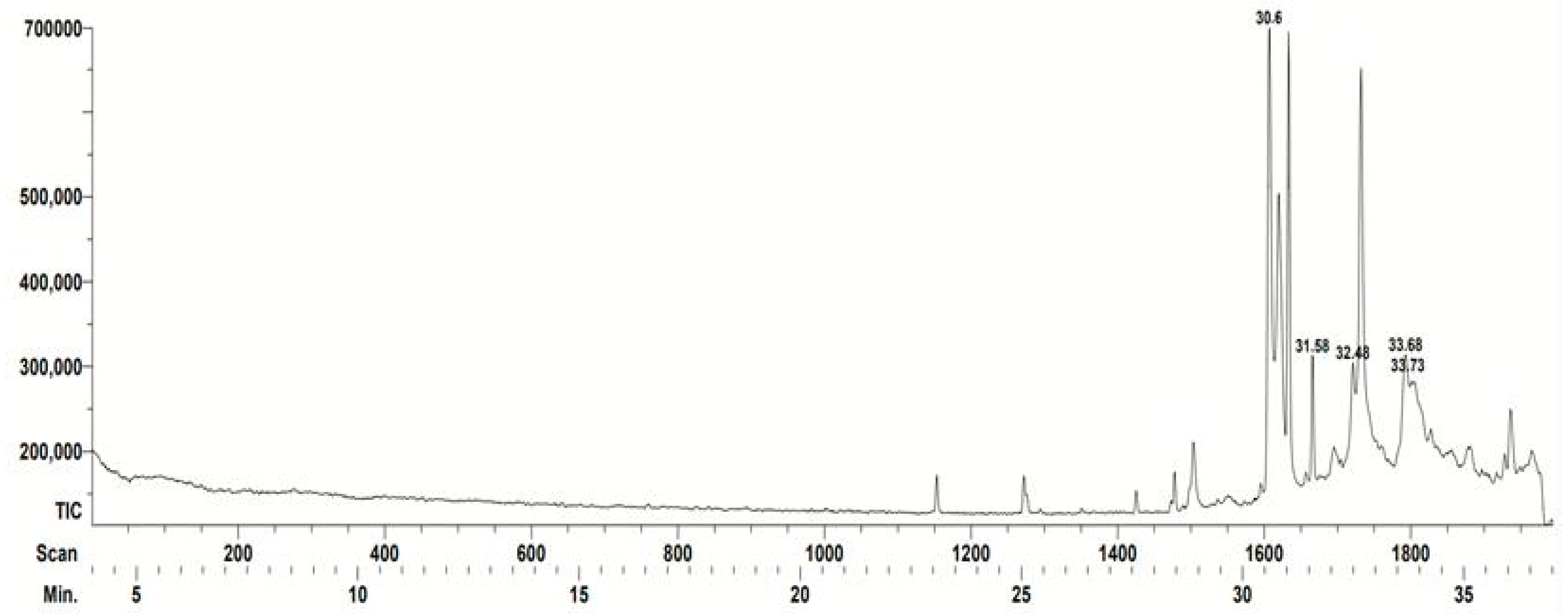
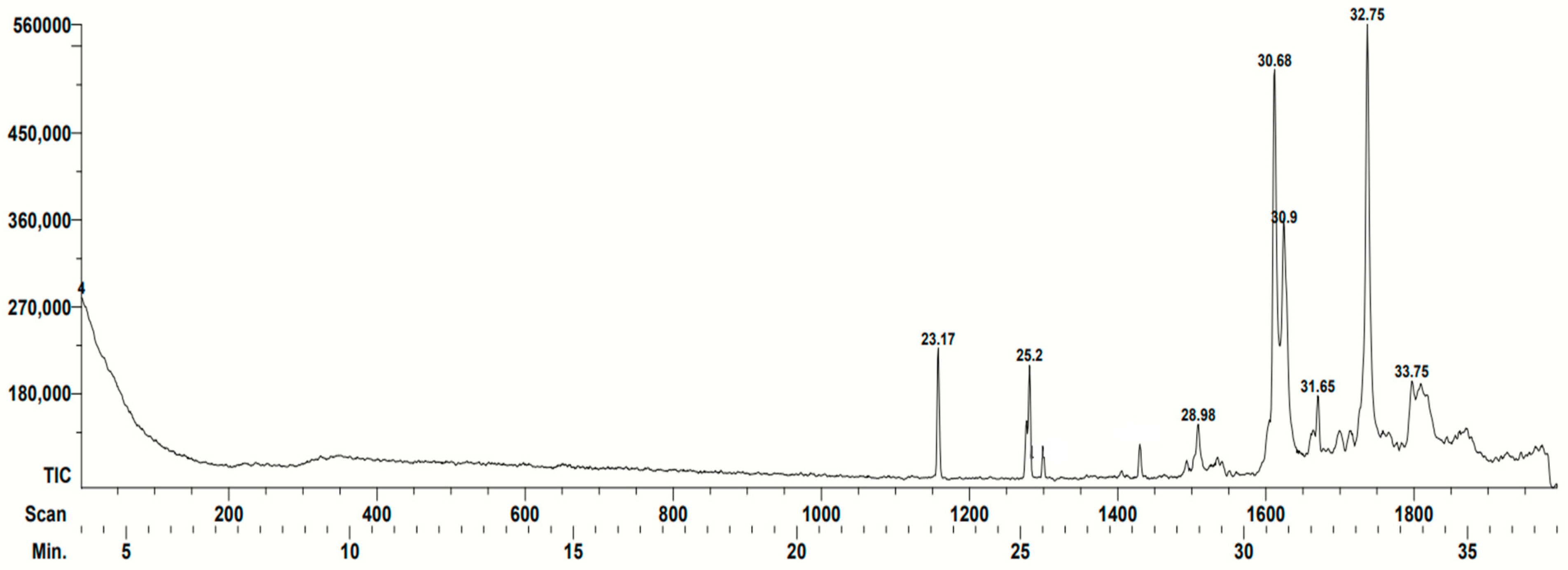
In the case of the T. angustula sample, only five compounds with biological activity were identified (Table 2). Terpenes with antifungal activity can be appreciated. It is also a compound with antibacterial activity (1,3-Benzenediol, 5-hexyl) as well as one with nematicide capacity (Hexadecanoic acid ethyl ester) The main peaks identified are shown in Figure 2.
Table 2.
Main constituents of propolis of Tetragonisca angustula characterised by CG-MS.
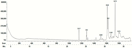
Figure 2.
Gas chromatogram corresponding to the Tetragonisca angustula propolis. The retention time of each compound identified by the database is indicated (minutes): 23.17: Hexadecanoic acid ethyl ester; 25.20: 9-Octadecenoic acid, ethyl ester; 28.98: 1-(1,1-dimethylethoxy)-4-methylbenzene; 30.68: Solavetivone; 30.90: Benzene, 1-(1,1-dimethylpropoxy)-4-methyl; 31.65: 2-Methyl-3-(3-methyl-but-2-enyl)-2-(4-methyl-pent-3-enyl)-oxethane; and 32.75: (1S,6R,9S)-5,5,9,10-etramethyltricy-cle[7.3.0.0(1,6)]dodec-10(11)-ene. The characteristics of the compounds can be found in Table 2. Spikes that are not numbered were not identified by the team database.
3.2. Evaluation of the Antimycotic Activity
All the tested Malassezia pachydermatis strains were susceptible to the propolis extracts. The minimum inhibitory concentration (MIC) was 7.11 mg/mL, and the minimum fungicidal concentration (MFC) was 21.33 mg/mL (Table 3).
Table 3.
Minimum inhibitory concentration (MIC) and minimum fungicidal concentration (MFC) values of stingless bee propolis extracts from two regions of the Mexican Republic on the reference strain M. pachydermatis ATCC 14522 and strains isolated from clinical samples.
3.3. Structural Damage
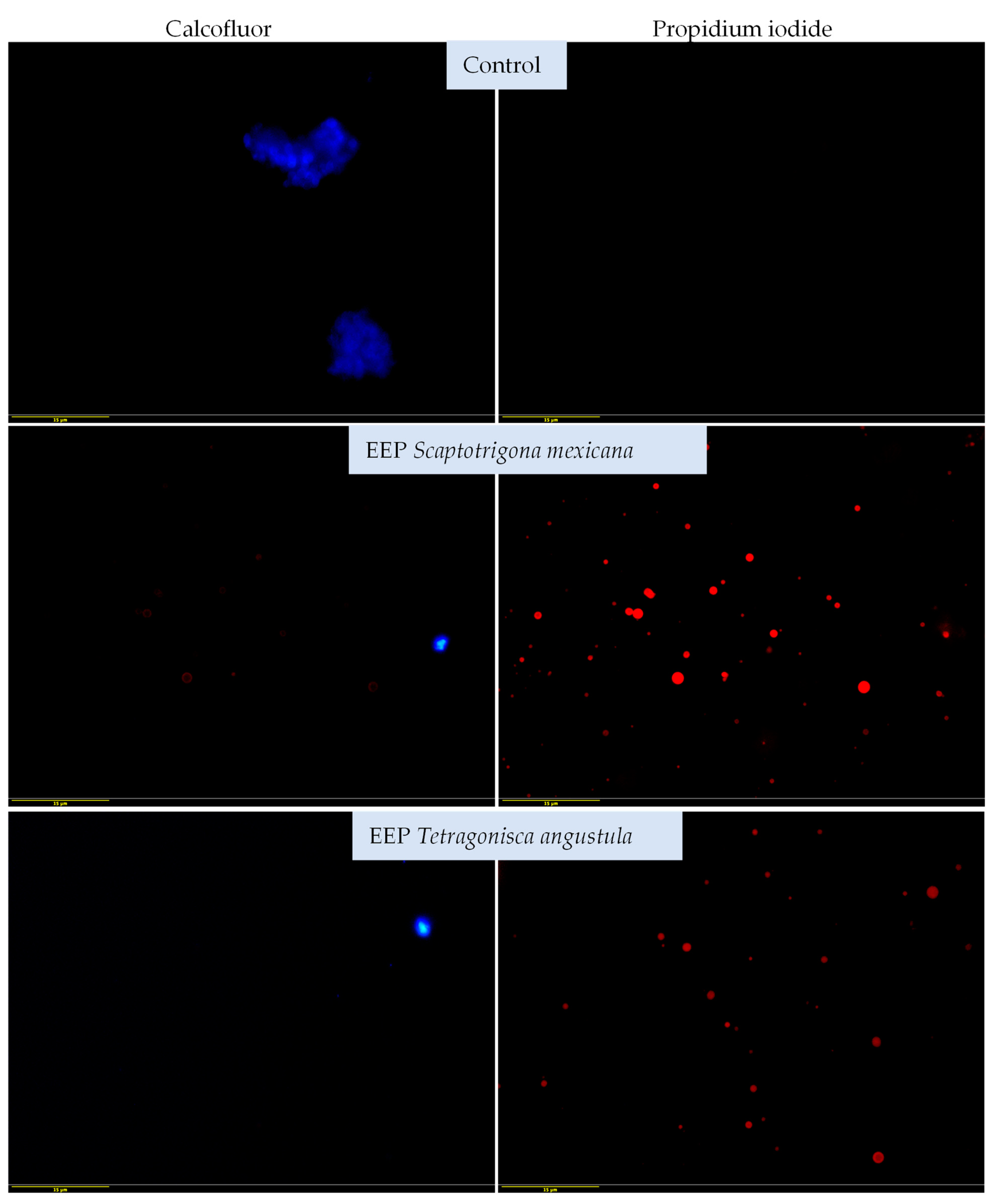
The structural damage to the yeasts was determined by using different stains: calcofluor white and propidium iodide. The structural damage of yeasts was determined by using different stains: calcofluor white and propidium iodide. Yeasts stained blue (calcofluor white) indicate integrity of the cell wall (control), those stained red (propidium iodide) indicate damage to the cell wall, since the damage wall allows the entry of propidium iodide (red) (EEP exposed). Propidium iodide penetration indicates damage to the yeast and with calcofluor-white stain, only morphology deformation was observed. In the untreated control cultures, the yeasts were clearly stained blue with calcofluor-white, but not with propidium iodide, which indicates the integrity of the yeast cell.
Figure 3 illustrates the effects of both propolis extracts on the reference strain of M. pachydermatis (ATCC 14522). The yeast samples treated with the EEP of S. mexicana did not show staining with calcofluor white but showed staining with propidium iodide in the form of red colouration, with the most severe damage being that produced by the EEP of S. mexicana.
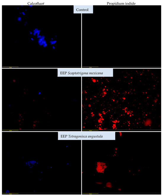
Figure 3.
Effects of EEP of Scaptotrigona mexicana and Tetragonisca angustula on reference strain Malassezia pachydermatis ATCC 14522 were obtained by fluorescence microscopy and dyeing with calcofluor white and propidium iodide. Cultures were exposed to EEP at a concentration of 21.3 mg/mL for 48 h at 28 °C. Yeast samples stained in blue (calcofluor white) indicate the integrity of the cell wall (control), while those stained in red (propidium iodide) indicate damage to the cell wall. They do not stain in blue due to damage to the cell wall, which allows the entry of propidium iodide (red) (EEP exposed). Propidium iodide penetration was observed, indicating damage to the yeast. With the calcofluor white stain, only morphology deformation was observed (40× magnification).
Figure 4 shows the effects of both EEPs on the clinical strain and also shows the staining of the untreated yeasts with calcofluor white but not with propidium iodide, which indicates the integrity of the plasma membrane and cell wall. The yeast samples treated with the EEPs of S. mexicana and T. angustula exhibited a similar effect. In both cases, the yeast samples were not stained with calcofluor white but were stained with propidium iodide, which indicates that the EEP damaged the structure of the fungi. Considering both the reference strain and the clinical one, we can conclude that the EEPs of S. mexicana and T. angustula affect the structural integrity of the yeasts, and this is more evident in the clinical strain.
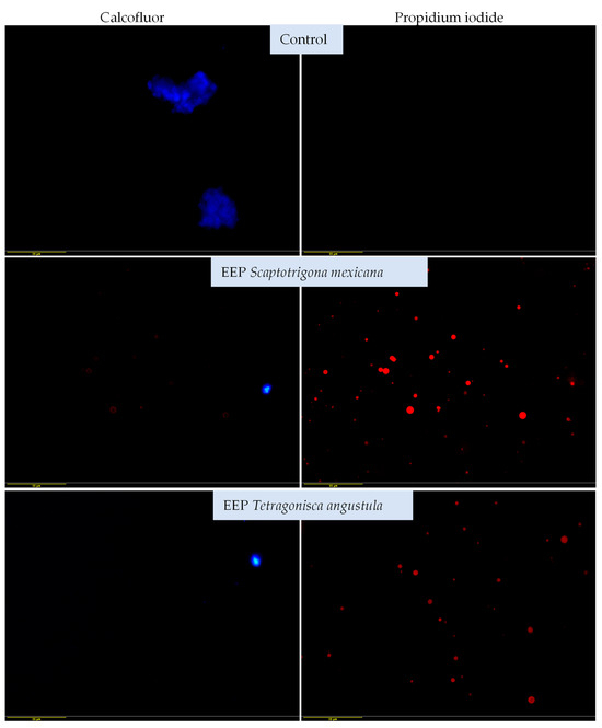
Figure 4.
Effects of the EEPs of Scaptotrigona mexicana and Tetragonisca angustula on the clinical strain of Malassezia pachydermatis, as determined by fluorescence microscopy and dyeing with calcofluor-white and propidium iodide. Cultures were exposed to 21.3 mg/mL of EEP for 48 h at 28 °C. Alteration of morphology is observed with the calcofluor white stain. As in Figure 3, yeast samples stained in blue (calcofluor white) indicate the integrity of the cell wall (control), while those stained in red (propidium iodide) indicate damage to the cell wall. They do not stain in blue due to damage to the cell wall, which allows the entry of propidium iodide (red) (EEP exposed). The red colouration of propidium iodide in both extracts indicates greater damage to the yeast cells (40× magnification).
4. Discussion
Many of the identified metabolites of stingless bee propolis were reported to exhibit a myriad of different chemical compositions, which is consistent with other studies that have biological activities, including antimicrobial, antiinflammatory, cytotoxic, antioxidant, hepatoprotective, and antiulcer effects [41,42,43,44,45]. The propolis extracts analysed show a diversity of compounds [46,47]. The floral diversity, time of collection, and bee species are all determinant factors for the final composition of each propolis, where sesquiterpene compounds predominated; these compounds are known for their antimicrobial activity [1,3].
Regarding the propolis of Scaptotrigona mexicana, bibliographic research of furan-2,5-dicarbaldehyde (a heterocyclic compound with two aldehyde groups) was unsuccessful using that exact denomination; however, we found mention of a similar compound, 2-acetyl-5-methylfuran, which was reported to exhibit antimicrobial activity against Escherichia coli, Candida albicans, and Staphylococcus aureus [32,33,39].
On the other hand, as the propolis of Tetragonisca angustula, the antibacterial and antifungal activity of solavetivone has been reported [35,36]. There is no specific information on the activity of l-(1S,6R,9S)-5,5,9,10-tetramethyltricyclo[7.3.0.0(1,6)]dodec-10(11)-ene; however, antifungal and antibacterial activities have been reported for a similar compound, 3,3,7,7-tetramethyl-5-(2-methyl-1-propenyl)-tricyclo[4.1.0.0(2,4)]heptane [38].
A notable difference was observed between the chemical composition of the analysed propolis and that of Mexican Apis mellifera. The antimicrobial activity of Apis mellifera propolis is related to the presence of flavonoids such as pinocembrin, tectochrysin (flavone), and the flavonoid precursor cardamomin (chalcone), as well as 2-methoxy-4-vinylphenol and a few terpenoids [10]. On the other hand, in this work, sesquiterpenes were the most important compounds, which is in agreement with a study by Bankova [2], which showed that terpenoids predominate in the propolis of native bees from various parts of the world. This seems to be a marked difference between the propolis of honeybees and native bees.
The antimycotic activity of propolis extracts, mainly from Apis mellifera, was demonstrated against Candida albicans. Moreover, the fungicidal and fungistatic properties of green and red propolis extracts from Brazil against other fungi genera were reported [43]. In addition, the inhibition and morphologic alterations of Cryptococcus neoformans when exposed to propolis were described [46].
The antimycotic activity of propolis from stingless bees, mainly against Candida albicans, has been reported for the following species: Lestrimellata spp., Melipona favora orlinge, Melipona marginata, Melipona quadrifasciata, Melipona scutellaris, Nannotrigona testaceicornis, Plebeia droryana, Plebeia remota, Scaptotrigona bipunctata, Tetragona clavipes, Tetragonisca angustula, and Tetragonisca fiebrigi (against Candida glabrata) [2,47]. The propolis from the Malaysian stingless bee Trigona thoracica was demonstrated to act against Cryptococcus neoformans [47]. Furthermore, an Indonesian propolis from Tetragonula sp. was evaluated as a possible therapeutic agent for the treatment of vaginal candidiasis [48]. However, no studies on the use of the propolis from this stingless bee for antifungal applications in animals were found.
Some reports have evaluated the activity of the propolis from Apis mellifera against Malassezia pachydermatis, but there are no such reports focusing on native bees. In a recent study, a correlation was established between the antimycotic activity of the ethanolic extract of Brazilian green and red propolis against M. pachydermatis, with an MIC between 4 and 8 mg/mL and an MFC of 8–16 mg/mL [49]. The study reported that, as the total content of phenols and flavonoids increased, propolis exhibited an enhanced biological effect, suggesting that the mechanism of action of EEP is based on the rupture of the cell wall. This idea is reinforced by the observation that some azole-resistant M. pachydermatis strains were inhibited by the EEP. The efficacy of an Argentinian propolis against M. pachydermatis was evaluated by different in vitro techniques; the results demonstrated that the yeast was vulnerable to all tested propolis concentrations, with an MIC of 0.30 mg/mL; however, the researchers were unable to determine the MFC [17]. The efficacy of a 2.5% EEP solution against 48 clinical strains of M. pachydermatis isolated from dogs diagnosed with otitis externa was also proven, as it was found that all the strains were susceptible to the EEP solution [17]. An EEP from Rio Grande do Soul, Brazil, demonstrated antimycotic activity against clinical isolates from dogs with otitis externa, with an MIC of 2.6 mg/mL and an MFC of 5.3 mg/mL. In this work, an MFC of 21.3 mg/mL was determined, being higher than what was found with Apis mellifera propolis, but the MIC of 7.11 mg/mL determined was similar to what was reported for this propolis; however, it is unclear whether high EPP concentrations could induce cytotoxicity. Therefore, more research is needed to identify the active principles of propolis as well as their action mechanisms. Currently, there are two theories aiming to explain the antifungal activity of propolis: the first proposes that propolis elicits cellular wall lysis. and the other proposes that propolis damages the plasma membrane by inhibiting ergosterol synthesis [16].
The photomicrographs obtained in the present report revealed that the EEP was able to penetrate the plasma membrane, which was found using the minimum fungicidal concentration (21.33 mg/mL), causing severe damage and eventually the death of yeast samples through structural and functional damage caused by membrane disruption. Calcofluor white exhibits a high affinity for fungal wall components [50,51]. The alterations observed with this stain were mainly deformed morphologies. In some cases, we hypothesise that the complete destruction of the cell wall prevented the observation of the yeasts, which would be a possible effect of the sesquiterpenes present in the EEP, as previously described [52]. It would be advisable to perform computational chemistry studies to establish the extent of the damage to the yeast’s cell wall caused by these compounds.
On the other hand, propidium iodide binds to nucleic acids and increases red colouration when there is damage to the cell membranes. This red colouration indicates severe cell damage and death, which was observed in the reference strain and the clinical strains treated with both EEPs, demonstrating the effectiveness of this type of propolis. This effect with propidium iodide has been observed in other fungi, such as Fusarium, by evaluating naturally occurring compounds’ efficacy against fungal growth [19]. It is likely that using higher concentrations of EPP would have resulted in more damage being detected, so it would be advisable to use higher concentrations in future research.
The above-mentioned stains have been used to detect cellular damage by Apis mellifera propolis in yeasts of medical importance, such as Candida albicans [53,54,55] as well as the bacterium Staphylococcus aureus [56]. There is scant research on the antimicrobial activity of stingless bee propolis against infectious agents in animals, which represents an opportunity to improve animal health.
Our team demonstrated the antiviral activity of the propolis of the stingless bee Plebeia frontalis against the canine distemper virus. This encourages further investigation into its effects on other viruses that affect animals [57].
Therefore, this work demonstrates that the propolis of the Mexican stingless bees Scaptotrigona mexicana and Tetragonisca angustula has antimycotic effects and causes structural damage to Malassezia pachydermatis. This result supports the use of these types of propolis for therapeutic purposes. The findings of this study and their implications should be discussed in the broadest possible context. Future research directions may also be highlighted.
5. Conclusions
In conclusion, the present study has shown the antifungal properties and extent of structural damage caused by two propolis ethanolic extracts from two stingless bee species (Scaptotrigona mexicana and Tetragonisca angustula) found in the Mexican municipalities of Yecuatla, Veracruz, and Chalchihuitan, Chiapas, against different Malazessia pachydermatis strains, one as a reference (ATCC 14522) and three clinical isolates. Sesquiterpenes, along with other compounds, are possibly the reason for the antimycotic activity of both extracts. It is important to mention that, to our knowledge, the present work is the first to demonstrate the structural damage caused by and antifungal effects of Mexican stingless bee propolis against Malazessia pachydermatis.
Nonetheless, further research must be undertaken in order to provide a more solid scientific basis for the future employment of propolis as an alternative treatment for canine external otitis.
Author Contributions
Evaluation of antimicotic activity, D.B.F.E.; analysis of propolis samples, B.R.P.; collection of propolis in the meliponaries, F.H.G.; collection of clinical and reference strains and conservation of microorganisms, N.T.B.; gas chromatography–mass spectrometry, J.P.F.; performance of chemical determinations, J.G.P.C.; fluorescence microscopy and revision of manuscript, B.R.P. and C.G.G.T.; funding acquisition and revision of manuscript, T.A.C.S. All authors have read and agreed to the published version of the manuscript.
Funding
This research was funded by National Autonomous University of México Programa de Apoyo a Proyectos de Investigación e Innovación Tecnológica (PAPIIT) IN 223719 project and IN215723 project. Facultad de Estudios Superiores Cuautitán: Research Chairs CI 2237, CI 2222, CI 2267, and the Bee Team from the South Border College (ECOSUR).
Institutional Review Board Statement
Not applicable.
Informed Consent Statement
Not applicable.
Data Availability Statement
Data are contained within the article.
Acknowledgments
The authors would like to thank Lázaro Arroyo and Yliana Delfin Fuentes for their support in propolis sample recollection from stingless bee families, and Amparo Londoño Orozco, Samantha Jardon Xicotencatl, and Francisco Rodolfo González Diaz for their technical support.
Conflicts of Interest
The authors declare no conflicts of interest.
References
- Bakchiche, B.; Temizer, I.K.; Güder, A.; Gencay, Ö.; Yegin, S.; Bardaweel, S.; Ghareeb, M. Chemical Composition and Biological Activities of Honeybee Products From Algeria. J. Appl. 2020, 7, 93. [Google Scholar] [CrossRef]
- Bankova, V.; Popova, M. PHCOG REV: Review Article Propolis of Stingless Bees: A Promising Source of Biologically Active Compounds. Pharmacogn. Rev. 2007, 1, 88–92. [Google Scholar]
- Ramón-Sierra, J.; Peraza-López, E.; Rodríguez-Borges, R.; Yam-Puc, A.; Madera-Santana, T.; Ortiz-Vázquez, E. Partial Characterization of Ethanolic Extract of Melipona beecheii Propolis and in vitro Evaluation of Its Antifungal Activity. Rev. Bras. Farmacogn. 2019, 29, 319–324. [Google Scholar] [CrossRef]
- Ayala, R. Revisión de Las Abejas Sin Aguijón de México (Hymenoptera: Apidae: Meliponini). Folia Entomol. Mex. 1999, 10, 106–123. [Google Scholar]
- Arnold, N.; Zepeda, R.; Vázquez, M.; Aldasoro, M. Las Abejas sin Aguijón y su Cultivo en Oaxaca, México Con Catálogo de Especies; Libros ECOSUR-CONABIO: San Cristóbal de las Casas, Mexico, 2018; pp. 10–58. [Google Scholar]
- Pardini, R.; Rocha, P.; El-Hani, C.; Pardini, F. Challenges and Opportunities for Bridging the Research—Implementation Gap in Ecological Science and Management in Brazil. In Conservation Biology: Voices from the Tropics; John Wiley & Sons, Ltd.: Hoboken, NJ, USA, 2013; pp. 75–85. [Google Scholar] [CrossRef]
- Hurtado Burillo, M.; Jara, L.; May-Itzá, W.; Quezada-Euán, J.J.; Ruiz, C.; Rúa, P.A. Geometric Morphometric and Microsatellite Analyses of Scaptotrigona mexicana and S. pectoralis (Apidae: Meliponini) Sheds Light on the Biodiversity of Mesoamerican Stingless Bees. J. Insect Conserv. 2016, 20, 753–763. [Google Scholar] [CrossRef]
- Aldama, J.; Rodríguez Pérez, B.; Gallardo, S.; Sánchez, T. Structural damage in Cryptococcus neoformans caused by a Mexican propolis. Nova Sci. 2020, 12, 1–17. [Google Scholar] [CrossRef]
- Quintero-Mora, M.L.; Londoño, O.A.; Hernández, H.F.; Manzano, G.P.; Lópe, M.R.; Soto, Z.C.I.; Carrillo, M.L.; Penieres, C.G.; García, C.G.; Cruz, T.A. Effect of Mexican propolis extracts from Apis mellifera on Candida albicans in vitro growth. Rev. Iberoam. Micol. 2008, 25, 22–26. [Google Scholar] [CrossRef]
- Rodríguez, P.B.; Canales, M.M.; Penieres, C.J.; Cruz, S.T. Chemical composition, antioxidant properties and antimicrobial activity of Mexican propolis. Acta Univ. 2019, 30, 1–29. [Google Scholar] [CrossRef]
- Flores, I.S.; Moreno, M.; Londoño, A.; Cruz, T.A. Use of mexican propolis for the tropical treatment of dermatomycosis in horses. Open J. Vet. Med. 2016, 6, 62770. [Google Scholar] [CrossRef]
- Tovar, N.; García, L.; Cruz, T.A. Propolis in dogs: Clinical experiences and perspectives (a brief review). Open J. Vet. Med. 2016, 5, 11–17. [Google Scholar] [CrossRef]
- Girão, M.D.; Prado, M.R.; Brilhante, R.S.N.; Cordeiro, R.A.; Monteiro, A.J.; Sidrim, J.J.C.; Rocha, M.F.G. Malassezia pachydermatis Isolated from Normal and Diseased External Ear Canals in Dogs: A Comparative Analysis. Vet. J. 2006, 172, 544–548. [Google Scholar] [CrossRef]
- Lozina, L.; Boehringer, S.; Daquino, M.; Acosta, O. Eficacia del Propóleo sobre Malassezia pachydermatis. Correlación de distintas Técnicas in vitro. Acta Farm. Bonaerense 2006, 25, 560. [Google Scholar]
- Cabañes, F.J. Diagnosis of Malassezia dermatitis and otitis in dogs and cats, is it just a matter of counting? Rev. Iberoam. Micol. 2021, 38, 3–4. [Google Scholar] [CrossRef]
- Deegan, K.R.; Fonseca, M.S.; Oliveira, D.C.P.; Santos, L.M.; Fernandez, C.C.; Hanna, S.A.; Machado, B.A.S.; Umsza-Guez, M.A.; Meyer, R.; Portela, R.W. Susceptibility of Malassezia pachydermatis Clinical Isolates to Allopathic Antifungals and Brazilian Red, Green, and Brown Propolis Extracts. Front. Vet. Sci. 2019, 6, 460. [Google Scholar] [CrossRef] [PubMed]
- Lozina, L.A.; Peichoto, M.E.; Boehringer, S.I.; Koscinczuk, P.; Granero, G.E.; Acosta, O.C. Efficacy of Argentine Propolis Formulation for Topical Treatment of Canine Otitis Externa. Arq. Bras. Med. Vet. Zootec. 2010, 62, 1359–1366. [Google Scholar] [CrossRef]
- Tovar Betancourt, N. Evaluación Antimicótica in vitro del Propóleo Mexicano Sobre Malassezia pachydermatis. Master’s thesis, Universidad Nacional Autónoma de México, Facultad de Estudios Superiores Cuautitlán, Izcalli, Mexico, 2016. Available online: https://ru.dgb.unam.mx/bitstream/20.500.14330/TES01000753315/3/0753315.pdf (accessed on 5 October 2023).
- Medina, R.Y.M.; Rodríguez, C.M.; Rodríguez, M.M.A.; Hernández, H.A.B.; Delgado, B.N.L.; Chirino, Y.I.; Cruz, S.T.; Garcia, T.C.G.; Canales, M.M.M. Effect of the Essential Oils of Bursera morelensis and Lippia graveolens and Five Pure Compounds on the Mycelium, Spore Production, and Germination of Species of Fusarium. J. Fungi 2022, 8, 617. [Google Scholar] [CrossRef] [PubMed]
- Diario Oficial de la Federación. Norma Oficial Mexicana NOM-003-AG/GAN-2017: Propóleos, Producción y Especificaciones para su Procesamiento. 2017. Available online: https://www.dof.gob.mx/nota_detalle.php?codigo=5500103&fecha=06/10/2017#gsc.tab=0 (accessed on 15 April 2023).
- Dos Santos, L.; Hochheim, S.; Boeder, A.; Kroger, A.; Tomazzoli, M.; Neto, R.; Maraschin, M.; Guedes, A.; Cordova, C. Chemical Characterization, Antioxidant, Cytotoxic and Antibacterial Activity of Propolis Extracts and Isolated Compounds from the Brazilian Stingless Bees Melipona quadrifasciata and Tetragonisca angustula. J. Apic. Res. 2017, 56, 543–558. [Google Scholar] [CrossRef]
- Rivera, Y.C.R.; Terrazas, L.I.; Jiménez, E.M.; Campos, J.E.; Flores, O.C.M.; Hernández, L.B.; Cruz, S.T.; Garrido, F.G.I.; Rodríguez, M.M.A.; Canales, M.M.M. Anti-Candida Activity of Bursera morelensis Ramírez Essential Oil and Two Compounds, α-Pinene and γ-Terpinene—An in vitro Study. Molecules 2017, 22, 2095. [Google Scholar] [CrossRef] [PubMed]
- Kaneko, T.; Makimura, K.; Abe, M.; Shiota, R.; Nakamura, Y.; Kano, R.; Hasegawa, A.; Sugita, T.; Shibuya, S.; Watanabe, S.; et al. Revised Culture-Based System for Identification of Malassezia Species. J. Clin. Microbiol. 2007, 45, 3737–3742. [Google Scholar] [CrossRef]
- Aspíroz, C.; Gilaberte, Y.; Rezusta, A.; Boekhout, T.; Rubio, M.A.C. Gentamycin Inhibits the Growth of Malassezia pachydermatis in Culture. Rev. Iberoam. Micol. 2010, 27, 20–21. [Google Scholar] [CrossRef] [PubMed]
- Balouiri, M.; Sadiki, M.; Ibnsouda, S.K. Methods for in vitro Evaluating Antimicrobial Activity: A Review. J. Pharm. Anal. 2016, 6, 71–79. [Google Scholar] [CrossRef]
- Sánchez, A.K.; Fernández, M.R.F.; Moreno, M.M.; Villegas, A.L.; Meneses, G.F.; Arenas, G.R. Sensitivity and specificity of mycological direct examination with calcofluor white for the diagnosis of onychomycosi. Med. Cut. Ibero-Lat. Am. 2013, 6, 261–266. [Google Scholar] [CrossRef]
- Phillips, A.J.; Sudbery, I.; Ramsdale, M. Apoptosis Induced by Environmental Stresses and Amphotericin B in Candida albicans. Proc. Natl. Acad. Sci. USA 2003, 100, 14327–14332. [Google Scholar] [CrossRef]
- Urbizu, G.A.L.; Castillo, R.O.; Martínez, A.G.C.; Torres, C.J.A. Natural Variability of Essential Oil and Antioxidants in the Medicinal Plant Turnera diffusa. Asian Pac. J. Trop. Med. 2017, 10, 121–125. [Google Scholar] [CrossRef] [PubMed]
- Delmondes, G.A.; Santiago, L.I.C.; Días, D.Q.; Cunha, G.L.D.; Araujo, I.M.; Barbosa, R.; Coutinho, H.D.M.; Felipe, C.F.B.; Barbosa, F.J.M.; Lima, N.T.R.; et al. Pharmacological Applications of Farnesol (C15H26O): A patent review. Expert. Opin. Ther Pat. 2020, 30, 227–234. [Google Scholar] [CrossRef]
- Fatnassi, S.; Zarrouk, H.; Chatti, S. Chemical Composition and Antimicrobial Activity of Volatile Fraction of the Peel of Maclura pomifera Fruit Growing in Tunisia. J. Soc. Chim. Tunisia 2011, 13, 1–6. [Google Scholar]
- Chen, C.; Change, H.C.; Kirk, K. Betulachrysoquinone Hemiketal: A p-Benzoquinone Hemiketal Macrocyclic Compound Produced by Phanerochaete chrysosporium. Phytochemistry 1977, 16, 1983–1985. [Google Scholar] [CrossRef]
- Murira, K.G.; Njagi, E.N.M.; Machocho, A.K.; Nyawira, W.L.; Mungiria, J.N.M. Chemical Composition and in vitro Antioxidant Activities of Ocimum americanum. Adv. Anal. Chem. 2015, 5, 42–49. [Google Scholar]
- Otieno, A.J. Antimicrobial Activity and Phytochemical Profiles of Warburgia ugandensis Sprague (Canellaceae) Extracts from Different Populations across the Kenyan Rift Valley. Master’s thesis, Kenyatta University, Nairobi, Kenia, 2016. Available online: https://ir-library.ku.ac.ke/handle/123456789/17924 (accessed on 6 December 2023).
- Siswadi, S.; Saragih, G.S. Phytochemical Analysis of Bioactive Compounds in Ethanolic Extract of Sterculia quadrifida R. Br. AIP Conf. Proc. 2021, 2353, 030098. [Google Scholar] [CrossRef]
- Xie, C.; Wang, S.; Cao, M.; Xiong, W.; Wu, L. (E)-9-Octadecenoic Acid Ethyl Ester Derived from Lotus seedpod Ameliorates Inflammatory Responses by Regulating MAPKs and NF-κB Signalling Pathways in LPS-Induced RAW264.7 Macrophages. Evid. Based Complement. Altern. Med. 2022, 2022, 6731360. [Google Scholar] [CrossRef] [PubMed]
- Takahashi, S.; Yeo, Y.-S.; Zhao, Y.; O’Maille, P.E.; Greenhagen, B.T.; Noel, J.P.; Coates, R.M.; Chappell, J. Functional Characterization of Premnaspirodiene Oxygenase, a Cytochrome P450 Catalyzing Regio- and Stereo-Specific Hydroxylations of Diverse Sesquiterpene Substrates. Int. J. Biol. Chem. 2007, 282, 31744–31754. [Google Scholar] [CrossRef] [PubMed]
- Peng, D.Q.; Yu, Z.X.; Wang, C.H.; Gong, B.; Liu, Y.Y.; Wei, J.H. Chemical Constituents and Anti-Inflammatory Effect of Incense Smoke from Agarwood Determined by GC-MS. Int. J. Anal. Chem. 2020, 2020, 4575030. [Google Scholar] [CrossRef]
- Bruzual Villarroel, H.Y.; Henríquez Guzmán, W.; Crescente, O.; Lanza, J.G. Aceite esencial de Wedelia calycina (Asteraceae): Composición química, actividad antibacteriana y antifúngica. Saber 2015, 27, 87–93. [Google Scholar]
- Sarvesan, R.; Eganathan, P.; Saranya, J.; Sujanapa, P. Chemical Composition and Antibacterial Activity of Leaf Essential Oil of Eugenia cotinifolia ssp. Codyensis (Munro Ex Wight) Ashton. Int. J. Pharm. Sci. 2015, 6, 3981–3985. [Google Scholar]
- Ambriz-Pérez, D.; Leyva-López, N.; Gutiérrez-Grijalva, E.P. Phenolic Compounds: Natural Alternative in Inflammation Treatment. A Review. Cogent Food Agric. 2016, 2, 1131412. [Google Scholar] [CrossRef]
- Badiazaman, A.A.M.; Zin, N.B.M.; Annisava, A.R.; Nafi, N.E.M.; Mohd, K.S. Phytochemical Screening and Antioxidant Properties of Stingless Bee Geniotrigona thoracica Propolis. Malays. J. Fundam. Appl. Sci. 2019, 15, 330–335. [Google Scholar] [CrossRef]
- Surek, M.; Fachi, M.M.; de Fátima Cobre, A.; de Oliveira, F.F.; Pontarolo, R.; Crisma, A.R.; de Souza, W.M.; Felipe, K.B. Chemical composition, cytotoxicity and antibacterial activity of propolis from Africanized Honeybees and three different Meliponini species. J. Ethnopharmacol. 2021, 269, 113662. [Google Scholar] [CrossRef] [PubMed]
- Torres, A.R.; Sandjo, L.P.; Friedemann, M.T.; Tomazzoli, M.M.; Maraschin, M.; Mello, C.F.; Santos, A.R.S. Chemical characterization, antioxidant and antimicrobial activity of propolis obtained from Melipona quadrifasciata and Tetragonisca angustula Stingless Bees. Braz. J. Med. Biol. Res. 2018, 51, e7118. [Google Scholar] [CrossRef] [PubMed]
- Yam, P.A.; Santana, H.A.; Yah, P.N.; Ramón, S.J.M.; Cáceres, M.R.; Borges, A.R.L.; Ortiz-Vázquez, E. Pentacyclic triterpenes and other constituents in propolis extract from Melipona beecheii collected in Yucatán, México. Rev. Bras. Farmacogn. 2019, 29, 358–363. [Google Scholar] [CrossRef]
- Grajales, C.J.; Elias, C.J.; Lozano, G.E.; Moreno, C.F.; Albores, F.V.; Lópe, G.A. Actividad antimicrobiana de propóleos de abejas sin aguijón en combinación con ajo, Allium sativum (Amaryllidaceae). Rev. Biol. Trop. 2020, 69, 23–35. [Google Scholar] [CrossRef]
- Hee, Y.C. In vitro evaluation of the antifungal activity of propolis extract on Cryptococcus neoformans and Candida albicans. Mycobiology 2002, 30, 93–95. [Google Scholar] [CrossRef]
- Shehu, A.; Ismail, S.; Rohin, M.; Harun, A.; Aziz, A.; Haque, M. Antifungal properties of Malaysian Tualang Honey and stingless bee propolis against Candida albicans and Cryptococcus neoformans. J. App. Pharm. Sci 2016, 6, 44–50. [Google Scholar] [CrossRef]
- Farida, S.; Sahlan, M.; Rohmatin, E.; Adawiyah, R. The beneficial effect of Indonesian Propolis wax from Tetragonula sp. as a therapy in limited vaginal candidiasis patients. Saudi J. Biol. Sci. 2020, 27, 142–146. [Google Scholar] [CrossRef] [PubMed]
- Cardoso, R.L.; Maboni, F.; Machado, G.; Alves, S.H.; de Vargas, A.C. Antimicrobial activity of propolis extract against Staphylococcus coagulase positive and Malassezia pachydermatis of canine otitis. Vet. Microbiol. 2010, 142, 432–434. [Google Scholar] [CrossRef] [PubMed]
- Hageage, G.J.; Harrington, B.J. Use of calcofluor white in clinical mycology. Lab. Med. 1984, 15, 109–112. [Google Scholar] [CrossRef]
- Ram, A.F.J.; Klis, F.M. Identification of fungal cell wall mutants using susceptibility assays based on calcofluor white and Congo red. Nat. Protoc. 2006, 1, 2253–2256. [Google Scholar] [CrossRef]
- Medina-Romero, Y.M.; Hernández-Hernández, A.B.; Rodríguez-Monroy, M.A.; Canales-Martínez, M.M. Essential oils of Bursera morelensis and Lippia graveolens for the development of a new biopesticides in postharvest control. Sci. Rep. 2021, 11, 20135. [Google Scholar] [CrossRef]
- De Castro, P.A.; Bom, V.L.P.; Brown, N.A.; Almeida, R.S.C.D.; Ramalho, L.N.Z.; Savoldi, M.; Goldman, M.H.S.; Berretta, A.A.; Goldman, G.H. Identification of the cell targets important for propolis-induced cell death in Candida albicans. Fungal Gent. Biol. 2013, 60, 74–86. [Google Scholar] [CrossRef] [PubMed]
- Adomavičiūtė, E.; Pupkevičiūtė, S.; Juskaite, V.; Zilius, M.; Stanys, S.; Pavilonis, A.; Briedis, V. Formation and investigation of electrospun PLA Materials with propolis extracts and silver nanoparticles for biomedical applications. J. Nanomater. 2017, 2017, 8612819. [Google Scholar] [CrossRef]
- Alfarrayeh, I.; Pollák, E.; Czéh, Á.; Vida, A.; Das, S.; Papp, G. Antifungal and anti-biofilm effects of caffeic acid phenethyl ester on different Candida species. Antibiotics 2021, 10, 1359. [Google Scholar] [CrossRef]
- Grecka, K.; Xiong, Z.R.; Chen, H.; Pełka, K.; Worobo, R.W.; Szweda, P. Effect of ethanol extracts of propolis (EEPs) against Staphylococcal biofilm—microscopic studies. Pathogens 2020, 9, 646. [Google Scholar] [CrossRef] [PubMed]
- Jiménez, O.V.D.; Pérez, B.R.; Cruz-Sánchez, T.A.; Tovar, C.G.G.; Bordes, J.L.N.; Zárate, C.I.S. Evaluation of the antiviral activity of propolis from native bees (Plebeia frontalis) against canine distemper virus. Open J. Vet. Med. 2020, 10, 207–218. [Google Scholar] [CrossRef]

Disclaimer/Publisher’s Note: The statements, opinions and data contained in all publications are solely those of the individual author(s) and contributor(s) and not of MDPI and/or the editor(s). MDPI and/or the editor(s) disclaim responsibility for any injury to people or property resulting from any ideas, methods, instructions or products referred to in the content. |
© 2024 by the authors. Licensee MDPI, Basel, Switzerland. This article is an open access article distributed under the terms and conditions of the Creative Commons Attribution (CC BY) license (https://creativecommons.org/licenses/by/4.0/).