Article

Development of Multiple Real-Time Fluorescent Quantitative PCR for Vibrio Pathogen Detection in Aquaculture

1
School of Ocean, Yantai University, Yantai 264005, China
2
Shandong Engineering Research Center of Healthy Land-Sea Relay Farming of Economic Fish, Yantai 264005, China
3
Yantai Engineering Research Center of Deep-Sea Aquaculture of Economic Fish, Yantai 264005, China
*
Author to whom correspondence should be addressed.
Vet. Sci. 2025, 12(4), 327; https://doi.org/10.3390/vetsci12040327
Submission received: 3 February 2025 / Revised: 31 March 2025 / Accepted: 1 April 2025 / Published: 2 April 2025

Simple Summary

Vibriosis, caused by various Vibrio species, significantly impacts marine biomass and leads to considerable economic losses in the aquaculture industry. Notably, Vibrio anguillarum, Vibrio alginolyticus, Vibrio harveyi, and Vibrio scophthalmi are major bacterial pathogens affecting the marine aquaculture sector. Thus, this study aimed to develop a rapid, accurate, and sensitive multiplex diagnostic method for the early detection of these four Vibrio species. A TaqMan probe-based multiplex real-time PCR method was developed, exhibiting four key characteristics: (1) high sensitivity, with detection limits 100 times more sensitive than conventional PCR assays; (2) high specificity, accurately and specifically detecting V. anguillarum, V. alginolyticus, V. harveyi, and V. scophthalmi; (3) multiplex detection, enabling the simultaneous detection of V. anguillarum, V. alginolyticus, V. harveyi, and V. scophthalmi in a single reaction; and (4) time efficiency, with detection results obtainable within one hour. These advancements facilitate the early detection and monitoring of these bacterial pathogens in both single and co-infected samples.

Abstract

The Vibrio genus represents a critical group of bacterial pathogens in the marine environment globally, leading to massive mortality in the aquaculture industry. Diagnosing vibriosis, an infection caused by Vibrio species, in clinical samples poses challenges due to its non-specific clinical manifestations. In this study, we developed a TaqMan probe-based multiplex real-time PCR method for the simultaneous detection and quantification of four Vibrio pathogens: Vibrio anguillarum (Va), Vibrio alginolyticus (Val), Vibrio harveyi (Vh), and Vibrio scophthalmi (Vsc). The assay targets conserved intra-species regions and specific inter-species regions using specific primers and TaqMan probes to ensure specificity. Sensitivity analysis demonstrated that the multiplex real-time PCR assay could simultaneously detect the four different bacteria, with detection limits of 26–60 copies per reaction, making it 100 times more sensitive than conventional PCR assays. Additionally, the assay exhibited high reproducibility, with intra- and inter-group coefficients of variation below 1.4%. A total of 63 clinical samples was analyzed using this established assay, which successfully detected both single and mixed infections. These results demonstrate that the multiplex quantitative PCR assay is a rapid, specific, and sensitive diagnostic tool for the detection of Va, Val, Vh, and Vsc, making it suitable for monitoring these bacteria in both single- and co-infected clinical samples.

1. Introduction

With the ongoing development of the mariculture industry, a diverse array of pathogens continues to emerge, leading to significant economic losses. Among these, bacterial infections are the most prevalent in aquaculture, characterized by their diversity and global distribution, with the potential to infect humans and other mammals [1,2]. The genus Vibrio is a major pathogenic microorganism, widely present in marine and freshwater ecosystems. Outbreaks of vibriosis have a severe impact on marine biomass and result in substantial economic losses in the aquaculture industry [3]. Early diagnosis is essential for the prevention and effective treatment of vibriosis; therefore, a rapid, accurate, multiplex, and sensitive diagnostic method is imperative.
Vibrio anguillarum, Vibrio alginolyticus, Vibrio harveyi, and Vibrio scophthalmi are significant bacterial pathogens affecting marine fish. Vibrio anguillarum is a halophilic pathogenic bacterium that is widely distributed in coastal and estuarine seawater [4]. It serves as a representative opportunistic bacterial pathogen for marine fish, shellfish, mollusks, and crustaceans, contributing to substantial economic losses in aquaculture [5]. This bacterium is capable of infecting at least 50 fish species, including Atlantic salmon (Salmo salar), rainbow trout (Oncorhynchus mykiss), turbot (Scophthalmus maximus), black sea bream (Sparus macrocephalus) and Japanese flounder (Parallichthys olivaceus) [6,7]. V. alginolyticus, initially classified as V. parahaemolyticus, is another halophilic bacterium predominantly found in marine and estuarine environments. It is regarded as one of the most detrimental Vibrio species to aquatic animals [8,9]. Systemic infections caused by V. alginolyticus have been documented in numerous fish and shrimp species, such as alfonsino (Sparus aurata), cobia (Rachycentron canadum), giant river prawn (Macrobrachium rosenbergii, Epinephelus malabaricus), South America white shrimp (Penaeus vannamei), and Japanese prawn (Penaeus japonicas), resulting in significant economic losses in aquaculture globally [10,11,12,13,14,15]. V. harveyi is recognized as a significant pathogen responsible for considerable mortality rates among various wild and cultured fish and invertebrates across a broad geographical range [16]. To date, V. harveyi has been isolated from a diverse array of marine fish and shrimp species worldwide, including Atlantic flounder (Paralichthys dentatus), S. salar, O. mykiss, silver perch (Lates calcarifer), orange-spotted grouper (Epinephelus coioides), sugpo prawn (Penaeus monodon) [17], and P. vannamei [17,18,19,20]. V. scophthalmi is an opportunistic pathogen of aquatic animals which was first isolated from the intestine of turbot (S. maximus) in 1997 [21]. Fish subjected to stressful conditions exhibit increased susceptibility to this bacterial strain [22]. To date, V. scophthalmi has also been isolated from other fish species, such as P. olivaceus, Japanese eel (Anguilla japonica), manila clam (Ruditapes philippinarum), and bluefin tuna (Thunnus maccoyii), resulting in mortality rates ranging from 30% to 90% among infected fish [22,23,24,25].
Due to the significant economic impact of these pathogens, the development of rapid and reliable detection methods is essential for preventing their further dissemination and facilitating appropriate therapeutic interventions. Vibrio species exhibit numerous similarities within this genus, including morphology, virulence factors, genome sequences, and clinical symptoms, which complicate the specific differentiation of one Vibrio species from another [26,27]. Current detection methodologies primarily rely on traditional culture-based techniques, as well as immunological and molecular biological detection technologies. While conventional symptom-based or culture-based methods can identify Vibrio infections, they often lack the specificity required to accurately distinguish between different Vibrio species [28,29]. PCR-based molecular diagnostics are widely employed in clinical settings due to their high specificity [30]. Numerous conventional PCR-based methods have been developed for the detection of V. anguillarum, V. alginolyticus, V. harveyi, and V. scophthalmi. However, these methods tend to be labor intensive and less sensitive [31,32,33,34]. Moreover, isothermal amplification techniques and real-time PCR have also been developed for the detection of certain bacterial pathogens [34,35,36,37,38,39]. Although these methods offer increased specificity, sensitivity, and time efficiency, they do not fully meet the clinical requirements for the detection of multiple pathogens.
TaqMan probe-based qPCR offers a rapid, specific, sensitive, and reproducible approach for pathogen detection and quantification, while multiplex PCR provides the advantages of high throughput and efficiency. Although the qPCR method is more expensive than conventional PCR, culturing methods, and the immune colloidal gold technique, it offers the advantage of detecting bacterial pathogens at the early stages of disease, thereby facilitating early prevention and treatment. TaqMan probe-based multiplex real-time PCR (multiplex qPCR) integrates multi-PCR and TaqMan probe-based qPCR technologies, offering advantages such as high sensitivity, the simultaneous detection of multiple targets, time efficiency, and reagent conservation [40]. In this study, a TaqMan probe-based multiplex real-time PCR assay was developed using the empA, toxR, vhhP2, and luxR genes as target markers to simultaneously detect V. anguillarum, V. alginolyticus, V. harveyi, and V. scophthalmi. This method demonstrates high specificity and sensitivity, is time-efficient, and possesses strong quantitative capabilities, making it a promising tool for the early diagnosis of infections caused by multiple pathogens.

2. Materials and Methods

2.1. Ethics Statement

Live animal experiments were performed in accordance with the guidelines of “Regulations for the Administration of Affairs Concerning Experimental Animals” promulgated by Shandong Province. Experiments involving live animals were approved by the Ethics Committee of Yantai University, with the ethical approval code No. 20230503.

2.2. Bacteria, Clinical, and Environmental Samples

Bacterial strains Edwardsiella piscicida (Ep), Photobacterium damselae (Pd), Pseudomonas fluorescens (Pf), Vibrio anguillarum (Va), and Vibrio harveyi (Vh) have been described previously [41,42,43,44]. Strains Vibrio alginolyticus (Val), Vibrio rotiferianus (Vro), Leclercia adecarboxylata (La), Vibrio hyugaensis (Vhy), Haemophilus piscium (Hp), Vibrio azureus (Vaz), and Vibrio scophthalmi (Vsc) were isolated and stored in our lab. All bacterial strains were cultured in Luria–Bertani broth (LB) medium at 28 °C for 12 h with constant shaking at 180 rpm under aerobic conditions with a rotary shaking (Zhichu, Shanghai, China). All bacterial species were further confirmed via 16S rDNA gene sequencing, conducted by Sangon Biotech Co., Ltd. (Shanghai, China).
For the preparation of clinical samples, 63 fish tissue samples were obtained. Briefly, 45 clinically healthy black rockfish (Sebastes schlegeli) with an average weight of 24.5 g were purchased from a commercial fish farm in Yantai, Shandong Province. The fish were maintained in aerated tanks (9 fish/tank) containing freshly prepared artificial seawater (changed daily) with a salinity of 30‰, a pH of 8.5, and a temperature of 20 °C. The bacterial strains Va, Val, Vh, and Vsc were cultured in LB medium as described above, and the turbidity of each strain was measured in a spectrophotometer (ThermoFisher, Waltham, MA, USA). When the optical density at 600 nm (OD600) reached 0.8–1.0, the bacterial cells were harvested through centrifugation at 5000× g for 2 min. The bacterial pellet was then washed 3 times with sterile phosphate-buffered saline (PBS) and adjusted to a concentration of 107 cfu/mL with PBS. The fish were divided into five groups and subjected to intraperitoneal injection with Va, Val, Vh, Vsc, or PBS, respectively. At 12, 24, and 48 h post-challenge (hpc), the fish (3 per time point) were euthanized using an overdose of tricaine methanesulfonate (Sigma, St. Louis, MO, USA). Then, the surfaces of the fish were disinfected with ethanol, and the liver, spleen, and kidney were collected under sterile conditions. For each fish, the three tissues were pooled, homogenized in a ten-fold volume of PBS, and subjected to bacterial examination via plate counts, as previously described [45]. Subsequently, positive samples containing different bacterial pathogens were selected and combined to create double-positive, triple-positive and quadruple-positive samples.
For the preparation of environmental samples, 21 sea-water and 21 sediment samples were collected from the intertidal zone of Yantai, China. Prior to experimentation, these samples were assessed for the presence of Va, Val, Vh, and Vsc using bacterial enrichment culture and PCR analysis. Subsequently, varying concentrations of Va, Val, Vh, and Vsc were added, and were verified as described above. A total of 42 positive samples (comprising 15 sea-water and 15 sediment samples) and 12 negative samples (comprising 6 sea-water and 6 sediment samples) were prepared for further qPCR analysis.

2.3. DNA Extraction

For the isolation of bacterial DNA, the bacterial strains were cultured in LB medium to an OD600 = 1.5. Subsequently, 1 mL bacterial suspension from each strain was collected and centrifuged at 5000× g for 2 min, and the bacterial pellet was subjected to DNA extraction using the TIANamp Bacteria DNA Kit (Tiangen Biotech Co, Ltd., Beijing, China) in accordance with the manufacturer’s instructions. For clinical samples, 200 μL of tissue homogenate was utilized for DNA extraction employing the CTAB method [46]. The extracted DNA was dissolved in 100 μL of nuclease-free water and stored at −20 °C for subsequent analyses.

2.4. Standard Recombinant Plasmids Construction

The empA gene (GenBank accession no. L02528), toxR gene (KJ579443), vhhP2 gene (FJ025787), and luxR gene (JN684210) were selected as target genes for the detection of Va, Val, Vh, and Vsc, respectively. Partial sequences of the empA, toxR, vhhP2, and luxR genes were amplified using the primer pairs empA-F/R, toxR-F/R, vhhP2-F/R, and luxR-F/R (Table 1), respectively, and subsequently inserted into the pMD-19T vector (TAKARA, Dalian, China). The recombinant plasmids were verified through sequencing in Sangon Biotech Co., Ltd., and were designated as pVa, pVal, pVh, and pVsc, respectively. The concentration of the recombinant plasmids was measured using a Nano-500 micro-spectrophotometer (Allsheng, Hangzhou, China). The DNA copy numbers were calculated using the following formula: DNA copy number (copies/μL) = [6.02 × 10²³ × plasmid concentration (ng/μL) × 10−9]/[DNA length × 660].
The standard recombinant plasmids were serially diluted (10-fold) ranging from 1 × 10⁸ to 1 × 100 copies/μL and stored at −20 °C for further use.

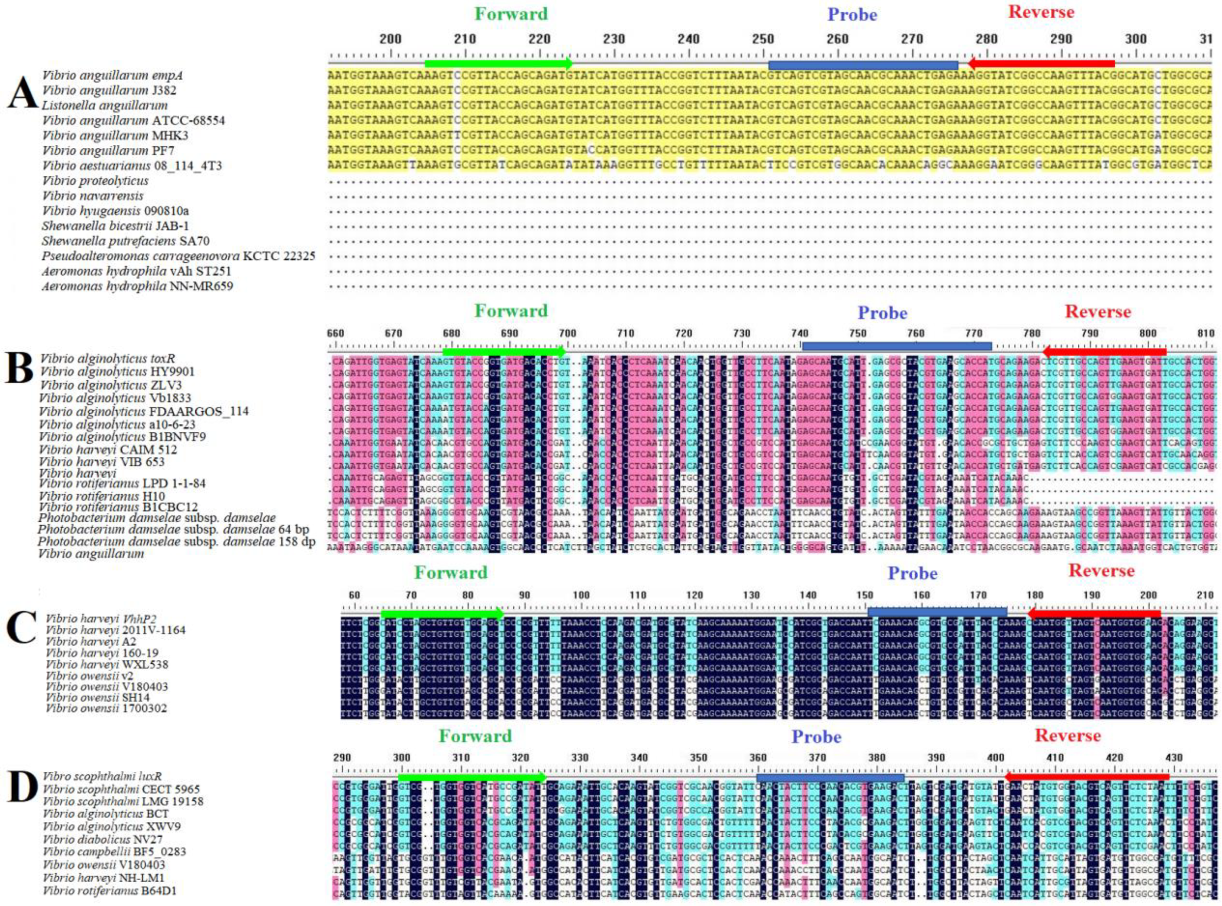
2.5. TaqMan Real-Time Fluorescent Quantitative PCR (qPCR) Primer Design

For the analysis of intraspecific conserved sequences and interspecific specific sequences, multiple sequence alignment was conducted using gene sequences obtained from the National Center for Biotechnology Information (NCBI) database, employing DNAMAN software (v7.0) (Table S1). Primers (empA-qF/qR, toxR-qF/qR, vhhP2-qF/qR, and luxR-qF/qR) and TaqMan fluorescence probes (empA-P, toxR-P, vhhP2-P, and luxR-P) were designed for qPCR (Table 1). Different fluorescence labels, FAM (6-carboxyfluorescein), HEX (6-carboxy-4′,5′-dichloro-2′,7′-dimethoxyfluorescein ester dye), ROX (Carboxy-X-Rhodamine), and CY5 (Cy Dye 5), were incorporated into the probes to distinguish different bacteria. Specificity of the primers and probes was confirmed through a Primer-BLAST search against the NCBI database. Additionally, OligoEvaluator software “http://www.oligoevaluator.com (accessed on 13 September 2023)” was utilized to assess the potential formation of primer dimers and hairpins. Finally, the synthesis and purification of the primers and probes were carried out by Sangon Biotech Co., Ltd., employing high-performance liquid chromatography techniques.

2.6. Optimization of Reaction Conditions for Multiplex qPCR

The reaction system and conditions for the multiplex qPCR were optimized by referencing the singleplex assay, utilizing the Pro Taq HS Premix Probe real-time PCR Kit III (Accurate Biology, Changsha, China), and conducted on the Bio-Rad CFX96 platform (Bio-Rad, Hercules, CA, USA). A series of ten concentration gradients for both forward and reverse primers (ranging 0.1 μmol/L~1 μmol/L, with increments of 0.1 μmol/L), and eight concentration gradients of probe (concentration range: 0.1 μmol/L~0.8 μmol/L; concentration gradient difference: 0.1 μmol/L) were set to determine the optimal primer and probe concentrations. The optimal annealing/extension temperature was evaluated across three temperatures (58 °C, 60 °C, and 62 °C). The temperature of optimal primers and probes, as well as the annealing/extension temperature, was selected based on amplification efficiency, determined from the Cq (cycle of quantification) values and the fluorescence intensity.

2.7. Establishment of Standard Curves of the qPCR

To construct standard curves, each standard recombinant plasmid was subjected to a 10-fold serial dilution, resulting in 7 dilution gradients ranging from 1 × 10⁹ to 1 × 10⁴ copies/μL. qPCR was conducted using these diluted plasmids in accordance with the optimized reaction system and procedures. The standard curve for qPCR was generated by plotting the logarithmic value of the plasmid copy number on the x-axis against the corresponding Cq value on the y-axis.

2.8. Specificity Test

To avoid false positives caused by other bacteria that could be present in this multiplex qPCR assay, the DNA templates extracted from other 8 bacterial strains, i.e., Ep, Pd, Vro, Pf, La, Vhy, Hp, and Vaz, were amplified, and ddH2O was used as a negative control. The specificity tests were performed under optimal conditions and system and repeated more than thrice. The templates were verified via 16S rRNA gene amplification and sequencing.

2.9. Sensitivity Test

False negatives are typically identified using the detection limit. Herein, the standard plasmids, pVa, pVal, pVh, and pVsc, were subjected to a 10-fold dilution to achieve a final concentration ranging from 105 to 100 copies/µL in nuclease-free water, in order to evaluate the sensitivity of the established multiplex qPCR. Optimal reaction conditions were used for multiplex qPCR amplification, with diluted standard plasmids serving as templates, and ddH2O as a negative control. Each concentration or negative control was analyzed in triplicate. For the conventional PCR analysis, diluted standard plasmids pVa, pVal, pVh, and pVsc were amplified using the primer pairs empA-F/R, toxR-F/R, vhhP2-F/R, and luxR-F/R, respectively. The conventional PCR was performed on a Bio-rad MyCycler PCR Thermal Cycler System (Bio-Rad, Hercules, WA, USA), and the reaction conditions were as follows: an initial denaturation at 95 °C for 3 min, followed by 35 cycles of denaturation at 95 °C for 30 s, annealing at 55 °C for 30 s, an extension at 72 °C for 90 s, and a final extension at 72 °C for 10 min. For agarose gel electrophoresis, a gel was prepared using 1 × TAE buffer (Solarbio, Beijing, China) with 1.5% agarose (Solarbio). The D2000 DNA Ladder (Solarbio) was employed as a standard marker to estimate the sizes of the amplified products.

2.10. Repeatability Test

The standard plasmids, pVa, pVal, pVh, and pVsc, were subjected to a 10-fold serial dilution to achieve final concentrations ranging from 106 to 104 copies/µL in nuclease-free water. This was conducted to evaluate the repeatability and reproducibility of the established multiplex qPCR. For multiplex real-time qPCR amplification, diluted standard plasmids were employed as templates. For intra-group repeatability analysis, qPCR was performed in triplicate for each concentration gradient. For inter-group repeatability analysis, each template was analyzed thrice with different participants every other week under the same conditions. The repeatability and reproducibility of the method were subsequently assessed using the coefficient of variation (CV), where a smaller CV value indicates greater data stability. The more stable the data, the smaller the CV value. The CV values were determined using the following formula: CV = [Standard Deviation]/[Mean] × 100.

2.11. Clinical Sample Testing

To evaluate the applicability of the multiplex qPCR assay for Va, Val, Vh, and Vsc detection in clinical and environmental samples, total DNA was extracted from the 105 samples prepared above. Among the clinical samples, 9 were negative control samples, 24 were single-positive samples, 18 were double-positive samples, 9 were triple-positive samples, and 3 were quadruple-positive samples. Among the sea-water samples, 6 were negative control samples, 12 were single-positive samples, and 3 were quadruple-positive samples. Among the sediment samples, 6 were negative control samples, 12 were single-positive samples, and 3 were quadruple-positive samples. Subsequently, the multiple qPCR assay was conducted on all 105 samples under the optimal reaction system and reaction conditions, and the coincidence rate between the detection results and the expected results was calculated. During these experiments, the standard plasmids, pVa, pVal, pVh, and pVsc, were used as positive controls.

2.12. Statistical Analysis

Standard curves, correlation coefficient (R2) values, and amplification efficiency (E) were analyzed using software supplied by the ABI PRISM 7500. The repeatability analysis was conducted using Microsoft Excel 2013 (Microsoft, Redmond, WA, USA).

3. Results

3.1. Primers and Probe Designed for qPCR Assay

The genes empA, toxR, vhhP2, and luxR were selected for the detection of Va, Val, Vh, and Vsc, respectively. Sequences of the empA, toxR, vhhP2, and luxR genes were retrieved from GenBank for multiple sequence alignment using DNAMAIN. To develop specific primers and probes, highly conserved intraspecific regions and specific interspecific regions were identified, and primers were designed utilizing AllelelD 6.0 software (Figure 1 and Table 1).

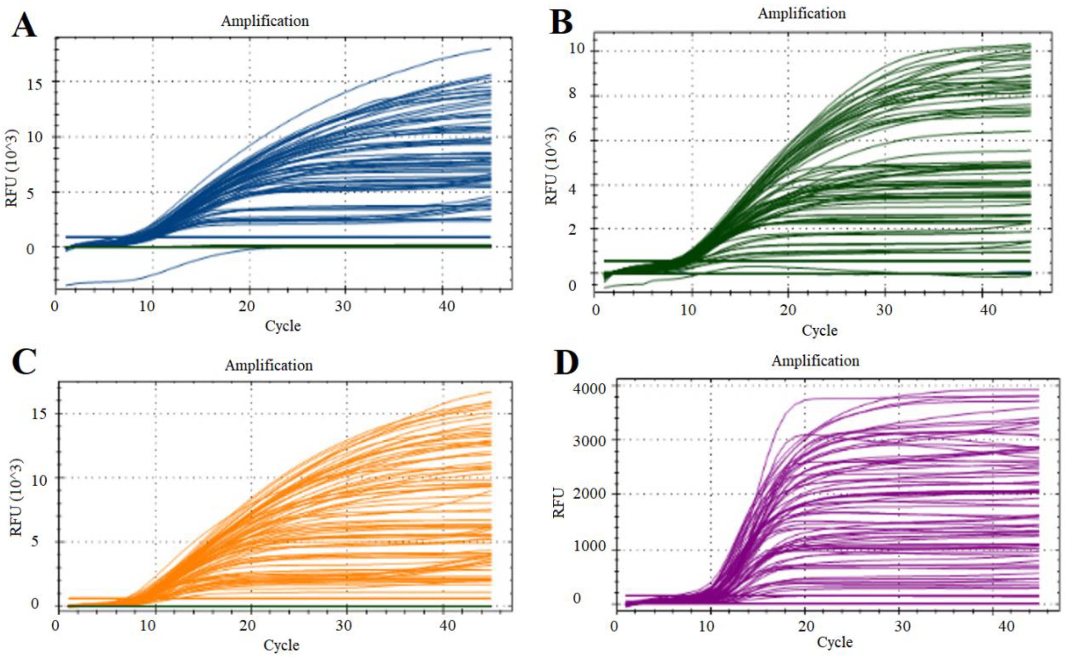
3.2. Optimization of qPCR Reaction Conditions

Through the optimization of primer and probe concentrations via single qPCR, the optimal working concentrations of primers for Va, Val, Vh, and Vsc were established at 0.3, 0.2, 0.4, and 0.2 μmol, respectively, while the optimal concentrations for the probes were 0.5, 0.6, 0.7, and 0.3 μmol, respectively (Figure 2 and Table 2). The reaction mixture comprised 10 μL of 2 × universal probe mix (Accurate Biology, Beijing, China), the specified primers and probes, 1 μL of each standard recombinant plasmid, and nuclease-free water to a total volume of 20 μL (Table 2). Variations in annealing temperatures did not significantly affect the results, leading to the selection of 60 °C as the standard annealing temperature. The final optimized reaction program was conducted as follows: pre-denaturation at 95 °C for 5 min, followed by 40 cycles at 95 °C for 15 s, and 60 °C for 30 s. The fluorescence channels for multiple qPCR analysis were set as follows: channel 1, FAM; channel 2, HEX; channel 3, ROX; and channel 4, Cy5.

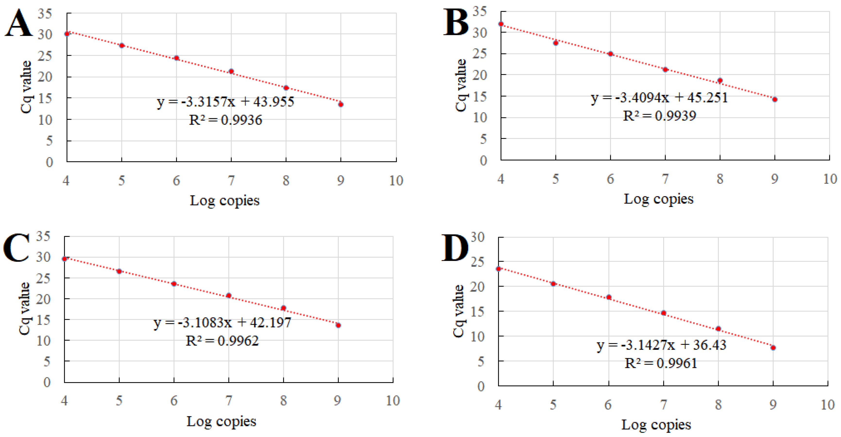
3.3. Standard Plasmid Construction and Standard Curve Establishment

For the construction of standard plasmids, partial sequences of the empA gene (800 bp), toxR gene (734 bp), vhhP2 gene (588 bp), and luxR gene (615 bp) were amplified and subsequently inserted into the pMD-19T vector (Figure S1). The resultant positive recombinant plasmids were extracted, sequenced, and designated as pVa, pVal, pVh, and pVsc. The purity and concentration of these plasmids were assessed using an Ultramicro spectrophotometer Nano 300 (Allsheng, Hangzhou, China). The copy numbers of pVa, pVal, pVh, and pVsc were determined to be 4.03 × 10¹⁰, 3.35 × 10¹⁰, 6.01 × 10¹⁰, and 2.59 × 10¹⁰ copies/μL, respectively.
For the establishment of standard curves, the standard plasmids were serially diluted in a 10-fold gradient ranging from 109 to 104 copies/µL and subsequently amplified via quantitative PCR (qPCR) under optimized reaction conditions. Four standard curves were generated by plotting the obtained quantification cycle (Cq) values on the y-axis against the logarithm of the plasmid concentration on the x-axis. The optimal standard formula, R2, and E values were as follows: Va, y = −3.3157x + 43.955, R2 = 0.9963, E = 1.00; Val, y = −3.4094x + 45.251, R2 = 0.9939, E = 0.96; Vh, y = −3.1083x + 42.197, R2 = 0.9962, E = 1.09; Vsc, y = −3.1427x + 36.43, R2 = 0.9961, E = 1.08 (Figure 3). The established curves exhibit excellent correlation coefficients, indicating that the TaqMan probe-based multiplex qPCR method developed is both reliable and valid.

3.4. Specificity of the Multiplex qPCR

The multiplex qPCR assay was conducted to detect various bacterial DNA sequences under uniform optimal reaction conditions. The results demonstrate that only DNA isolated from Va, Val, Vh, and Vsc was successfully detected in its specific fluorescence channel. Importantly, the DNA from other bacterial species, including Ep, Pd, Vro, Pf, La, Vhy, Hp, and Vaz, was not amplified, underscoring the high specificity of the developed multiplex qPCR method (Figure 4).

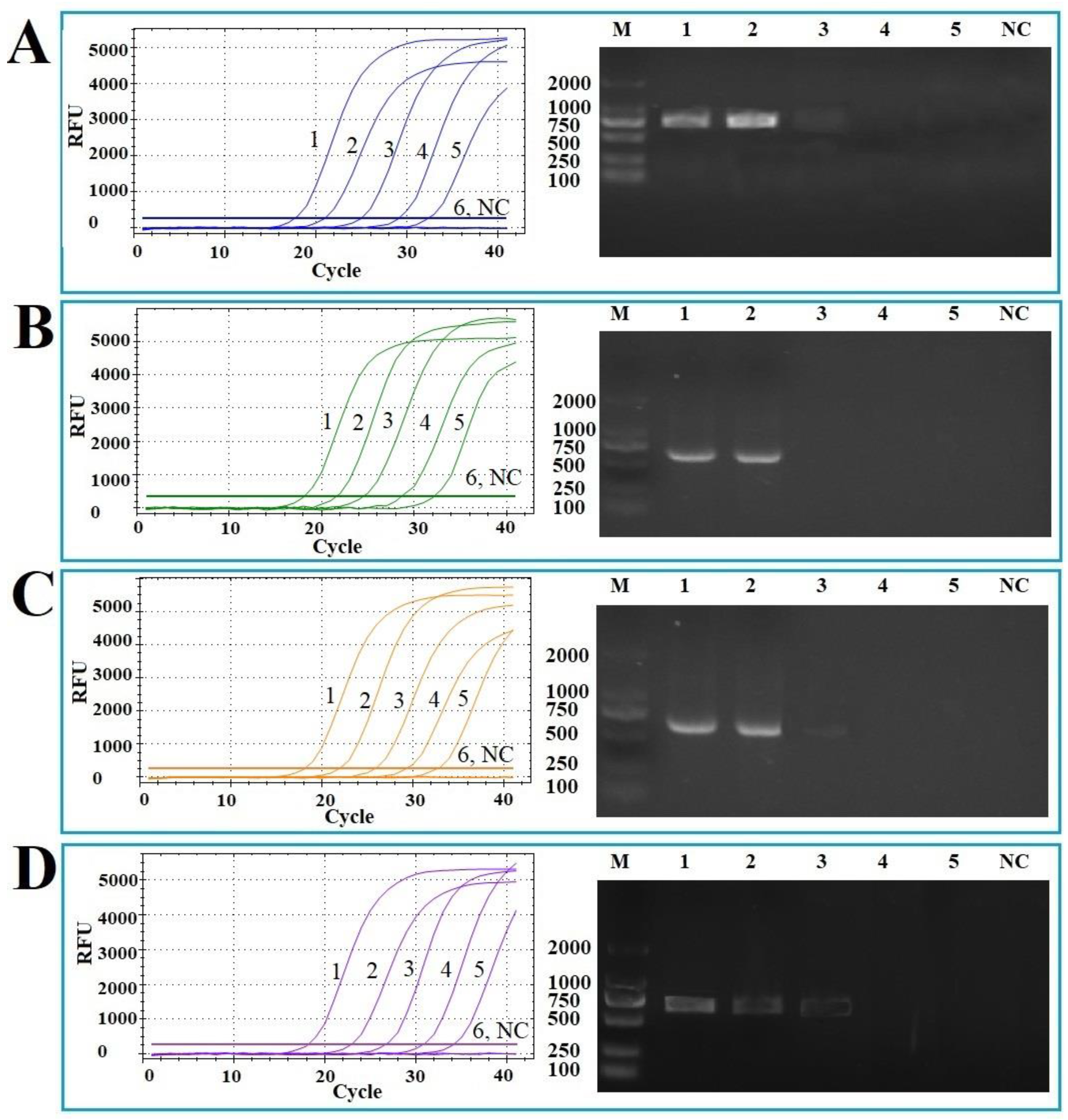
3.5. Sensitivity of the Multiplex qPCR

For sensitivity analysis, six concentrations (105 copies/μL–100 copies/μL) of different standard plasmids were evaluated and compared using both the qPCR and conventional PCR techniques. The detection limits of the qPCR for Va, Val, Vh, and Vsc were determined to be 4.0 × 101, 3.4 × 101, 6.0 × 101, and 2.6 × 101 copies/uL, respectively. In contrast, the detection limits for conventional PCR were 4.0 × 10³, 3.4 × 10³, 6.0 × 10³, and 2.6 × 10³ copies/uL, respectively. The average Cq values for all standard plasmids at 101 copies/uL were greater than 35, and thus Cq values ≥ 35 were defined as the critical point of negative. Compared with the traditional PCR, the sensitivity of the developed qPCR increased by 100 times (Figure 5).

3.6. Repeatability of the Multiplex qPCR

The repeatability and reproducibility of the developed multiplex quantitative PCR (qPCR) assay were assessed utilizing four standard plasmids, diluted ten-fold, ranging from 106 to 104 copies/µL, as templates. The coefficients of variation (%CV) were employed as the metric for evaluation. The findings revealed that the intra-assay and inter-assay CVs ranged from 0.20% to 1.39% and from 0.20% to 1.26%, respectively (Table 3), demonstrating the assay’s excellent repeatability and high accuracy.

3.7. Testing of Clinical and Environmental Samples Using the Multiplex qPCR

The applicability of the multiplex qPCR was evaluated using 63 clinical samples and 42 environmental samples. In clinical samples, the assay identified 14.29% as negative, 38.10% as single-positive, 28.57% as double-positive, 14.29% as triple-positive, and 4.76% as quadruple-positive (Table 4 and Table S2). In environmental samples, the assay identified 28.57% as negative, 57.14% as single-positive, and 14.29% as quadruple-positive (Table 4 and Table S3). The detection method exhibited an accuracy rate of 100%, underscoring the robust accuracy of the established multiplex qPCR method in the detection of clinical samples.

4. Discussion

Vibriosis, caused by Vibrio species, represents one of the most prevalent diseases in marine aquaculture, posing a significant threat to both wild and farmed marine species globally [47]. The rapid expansion of the aquaculture industry has facilitated numerous outbreaks and the spread of various Vibrio species, especially Va, Val, Vh, and Vhc, resulting in substantial economic losses in mariculture worldwide [4,8,16,22]. These bacterial pathogens exhibit a high likelihood of co-infection and cross-species transmission, and share similar clinical symptoms, complicating accurate diagnosis [3,48]. Consequently, the development of effective and specific diagnostic tools is essential for the detection of these bacteria. Over recent years, molecular diagnostic methods have undergone significant advancements. Currently, several traditional PCR, quantitative PCR (qPCR), multiplex PCR, and isothermal amplification techniques have been developed for the detection of Va, Val, Vh, or Vsc [34,49,50,51,52]. Nonetheless, no existing method can simultaneously detect all four bacteria in a single reaction.
TaqMan probe-based qPCR is widely regarded as the most reliable method for detecting pathogens in humans, livestock, and aquatic organisms [53]. In this study, we integrated the benefits of both quantitative PCR (qPCR) and multiplex PCR for the detection of Vibrio species Va, Val, Vh, and Vsc, selecting the genes empA, toxR, vhhP2, and luxR as the respective target genes. The empA gene, which encodes a zinc metalloprotease, is recognized as a crucial virulence factor of the fish pathogen V. anguillarum [54]. The empA gene holds potential for bacterial species identification; to date, PCR and Loop-mediated Isothermal Amplification (LAMP) methods have been developed for the specific detection of V. anguillarum using empA as a target gene [33,55]. For the detection of V. alginolyticus, rapid detection methods such as LAMP and Recombinase-Aided Amplification (RAA) have been developed, targeting the virulence gene toxR [39,56,57]. The toxR gene is a transcriptional regulatory gene which is exhibited in all pathogenic strains of V. alginolyticus and plays a significant role in regulating the transcription of virulence genes [58]. The toxR gene exhibits high interspecific conservation and intraspecific hypervariability, making it a reliable molecular target for the identification of Vibrio species [39,56,57,59]. VhhP2 is an outer membrane protein ubiquitously found in the pathogenic strains of V. harveyi, but it is absent in most non-V. harveyi species. This characteristic makes vhhP2 a specific marker for the precise identification of V. harveyi [53,60]. Several PCR-based methods have been developed utilizing the conserved sequence of vhhP2 for the rapid and specific detection of V. harveyi [60,61]. LuxR serves as a master quorum-sensing regulator, influencing biofilm formation in V. scophthalmi and certain pathogenic Vibrio species [62]. The luxR gene of V. scophthalmi exhibits relatively low similarity with those of other bacterial species, making it a suitable target gene for V. scophthalmi detection using a LAMP method [63,64]. In this study, multiple sequence alignments were conducted, and both intraspecific conserved and interspecific variant regions of these genes were utilized to design species-specific primers and probes. Specificity analysis demonstrated that the multiplex qPCR method exhibited no cross-reactivity with each other or with eight other pathogens, including Ep, Pd, Vro, Pf, La, Vhy, Hp, and Vaz, thereby indicating high specificity.
In this study, a TaqMan probe-based multiplex quantitative PCR (qPCR) method was developed to enable the simultaneous detection of Va, Val, Vh, and Vsc within a single reaction system. The minimum detection limits for the multiplex qPCR were determined to be 4.0 × 101, 3.4 × 101, 6.0 × 101, and 2.6 × 101 copies/uL for Va, Val, Vh, and Vsc, respectively. This represents an enhancement in detection sensitivity ranging from 10- to 100-fold compared to traditional PCR methods [31,32,33,34]. Unlike single qPCR assays, multiplex qPCR is performed within the same reaction mixture, leading to more rapid substrate consumption and the generation of internal competition, which can result in a slight reduction in sensitivity. The detection limits achieved with the developed multiplex qPCR were comparable to or marginally higher than those reported for single qPCR assays in previous studies [34,36,37,65]. Similar findings were observed in a multiplex qPCR method designed for the detection of four feline diarrhea-associated viruses, suggesting that competition among primers, probes, and templates of different pathogens may reduce the sensitivity of multiplex detection methods [66].
The standard curves for Va, Val, Vh, and Vsc exhibit strong linearity and correlation, with the coefficient of determination (R2) exceeding 0.99, indicative of extremely high correlations. According to the MIQE guidelines [67], optimal amplification efficiencies should range between 90% and 110%. In this study, the amplification efficiency of the standard plasmids ranged from 96% to 108%, demonstrating high amplification efficiencies. Furthermore, the coefficients of variation (CVs) for both inter-assay and intra-assay replicates were below 1.4%, significantly lower than those reported for other multiplex qPCR methods [68,69,70], suggesting that the established qPCR method possesses high repeatability and stability.
A notable advantage of the multiplex qPCR is its capability to detect mixed target bacteria in complex clinical and environmental samples. To evaluate this advantage, 63 distinct artificial clinical samples and 42 environmental samples were prepared, encompassing single-, double-, triple-, and quadruple-infected samples. The established multiplex qPCR method successfully detected and differentiated the mixed bacteria in all 63 clinical samples and 42 environmental samples, demonstrating high accuracy. These findings of this study demonstrate that multiplex quantitative PCR (qPCR) methods are effective for the detection of Va, Val, Vh, and Vsc infections in fish or their existence in water and sedimental samples.

5. Conclusions

In this study, we have successfully developed, for the first time, a TaqMan multiplex qPCR technique capable of simultaneously and accurately detecting these pathogens. This method is characterized by high specificity and sensitivity, as well as rapid processing capabilities, making it an efficient tool for identifying bacterial pathogens with a substantial detection throughput. Furthermore, the method is cost-effective and time-efficient, rendering it particularly suitable for the detection of mixed pathogen infections in mariculture. This advancement facilitates early and rapid clinical detection and treatment in both clinical and field settings.

Supplementary Materials

The following supporting information can be downloaded at: https://www.mdpi.com/article/10.3390/vetsci12040327/s1, Figure S1: Construction of standard plasmids; Table S1: Bacterial strains used for multiple sequence alignment.; Table S2: Cq values of the clinical samples; Table S3: Cq values of the environmental samples.

Author Contributions

Investigation, B.Z., Y.Q. and C.S.; mMethodology, B.Z.; data curation, B.Z. and C.S.; writing—original draft, B.Z.; software, Y.Q.; validation, Y.Q.; conceptualization, formal analysis, funding acquisition, writing—review and editing, and supervision, J.Z. All authors have read and agreed to the published version of the manuscript.

Funding

This work was supported by grants from the National Natural Science Foundation of China (U24A20463) and the Graduate Innovation Foundation of Yantai University (GGIFYTU2340).

Institutional Review Board Statement

The animal study protocol was approved by the Ethics Committee of Yantai University (approval code No. 20230503) for studies involving animals, date 15 May 2023.

Informed Consent Statement

Not applicable.

Data Availability Statement

The data presented in this study are available upon request from the corresponding author.

Conflicts of Interest

The authors declare no conflicts of interest.

References

  1. Oktariani, A.F.; Ramona, Y.; Sudaryatma, P.E.; Dewi, I.A.M.M.; Shetty, K. Role of marine bacterial contaminants in histamine formation in seafood products: A review. Microorganisms 2022, 10, 1197. [Google Scholar] [CrossRef] [PubMed]
  2. Janda, J.M.; Duman, M. Expanding the spectrum of diseases and disease associations caused by Edwardsiella tarda and related species. Microorganisms 2024, 12, 1031. [Google Scholar] [CrossRef] [PubMed]
  3. Xu, K.; Wang, Y.; Yang, W.; Cai, H.; Zhang, Y.; Huang, L. Strategies for prevention and control of Vibriosis in Asian fish culture. Vaccines 2022, 11, 98. [Google Scholar] [CrossRef]
  4. Lages, M.A.; Balado, M.; Lemos, M.L. The expression of virulence factors in Vibrio Anguillarum is dually regulated by iron levels and temperature. Front. Microbiol. 2019, 10, 2335. [Google Scholar] [CrossRef] [PubMed]
  5. Akinbowale, O.L.; Peng, H.; Grant, P.; Barton, M.D. Antibiotic and heavy metal resistance in motile aeromonads and pseudomonads from rainbow trout (Oncorhynchus mykiss) farms in Australia. Int. J. Antimicrob. Agents 2007, 30, 177–182. [Google Scholar] [CrossRef]
  6. Fauzi, I.A.; Haga, Y.; Kondo, H.; Hirono, I.; Satoh, S. Dietary citrulline improves survival of rainbow trout Oncorhynchus mykiss juveniles challenged with Vibrio anguillarum. Aquaculture 2020, 528, 735491. [Google Scholar] [CrossRef]
  7. Akter, T.; Lindegaard, M.; Pedersen, K.; Strube, M.L.; Ronco, T.; Dalsgaard, I. Sequence analysis of plasmids in Vibrio anguillarum from different fish and locations. J. Aquat. Anim. Health 2020, 32, 21–27. [Google Scholar] [CrossRef]
  8. Austin, B. Vibrios as causal agents of zoonoses. Vet. Microbiol. 2010, 140, 310–317. [Google Scholar] [CrossRef]
  9. Citil, B.E.; Derin, S.; Sankur, F.; Sahan, M.; Citil, M.U. Vibrio alginolyticus associated chronic myringitis acquired in mediterranean waters of Turkey. Infect. Disnor. 2015, 2015, 187212. [Google Scholar]
  10. Abbate, F.; Guerrera, M.C.; Montalbano, G.; Ciriaco, E.; Germana, A. Morphology of the tongue dorsal surface of gilthead seabream (Sparus aurata). Microsc. Res. Tech. 2012, 75, 1666–1671. [Google Scholar] [CrossRef]
  11. Liu, P.C.; Lin, J.Y.; Hsiao, P.T.; Lee, K.K. Isolation and characterization of pathogenic Vibrio alginolyticus from diseased cobia Rachycentron canadum. J. Basic. Microbiol. 2004, 44, 23–28. [Google Scholar] [PubMed]
  12. Jayaprakash, N.S.; Pai, S.S.; Philip, R.; Singh, I.S. Isolation of a pathogenic strain of Vibrio alginolyticus from necrotic larvae of Macrobrachium rosenbergii (de man). J. Fish Dis. 2006, 29, 187–191. [Google Scholar] [CrossRef] [PubMed]
  13. Lee, K.K. Pathogenesis studies on Vibrio alginolyticus in the grouper, Epinephelus malabaricus. Blochet. Schneider Microb. Pathog. 1995, 19, 39. [Google Scholar]
  14. Selvin, J.; Lipton, A.P. Vibrio alginolyticus associated with white spot disease of Penaeus monodon. Dis. Aquat. Org. 2003, 57, 147–150. [Google Scholar]
  15. Lee, K.K.; Yu, S.R.; Yang, T.I.; Liu, P.C.; Chen, F.R. Isolation and characterization of Vibrio alginolyticus isolated from diseased kuruma prawn, Penaeus japonicus. Lett. Appl. Microbiol. 1996, 22, 111–114. [Google Scholar] [CrossRef] [PubMed]
  16. Austin, B.; Zhang, X.H. Vibrio harveyi: A significant pathogen of marine vertebrates and invertebrates. Lett. Appl. Microbiol. 2006, 43, 119–124. [Google Scholar]
  17. Ransangan, J.; Mustafa, S. Identification of Vibrio harveyi isolated from diseased Asian seabass Lates calcarifer by use of 16S ribosomal DNA sequencing. J. Aquat. Anim. Health 2009, 21, 150–155. [Google Scholar]
  18. Ruwandeepika, H.A.D.; Jayaweera, T.S.P.; Bhowmick, P.P.; Karunasagar, I.; Bossier, P.; Defoirdt, T. Pathogenesis, virulence factors and virulence regulation of vibrios belonging to the Harveyi clade. Rev. Aquacult. 2012, 4, 59–74. [Google Scholar]
  19. Shen, G.M.; Shi, C.Y.; Fan, C.; Jia, D.; Wang, S.Q.; Xie, G.S.; Li, G.Y.; Mo, Z.L.; Huang, J. Isolation, identification and pathogenicity of Vibrio harveyi, the causal agent of skin ulcer disease in juvenile hybrid groupers Epinephelus fuscoguttatus×Epinephelus lanceolatus. J. Fish Dis. 2017, 40, 1351–1362. [Google Scholar]
  20. Defoirdt, T.; Boon, N.; Sorgeloos, P.; Verstraete, W.; Bossier, P. Alternatives to antibiotics to control bacterial infections: Luminescent vibriosis in aquaculture as an example. Trends Biotechnol. 2007, 25, 472–479. [Google Scholar] [CrossRef]
  21. Cerdà-Cuéllar, M.; Rossellò-Mora, R.A.; Lalucat, J.; Jofre, J.; Blanch, A. Vibrio scophthalmi sp. nov., a new species from turbot (Scophthalmus maximus). Int. J. Syst. Bacteriol. 1997, 47, 58–61. [Google Scholar]
  22. Qiao, G.; Lee, D.C.; Woo, S.H.; Li, H.; Xu, D.H.; Park, S.I. Microbiological characteristics of Vibrio scophthalmi isolates from diseased olive flounder Paralichthys olivaceus. Fish. Sci. 2012, 78, 853–863. [Google Scholar]
  23. Soffientino, B.; Gwaltney, T.; Nelson, D.R.; Specker, J.L.; Mauel, M.; Gómez-Chiarri, M. Infectious necrotizing enteritis and mortality caused by Vibrio carchariae in summer flounder Paralichthys dentatus during intensive culture. Dis. Aquat. Org. 1999, 38, 201–210. [Google Scholar]
  24. Hidalgo, B.R.; Cleenwerck, I.; Balboa, S.; Wachter, M.D.; Thompson, F.L.; Swings, J.; De Vos, P.; Romalde, J. Diversity of Vibrios associated with reared clams in Galicia (NW Spain). Syst. Appl. Microbiol. 2008, 31, 215–222. [Google Scholar]
  25. Valdenegro-Vega, V.; Naeem, S.; Carson, J.; Bowman, J.P.; Tejedor, R.J.L.; Nowak, B. Culturable microbiota of ranched southern bluefin tuna (Thunnus maccoyii Castelnau). J. Appl. Microbiol. 2013, 115, 923–932. [Google Scholar]
  26. Rui, H.; Liu, Q.; Ma, Y.; Wang, Q.; Zhang, Y. Roles of LuxR in regulating extracellular alkaline serine protease a, extracellular polysaccharide and mobility of Vibrio alginolyticus. FEMS Microbiol. Lett. 2008, 285, 155–162. [Google Scholar]
  27. Zhou, Z.; Pang, H.; Ding, Y.; Cai, J.; Huang, Y.; Jian, J.; Wu, Z. VscO, a putative T3SS chaperone escort of Vibrio alginolyticus, contributes to virulence in fish and is a target for vaccine development. Fish Shellfish Immunol. 2013, 35, 1523–1531. [Google Scholar] [PubMed]
  28. Vandenberghe, J.; Thompson, F.L.; Gomez-Gil, B.; Swings, J. Phenotypic diversity among Vibrio isolates from marine aquaculture systems. Aquaculture 2003, 219, 9–20. [Google Scholar]
  29. Ina-Salwany, M.Y.; Al-Saari, N.; Aslah, M.; Mursidi, F.A.; Mohd-Aris, A.; Amal, M.N.A.; Kasai, H.; Mino, S.; Sawabe, T.; Zamri-Saad, M. Vibriosis in Fish: A review on disease development and prevention. J. Aquat. Anim. Health 2019, 31, 3–22. [Google Scholar]
  30. Kim, H.J.; Ryu, J.O.; Lee, S.Y.; Kim, E.S.; Kim, H.Y. Multiplex PCR for detection of the Vibrio genus and five pathogenic Vibrio species with primer sets designed using comparative genomics. BMC Microbiol. 2015, 15, 239. [Google Scholar]
  31. O’Hara, C.M.; Sowers, E.G.; Bopp, C.A.; Duda, S.B.; Strockbine, N.A. Accuracy of six commercially available systems for identification of members of the family Vibrionaceae. J. Clin. Microbiol. 2003, 41, 5654–5659. [Google Scholar] [PubMed]
  32. Pang, L.; Zhang, X.H.; Zhong, Y.; Chen, J.; Li, Y.; Austin, B. Identification of Vibrio harveyi using PCR amplification of the toxR gene. Lett. Appl. Microbiol. 2006, 43, 249–255. [Google Scholar]
  33. Xiao, P.; Mo, Z.L.; Mao, Y.X.; Wang, C.L.; Zou, Y.X.; Li, J. Detection of Vibrio anguillarum by PCR amplification of the empA gene. J. Fish Dis. 2009, 32, 293–296. [Google Scholar]
  34. Osman, E.; Kim, N.; Lee, Y.; Yoo, J.; Kim, S.H.; Kim, D.H. Molecular approaches for detection and quantification of Vibrio scophthalmi based on recA. J. Fish Dis. 2022, 45, 373–378. [Google Scholar]
  35. Cao, Y.T.; Wu, Z.H.; Jian, J.C.; Lu, Y.S. Evaluation of a loop-mediated isothermal amplification method for the rapid detection of Vibrio harveyi in cultured marine shellfish. Lett. Appl. Microbiol. 2010, 51, 24–29. [Google Scholar] [PubMed]
  36. Fukui, Y.; Sawabe, T. Rapid detection of Vibrio harveyi in seawater by real-time PCR. Microbes Environ. 2008, 23, 172–176. [Google Scholar]
  37. Zhao, J.J.; Chen, C.; Luo, P.; Ren, C.H.; Jiang, X.; Zhao, Z.; Hu, C.Q. SYBR Green I-based real-time PCR targeting the rpoX gene for sensitive and rapid detection of Vibrio alginolyticus. Mol. Cell Probes 2011, 25, 137–141. [Google Scholar]
  38. Crisafi, F.; Denaro, R.; Genovese, M.; Yakimov, M.; Genovese, L. Application of relative real-time PCR to detect differential expression of virulence genes in Vibrio anguillarum under standard and stressed growth conditions. J. Fish Dis. 2014, 37, 629–640. [Google Scholar]
  39. Li, D.R.; Zhao, J.Y.; Lan, W.Q.; Zhao, Y.; Sun, X.H. Effect of food matrix on rapid detection of Vibrio parahaemolyticus in aquatic products based on toxR gene. World J. Microbiol. Biotechnol. 2023, 39, 188. [Google Scholar]
  40. Law, J.W.F.; Ab Mutalib, N.S.; Chan, K.G.; Lee, L.H. Rapid methods for the detection of foodborne bacterial pathogens: Principles, applications, advantages and limitations. Front. Microbiol. 2015, 5, 770. [Google Scholar]
  41. Hu, Y.H.; Deng, T.; Sun, B.G.; Sun, L. Molecular analysis of the copper-responsive CopRSCD of a pathogenic Pseudomonas fluorescens strain. Fish Shellfish Immunol. 2012, 32, 1155–1161. [Google Scholar]
  42. Yu, L.P.; Hu, Y.H.; Sun, B.G.; Sun, L. C312M: An attenuated Vibrio anguillarum strain that induces immunoprotection as an oral and immersion vaccine. Dis. Aquat. Organ. 2012, 102, 33–42. [Google Scholar] [PubMed]
  43. Dong, Y.; Zhou, D.; Zhang, B.; Xu, X.; Zhang, J. Development of a real-time recombinase-aided amplification assay for rapid and sensitive detection of Edwardsiella piscicida. Front. Cell. Infect. Microbiol. 2024, 14, 1355056. [Google Scholar]
  44. Zhou, D.D.; Zhang, B.Z.; Dong, Y.C.; Li, X.P.; Zhang, J. Coinfection of cage-cultured spotted sea bass (Lateolabrax maculatus) with Vibrio harveyi and Photobacterium damselae subsp. piscicida associated with skin ulcer. Microorganisms 2024, 12, 503. [Google Scholar]
  45. Zhang, J.; Zhang, M.; Sun, L. Junctional adhesion molecule A of red drum (Sciaenops ocellatus): A possible immunomodulator and a target for bacterial immune evasion. Vet. Immunol. Immunopathol. 2014, 161, 99–107. [Google Scholar] [PubMed]
  46. Guerra, V.; Beule, L.; Lehtsaar, E.; Liao, H.L.; Karlovsky, P. Improved protocol for DNA extraction from subsoils using phosphate lysis buffer. Microorganisms 2020, 8, 532. [Google Scholar] [CrossRef] [PubMed]
  47. Kah Sem, N.A.D.; Abd Gani, S.; Chong, C.M.; Natrah, I.; Shamsi, S. Management and mitigation of Vibriosis in aquaculture: Nanoparticles as promising alternatives. Int. J. Mol. Sci. 2023, 24, 12542. [Google Scholar] [CrossRef]
  48. de Souza Valente, C.; Wan, A.H.L. Vibrio and major commercially important vibriosis diseases in decapod crustaceans. J. Invertebr. Pathol. 2021, 181, 107527. [Google Scholar]
  49. Frans, I.; Michiels, C.W.; Bossier, P.; Willems, K.A.; Lievens, B.; Rediers, H. Vibrio anguillarum as a fish pathogen: Virulence factors, diagnosis and prevention. J. Fish Dis. 2011, 34, 643–661. [Google Scholar]
  50. Wang, J.X.; Tang, W.L.; Chen, S.Q.; Zhang, J.; Ji, J.; Dong, J.Q.; Liu, G.; Gao, S. Rapid and sensitive detection of Vibrio alginolyticus pathogenic strains by real-time recombinase polymerase amplification. Acta Biochim. Biophys. Sin. 2021, 53, 950–954. [Google Scholar]
  51. Siddique, M.P.; Jang, W.J.; Lee, J.M.; Hasan, M.T.; Kim, C.H.; Kong, I.S. Detection of Vibrio anguillarum and Vibrio alginolyticus by singleplex and duplex loop-mediated isothermal amplification (LAMP) assays targeted to groEL and fklB genes. Int. Microbiol. 2019, 22, 501–509. [Google Scholar] [CrossRef] [PubMed]
  52. Zhang, X.H.; He, X.X.; Austin, B. Vibrio harveyi: A serious pathogen of fish and invertebrates in mariculture. Mar. Life Sci. Technol. 2020, 2, 231–245. [Google Scholar]
  53. Saha, R.; Bestervelt, L.L.; Donofrio, R.S. Development and validation of a real-time TaqMan assay for the detection and enumeration of Pseudomonas fluorescens ATCC 13525 used as a challenge organism in testing of food equipments. J. Food Sci. 2012, 77, M150–M155. [Google Scholar] [CrossRef] [PubMed]
  54. Denkin, S.M.; Nelson, D. Regulation of Vibrio anguillarum empA metalloprotease expression and its role in virulence. Appl. Environ. Microbiol. 2004, 70, 4193–4204. [Google Scholar] [PubMed]
  55. Gao, H.; Li, F.; Zhang, X.; Wang, B.; Xiang, J. Rapid, sensitive detection of Vibrio anguillarum using loop-mediated isothermal amplification. Chin. J. Oceanol. Limnol. 2010, 28, 62–66. [Google Scholar]
  56. Dong, Y.; Zhao, P.; Chen, L.; Wu, H.; Si, X.; Shen, X.; Shen, H.; Qiao, Y.; Zhu, S.Y.; Chen, Q.; et al. Fast, simple and highly specific molecular detection of Vibrio alginolyticus pathogenic strains using a visualized isothermal amplification method. BMC Vet. Res. 2020, 16, 76. [Google Scholar]
  57. Fu, K.; Li, J.; Wang, Y.; Liu, J.; Yan, H.; Shi, L.; Zhou, L. An innovative method for rapid identification and detection of Vibrio alginolyticus in different infection models. Front. Microbiol. 2016, 7, 651. [Google Scholar]
  58. Cai, S.; Cheng, H.; Pang, H.; Lu, Y.; Jian, J. Role of the toxR gene from fish pathogen Vibiro alginolyticus in the physiology and virulence. Indian J. Microbiol. 2017, 57, 477–484. [Google Scholar] [CrossRef]
  59. He, P.; Chen, Z.; Luo, J.; Wang, H.; Yan, Y.; Chen, L.; Gao, W. Multiplex real-time PCR assay for detection of pathogenic Vibrio parahaemolyticus strains. Mol. Cell Probes 2014, 28, 246–250. [Google Scholar] [CrossRef]
  60. Sun, K.; Hu, Y.H.; Bai, F.F.; Sun, L. Identification of vhhP2, a novel genetic marker of Vibrio harveyi, and its application in the quick detection of V. harveyi from animal specimens and environmental samples. J. Appl. Microbiol. 2009, 107, 1251–1257. [Google Scholar] [CrossRef]
  61. Cano-Gomez, A.; Hoj, L.; Owens, L.; Andreakis, N. Multilocus sequence analysis provides basis for fast and reliable identification of Vibrio harveyi-related species and reveals previous misidentification of important marine pathogens. Syst. Appl. Microbiol. 2011, 34, 561–565. [Google Scholar] [PubMed]
  62. García-Aljaro, C.; Melado-Rovira, S.; Milton, D.L.; Blanch, A.R. Quorum-sensing regulates biofilm formation in Vibrio scophthalmi. BMC Microbiol. 2012, 12, 287. [Google Scholar]
  63. Seok, B.; Kim, M.S.; Kim, B.S. Genome-wide analysis of quorum sensing regulon in marine fish pathogen Vibrio scophthalmi. Sci. Rep. 2024, 14, 27740. [Google Scholar] [CrossRef]
  64. Zhou, S.; Gao, Z.X.; Zhang, M.; Liu, D.Y.; Zhao, X.P.; Liu, Y. Development of a quadruplex loop-mediated isothermal amplification assay for field detection of four Vibrio species associated with fish disease. Springerplus 2016, 5, 1104. [Google Scholar] [CrossRef] [PubMed]
  65. Hickey, M.E.; Richards, G.P.; Lee, J.L. Development of a two-step, non-probed multiplex real-time PCR for surveilling Vibrio anguillarum in seawater. J. Fish Dis. 2015, 38, 551–559. [Google Scholar]
  66. Zou, J.; Yu, J.; Mu, Y.; Xie, X.; Wang, R.; Wu, H.; Liu, X.; Xu, F.; Wang, J.; Wang, Y. Development of a TaqMan-based multiplex real-time PCR for simultaneous detection of four feline diarrhea-associated viruses. Front. Vet. Sci. 2022, 9, 1005759. [Google Scholar]
  67. Bustin, S.A.; Benes, V.; Garson, J.A.; Hellemans, J.; Huggett, J.; Kubista, M.; Mueller, R.; Nolan, T.; Pfaffl, M.W.; Shipley, G.L.; et al. The MIQE guidelines: Minimum information for publication of quantitative real-time PCR experiments. Clin. Chem. 2009, 55, 611–622. [Google Scholar] [CrossRef] [PubMed]
  68. Wang, R.Y.; Zhang, W.Y.; Ye, R.; Pan, Z.Z.; Li, G.R.; Su, S. One-step multiplex TaqMan probe-based method for real-time PCR detection of four canine diarrhea viruses. Mol. Cell. Probes 2020, 53, 101618. [Google Scholar]
  69. Fürer, F.; Fraefel, C.; Lechmann, J. Multiplex real-time PCR for the detection and differentiation of equid gammaherpesvirus 2 and 5. J. Virol. Methods 2022, 310, 114615. [Google Scholar]
  70. Xu, Z.X.; Li, B.S.; Jiang, Y.S.; Huang, J.; Su, L.B.; Wu, W.B.; Pang, Q.L.; Li, Z.L.; Zhang, J.Q.; Li, X.H.; et al. Development of a quadruple qRT-PCR assay for simultaneous identification of hypervirulent and carbapenem-resistant Klebsiella pneumoniae. Microbiol. Spectr. 2024, 12, e0071923. [Google Scholar] [CrossRef]
Figure 1. Multiple sequence alignment of target genes with their homologous sequences. (A) empA gene; (B) toxR gene; (C) vhhP2 gene; (D) luxR gene. Black indicates the consensus residues; pink indicates the residues that are ≥75% identical among the aligned sequences; blue indicates the residues that are ≥50% and <75% identical among the aligned sequences; and yellow indicates residues that are ≥25% and <50% identical among the aligned sequences. The red/green arrows and blue horizontal lines indicate primer and probe sequences, respectively.
Vetsci 12 00327 g001
Figure 2. Optimization of the single qPCR conditions. Amplification curves based on the qPCR amplification of plasmids pempA (A), ptoxR (B), pvhhP2 (C), and pluxR (D). RFU: relative fluorescence unit.
Vetsci 12 00327 g002
Figure 3. Standard curves of the multiplex qPCR method. Ten-fold serially diluted plasmids (from 109 to 104 copies/µL) were amplified with different primer/probe sets targeting Va (A), Val (B), Vh (C), and Vsc (D). The Cq values were then plotted against the logarithmic plasmid copy number. R2 = correlation coefficient.
Vetsci 12 00327 g003
Figure 4. Specificity analysis of the multiplex qPCR method. Genomic DNA of (1) Va, (2) Val, (3) Vh, (4) Vsc, (5) Ep, (6) Pd, (7) Vro, (8) Pf, (9) La, (10) Vhy, (11) Hp, (12) Vaz, and (13) negative control amplified by the multiplex qPCR.
Vetsci 12 00327 g004
Figure 5. Sensitivity analysis of the multiplex qPCR method. Ten-fold serially diluted plasmids (from 105 to 100 copies/µL) were amplified with different primer/probe sets targeting Va (A), Val (B), Vh (C), and Vsc (D), and the multiplex qPCR results were compared with the results of conventional PCR. 1–6: 105–100 copies/µL standard plasmids; NC: negative control.
Vetsci 12 00327 g005
Table 1. Primers and probes used in this study.
TargetGenePrimer/ProbeSequence (5′-3′)
VaempAempA-FTTATATTGATAGTTATGTGCACTATTAA
empA-RACAAAGAAGTCGACTAAATAAACCAT
empA-qFAAGTCCGTTACCAGCAGATG
empA-qRCGTAAACTTGGCCGATACCT
empA-P[6-FAM]TCTCAGTTTGCGTTGCTACGACTGAC[BHQ1]
ValtoxRtoxR-FGTGGAACGCTTGAGCCCATT
toxR-RGCGTAGTGGGCCGACAGTAT
gyrB-qFGTGTACCGGTGATGACACCTG
gyrB-qRAATCACTTCAACTGGCAACGAG
gyrB-P[HEX]CACGTAGCGCTCAATGCATTGCTC[BHQ1]
VhvhhP2vhhP2-FATGAAGAGAAGGAATCCTCAAGG
vhhP2-RTTATTCCAATCTAGTTGGTTTTGATG
vhhR2-qFCATCCTAGCTGTTGTTGCAGCT
vhhR2-qRGTTCCACCATTGACTAACCATTGG
vhhR2-P[ROX]TGGGTAAATCGCACGCCTGTTTCGA[BHQ2]
VscluxRluxR-FATGGACTCTATAGCAAAAAGACCC
luxR-RTTACGCTTCTTCTTTGTAAATACACAG
luxR-qFGTCGTGGTCATGCCGATATT
luxR-qRATTAGAGAACTGACGTACCACATAGTTC
luxR-P[Cy5] CAACTACTTCCCAACACGTGAAGAC[BHQ2]
Table 2. Optimal qPCR system.
ComponentVolume (μL)
empA-qF(20 μM)0.3
empA-qR(20 μM)0.3
empA-qP(20 μM)0.5
toxR-qF(20 μM)0.2
toxR-qR(20 μM)0.2
toxR-qP(20 μM)0.6
vhhR2-qF(20 μM)0.4
vhhR2-qR(20 μM)0.4
vhhR2-qP(20 μM)0.5
luxR-qF(20 μM)0.2
luxR-qR(20 μM)0.2
luxR-qP(20 μM)0.3
DNA-Va1
DNA-Val1
DNA-Vh1
DNA-Vsc1
Pro Taq HS Premix Probe real-time PCR Kit III10
ddH2OUp to 20
Table 3. Reproducibility of the multiplex qPCR method.
Intra-AssayInter-Assay
TargetsTemplates (Copies/µL)Cq Value (Mean ± SD)CV/%Cq Value (Mean ± SD)CV/%
empA10622.02 ± 0.180.8223.33 ± 0.210.90
10524.83 ± 0.261.0526.49 ± 0.240.91
10427.05 ± 0.130.4829.11 ± 0.140.48
toxR10625.12 ± 0.351.3924.99 ± 0.311.24
10528.22 ± 0.621.2027.73 ± 0.351.26
10431.00 ± 0.771.4830.46 ± 0.270.88
vhhR210623.47 ± 0.070.3021.97 ± 0.090.41
10526.44 ± 0.110.4225.35 ± 0.050.20
10429.76 ± 0.060.2028.82 ± 0.140.49
luxR10618.04 ± 0.140.7818.85 ± 0.100.53
10521.41 ± 0.170.7921.71 ± 0.190.86
10424.54 ± 0.210.8624.59 ± 0.170.69
Table 4. Analysis of clinical and environmental samples using the multiplex qPCR method.
Different SampleSample SizeTest Result (Positive/Negative)Accuracy Rate/%
Clinical Sample
NC a90/9100
Va66/0100
Val66/0100
Vh66/0100
Vsc66/0100
Va+Val33/0100
Va+Vh33/0100
Va+Vsc33/0100
Val+Vh33/0100
Val+Vsc33/0100
Vh+Vsc33/0100
Va+Val+Vh33/0100
Va+Val+Vsc33/0100
Val+Vh+Vsc33/0100
Va+Val+Vh+Vsc33/0100
Water Sample
NC a60/6100
Va33/0100
Val33/0100
Vh33/0100
Vsc33/0100
Va+Val+Vh+Vsc33/0100
Sediment Sample
NC a60/6100
Va33/0100
Val33/0100
Vh33/0100
Vsc33/0100
Va+Val+Vh+Vsc33/0100
Positive Control
pVa33/0100
pVal33/0100
pVh33/0100
pVsc33/0100
a Samples from the fish injected with PBS.
Disclaimer/Publisher’s Note: The statements, opinions and data contained in all publications are solely those of the individual author(s) and contributor(s) and not of MDPI and/or the editor(s). MDPI and/or the editor(s) disclaim responsibility for any injury to people or property resulting from any ideas, methods, instructions or products referred to in the content.

