
Comparison of the Fecal Microbiota of Horses with Intestinal Disease and Their Healthy Counterparts
by
 , ,
, ,
Taemook Park
1,2 ,
,
Heetae Cheong
3,
Jungho Yoon
1,
Ahram Kim
1,
Youngmin Yun
2,* and
Tatsuya Unno
4,5,* 1
Equine Clinic, Jeju Stud Farm, Korea Racing Authority, Jeju 63346, Korea
2
College of Veterinary Medicine and Veterinary Medical Research Institute, Jeju National University, Jeju 63243, Korea
3
College of Veterinary Medicine and Institute of Veterinary Science, Kangwon National University, Chuncheon 24341, Korea
4
Faculty of Biotechnology, School of Life Sciences, SARI, Jeju 63243, Korea
5
Subtropical/Tropical Organism Gene Bank, Jeju National University, Jeju 63243, Korea
*
Authors to whom correspondence should be addressed.
Vet. Sci. 2021, 8(6), 113; https://doi.org/10.3390/vetsci8060113
Submission received: 20 April 2021
/
Revised: 12 June 2021
/
Accepted: 15 June 2021
/
Published: 17 June 2021
Abstract
:(1) Background: The intestinal microbiota plays an essential role in maintaining the host’s health. Dysbiosis of the equine hindgut microbiota can alter the fermentation patterns and cause metabolic disorders. (2) Methods: This study compared the fecal microbiota composition of horses with intestinal disease and their healthy counterparts living in Korea using 16S rRNA sequencing from fecal samples. A total of 52 fecal samples were collected and divided into three groups: horses with large intestinal disease (n = 20), horses with small intestinal disease (n = 8), and healthy horses (n = 24). (3) Results: Horses with intestinal diseases had fewer species and a less diverse bacterial population than healthy horses. Lactic acid bacteria, Lachnospiraceae, and Lactobacillaceae were overgrown in horses with large intestinal colic. The Firmicutes to Bacteroidetes ratio (F/B), which is a relevant marker of gut dysbiosis, was 1.94, 2.37, and 1.74 for horses with large intestinal colic, small intestinal colic, and healthy horses, respectively. (4) Conclusions: The overgrowth of two lactic acid bacteria families, Lachnospiraceae and Lactobacillaceae, led to a decrease in hindgut pH that interfered with normal fermentation, which might cause large intestinal colic. The overgrowth of Streptococcus also led to a decrease in pH in the hindgut, which suppressed the proliferation of the methanogen and reduced methanogenesis in horses with small intestinal colic.
1. Introduction
Horses are nonruminant herbivores whose digestive system has evolved to utilize the fibers in the roughages in their hindgut [1,2,3,4]. The large intestine of horses is an anaerobic fermentation chamber filled with fibrolytic bacteria. Therefore, the large intestinal microbiota of horses plays an essential role in the utilization of plant fibers by producing volatile fatty acids (VFAs), such as acetate, propionate, and butyrate, which are absorbed through the cecal and colonic epithelium and distributed for use throughout the body [5]. In addition to metabolic benefits, the intestinal microbiota provides the host with other advantages, including protection against pathogen overgrowth, stimulation of the immune response in the gut, and enhanced intestinal barrier function by regulating gene expression in the host intestinal epithelial tissue [6,7,8,9,10,11].
The relationship between the gut microbiota and clinical conditions, such as inflammatory bowel disease, colorectal cancer, or diabetes, have been examined in large-scale studies involving humans [12,13,14,15,16]. Studies performed on different animal species have shown that the gut microbial dynamics can be influenced by various factors, including the environment, diet, gestational age, hospitalization, antibiotics, delivery mode, stress, and feeding method [7,8,9,12,17,18]. Similarly, differences in the fecal bacterial communities between horses with gastrointestinal diseases and their healthy counterparts have been reported [6,19,20,21,22]. Furthermore, an acute change in the colonic microbiota was observed in horses that underwent an exploratory laparotomy to treat equine colic [23]. A disturbance in the equine hindgut microbiota can alter the fermentation patterns and ultimately lead to metabolic disorders [24].
Carbohydrate fermentation is the main source of lactic acid production in the equine hindgut. In healthy horses, the luminal lactate is converted to VFAs by commensal bacteria. Therefore, very little lactate is present in their hindgut [25]. Rapid dietary changes, such as grain overload, have long been recognized to disrupt normal fermentation in the horse hindgut. Consequently, excessive carbohydrate fermentation leads to lactate accumulation in the hindgut, which can induce subclinical acidosis. Low pH in the gut lumen alters the hindgut microbiota, which causes the release of endotoxin from the death of acid-sensitive Gram-negative bacteria and compromises the intestinal barrier function. Such changes linked to development of potentially life-threatening complications include colitis [6,22,26], laminitis [11,20,27,28], and systemic inflammatory response syndrome [29].
Alteration of the equine hindgut microbiota in clinical conditions has been reported in many studies [2,6,11,19,20,21]. Similarly, discrepancies in the hindgut microbiota in horses with intestinal disease and healthy horses have been reported [2,6,20,21,22]. On the other hand, the associations of equine hindgut microbiota with health and disease have not been entirely understood [6,20,21,22,26,30]. The present study was conducted to determine the differences between the horse fecal microbiota of healthy and diseased horses. In this study, we differentiated the diseased horses in large and small intestinal colic diseased animals. Our results further contribute to our understanding of how horse fecal microbiota is associated with different intestinal colic disorders.
2. Materials and Methods
2.1. Horse Descriptions and Fecal Sampling
All animal protocols were approved by the Institutional Animal Care and Use Committee of Korea Racing Authority (KRA IACUC-2009-AEC-2007). A total of 28 adult horses admitted to the Jeju Stud Farm Equine Clinic of Korea Racing Authority to evaluate gastrointestinal diseases were included in this study.
The 28 horses showing signs of colic were divided further into two study groups: horses with large intestinal colic (LC, n = 20) and horses with small intestinal colic (SC, n = 8). A total of 24 clinically healthy adult horses (HH, n = 24) from seven independent farms in Jeju island, Korea, were also included in the study (6.2 ± 3.1 years, 8 male, 14 female, 2 gelded). The horses in the control group did not receive any antimicrobials or anti-inflammatory drugs. They had no history of gastrointestinal diseases for the two months prior to the study. All horses included in this study were thoroughbreds.
The fecal samples were collected from horses with gastrointestinal diseases within two hours after admission to the clinic. The fecal samples were collected directly from the rectum to minimize environmental contamination using clean rectal gloves and sterile lubrication (Kruuse, Langeskov, Denmark) as described previously [31]. Each sample was placed in a sealed collection bag and stored at −80 °C until DNA extraction. Fresh fecal samples were obtained from the healthy control horses in a similar manner.
2.2. Microbial Community Analysis
The fecal DNA was extracted using a PowerFecal DNA extraction kit (Qiagen, Hilden, Germany). The V3 and V4 regions of the partial 16S rRNA gene were amplified by a polymerase chain reaction (PCR) using the 341F (5′-TCGTCGGCAGCGTCAGATGTGTATAAGAGACAGCCTACGGGNGGCWGCAG-3′) and 806R (5′-GTCTCGTGGGCTCGGAGATGTGTATAAGAGACAGGACTACHVGGGTATCTAATCC-3′) primer sets. Two-step PCR was performed to construct the MiSeq library. Sequencing was performed at Macrogen Inc. (Seoul, Korea) according to the manufacturer’s instruction. The sequence data were processed using MOTHUR according to the standard operational protocol as previously described online (https://mothur.org/wiki/miseq_sop/) (accessed on 14 April 2021) with a minor modification of singleton removal after the pre.cluster subroutine. Silva.nr_v132 was used for alignment, and RDP version 11.5 was used for the taxonomic classification. The operational taxonomic units (OTUs) were assigned using the opti.clust algorithm with a sequence distance at 0.03. PICRUSt2 was used to predict the metabolic activities based on 16S rRNA gene sequences. The MetaCyc database [32] was used to define the differentially abundant metabolic pathways indicated by PICRUSt2.
2.3. Statistics
MOTHUR was used to calculate the ecological indices (Chao I and Shannon) for species richness and evenness. Nonmetric multidimensional scaling (NMDS) was performed and plotted with ellipses at the 95% confidence level using the vegan R package. MOTHUR was used to analyze the molecular variances (AMOVA) to determine the significant differences in fecal microbiota in the study. Differential abundance analysis was performed using the liner discriminant analysis effect size (LEfSe) and ALDEx2 for the OTUs and predicted metabolic activities, respectively. A Wilcoxon rank-sum test was applied to compare the ecological indices. The differences were considered significant at p < 0.05.
3. Results
3.1. α-Diversity Analysis
The differences in the alpha-diversities between horses with intestinal disease and healthy horses were analyzed using the Chao I and Shannon indices for species richness and evenness estimation, respectively. All samples showed a Good’s coverage greater than 98%, suggesting that sequence depth was sufficient to cover most of the species in the samples (Figure S1). The species richness of the horses with large intestinal colic was lower than that of healthy horses (p < 0.0001). In contrast, there was no difference between healthy horses and horses with small intestinal colic (Figure 1A). On the other hand, the species evenness was lower in both colic groups compared to the healthy horses (Figure 1B) (p < 0.05). These results suggest that the intestinal disease status affects the alpha-diversity of the fecal microbiota.
3.2. β-Diversity and Taxonomic Composition Analysis
The fecal microbiota of horses with small intestinal colic was more distant from healthy horses or horses with large intestinal colic (Figure 2). AMOVA revealed significant differences in the intestinal microbiota (p < 0.01). A comparison of the fecal microbial communities at the phylum level in previous studies showed that healthy horses possessed a consistent portion of Firmicutes and Bacteroidetes [33,34,35,36,37], whereas a high abundance of Firmicutes was observed among horses with small intestinal colic.
Horses with large intestinal colic appeared to have a higher abundance of Bacteroidetes and a lower abundance of Verrucomicrobia than healthy horses (Figure 3A). At the family level (Figure 3B), a higher abundance of Lachnospiraceae and Streptococcaceae was observed in horses with large intestinal colic, and horses with small intestinal colic had a significantly lower Subdivision5_unclassified family belonging to the phylum Verrucomicrobia compared to healthy horses (p < 0.05). Lactobacillaceae and Coriobacteriaceae were significantly more abundant in horses with large intestinal colic than healthy horses (p < 0.05). On the other hand, Methanobacteriaceae was significantly lower in horses with small intestinal colic than healthy horses (p < 0.05).
3.3. Differentially Abundant Genera
The differentially abundant genera in each group were identified by LEfSe (Figure 4). In horses with intestinal disease groups, the density of Enterococcus and Acinetobacter were increased significantly, whereas the presence of Methanobrevibacter was reduced significantly (p < 0.05). Interestingly, the well-known probiotics Lactobacillus and Bifidobacterium, which are commonly known probiotics in horses, were increased in horses with large intestinal colic and small intestinal colic, respectively. Some Blautia, Enterococcus, and Streptococcus species were more abundant in horses with intestinal disease, even though these are known as probiotics for humans [38,39,40]. In horses with small intestinal colic, genera Kurthia, Weissella, and Rummeliibacillus were abundant, but their roles in the gut are yet to be discovered. Most of the fecal microbiota genera that were reduced in horses with intestinal disease were unclassified genera except for Methanobrevibacter in both horses with large intestinal colic and small intestinal colic and Coprococcus, Faecalitalea, Treponema, and Akkermansia in horses with small intestinal colic. Treponema is a known human pathogen, whereas Akkermansia and Coprococcus are beneficial to humans. Faecalitalea is not a well-known bacteria.
3.4. Comparison of the Metabolic Activities between Horses with Intestinal Disease and Healthy Horses
Table 1 and Table 2 list the significantly enriched and depleted metabolic activities in horses with small intestinal colic compared to healthy horses, respectively. The enriched pathways were involved in two functions, enterobactin biosynthesis and the TCA cycle. On the other hand, the depleted pathways were also involved in two functions, methanogen lipid membrane biosynthesis and methanogenesis. Because both functions are related to methanogenic bacteria, likely Methanobrevibacter, a loss of Methanobrevibacter may be associated with small intestinal colic. Nevertheless, the intestinal metabolic activities were similar in healthy horses and those with large intestinal colic.
4. Discussion
As demonstrated in other studies [19,21,29], this study confirms that the bacterial community compositions of horses with intestinal diseases are considerably different from that of their clinically healthy counterparts. In particular, horses with large intestinal colic had lower species evenness and richness than the healthy horses, with some bacterial species no longer detectable and the generation of greater evenness.
In this study, a clear difference in the bacterial compositions was observed between horses with intestinal disease and healthy horses at the phylum level. The Firmicutes to Bacteroidetes (F/B) ratio, which has been reported to indicate gut dysbiosis in humans [41], was increased in horses with an intestinal condition compared to healthy controls, which is consistent with previous reports [21,42]. In addition, the average F/B ratios were 1.94, 2.37, and 1.74 for horses with large intestinal disease, small intestinal disease, and healthy controls, respectively. In contrast to previous reports, increased Bacteroidetes in horses admitted for colic was not observed in the current research [19,42]. This suggests that the F/B ratio alone is not enough to evaluate the intestinal disease status of horses.
At the family level, horses with large intestinal illnesses had larger numbers of two lactic acid bacteria, Lachnospiraceae and Lactobacillaceae. This observation may support previous findings that excessive lactate production and decreases in the hindgut luminal pH are associated with an increased relative abundance of Streptococcus and lactic acid bacteria in horses with colic. Furthermore, a decrease in luminal pH negatively affects fiber digestion and volatile fatty acid production [11,43,44], which might have decreased Methanobacteriaceae. This is likely because methanogens are quite sensitive to the acidic environment, as reported previously [45].
At the genus level, overgrowth of Lactobacillus or Streptococcus was observed in horses with colic with large or small intestinal origin, respectively. Similar to the current findings, an increased abundance of lactic acid bacteria was reported to be a major cause of intestinal dysbiosis and colic [11,43,46]. Moreover, a decrease in pH may decrease the abundance of methanogens in intestinal disease horses [47]. In the current study, both Escherichia and Streptococcus were increased in horses with intestinal diseases, similar to previously reported findings [11,26,44]. In detail, Escherichia was increased markedly in horses with large intestinal colic, while an increased number of Streptococcus was noted in horses with small intestinal disease [11,44].
Although the beneficial effects of Lactobacillus and Bifidobacterium are well documented in humans [48], both genera were more abundant in horses with colic in the current study. Such a discrepancy may suggest that the role of specific microbes can vary in different animal species. Further investigation on equine fecal microbiota and their functional role in health and disease will be needed to understand the benefits and dynamics of probiotics of horses.
Moreover, the abundance of Methanobrevibacter could be used to monitor the health status of horses because the results showed that Methanobrevibacter decreased significantly in horses with large and small intestinal colic compared to healthy horses. Methanogenic archaea are often abundant in healthy equine colon [49], which metabolizes H2 and CO2 to produce methane and likely supports the degradation of cellulolytic bacteria in the lower gut [50,51]. These results indicate that the abundance of Methanobrevibacter is associated with a healthy horse status. Steinberg and Regan reported that the quantification of methyl coenzyme M reductase α-subunit (mcrA) genes by real-time PCR successfully quantified different phylogeny of methanogens [52]. Further investigations with such qPCR-based quantification of methanogenic bacteria and diagnostics of horse physiological conditions, such as colic, enteritis, and other metabolic diseases, could verify if the abundance of methanogens can be used to indicate horse intestinal health.
While several genera were differentially abundant between horses with large intestinal colic and healthy horses, no significant difference was observed in intestinal metabolic activities predicted by PICRUSt2. This might be because nonhuman samples are less accurate in predicting metabolic activities through PICRUSt algorithms. On the other hand, metabolic activity prediction also indicate that decreased methanogenic activities and increased activities of enterobactin biosynthesis are associated with the disease status of horses.
The limitations of this study include the small number of horses recruited in colic groups. A follow-up study using a larger number of horses with different types of intestinal disease will be needed to better understand the differential distribution of microbiota richness in different pathogenesis of colic.
5. Conclusions
The bacterial community composition in horses with intestinal disease was substantially different from that of healthy horses. Horses with intestinal disease had fewer species and a less diverse bacterial population than healthy horses. The overgrowth of lactic acid-producing bacteria, such as Lachnospiraceae, Lactobacillaceae, and even probiotics for humans, can decrease the hindgut pH, which subsequently interferes with fermentation and produces excessive gas in the hindgut, eventually causing large intestinal colic. The abundance of methanogen, however, might be negatively associated with horse intestinal health.
Supplementary Materials
The following are available online at https://www.mdpi.com/article/10.3390/vetsci8060113/s1, Figure S1: Good’s coverage obtained for each horse fecal sample in this study.
Author Contributions
Conceptualization, T.P. and J.Y.; investigation, T.P., H.C., A.K., and T.U.; writing—original draft preparation, T.P.; writing—review and editing, T.P., H.C., and T.U.; visualization, T.P. and T.U.; supervision, Y.Y. and T.U. All authors have read and agreed to the published version of the manuscript.
Funding
This study was carried out with the support of a research project through the Horse Industry Research Center of Korea Racing Authority in Korea. This study was also supported, in part, by the Basic Science Research Program through the National Research Foundation of Korea (NRF), funded by the Ministry of Education (2016R1A6A1A03012862).
Institutional Review Board Statement
The Institutional Animal Care and Use Committee of Korea Racing Authority approved the animal protocols for this study (KRA IACUC-2009-AEC-2007).
Informed Consent Statement
Not applicable.
Data Availability Statement
Publicly available datasets were analyzed in this study. These data are available in the NCBI SRA database (accession number; PRJNA728810).
Acknowledgments
The authors wish to thank So Young Kwon for her expert comments and suggestions.
Conflicts of Interest
The authors declare no conflict of interest.
References
- Costa, M.C.; Weese, J.S. The equine intestinal microbiome. Anim. Health Res. Rev. 2012, 13, 121–128. [Google Scholar] [CrossRef]
- Julliand, V.; Grimm, P. Horse Species Symposium: The microbiome of the horse hindgut: History and current knowledge. J. Anim. Sci. 2016, 94, 2262–2274. [Google Scholar] [CrossRef] [PubMed]
- Julliand, V.; De Fombelle, A.; Drogoul, C.; Jacotot, E. Feeding and microbial disorders in horses: Part 3—Effects of three hay: Grain ratios on microbial profile and activities. J. Equine Vet. Sci. 2001, 21, 543–546. [Google Scholar] [CrossRef]
- Coverdale, J. Horse Species Symposium: Can the microbiome of the horse be altered to improve digestion? J. Anim. Sci. 2016, 94, 2275–2281. [Google Scholar] [CrossRef]
- Jensen, R.; Austbø, D.; Blache, D.; Knudsen, K.B.; Tauson, A.-H. The effect of feeding barley or hay alone or in combination with molassed sugar beet pulp on the metabolic responses in plasma and caecum of horses. Anim. Feed Sci. Technol. 2016, 214, 53–65. [Google Scholar] [CrossRef]
- Chapman, A.M. Acute diarrhea in hospitalized horses. Vet. Clin. N. Am. Equine Pract. 2009, 25, 363–380. [Google Scholar] [CrossRef] [PubMed]
- Vieira, S.M.; Hiltensperger, M.; Kumar, V.; Zegarra-Ruiz, D.; Dehner, C.; Khan, N.; Costa, F.R.C.; Tiniakou, E.; Greiling, T.; Ruff, W.; et al. Translocation of a gut pathobiont drives autoimmunity in mice and humans. Science 2018, 359, 1156–1161. [Google Scholar] [CrossRef] [Green Version]
- Mazmanian, S.K. Capsular polysaccharides of symbiotic bacteria modulate immune responses during experimental colitis. J. Pediatric Gastroenterol. Nutr. 2008, 46, E11–E12. [Google Scholar] [CrossRef]
- Paun, A.; Danska, J.S. Immuno-ecology: How the microbiome regulates tolerance and autoimmunity. Curr. Opin. Immunol. 2015, 37, 34–39. [Google Scholar] [CrossRef] [PubMed]
- Wu, H.J.; Wu, E. The role of gut microbiota in immune homeostasis and autoimmunity. Gut Microbes 2012, 3, 4–14. [Google Scholar] [CrossRef] [Green Version]
- Milinovich, G.J.; Klieve, A.V.; Pollitt, C.C.; Trott, D.J. Microbial events in the hindgut during carbohydrate-induced equine laminitis. Vet. Clin. N. Am. Equine Pract. 2010, 26, 79–94. [Google Scholar] [CrossRef] [PubMed]
- Fujimoto, T.; Imaeda, H.; Takahashi, K.; Kasumi, E.; Bamba, S.; Fujiyama, Y.; Andoh, A. Decreased abundance of Faecalibacterium prausnitzii in the gut microbiota of Crohn’s disease. J. Gastroenterol. Hepatol. 2013, 28, 613–619. [Google Scholar] [CrossRef] [PubMed]
- Giamarellos-Bourboulis, E.; Tang, J.; Pyleris, E.; Pistiki, A.; Barbatzas, C.; Brown, J.; Lee, C.C.; Harkins, T.T.; Kim, G.; Weitsman, S. Molecular assessment of differences in the duodenal microbiome in subjects with irritable bowel syndrome. Scand. J. Gastroenterol. 2015, 50, 1076–1087. [Google Scholar] [CrossRef] [PubMed]
- Ahn, J.; Sinha, R.; Pei, Z.; Dominianni, C.; Wu, J.; Shi, J.; Goedert, J.J.; Hayes, R.B.; Yang, L. Human gut microbiome and risk for colorectal cancer. J. Natl. Cancer Inst. 2013, 105, 1907–1911. [Google Scholar] [CrossRef] [Green Version]
- Zheng, P.; Li, Z.; Zhou, Z. Gut microbiome in type 1 diabetes: A comprehensive review. Diabetes Metab. Res. Rev. 2018, 34, e3043. [Google Scholar] [CrossRef]
- Aydin, Ö.; Nieuwdorp, M.; Gerdes, V. The gut microbiome as a target for the treatment of type 2 diabetes. Curr. Diabetes Rep. 2018, 18, 1–11. [Google Scholar] [CrossRef] [Green Version]
- Asano, Y.; Hiramoto, T.; Nishino, R.; Aiba, Y.; Kimura, T.; Yoshihara, K.; Koga, Y.; Sudo, N. Critical role of gut microbiota in the production of biologically active, free catecholamines in the gut lumen of mice. Am. J. Physiol. Gastrointest. Liver Physiol. 2012, 303, G1288–G1295. [Google Scholar] [CrossRef] [PubMed] [Green Version]
- Sudo, N.; Chida, Y.; Aiba, Y.; Sonoda, J.; Oyama, N.; Yu, X.N.; Kubo, C.; Koga, Y. Postnatal microbial colonization programs the hypothalamic–pituitary–adrenal system for stress response in mice. J. Physiol. 2004, 558, 263–275. [Google Scholar] [CrossRef]
- Stewart, H.L.; Pitta, D.; Indugu, N.; Vecchiarelli, B.; Hennessy, M.L.; Engiles, J.B.; Southwood, L.L. Changes in the faecal bacterial microbiota during hospitalisation of horses with colic and the effect of different causes of colic. Equine Vet. J. 2020, 1–13. [Google Scholar] [CrossRef]
- Al Jassim, R.A.; Andrews, F.M. The bacterial community of the horse gastrointestinal tract and its relation to fermentative acidosis, laminitis, colic, and stomach ulcers. Vet. Clin. N. Am. Equine Pract. 2009, 25, 199–215. [Google Scholar] [CrossRef]
- Weese, J.S.; Holcombe, S.J.; Embertson, R.M.; Kurtz, K.A.; Roessner, H.A.; Jalali, M.; Wismer, S.E. Changes in the faecal microbiota of mares precede the development of post partum colic. Equine Vet. J. 2015, 47, 641–649. [Google Scholar] [CrossRef]
- Costa, M.C.; Arroyo, L.G.; Allen-Vercoe, E.; Stampfli, H.R.; Kim, P.T.; Sturgeon, A.; Weese, J.S. Comparison of the fecal microbiota of healthy horses and horses with colitis by high throughput sequencing of the V3-V5 region of the 16S rRNA gene. PLoS ONE 2012, 7, e41484. [Google Scholar] [CrossRef] [Green Version]
- Salem, S.E.; Maddox, T.W.; Antczak, P.; Ketley, J.M.; Williams, N.J.; Archer, D.C. Acute changes in the colonic microbiota are associated with large intestinal forms of surgical colic. BMC Vet. Res. 2019, 15, 1–13. [Google Scholar] [CrossRef] [Green Version]
- Dougal, K.; de la Fuente, G.; Harris, P.A.; Girdwood, S.E.; Pinloche, E.; Newbold, C.J. Identification of a core bacterial community within the large intestine of the horse. PLoS ONE 2013, 8, e77660. [Google Scholar] [CrossRef] [PubMed] [Green Version]
- Biddle, A.S.; Black, S.J.; Blanchard, J.L. An in vitro model of the horse gut microbiome enables identification of lactate-utilizing bacteria that differentially respond to starch induction. PLoS ONE 2013, 8, e77599. [Google Scholar] [CrossRef] [PubMed] [Green Version]
- Rodriguez, C.; Taminiau, B.; Brévers, B.; Avesani, V.; Van Broeck, J.; Leroux, A.; Gallot, M.; Bruwier, A.; Amory, H.; Delmée, M. Faecal microbiota characterisation of horses using 16 rdna barcoded pyrosequencing, and carriage rate of clostridium difficile at hospital admission. BMC Microbiol. 2015, 15, 1–14. [Google Scholar] [CrossRef] [Green Version]
- Milinovich, G.J.; Trott, D.J.; Burrell, P.C.; Croser, E.L.; Al Jassim, R.A.; Morton, J.M.; Van Eps, A.W.; Pollitt, C.C. Fluorescence in situ hybridization analysis of hindgut bacteria associated with the development of equine laminitis. Environ. Microbiol. 2007, 9, 2090–2100. [Google Scholar] [CrossRef] [PubMed]
- Steelman, S.M.; Chowdhary, B.P.; Dowd, S.; Suchodolski, J.; Janečka, J.E. Pyrosequencing of 16S rRNA genes in fecal samples reveals high diversity of hindgut microflora in horses and potential links to chronic laminitis. BMC Vet. Res. 2012, 8, 1–11. [Google Scholar] [CrossRef] [Green Version]
- Daly, K.; Proudman, C.J.; Duncan, S.H.; Flint, H.J.; Dyer, J.; Shirazi-Beechey, S.P. Alterations in microbiota and fermentation products in equine large intestine in response to dietary variation and intestinal disease. Br. J. Nutr. 2012, 107, 989–995. [Google Scholar] [CrossRef] [Green Version]
- Uzal, F.A.; Diab, S.S. Gastritis, enteritis, and colitis in horses. Vet. Clin. N. Am. Equine Pract. 2015, 31, 337–358. [Google Scholar] [CrossRef] [PubMed] [Green Version]
- Stewart, H.L.; Pitta, D.; Indugu, N.; Vecchiarelli, B.; Engiles, J.B.; Southwood, L.L. Characterization of the fecal microbiota of healthy horses. Am. J. Vet. Res. 2018, 79, 811–819. [Google Scholar] [CrossRef]
- Caspi, R.; Billington, R.; Keseler, I.M.; Kothari, A.; Krummenacker, M.; Midford, P.E.; Ong, W.K.; Paley, S.; Subhraveti, P.; Karp, P.D. The MetaCyc database of metabolic pathways and enzymes—A 2019 update. Nucleic Acids Res. 2020, 48, D445–D453. [Google Scholar] [CrossRef] [Green Version]
- O’Donnell, M.M.; Harris, H.M.; Ross, R.P.; O’Toole, P.W. Core fecal microbiota of domesticated herbivorous ruminant, hindgut fermenters, and monogastric animals. Microbiologyopen 2017, 6, e00509. [Google Scholar] [CrossRef]
- Salem, S.E.; Hough, R.; Probert, C.; Maddox, T.W.; Antczak, P.; Ketley, J.M.; Williams, N.J.; Stoneham, S.J.; Archer, D.C. A longitudinal study of the faecal microbiome and metabolome of periparturient mares. PeerJ 2019, 7, e6687. [Google Scholar] [CrossRef] [PubMed]
- Zhao, Y.; Li, B.; Bai, D.; Huang, J.; Shiraigo, W.; Yang, L.; Zhao, Q.; Ren, X.; Wu, J.; Bao, W. Comparison of fecal microbiota of Mongolian and Thoroughbred Horses by high-throughput sequencing of the V4 Region of the 16S rRNA gene. Asian Aust. J. Anim. Sci. 2016, 29, 1345. [Google Scholar] [CrossRef] [PubMed] [Green Version]
- Costa, M.; Silva, G.; Ramos, R.; Staempfli, H.; Arroyo, L.; Kim, P.; Weese, J.S. Characterization and comparison of the bacterial microbiota in different gastrointestinal tract compartments in horses. Vet. J. 2015, 205, 74–80. [Google Scholar] [CrossRef] [PubMed]
- Mach, N.; Foury, A.; Kittelmann, S.; Reigner, F.; Moroldo, M.; Ballester, M.; Esquerré, D.; Rivière, J.; Sallé, G.; Gérard, P. The effects of weaning methods on gut microbiota composition and horse physiology. Front. Physiol. 2017, 8, 535. [Google Scholar] [CrossRef] [Green Version]
- Liu, X.; Mao, B.; Gu, J.; Wu, J.; Cui, S.; Wang, G.; Zhao, J.; Zhang, H.; Chen, W. Blautia—A new functional genus with potential probiotic properties? Gut Microbes 2021, 13, 1–21. [Google Scholar]
- Nueno-Palop, C.; Narbad, A. Probiotic assessment of Enterococcus faecalis CP58 isolated from human gut. Int. J. Food Microbiol. 2011, 145, 390–394. [Google Scholar] [CrossRef]
- Zupancic, K.; Kriksic, V.; Kovacevic, I.; Kovacevic, D. Influence of oral probiotic Streptococcus salivarius K12 on ear and oral cavity health in humans: Systematic review. Probiotics Antimicrob. Proteins 2017, 9, 102–110. [Google Scholar] [CrossRef]
- Magne, F.; Gotteland, M.; Gauthier, L.; Zazueta, A.; Pesoa, S.; Navarrete, P.; Balamurugan, R. The Firmicutes/Bacteroidetes Ratio: A Relevant Marker of Gut Dysbiosis in Obese Patients? Nutrients 2020, 12, 1474. [Google Scholar] [CrossRef]
- Stewart, H.L.; Southwood, L.L.; Indugu, N.; Vecchiarelli, B.; Engiles, J.B.; Pitta, D. Differences in the equine faecal microbiota between horses presenting to a tertiary referral hospital for colic compared with an elective surgical procedure. Equine Vet. J. 2019, 51, 336–342. [Google Scholar] [CrossRef] [PubMed]
- Hussein, H.; Vogedes, L.; Fernandez, G.; Frankeny, R. Effects of cereal grain supplementation on apparent digestibility of nutrients and concentrations of fermentation end-products in the feces and serum of horses consuming alfalfa cubes. J. Anim. Sci. 2004, 82, 1986–1996. [Google Scholar] [CrossRef]
- Geor, R.J. Current concepts on the pathophysiology of pasture-associated laminitis. Vet. Clin. N. Am. Equine Pract. 2010, 26, 265–276. [Google Scholar] [CrossRef] [PubMed]
- Horn, M.A.; Matthies, C.; Küsel, K.; Schramm, A.; Drake, H.L. Hydrogenotrophic methanogenesis by moderately acid-tolerant methanogens of a methane-emitting acidic peat. Appl. Environ. Microbiol. 2003, 69, 74–83. [Google Scholar] [CrossRef] [PubMed] [Green Version]
- Venable, E.; Kerley, M.; Raub, R. Assessment of equine fecal microbial profiles during and after a colic episode using pyrosequencing. J. Equine Vet. Sci. 2013, 5, 347–348. [Google Scholar] [CrossRef]
- Kim, I.S.; Hwang, M.H.; Jang, N.J.; Hyun, S.H.; Lee, S.T. Effect of low pH on the activity of hydrogen utilizing methanogen in bio-hydrogen process. Int. J. Hydrog. Energy 2004, 29, 1133–1140. [Google Scholar] [CrossRef]
- Candela, M.; Perna, F.; Carnevali, P.; Vitali, B.; Ciati, R.; Gionchetti, P.; Rizzello, F.; Campieri, M.; Brigidi, P. Interaction of probiotic Lactobacillus and Bifidobacterium strains with human intestinal epithelial cells: Adhesion properties, competition against enteropathogens and modulation of IL-8 production. Int. J. Food Microbiol. 2008, 125, 286–292. [Google Scholar] [CrossRef]
- Dougal, K.; Harris, P.A.; Edwards, A.; Pachebat, J.A.; Blackmore, T.M.; Worgan, H.J.; Newbold, C.J. A comparison of the microbiome and the metabolome of different regions of the equine hindgut. FEMS Microbiol. Ecol. 2012, 82, 642–652. [Google Scholar] [CrossRef] [Green Version]
- Joblin, K.; Campbell, G.P.; Richardson, A.J.; Stewart, C. Fermentation of barley straw by anaerobic rumen bacteria and fungi in axenic culture and in co-culture with methanogens. Lett. Appl. Microbiol. 1989, 9, 195–197. [Google Scholar] [CrossRef]
- Flint, H.J.; Scott, K.P.; Duncan, S.H.; Louis, P.; Forano, E. Microbial degradation of complex carbohydrates in the gut. Gut Microbes 2012, 3, 289–306. [Google Scholar] [CrossRef] [PubMed] [Green Version]
- Steinberg, L.M.; Regan, J.M. mcrA-targeted real-time quantitative PCR method to examine methanogen communities. Appl. Environ. Microbiol. 2009, 75, 4435–4442. [Google Scholar] [CrossRef] [PubMed] [Green Version]
Figure 1.
Comparison of the horse fecal microbiota ecological indices for species richness using Chao I (A) and species evenness using Shannon (B). Significant mean difference evaluated by Wilcoxon test are indicated with * and **** for p < 0.05 and p < 0.0001, respectively. HH, healthy horses; LC, horses with large intestinal colic; SC, horses with small intestinal colic.

Figure 2.
Nonmetric multidimensional scaling (NMDS) analysis for a beta-diversity comparison of the horse fecal microbiota. HH, healthy horses; LC, horses with large intestinal colic; SC, horses with small intestinal colic.
Figure 2.
Nonmetric multidimensional scaling (NMDS) analysis for a beta-diversity comparison of the horse fecal microbiota. HH, healthy horses; LC, horses with large intestinal colic; SC, horses with small intestinal colic.
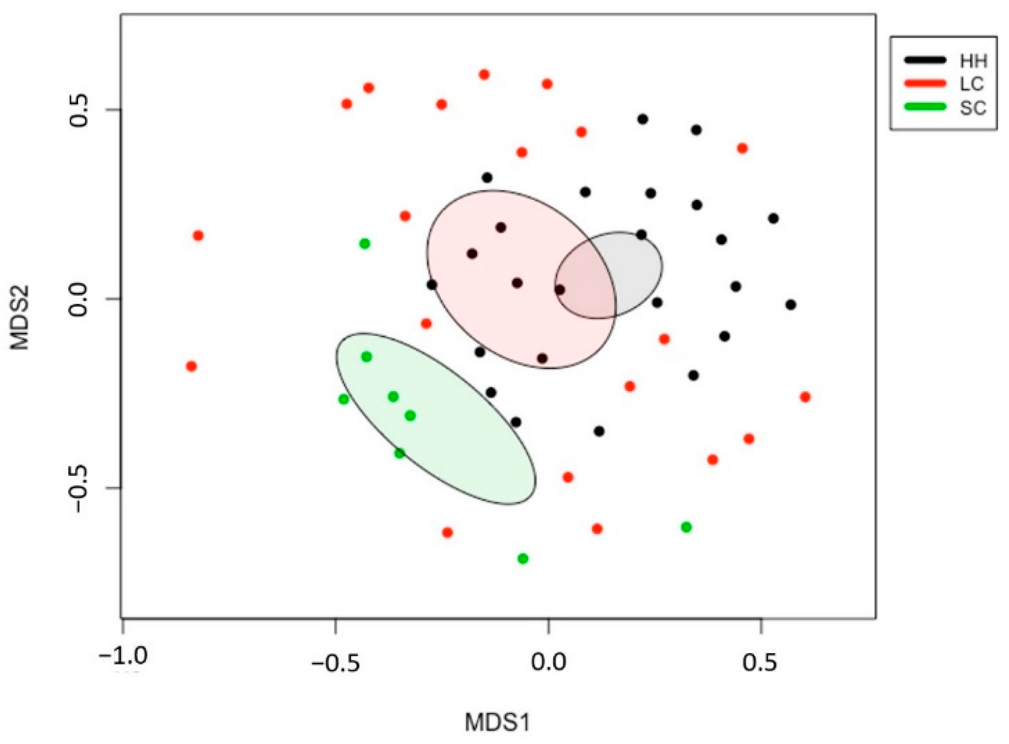
Figure 3.
Comparison of the horse fecal microbiota bacterial composition at the phylum (A) and family (B) level. HH, healthy horses; LC, horses with large intestinal colic; SC, horses with small intestinal colic.
Figure 3.
Comparison of the horse fecal microbiota bacterial composition at the phylum (A) and family (B) level. HH, healthy horses; LC, horses with large intestinal colic; SC, horses with small intestinal colic.

Figure 4.
Differentially abundant genera in horse fecal microbiota among LC (A) and SC (B) compared to HH. HH, healthy horses; LC, horses with large intestinal colic; SC, horses with small intestinal colic.
Figure 4.
Differentially abundant genera in horse fecal microbiota among LC (A) and SC (B) compared to HH. HH, healthy horses; LC, horses with large intestinal colic; SC, horses with small intestinal colic.


Table 1.
Enriched metabolic pathways in the SC compared to the HH.
| Pathways (MetaCyc) | Enriched Pathways in SC | ALDEx Diff. | Metabolism |
|---|---|---|---|
| ALL-CHORISMATE-PWY | Superpathway of chorismate metabolism | 8.16 | Enterobactin biosynthesis |
| ENTBACSYN-PWY | Enterobactin biosynthesis | 8.02 | |
| PWY0-321 | Phenylacetate degradation I (aerobic) | 5.05 | TCA cycle |
| PWY-5178 | Toluene degradation IV (aerobic) (via catechol) | 5.01 | |
| PWY-6185 | 4-methylcatechol degradation (ortho cleavage) | 4.77 | |
| PWY0-1277 | 3-phenylpropanoate and 3-(3-hydroxyphenyl) propanoate degradation | 4.60 | |
| PWY-5417 | Catechol degradation III (ortho-cleavage pathway) | 4.53 | |
| PWY-6182 | Superpathway of salicylate degradation | 4.38 | |
| PWY-5431 | Aromatic compounds degradation via & beta;-ketoadipate | 4.21 | |
| TCA-GLYOX-BYPASS | Superpathway of glyoxylate bypass and TCA | 4.11 |
HH, healthy horses; SC, horses with small intestinal colic.
Table 2.
Depleted metabolic pathways in the SC compared to the HH.
| Pathways (MetaCyc) | Depleted Pathways in SC | ALDEx Diff. | Metabolism |
|---|---|---|---|
| PWY-6141 | Archaetidylserine and archaetidylethanolamine biosynthesis | −2.52 | Methanogen lipid membrane biosynthesis |
| PWY-7286 | 7-(3-amino-3-carboxypropyl)-wyosine biosynthesis | −2.39 | |
| PWY-6167 | Flavin biosynthesis II (archaea) | −2.35 | |
| PWY-6350 | Archaetidylinositol biosynthesis | −2.32 | |
| PWY-6349 | CDP-archaeol biosynthesis | −2.31 | |
| P261-PWY | Coenzyme M biosynthesis I | −2.49 | Methanogenesis |
| METHANOGENESIS-PWY | Methanogenesis from H2 and CO2 | −2.40 | |
| PWY-6148 | Tetrahydromethanopterin biosynthesis | −2.29 | |
| PWY-5198 | Factor 420 biosynthesis | −2.27 | |
| TCA-GLYOX-BYPASS | Coenzyme B biosynthesis | −2.21 |
HH, healthy horses; SC, horses with small intestinal colic.
Publisher’s Note: MDPI stays neutral with regard to jurisdictional claims in published maps and institutional affiliations. |
© 2021 by the authors. Licensee MDPI, Basel, Switzerland. This article is an open access article distributed under the terms and conditions of the Creative Commons Attribution (CC BY) license (https://creativecommons.org/licenses/by/4.0/).
Share and Cite
MDPI and ACS Style
Park, T.; Cheong, H.; Yoon, J.; Kim, A.; Yun, Y.; Unno, T. Comparison of the Fecal Microbiota of Horses with Intestinal Disease and Their Healthy Counterparts. Vet. Sci. 2021, 8, 113. https://doi.org/10.3390/vetsci8060113
AMA Style
Park T, Cheong H, Yoon J, Kim A, Yun Y, Unno T. Comparison of the Fecal Microbiota of Horses with Intestinal Disease and Their Healthy Counterparts. Veterinary Sciences. 2021; 8(6):113. https://doi.org/10.3390/vetsci8060113
Chicago/Turabian StylePark, Taemook, Heetae Cheong, Jungho Yoon, Ahram Kim, Youngmin Yun, and Tatsuya Unno. 2021. "Comparison of the Fecal Microbiota of Horses with Intestinal Disease and Their Healthy Counterparts" Veterinary Sciences 8, no. 6: 113. https://doi.org/10.3390/vetsci8060113
Note that from the first issue of 2016, this journal uses article numbers instead of page numbers. See further details here.