Article

Prevalence, Diagnosis and Improving the Effectiveness of Therapy of Mastitis in Cows of Dairy Farms in East Kazakhstan

by
Nurzhamal Mukhamadieva
1,
Mardan Julanov
2,
Dinara Zainettinova
1,
Vasyl Stefanik
3,
Zhanat Nurzhumanova
1,
Aitbek Mukataev
1 and
Anuarbek Suychinov
4,*
1
Veterinary Department, Shakarim University, Semey 071412, Kazakhstan
2
Faculty of Veterinary, Kazakh National Agrarian Research University, Almaty 050000, Kazakhstan
3
Department of Obstetrics, Gynecology and Biotechnology of Animal Reproduction, Lviv National University of Veterinary Medicine and Biotechnology Named after S.Z. Gzhitsky, 79000 Lviv, Ukraine
4
Kazakh Research Institute of Processing and Food Industry (Semey Branch), Semey 071410, Kazakhstan
*
Author to whom correspondence should be addressed.
Vet. Sci. 2022, 9(8), 398; https://doi.org/10.3390/vetsci9080398
Submission received: 16 June 2022 / Revised: 8 July 2022 / Accepted: 25 July 2022 / Published: 30 July 2022

Abstract

:

Simple Summary

Mastitis is an inflammation of the mammary gland that occurs in response to adverse mechanical, physical, chemical and biological factors. The use of antibiotics as a treatment has a negative impact on milk quality. This paper proposes the treatment of mastitis with a drug that contains natural, environmentally friendly and pharmacologically active preparations. The inclusion of the preparation into the scheme of treatment allows to treat effectively inflammatory processes in the udder of cows and restore their productivity with minimal time and expenses. The drug has anti-inflammatory, antimicrobial and stimulating healing effects.

Abstract

In the present work, the prevalence, etiological factors and effective treatment scheme of mastitis in cows of dairy farms “Balke” and “Madi-R” in Eastern Kazakhstan were investigated. In total, 210 heads were investigated on two farms. The incidence of mastitis in cows on dairy farms is not the same in different years. Average clinical mastitis was detected in 35.4% of cows in 2016, 19.6% in 2017, 28.5% in 2018, and in 2019 in 16.4% of cows. The prevalence rates of subclinical mastitis by year had some differences. So, in 2016—36.5% of cows, then in 2017—21.5%, 2018—19.3% and in 2019—22.6%. In cows with udder inflammation, serum calcium 9.37 ± 0.15 mg/% with a range of 8.0 to 10.8 mg/%, phosphorus 3.58 ± 0.07 mg/% (3.0 to 4.3 mg/%), reserve alkalinity 363.46 ± 6.69 mg/% (320 to 440), carotene 0.49 ± 0.03 mg/% (0.220 to 0.988 mg/%), which are in the lower limit of physiological parameters. The drug “Dorob” was tested during the study of comparative effectiveness of treatment methods. The results of the study showed that this drug has anti-inflammatory, antimicrobial and stimulating healing actions. The treatment of the sick cows with catarrhal mastitis has shown that a total of 8 cows have recovered in the control group and 10 cows in the experimental group with the preparation “Dorob”. The period of recovery in the control group was 8.8 ± 0.39, and in the experimental group—6.2 ± 0.28 (p < 0.05). The drug does not contain antibiotics and hormonal preparations. The inclusion of the drug in the scheme of treatment allows for effectively treating inflammatory processes in the udder of cows and restoring their productivity with minimal cost of time and money.

1. Introduction

The main sector of agriculture in Kazakhstan, including the East Kazakhstan region, is cattle breeding, and its development is conditioned by environmental, climatic and geographical conditions. The high number of livestock on large farms and complexes, and mechanization of basic production processes, including milking, revealed a number of critical problems in the prevention of serious diseases. A specific place among diseases is occupied by mammary gland diseases, which significantly affect animal productivity and reduce the quality of milk [1,2].
Mastitis in cows is an inflammation of the udder that causes pus and results in swelling and redness of the cow’s mammary gland. This disease can cause significant damage to large-scale milk production facilities [3,4]. For example, mastitis is estimated to cost the U.S. dairy industry approximately $2 billion in economic harm per year [5].
Udder inflammation occurs with violations of the rules of machine milking and poor conditions of housing and feeding of cows, which can facilitate the penetration and development of microbes in the mammary gland [6,7]. Mastitis can be caused by various reasons, primarily by inadequate or insufficient feeding, poor husbandry, improper housing and handling of animals, careless organization and performance of artificial insemination, due to various diseases of the reproductive organs, which appear most often during calving and in the postpartum period [8,9]. When veterinary and sanitary rules for maintenance of milking facilities and milking hygiene are violated, as well as the general resistance of the animal and mammary gland being reduced, microorganisms colonize the teat head and the outer opening of the teat canal. Then, microorganisms penetrate into the teat and suprapapillary cisterns (sinuses) and cause an infection process of varying severity from subclinical to clinically expressed inflammation of various forms [10].
Mastitis is a widespread disease in dairy complexes and farms. The incidence in some herds can reach 35–60% and up to 60–75% of cows in a herd can be affected within a year. Mortality rates from mastitis can in some cases exceed those from other diseases. During the disease period and after clinical recovery, the natural loss of milk per cow averages from 10 to 15% of the annual milk yield [11,12].
Mastitis can be caused by more than 140 different species of microorganisms. The most common of them can be grouped into the following groups: contagious (infectious), opportunistic and pathogenic microbes of the animal organism and environmental microorganisms [13].
Mastitis is categorized as clinical and subclinical mastitis. During the milking of cows with clinical mastitis, the properties of milk deteriorate (flakes, clots, watery or discolored milk) or the presence of swollen or indurated mammary quarters before milking [14]. During subclinical mastitis, an udder infection shows no external changes. Subclinical mastitis can result in an elevated milk somatic cell count and decrease milk yield, changing the composition of milk to influence milk quality. The somatic cell count (SCC) increases from about 200,000 SC/mL of milk (uninfected) to over 300,000 SC/mL of milk from mastitis cows [15,16].
In addition, data from many scientists show that not only staphylococci, streptococci and Escherichia coli cause mastitis, but also coronobacteria, salmonellae, mycobacteria, mycoplasmas, viruses, and fungi [17,18,19].
Getting quality and safe cow milk is an important task for dairy farms. Most scientists claim that mastitis in cattle is mainly caused by biological factors and therefore consider it an infectious disease of the mammary gland [20,21,22].
For treatment of mastitis, preparations containing (1) antiseptics (iodine, revanol, etc.), which are substances of non-selective action on pathogens, and (2) chemotherapeutic agents, which are substances of selective action on certain types of microorganisms are used. This group includes: sulfonamides, antibiotics, nitrofurans and new drugs of different groups (quinolones, quinoxaline derivatives, etc.) [23].
The main treatment for mastitis is usually administered by infusion of intramammary ointment or intramuscular or intravenous injections of antibiotics such as streptomycin, ampicillin, cloxacillin, penicillin and tetracycline [24,25]. However, treatment is expected to become problematic in the near future because of the rapid growth of antibiotic-resistant pathogens [26,27,28]. Therefore, it is necessary to introduce a set of effective preventive steps in the technology of milk production to control mammary gland diseases in cows, in particular, mastitis.
Drugs for intracisternal application in mastitis of cows: mastisan A, mastisan B, mastisan E, masticide, etc. [29,30]. They contain various combinations of antibiotics or antibiotics in combination with sulfonamides and oil bases. Usually, the oil base of the drugs prevents the contact of antibiotics with mastitis pathogens and their penetration into udder tissues. At the same time, about 50% of the antibiotics in the drug are excreted with milk, which creates environmental problems and limits the use of milk obtained from cows treated with such drugs [31,32].
Antimastitis drugs based on 3rd and 4th generations of cephalosporins have the shortest excretion period—no more than 60 h (for comparison, drugs based on lincomycin or gentamicin are excreted in five to seven days), and injectable drugs of the 3rd generation of cephalosporins (preparations based on ceftiofur) have no milk waiting period [33].
“Dioxilin gel” contains a complex that includes antibiotics and antiseptics in low concentrations. This makes it possible to fight effectively against the inflammatory process in the mammary glands of cows. The drug is a polymeric hydrogel, when applied to the udder of the animal, it dries quickly. The film that forms after drying ensures the long-lasting effect of active ingredients on inflammation. In addition, it protects against harmful external influences. “Dioxilin gel” is not toxic and has no effect on the quality of milk [34].
It is known in medicine and veterinary medicine sea that buckthorn oil contains vitamins A provitamin (carotene) 0.9–10.9 mg%, B1 (thiamine) 0.016–0.085 mg%, B2 (riboflavin) 0,030–0, 056 mg%, B3, B6, B9, K, macro-and micronutrients, folic acid 0.79 mg%, vitamin C (ascorbic acid) 54–316 mg%, P (bioflavonoids) 75–100 mg%, vitamin K (phylloquinones) 0.9–1.5 mg%, E (tocopherol) 8–18 mg. Sea buckthorn oil is recommended for preventing the development of inflammatory processes and has a protective effect on mucous membranes [35].
It is known that the method for the treatment of mastitis in cows, including biologically active components and diluting medium, in which, according to the invention, Vivaton—veterinary 10%; ASD-2f—4%; “Bursanatal”—liquid immunomodulator made from broiler Fabrica bursa 6%, and 0.9% isotonic solution is used as diluting medium [36]. The disadvantage of the prototype is its multicomponent composition, as well as the complexity of implementation of the invention. Virbac New Zealand Limited produces mastitis treatment preparation called Masticillin, which contains procaine penicillin. It is used to treat early season, multi-quarter mastitis, particularly Strep. uberis [37].
The main method of udder disease prevention is a science-based, cost-effective livestock breeding while complying with zoohygienic norms of maintenance, feeding and milking, providing a high level of natural resistance of the organism and animal productivity.
The purpose of the work is to determine the incidence, etiological factors and development of effective treatment of mastitis in cows on dairy farms in Eastern Kazakhstan. The practical significance of the work is the development of a drug for the treatment of mastitis in cows, which is easy to prepare and use. The drug does not contain antibiotics and hormonal drugs, allowing the effective treatment of mastitis in cows and restoring their productivity with minimal time and money. The drug does not require special expensive imported materials and expands the range of domestic mastitis drugs.

2. Materials and Methods

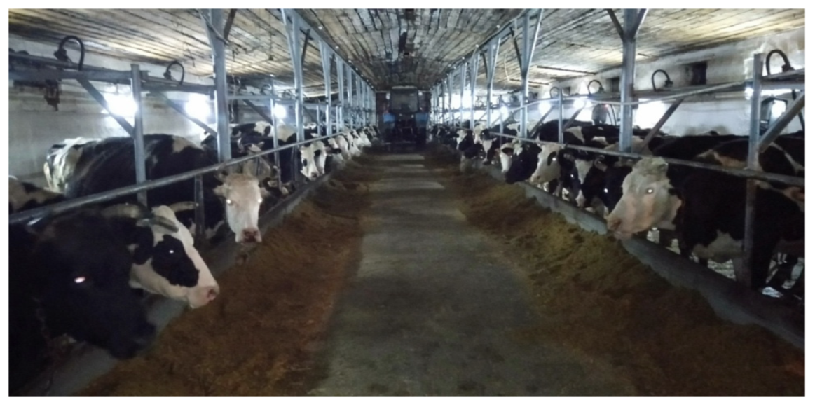
2.1. Experimental Base of the Study

The work was carried out as part of the research work of applied scientific research in the field of agriculture for 2018–2020 (0.0879) on the scientific and technical program: “Increasing the effectiveness of breeding methods in cattle breeding “on the project: “Development of effective breeding methods in dairy cattle breeding industry “on the measure: “Improvement of reproductive ability of dairy cows in the southern region”.
The experimental base of the study was the cattle farm “Balke” of the Beskaragai district and the cattle farm “Madi-R”, of the Znamenski rural district of Semey in East Kazakhstan region of the Republic of Kazakhstan. Cows in these farms were tested for subclinical and clinical forms of mastitis in all seasons of the year in the period from December 2016 to November 2019.
Peasant farm “Balke” of vegetable and dairy production is actively developing in the field of agriculture. Today the farm has more than 500 heads of black-motley breeding cows of the Kostroma breed (Figure 1). The total land area of the farm “Balke” is 2200 hectares (Table 1). In the farm “Balke” breeding stock of cattle: dairy, black-motley, Holstein–Friesian breeds.
For the studies, 210 milking cows of the black-motley breed at the age of 5–6 years with a live weight of 450–500 kg from the farm “Balke” and milking cows of the local Kazakh white-headed breed at the age of 5–6 years with a live weight of 350–470 kg from the farm “Madi-R” were selected.

2.2. Stages of Research and Treatment of Cows

The research was conducted from December 2016 to November 2019. The work was conducted in two stages. In the first stage, diagnostic research was carried out. In the second, the scheme of treatment was developed, and the results obtained during the experimental work were analyzed, checked and clarified.
During the study, the following methods were used: anamnesis collection, and clinical and laboratory methods of investigation. A medical history was created for each animal.
To study the effectiveness of the treatment protocols and testing the drug “Dorob” sick cows (20 animals) diagnosed with catarrhal mastitis were divided into two groups.
The first group consisted of cows treated with traditional methods of treatment (control group of animals, 10 cows). The treatment protocol of the control group of cows: the drug Mastiet forte, also cows of this group used the drug Ketoprof as an anti-inflammatory agent intramuscularly. The application of the above remedies was combined with biologically active supplements—Healthivit, which was injected subcutaneously in a dose of 6 mL once (Table 2).
The second group consisted of cows treated with the drug “Dorob” (10 cows). Cows in the second group were treated with the drug “Dorob” (Table 3). The drug “Dorob” was used intracisternally in a dose of 5 mL, twice a day with an interval of 10–12 h after completion of milking. The composition of this preparation: sea buckthorn oil in a ratio of 10:1 with ASD fraction 2. This preparation is an orange transparent oily liquid. Sea buckthorn oil, as well as ASD fraction 2, have vasoconstrictor, antiseptic, anti-inflammatory and analgesic effects and promote tissue regeneration. In addition, ASD fraction 2 as a biogenic stimulant increases the protective function and increases the body’s resistance to pathogenic and toxic influences. ASD fraction 2 contains low molecular weight organic compounds, including lower carboxylic acids, their amides and ammonium salts, choline esters of carboxylic acids, choline, primary and secondary amines, peptides, as well as inorganic nitrogenous compounds (salts of ammonium carbonate, ammonium acetic acid) and water [38].
As stated above, cows with catarrhal mastitis were divided into two groups (Figure 2). Animals of the first group were treated according to the scheme presented in Table 1. That is Mastiet forte—intracisternally for five days at a dose of 10 mL 2 times a day for five days. After injecting the drug, a slight massage of the affected quarter of the udder was carried out to distribute the drug evenly.
Moreover, cows of this group were treated with the drug Ketoprof as an anti-inflammatory agent intramuscularly. The dose of the drug Ketoprof—3 mL per 100 kg of animal weight once a day for three days. Application of the above drugs was combined with a complex vitamin-containing preparation—Healthivit subcutaneously in a dose of 6 mL once a day.
The therapeutic efficacy of the applied treatment protocols was determined by the number of cows recovered, the number of days of recovery, and the manifestation of relapses. Economic efficiency was determined by taking into account the cost of drugs, labor costs of veterinarians and service personnel, and the prevented damage from the loss of dairy products.

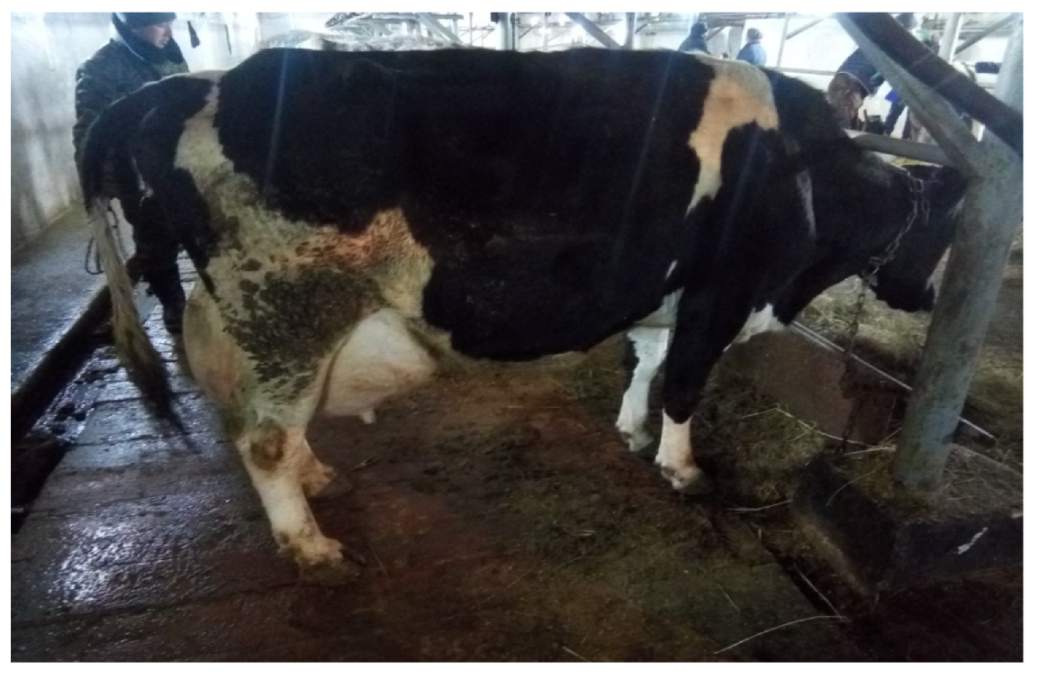
2.3. Testing for Mastitis

Diagnosis was based on the classification of mastitis according to A.P. Studentsov [36]. To detect subclinical mastitis the following tests were used: bromothymol, mastidine, dimestine, Whiteside, sedimentation of milk, California mastitis test. Furthermore, in work the device of express mastitis diagnosis (PEDM), Lactan was used. Lactan 4,1-mini device was used to check the amount of dry skimmed milk residue, density, the water–fat ratio in milk, and the number of somatic cells, and the Miltek device was used to determine mastitis milk.

2.4. Biochemical Blood Tests

To determine the number of hemoglobin, erythrocytes and leukocytes in the blood we conducted research using a PCE 90 Vet hematology analyzer. A hematological analyzer is a device for determining the number of blood cells in animals and the amount of hemoglobin for diagnosis in veterinary laboratories. The modern apparatus is very convenient and fast, it takes a few minutes to carry out the analysis, and the response time of the general analyzers is 7–10 min.
Hemanalyzer standard reagents are intended for the analysis of 170–200. Defined parameters: erythrocytes (RBC), hemaglobin (HGB), hematocrit (HCT), white blood cells (WBC), mean erythrocyte volume (MCV), mean hemaglobin volume per erythrocyte (MCH), mean hemaglobin concentration per erythrocyte (MSHC), erythrocyte volume ratio change (RDW %), platelets (PLT), mean platelet volume (MPV), thrombocrit (PCT), platelet length particles (PDW). The histogram displays the volume of erythrocytes, white blood cells, and platelets.

2.5. Determination of the Chemical Composition of the Feed

Moisture content was determined according to GOST 13496.3-92 [39]. Opened beakers with feed are placed in a drying cabinet preheated to a temperature of (130 ± 2) °C. Drying is carried out for 40 min, counting from the moment when the temperature reaches 130 °C.
After drying and cooling the beakers with feed are weighed to the second decimal place.
The mass fraction of moisture (W) in percent is calculated according to the formula
W = m 1 m 2 m 1 m × 100
where
  • m1 is the mass of the beaker with the sample before drying, g;
  • m2—mass of the beaker with the sample after drying and cooling, g;
  • m—mass of empty beaker, g.

2.6. Determination of Carotene in Feeds

The essence of the method consists of the extraction of carotene with petroleum ether or gasoline and photometric measurement of the color intensity of the extract, which depends on the carotene content [40].

2.7. Determination of the Reserve Alkalinity of Blood by Kondrakhin’s Method

Principle of the method. In one half of the flask, blood plasma is treated with sulfuric acid, which results in the release of carbon dioxide, which is part of the bicarbonates. The released carbon dioxide is absorbed by a sodium hydroxide solution, which is in the other half of the flask. The excess sodium hydroxide that was not reacted with carbon dioxide and half of the sodium hydrocarbonate (NaHCO3) formed during the absorption of CO2 is titrated with a sulfuric acid solution. The amount of initially bound sodium hydroxide is used to determine the amount of carbon dioxide released from the plasma, which is equivalent to the bicarbonate content [41].

2.8. Statistics

Each experiment was performed in triplicate and values were expressed as mean ± SEM of three independent observations. Statistical analysis was performed using Statistica 12.0 (STATISTICA, 2014; StatSoft Inc., Tulsa, OK, USA). The differences between samples were evaluated using one-way ANOVA. The Tukey HSD test was used for means comparisons. A p-value < 0.05 was considered statistically significant.

3. Results

3.1. Incidence of Mastitis in Cows on Dairy Farms of Balke and Madi-R

The incidence of mastitis in cows on dairy farms of Balke and Madi-R farms showed different incidences in different years (Table 4). A total of 210 cows were examined on two farms. On average, clinical mastitis was registered in 35.4% of cows in 2016, 19.6% in 2017, 28.5% in 2018 and 16.4% in 2019. Furthermore, the prevalence rates of subclinical mastitis had some differences by year. For example, in 2016 it was 36.5%, then in 2017—21.5%, in 2018—19.3 and in 2019—22.6%.
Out of 90 cows, 56 cows had clinical mastitis, which amounted to 35.4%. Subclinical mastitis was present in 34 cows, which amounted to 36.5%. Catarrhal mastitis occurred in 24 cows or 32%.
Analyzing the data in Table 5, we revealed the prevalence of subclinical forms of mastitis. In winter, the rate of subclinical mastitis gradually increased. In spring, there was a dramatic rise. A decline was observed in summer, and a high level of subclinical mastitis occurred in the autumn season. The incidence of subclinical mastitis in cows was 15 animals in winter, 29 animals in spring, 19 animals in summer, and 30 animals in fall.
According to the data (Table 6), the highest number of clinical mastitis occurs in the spring and autumn periods. Clinical mastitis in winter was detected in 38 animals, in spring—48 animals, in summer—27 animals, in autumn—45 animals.
Our research showed the occurrence by season of catarrhal mastitis in 2016 was—32%, in 2017—21.3%, in 2018—25.3%, and in 2019—21.3% (Table 7). The rate of catarrhal mastitis gradually increased during the winter and spring seasons. There was a gradual decline in the summer. The increase in clinical mastitis was observed in autumn.
The incidence of mastitis in cows was 15 head in winter, 19 head in spring, 10 head in summer, and 31 head in fall.

3.2. Feeding Diets of Cows

Moreover, when identifying predisposing factors of mastitis, we studied the feeding diets of cows in the summer-autumn and winter-spring periods of the year. So, in the summer-autumn period, animals were on pasture—grass pasture hay (steppe, meadow), additional feeding, salt, chalk, water at will, mixed fodder 3.5 kg per head during milking.
During the winter period, the cows’ ration consisted of 25 kg of silage, 10 kg of mono-fodder, 5 kg of hay, 3.5 mixed fodder, 1.5 kg of sunflower meal, 4 kg of vitamin-mineral additive, 150 g of molasses, 25 g of chalk, 60 g of salt. The daily ration of the first heifers was 14.6 fodder units (Table 8).
The diet of the first heifers in the summer-autumn periods consisted of 20 kg of silage, 10 kg of mono-fodder, 8 kg of hay, and 350 g of mixed fodder. Per liter of milk, 1.5 kg of sunflower meal, 3 kg of vitamin-mineral additive, 100 g of molasses, 25 g of chalk, and 50 g of salt. The daily ration was 8.8 fodder units.
The analysis of our studies showed that in the summertime, with sufficient proteins, carbohydrates, vitamins and minerals in green feed, mastitis diseases in cows are less common.
Chemical analysis of forages was characterized by the following indicators (Table 9). Carotene content in hay was 8.67 ± 1.60 mg/kg (with variation from 4.2 mg/kg to 10.3 mg/kg). The carotene content of the silage was 5.30 ± 2.15 mg/kg (ranging from 2 mg/kg to 8 mg/kg). The crude protein level in the silage was 20.60 ± 3.47 g/kg (15.6 to 25.4 g/kg). Digestible protein content in hay was 50.50 ± 4.08 g/kg (39.1 to 64.2 g/kg) and in silage was 11.00 ± 2.50 g/kg (7.2 to 14.2 g/kg). Calcium and phosphorus in hay contained between 4.48 ± 0.73 g/kg (1.7–6.2 g/kg and 0.92–2.00 g/kg), and in silage their content was 0.56 ± 0.15 g/kg (0.326 to 0.757 g/kg and 0.38 ± 0.12 g/kg (0.236 to 0.565 g/kg), respectively.
The results of the analysis of fodder of the farm “Balke” of the Beskaragai district in the East Kazakhstan region showed that the nutrient value of hay was on average 0.35 ± 0.04 f.u./kg (from 0.35 to 0.58 f.u./kg). The results of the study of hay and silage are presented in Table 8.

3.3. Analysis of Cow’s Blood

As it is known, in mastitis, the pathological process is not limited to the tissues of the affected quarter of the mammary gland, it covers the whole organism, which as a whole participates in the elimination of the disease [42,43]. Accordingly, during inflammation of the mammary gland, there are biochemical changes in the secretion from the affected udder, as well as in blood. These changes may occur even before the clinical manifestation of mastitis, therefore, having great diagnostic value in practical veterinary medicine [44,45].
We also examined blood serum from 26 cows with mastitis for some biochemical parameters, specifically for calcium, phosphorus, reserve alkalinity and carotene content (Table 10). If we take into account the fact that normally the blood of cows should contain in mg/% of calcium—11.3–13.1, phosphorus—4.0–4.6, reserve alkalinity—460–540, carotin—0.5–2.28, then according to the research these indices were reduced or were at the lower limit of the physiological characteristics in the blood of the cows with mastitis.
In cows with udder inflammation, serum calcium was found to be 9.37 ± 0.15 mg/% with a range of 8.0 to 10.8 mg/%, phosphorus 3.58 ± 0.07 mg/% (3.0 to 4.3 mg/%), reserve alkalinity 363.46 ± 6.69 mg/% (320 to 440), carotene 0.49 ± 0.03 mg/% (0.220 to 0.988 mg/%).
Cows with mastitis were divided into two groups for treatment. Before treatment, we determined the level of erythrocytes (RBC), hemoglobin (HGB), hematocrit (HCT), leukocytes (WBC), mean erythrocyte volume (MCV), mean hemoglobin volume in erythrocyte (MCH), mean erythrocyte hemoglobin concentration (MCHC), change in erythrocyte volume ratio (RDW %), platelets (PLT), mean platelet volume (MPV), thrombocrit (PCT), platelet distribution width (PDW).
The results of blood tests of cows with mastitis before treatment were (Table 11): White blood cell (WBC)—9.86 ± 1.59 × 10⁹/L, red blood cell (RBC)—6.50 ± 0.39 × 10¹²/L, hemoglobin (HGB)—79.50 ± 1.83 g/L, hematocrit (HCT)—17.79 ± 0.49%, mean red blood cell volume (MCV)—29.11 ± 0.64 fL, mean hemoglobin per red blood cell (MCH)—12, 27 ± 0.55 pg, mean erythrocyte hemoglobin concentration (MCNS)—417.60 ± 5.28 g/L, change in erythrocyte volume ratio (RDW %)—15.67 ± 0.34%, platelet (PLT)—240.10 ± 24.02 × 10⁹/L.
According to the results of the study (Table 12) the blood of cows with catarrhal mastitis was as follows: leukocyte (WBC) in the experimental group was: −13.88 ± 1.80 × 10⁹/L, in the control group—8.99 ± 0.47, erythrocyte (RBC)—8.10 ± 0.73 × 10¹²/L, in the control group—7.89 ± 0.46. Hemoglobin (HGB)—98.40 ± 4.58 g/L, in the control group 133.20 ± 31.94. Hematocrit (HCT)—24.44 ± 1.10%, in controls—35.06 ± 1.60. Mean erythrocyte volume (MCV)—31.66 ± 1.04 fL, in controls—46.88 ± 1.36. Mean erythrocyte hemoglobin volume (MCH)—11.06 ± 1.09, in controls 15.45 ± 0.43 pg. Mean erythrocyte hemoglobin concentration (MSHC)—413.80 ± 5.28 g/L, change in erythrocyte volume ratio (RDW %)—15.53 ± 0.29%, platelet (PLT)—243.50 ± 27.20 × 10⁹/L.
As the results of the treatment of sick cows with catarrhal mastitis (Table 13) showed, 8 cows in the control group and 10 cows in the experimental group recovered.
There were two cows that did not recover. Terms of recovery in the control group were 8.8 ± 0.39, in the experimental group 6.2 ± 0.28 (p < 0.05). Two cases of relapses in the control group were detected.
Thus, during the comparative effectiveness of the treatment methods, the drug “Dorob” proved to be effective. The drug had anti-inflammatory, antimicrobial and stimulating healing effects. It should be noted that the drug did not contain antibiotics and hormones. Therefore, the inclusion of the drug in the scheme of treatment allows, with minimal time and money, effectively treating inflammatory processes in the udder of cows, restoring their function and productivity.

4. Discussion

Despite the constant improvement of methods to prevent mastitis, mammary gland inflammation remains the most common disease in cows on dairy farms and cattle complexes to this day [46,47,48,49]. In our research, we found out that mastitis in conditions of the East Kazakhstan region is widespread. The main causes of mastitis are inadequate feeding, violation of keeping conditions, milking improprieties, udder trauma and a variety of stresses [50].
We also studied the occurrence of subclinical and clinical mastitis by seasons of the year for the period 2016–2019. Clinical mastitis was recorded in 2016 in 35.4% of cows, in 2017 in 19.6%, 2018 in 28.5% and in 2019 in 16.4%. Furthermore, the prevalence rates of subclinical mastitis had some variation by year. Thus, in 2016—36.5%, in 2017—21.5%, 2018—19.3 and in 2019—22.6%.
Analyzing the data of the conducted study, it can be concluded that high levels of subclinical and clinical mastitis are revealed in the autumn time of the year.
The prevalence of mastitis among cows in different years is not the same. The results of the data show that with low air temperature and high precipitation the incidence of mastitis among cows increases. At the beginning of spring (draughts, coldness, dampness), seasonal mastitis outbreaks are observed. Thus, the deterioration of weather conditions negatively affects milking animals, which leads to an increase in the number of all forms of mastitis in cows [51,52,53]. (Cervinkova et al., 2013) studied that half of the collected milk from 16 dairy farms in the Czech Republic was contaminated by microorganisms with the potential to cause mastitis [54]. In the United Kingdom, the annual prevalence of clinical mastitis has been estimated at 35% of the total dairy cow population [55].
The most common forms of mastitis were subclinical and catarrhal mastitis [56,57]. Therefore, in our work, we selected animals with the catarrhal form of mastitis to determine treatment efficacy. The feeding diets of cows in the summer-autumn and winter-spring periods of the year were studied for the manifestation of mastitis. Analysis of our studies showed that in summer, with sufficient proteins, carbohydrates, vitamins and minerals in green fodder, mastitis diseases in cows occur less frequently.
The results of the analysis of forages of the farm “Balke” showed that the nutritive value of hay on average was 0.35 ± 0.04 f.u./kg.
We also examined blood serum from 26 cows with mastitis for some biochemical parameters, namely calcium, phosphorus, reserve alkalinity and carotene.
Such a violation of mineral metabolism, especially in combination with protein and vitamin deficiencies in the diet, in our opinion, leads to a decrease in the resistance of the body of cows and subsequently predisposes the body to the manifestation of pathological processes, including mastitis.
To treat cows with catarrhal mastitis they were divided into two groups.
According to the results of blood tests of cows with catarrhal mastitis before treatment, it was found: White blood cell (WBC)—9.86 ± 1.59 × 10⁹/L, red blood cell (RBC)—6.50 ± 0.39 × 10¹²/L, hemoglobin (HGB)—79.50 ± 1.83 g/L, hematocrit (HCT)—17.79 ± 0.49%, mean red blood cell volume (MCV)—29.11 ± 0.64 fL, mean hemoglobin per red blood cell (MCH)—12.27 ± 0.55 pg, mean erythrocyte hemoglobin concentration (MCNS)—417.60 ± 5.28 g/L, erythrocyte volume ratio change (RDW %)-15.67 ± 0.34%, platelet count (PLT)—240.10 ± 24.02 × 10⁹/L. Blood values were in the lower limit of physiological indices.
The results of blood analysis of cows with catarrhal mastitis revealed: that white blood cell (WBC) in the experimental group was: −13.88 ± 1.80 × 10⁹/L, in the control group—8.99 ± 0.47, erythrocyte (RBC)—8.10 ± 0.73 × 10¹²/L, in the control group—7.89 ± 0.46. Hemoglobin (HGB)—98.40 ± 4.58 g/L, in the control group 133.20 ± 31.94. Hematocrit (HCT)—24.44 ± 1.10%, in controls—35.06 ± 1.60. Mean erythrocyte volume (MCV)—31.66 ± 1.04 fL, in controls—46.88 ± 1.36. Mean erythrocyte hemaglobin volume (MCH)—11.06 ± 1.09, in controls 15.45 ± 0.43 pg, Mean erythrocyte hemaglobin concentration (MSHC)—417.60 ± 5.28 g/L, change in erythrocyte volume ratio (RDW %)—15.67 ± 0.34%, platelet (PLT)—240.10 ± 24.02 × 10⁹/L.
Alhussien et al. (2015) observed that the blood and milk lymphocytes and monocytes of cows with mastitis were significantly decreased [58].
One of the tasks of this work is to develop treatment protocols for cows with catarrhal mastitis. For this purpose, we selected such a scheme, which, firstly, would be simple, easily performed in production conditions, secondly, would not contain antibiotics and hormonal agents, and, thirdly, would be effective both therapeutically and economically.
Such a remedy was the drug “Dorob”. This remedy for the treatment of cows with mastitis included ASD fraction 2 and sea buckthorn oil. The ratio of ASD fraction 2 to sea buckthorn oil was 1:10.
Studies of compatibility of the drug components on the animal body, as well as the pharmacotherapeutic effect in the treatment of mammary gland (mucous membranes) diseases, were carried out. Materials used in the experiments were: egg white, fresh milk, sea buckthorn oil, and ASD fraction 2. We studied the interaction of milk and sea buckthorn oil, then egg albumen with sea buckthorn oil, then egg albumen with ASD fraction 2, and a water-milk solution in the ratio 1:10 with ASD fraction 2. There were no flakes or clots formed in the udder secretion. The solution turned out homogeneous, dispersedly thick.
Abboud M. et al. (2015) reported that essential oils from Thymus Vulgaris and Lavandula Angustifolia showed strong antibacterial activity against the main strains of mastitis Staphylococcus and Streptococcus [59]. Dilshad et al. (2010) analyzed the practices for mastitis treatment and reported that 25 different plant species were being used for the treatment and control of mastitis in cows and buffaloes in Pakistan [60]. Pașca et al. (2020) proposed the treatment of mastitis caused by Gram-positive bacteria with natural products, containing different plant extracts and propolis [61].
From the data, we can conclude that the fluids obtained in the interaction of the drug components with the animal’s body have a high viscosity. Chemical processes in their environment proceed slowly, diffusion and resorption are delayed, the fluids have adsorptive properties, decrease the sensitivity of nerve endings and promote faster healing of inflammatory processes.

5. Conclusions

Analyzing the data of the study we can conclude that a high level of subclinical and clinical mastitis is revealed in autumn time of the year. The main reasons for mastitis in milking cows are poor quality of feeding affected by mold fungi and incomplete feeding, violation of keeping conditions and milking inaccuracies. The inclusion of the preparation “Dorob” into the scheme of treatment allows to treat effectively inflammatory processes in the udder of cows and restore their productivity with minimal time and expenses. The drug has anti-inflammatory, antimicrobial and stimulating healing effects. The product does not contain antibiotics and hormones.

Author Contributions

Conceptualization, N.M.; Formal analysis, A.S.; Investigation, N.M. and D.Z.; Methodology, N.M., D.Z. and Z.N.; Resources, Z.N. and A.M.; Supervision, M.J.; Validation, M.J. and V.S.; Visualization, D.Z.; Writing—original draft, V.S.; Writing—review & editing, A.S. All authors have read and agreed to the published version of the manuscript.

Funding

This research received no external funding.

Institutional Review Board Statement

The animal study protocol was approved by the Institutional Review Board of Shakarim University of Semey (protocol code P042-2.11 from 20.04.2021). Research and educational work using experimental animals are regulated by the Law of the Republic of Kazakhstan on Veterinary Medicine of 10 July 2002, No. 339, the Rules of preclinical studies, medical and biological experiments and clinical trials in the Republic of Kazakhstan approved by Order No. 442 of the Minister of Health of the Republic of Kazakhstan dated 25 July 2007. In research we created conditions for humane treatment of laboratory animals in experiments and proper ethical control over their maintenance.

Informed Consent Statement

Informed consent was obtained from all subjects involved in the study.

Data Availability Statement

All data generated or analyzed during this study are included in this published article.

Conflicts of Interest

The authors declare no conflict of interest.

References

  1. Zainettinova, D.; Muhamadieva, N.; Julanov, M.; Stefanyk, V.; Zaviruha, V.; Sarhambaeva, I. The effectiveness of the treatment of cows with mastitis. Sci. Messenger Lviv. Natl. Univ. Vet. Med. Biotechnologies. Ser. Vet. Sci. 2019, 21, 178–181. [Google Scholar] [CrossRef] [Green Version]
  2. Oraz, G.T.; Ospanov, A.B.; Chomanov, U.C.; Kenenbay, G.S.; Tursunov, A.A. Study of beef nutritional value of meat breed cattle of Kazakhstan. J. Hyg. Eng. Des. 2019, 29, 99–105. [Google Scholar]
  3. Arkhipov, A.A.; Stollar, A.T. Adequate treatment for acute mastitis-the key to herd well-being. Vet. Med. 2008, 11, 15–17. [Google Scholar]
  4. Van Hoeij, R.J.; Lam, T.J.G.M.; Bruckmaier, R.M.; Dijkstra, J.; Remmelink, G.J.; Kemp, B.; van Knegsel, A.T.M. Udder health of dairy cows fed different dietary energy levels after a short or no dry period without use of dry cow antibiotics. J. Dairy Sci. 2018, 101, 4570–4585. [Google Scholar] [CrossRef]
  5. Dalanezi, F.M.; Joaquim, S.F.; Guimarães, F.F.; Guerra, S.T.; Lopes, B.C.; Schmidt, E.M.S.; Cerri, R.L.A.; Langoni, H. Influence of pathogens causing clinical mastitis on reproductive variables of dairy cows. J. Dairy Sci. 2020, 103, 3648–3655. [Google Scholar] [CrossRef] [PubMed]
  6. Pyörälä, S. Indicators of inflammation in the diagnosis of mastitis. Vet Res. 2003, 34, 565–578. [Google Scholar] [CrossRef] [Green Version]
  7. Chelnokova, M.I.; Shcherbakova, N.A. Diagnosis and therapy of mastitis of cows. Proc. Velikoluk. State Agric. Acad. 2018, 1, 20–24. [Google Scholar]
  8. Procópio, T.F.; Moura, M.C.; Bento, E.F.; Soares, T.; Coelho, L.C.; Bezerra, R.P.; Mota, R.A.; Porto, A.L.; Paiva, P.M.; Napoleão, T.H. Looking for alternative treatments for bovine and caprine mastitis: Evaluation of the potential of Calliandra surinamensis leaf pinnulae lectin (CasuL), both alone and in combination with antibiotics. MicrobiologyOpen 2019, 8, e869. [Google Scholar] [CrossRef] [Green Version]
  9. Waminal, Y.O.; Gambol, M.R.; Sabado, A.C.; Vallejos, J.P.M.; Briones, R.C.; Estabillo, E.J.M. Mastitis Detection in Holstein Sahiwal Crossbred Cattle (Bos taurus) Using Different Brands and Dilution Levels of Liquid Anionic Surfactants. Anim. Prod. 2021, 23, 144–150. [Google Scholar] [CrossRef]
  10. Qaisar, H.M.U.; Ahmad, T.; Rizwan, M.; Saqib, M. Antimicrobial efficacy of combination of lincomycin and spiramycin (Lispiracin™) as systemic dry cow therapy for controlling bovine mastitis. Punjab Univ. J. Zool. 2017, 32, 197–201. [Google Scholar]
  11. Zhumanov, K.T.; Biyashev, K.B.; Biyashev, B.K.; Sansyzbai, A.R.; Valdovska, A.; Koshkinbaev, S.S. The study of morphological properties of mastitis pathogens cows. Res. Results 2015, 3, 35–39. [Google Scholar]
  12. Gao, J.; Liu, Y.C.; Wang, Y.; Li, H.; Wang, X.M.; Wu, Y.; Zhang, D.R.; Gao, S.; Qi, Z.L. Impact of yeast and lactic acid bacteria on mastitis and milk microbiota composition of dairy cows. AMB Express 2020, 10, 1–12. [Google Scholar] [CrossRef] [PubMed]
  13. Biziulevichius, G.A.; Lukauskas, K. In vivo studies on lysosubtilin. 3. Efficacy for treatment of mastitis and superficial lesions of the udder and teats in cows. Vet. Res. 1998, 29, 441–456. [Google Scholar] [PubMed]
  14. Malek dos Reis, C.B.; Barreiro, J.R.; Mestieri, L.; Porcionato, M.A.; dos Santos, M.V. Effect of somatic cell count and mastitis pathogens on milk composition in Gyr cows. BMC Vet. Res. 2013, 9, 67. [Google Scholar] [CrossRef] [Green Version]
  15. Piepers, S.; De Meulemeester, L.; de Kruif, A.; Opsomer, G.; Barkema, H.W.; De Vliegher, S. Prevalence and distribution of mastitis pathogens in subclinically infected dairy cows in Flanders. Belgium. J Dairy Res. 2007, 74, 478–483. [Google Scholar] [CrossRef]
  16. Blowey, R.W.; Edmondson, P. Mastitis Control in Dairy Herds; CABI: Cambridge, MA, USA, 2010. [Google Scholar]
  17. Kakimov, A.; Muratbayev, A.; Zharykbasova, K.; Zharykbasov, Y.; Kassymov, S.; Zhumadilova, G.; Jumazhanova, M.; Utegenova, A. Developing HACCP plan for a fermented milk drink with encapsulated biologically active supplements. Eur. Asian J. Biosci. 2020, 14, 889–895. [Google Scholar]
  18. Sutra, L.; Poutrel, B. Virulence factors involved in the pathogenesis of bovine intramammary infections due to Staphylococcus aureus. J. Med. Microbiol. 1994, 40, 79–89. [Google Scholar] [CrossRef] [Green Version]
  19. Lundberg, A.; Aspán, A.; Nyman, A.; Unnerstad, H.E.; Waller, K.P. Associations between bacterial genotype and outcome of bovine clinical Staphylococcus aureus mastitis. Acta Vet. Scand. 2014, 56, 2. [Google Scholar] [CrossRef] [Green Version]
  20. Riekerink, R.O.; Barkema, H.W.; Kelton, D.F.; Scholl, D.T. Incidence rate of clinical mastitis on Canadian dairy farms. J. Dairy Sci. 2008, 91, 1366–1377. [Google Scholar] [CrossRef]
  21. Mekibib, B.; Furgasa, M.; Abunna, F.; Megersa, B.; Regassa, A. Bovine mastitis: Prevalence, risk factors and major pathogens in dairy farms of holeta town, central Ethiopia. Vet. World 2010, 3, 397–403. [Google Scholar] [CrossRef]
  22. Nyman, A.K.; Ekman, T.; Emanuelson, U.; Gustafsson, A.H.; Holtenius, K.; Waller, K.P.; Sandgren, C.H. Risk factors associated with the incidence of veterinary-treated clinical mastitis in Swedish dairy herds with a high milk yield and a low prevalence of subclinical mastitis. Prev. Vet. Med. 2007, 78, 142–160. [Google Scholar] [CrossRef] [PubMed]
  23. Krömker, V.; Leimbach, S. Mastitis treatment—Reduction in antibiotic usage in dairy cows. Reprod. Domest. Anim. 2017, 52, 21–29. [Google Scholar] [CrossRef] [PubMed] [Green Version]
  24. Shirazi-Beheshtiha, S.H.; Safi, S.; Rabbani, V.; Bolourchi, M.; Ameri, M.; Khansari, M.R. The diagnostic value of determination of positive and negative acute phase proteins in milk from dairy cows with subclinical mastitis. Comp. Clin. Path. 2012, 21, 999–1003. [Google Scholar] [CrossRef]
  25. Barnouin, J.; Bord, S.; Bazin, S.; Chassagne, M. Dairy management practices associated with incidence rate of clinical mastitis in low somatic cell score herds in France. J. Dairy Sci. 2005, 88, 3700–3709. [Google Scholar] [CrossRef] [Green Version]
  26. Hoque, M.N.; Das, Z.C.; Rahman, A.N.; Hoque, M.M. Effect of administration of vitamin E, selenium and antimicrobial therapy on incidence of mastitis, productive and reproductive performances in dairy cows. Int. J. Vet. Sci. 2016, 4, 63–70. [Google Scholar] [CrossRef] [PubMed]
  27. Wang, W.; Song, Y.; Petrovski, K.; Eats, P.; Trott, D.J.; San Wong, H.; Page, S.W.; Perry, J.; Garg, S. Development of intramammary delivery systems containing lasalocid for the treatment of bovine mastitis: Impact of solubility improvement on safety, efficacy, and milk distribution in dairy cattle. Drug Des. Dev. Ther. 2015, 9, 631. [Google Scholar]
  28. McDougall, S. Intramammary treatment of clinical mastitis of dairy cows with a combination of lincomycin and neomycin, or penicillin and dihydrostreptomycin. N. Z. Vet. J. 2003, 51, 111–116. [Google Scholar] [CrossRef]
  29. Oliver, S.P.; Murinda, S.E. Antimicrobial resistance of mastitis pathogens. Vet. Clin. North Am. Food Anim. 2012, 28, 165–185. [Google Scholar] [CrossRef]
  30. Marimuthu, M.; Abdullah, F.F.J.; Mohammed, K.; Poshpum, S.D.S.; Adamu, L.; Osman, A.Y.; Abba, Y.; Tijjani, A. Prevalence and antimicrobial resistance assessment of subclinical mastitis in milk samples from selected dairy farms. Am. J. Anim. Vet. Sci. 2014, 9, 65–70. [Google Scholar] [CrossRef] [Green Version]
  31. Smulski, S.; Gehrke, M.; Libera, K.; Cieslak, A.; Huang, H.; Patra, A.K.; Szumacher-Strabel, M. Effects of various mastitis treatments on the reproductive performance of cows. BMC Vet. Res. 2020, 16, 1–10. [Google Scholar] [CrossRef] [PubMed]
  32. Kartashova, V.M.; Ivashura, A.I. Mastitis of Cows; Agropromizdat: Moscow, Russia, 1988. [Google Scholar]
  33. Sidorkin, E.A.; Ulizko, M.A.; Gritsay, O.S.; Koptsev, E.A.; Gostev, E.E. Mastomycin for the prevention of mastitis in cows in the dry period. Vet. Med. 2009, 2, 20–21. [Google Scholar]
  34. Pristromova, Y.I.; Yurkshtovich, Y.N.; Yurkshtovich, N.K.; Alinovskaya, V.A. Composition and physicochemical properties of antimicrobial gels based on carbopols. News Sci. Agroind. Complex 2018, 2, 452–454. [Google Scholar]
  35. Rysuly, M.; Parmankulova, T.N.; Sadvakas, A.S.; Hamzaeva, M.B.; Kurmangozhaeva, A.K.; Azymhan, D.T. Possibilities of application of local plants in treatment of oncological diseases. Vestn. KazNMU 2016, 1, 548. [Google Scholar]
  36. Studentsov, A.P. Diseases of the Udder of Cows; Selkhozgiz: Moscow, Russia, 1952. [Google Scholar]
  37. Available online: https://nz.virbac.com/products/mastitis-treatments/masticillin (accessed on 6 July 2022).
  38. Available online: http://armbio.bio/catalog/veterinary/stimulators/asd_fr_2 (accessed on 6 July 2022).
  39. GOST 13496.3 92; Compound Fodder, Feed Raw Materials. Methods of Determination of Moisture. IPK Publishing House of Standards: Moscow, Russia, 2000.
  40. GOST 13496.17 95; Feed. Method for the Determination of Carotene. Publishing House of Standards: Minsk, Belarus, 2000.
  41. Kondrakhin, I.P. Clinical Laboratory Diagnosis in Veterinary Medicine; Agropromizdat: Moscow, Russia, 1985. [Google Scholar]
  42. Muhee, A.; Malik, H.; Bhat, R.A.; Taifa, S.; Azad, M.N.; Rather, W. Phagocytic activity and the phagocytic index of milk PMN’s as a marker for diagnosis and monitoring of the therapeutic and prophylactic efficacy of antioxidant formulation in bovine mastitis. Vet. Arh. 2021, 91, 227–236. [Google Scholar] [CrossRef]
  43. Allore, H.G. A review of the incidence of mastitis in buffaloes and cattle. Pak. Vet. J. 1993, 13, 1–7. [Google Scholar]
  44. Khan, M.Z.; Khan, A. Basic facts of mastitis in dairy animals: A review. Pak. Vet. J. 2006, 26, 204–208. [Google Scholar]
  45. Thorberg, B.M.; Danielsson-Tham, M.L.; Emanuelson, U.; Persson Waller, K. Bovine subclinical mastitis caused by different types of coagulase-negative staphylococci. J Dairy Sci. 2009, 92, 4962–4970. [Google Scholar] [CrossRef] [Green Version]
  46. Abdrakhmanov, T.J. Development of Methods of Diagnosis, Therapy, Prevention of Postpartum Purulent-Catarrhal Endometritis and Subclinical Mastitis in Cows. PhD-thesis, S.Seifullin Kazakh Agrotechnical University, Astana, Kazakhstan, 2002. [Google Scholar]
  47. Bhatt, V.D.; Patel, M.S.; Joshi, C.G.; Kunjadia, A. Identification and antibiogram of microbes associated with bovine mastitis. Anim. Biotechnol. 2011, 22, 163–169. [Google Scholar] [CrossRef]
  48. Dingwell, R.T.; Leslie, K.E.; Schukken, Y.H.; Sargeant, J.M.; Timms, L.L. Evaluation of the California mastitis test to detect an intramammary infection with a major pathogen in early lactation dairy cows. Can. Vet. J. 2003, 44, 413–415. [Google Scholar]
  49. Loretts, O.G.; Barkova, A.S.; Elesin, A.V.; Khonina, T.G.; Shurmanova, E.I.; Barashkin, M.I.; Milstein, I.M. Dissemination, etiology, pathogenesis and treatment of cattle teat diseases in agricultural organizations of the Sverdlovsk region of Russian Federation. Res. J. Pharm. Biol. Chem. 2018, 9, 1867. [Google Scholar]
  50. Djulanov, M.N. The Role of Environmental Factors in the Etiology of Mastitis in Cows in Kazakhstan. Ph.D. Thesis, Lvov Zooveterinary Institute, Lvov, Ukraine, 1992. [Google Scholar]
  51. Oskolkova, M.V.; Kuzmina, E.V. Effect of physical and chemical factors on mastitis occurrence in cows. Izv. Orenbg. State Agrar. Univ. 2015, 2, 98–100. [Google Scholar]
  52. Saleem, H.D.; Razooqi, M.A.; Gharban, H.A.J. Cumulative Effect of Subclinical Mastitis on Immunological and Biochemical Parameters in Cow Milk. Arch. Razi Inst. 2021, 76, 1629–1638. [Google Scholar] [CrossRef] [PubMed]
  53. Broom, D.M. Behaviour and welfare in relation to pathology. Appl. Anim. Behav. Sci. 2006, 97, 73–83. [Google Scholar] [CrossRef]
  54. Cervinkova, D.; Vlkova, H.; Borodacova, I.; Makovcova, J.; Babak, V.; Lorencova, A.; Vrtkova, I.; Marosevic, D.; Jaglic, Z. Prevalence of mastitis pathogens in milk from clinically healthy cows. Vet. Med. 2013, 58, 567–575. [Google Scholar] [CrossRef] [Green Version]
  55. Witkowska, D.; Ponieważ, A. The Effect of Housing System on Disease Prevalence and Productive Lifespan of Dairy Herds—A Case Study. Animals 2022, 12, 1610. [Google Scholar] [CrossRef]
  56. Schukken, Y.H.; Weersink, A.; Leslie, K.E.; Martin, S.W. Dynamics and Regulation of Bulk Milk Somatic Cell Counts. Can. J. Vet. Res. 1993, 57, 131–135. [Google Scholar]
  57. Abdel Khalek, A.; Alanzi, J. Prevalence of Subclinical Mastitis in some Dairy Cattle Farms in Kuwait. Mansoura Vet. Med. J. 2019, 20, 46–51. [Google Scholar] [CrossRef]
  58. Alhussien, M.; Kaur, M.; Manjari, P.; Kimothi, S.P.; Mohanty, A.K.; Dang, A.K. A comparative study on the blood and milk cell counts of healthy, subclinical, and clinical mastitis Karan Fries cows. Vet. World. 2015, 8, 685–689. [Google Scholar] [CrossRef]
  59. Abboud, M.; El Rammouz, R.; Jammal, B.; Sleiman, M. In vitro and in vivo antimicrobial activity of two essential oils Thymus vulgaris and Lavandula angustifolia against bovine Staphylococcus and Streptococcus mastitis pathogen. Middle East J. Agric. Res. 2015, 4, 975–983. [Google Scholar]
  60. Dilshad, S.M.R.; Rehman, N.U.; Ahmad, N.; Iqbal, A. Documentation of ethnoveterinary practices for mastitis in dairy animals in Pakistan. Pak. Vet. J. 2010, 30, 167–171. [Google Scholar]
  61. Pașca, C.; Mărghitaș, L.A.; Dezmirean, D.S.; Matei, I.A.; Bonta, V.; Pașca, I.; Chirilă, F.; Cîmpean, A.; Fiț, N.I. Efficacy of natural formulations in bovine mastitis pathology: Alternative solution to antibiotic treatment. J. Vet. Res. 2020, 64, 523–529. [Google Scholar] [CrossRef] [PubMed]
Figure 1. Cows of the “Balke” farm.
Vetsci 09 00398 g001
Figure 2. Cow with mastitis.
Vetsci 09 00398 g002
Table 1. Average annual milk of the farm “Balke”.
Number of Milking Cows (Heads)Time (days) Milked During the YearMilk from One Cow per Day (Liters)Annual Milking per Cow (Liters)Annual Milking of Milk from All Cows (Tons)
2003052061001220
Table 2. Treatment protocol for control group cows with catarrhal mastitis.
Product NameApplicationSingle DoseTreatment Days
12345
Mastiet ForteIntracisternally, twice a day at 10–12 h intervals.1 syringe dispenser+++++
KetoprofIntramuscularly3 mL per 100 kg+++
Healthivitintramuscular6 mL+
Table 3. Treatment protocol for cows of the experimental group with catarrhal form.
Name of the DrugApplicationDoseTreatment Days
12345
“Dorob”Intracisternally twice a day at intervals of 10–12 h.5 mL per quarter of the udder+++++
Table 4. Analysis of the incidence of mastitis among cows of Balke and Madi-R farms.
Diseases of Cows2016201720182019
Number of Cases%Number of Cases%Number of Cases%Number of Cases%
Clinical mastitis5635.43119.64528.52616.4
Including:
Serous mastitis1314.4815.61320.6510.6
Fibrinous acute mastitis1617.747.8711.124.2
Catarrhal mastitis24321621.31925.31621.3
Hemorrhagic mastitis11.135.857.924.2
Purulent mastitis22.2 -11.512.1
Subclinical mastitis3436.52021.51819.32122.6
Total90100511006310047100
Table 5. Occurrence of subclinical mastitis in cows by season of the year for the period 2016–2019.
YearSeason of the Year
WinterSpringSummerAutumnTotal
Number of Cases%Number of Cases%Number of Cases%Number of Cases%Number of Cases%
2016426.61241.4842.11033.33436.5
2017320620.7526.36202021.5
2018320413.7421723.31819.3
2019533.3724.1210.5723.32122.6
Total1510029100191003010093100
Table 6. Occurrence of clinical mastitis in cows by season of the year for the period 2016–2019.
YearSeason of the Year
WinterSpringSummerAutumnTotal
Number of Cases%Number of Cases%Number of Cases%Number of Cases%Number of Cases%
20161436.9122510372044.45635.4
2017513.1918.7622.21124.43119.6
20181642.11837.5518.5613.34528.5
201937.8918.7622.2817.82616.4
Total38100481002710045100158100
Table 7. Occurrence of catarrhal mastitis of cows by seasons of the year for the period 2016–2019.
YearSeason of the Year
WinterSpringSummerAutumnTotal
Number of Cases%Number of Cases%Number of Cases%Number of Cases%Number of Cases%
2016533.3736.8440825.82432
2017320526.3220619.31621.3
2018320526.3330825.81925.3
2019426.6210.51109291621.3
Total1510019100101003110075100
Table 8. Feeding ration of cattle for the winter—stall period on the farm “ Balke”.
Groups of AnimalsUnitFodderSilageMonofeedHaySunflower MealVitamin
Supplement
Treacle, gChalk, gSalt, gFeed Units
Breeding cowskg3.5251051.541000256012.1
Heifers at calvingkg350 g per 1 L201081.531000255014.6
Dry cowbanekg2.01058--500020508.8
Table 9. Results of analysis of hay and silage.
NameCarotene, mg/kgCa,
g/kg
P,
g/kg
Protein, g/kgCrude Protein, g/kgFeed Units, kg/kgMoisture, %
Hay8.67 ± 1.604.48 ± 0.732.72 ± 1.3950.50 ± 4.08-0.49 ± 0.04-
Silage5.30 ± 2.150.56 ± 0.150.38 ± 0.1211.00 ± 2.5020.60 ± 3.47-78.77 ± 1.56
Table 10. Results of biochemical blood serum tests of cows with mastitis, n = 26.
IndicatorNumber of CalvingsResults, mg%
CaPAlkaline ReserveCarotene
M ± m4.88 ± 0.369.37 ± 0.153.58 ± 0.07363.46 ± 6.690.49 ± 0.03
Table 11. Results of blood tests before treatment.
Blood Parameters
WBCRBCHGBHCTMCVMCHMCHCRDWPLT
9.86 ± 1.59 × 10⁹/L6.50 ± 0.39 × 10¹²/L79.50 ± 1.83 g/L17.79 ± 0.49%29.11 ± 0.64 fL12.27 ± 0.55 pg417.60 ± 5.28 g/L15.67 ± 0.34%240.10 ± 24.02 × 10⁹/L
Table 12. Results of blood analysis after the test.
Blood Parameters
GroupsWBC
× 109/L
RBC
× 1012/L
HGB
g/L
HCT
%
MCV
fL
MCH
pg
MCHC
g/L
RDW
%
PLT
× 109/L
experimental, n = 1013.88 ± 1.808.10 ± 0.7398.40 ± 4.5824.44 ± 1.1031.66 ± 1.0411.06 ± 1.09413.80 ± 7.3916.53 ± 0.29243.50 ± 27.20
control, n = 108.99 ± 0.477.89 ± 0.46133.20 ± 31.9435.06 ± 1.6046.88 ± 1.3615.45 ± 0.43344.40 ± 8.7116.41 ± 0.40311.00 ± 20.46
Table 13. Results of treatment of cows with catarrhal mastitis.
IndicatorsGroups
Control, n = 10Experimental, n = 10
Cows recovered810
Cows not recovered2-
Terms of recovery, days8.8 ± 0.396.2 ± 0.28
p < 0.05
Manifestations of relapses2-
Publisher’s Note: MDPI stays neutral with regard to jurisdictional claims in published maps and institutional affiliations.

