
Percutaneous Balloon Dilation in Two Dogs with Cor Triatriatum Dexter

 , ,
, ,
Abstract
:Simple Summary
Abstract
1. Introduction
2. Case Description
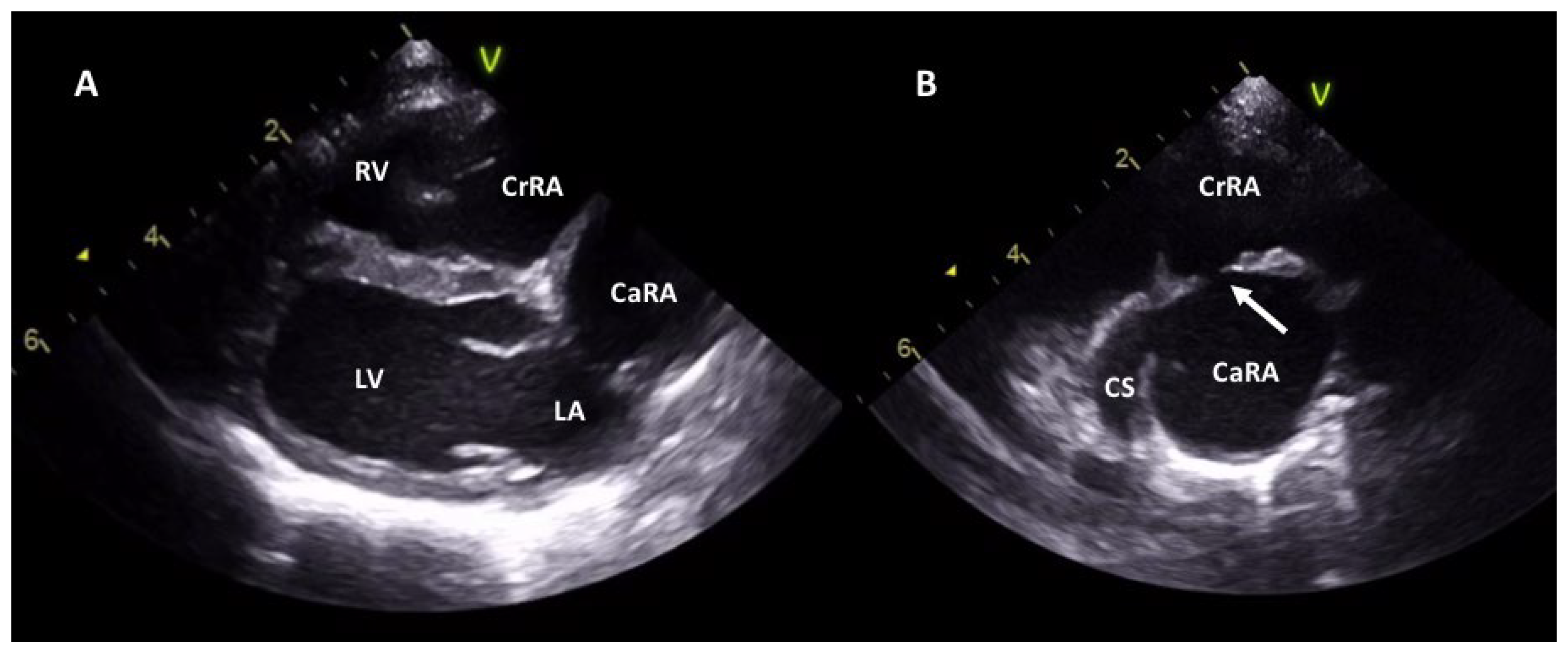
2.1. Case 1
2.2. Case 2
3. Discussion
4. Conclusions
Supplementary Materials
Author Contributions
Funding
Institutional Review Board Statement
Informed Consent Statement
Data Availability Statement
Acknowledgments
Conflicts of Interest
References
- Oliveira, P.; Domenech, O.; Silva, J.; Vannini, S.; Bussadori, R.; Bussadori, C. Retrospective review of congenital heart disease in 976 dogs. J. Vet. Intern. Med. 2011, 25, 477–483. [Google Scholar] [CrossRef] [PubMed]
- Alboliras, E.T.; Edwards, W.D.; Driscoll, D.J.; Seward, J.B. Cor triatriatum dexter: Two-dimensional echocardiographic diagnosis. J. Am. Coll. Cardiol. 1987, 9, 334–337. [Google Scholar] [CrossRef]
- Hoskins, J.D. The Cardiovascular System. In Veterinary Pediatrics, 3rd ed.; W.B. Saunders Company, Elsevier Health Sciences: Amsterdam, The Netherlands, 2001; pp. 103–134. [Google Scholar]
- Hokanson, C.M.; Rhinehart, J.D.; Scansen, B.A. Bidirectional flow across a perforate cor triatriatum dexter in a dog with concurrent pulmonary, tricuspid, and mitral valve dysplasia. J. Vet. Cardio. 2019, 21, 93–97. [Google Scholar] [CrossRef] [PubMed]
- Nadolny, K.E.; Kellihan, H.B.; Scansen, B.A.; Tjostheim, S.S.; Grint, K.A.; Forrest, L.J.; Stepien, R.L. Cor triatriatum dexter in 17 dogs. J. Vet. Cardiol. 2019, 23, 129–141. [Google Scholar] [CrossRef]
- Birettoni, F.; Caivano, D.; Bufalari, A.; Giorgi, M.E.; Miglio, A.; Paradies, P.; Porciello, F. Transthoracic ultrasound guided balloon dilation of cor triatriatum dexter in 2 Rottweiler puppies. J. Vet. Cardiol. 2016, 18, 385–390. [Google Scholar] [CrossRef] [PubMed]
- De Mandron, E.; Chetboul, V.; Bussadori, C. Congenital Cardiopathies. In Clinical Echocardiography of the Dog and Cat; Elsevier Health Sciences: Amsterdam, The Netherlands, 2015; Chapter 19; pp. 313–316. [Google Scholar]
- Morgan, K.R.S.; Stauthammer, C.D.; Gruenstein, D. Transmembrane stent placement for cor triatriatum dexter in 6 dogs. J. Vet. Cardiol. 2022, 41, 79–87. [Google Scholar] [CrossRef]
- Johnson, M.S.; Martin, M.; De Giovanni, J.V.; Boswood, A.; Swift, S. Management of cor triatriatum dexter by balloon dilatation in three dogs. J. Small Anim. Pract. 2004, 45, 16–20. [Google Scholar] [CrossRef] [PubMed]
- Leblanc, N.; Defrancesco, T.C.; Adams, A.K.; Atkins, C.E.; Tou, S.P.; Fudge, J.C.; Keene, B.W. Cutting balloon catheterization for interventional treatment of cor triatriatum dexter: 2 cases. J. Vet. Cardiol. 2012, 14, 525–530. [Google Scholar] [CrossRef] [PubMed]
- Tanaka, R.; Hoshi, K.; Shimizu, M.; Hirao, H.; Akiyama, M.; Kobayashi, M.; Machida, N.; Maruo, K.; Yamane, Y. Surgical correction of cor triatriatum dexter in a dog under extracorporeal circulation. J. Small Anim. Pract. 2003, 44, 370–373. [Google Scholar] [CrossRef] [PubMed]
- Chanoit, G.; Bublot, I.; Viguier, E. Transient tricuspid valve regurgitation following surgical treatment of cor triatriatum dexter in a dog. J. Small Anim. Pract. 2009, 50, 241–245. [Google Scholar] [CrossRef] [PubMed]
- Marchesotti, F.; Rondelli, V.; Pesaresi, M.; Nicoli, S.; Vezzosi, T.; Auriemma, E.; Lanzillo, G.; Cuccio, A.; Khouri, T.; Dejong, A.; et al. Combined interventional procedure and cardiopulmonary bypass surgery in a dog with cor triatriatum dexter, patent foramen ovale, and pulmonary stenosis. J. Vet. Intern. Med. 2019, 33, 2227–2234. [Google Scholar] [CrossRef] [Green Version]
- Caivano, D.; Corda, A.; Birettoni, F.; Pinna Parpaglia, M.L.; Mele, I.; Porciello, F. Management of re-stenosis following balloon catheter dilation of cor triatriatum dexter in a dog. Rev. Vétérinaire Clin. 2019, 54, 37–41. [Google Scholar] [CrossRef]
- Thomas, W.P.; Gaber, C.E.; Jacobs, G.J.; Kaplan, P.M.; Lombard, C.W.; Moise, N.S.; Moses, B.L. Recommendations for standards in transthoracic two-dimensional echocardiography in the dog and cat. Echocardiography Committee of the specialty of cardiology, American College of Veterinary Internal Medicine. J. Vet. Intern. Med. 1993, 7, 247–252. [Google Scholar] [CrossRef]
- Giraud, L.; Fernandes Rodrigues, N.; Lekane, M.; Farnir, F.; Kennedy, C.; Gommeren, K.; Merveille, A.C. Caudal vena cava point-of-care ultrasound in dogs with degenerative mitral valve disease without clinically important right heart disease. J. Vet. Cardiol. 2022, 41, 18–29. [Google Scholar] [CrossRef] [PubMed]
- Noone, K.E. Pleural effusions and diseases of the pleura. Vet. Clin. N. Am. Small Anim. Pract. 1985, 15, 1069–1084. [Google Scholar] [CrossRef]
- Partington, C.; Neves, J.; Navarro-Cubas, X.; Schiborra, F.; Dukes-McEwan, J. Cor triatriatum dexter of unusual morphology in a miniature schnauzer. J. Vet. Cardiol. 2022, 41, 165–171. [Google Scholar] [CrossRef]
- James, T.N. Normal and abnormal consequences of apoptosis in the human heart. Annu. Rev. Physiol. 1998, 60, 309–325. [Google Scholar] [CrossRef] [Green Version]
- Kornreich, B.G.; Moïse, N.S. Right atrioventricular valve malformation in dogs and cats: An electrocardiographic survey with emphasis on splintered QRS complexes. J. Vet. Intern. Med. 1997, 11, 226–230. [Google Scholar] [CrossRef] [PubMed] [Green Version]
- Zoia, A.; Augusto, M.; Drigo, M.; Caldin, M. Evaluation of hemostatic and fibrinolytic markers in dogs with ascites attributable to right-sided congestive heart failure. J. Am. Vet. Assoc. 2012, 241, 1336–1343. [Google Scholar] [CrossRef] [PubMed]
- Agarwal, S.; Joyner, K.A.; Swaim, M.W. Ascites as a possible origin for hyperfibrinolysis in advance liver disease. Am. J. Gastronterol. 2000, 95, 3218–3224. [Google Scholar] [CrossRef] [PubMed]
- Leduc, D.; De Troyer, A. Dysfunction of the canine respiratory muscle pump in ascites. J. Appl. Physiol. 2006, 102, 650–657. [Google Scholar] [CrossRef] [PubMed] [Green Version]
- Mayr, U.; Karsten, E.; Lahmer, T.; Rasch, S.; Thies, P.; Henschel, B.; Fischer, G.; Schmid, R.M.; Huber, W. Impact of large volume paracentesis on respiratory parameters including transpulmonary pressure and on transpulmonary thermodilution derived hemodynamics: A prospective study. PLoS ONE 2018, 13, e0193654. [Google Scholar] [CrossRef] [PubMed] [Green Version]
- Atkins, C.; DeFrancesco, T. Balloon dilation of cor triatriatum dexter in a dog. J. Vet. Intern. Med. 2000, 14, 471–472. [Google Scholar] [CrossRef] [PubMed] [Green Version]
- Adin, D.B.; Thomas, W.P. Balloon dilation of cor triatriatum dexter in a dog. J. Vet. Intern. Med. 1999, 13, 617–619. [Google Scholar] [CrossRef] [PubMed]
- López-Alvarez, J.; Dukes-McEwan, J.; Martin, M.W.; Killick, D.; Fonfara, S.; Fraser McConnell, J. Balloon dilation of an imperforate cor triatriatum dexter in a Golden Retriever with concurrent double-chambered right ventricle and subsequent evaluation by cardiac magnetic resonance imaging. J. Vet. Cardiol. 2011, 13, 211–218. [Google Scholar] [CrossRef] [PubMed]
- Barncord, K.; Stauthammer, C.; Moen, S.L.; Hanson, M.; Gruenstein, D.H. Stent placement for palliation of cor triatriatum dexter in a dog with suspected patent foramen ovale. J. Vet. Cardiol. 2016, 18, 79–87. [Google Scholar] [CrossRef] [PubMed]


| Before the Procedure | After the Procedure | |
|---|---|---|
| Membrane’s orifice (mm) | 3 | 10 |
| Diameter CaVC (mm) | 14.4 | 12.0 |
| CaRA (mm) | 23.1 | 15.5 |
| CrRA (mm) | 24.8 | 24.8 |
| Tricuspid annulus (mm) | 22.1 | 22.1 |
| Transmembrane Vmax (m/s) | 3 | 1.2 |
| Before the Procedure | After the Procedure | |
|---|---|---|
| Membrane’s orifice (mm) | 4 | 10 |
| Diameter CaVC (mm) | 19.5 | 17.0 |
| CaRA (mm) | 27.5 | 19.5 |
| CrRA (mm) | 26.8 | 26.8 |
| Tricuspid annulus (mm) | 27 | 27 |
| Transmembrane Vmax (m/s) | 3.2 | 1.2 |
Publisher’s Note: MDPI stays neutral with regard to jurisdictional claims in published maps and institutional affiliations. |
© 2022 by the authors. Licensee MDPI, Basel, Switzerland. This article is an open access article distributed under the terms and conditions of the Creative Commons Attribution (CC BY) license (https://creativecommons.org/licenses/by/4.0/).
Share and Cite
Patata, V.; Vezzosi, T.; Calogero, G.; Croce, M.; Broch, H.; Marchesotti, F.; Bini, M.; Domenech, O. Percutaneous Balloon Dilation in Two Dogs with Cor Triatriatum Dexter. Vet. Sci. 2022, 9, 419. https://doi.org/10.3390/vetsci9080419
Patata V, Vezzosi T, Calogero G, Croce M, Broch H, Marchesotti F, Bini M, Domenech O. Percutaneous Balloon Dilation in Two Dogs with Cor Triatriatum Dexter. Veterinary Sciences. 2022; 9(8):419. https://doi.org/10.3390/vetsci9080419
Chicago/Turabian StylePatata, Valentina, Tommaso Vezzosi, Giulia Calogero, Marta Croce, Helena Broch, Federica Marchesotti, Martina Bini, and Oriol Domenech. 2022. "Percutaneous Balloon Dilation in Two Dogs with Cor Triatriatum Dexter" Veterinary Sciences 9, no. 8: 419. https://doi.org/10.3390/vetsci9080419