Article

Genetic Analysis of Candida albicans Filamentation by the Iron Chelator BPS Reveals a Role for a Conserved Kinase—WD40 Protein Pair

Department of Molecular Microbiology, B. Rappaport Faculty of Medicine, Technion—I.I.T., Haifa 31096, Israel
*
Author to whom correspondence should be addressed.
J. Fungi 2024, 10(1), 83; https://doi.org/10.3390/jof10010083
Submission received: 17 December 2023 / Revised: 16 January 2024 / Accepted: 18 January 2024 / Published: 22 January 2024
(This article belongs to the Section Fungal Genomics, Genetics and Molecular Biology)

Abstract

:
Candida albicans is a major human pathogenic fungus that is distinguished by its capability to switch from a yeast to a hyphal morphology under different conditions. Here, we analyze the cellular effects of high concentrations of the iron chelator bathophenanthroline disulfonate (BPS). BPS inhibits cellular growth by withholding iron, but when iron chelation is overcome by the addition of hemoglobin as an iron source, the cells resume growth as hyphae. The BPS hyphal induction pathway was characterized by identifying the hyphal-specific transcription factors that it requires and by a forward genetic screen for mutants that fail to form hyphae in BPS using a transposon library generated in a haploid strain. Among the mutants identified are the DYRK1-like kinase Yak1 and Orf19.384, a homolog of the DYRK1-associated protein WDR68/DCAF7. Orf19.384 nuclear localization depends on Yak1, similar to their mammalian counterparts. We identified the hyphal suppressor transcription factor Sfl1 as a candidate target of Yak1-Orf19.384 and show that Sfl1 modification is similarly affected in the yak1 and orf19.384 mutant strains. These results suggest that DYRK1/Yak1 and WDR68/Orf19.384 represent a conserved protein pair that regulates cell differentiation from fungi to animals.

1. Introduction

Candida albicans is a commensal organism of humans as well as a major opportunistic pathogen, capable of causing superficial infections among susceptible populations and deep-seated infections in immunosuppressed patients [1]. One of the best-studied traits of C. albicans is its ability to switch between multiple morphologies, including yeast, pseudohyphae, and hyphae [2]. This ability contributes to C. albicans’ ability to cause disease in the susceptible host [3,4,5]. Many stimuli are known to induce the transition from yeast to hyphal growth, including external conditions such as an elevated temperature, the presence of serum, certain growth media formulations, N-acetylglucosamine, neutral to slightly alkaline pH, and interference with internal homeostatic regulations, such as cell cycle progression [6,7,8,9]. Here, we identified a new such stimulus, exposure to the iron chelator bathophenanthroline disulfonate (BPS) [10], and identified the conditions under which BPS induces filamentation.
C. albicans is a diploid organism lacking a complete sexual cycle. As a consequence, using forward genetics to study the biology of this organism has been precluded by the fact that most random mutations usually yield recessive loss-of-function mutants. Genetic analysis in C. albicans has therefore traditionally relied on reverse genetics: genes identified by bioinformatics or, occasionally, by screening of C. albicans plasmid libraries in S. cerevisiae are deleted by homologous recombination, one allele sequentially after the other [11]. Large-scale targeted gene deletion libraries have been constructed over the years and screened for various phenotypes, e.g., [12,13], but none of these libraries cover the entire C. albicans genome. The recent identification in a haploid line of C. albicans has, however, opened the prospect of forward genetic analysis in this organism [14]. Transposon mutagenesis systems have been established that enable rapid construction, screening, and analysis of mutant collections [15,16,17].
Here, we used a combination of mutant library screening and transposon mutagenesis to isolate mutants that are not responsive to BPS-induced filamentation. Among the genes identified are those encoding a set of known filamentation-related transcription factors, as well as the kinase Yak1 and the WD40 repeat protein Orf19.384.

2. Materials and Methods

2.1. Media and Chemicals

Cells were grown in YPD medium (1% yeast extract, 2% bacto-peptone, 2% glucose, and tryptophan at 150 mg/L) or in Synthetic Complete (SC) medium lacking specific amino acids, as indicated. SC medium contains, per liter, Yeast Nitrogen Base (USBiological, Salem, MA, USA) 1.7 g, (NH4)2SO4 5 g, the 20 amino acids, adenine and uridine, 0.1 g each, except leucine, 0.2 g, glucose, 20 g, and 0.2 mM inositol. Media were supplemented with the ion chelators ferrozine or bathophenanthroline sulfonate (BPS) or with the siderophore ferrichrome at the indicated concentrations or bovine hemoglobin (all from Sigma Aldrich, St. Louis, MO, USA) from a 0.5 mM stock in Dulbecco’s phosphate-buffered saline (Biological Industries, Beit Haemek, Israel).

2.2. Plasmids and Strains

Plasmids KB2734 and KB2735 contain the YAK1 5′ region (−400 to +80, primers 9 + 10 in Table S1, SacI-SpeI) and the 3′ region (2370 to 3047, primers 11 + 12, PstI-XhoI) at the two ends of the hisG-URA3-hisG “blaster” of plasmids KB985 and KB986, respectively [18,19]. KB2736 and KB2737 similarly contain the ORF19.384 5′ region (−273 to +80, primers 15 + 16, SacI-SpeI) and 3′ region (1607 to 2096, primers 17 + 18, PstI-XhoI) in plasmids KB985 and KB986, respectively. Plasmid KB2726 contains the YAK1 open reading frame (610 to 2427) fused to the CaGFPgamma ORF [20] (primers 7 + 8), and KB2752 contains the ORF19.384 ORF (302 to 1674) fused likewise (primers 13 + 14) [19].
The strains are listed in Table 1. KC1337 was generated by deleting the 1st allele of YAK1 with plasmid KB2735, followed by treatment with 5-fluoroorotic acid (5FOA) to select for cells that have ejected the URA3 marker. KC1339 was generated by transforming KC1337 with KB2734, followed by 5FOA treatment. KC1341 was generated by deleting the 1st allele of ORF19.384 with plasmid KB2736, followed by treatment with 5FOA. KC1381 was generated by transforming KC1341 with KB2737, followed by 5FOA treatment. KC1543, KC1544, KC1545 were generated by introducing the 13xMyc tag after the SFL1 open reading frame in KC274, KC1339, and KC1381, respectively, using a PCR product amplified from plasmid KB1541 [18] with primers 23, 24 (Table S1). KC1669 and KC1672 are KC274 and KC1381 transformed with KB2726 digested with BSP119I; KC1670 and KC1671 are KC274 and KC1339 transformed with KB2752 digested with HpaI.

2.3. Growth Assays

Overnight starter cultures grown in YPD were diluted into a series of two-fold dilutions of hemoglobin, or BPS, in the indicated media. Cells were inoculated in flat-bottomed 96-well plates at OD600 = 0.00001, 150 µL per well. Plates were incubated at 30 °C on an orbital shaker at 60 rpm, and growth was measured by optical density (OD600) after 1, 2, and 3 days with an ELISA reader. Cells were resuspended with a multi-pipettor before each reading. Each culture was performed in triplicate.

2.4. Enrichment and Screening for Filamentation-Defective Mutants

A starting mutant pool a haploid C. albicans strain mutagenized by random transposon insertion [16] was enriched for non-filamenting mutants as described [19]. Briefly, cells were grown for 24 h in 5 mL YPD + 2 mM BPS and 0.25 µM hemoglobin at 30° while shaking, and then the cultures were left to sediment in a test tube on the assumption that non-filamenting mutants would sediment slower than the hyphal wild-type cells. After 10 min, the top 1 mL of the tube was removed and diluted in fresh 5 mL of medium and left to sediment again, after which the top 1 mL was diluted in fresh medium and grown for 24 h as before. This procedure was repeated 10 times. Finally, the enriched pool was plated on YPD plates and 2 mM BPS and 1 µM hemoglobin, incubated or 24 h, and the colonies (200/plate) were visually scanned with a binocular microscope for reduced hyphal formation.

2.5. Identification of Transposon Insertion Sites

Single colonies were grown overnight in YPD medium. DNA was extracted as described in [27], and subjected to the FPNI DNA amplification protocol [28] using Ds-specific primers (Table S1, primers 1–6), as described in [17]. PCR reactions were tested for the presence of bands on an agarose gel, and the positive reaction (>90%) was cleaned using the GenElute kit (Sigma-Aldrich) and sequenced by Sanger sequencing. The sequence was used to determine the position and orientation of the Ds insertion in the C. albicans genome.

2.6. Microscopy

A Zeiss AxioImager M1 microscope (Carl Zeiss AG, Oberkochen, Germany) equipped with a Colibri 5 laser light source for epifluorescence was used throughout. For regular light microscopy, a 10× A-Plan objective or a 40× Plan-Neofluar objective with DIC optics were used. For visualization of fluorescently labeled cells, cultures were incubated for a final 5 min with 10 µM Hoechst 33342, then spun down and resuspended in a small volume of phosphate-buffered saline. Cells were then immediately visualized with a 100× plan-apochromat objective using a GFP filter set or a DAPI filter set for Hoechst 33342.

2.7. Protein Analysis

Proteins for Western blotting were extracted using NaOH/β-mercaptoethanol (βME). Culture aliquots were spun down and resuspended in 1 mL of 250 mM NaOH and 1% βME and incubated for 10 min on ice. Then, trichloroacetic acid was added to 5% and the cells were incubated for another 10 min, at least, on ice. The precipitate was pelleted, in a refrigerated Eppendorf centrifuge; the pellet was washed with cold 100% acetone, dried, and then resuspended in gel loading buffer with 4% βME, 40 µL/OD600 unit. The samples were run on a 6% polyacrylamide gel, transferred to a PVDF membrane, and reacted in TBST (Tris-buffered saline + 0.1% Tween 20) with the anti-Myc 9E10 monoclonal antibody (Invitrogen, Carlsbad, CA, USA, 1:500) and a secondary horseradish peroxidase-conjugated anti-mouse antibody (Sigma-Aldrich, 1:10,000). The membranes were reacted with an Amersham ECL Plus kit (GE Healthcare, Chicago, IL, USA) and visualized with a FUSION FX7 Edge imaging system (Witec AG, Sursee, Switzerland).

3. Results

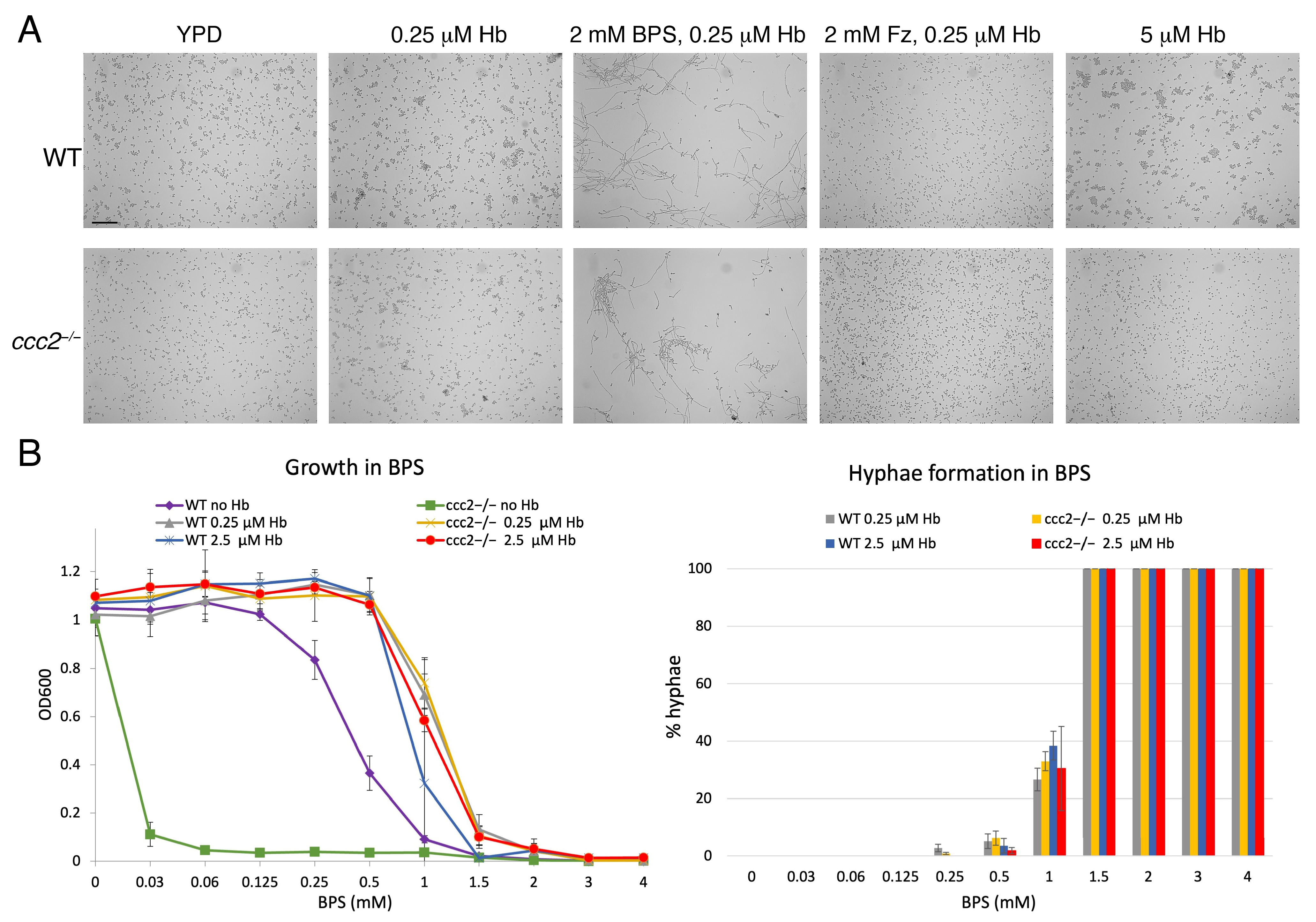
3.1. BPS Induces Filamentous Growth

When analyzing the pathway of heme-iron acquisition in C. albicans, we make extensive use of an experimental system where 1–2 mM BPS is used to chelate iron in the medium, and growth is recovered by addition of hemin or hemoglobin to the medium [29,30]. We noticed that under these conditions, cells that recovered growth in the presence of hemoglobin (or hemin) were largely hyphal after two days (Figure 1A). There have been some reports of hemin causing hyphal morphogenesis, particularly at high concentrations [31,32], but in these reports, BPS was already suggested to be an important contributing factor [32]. Hemoglobin has also been shown to cause hyphal morphogenesis [33], however, at much higher concentrations (1 mg/mL = 15 µM) than our own typical working concentrations (0.25–1 µM). To clarify the role of hemoglobin vs. BPS while maintaining conditions of utilization of heme as an iron source, we used an alternative iron chelator, ferrozine [34], which does not completely inhibit growth of wild-type strains but does completely inhibit growth of a ccc2−/− mutant, defective in high-affinity iron uptake [35]. Comparison of the morphologies of the ccc2−/− strain grown in the presence of hemoglobin and either BPS or ferrozine as iron chelators indicated that only BPS efficiently induced filament formation (Figure 1A), indicating that utilization of heme as an iron source is not the trigger for hyphal morphogenesis. Furthermore, under these conditions, hemoglobin alone, even at much higher concentrations, did not induce hyphal morphogenesis (Figure 1A). Lastly, to refute the remaining possibility that only the combination of BPS with hemoglobin can induce hyphal growth, we tested the effect of rescuing BPS inhibition with the siderophore ferrichrome pre-loaded with iron [36]. While growth rescue was only partial, the cells exhibited a mixture of elongated hyphae and short, germ tube-like hyphae, supporting the notion that BPS is itself an inducer of hyphal morphogenesis (Figure S1).
To further characterize the effect of BPS on filamentation, we tested different BPS concentrations and quantitated growth and filamentation in the absence and presence of hemoglobin at two different concentrations. As shown in Figure 1B, at low BPS concentrations, no filamentation was detected, but at concentrations that were inhibitory in the absence of heme-iron, namely above 1 mM, extensive filamentation could be measured after 2 days of incubation. In the iron uptake mutant ccc2−/−, while, as expected, a higher sensitivity to BPS was observed in the absence of hemoglobin, in its presence, the same extent of filamentation was detected at the same BPS concentrations (Figure 1B). Furthermore, no significant difference in filamentation was measured at 0.25 µM vs. 2.5 µM hemoglobin. Together, these results further confirm that, under these conditions, BPS is directly inducing filamentation.

3.2. Identification of Factors Required for BPS-Induced Hyphal Morphogenesis

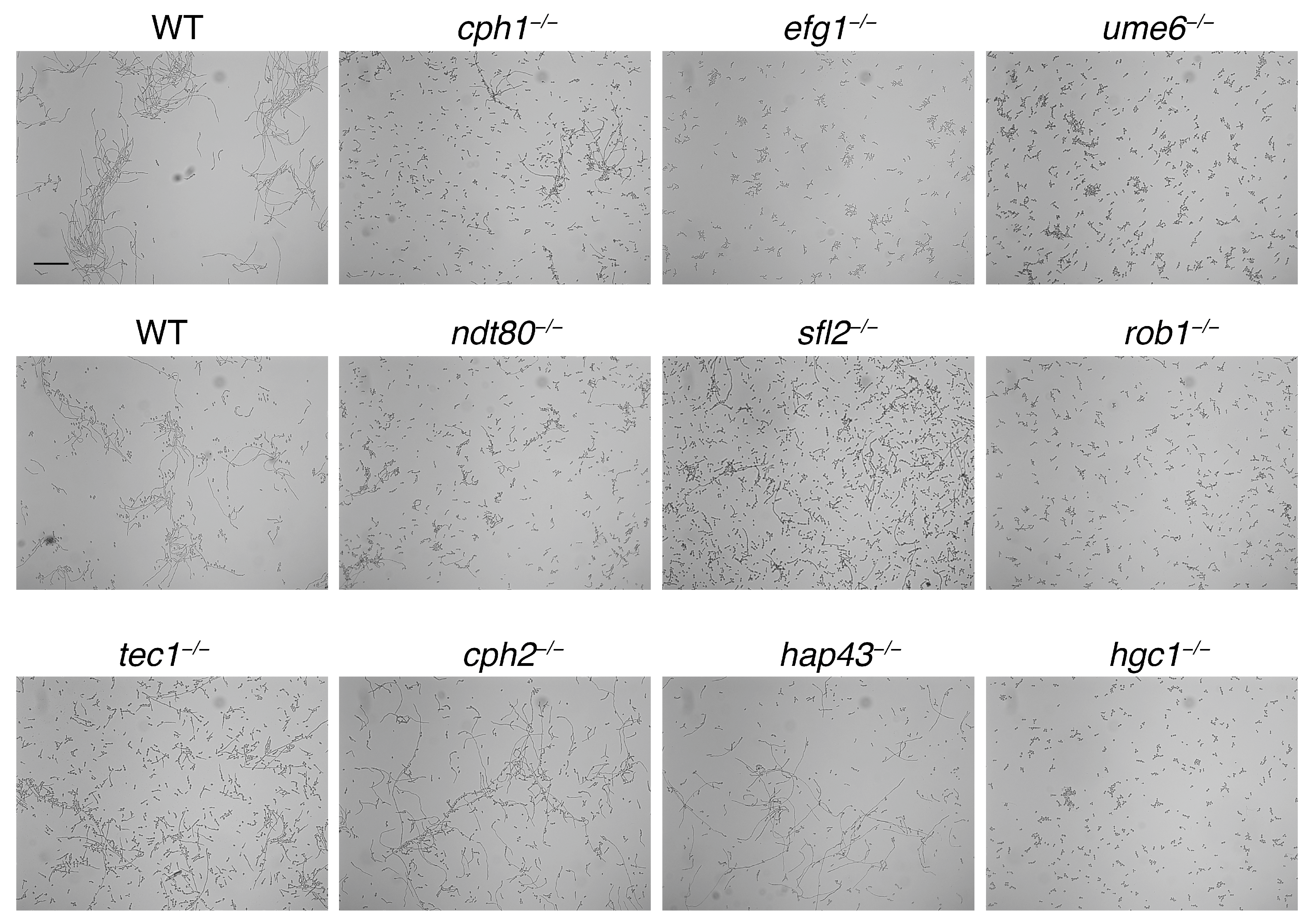
3.2.1. Screening of Transcription Factor Mutants

Different transcription factors and signal transduction pathways have been identified that are involved in hyphal morphogenesis in C. albicans, depending on the induction signals [7,8,37]. In order to identify mutants required for BPS-induced filamentation, we scanned the Homann library of transcription factor mutants [12], as well as a selection of mutants from our lab stock, including hgc1−/−, efg1−/−, cph1−/−, and ume6−/−, for defects in hyphal morphogenesis [19]. In a first pass, the clones were grown in 96-well plates in YPD + 2 mM BPS, 0.25 µM hemoglobin, for 48 h at 30°. Selected clones were re-checked in individual tubes (Figure 2). Mutants that showed a strong defect in hyphal formation include the transcription factors efg1−/−, ume6−/−, rob1−/− and the hyphal-specific cyclin hgc1−/−, whereas sfl2−/−, tec1−/−, ndt80−/−, and cph1−/− exhibited a partial defect. cph2−/− and hap43−/− are examples of transcription factor mutants that were unaffected in BPS-induced hyphal morphogenesis.

3.2.2. Unbiased Selection for Non-Filamenting Mutants

In order to identify additional factors in the BPS-induced filamentation pathway, we performed an unbiased, forward genetic screen using a transposon-mutagenized pool of haploid cells [16]. The pool was first enriched for non-filamenting mutants by repeated removal of slower-sedimenting cells over several subcultures, as described in Methods, and the enriched pool was plated on BPS and hemoglobin plates and visually scanned for clones exhibiting reduced hyphal formation [19]. Less-filamenting clones were then genotyped for the locus of transposon insertion. A total of 48 clones were characterized at the genotype level (Table S3). A total of 15 insertions were in unique loci, mostly in intergenic regions, whereas the other 35 were represented between 2 and 12 hits within the coding region or the promoter region of 7 genes: FLO8 (12), EFG1 (5), orf19.384 (5), YAK1 (4), UME6 (3), MSH6 (2), and EHT1 (2). Efg1 and Ume6 are transcription factors that were also identified in our directed screen (Figure 2). Flo8 is another known regulator of hyphal growth [38], which could not have been identified in our previous screen because it is not represented in the Homann library. Msh6 is a DNA mismatch repair gene homolog, and Eht1 is a putative fatty acid biosynthesis enzyme, neither of which had previously been linked to hyphal morphogenesis.

3.3. Yak1 and Orf19.384

Of the last two genes identified by multiple hits, YAK1 and orf19.384, the former encodes a kinase belonging to the DYRK family of dual-specificity kinases, conserved from fungi to mammals [39,40], which had been previously implicated in the initiation and maintenance of hyphal growth [41]. Orf19.384, in contrast, has not been characterized before. The analysis of the Orf19.384 sequence in the Interpro database [42] indicated that it is an ortholog of the conserved WDR68/DCAF7 (WD-40 Repeat 68/DDB1- and CUL4-Associated Factor 7) proteins, associated with developmental pathways in plants and animals [43,44,45]. Strikingly, WDR68/DCAF7 was found to interact with the Yak1 homologs DYRK1A and DYRK1B in animal cells [46,47,48].

3.3.1. Confirmation of the Phenotypes in the Standard Diploid Background

To confirm the role of these two genes in BPS-induced filamentation in C. albicans, we deleted them in the standard diploid strain and tested the phenotype of the heterozygous and homozygous mutants grown in BPS and hemoglobin. As shown in Figure 3A, the YAK1+/− heterozygote already exhibited a partial defect in filamentation, whereas the yak1−/− homozygote was completely defective in filamentation. For ORF19.384, in contrast, the heterozygote was as filamentous as the wild-type, but the orf19.384−/− homozygote was profoundly defective in filamentation. We confirmed that the reduced filamentation in the mutants is not due to reduced growth, e.g., to an inability to utilize hemoglobin (Figure S2).
Since Goyard et al. have shown that the yak1−/− mutant is defective in filamentation in Lee’s medium [41], we also tested the heterozygous and homozygous YAK1 and ORF19.384 mutant strains in this medium and monitored hyphal formation after 5 h and 24 h (Figure 3B). We confirmed that the yak1−/− mutant is completely defective in hyphal formation in Lee’s medium, and we found that the heterozygote is already partially defective, similar to the phenotype in BPS-induced filamentation. For ORF19.384, in the heterozygote, hyphae already appeared shorter and less prominent, whereas in the homozygous orf19.384−/− mutant, very few hyphae were visible.

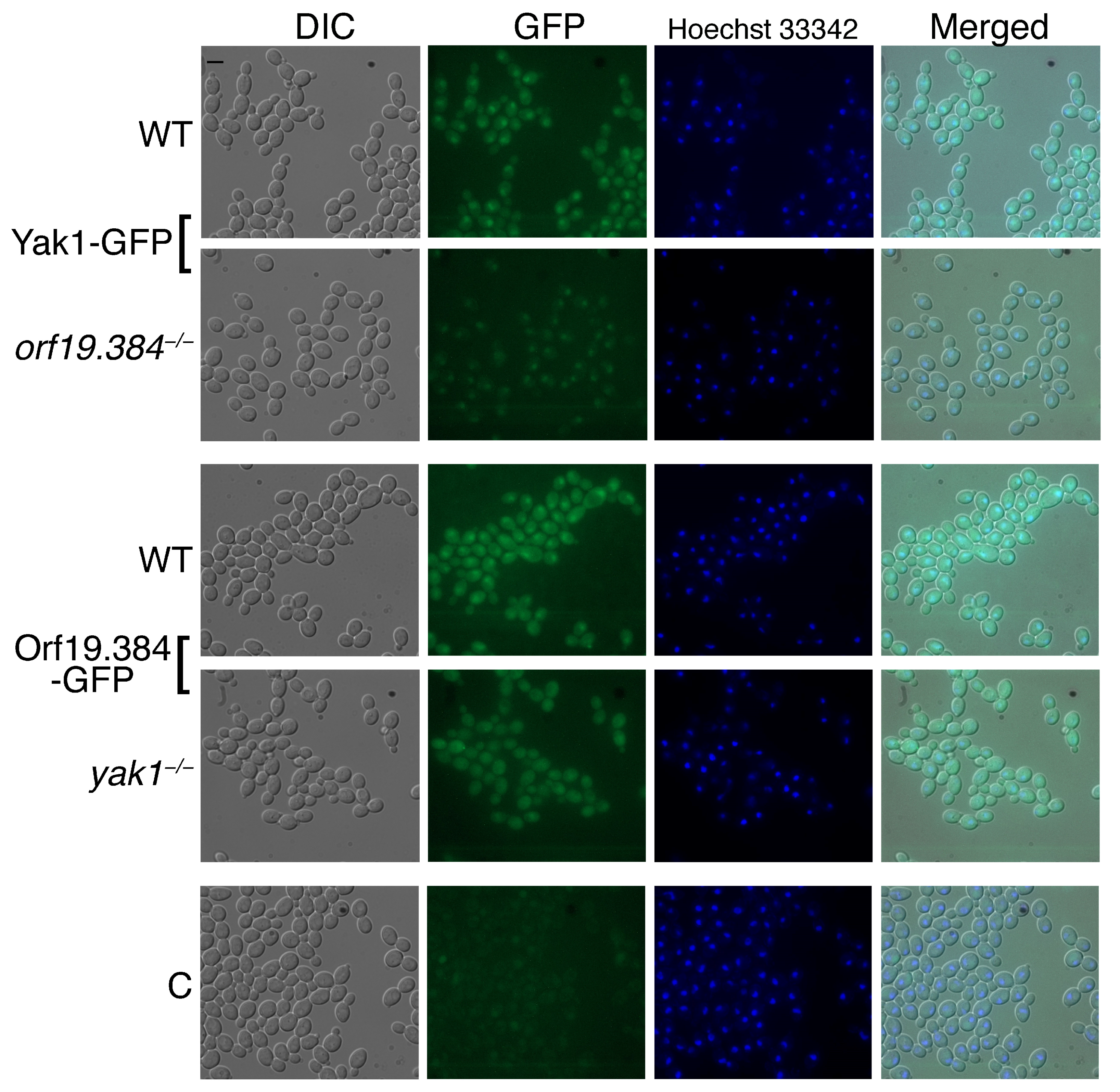
3.3.2. Subcellular Localization

To further analyze the interaction between Yak1 and Orf19.384, we fused the YAK1 and ORF19.384 open reading frames to GFP and expressed the fusion proteins under their native promoter in wild-type cells or in cells lacking the other partner. As shown in Figure 4, wild-type cells show cytoplasmic and nuclear localization of both Yak1-GFP and Orf19.384-GFP, with a higher concentration in the nucleus of both proteins. However, Yak1-GFP expression in the orf19.384−/− mutant showed lower levels overall and remained visible only in the nucleus. Conversely, Orf19.384-GFP expressed in yak1−/− cells was still visible in the cytoplasm but lost its specific nuclear localization. The growth of the cells in synthetic medium yielded a very similar picture (Figure S3). Thus, Yak1 and Orf19.384 affect each other’s levels or localization.

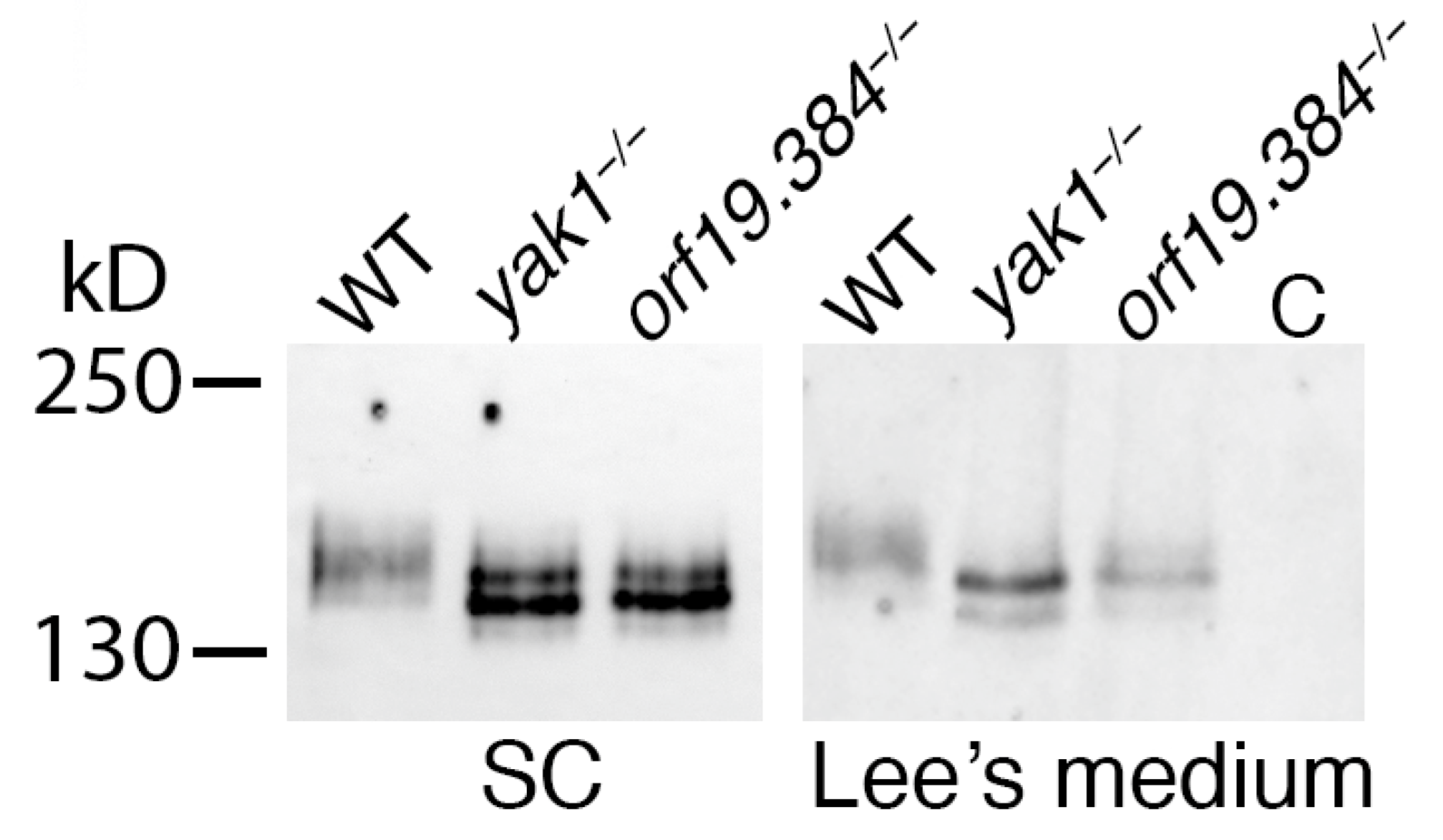
3.3.3. Sfl1 Is a Candidate Substrate of Yak1/Orf19.384

Based on the proposed DYRK1/Yak1 substrate consensus RPX(S/T)P [49], we scanned the C. albicans proteome for potential Yak1 targets. Among some 60 proteins having one potential target site each (Table S4) is Sfl1, a transcription factor that was identified as a suppressor of hyphal morphogenesis. In particular, Sfl1 was proposed to be antagonistic to Flo8 [50] and to Sfl2 [51], and to co-bind its targets with Efg1 and/or Ndt80 [51], all factors that were identified in our screens. We therefore tested whether Sfl1 could be a target of Yak1 together with Orf19.384. To this end, we tagged Sfl1 with a 13xMyc tag at its C-terminus in wild-type, yak1−/− and orf19.384−/− cells. As shown in Figure 5, in two different media, Sfl1 migration was slower in the wild-type cells than in the yak1−/− or orf19.384−/− cells, while the two mutant strains exhibited a similar band pattern. Importantly, the proteins were analyzed in yeast growth conditions (SC medium) as well as in hyphal-inducing conditions (Lee’s medium). The observation that even in SC medium, where the morphologies of the wild-type and mutant strains are indistinguishable, the modification pattern of Sfl1 was very different in the wild-type vs. the mutants indicates that it does not represent an indirect effect of cell morphology on Sfl1 modification. This observation therefore supports the possibility that Yak1 and Orf19.384 function together to modify this substrate.

4. Discussion

4.1. BPS and Filamentation

We have shown here that the iron chelator BPS induces, at high concentrations, hyphal morphogenesis in C. albicans. Since high BPS concentrations preclude growth by withholding essential iron from the cells, this filamentation is detectable only in the presence of hemoglobin (or hemin—our unpublished results), which restores growth of C. albicans cells by serving as an alternative iron source, thereby enabling the cells to manifest the BPS-induced hyphal morphology [35,52].
Notably, the addition of BPS had been previously shown by Hameed et al. to induce hyphal morphogenesis [53]. These authors interpreted the results as showing that iron deprivation, rather than BPS per se, was the proximal inducer of filamentation, based on the filamentous phenotype of the ftr1 and ccc2 high-affinity iron uptake mutants, even in the absence of BPS. A more recent report also found that BPS can induce hyphae formation on plates, dependent on a new iron regulator, the transcription factor Irf1 [54]. One factor that distinguishes these experimental systems from ours is that the BPS concentration used, 150 µM, was much lower and does not, in our hands, induce filamentation (Figure 1B). This discrepancy is probably due, in one case, to temperature. While we grew the cells at 30 °C, Hameed et al. used 37 °C, a condition known by itself to induce, or strongly contribute to, hyphal morphogenesis [6]. In the second case, filamentation was only detected after prolonged incubation on plates [54]. If iron withholding by BPS were inducing filamentation in our system, then the higher hemoglobin concentrations would have reduced filamentation. Thus, it is likely that Hameed et al. describe hyphal morphogenesis induced by the combination of high temperature and iron limitation, whereas we found a BPS-specific hyphal induction mechanism. Further indication that the two protocols induce filamentation by different pathways comes from the observation that while the Homann collection hap43−/− mutant is defective in hyphal induction in 150 µM BPS [12], this mutant is fully filamentous in 2 mM BPS at 30 °C (Figure 2).
Since hemoglobin had been shown to induce hyphal morphogenesis on its own, albeit at a higher concentration of 15 µM [33], it was important to show that in our experiments, the hemoglobin was not responsible for this phenotype. We did this in several ways: by showing that an alternate iron chelator, ferrozine, does not induce filamentation in the presence of the same hemoglobin concentrations, even in cells that are highly sensitive to iron limitation; and by showing that, by varying the BPS and hemoglobin concentrations, the extent of filamentation was strongly dependent upon BPS concentration but not hemoglobin concentration. Thus, neither the presence of hemoglobin, nor iron starvation per se or the utilization of hemoglobin as iron source, can explain the BPS-induced filamentation phenotype. What, then, is inducing this phenotype? We did notice that at high BPS concentrations, i.e., above 1 mM, growth becomes progressively inhibited until it is completely inhibited at 4 mM, even in the presence of hemoglobin (Figure 1). This suggests that at a high enough concentration, BPS exerts effects on the cell that go beyond the chelation of iron in the environment. It is possible that, at a high enough concentration, BPS can penetrate the cell and interfere with cellular pathways via chelation of intracellular iron or by other means, thereby inducing the switch to hyphal morphogenesis.

4.2. Mutants Defective in BPS-Dependent Filamentation

To try to understand the mechanism of action of BPS on cellular morphogenesis, we carried out a screen of mutants of known filamentation factors as well as an unbiased, forward genetics screen, looking for mutants exhibiting reduced filamentation in the presence of BPS. Taken together, the two screens identified a set of six transcription factors, namely Efg1, Ndt80, Rob1, Flo8, Sfl2, and Cph1, that are required for BPS-induced filamentation. Efg1 and Ndt80 are central transcription factors of both hyphal morphogenesis [55,56] and biofilm formation [57], and are usually found bound together to the same promoters [58]. Cph1 [23], Flo8 [38], and Sfl2 [59,60] were identified as positive regulators of hyphal morphogenesis as well: Flo8 interacts directly with Efg1 [38] and Sfl2 with Efg1 and Ndt80, and their binding sites usually co-occur on target promoters [51]. Rob1 was associated mainly with biofilm formation [57], and strikingly, it, together with Efg1, Ndt80, Flo8, and Brg1, were proposed to be among the core genes in the transcription network that regulates biofilm formation [57,61]. Thus, our screens have identified a coherent subset of interacting transcription factors involved in hyphal morphogenesis and in the related pathway of biofilm formation.

4.3. Yak1 and Orf19.384

Two additional genes that came up repeatedly in the transposon-mutagenized pool were YAK1 and ORF19.384. YAK1 was previously shown to affect hyphal morphogenesis and hyphal-specific transcription in C. albicans [41]. In S. cerevisiae, Yak1 is regulated by the protein kinase A (PKA) pathway, and it affects i.a. adhesive growth by activating expression of the flocculin gene FLO11, itself a target of S. cerevisiae Flo8 [62]. C. albicans Yak1 was recently shown to also function within the PKA pathway and to require the transcription factors Efg1 and Flo8 for the induction of filamentation [63]. In addition, C. albicans Yak1 can be inhibited by a lactobacillus metabolite, 1-ABC, that inhibits hyphal morphogenesis, and analysis of genomic suppressors of 1-ABC-mediated suppression of filamentation pointed to Rob1 as a possible Yak1 target [64].
Yak1 belongs to the conserved dual-specificity tyrosine-phosphorylated and regulated kinase (DYRK) family [39,40]. DYRK kinases are involved in the regulation of cellular growth and differentiation in invertebrates and vertebrates [48,65,66]. In several animal models, DYRK1-type kinases were found to interact with another conserved family of proteins, the WD40 repeat protein family WDR68/DCAF7 [46,47,48]. One proposed role for these proteins is to mediate the interaction of the DYRK kinases with their substrates [67]. The fact that Orf19.384 is the single apparent ortholog of WDR68 in C. albicans suggests that it may function together with Yak1. Furthermore, the single apparent Orf19.384/WDR68 ortholog in S. cerevisiae, Ypl247c, was shown in two different proteomics screens to physically interact with the S. cerevisiae Yak1 ortholog [68,69].
On the assumption that Yak1 and Orf19.384/Wdr68 interact as well, we first tested whether they affect each other’s subcellular localization. We found that both proteins are normally localized in the cytosol and concentrated in the nucleus, but in the absence of Yak1, Orf19.384 lost its nuclear localization. Conversely, in the absence of Orf19.384, Yak1 was only detectable in the nucleus. This, however, could be due to overall lower amounts of the Yak1 protein in cells lacking Orf19.384, rather than to relocalization from the cytosol to the nucleus (Figure 4). In any case, we find that Yak1 and Orf19.384 affect each other’s localization and/or levels. Interestingly, similar to the dependence of Orf19.384/Wdr68 on Yak1 for its nuclear accumulation, human WDR68 was found to depend on the Yak1 homolog DYRK1A for its nuclear accumulation as well [46].
To further support the conjecture that Yak1 and Orf19.384/Wdr68 function together in C. albicans, we attempted to identify a potential Yak1 target. Among some 60 proteins that contain the DYRK consensus RPX(S/T)P, Sfl1 stood out, based on its known relationship with many of the other genes identified in our screen. Sfl1 is a repressor of hyphal morphogenesis [50,70], which was suggested to antagonize Flo8 [50] as well as Sfl2 [51]. It was suggested to be centrally involved in the formation of biofilm-like microcolonies on an oral mucosa model, together with Sfl2, Rob1, and Ndt80 [71], and to suppress hyphal formation in an acidic medium [72]. In our hands, Sfl1 was indeed found to be differentially modified in wild-type vs. yak1−/− or orf19.384−/− cells, even in normal yeast growth medium. This supports the notion that Yak1 and Orf19.384 function together in C. albicans and links them to the transcription network defined by the other non-filamenting mutants identified here.
Taken together, our data suggest a deep conservation of the function of the DYRK-WDR68/DCAF7 and Yak1-Orf19.384 complexes across the animal and fungal kingdoms. Based on the sequence and functional homology of ORF19.384 to its mammalian homologs, we suggest renaming it WDR68.

5. Conclusions

We have identified a new protocol for hyphal induction in C. albicans: exposure to high BPS concentrations in the presence of hemoglobin as an alternative iron source. A genetic analysis of this hyphal induction pathway revealed a subset of hyphal- and biofilm-specific transcription factors that were known to interact physically and/or functionally, as well as a conserved protein kinase complex, Yak1-Orf19.386/Wdr68, whose homologs are involved in cellular differentiation in animals. Considering that the dimorphic switch from yeast to hyphal morphology represents a form of cellular differentiation, we conclude that the conservation of structure as well as function of this complex extends to the fungal kingdom. We propose that fungi represent a useful model system for the study of the function, mechanism, and regulation of DYRK kinases.

Supplementary Materials

The following supporting information can be downloaded at https://www.mdpi.com/article/10.3390/jof10010083/s1. Figure S1: Partial rescue of BPS growth inhibition by iron-ferrichrome induces filamentous growth. Figure S2: The yak1 and orf19.384 mutants are not defective in growth on BPS and hemoglobin. Figure S3: Effect of deletion of YAK1 or ORF19.384 on expression and localization of their partner protein.; Table S1: Primers; Table S2: Quantitation of Figure 2; Table S3: Tn insertion sites; Table S4: Candidate Yak1 target sites.

Author Contributions

Conceptualization, D.K.; Formal analysis, M.P. and D.K.; Funding acquisition, D.K.; Investigation, M.P. and D.K.; Supervision, D.K.; Visualization, M.P. and D.K.; Writing—original draft, D.K.; Writing—review and editing, D.K. All authors have read and agreed to the published version of the manuscript.

Funding

This research was funded by the Israel Science Foundation, grant number 3039/19.

Institutional Review Board Statement

Not applicable.

Informed Consent Statement

Not applicable.

Data Availability Statement

All the data appear in the manuscript figures and attached supplementary data.

Acknowledgments

We thank Judy Berman (Tel-Aviv University) for the haploid strains and transposon-mutagenized mutant pool, and Yue Wang (A*STAR, Singapore) and Haoping Liu (UC Irvine) for strains.

Conflicts of Interest

The authors declare no conflicts of interest. The funders had no role in the design of the study; in the collection, analyses, or interpretation of data; in the writing of the manuscript; or in the decision to publish the results.

References

  1. Calderone, R.A.; Clancy, C.J. Candida and Candidiasis, 2nd ed.; Calderone, R.A., Clancy, C.J., Eds.; ASM press: Washington, DC, USA, 2012; ISBN 9781555815394. [Google Scholar]
  2. Sudbery, P.; Gow, N.; Berman, J. The distinct morphogenic states of Candida albicans. Trends Microbiol. 2004, 12, 317–324. [Google Scholar] [CrossRef] [PubMed]
  3. O’Meara, T.R.; Veri, A.O.; Ketela, T.; Jiang, B.; Roemer, T.; Cowen, L.E. Global analysis of fungal morphology exposes mechanisms of host cell escape. Nat. Commun. 2015, 6, 6741. [Google Scholar] [CrossRef] [PubMed]
  4. Jacobsen, I.D.; Wilson, D.; Wachtler, B.; Brunke, S.; Naglik, J.R.; Hube, B. Candida albicans dimorphism as a therapeutic target. Expert Rev. Anti-Infect. Ther. 2012, 10, 85–93. [Google Scholar] [CrossRef] [PubMed]
  5. Vila, T.; Romo, J.A.; Pierce, C.G.; McHardy, S.F.; Saville, S.P.; Lopez-Ribot, J.L. Targeting Candida albicans filamentation for antifungal drug development. Virulence 2017, 8, 150–158. [Google Scholar] [CrossRef] [PubMed]
  6. Sudbery, P.E. Growth of Candida albicans hyphae. Nat. Rev. Microbiol. 2011, 9, 737–748. [Google Scholar] [CrossRef] [PubMed]
  7. Kornitzer, D. Regulation of candida albicans hyphal morphogenesis by endogenous signals. J. Fungi 2019, 5, 21. [Google Scholar] [CrossRef] [PubMed]
  8. Basso, V.; D’Enfert, C.; Znaidi, S.; Bachellier-Bassi, S. From genes to networks: The regulatory circuitry controlling candida albicans morphogenesis. In Current Topics in Microbiology and Immunology; Springer: Berlin/Heidelberg, Germany, 2019; Volume 422, pp. 61–99. [Google Scholar]
  9. Arkowitz, R.A.; Bassilana, M. Recent advances in understanding Candida albicans hyphal growth. F1000Research 2019, 8, 700. [Google Scholar] [CrossRef]
  10. Trinder, P. The improved determination of iron in serum. J. Clin. Pathol. 1956, 9, 170–172. [Google Scholar] [CrossRef]
  11. Berman, J.; Sudbery, P.E. Candida Albicans: A molecular revolution built on lessons from budding yeast. Nat. Rev. Genet. 2002, 3, 918–930. [Google Scholar] [CrossRef]
  12. Homann, O.R.; Dea, J.; Noble, S.M.; Johnson, A.D. A phenotypic profile of the Candida albicans regulatory network. PLoS Genet. 2009, 5, e1000783. [Google Scholar] [CrossRef]
  13. Roemer, T.; Jiang, B.; Davison, J.; Ketela, T.; Veillette, K.; Breton, A.; Tandia, F.; Linteau, A.; Sillaots, S.; Marta, C.; et al. Large-scale essential gene identification in Candida albicans and applications to antifungal drug discovery. Mol. Microbiol. 2003, 50, 167–181. [Google Scholar] [CrossRef] [PubMed]
  14. Hickman, M.A.; Zeng, G.; Forche, A.; Hirakawa, M.P.; Abbey, D.; Harrison, B.D.; Wang, Y.M.; Su, C.H.; Bennett, R.J.; Wang, Y.; et al. The “obligate diploid” Candida albicans forms mating-competent haploids. Nature 2013, 494, 55–59. [Google Scholar] [CrossRef] [PubMed]
  15. Gao, J.; Chow, E.W.L.; Wang, H.; Xu, X.; Cai, C.; Song, Y.; Wang, J.; Wang, Y. LncRNA DINOR is a virulence factor and global regulator of stress responses in Candida auris. Nat. Microbiol. 2021, 6, 842–851. [Google Scholar] [CrossRef]
  16. Segal, E.S.; Gritsenko, V.; Levitan, A.; Yadav, B.; Dror, N.; Steenwyk, J.L.; Silberberg, Y.; Mielich, K.; Rokas, A.; Gow, N.A.R.; et al. Gene essentiality analyzed by in vivo transposon mutagenesis and machine learning in a stable haploid isolate of candida albicans. MBio 2018, 9, 1–21. [Google Scholar] [CrossRef] [PubMed]
  17. Mielich, K.; Shtifman-Segal, E.; Golz, J.C.; Zeng, G.; Wang, Y.; Berman, J.; Kunze, R. Maize transposable elements Ac/Ds as insertion mutagenesis tools in Candida albicans. G3 Genes Genomes Genet. 2018, 8, 1139–1145. [Google Scholar] [CrossRef]
  18. Atir-Lande, A.; Gildor, T.; Kornitzer, D. Role for the SCFCDC4 ubiquitin ligase in Candida albicans morphogenesis. Mol. Biol. Cell 2005, 16, 2772–2785. [Google Scholar] [CrossRef]
  19. Avitan, D. A Novel Inducer of Hyphal Morphogenesis in Candida Albicans; Technion-Israel Institute of Technology: Haifa, Israel, 2021. [Google Scholar]
  20. Zhang, C.; Konopka, J.B. A photostable green fluorescent protein variant for analysis of protein localization in Candida albicans. Eukaryot. Cell 2010, 9, 224–226. [Google Scholar] [CrossRef]
  21. Fonzi, W.A.; Irwin, M.Y. Isogenic strain construction and gene mapping in Candida albicans. Genetics 1993, 134, 717–728. [Google Scholar] [CrossRef]
  22. Liu, H.; Kohler, J.; Fink, G.R. Suppression of hyphal formation in Candida albicans by mutation of a STE12 homolog. Science 1994, 266, 1723–1726. [Google Scholar] [CrossRef]
  23. Lo, H.-J.; Kohler, J.; DiDomenico, B.; Loebenberg, D.; Cacciapuoti, A.; Fink, G.R. Nonfilamentous C. albicans mutants are avirulent. Cell 1997, 90, 939–950. [Google Scholar] [CrossRef]
  24. Noble, S.M.; Johnson, A.D. Strains and strategies for large-scale gene deletion studies of the diploid human fungal pathogen Candida albicans. Eukaryot. Cell 2005, 4, 298–309. [Google Scholar] [CrossRef] [PubMed]
  25. Mendelsohn, S.; Pinsky, M.; Weissman, Z.; Kornitzer, D. Regulation of the Candida albicans Hypha-Inducing Transcription Factor Ume6 by the CDK1 Cyclins Cln3 and Hgc1. mSphere 2017, 2, e00248-16. [Google Scholar] [CrossRef] [PubMed]
  26. Zheng, X.; Wang, Y. Hgc1, a novel hypha-specific G1 cyclin-related protein regulates Candida albicans hyphal morphogenesis. EMBO J. 2004, 23, 1845–1856. [Google Scholar] [CrossRef] [PubMed]
  27. Hoffman, C.S.; Winston, F. A ten-minute DNA preparation from yeast efficiently releases autonomous plasmids for transformaion of Escherichia coli. Gene 1987, 57, 267–272. [Google Scholar] [CrossRef] [PubMed]
  28. Wang, Z.; Ye, S.; Li, J.; Zheng, B.; Bao, M.; Ning, G. Fusion primer and nested integrated PCR (FPNI-PCR): A new high-efficiency strategy for rapid chromosome walking or flanking sequence cloning. BMC Biotechnol. 2011, 11, 109. [Google Scholar] [CrossRef] [PubMed]
  29. Weissman, Z.; Kornitzer, D. A family of Candida cell surface haem-binding proteins involved in haemin and haemoglobin-iron utilization. Mol. Microbiol. 2004, 53, 1209–1220. [Google Scholar] [CrossRef] [PubMed]
  30. Weissman, Z.; Shemer, R.; Conibear, E.; Kornitzer, D. An endocytic mechanism for haemoglobin-iron acquisition in Candida albicans. Mol. Microbiol. 2008, 69, 201–217. [Google Scholar] [CrossRef]
  31. Casanova, M.; Cervera, A.M.; Gozalbo, D.; Martinez, J.P. Hemin induces germ tube formation in Candida albicans. Infect. Immun. 1997, 65, 4360–4364. [Google Scholar] [CrossRef]
  32. Santos, R.; Buisson, N.; Knight, S.; Dancis, A.; Camadro, J.-M.; Lesuisse, E. Haemin uptake and use as an iron source by Candida albicans: Role of CaHMX1-encoded haem oxygenase. Microbiology 2003, 149, 579–588. [Google Scholar] [CrossRef]
  33. Pendrak, M.L.; Roberts, D.D. Hemoglobin is an effective inducer of hyphal differentiation in Candida albicans. Med. Mycol. 2007, 45, 61–71. [Google Scholar] [CrossRef]
  34. Stookey, L.L. Ferrozine-A New Spectrophotometric Reagent for Iron. Anal. Chem. 1970, 42, 779–781. [Google Scholar] [CrossRef]
  35. Weissman, Z.; Shemer, R.; Kornitzer, D. Deletion of the copper transporter CaCCC2 reveals two distinct pathways for iron acquisition in Candida albicans. Mol. Microbiol. 2002, 44, 1551–1560. [Google Scholar] [CrossRef] [PubMed]
  36. Ardon, O.; Bussey, H.; Philpott, C.; Ward, D.M.; Davis-Kaplan, S.; Verroneau, S.; Jiang, B.; Kaplan, J. Identification of a Candida albicans ferrichrome transporter and its characterization by expression in Saccharomyces cerevisiae. J. Biol. Chem. 2001, 276, 43049–43055. [Google Scholar] [CrossRef]
  37. Villa, S.; Hamideh, M.; Weinstock, A.; Qasim, M.N.; Hazbun, T.R.; Sellam, A.; Hernday, A.D.; Thangamani, S. Transcriptional control of hyphal morphogenesis in Candida albicans. FEMS Yeast Res. 2020, 20, 5. [Google Scholar] [CrossRef]
  38. Cao, F.; Lane, S.; Raniga, P.P.; Lu, Y.; Zhou, Z.; Ramon, K.; Chen, J.; Liu, H. The Flo8 Transcription Factor Is Essential for Hyphal Development and Virulence in Candida albicans. Mol. Biol. Cell 2006, 17, 295–307. [Google Scholar] [CrossRef] [PubMed]
  39. Becker, W.; Joost, H.G. Structural and Functional Characteristics of Dyrk, a Novel Subfamily of Protein Kinases with Dual Specificity. Prog. Nucleic Acid Res. Mol. Biol. 1998, 62, 1–17. [Google Scholar] [CrossRef]
  40. Aranda, S.; Laguna, A.; de la Luna, S. DYRK family of protein kinases: Evolutionary relationships, biochemical properties, and functional roles. FASEB J. 2011, 25, 449–462. [Google Scholar] [CrossRef]
  41. Goyard, S.; Knechtle, P.; Chauvel, M.; Mallet, A.; Prévost, M.-C.; Proux, C.; Coppée, J.-Y.; Schwarz, P.; Dromer, F.; Park, H.; et al. The Yak1 Kinase Is Involved in the Initiation and Maintenance of Hyphal Growth in Candida albicans. Mol. Biol. Cell 2008, 19, 2251–2266. [Google Scholar] [CrossRef]
  42. Paysan-Lafosse, T.; Blum, M.; Chuguransky, S.; Grego, T.; Pinto, B.L.; Salazar, G.A.; Bileschi, M.L.; Bork, P.; Bridge, A.; Colwell, L.; et al. InterPro in 2022. Nucleic Acids Res. 2023, 51, D418–D427. [Google Scholar] [CrossRef]
  43. De Vetten, N.; Quattrocchio, F.; Mol, J.; Koes, R. The an11 locus controlling flower pigmentation in petunia encodes a novel WD-repeat protein conserved in yeast, plants, and animals. Genes Dev. 1997, 11, 1422–1434. [Google Scholar] [CrossRef]
  44. Nissen, R.M.; Amsterdam, A.; Hopkins, N. A zebrafish screen for craniofacial mutants identifies wdr68 as a highly conserved gene required for endothelin-1 expression. BMC Dev. Biol. 2006, 6, 28. [Google Scholar] [CrossRef] [PubMed]
  45. Jin, J.; Arias, E.E.; Chen, J.; Harper, J.W.; Walter, J.C. A Family of Diverse Cul4-Ddb1-Interacting Proteins Includes Cdt2, which Is Required for S Phase Destruction of the Replication Factor Cdt1. Mol. Cell 2006, 23, 709–721. [Google Scholar] [CrossRef] [PubMed]
  46. Miyata, Y.; Nishida, E. DYRK1A binds to an evolutionarily conserved WD40-repeat protein WDR68 and induces its nuclear translocation. Biochim. Biophys. Acta-Mol. Cell Res. 2011, 1813, 1728–1739. [Google Scholar] [CrossRef] [PubMed]
  47. Yousefelahiyeh, M.; Xu, J.; Alvarado, E.; Yu, Y.; Salven, D.; Nissen, R.M. DCAF7/WDR68 is required for normal levels of DYRK1A and DYRK1B. PLoS ONE 2018, 13, e0207779. [Google Scholar] [CrossRef] [PubMed]
  48. Mazmanian, G.; Kovshilovsky, M.; Yen, D.; Mohanty, A.; Mohanty, S.; Nee, A.; Nissen, R.M. The zebrafish dyrk1b gene is important for endoderm formation. Genesis 2010, 48, 20–30. [Google Scholar] [CrossRef] [PubMed]
  49. Himpel, S.; Tegge, W.; Frank, R.; Leder, S.; Joost, H.G.; Becker, W. Specificity determinants of substrate recognition by the protein kinase DYRK1A. J. Biol. Chem. 2000, 275, 2431–2438. [Google Scholar] [CrossRef] [PubMed]
  50. Li, Y.; Su, C.; Mao, X.; Cao, F.; Chen, J. Roles of Candida albicans Sfl1 in hyphal development. Eukaryot. Cell 2007, 6, 2112–2121. [Google Scholar] [CrossRef]
  51. Znaidi, S.; Nesseir, A.; Chauvel, M.; Rossignol, T.; d’Enfert, C. A Comprehensive Functional Portrait of Two Heat Shock Factor-Type Transcriptional Regulators Involved in Candida albicans Morphogenesis and Virulence. PLOS Pathog. 2013, 9, e1003519. [Google Scholar] [CrossRef]
  52. Moors, M.A.; Stull, T.L.; Blank, K.J.; Buckley, H.R.; Mosser, D.M. A role for complement receptor-like molecules in iron acquisition by Candida albicans. J. Exp. Med. 1992, 175, 1643–1651. [Google Scholar] [CrossRef]
  53. Hameed, S.; Prasad, T.; Banerjee, D.; Chandra, A.; Mukhopadhyay, C.K.; Goswami, S.K.; Lattif, A.A.; Chandra, J.; Mukherjee, P.K.; Ghannoum, M.A.; et al. Iron deprivation induces EFG1-mediated hyphal development in Candida albicans without affecting biofilm formation. FEMS Yeast Res. 2008, 8, 744–755. [Google Scholar] [CrossRef]
  54. van Wijlick, L.; Znaidi, S.; Hernández-Cervantes, A.; Basso, V.; Bachellier-Bassi, S.; d’Enfert, C. Functional Portrait of Irf1 (Orf19.217), a Regulator of Morphogenesis and Iron Homeostasis in Candida albicans. Front. Cell. Infect. Microbiol. 2022, 12, 960884. [Google Scholar] [CrossRef] [PubMed]
  55. Stoldt, V.R.; Sonneborn, A.; Leuker, C.E.; Ernst, J.F. Efg1p, an essential regulator of morphogenesis of the human pathogen Candida albicans, is a member of a conserved class of bHLH proteins regulating morphogenetic processes in fungi. EMBO J. 1997, 16, 1982–1991. [Google Scholar] [CrossRef] [PubMed]
  56. Sellam, A.; Askew, C.; Epp, E.; Tebbji, F.; Mullick, A.; Whiteway, M.; Nantel, A. Role of transcription factor CaNdt80p in cell separation, hyphal growth, and virulence in Candida albicans. Eukaryot. Cell 2010, 9, 634–644. [Google Scholar] [CrossRef] [PubMed]
  57. Nobile, C.J.; Fox, E.P.; Nett, J.E.; Sorrells, T.R.; Mitrovich, Q.M.; Hernday, A.D.; Tuch, B.B.; Andes, D.R.; Johnson, A.D. A Recently Evolved Transcriptional Network Controls Biofilm Development in Candida albicans. Cell 2012, 148, 126–138. [Google Scholar] [CrossRef] [PubMed]
  58. Mancera, E.; Nocedal, I.; Hammel, S.; Gulati, M.; Mitchell, K.F.; Andes, D.R.; Nobile, C.J.; Butler, G.; Johnson, A.D. Evolution of the complex transcription network controlling biofilm formation in candida species. Elife 2021, 10, e64682. [Google Scholar] [CrossRef] [PubMed]
  59. Spiering, M.J.; Moran, G.P.; Chauvel, M.; MacCallum, D.M.; Higgins, J.; Hokamp, K.; Yeomans, T.; d’Enfert, C.; Coleman, D.C.; Sullivan, D.J. Comparative transcript profiling of Candida albicans and Candida dubliniensis identifies SFL2, a C. albicans gene required for virulence in a reconstituted epithelial infection model. Eukaryot. Cell 2010, 9, 251–265. [Google Scholar] [CrossRef]
  60. Song, W.; Wang, H.; Chen, J. Candida albicans Sfl2, a temperature-induced transcriptional regulator, is required for virulence in a murine gastrointestinal infection model. FEMS Yeast Res. 2011, 11, 209–222. [Google Scholar] [CrossRef]
  61. Fox, E.P.; Bui, C.K.; Nett, J.E.; Hartooni, N.; Mui, M.C.; Andes, D.R.; Nobile, C.J.; Johnson, A.D. An expanded regulatory network temporally controls Candida albicans biofilm formation. Mol. Microbiol. 2015, 96, 1226–1239. [Google Scholar] [CrossRef]
  62. Lee, P.; Cho, B.R.; Joo, H.S.; Hahn, J.S. Yeast Yak1 kinase, a bridge between PKA and stress-responsive transcription factors, Hsf1 and Msn2/Msn4. Mol. Microbiol. 2008, 70, 882–895. [Google Scholar] [CrossRef]
  63. MacAlpine, J.; Liu, Z.; Hossain, S.; Whitesell, L.; Robbins, N.; Cowen, L.E. DYRK-family kinases regulate Candida albicans morphogenesis and virulence through the Ras1/PKA pathway. MBio 2023, 14, e02183-23. [Google Scholar] [CrossRef]
  64. MacAlpine, J.; Daniel-Ivad, M.; Liu, Z.; Yano, J.; Revie, N.M.; Todd, R.T.; Stogios, P.J.; Sanchez, H.; O’Meara, T.R.; Tompkins, T.A.; et al. A small molecule produced by Lactobacillus species blocks Candida albicans filamentation by inhibiting a DYRK1-family kinase. Nat. Commun. 2021, 12, 6151. [Google Scholar] [CrossRef] [PubMed]
  65. Guedj, F.; Pereira, P.L.; Najas, S.; Barallobre, M.J.; Chabert, C.; Souchet, B.; Sebrie, C.; Verney, C.; Herault, Y.; Arbones, M.; et al. DYRK1A: A master regulatory protein controlling brain growth. Neurobiol. Dis. 2012, 46, 190–203. [Google Scholar] [CrossRef] [PubMed]
  66. Tejedor, F.; Zhu, X.R.; Kaltenbach, E.; Ackermann, A.; Baumann, A.; Canal, I.; Heisenberg, M.; Fischbach, K.F.; Pongs, O. minibrain: A new protein kinase family involved in postembryonic neurogenesis in Drosophila. Neuron 1995, 14, 287–301. [Google Scholar] [CrossRef] [PubMed]
  67. Glenewinkel, F.; Cohen, M.J.; King, C.R.; Kaspar, S.; Bamberg-Lemper, S.; Mymryk, J.S.; Becker, W. The adaptor protein DCAF7 mediates the interaction of the adenovirus E1A oncoprotein with the protein kinases DYRK1A and HIPK2. Sci. Rep. 2016, 6, 28241. [Google Scholar] [CrossRef]
  68. Ho, Y.; Gruhler, A.; Heilbut, A.; Bader, G.D.; Moore, L.; Adams, S.L.; Millar, A.; Taylor, P.; Bennett, K.; Boutilier, K.; et al. Systematic identification of protein complexes in Saccharomyces cerevisiae by mass spectrometry. Nature 2002, 415, 180–183. [Google Scholar] [CrossRef]
  69. Breitkreutz, A.; Choi, H.; Sharom, J.R.; Boucher, L.; Neduva, V.; Larsen, B.; Lin, Z.Y.; Breitkreutz, B.J.; Stark, C.; Liu, G.; et al. A global protein kinase and phosphatase interaction network in yeast. Science 2010, 328, 1043–1046. [Google Scholar] [CrossRef]
  70. Bauer, J.; Wendland, J. Candida albicans Sfl1 suppresses flocculation and filamentation. Eukaryot. Cell 2007, 6, 1736–1744. [Google Scholar] [CrossRef]
  71. McCall, A.D.; Kumar, R.; Edgerton, M. Candida albicans Sfl1/Sfl2 regulatory network drives the formation of pathogenic microcolonies. PLoS Pathog. 2018, 14, e1007316. [Google Scholar] [CrossRef]
  72. Unoje, O.; Yang, M.; Lu, Y.; Su, C.; Liu, H. Linking Sfl1 Regulation of Hyphal Development to Stress Response Kinases in Candida albicans. mSphere 2020, 5, e00672-19. [Google Scholar] [CrossRef]
Figure 1. Induction of filamentous growth in the presence of BPS. (A). Wild-type (KC2) or ccc2−/− mutant cells (KC68) were grown for 48 h at 30° in YPD medium in aerated test tubes with the indicated additions: 2 mM BPS, 0.25 µM hemoglobin (Hb), or 2 mM ferrozine (Fz). Scale bar = 100 µm. (B). BPS sensitivity of growth and hyphae formation in the wild-type (KC2) or ccc2−/− mutant cells (KC68). The strains were diluted to OD600 = 0.0001 in the indicated media and grown for 48 h in 96-well plates at 30° while shaking. The optical densities represent the average of three different cultures. The error bars indicate the standard deviations. For morphology, a sample was placed under the microscope at 10X magnification, and fields were scanned until at least 100 cells were counted for each condition.
Jof 10 00083 g001
Figure 2. Screen for mutants that are defective in BPS-induced filamentation. Selected strains were incubated in YPD with 2 mM BPS and 0.25 µM hemoglobin at 30 °C for 48 h. The strains shown include mutants defective in BPS-induced filamentation as well as mutants that do not show a phenotype under these conditions. Top row: selected strains from our lab stock (KC2, KC148, KC149, and KC445). Middle and bottom row: selected strains from the Homann library [12], except the hgc1−/− mutant [26]. The scale bar is 100 µm. Quantitation of the percentage hyphae is shown in Table S2.
Jof 10 00083 g002
Figure 3. Phenotype of the yak1 and orf19.384 mutants. The indicated strains, wild-type (KC1175), YAK1+/− (KC1336), yak1−/− (KC1338), ORF19.384+/− (KC1340), and orf19.384−/− (KC1363), were incubated (A) in YPD with 2 mM BPS and 0.25 µM hemoglobin at 30 °C for 48 h or (B) in Lee’s medium at 37 °C for 5 h (top) or 24 h (bottom). The numbers indicate the percentage of hyphal cells in the BPS-induced cultures. The scale bars are 50 µm.
Jof 10 00083 g003
Figure 4. Effect of deletion of YAK1 or ORF19.384 on expression and localization of their partner protein. Wild-type (KC1669) and orf19.384−/− (KC1672) cells expressing YAK1 fused to GFP and wild-type (KC1670) and yak1−/− (KC1671) cells expressing ORF19.384 fused to GFP were grown to log phase in YPD medium at 30 °C, incubated for 5 min with Hoechst 33342 for nuclear staining, and visualized by DIC and epifluorescence microscopy. C = untagged control strain KC1175. Scale bar = 5 µM.
Jof 10 00083 g004
Figure 5. Reduced modification of Sfl1-13xMyc in yak1−/− and orf19.384−/− cells. Wild-type (KC1543), yak1−/− (KC1544), and orf19.384−/− (KC1545) cells expressing an SFL1 allele fused to the 13xMyc epitope tag were grown 4 h in SC medium or 5 h in Lee’s medium at 30 °C, and the protein extracts were submitted to Western blotting and reacted with the anti-Myc 9E10 antibody. C indicates the extract of a non-tagged control strain (KC274).
Jof 10 00083 g005
Table 1. List of Candida albicans strains.
NameGenotype/Strain NumberOrigin
Diploid strains
KC2 = CAI4ura3Δ::imm434/ura3Δ::imm434[21]
KC148 = JKC18ura3Δ::imm434/ura3Δ::imm434 cph1Δ/cph1Δ[22]
KC149 = HLC52ura3Δ::imm434/ura3Δ::imm434 efg1Δ/efg1Δ[23]
KC274 = SN148ura3Δ::imm434/ura3Δ::imm434, his1Δ/his1Δ, leu2Δ/leu2Δ, arg4Δ/arg4Δ [24]
KC445ura3Δ::imm434/ura3Δ::imm434 ume6Δ::hisG/ume6Δ::hisG[25]
KC532KC274 hgc1Δ::HIS1/hgc1Δ::ARG4[26]
KC1175KC274 ADE2/ade2::URA3This work
KC1336KC274 YAK1/yak1Δ::hisG-URA3-hisG[19]
KC1337KC274 YAK1/yak1Δ::hisG[19]
KC1338KC274 yak1Δ::hisG/yak1Δ::hisG-URA3-hisG[19]
KC1339KC274 yak1Δ::hisG/yak1Δ::hisG[19]
KC1340KC274 ORF19.384/orf19.384Δ::hisG-URA3-hisG[19]
KC1341KC274 ORF19.384/orf19.384Δ::hisG[19]
KC1363KC274 orf19.384Δ::hisG/orf19.384Δ::hisG[19]
KC1381KC274 orf19.384Δ::hisG/orf19.384Δ::hisG[19]
KC1543KC274 SFL1-13xMyc URA3This work
KC1544KC1339 SFL1-13xMyc URA3This work
KC1545KC1381 SFL1-13xMyc URA3This work
KC1669KC274 YAK1/YAK1-GFP URA3This work
KC1670KC274 ORF19.384/ORF19.384-GFP URA3This work
KC1671KC1339 ORF19.384/ORF19.384-GFP URA3This work
KC1672KC1381 YAK1/YAK1-GFP URA3This work
Haploid strains
KC1139ura3Δ ade2::Ds-NAT1 NEU5tl::AcTPase4x URA3[16]
KC1140ura3Δ[16]
Disclaimer/Publisher’s Note: The statements, opinions and data contained in all publications are solely those of the individual author(s) and contributor(s) and not of MDPI and/or the editor(s). MDPI and/or the editor(s) disclaim responsibility for any injury to people or property resulting from any ideas, methods, instructions or products referred to in the content.

