Abstract
In this study, five new species from China, Hymenogaster latisporus, H. minisporus, H. papilliformis, H. perisporius, and H. variabilis, are described and illustrated based on morphological and molecular evidence. Hymenogaster latisporus was distinguished from other species of the genus by the subglobose, broad ellipsoidal, ovoid basidiospores (average = 13.7 μm × 11.6 μm) with sparse verrucose and ridge-like ornamentation (1–1.2 μm high); H. minisporus by the ellipsoidal to broadly ellipsoidal and small basidiospores (average = 11.7 μm × 9.5 μm); H. papilliformis was characterized by the whitish to cream-colored basidiomes, and broadly fusiform to citriform basidiospores with a pronounced apex (2–3 μm, occasionally up to 4 μm high), papillary, distinct warts and ridges, and pronounced appendix (2–3 μm long); H. perisporius by the dirty white to pale yellow basidiomes, broad ellipsoidal to ellipsoidal, and yellow-brown to dark-brown basidiospores with warts and gelatinous perisporium; H. variabilis by the peridium with significant changes in thickness (167–351 μm), and broad ellipsoidal to subglobose basidiospores ornamented with sparse warts and ridges. An ITS/LSU-based phylogenetic analysis supported the erection of the five new species. A key for Hymenogaster species from northern China is provided.
1. Introduction
Hymenogaster (Hymenogastraceae, Agaricales), established by Carolo Vittadini based on eight species found in Europe [1], is one of the most species-rich genera of false truffles [2,3,4]. Hymenogaster citrinus was designated as the type species [5]. Species within this genus can form ectomycorrhiza with a wide range of tree species, mainly including Betulaceae, Ericaceae, Fagaceae, Myrtaceae, Pinaceae, Salicaceae, and Tiliaceae, and display no significant host specificity [6,7,8,9,10,11,12,13,14]; thus, they can assist host plants in nutrient uptake and play an important role in the conservation, restoration, or rebuilding of ecosystems.
Since Vittadini’s original description of this genus, the reliance on morphological features alone has led to persistent taxonomic errors and confusion [5,15,16,17]. The primary morphological characteristics for species delimitation are the color or discoloration of the basidiomes when fresh, and the features of basidiospores (including color, shape, and ornamentation). Molecular sequencing technology significantly altered our understanding of the species delineation within this genus. For instance, Stielow et al. [2] re-examined the genus using ITS analysis and described two new species; Smith et al. [18] employed multiple gene regions to demonstrate that H. mcmurphyi was in fact a sequestrate species of Xerocomellus (Boletineae, Boletales), rather than a Hymenogaster species as previously believed based solely on morphological data. However, molecular sequence-based studies of Chinese Hymenogaster remain scarce.
A total of 32 species and variants of Hymenogaster have been reported in China based on morphology [19]. Clearly, it is necessary that the occurrence of these Hymenogaster species be re-examined and verified with molecular data.
In this study, we employed a combination of morphological and molecular methods to systematically investigate Hymenogaster species collected from Beijing and Shanxi Province in China. This approach led to the identification and description of five new species. Two known species reported in previous studies in China [19], H. arenarius and H. citrinus, were confirmed with morphological and molecular evidence (Figure 1 and Figure 2).
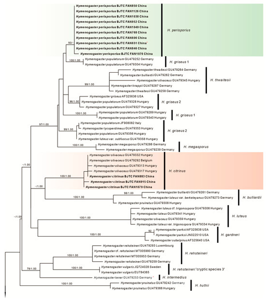
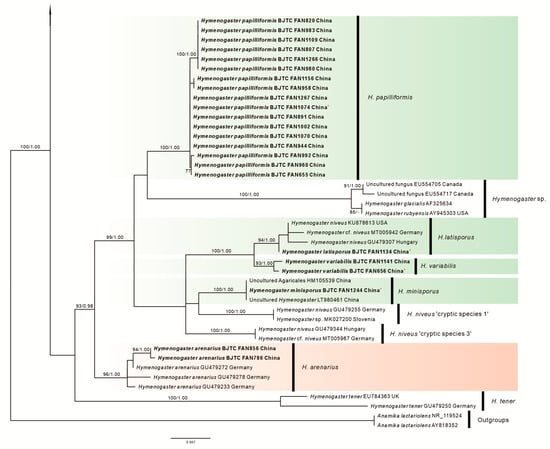
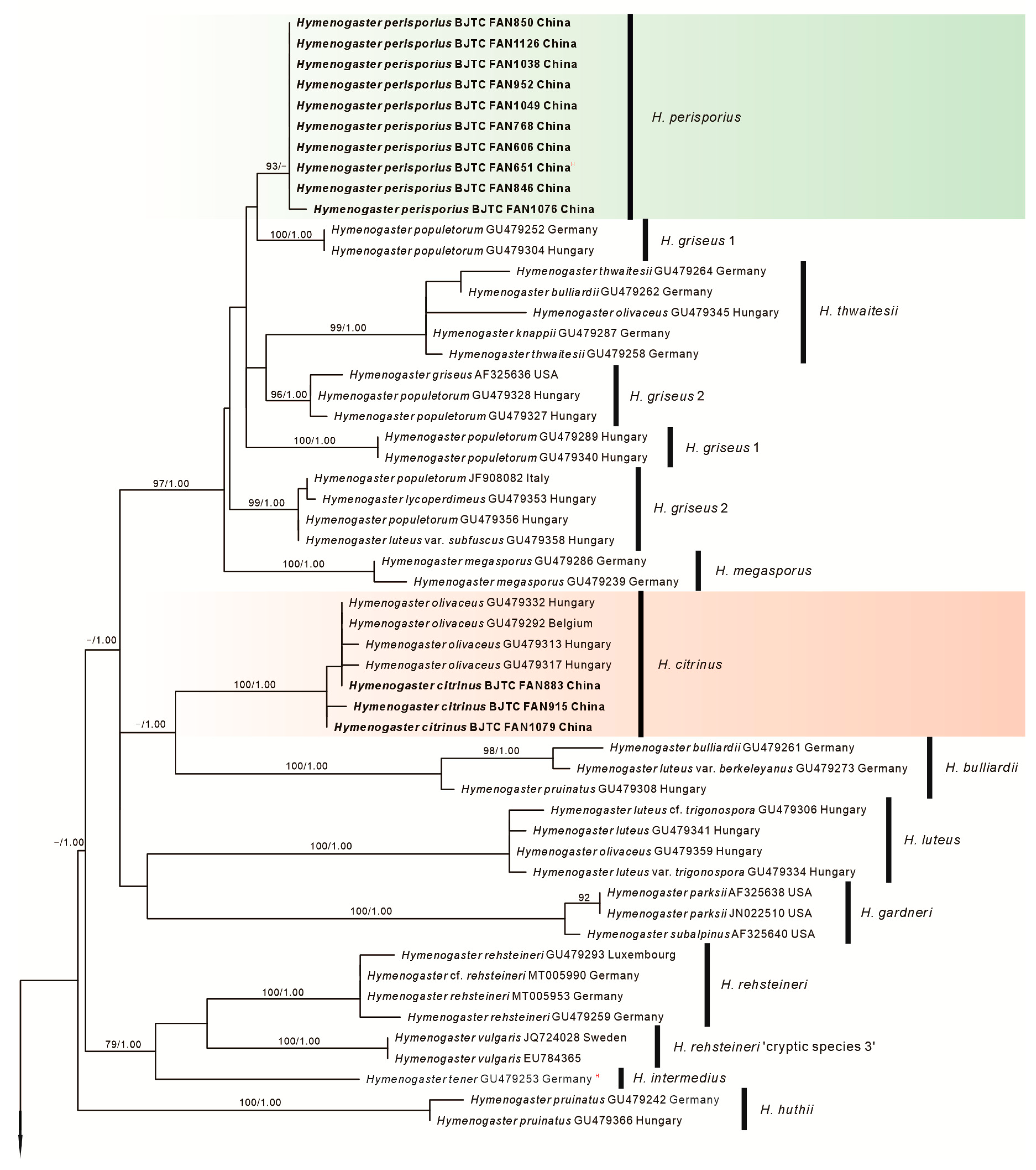
Figure 1.
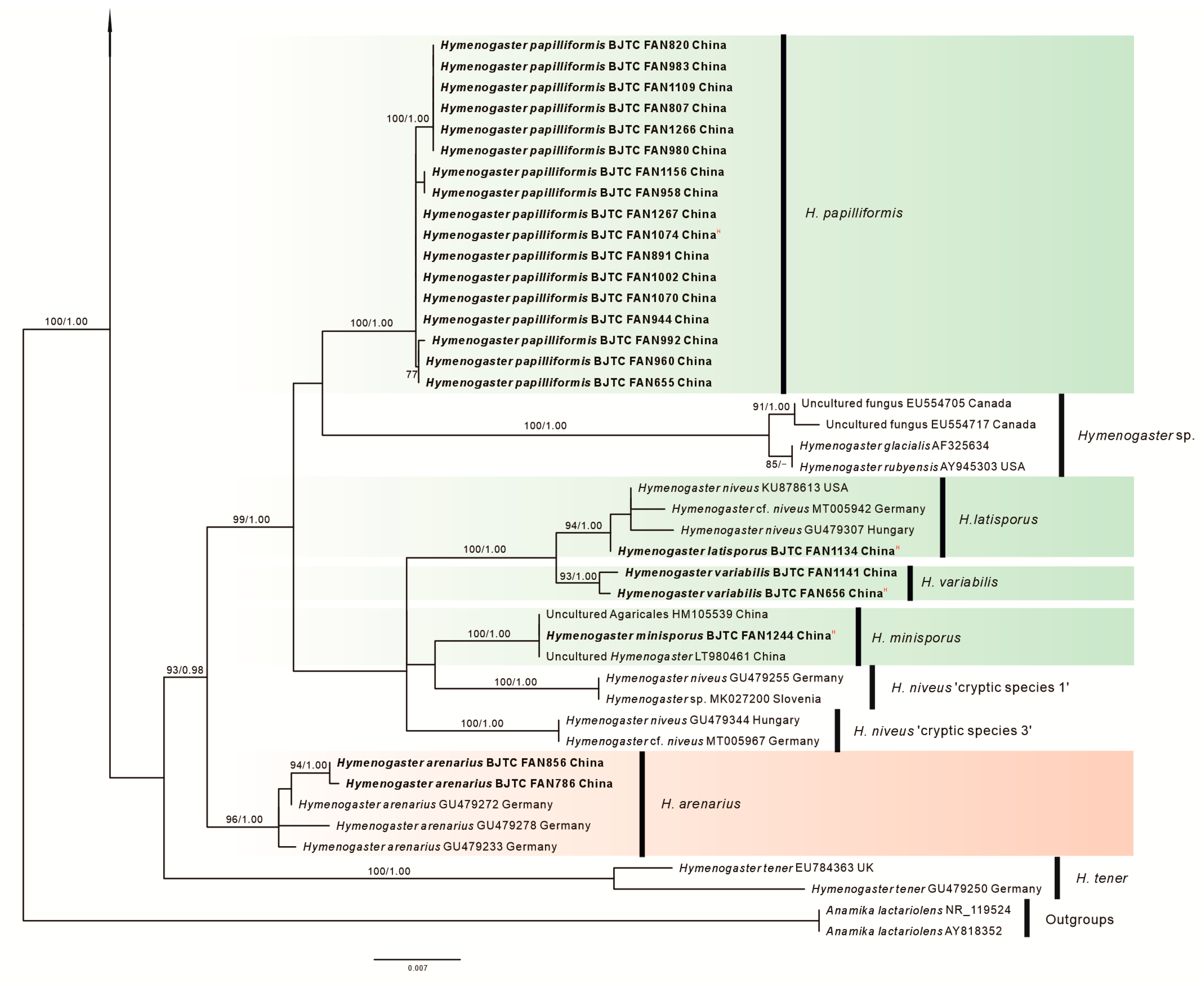
Phylogeny derived from maximum likelihood analyses of the ITS/LSU sequences from Hymenogaster and related species. Two sequences of Anamika lactariolens were selected as the outgroup. Values on the left represent the likelihood bootstrap support values (≥70%). Values on the right represent significant Bayesian posterior probability values (≥0.95). Novel sequences are in bold. Super index “H” means “Holotype”. The green background represents the five new species described in this study, the red background represents the two old species from China supported by molecular data.
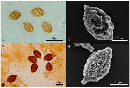
Figure 2.
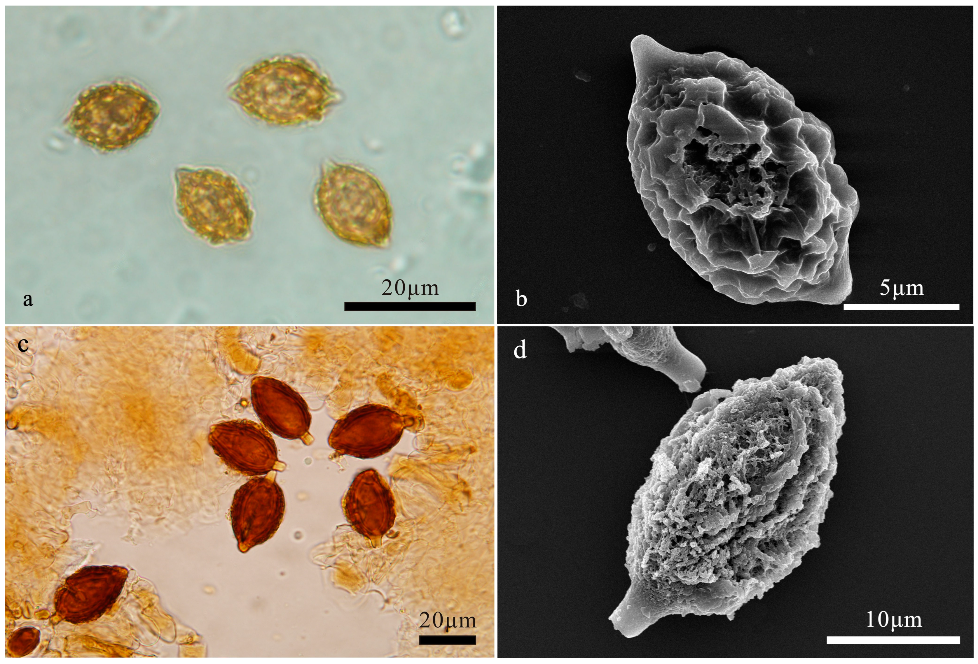
(a,b) Hymenogaster arenarius. (a) Basidiospores under LM. (b) Basidiospore under SEM. (c,d) Hymenogaster citrinus. (c) Basidiospores under LM. (d) Basidiospore under SEM.
2. Materials and Methods
2.1. Sample Collections
Samples were systematically collected over a period of six years in China and subsequently examined. These voucher specimens have been accessioned in the Herbarium of the Biology Department at Capital Normal University (BJTC). Macroscopic characteristics of these specimens were described from both fresh and dried materials. For microscopic analysis, thin sections were prepared from dried specimens by hand. These sections were then immersed in a 3% KOH (w/v) solution and Melzer’s reagent [20] for detailed study. For the scanning electron microscopy (SEM) analysis, basidiospores were positioned onto double-sided adhesive tape affixed to the SEM stub. Subsequently, these samples were uniformly coated with a platinum–palladium film utilizing an ion sputter-coater (HITACHI E-1010). The coated samples were then examined and documented using a Hitachi S-4800 SEM (Hitachi, Tokyo, Japan).
2.2. DNA Extraction, PCR Amplification, Sequencing and Nucleotide Alignment
Dried gleba was ground by shaking for 3 min at 30 Hz (Mixer Mill MM 301, Retsch, Haan, Germany) in a 1.5 mL tube together with one 3 mm diameter tungsten carbide ball, and total genomic DNA was extracted using the modified CTAB method [21]. The internal transcribed spacer (ITS) region of nuclear ribosomal DNA (nrDNA) was amplified using primers ITS1f/ITS4 [21,22]. The 28S large subunit (nrLSU) nrDNA region was amplified using primers LR0R/LR5 [23]. PCRs were performed in 50 µL reactions containing 4 µL of DNA template, 2 µL of each primer (10 µM), and 25 µL 2 × Master Mix [Tiangen Biotech Co., Beijing, China]. Amplification reactions were performed as follows: for the ITS gene, initial denaturation at 95 °C for 4 min, followed by 35 cycles at 95 °C for 30 s, 55 °C for 45 s, 72 °C for 1 min, and a final extension at 72 °C for 10 min; for the nrLSU gene, initial denaturation at 95 °C for 4 min, followed by 35 cycles at 95 °C for 30 s, 55 °C for 60 s, 72 °C for 1 min, and a final extension at 72 °C for 10 min. The PCR products were sent to Beijing Zhongkexilin Biotechnology Co., Ltd. (Beijing, China) for purification and sequencing. Validated sequences are stored in the NCBI database (http://www.ncbi.nlm.nih.gov/) (accessed on 30 April 2023) under the accession numbers provided (Table 1).

Table 1.
Sources of specimens and GenBank accession numbers for sequences used in this study. Newly generated sequences are in bold.
2.3. Phylogenetic Analysis
The ITS-LSU combined dataset was assembled and aligned utilizing the MAFFT algorithm [24], adhering to default parameters. This alignment was further refined through manual adjustments in Se-Al v2.03a [25], ensuring optimal sequence similarity. Alignments of all datasets used in this study were submitted to TreeBASE (No. 31242). ML and BI analysis were used together to construct phylogenetic tree. Maximum likelihood (ML) analysis was performed with RAxML 8.0.14 [26] employing the GTRGAMMAI substitution model with parameters unlinked. ML bootstrap replicates (1000) were computed in RAxML using a rapid bootstrap analysis and search for the best-scoring ML tree. The ML trees were visualized with TreeView32 [27]. Clades with bootstrap support (BS) ≥ 70% were considered significant [28]. Bayesian inference (BI) was conducted using MrBayes v3.1.2 [29] as an additional method of evaluating branch support. In BI analysis, after selecting the best substitution models (GTR + I + G for all positions) determined by MrModeltest v2.3 [30], two independent runs of four chains were conducted for 1,065,000 Markov chain Monte Carlo (MCMC) generations with the default settings. Average standard deviations of split frequency (ASDSF) values were far less than 0.01 at the end of the generations. Trees were sampled every 100 generations after burn-in (well after convergence), and a 50% majority-rule consensus tree was constructed and visualized with TreeView 32 [27]. Clades with Bayesian posterior probability (PP) ≥ 0.95 were considered significantly supported [31].
3. Results
3.1. Molecular Phylogenetics
The ITS-LSU combined dataset was compiled to elucidate the phylogenetic position of the new species in this study. This comprehensive dataset comprises 97 sequences from 23 different species, inclusive of 72 newly generated sequences derived from Chinese collections. The length of the aligned dataset was 1465 bp after the exclusion of poorly aligned sites, with 651 bp for ITS and 814 bp for nrLSU. Both Maximum Likelihood (ML) and Bayesian Inference (BI) analyses resulted in similar phylogenetic tree topologies. The tree, as deduced from the ML analysis, is presented here, which showed robust statistical bootstrap support from ML and posterior probability values from BI, confirming the reliability of the findings (Figure 1). Our collections were resolved in seven independent clades with strong statistical bootstrap support, indicating they represented seven distinct species. Two of them represented the known species H. arenarius and H. citrinus, respectively. The remaining five species are novel to science.
3.2. Taxonomy
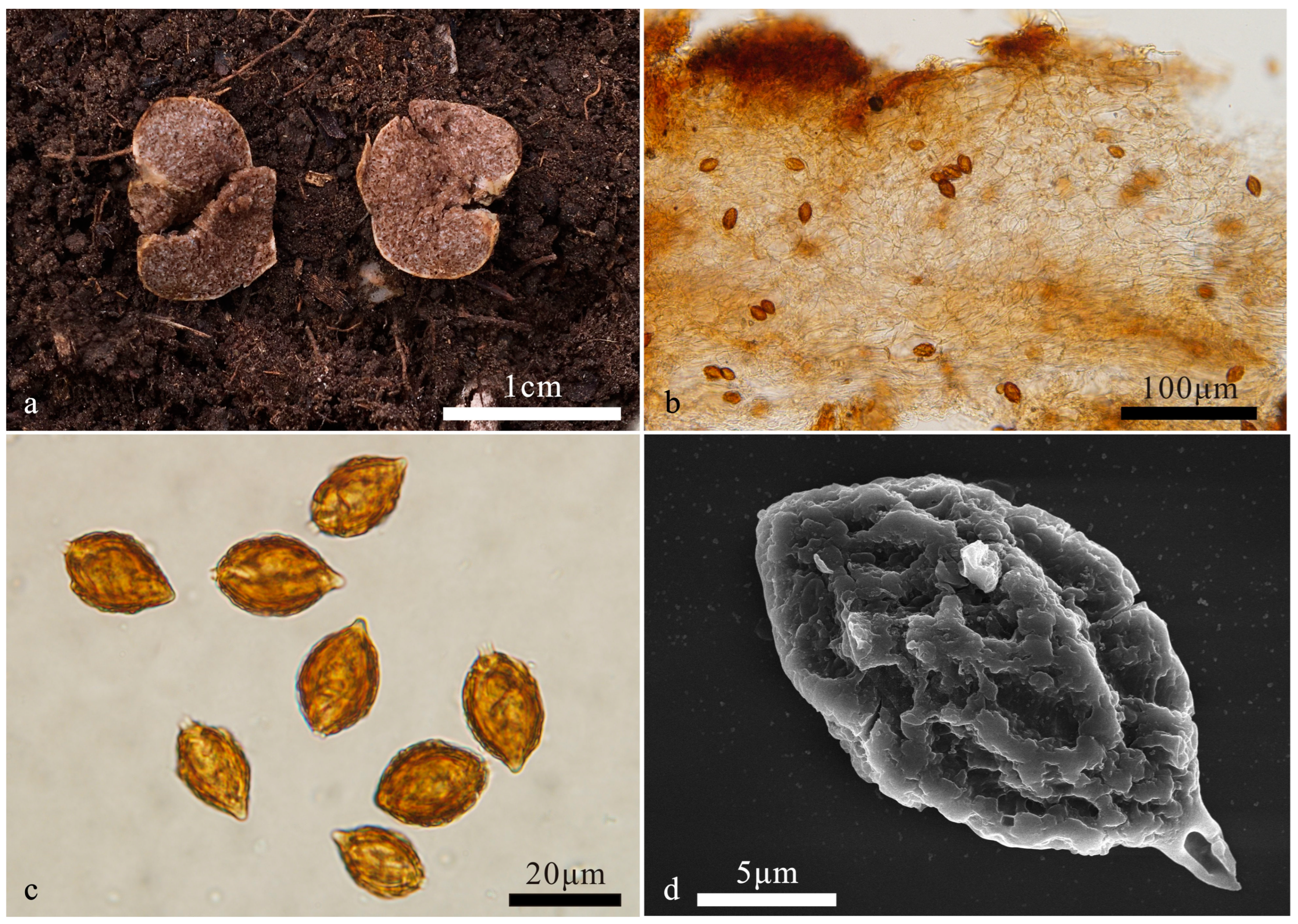
Hymenogaster latisporus L. Fan and T. Li, sp. nov. (Figure 3)
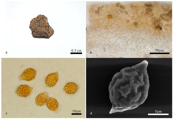
Figure 3.
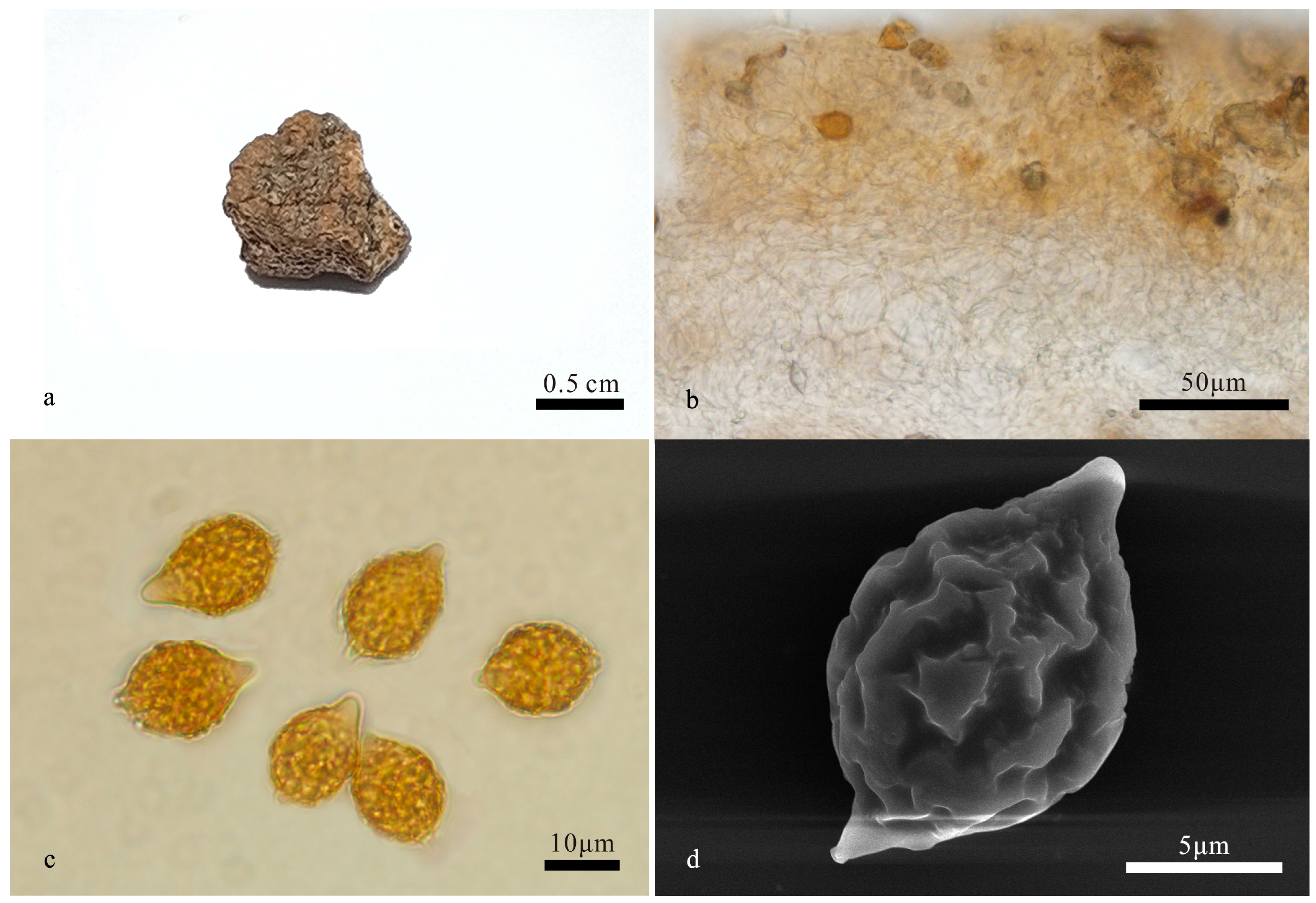
Hymenogaster latisporus (BJTC FAN1134, holotype). (a) Basidiome. (b) Peridium under LM. (c) Basidiospores under LM. (d) Basidiospore under SEM.
MycoBank: MB852599
Etymology: latisporus, referring to the spores with a wider width.
Holotype: China. Shanxi Province, Yuncheng City, Yuanqu County, Lishan Town, Shunwangping, alt. 1744 m, 30 October 2017, YXY144 (BJTC FAN1134).
Description—the basidiome is subglobose to irregular globose, 1.2–1.8 cm in diameter, soft and elastic, earth yellow when fresh, yellow-brown to brown when dry, with a distinct depression at the sterile base. Its surface is smooth and glabrous.
The peridium is 127–246 μm thick, prosenchymatous, interwoven, light yellow-brown, 2–3 μm broad hyphae, and mixed with inflated ellipsoid to subglobose cells of 8–13 μm near to hymenium, pale yellow to nearly hyaline. Gleba light yellow-brown to brown when fresh, loculate, locules irregular, empty, filled with spores at maturity. Hymenium 19–28 μm thick. Hymenial cystidia not seen. The basidia are narrow clavate, two-spored, 28.5–40.5 μm long; the sterigmata are short, 1–2 μm long, and the basidia are collapsed and disappeared at maturity. The basidiospores are subglobose, broad ellipsoidal, ovoid, pale yellow-brown to yellow-brown at maturity, and ornamented with warts and ridges 1–1.2 μm high, with ridges short, irregular, and interwoven, (11.6–)12–15.3(–16) × 10.6–12.6 μm (Lm × Wm = 13.7 ± 0.8 × 11.6 ± 0.5, n = 30), Q = 1.1–1.3 (Qav = 1.2), excluding ornamentations, with gelatinous perisporium, with apex, obtuse, papillary, nearly hyaline, 2.5–3 μm high, appendix evident, 1.5–2.5 μm long.
Habit and habitat: hypogeous, gregarious, under the soil of Pinus tabulaeformis Carr., alt. 1744 m, Shanxi, northern China.
Notes: Hymenogaster latisporus is characterized by the earth yellow basidiome, large and subglobose, broad ellipsoidal, ovoid basidiospores with sparse verrucose and ridge-like ornamentations. Compared with H. minisporus sp. nov (spores 10.8–12.6 × 8.5–10.4 μm), another new species in this study, the former has larger spores (12–15.3(–16) × 10.6–12.6 μm), and with sparse spore ornamentations; compared with H. variabilis sp. nov (peridium 167–351 μm thick), this species has a thinner peridium (127–246 μm thick). The ITS-LSU-based phylogeny supported the description of this new species. DNA analysis showed that H. latisporus shared less than 97% identity in ITS sequence to other Hymenogaster species.
Hymenogaster minisporus T. Li & L. Fan, sp. nov. (Figure 4)
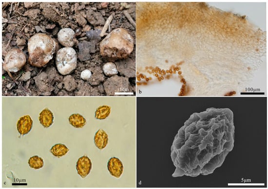
Figure 4.
Hymenogaster minisporus (BJTC FAN1244, holotype). (a) Basidiomes. (b) Peridium under LM. (c) Basidiospores under LM. (d) Basidiospores under SEM.
MycoBank: MB852601
Etymology: minisporus, referring to small basidiospores.
Holotype: China. Beijing, Miyun County, Bulaotun Town, alt. 273 m. 27 July 2020, in soil under Castanea mollissima Blume, ZH571 (BJTC FAN1244).
Description—the basidiome is subglobose to globose, 0.5–1.7 cm in diameter, soft and elastic, dirty white when fresh, stained pale brown, with a distinct depression at the sterile base. The surface is smooth and glabrous.
The peridium is 110–288 μm thick, pseudoparenchymatous, composed of elliptic cells of 11–16 × 8–11 μm, light yellow-brown to pale yellow; the outer surface of the peridium locally exhibits a layer of more-or-less parallel interwoven hyphae of 4.8–7.5 μm broad, light yellow-brown. The gleba are light brown when fresh, brown when dry, loculate, with locules irregular or oblong, empty, and filled with spores at maturity. The hymenium is 16–22 μm broad. The hymenial cystidia are clavate, 29–38 µm long, and only present when young, collapsed, and disappeared at maturity. The basidia are cylindrical, not inflate on the apex, two-to-four-spored, mostly two-spored, 23–35 μm, sterigmata short, 2–3 μm long, basidia collapsed and disappeared at maturity. The basidiospores are ellipsoidal to broadly ellipsoidal, occasionally subglobose, yellow-brown at maturity, ornamented with warts and irregular short ridges of 1 μm high, (10–)10.8–12.6(–13.3) × 8.5–10.4(–10.9) μm (Lm × Wm = 11.7 ± 0.6 × 9.5 ± 0.7, n = 30), Q = 1.2–1.3 (Qav = 1.2), excluding ornamentations, without gelatinous perisporium, with a small apex, obtuse, papillary, 1–1.3 μm high, with appendix, 1–1.5 μm long.
Habit and habitat: hypogeous, gregarious, in the soil under Castanea mollissima.
Notes: Hymenogaster minisporus is characterized by the ellipsoidal to broadly ellipsoidal and small basidiospores. This new species was grouped into a clade with the ‘cryptic species 1’ of H. niveus provisionally proposed by Stielow et al. [2] but without statistical support (Figure 1). Morphologically, the latter has white basidiomes that change to red when touched or bruised [2]. ITS-LSU-based phylogeny supports the erection of this new species. DNA analysis showed that H. minisporus shared less than 97% identity in the ITS sequence to other Hymenogaster species.
Hymenogaster papilliformis L. Fan & T. Li, sp. nov. (Figure 5)
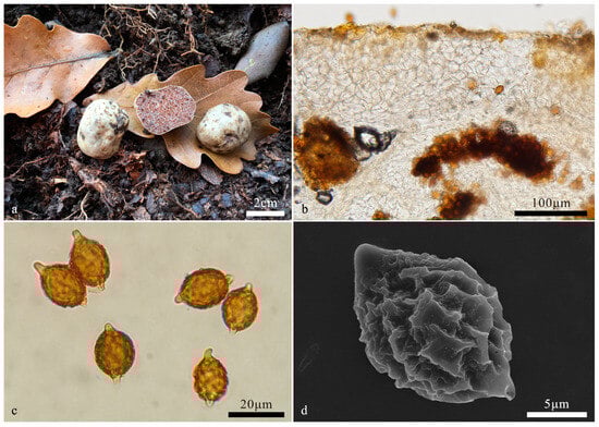
Figure 5.
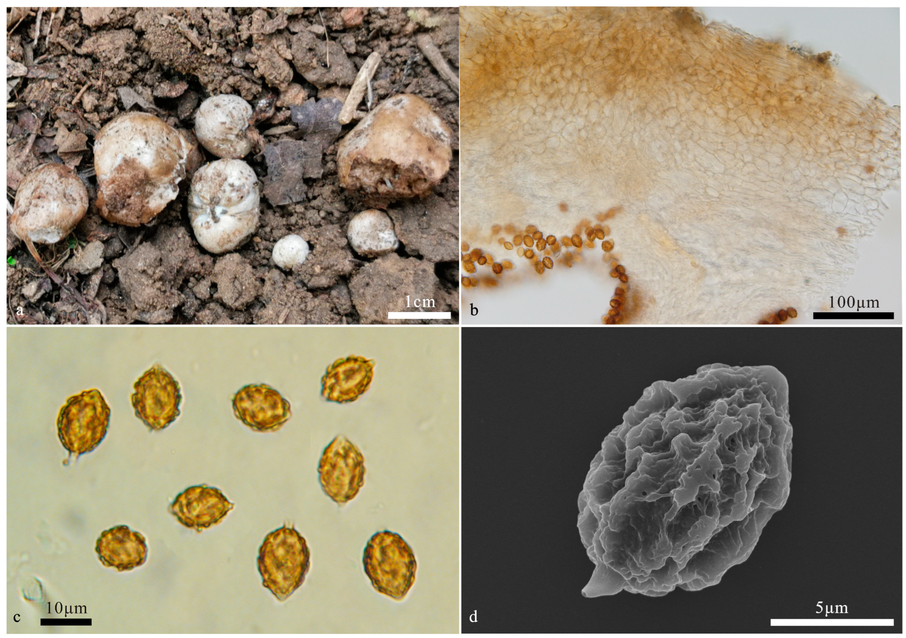
Hymenogaster papilliformis (BJTC FAN1074, holotype). (a) Basidiomes. (b) Peridium under LM. (c) Basidiospores under LM. (d) Basidiospores under SEM.
MycoBank No: MB852600
Etymology: papilliformis, referring to the papillia-shaped apex of basidiospores.
Holotype: China. Shanxi Province, Linfen City, Xi Country, Wulu Mountain National Nature Reserve, 37°32.57′ N, 111°12.10′ E, alt. 1730 m, 26 October 2017, in soil under Quercus sp., LT051 (BJTC FAN1074).
Description—the basidiome is subglobose to irregular globose, 0.8–2.5 cm in diameter, soft and elastic, whitish to cream-colored when fresh, with pale yellow to pale taupe spots, yellowish to yellow-brown when dry, with a distinct depression at the sterile base. The surface is smooth and glabrous.
The peridium is 140–300 μm thick, pseudoparenchymatous, composed of elliptic cells of 14–20 μm in diam, pale yellow to nearly hyaline. The gleba are reddish-brown to brown when fresh, deep brown when dry, and loculate; the locules are rectangle to irregular shape, empty, filled with spores at maturity. The hymenium is 40–70 μm thick. The hymenial cystidia are not seen. The basidia are rare, clavate, one-to-two-spored, mostly two-spored, sterigmata short, 2–4 μm long, basidia collapsed and disappeared at maturity. The basidiospores are broadly fusiform to citriform, yellow-brown at maturity, ornamented with distinct warts and irregular short ridges 1 μm high, ridges interwoven, (13.3–)14–18.6(–19.3) × (10–)10.6–13.6(–15) μm (Lm × Wm = 15.7 ± 1.2 × 11.7 ± 0.8, n = 30), Q = 1.3–1.4 (Qav = 1.34), excluding ornamentations, without gelatinous perisporium, with a pronounced apex, obtuse, papillary, nearly hyaline, 2–3 μm high, occasionally up to 4 μm, appendix very evident, truncate, nearly hyaline, 2–3 μm long.
Habit and habitat: hypogeous, gregarious, in coniferous and broadleaf mixed forest, in the soil under Betula platyphylla Sukaczev, Larix gmelini (Rupr) Rupr., Pinus tabulaeformis, P. bungeana Zucc. ex Endl., and Quercus liaotungensis Koidz.
Additional specimens examined. China. Beijing, Mentougou District, Baihuashan Mountain, 39°49.42′ N, 115°35.14′ E, alt. 1593 m, 14 October 2016, in soil under Pinus tabulaeformis, SXY009 (BJTC FAN655); Yanqing District, Badaling Great Wall, 40°33.10′ N, 115°97.40′ E, alt. 809 m, 16 September 2017, in soil under Quercus liaotungensis, HKB170 (BJTC FAN1155), HKB171 (BJTC FAN1156). Shanxi Province, Lvliang City, Jiaocheng Country, Pangquangou, 37°51.37′ N, 111°27.18′ E, alt. 1879 m, 6 September 2017, in soil under Betula platyphylla, XYY009 (BJTC FAN807), YXY056 (BJTC FAN819), YXY057 (BJTC FAN820), YXY058 (BJTC FAN821); Linfen City, Pu Country, Gelaozhang, 37°32.57′ N, 111°12.10′ E, alt. 1698 m, 10 September 2017, in soil under Pinus tabulaeformis, YXY074 (BJTC FAN888), XYY026 (BJTC FAN902), LT025 (BJTC FAN917), HKB109 (BJTC FAN935); Xi Country, Shenjiagou, 36°36.4′ N, 111°10.34′ E, alt. 1321 m, 10 September 2017, in soil under Pinus bungeana, YXY077 (BJTC FAN891), XYY034 (BJTC FAN910), LT034 (BJTC FAN925), LT035 (BJTC FAN926); Pu Country, Megou, 36°36.57′ N, 111°13.10′ E, alt. 1443 m, 11 September 2017, in soil under Larix gmelini, HKB117 (BJTC FAN944); Chaoyanggou, 36°34.3′ N, 111°11.55′ E, alt. 1645 m, 11 September 2017, in soil under Pinus tabulaeformis, HKB120 (BJTC FAN947), YXY046 (BJTC FAN971); ibid., in soil under mixed forest, LT036 (BJTC FAN954), LT037 (BJTC FAN955), in soil under Quercus liaotungensis, LT040 (BJTC FAN958), LT (BJTC FAN959), LT042 (BJTC FAN960), LT043 (BJTC FAN961), XYY039 (BJTC FAN965), XYY043 (BJTC FAN969), YXY088 (BJTC FAN979), X.Y. Yan089 (BJTC FAN980), YXY092 (BJTC FAN983); Megou, 36°36.57′ N, 111°13.10′ E, alt. 1443 m, 11 September 2017, in soil under Quercus liaotungensis, YXY081 (BJTC FAN972); Gelaozhang, 36°34.8′ N, 111°11.33′ E, alt. 1770 m, 12 September 2017, in soil under Quercus liaotungensis, LT046 (BJTC FAN991), XYY047 (BJTC FAN998), in soil under Pinus tabulaeformis, YXY099 (BJTC FAN992), YXY100 (BJTC FAN993), YXY101 (BJTC FAN994), YXY102 (BJTC FAN1002), in soil under Quercus sp., 36°32.50′ N, 111°12.17′ E, alt. 1700 m, 26 October 2017, XYY070 (BJTC FAN1070); Huozhou country, Qilishangu, 36°35.44′ N, 112°1.46′ E, alt. 1800 m, 7 October 2020, in soil under Quercus sp., LT151 (BJTC FAN1266); Yuncheng City, Xia country, 35°4.44′ N, 111°23.41′ E, alt. 990 m, 7 October 2020, in soil under Quercus sp., LT152 (BJTC FAN1267), LT153 (BJTC FAN1268).
Notes: Hymenogaster papilliformis is characterized by the whitish to cream-colored basidiomata, and broadly fusiform to citriform basidiospores with a pronounced apex (2–3 μm, occasionally up to 4 μm long), distinct warts and ridges, and pronounced truncate appendix (2–3 μm long). DNA analysis showed that H. papilliformis shared less than 96.74% identity in the ITS sequence to other Hymenogaster species. The sequences of H. papilliformis clustered together on an independent branch in the ITS/LSU-based phylogenetic tree (Figure 1) with high supports, further supporting the erection of this new species.
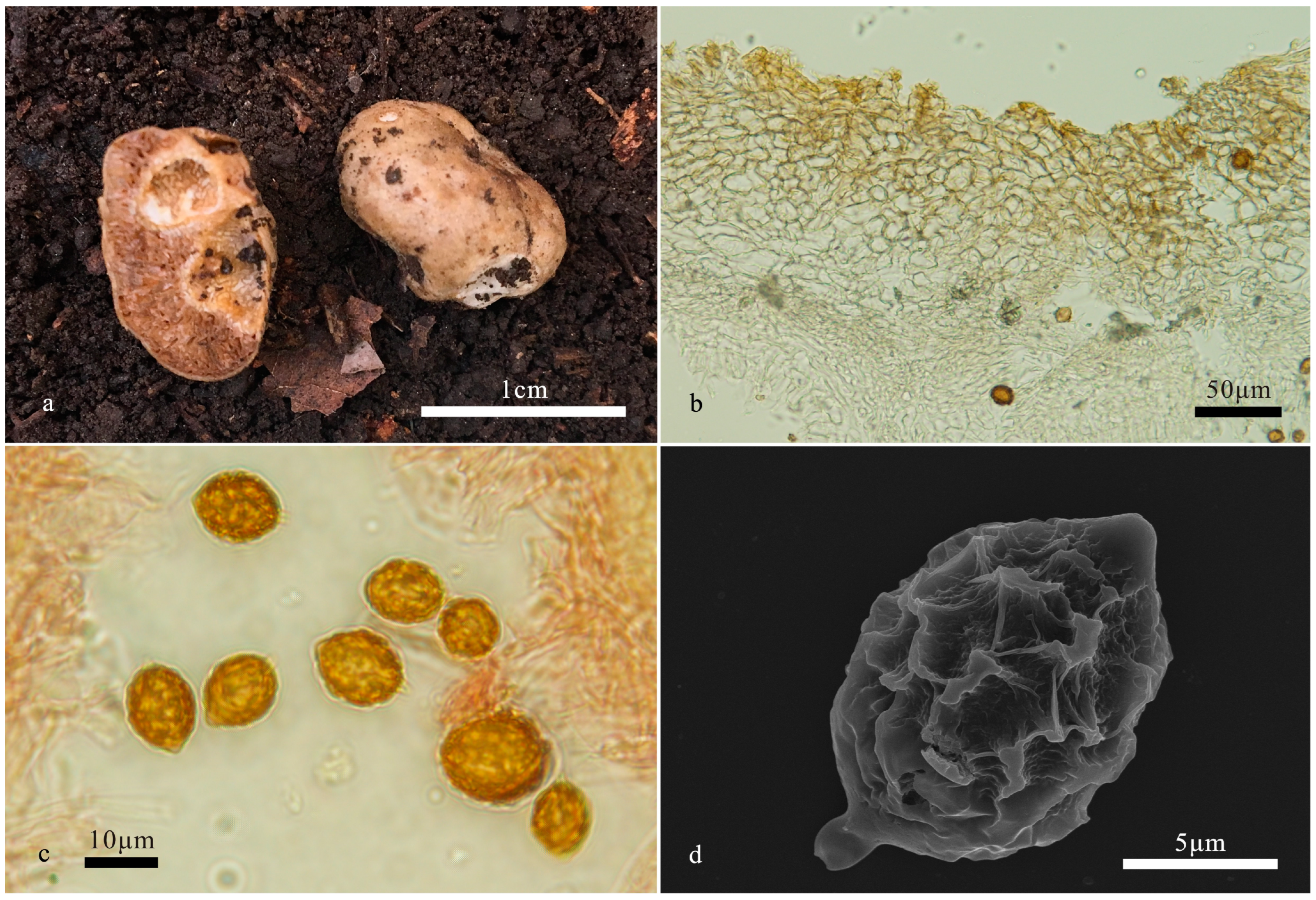
Hymenogaster perisporius T. Li & L. Fan, sp. nov. (Figure 6)
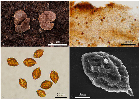
Figure 6.
Hymenogaster perisporius (BJTC FAN651, holotype). (a) Basidiomes. (b) Peridium under LM. (c) Basidiospores under LM. (d) Basidiospores under SEM.
MycoBank: MB852602
Etymology: perisporius, referring to the gelatinous perisporium of basidiospores.
Holotype: China. Beijing, Mentougou County, Qingshui Town, Baihuashan Nature Reserve, alt. 738 m, 13 October 2016, in soil under Populus beijingensis W. Y. Hsu, WYW021 (BJTC FAN651).
Description—the basidiome is subglobose to irregular globose, 0.5–2 cm diameter, soft and elastic, dirty white to pale yellow when fresh, sometimes brownish, with a distinct depression at the white sterile base. Surface smooth, glabrous.
The peridium is 223–310 μm thick, pseudoparenchymatous, composed of elliptic cells of 8–12 μm in diameter and interwoven hyphae of 6.8–8.5 μm broad, light yellowish to nearly hyaline. The gleba are reddish-brown to brown when fresh, deep reddish-brown when dry, loculate, with locules irregular, empty, and filled with spores at maturity. The hymenium is 21–29 μm thick. The hymenial cystidia are clavate, 26–36 µm long, only present when young, collapsed, and disappeared at maturity. The basidia clavate are not inflate on the apex, two-to-three-spored, mostly two-spored, with sterigmata short, 1–2 μm long, and basidia collapsed and disappeared at maturity. The basidiospores are ellipsoidal, yellow-brown to dark brown at maturity, ornamented with warts and ridges of 1–2 μm high, (15–)17–22(–23) × (11–)12–15(–17) μm (Lm × Wm = 19.1 ± 1.3 × 13.3 ± 0.9, n = 30), Q = 1.3–1.6 (Qav = 1.44), excluding ornamentations, with gelatinous perisporium, with apex, obtuse, nearly hyaline, 1–2 μm high, with appendix, truncate, (1–)2–3 μm long.
Habit and habitat: hypogeous, gregarious, in the soil under Betula platyphylla, Castanea mollissima, Larix principis-rupprechtii Mayr., Pinus armandii Franch., P. tabuliformis Carrière, Populus beijingensis, Quercus liaotunggensis Koidz, and Q. mongolica Fisch. ex Ledeb.
Additional specimens examined. China. Beijing, Mentougou County, Qingshui Town, Baihuashan Nature Reserve, alt. 738 m, 4 August 2016, in soil under Quercus mongolica, HBD017 (BJTC FAN546), alt. 752 m, in soil under Populus beijingensis, X.Y. Yan 025 (BJTC FAN650), Y.W. Wang 022 (BJTC FAN653), Y.W. Wang 023 (BJTC FAN654). Shanxi Province: Yuncheng City, Yuanqu County, Lishan Town, Shunwangping scenic spot, alt. 2209 m, 16 August 2016, in soil under Pinus armandii, K.B. Huang 011 (BJTC FAN553), K.B. Huang 001 (BJTC FAN561), X.Y. Yan 014 (BJTC FAN569), Y.W. Wang 007 (BJTC FAN570), X.Y. Sang 004 (BJTC FAN589), B.D. He 002 (BJTC FAN592), B.D. He 003 (BJTC FAN6046), K.B. Huang 015 (BJTC FAN608), alt. 2276 m, 17 October 2016, K.B. Huang 041 (BJTC FAN694); Xinzhou City, Qiuqiangou, alt. 2099 m, 17 October 2016, in soil under Larix principis-rupprechtii, K.B. Huang 063 (BJTC FAN768); Lvliang City, Jiaocheng County, Shenweigou, alt. 2003 m, 7 September 2017, in soil under Betula platyphylla, X.Y. Yan 059 (BJTC FAN846), in soil under Larix principis-rupprechtii, X.Y. Yan 063 (BJTC FAN850); Linfen City, Pu Country, Chaoyanggou, alt. 1660 m, 11 September 2017, in soil under Pinus tabuliformis, K.B. Huang 125 (BJTC FAN952), X.Y. Yan 086 (BJTC FAN977); Guancen Mountain, Liaowangtai, Yingbeimian, alt. 2120 m, 13 October 2017, in soil under Betula platyphylla, K.B. Huang 139 (BJTC FAN1038), X.Y. Yan 118 (BJTC FAN1049); Linfen City, Pu County, Wulu Mountain, alt. 1555 m, 26 October 2017, in soil under Quercus liaotunggensis, K.B. Huang 143 (BJTC FAN1076); Yuncheng City, Xia County, Sijiao Town, Jialu Village, alt. 1057 m, 29 October 2017, in soil under Castanea mollissima, K.B. Huang 162 (BJTC FAN1126).
Notes: Hymenogaster perisporius is characterized by the dirty white to pale yellow basidiomes, broad ellipsoidal to ellipsoidal, yellow-brown to dark brown basidiospores with warts and ridges, with gelatinous perisporium. Hymenogaster bulliardii Vittad. and H. thwaitesii Berk. and Broome are similar to H. perisporius in spore size and gleba color, but H. bulliardii has smooth spores and H. thwaitesii has a yellow-brown peridium. Hymenogaster citrinus is similar to H. perisporius in the appearance of its basidiospores, but the spores of H. citrinus are larger (21.1–25.9 × 14.0–18.4 μm). DNA analysis showed that sequences of H. perisporius clustered with H. griseus s. l. [2] (Figure 1); however, the spores are slender fusiform (Q = 1.9) in the latter [2], quite different from this new species. The ITS-LSU-based phylogeny supports the erection of this new species. The DNA analysis showed that H. perisporius shared less than 97% identity in ITS sequence to other Hymenogaster species.
Hymenogaster variabilis L. Fan & T. Li, sp. nov. (Figure 7)

Figure 7.
Hymenogaster variabilis (BJTC FAN656, holotype). (a) Basidiomes. (b) Peridium under LM. (c) Basidiospores under LM. (d) Basidiospores under SEM.
MycoBank: MB852603
Etymology: variabilis, referring to the peridium of very variable thickness.
Holotype: China. Beijing, Mentougou County, Qingshui Town, Baihuashan Nature Reserve, alt. 1593 m, 14 October 2016, in soil under Pinus tabulaeformis, WYW024 (BJTC FAN656).
Description—the basidiome is subglobose to irregular globose, 1–1.8 cm diameter, soft and elastic, pale yellow to earth yellow when fresh, yellow-brown to brown when dry, with a distinct depression at the sterile base. The surface is smooth and glabrous.
The peridium is 167–351 μm thick, pseudoparenchymatous, composed of elliptic to subglobose cells of 8–19 μm in diameter, light yellowish to nearly hyaline. The gleba are light yellow-brown to brown when fresh, loculate, with locules irregular, empty, and filled with spores at maturity. The hymenium are 14–23 μm thick. The hymenial cystidia are clavate, 25–40 µm long, only present when young, collapsed, and disappeared at maturity. The basidia are narrow clavate, occasionally inflate at the apex, two-spored, 27–36 μm, sterigmata short, 1–3 μm long, basidia collapsed and disappeared at maturity. The basidiospores are broadly ellipsoidal to subglobose, pale yellow-brown to yellow-brown at maturity, ornamented with sparse warts and ridges of 1–1.2 μm high, with ridges short, interwoven, (8.5–)10.6–13.6(–15) × (7.4–)8.8–10.9(–13) μm (Lm × Wm = 11.5 ± 1.7 × 9.7 ± 1.4, n = 30), Q = 1.1–1.3 (Qav = 1.2), excluding ornamentations, without gelatinous perisporium, with apex, obtuse, papillary, nearly hyaline, 1–3 μm high, with appendix, truncate, 1–2 μm long.
Habit and habitat: hypogeous, gregarious, in the soil under Pinus tabulaeformis.
Additional specimen examined. China. Shanxi Province, Yuncheng City, Yuanqu County, Lishan Town, Shunwangping, alt. 1744 m, 30 October 2017, LT071 (BJTC FAN1141).
Notes: Hymenogaster variabilis is characterized by the peridium with significant changes in thickness, broad ellipsoidal to subglobose basidiospores ornamented with sparse warts and ridges. Hymenogaster minisporus is similar to H. variabilis in spore size, but the former is distinguished by its densely ornamented spores, and its peridium of 110–288 μm thickness. The ITS-LSU-based phylogeny supports the erection of this new species. The DNA analysis showed that H. variabilis shared less than 95.65% identity in the ITS sequence to other Hymenogaster species.
4. Discussion
Among the 32 species of Hymenogaster reported in previous studies from China, 14 species were recorded from Shanxi Province [19], but none from Beijing. We re-examined those voucher specimens collected at that time and confirmed that morphologically they represented species of Hymenogaster, but unfortunately their DNA sequences could not be successfully sequenced. Among our newly collected specimens from Shanxi Province, two species reported in the previous studies were confirmed with molecular data in this study, namely, H. arenarius and H. citrinus (Figure 1 and Figure 2). DNA analysis showed that the two species shared less than 95.43% and 95.26% sequence identity, respectively, with other Hymenogaster species. Consequently, there are a total of seven species currently confirmed using morphological and molecular data in Shanxi and Beijing, including five new species described in this study. A key to these species is provided as follows.
Key to the species of Hymenogaster from Shanxi and Beijing in China:
| 1 | Basidiome pale yellow, white to dirty white when fresh | 2 |
| 1 | Basidiome earth yellow to yellow-brown when fresh | 3 |
| 2 | Gleba reddish brown to brown when fresh | 4 |
| 2 | Gleba light brown when fresh | H. minisporus |
| 3 | Peridium very variable in thickness and more than >130 μm thick | 5 |
| 3 | Peridium without major changes in thickness and <130 μm thick | 6 |
| 4 | Basidiospores 21–26 × 14–18.5 μm | H. citrinus |
| 4 | Basidiospores 17–22 × 12–15 μm | H. perisporius |
| 5 | Basidiospores broad ellipsoidal to subglobose, Q = 1.1–1.3 | H. variabilis |
| 5 | Basidiospores broad fusiform to citriform, Q = 1.3–1.4 | H. apilliformis |
| 6 | Basidiospores fusiform, Q = 1.2–1.4 | H. arenarius |
| 6 | Basidiospores broadly ellipsoidal to broadly ovoid, Q = 1.1–1.3 | H. latisporus |
Author Contributions
L.F. conceived and designed the study; T.L. wrote the manuscript and conducted phylogenetic analyses and morphological observations; N.M., H.F. and Y.Z. conducted the experiments. All authors have read and agreed to the published version of the manuscript.
Funding
This study was supported by the National Natural Science Foundation of China (No. 32370010), the BJAST Budding Talent Program (Grant No. 24CE-BGS-19), and the Beijing Government.
Institutional Review Board Statement
Not applicable.
Informed Consent Statement
Not applicable.
Data Availability Statement
The sequencing data has been submitted to GenBank.
Acknowledgments
J.Z. Cao is appreciated for collecting specimens and providing valuable suggestions.
Conflicts of Interest
The authors declare no conflicts of interest.
References
- Vittadini, C. Monographia Tuberacearum; Ex Typographia, F. Rusconi: Milian, Italy, 1831; pp. 1–88. [Google Scholar]
- Stielow, B.; Bratek, Z.; Orczán, A.K.I.; Rudnoy, S.; Hensel, G.; Hoffmann, P.; Klenk, H.P.; Göker, M. Species delimitation in taxonomically difficult fungi: The case of Hymenogaster. PLoS ONE 2011, 6, e15614. [Google Scholar] [CrossRef] [PubMed]
- Montecchi, A.; Sarasini, M. Funghi Ipogei d’Europa; AMB Fondazione Centro Studi Micologici: Vicenza, Italy, 2000; pp. 52–501. [Google Scholar]
- Pegler, D.N.; Spooner, B.M.; Young, T.W.K. British truffles: A revision of British Hypogeous fungi. Kew Bull. 1993, 49, 167. [Google Scholar]
- Dodge, C.W.; Zeller, S.M. Hymenogaster and related genera. Ann. Mo. Bot. Gard. 1934, 21, 625–708. [Google Scholar] [CrossRef]
- Harkness, H.W. Californian hypogaeous fungi. Calif. Acad. Sci. 1899, 1, 241–291. [Google Scholar]
- Smith, A.H. Notes on Dendrogaster, Gymnoglossum, Protoglossum and species of Hymenogaster. Mycologia 1966, 58, 100–124. [Google Scholar] [CrossRef]
- Cázares, E.; Trappe, J.M. Alpine and subalpine fungi of the Cascade Mountains. I. Hymenogaster glacialis sp. nov. Mycotaxon 1990, 38, 245–249. [Google Scholar]
- Fogel, R.; States, J. Materials for a hypogeous mycoflora of the Great Basin and adjacent cordilleras of the western United States VI: Hymenogaster rubyensis sp. nov. (Basidiomycota, Cortinariaceae). Mycotaxon 2001, 80, 333–337. [Google Scholar]
- Julou, T.; Burghardt, B.; Gebauer, G.; Berveiller, D.; Damesin, C.; Selosse, M.A. Mixotrophy in orchids: Insights from a comparative study of green individuals and nonphotosynthetic individuals of Cephalantera damasonium. N. Phytol. 2005, 166, 639–653. [Google Scholar] [CrossRef] [PubMed]
- Trappe, J.M.; Molina, R.; Luoma, D.L.; Cázares, E.; Pilz, D.; Smith, J.E.; Castellano, M.A.; Miller, S.L.; Trappe, J.M. Diversity, Ecology, and Conservation of Truffle Fungi in Forests of the Pacific Northwest; Pacific Northwest Research Station, USDA Forest Service: Corvallis, OR, USA, 2009; pp. 1–194.
- Türkoğlu, A.; Castellano, M.A. New records of truffle fungi (Basidiomycetes) from Turkey. Turk. J. Bot. 2013, 37, 970–976. [Google Scholar] [CrossRef]
- Pecoraro, L.; Huang, L.; Caruso, T.; Perotto, S.; Girlandam, M.; Cai, L.; Liu, Z.J. Fungal diversity and specificity in Cephalanthera damasonium and C. longifolia (Orchidaceae) mycorrhizas. J. Syst. Evol. 2017, 55, 158–169. [Google Scholar] [CrossRef]
- Rudawska, M.; Kujawska, M.; Leski, T.; Janowski, D.; Karliński, L.; Wilgan, R. Ectomycorrhizal community structure of the admixture tree species Betula pendula, Carpinus betulus, and Tilia cordata grown in bare-root forest nurseries. For. Ecol. Manag. 2019, 437, 113–125. [Google Scholar] [CrossRef]
- Hawker, L.E. British hypogeous fungi. Philos. Trans. R. Soc. Biol. Sci. 1954, 237, 429–546. [Google Scholar]
- Soehner, V.E. Die Gattung Hymenogaster Vitt: Eine monographische Studie mit besonderer. Nova Hedwigia. 1962, 2, 1–113. [Google Scholar]
- Bougher, N.L.; Castellano, M.A. Delimitation of Hymenogaster sensu stricto and four new segregate genera. Mycologia 1993, 85, 273–293. [Google Scholar] [CrossRef]
- Smith, M.E.; Castellano, M.A.; Frank, J.L. Hymenogaster macmurphyi and Splanchnomyces behrii are sequestrate species of Xerocomellus from the western United States. Mycologia 2018, 110, 605–617. [Google Scholar] [CrossRef] [PubMed]
- Liu, B.; Liu, Y.H.; Liu, J.H. The hypogeous macrofungi of China. Edible Fungi China 2002, 21, 14–15. [Google Scholar]
- Dring, D.M. Techniques for microscopic preparation. In Methods in Microbiology; Booth, C., Ed.; Academic Press Inc.: New York, NY, USA, 1971; pp. 95–111. [Google Scholar]
- Gardes, M.; Bruns, T.D. ITS primers with enhanced specificity for Basidiomycetes—Application to the identification of mycorrhizae and rusts. Mol. Ecol. 1993, 2, 113–118. [Google Scholar] [CrossRef] [PubMed]
- White, T.J.; Bruns, T.; Lee, S.; Taylor, F.J.R.M. Amplification and direct sequencing of fungal ribosomal RNA genes for phylogenetics. In PCR Protocols, A Guide to Methods and Applications; Seliger, H., Gelfand, D.H., Sinsky, J.J., White, T.J., Eds.; Academic: San Diego, CA, USA, 1990; pp. 315–322. [Google Scholar]
- Vilgalys, R.; Hester, M. Rapid genetic identification and mapping of enzymatically amplified ribosomal DNA from several Cryptococcus species. J. Bacteriol. 1990, 172, 4238–4246. [Google Scholar] [CrossRef] [PubMed]
- Katoh, K.; Frith, M.C. Adding unaligned sequences into an existing alignment using MAFFT and LAST. Bioinformatics 2012, 28, 3144–3146. [Google Scholar] [CrossRef] [PubMed]
- Rambaut, A. Estimating the rate of molecular evolution: Incorporating non-contemporaneous sequences into maximum likelihood phylogenies. Bioinformatics 2000, 16, 395–399. [Google Scholar] [CrossRef] [PubMed]
- Stamatakis, A. RAxML version 8: A tool for phylogenetic analysis and post-analysis of large phylogenies. Bioinformatics 2014, 30, 1312–1313. [Google Scholar] [CrossRef] [PubMed]
- Page, R.D.M. TreeView; Glasgow University: Glasgow, UK, 2001. [Google Scholar]
- Hillis, D.M.; Bull, J.J. An empirical test of bootstrapping as a method for assessing confidence in phylogenetic analysis. Syst. Biol. 1993, 42, 182–192. [Google Scholar] [CrossRef]
- Ronquist, F.; Huelsenbeck, J.P. MrBayes 3: Bayesian phylogenetic inference under mixed models. Bioinformatics 2003, 19, 1572–1574. [Google Scholar] [CrossRef] [PubMed]
- Nylander, J. MrModeltest 2.2. Computer Software Distributed by the University of Uppsala; Evolutionary Biology Centre: Uppsala, Sweden, 2004. [Google Scholar]
- Alfaro, M.E.; Zoller, S.; Lutzoni, F. Bayes or bootstrap? A simulation study comparing the performance of Bayesian Markov chain Monte Carlo sampling and bootstrapping in assessing phylogenetic confidence. Mol. Biol. Evol. 2003, 20, 255–266. [Google Scholar] [CrossRef] [PubMed]

Disclaimer/Publisher’s Note: The statements, opinions and data contained in all publications are solely those of the individual author(s) and contributor(s) and not of MDPI and/or the editor(s). MDPI and/or the editor(s) disclaim responsibility for any injury to people or property resulting from any ideas, methods, instructions or products referred to in the content. |
© 2024 by the authors. Licensee MDPI, Basel, Switzerland. This article is an open access article distributed under the terms and conditions of the Creative Commons Attribution (CC BY) license (https://creativecommons.org/licenses/by/4.0/).