Article

Morphological and Phylogenetic Analyses Reveal Three Novel Species of Sanguinoderma (Ganodermataceae, Basidiomycota) from Yunnan Province, China

1
College of Agriculture and Biological Science, Dali University, Dali 671003, China
2
College of Biotechnology and Engineering, West Yunnan University, Lincang 677000, China
3
Cangshan Forest Ecosystem Observation and Research Station of Yunnan Province, Dali University, Dali 671003, China
*
Authors to whom correspondence should be addressed.
J. Fungi 2024, 10(8), 589; https://doi.org/10.3390/jof10080589
Submission received: 22 July 2024 / Revised: 14 August 2024 / Accepted: 16 August 2024 / Published: 19 August 2024
(This article belongs to the Special Issue Diversity, Phylogeny and Ecology of Forest Fungi)

Abstract

:
Sanguinoderma (Ganodermataceae) is recognized as a valuable medicinal resource in Taiwan, China. Additionally, it serves as a traditional folk medicine for treating neurotic epilepsy in Malaysia. This study involved the collection of six specimens of Sanguinoderma from Yunnan Province, China. Employing multigene phylogenetic analysis of DNA sequences, including internal transcribed spacer (ITS), nuclear large subunit (LSU), RNA polymerase II second largest subunit (rpb2), translation elongation factor 1-alpha (tef1-α), mitochondrial small subunit (mtSSU), nuclear small subunit (nSSU) and morphological examinations, three new species, viz. Sanguinoderma concentricum, S. dehongense and S. ovisporum, are introduced. Sanguinoderma concentricum is characterized by a central stipe basidiomata, an orbicular to suborbicular pileus, a grayish-yellow surface with alternating concentric zones and wavy margin-like petals and regular pileipellis cells (4–8 × 17–28 μm). Sanguinoderma dehongense is characterized by a long stipe and flabelliform basidiomata, a dark-grayish yellow-to-dark-yellow pileus surface, irregular pileipellis cells and wavy margin and ellipsoid basidia (8–11 × 9–13 μm). Sanguinoderma ovisporum is characterized by a reniform basidiomata, a heterogeneous context and ovoid basidiospores (7.5–8.6 × 5.5–7.2 µm). A detailed description and illustrations of these new species are provided, as well as a morphological comparison with similar taxa.

1. Introduction

Ganodermataceae is a genera-rich and highly diverse group of macro-fungi, widely distributed in tropical and subtropical regions around the world. Up to date, fourteen genera are accepted in Ganodermataceae. Previous studies have confirmed that species within the genera Ganoderma P. Karst and Sanguinoderma Y.F. Sun, D.H. Costa and B.K. Cui possess significant medicinal value, including anti-cancer, anti-bacterial, anti-aging, immune-enhancing and lipid-lowering effects [1,2,3,4,5,6,7].
Sanguinoderma (Ganodermataceae, Polyporales) was introduced to accommodate the species in which the fresh pore surface changes to blood red when bruised, with S. rude Y.F. Sun, D.H. Costa and B.K. Cui as type species. Sanguinoderma is characterized by an annual basidiomata, central or lateral stipitate with an almost sessile, corky-to-woody hard texture. The pileus is single, suborbicular to flatly reniform and glabrous to tomentose, with concentric zonation or furrowing and radial rugosity; the pore surface changes to blood red when bruised [8]. There are 18 epithets of Sanguinoderma in the Species Fungorum (http://www.speciesfungorum.org/; accessed date: 13 July 2024). Before the establishment of this genus, the species had consistently been classified within the Amauroderma Murrill. In 1905, Murrill [9] established Amauroderma, classifying it within the Ganodermataceae. Costa-Rezende et al. [10,11] suggested that Amauroderma was polyphyletic based on phylogenetic analysis, analyzing the ITS, LSU, rpb1, and tef1-α sequences of Ganodermataceae, and described two new genera, Furtadoa Costa-Rez., Drechsler-Santos and Reck and Foraminispora Costa-Rez., Drechsler-Santos and Robledo from Amauroderma. The rest of the species previously considered Amauroderma to be clustered in unrelated clades, one of them considered to be Amauroderma s.str., and the other as the ‘Amauroderma rude’ clade, but without morphological differences between the two clades. Until 2020, Sun et al. [8] clarified the taxonomy and phylogeny of Amauroderma s.lat, when Sanguinoderma was established as a new genus.
Sanguinoderma is the largest group of species aside from Ganoderma P. Karst and Amauroderma in Ganodermataceae. Sanguinoderma rugosum (Blume and T. Nees) Y.F. Sun, D.H. Costa and B.K. Cui is regarded as a precious traditional medicine with anti-cancer properties in Taiwan, China [12], and the indigenous peoples in Peninsular Malaysia used S. rugosum to treat epilepsy [13]. Most Sanguinoderma species are soil-inhabiting, distributed in tropical and subtropical regions of Africa, Asia, North America, Oceania and South America [8,14].
In China, species of Sanguinoderma have been reported in 13 provinces (Chongqing, Fujian, Guangdong, Guangxi, Guizhou, Hainan, Hubei, Hunan, Jiangxi, Sichuan, Taiwan, Yunnan and Zhejiang), and Yunnan Province has the most abundant diversity of species distribution, with a total of seven species [S. elmerianum (Murrill) Y.F. Sun and B.K., S. guangdongense B.K. Cui and Y.F. Sun, S. laceratum Y.F. Sun and B.K. Cui, S. leucomarginatum B.K. Cui and Y.F. Sun, S. longistipitum B.K. Cui and Y.F. Sun, S. preussii (Henn.) B.K. Cui and Y.F. Sun and S. rugosum.]; S. laceratum and S. leucomarginatum are reported exclusively in Yunnan Province [10,11]. In this study, six Sanguinoderma-like samples collected from Yunnan Province were identified as three new species based on phylogenetic analysis and morphological study, namely S. concentricum, S. dehongense and S. ovisporum.

2. Materials and Methods

2.1. Specimen Collection

Six Sanguinoderma-like specimens were collected during July and August of 2023 in the Yunnan Province, China. The collection and documentation process for Sanguinoderma specimens entail capturing photographs and recording essential information, such as habitat, altitude, collection time and location. Detailed descriptions of morphological features were also documented. In the benchtop (AIRTECH SW-CJ-2F), a 1–2 mm2 of context comprised inoculated tissue on a PDA (peel potatoes: 200 g/L, glucose: 20 g/L, agar: 20 g/L and distilled water: 1000 mL) agar medium to obtain a pure culture. The specimens are then dried in an oven at 40~50 °C and placed with an appropriate amount of silica gel in self-sealing bags to prevent moisture regain. Specimens were deposited in the herbarium of Cryptogams, Kunming Institute of Botany, Academia Sinica (KUN-HKAS).

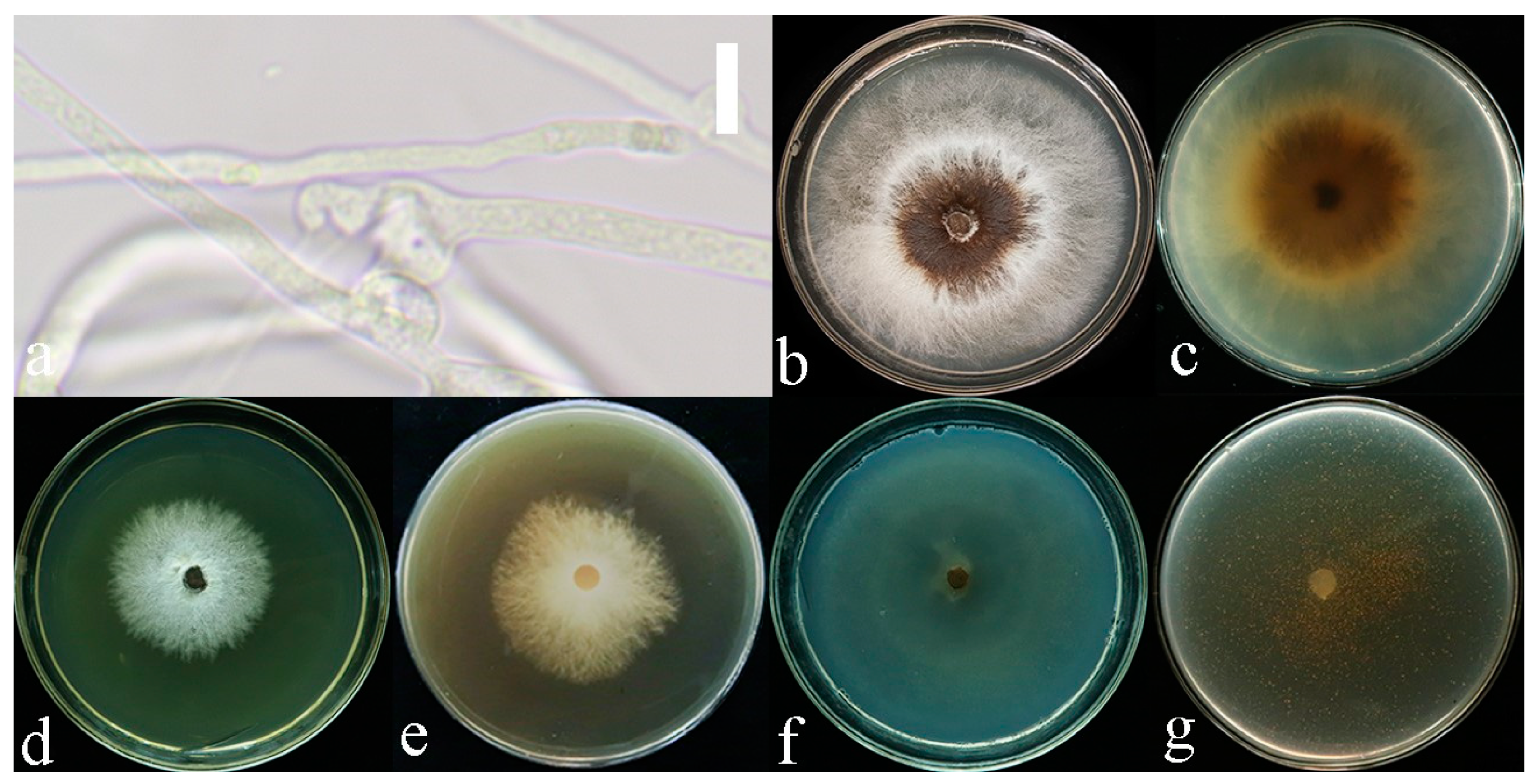
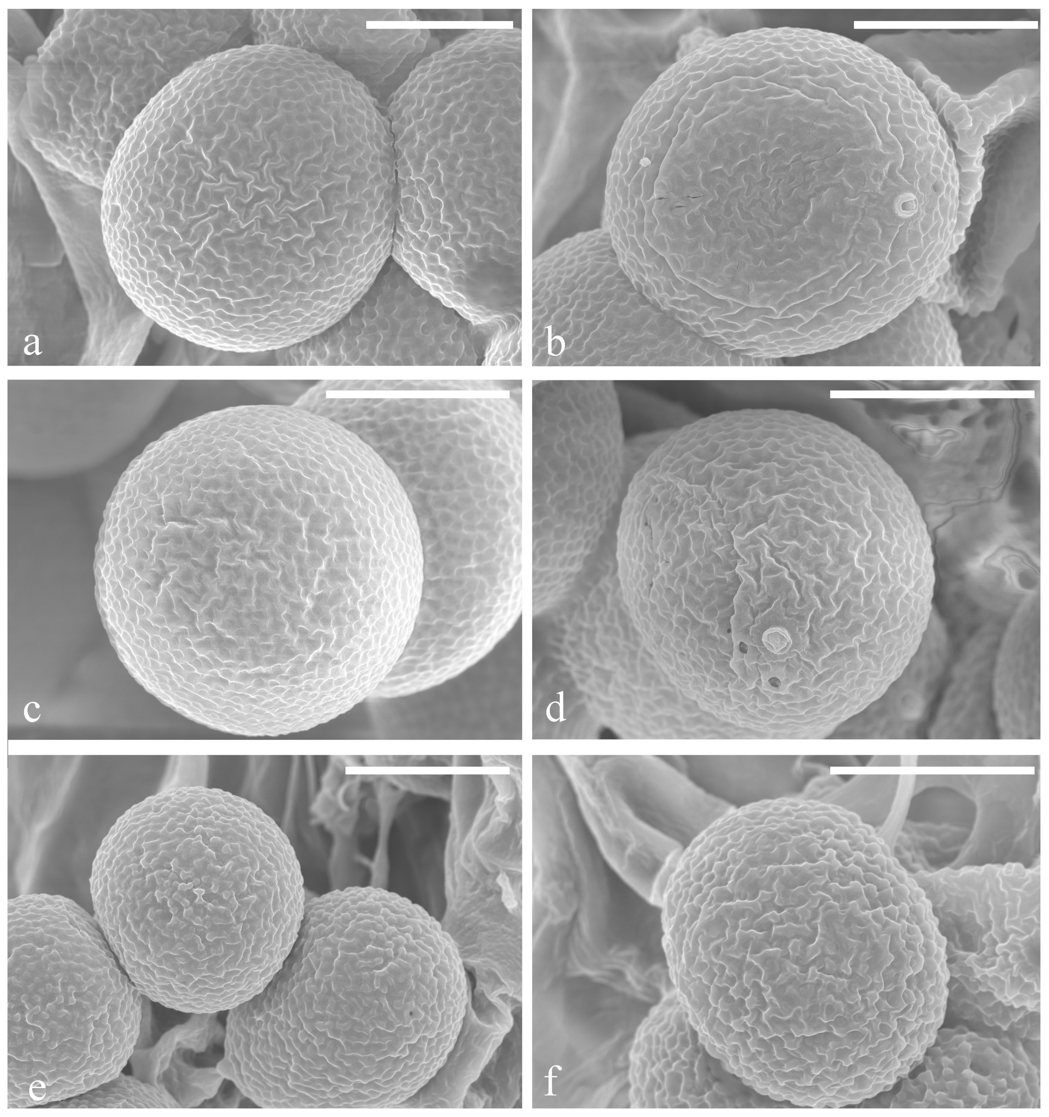
2.2. Morphological Observations

Macro-morphological studies were conducted following the protocols provided by Sun et al. [14]; the color was compared to the standard colors on the colorhexa website (https://www.colorhexa.com (accessed on 10 July 2024)). Micro-morphological structures were obtained from the dried specimens and then photographed by using a Nikon ECLIPSE Ni-U compound microscope fitted with a Nikon DS-Ri2 digital camera. Microscopic observations and color reactions were, respectively, made from slide preparations using 5% potassium hydroxide (KOH), Melzer’s reagent, and Cotton Blue. Measurements were made using the Image Frame work v.0.9.7. At least 20 basidiospores were measured in each specimen, 5% of measurements were excluded from each end of the range and extreme values were provided in parentheses [15]. The following abbreviations were used: IKI = Melzer’s reagent; IKI− = neither amyloid nor dextrinoid; CB = Cotton Blue; CB+ = cyanophilous; L = mean spore length (arithmetic average of all spores); W = mean spore width (arithmetic average of all spores); Q = L/W ratio and (a) = number of spores measured and (b) = specimens number [8]. Ultrastructures of basidiospores were observed via Scanning Electron Microscopy (SEM) at Yunnan Academy of Agricultural Sciences, China.

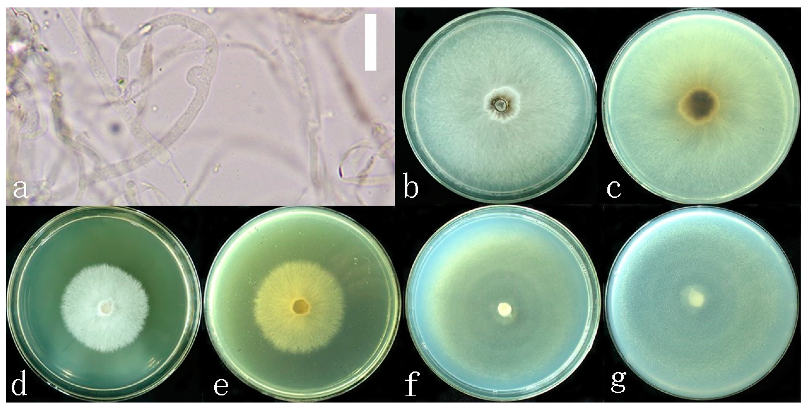
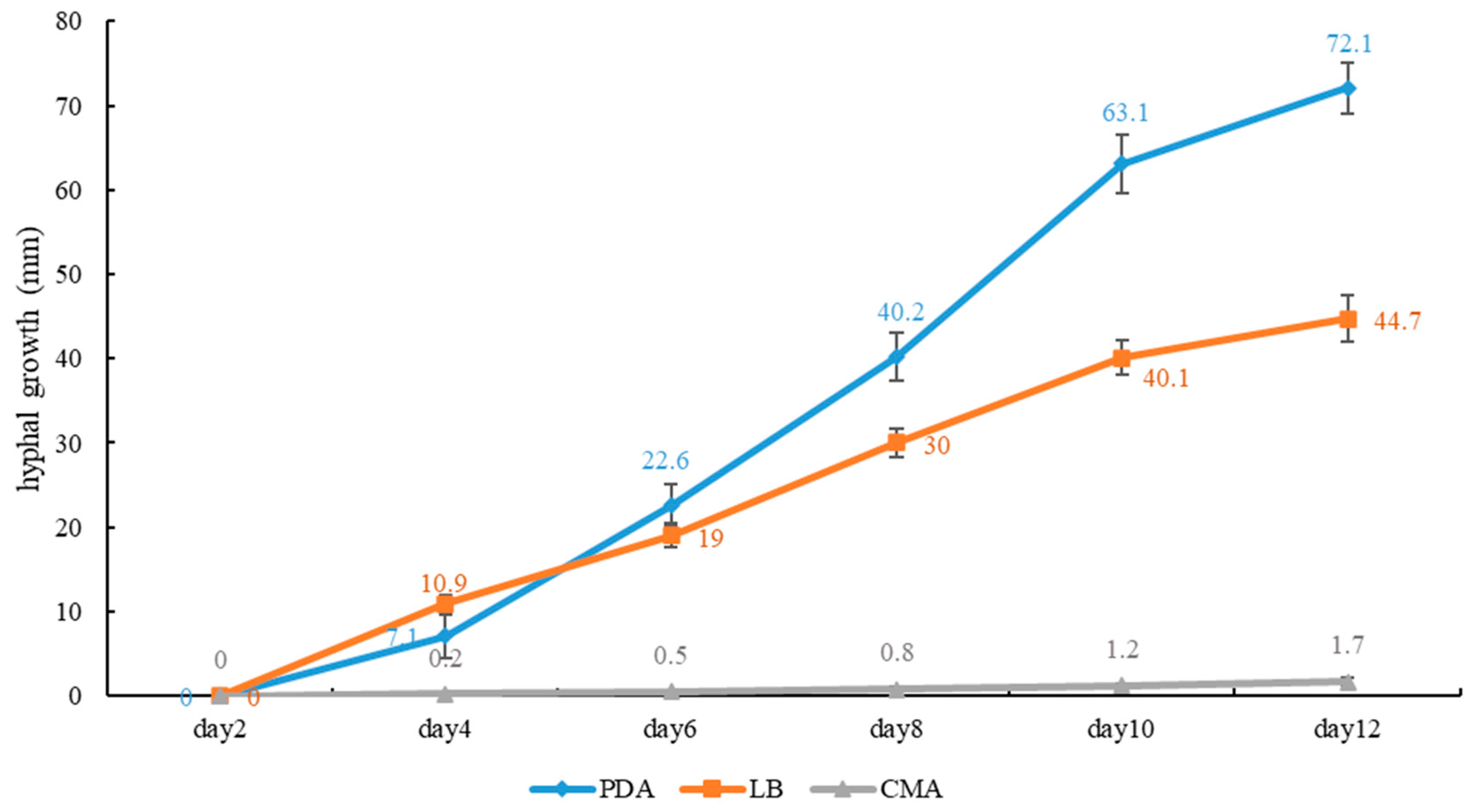
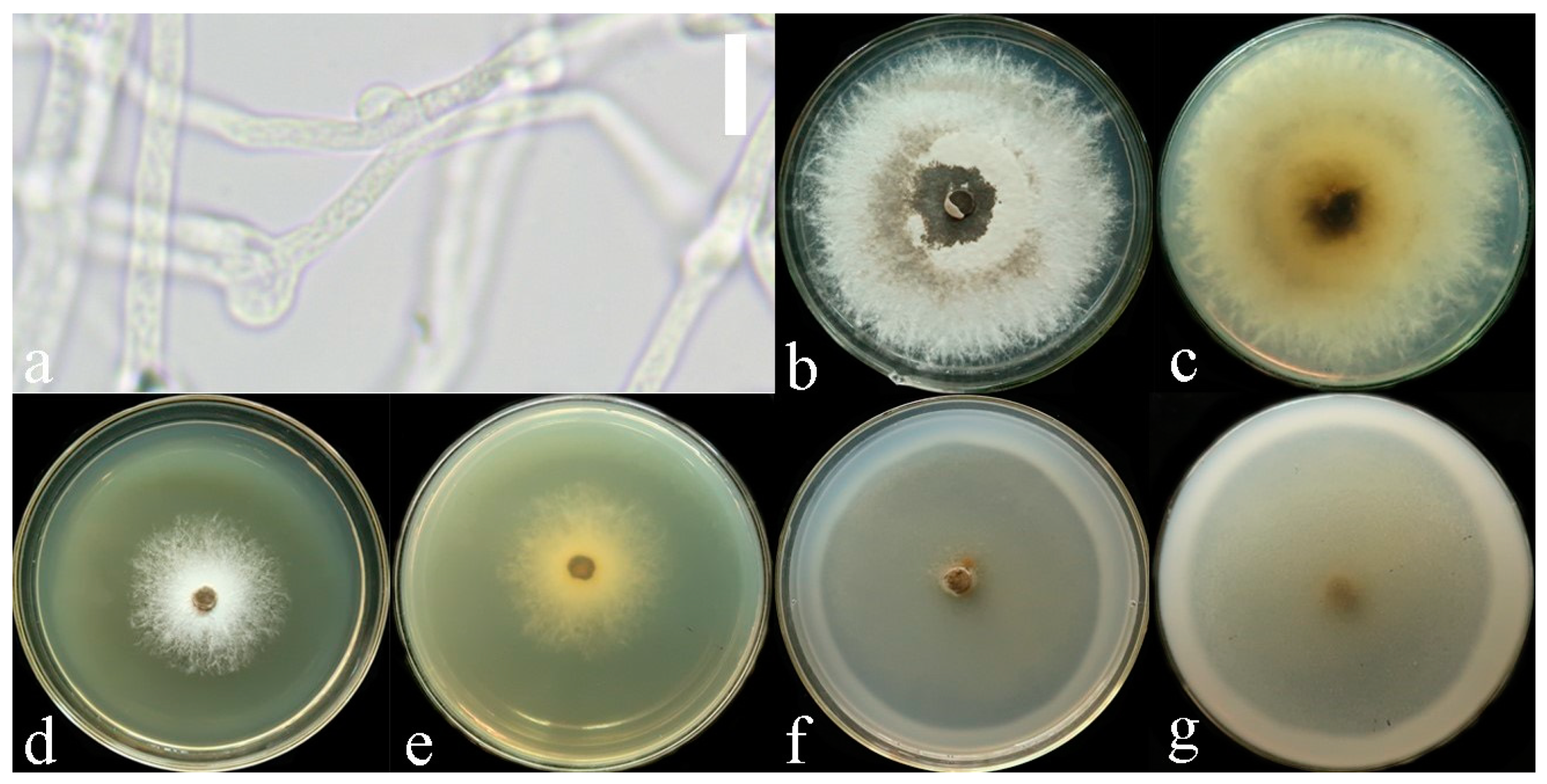
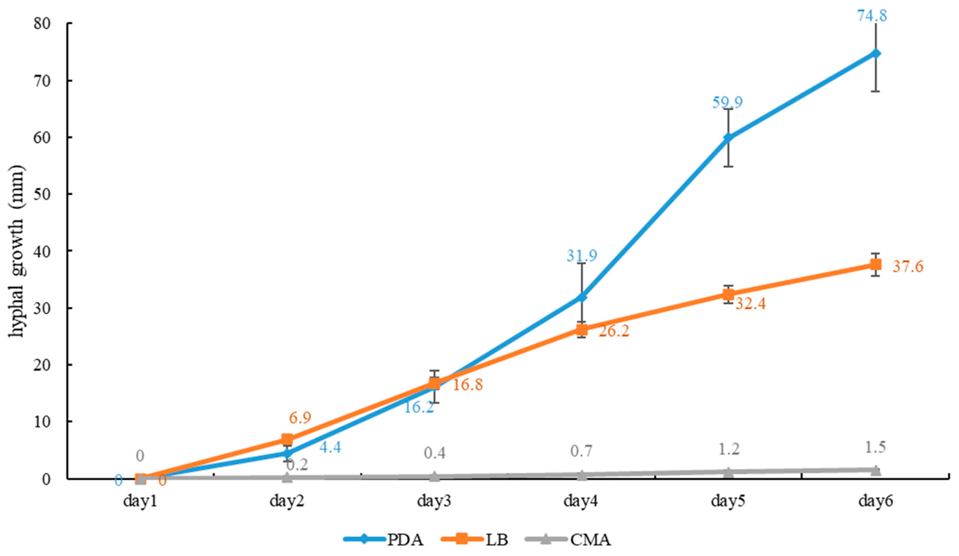
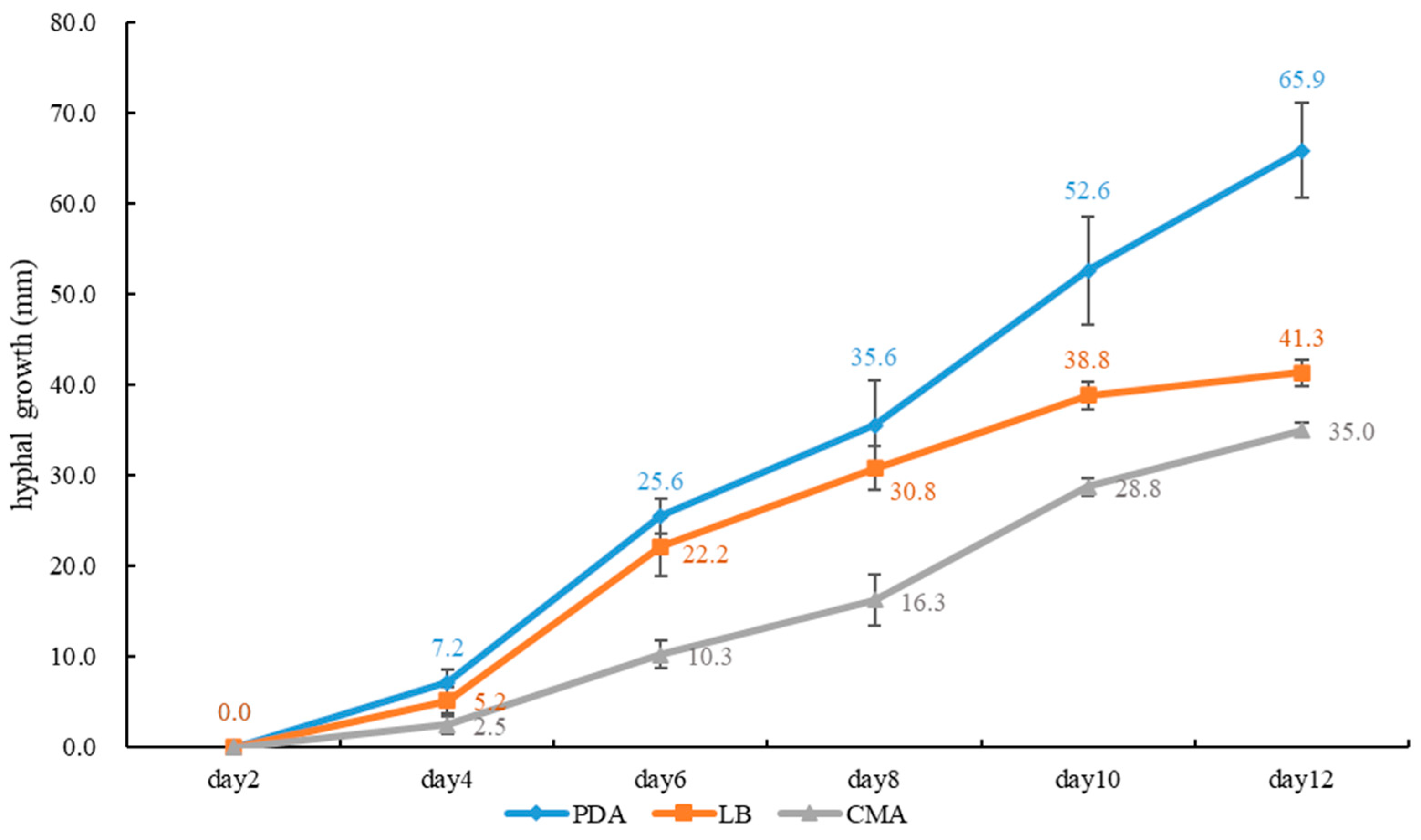
2.3. Growth Rate of Mycelium on Culture Media

A 7 mm diameter culture was taken from the edge of the PDA medium and inoculated onto new PDA, CMA (cornstarch 30 g, agar 20 g and distilled water 1000 mL) and LB (beef extract 3 g/L, peptone 10 g/L, NaCl 5 g/L, agar 20 g/L and distilled water 1000 mL) media. The cultures were incubated at a constant temperature of 24 °C, avoiding light. The growth diameter was measured every 24 h. Each experiment was repeated five times, and the growth rate of the pure culture was recorded.

2.4. DNA Extraction, PCR Amplification and Sequencing

Genomic DNA was extracted from dried specimens using Ezup Column Fungi Genomic DNA Purification Kit (Sangon Biotech Limited Company, Kunming, Yunnan, China) based on the manufacturer’s protocol. The internal transcribed spacer (ITS) regions were amplified with primer pairs ITS5 and ITS4 [16], LR0R and LR5 for the large subunit of nuclear ribosomal RNA gene (LSU) [17] and primer pairs fRPB2-5F and fRPB2-7CR [18] were used to amplify the second subunit of RNA polymerase II (rpb2). The translation elongation factor 1-α gene (tef1-α) was amplified with primer pairs EF1-983F and EF1-1567R [19]. The small subunit mitochondrial rRNA gene (mtSSU) was amplified with primer pairs MS1 and MS2 [16], and the small subunit nuclear ribosomal RNA gene (nSSU) was amplified with primer pairs PNS1 and NS41 [16].
The PCR volume contained 1 µL of each primer, 1 µL of extracted DNA, 9.5 µL of ddH2O and 12.5 µL of 2× EasyTaq PCR SuperMix (Sangon Biotechnology Co., Kunming, China). PCR cycling schedules for four-gene regions of ITS, LSU, mtSSU and nSSU were based on Sun et al. [20]. The PCR cycle of tef1-α was as follows: initial denaturation at 94 °C for 5 min, denaturation at 94 °C for 30 s, denaturation at 55 °C for 30 s, denaturation at 72 °C for 50 s and extension at 72 °C for 10 min; 35 cycles were repeated. The PCR cycle of rpb2 was as follows: initial denaturation at 95 °C for 5 min, 95 °C for 1 min, 51 °C for 2 min, 72 °C for 1.5 min for 35 cycles and, finally, extension for 10 min at 72 °C. The PCR amplicons were sent to Sangon Biotech (Kunming, Yunnan, China) for Sanger sequencing. Raw DNA sequences were assembled and edited in Sequencher v.4.1.4, and the assembled DNA sequences were deposited in GenBank (Table 1).

2.5. Sequence Alignment and Phylogenetic Analysis

The ITS, LSU, rpb2, tef1-α, mtSSU and nSSU sequences used in this study were combined into a dataset. Magoderna subresinosum was used as the outgroup [20], which is the sister clade of Sanguinoderma. Sequences were aligned using the online version of MAFFT v.7 (https://mafft.cbrc.jp/alignment/server/ (accessed on 6 March 2024)) [24] and were manually adjusted in BioEdit v.7.1.3 [25]. Ambiguous aligned regions were excluded from the analyses, and gaps were treated as missing data. The phylogeny website tool “ALTER” [26] was used to convert the Fasta file to Phylip format for RAxML analysis, Aliview and PAUP v.4.0b 10 were used to convert the Fasta alignment file to a Nexus file for Bayesian analysis [27].

2.6. Phylogenetic Inference

Based on the combined dataset, the maximum likelihood (ML) analysis was conducted in RAxML-HPC2 v.8.2.3 [28] and implemented on the CIPRES portal (https://www.phylo.org/portal2/login.action (accessed on 6 March 2024)) [29] with the GTR + G model for each gene and 1000 rapid bootstrap (BS) replicates. Since no supported conflict (BS ≥ 75%) was detected among the topologies, the six single-gene alignments were concatenated using Sequence Matrix [30].
Bayesian analysis was performed in MrBayes 3.2 [31] and the best-fit models of sequences’ evolution were estimated with MrModeltest 2.3 [32,33,34]; the selected models were HKY+G for ITS, GTR+I for LSU and nSS, GTR+I+G for mtSSU and rpb2 and HKY+I+G for tef1-α. The Markov Chain Monte Carlo (MCMC) sampling approach was used to calculate posterior probabilities (PP) [35]. Bayesian analysis of six simultaneous Markov chains was run for 10,000,000 generations, and trees were sampled every 1000 generations. The first 5000 trees, representing the burn-in phase of the analyses, were discarded, while the remaining 1500 trees were used for calculating posterior probabilities in the majority rule consensus tree (the critical value for the topological convergence diagnostic is 0.01).
Bootstrap support values in maximum likelihood (ML) equal to or greater than 75% and Bayesian posterior probabilities (PP) equal to or greater than 0.95 are provided above the nodes. All trees were viewed in FigTree v. 1.4.0 (http://tree.bio.ed.ac.uk/software/figtree/ (accessed on 12 March 2024)) and were edited using Adobe Illustrator CS5 (Adobe Systems Inc., San Jose, CA, USA) [15]. Sequences derived from this study were deposited in GenBank. The final sequence alignments and the phylogenetic trees are available at Figshare (https://figshare.com/ DIO: 10.6084/m9.figshare.26232650 (accessed on 20 July 2024)).

3. Results

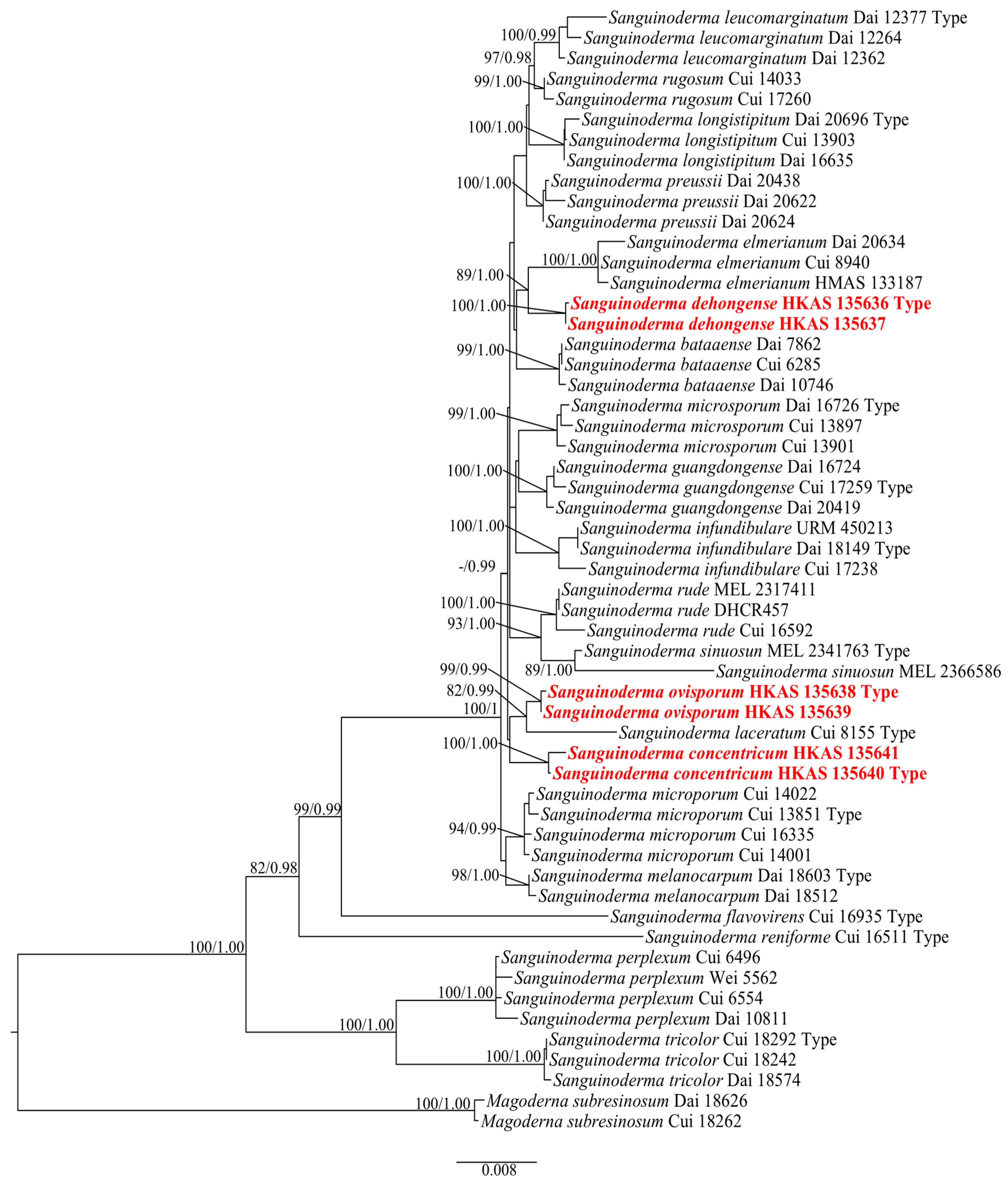
3.1. Phylogenetic Analyses
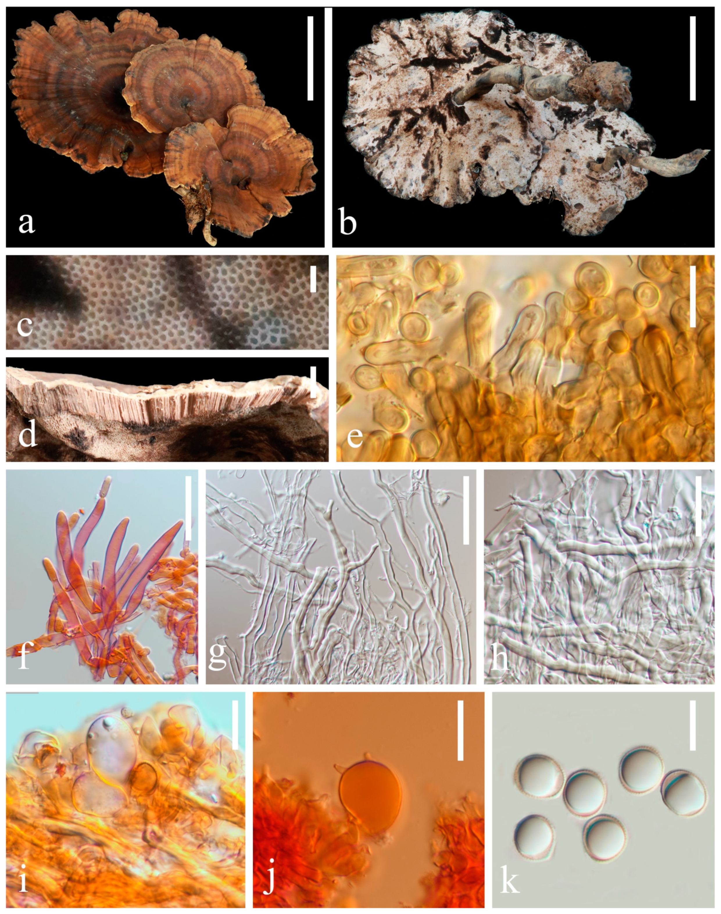
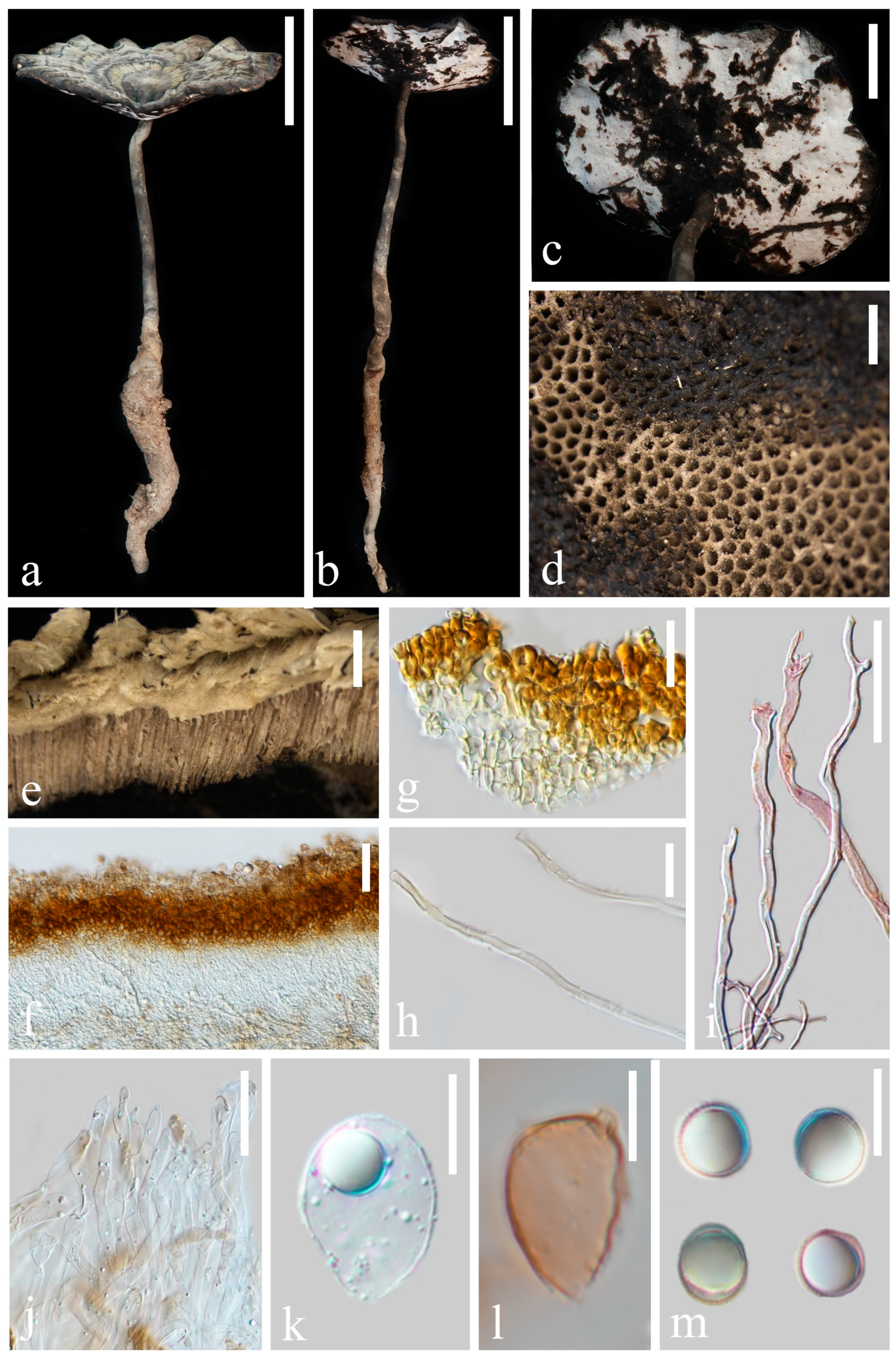
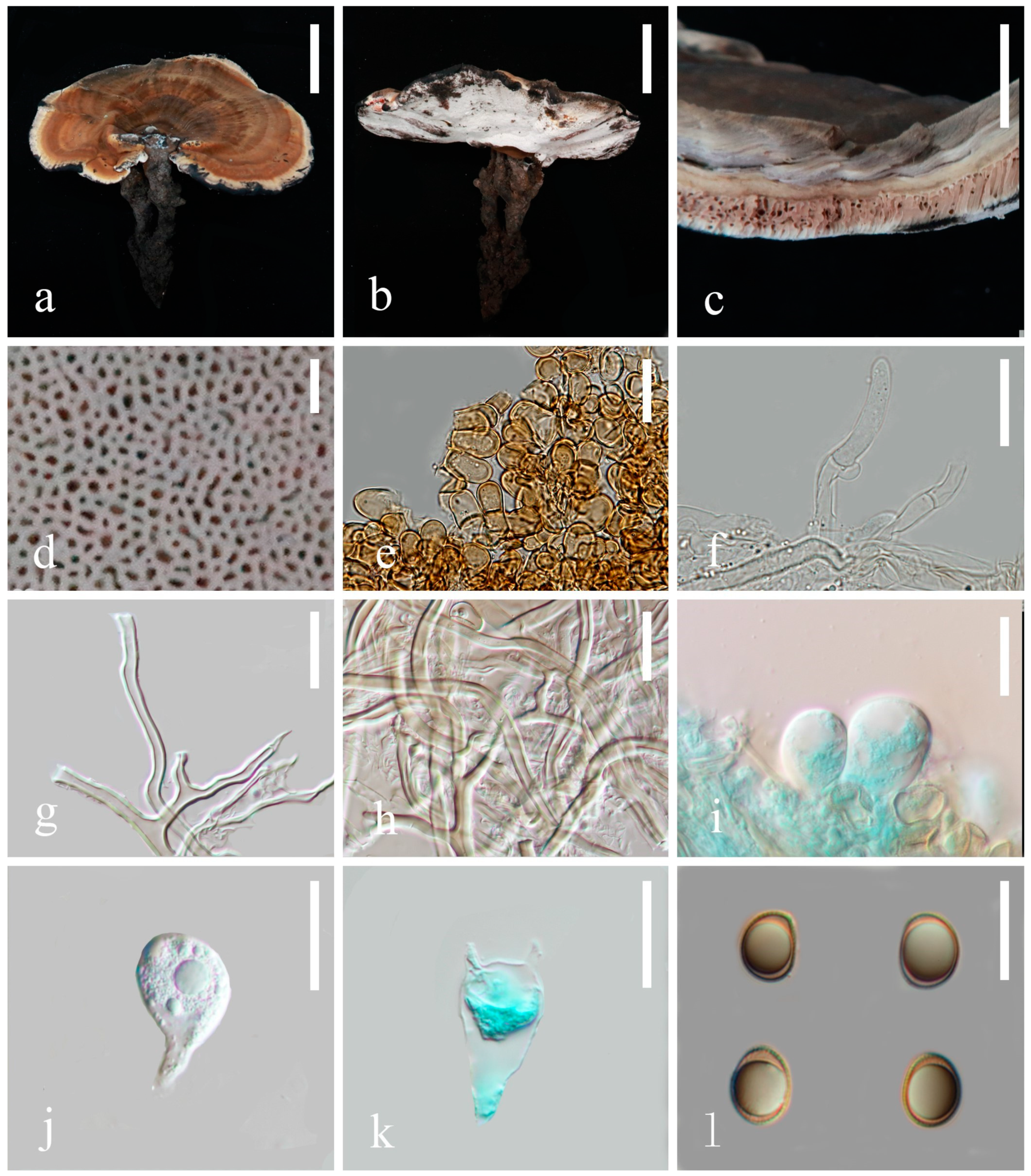
In this study, 290 sequences of ITS, LSU, rpb2, tef1-α, mtSSU and nSSU were used to construct phylogenetic trees of Sanguinoderma, including 52 ITS, 51 LSU, 37 rpb2, 48 tef1-α, 51 mtSSU and 51 nSSU. The sequences were obtained from 53 specimens representing 21 taxa in Sanguinoderma. The combined six-gene sequence dataset had an aligned length of 5012 total characters including gaps (ITS: 1–542; LSU: 543–1863; mtSSU: 1864–2371; nSSU: 2372–3454; rpb2: 3455–4491 and tef1-α: 4492–5011).
The tree topologies of the maximum likelihood analysis and the Bayesian analysis were similar. The RAxML analysis of the combined dataset yielded the best scoring tree with a final maximum likelihood value of −13,811.532268. The matrix had 664 distinct alignment patterns, with 20.32% undetermined characters or gaps. ML and BI analyses generated nearly identical tree topologies with minimal variations in statistical support values. Thus, only a ML tree is shown (Figure 1).
In the phylogenetic analysis, six specimens collected from Yunnan, China formed three monophyletic clades, S. concentricum (100% ML/1.00 PP), S. dehongense (100% ML/1.00 PP) and S. ovisporum (99% ML/0.99 PP). Sanguinoderma ovisporum and S. laceratum (Cui 8155) form sister clades with a good support value (82% ML/0.99 PP, Figure 1). A comparison of the LSU nucleotide bases between S. ovisporum (HKAS 135638) and S. laceratum (Cui 8155) revealed differences of 21 bps (21/915, including one gap). A comparison of the ITS and tef1-α nucleotide bases between S. dehongense (HKAS 135636) and S. elmerianum (Dai 20634) revealed differences of 12 bps (542/12, no gaps) and 15 bps (15/510, no gaps), respectively. A comparison of the LSU nucleotide bases between S. concentricum (HKAS 135640) and S. laceratum (Cui 8155) revealed differences of 19 bps (19/896, no gaps).
Figure 1. Tree results from the maximum likelihood (ML) phylogenetic tree of Sanguinoderma based on the dataset of ITS + LSU + rpb2 + tef1-α + mtSSU + nSSU. Branches are labeled with maximum likelihood bootstrap values equal to or higher than 75% and Bayesian posterior probability values equal to or higher than 0.95. New species are indicated in bold red.
Jof 10 00589 g001

3.2. Taxonomy

Sanguinoderma concentricum K.Y. Niu, J. He and Z.L. Luo sp. nov.
Fungal Names number: FN 572020
Diagnosis. Sanguinoderma concentricum differs from other species by its central stipe basidiomata, with concentrically zonate pileus surface, wavy margin lacerated like petals and broadly clavate basidia (16–18 × 18–21 μm).
Etymology. The epithet ‘concentricum’ refers to the pileus surface with obvious concentric zones.
Holotype. China, Yunnan Province, Dehong Prefecture, on the ground with humus, 1245 m, Kai-Yang Niu, 7 August 2023, HKAS 135640.
Description. Basidiomata is annual, centrally stipitate and occasionally imbricate; coriaceous to corky. Pileus orbicular to suborbicular, up to 9.7 cm diameter and 5 mm thick; surface is dark and moderately orange (#9d6c4b), dull and tomentose, with alternating concentric zones that are grayish yellow (#d3d2bd) to dark-grayish lime green (#5a605a); dense and radial fine wrinkles; margin is moderately orange (#c29144), obtuse, lacerated-like petals, acute, wavy and obviously incurved when dry. Context is up to 1 mm thick, homogeneous, light-grayish orange (#f4e8d0) and soft and corky without black melanoid lines. Tubes are up to 4 mm long, mostly desaturated dark orange (#b19570), hard and corky and unstratified. Pores are 3–4 per mm, circular to angular, grayish yellow (#c8c9c3) when fresh, changing to brown, dark and moderately red (#753a37) when bruised, then quickly darkening, without discoloration and dissepiments entire when dry. Stipe is up to 5.1 cm long and 8 mm in diameter, central, cylindrical, hollow, slightly curved, dark-grayish orange (#9c8c7c) and fibrous to woody.
Hyphal system trimitic, with generative hyphae 3–6 μm in diameter, hyaline, thin walled and with clamp connections; skeletal hyphae are 5–9 μm in diameter, hyaline, thick walled with a wide-to-narrow lumen, flexuous and arboriform; binding hyphae are 1–3 μm in diameter, hyaline, flexuous and branched. All hyphae are IKI− and CB; tissue darkening in KOH. Pileipellis is a regular palisade, apical cells are 4–8 × 17–28 μm, short clavate and yellowish brown. Basidiospores are broadly ellipsoid, pale gray, IKI− and CB+ with double and slightly thin walls; exospore wall is smooth; endospore wall features conspicuous pillars, (9.4) 9.6–10.7 (10.9) × (8.1) 8.2–9.2 (9.6), L = 10.0 μm, W = 8.7 μm and Q = 1.15 (40/2); under SEM, exospore wall reticulates (Figure 11a,b). Basidia: broadly clavate, hyaline, thin walled and 16–18 × 18–21 μm. Basidioles similar to basidia; 18–20 × 23–25 μm.
Culture features. Circular, slight smell of corruption, initially white to grayish white, gradually turns grayish black, changing to blood red when bruised, color of the agar does not change with the growth of the culture; generative hyphae feature multiple branches, with irregularly thickened walls or with scattered thick-walled, refractive areas on walls; texture is sub felty and farinaceous (Figure 3). On PDA, at 24 °C, growth is fast, reaching 74.8 mm in 6 days; culture hyphae are sparse. On LB, at 24 °C, growth is slow, reaching 37 mm at 6 days; culture hyphae are dense. On CMA, at 24 °C, growth is extremely slow, reaching 1.5 mm in 6 days; culture hyphae are particularly sparse (Figure 4).
Additional specimens examined. China, Yunnan Province, Dehong Prefecture, on the ground with humus, 1539 m, Kai-Yang Niu, 20 August 2023, HKAS 135641.
Notes: Sanguinoderma infundibulare described from subtropical areas of China is morphologically similar to S. concentricum by having the centrally to laterally stipitate basidiomata with a funnel-shape to orbicular pileus. However, S. infundibulare differs by the smaller pores (4–6 per mm), pale wood-brown context with dark melanoid lines and larger basidiospore (10–12.2 × 8.5–10.6 μm) [20]. Sanguinoderma preussii is another species that also has an orbicular pileus in Sanguinoderma, but it differs from S. concentricum by the grayish-brown pileus surface, smaller pores (6–7 per mm) and buff yellow context [10]. Moreover, S. concentricum and S. preussii were supported as two distinct lineages in the phylogenetic tree (Figure 1). In culture, S. concentricum exhibited a more uniform mycelial density compared to S. ovisporum and S. dehongense, with neat colony edges (Figure 3, Figure 6 and Figure 9). On PDA and LB media, the mycelial growth rate of S. concentricum was higher than that of S. ovisporum and S. dehongense (Figure 4, Figure 7 and Figure 10).
Figure 2. Basidiomata and microscopic structures of Sanguinoderma concentricum (HKAS 135640, holotype). (a,b) Basidiomata. (c) Pores. (d) Pileus cross-section. (e) Apical cells from pileipellis. (f) Terminal generative hyphae from tubes. (g) Binding and skeletal hyphae from context. (h) Skeletal hyphae from context. (i) Basidioles. (j) Basidia. (k) Basidiospores. Scale bars: a, b = 5 cm; c = 1 mm; d = 5 mm; e, i, j = 5 μm; f, h, k = 10 μm and g = 15 μm.
Jof 10 00589 g002
Figure 3. Sanguinoderma concentricum culture characters. (a) Clamped generative hyphae; (b,c) colony on PDA; (b) obverse; (c) reverse; (d,e) colony LB, (d) obverse; (e) reverse; (f,g) colony on CMA; (f) obverse; (g) reverse. Scale bars: a = 20 μm.
Jof 10 00589 g003
Figure 4. Average daily hyphal growth of Sanguinoderma concentricum on different culture media.
Jof 10 00589 g004
Sanguinoderma dehongense K.Y. Niu, J. He and Z.L. Luo sp. nov.
Fungal Names number: FN 572019
Diagnosis. Sanguinoderma dehongense differs from other species in its pileus surface being dark grayish yellow to dark yellow with concentrically zonate, long stipe and irregular pileipellis cells and wavy margin.
Etymology. The epithet ‘dehongense’ refers to the Dehong Prefecture of Yunnan Province, where the holotype was collected.
Holotype. China, Yunnan Province, Dehong Prefecture, on the ground covered with humus, 1339 m, Kai-Yang Niu, 7 August 2023, HKAS 135636.
Description. Basidiomata is annual, laterally stipitate and is hard and corky to woody and hard. Pileus is single, flabelliform to reniform, up to 7 cm in diameter and 5 mm thick; surface is grayish yellow (#d1d0be), dull and glabrous with alternating concentric zones that are dark-grayish yellow (#46473f) to dark yellow (#161614); dense and radial fine wrinkles; margin is very dark gray (#262626), obtuse, entire, strongly wavy and incurved when dry. Context is up to 2 mm thick, homogeneous, slightly desaturated orange (#b59a6b) when dry and soft and corky without black melanoid lines. Tubes are up to 3 mm long, dark and moderately orange (#7b5f3d) when dry, hard and woody and unstratified. Pores are 3–4 per mm, subregular pentagonal to oblong, very light gray (#fafafa) when fresh, changing to mostly desaturated dark red (#95746c) when bruised, then quickly darkening; mostly desaturated dark orange (#b0926e) and dissepiments entire when dry. Stipe is up to 21 cm long and 6 mm diameter, cylindrical, hollow, slightly curved, swollen at base, dark-grayish orange (#a8a190) to very dark-grayish yellow (#33322e) and fibrous to woody.
Hyphal system trimitic, with generative hyphae 2–5 μm in diameter, hyaline and thin walled with clamp connections; skeletal hyphae are 4–8 μm in diameter, pale grey to pale yellow and thick walled with a wide to narrow lumen or subsolid, straight and little branched; binding hyphae are 2–5 μm in diameter, pale grey, thick walled, branched and flexuous; all hyphae IKI−, CB+; tissue darkening in KOH. Pileipellis is irregular palisade; apical cells are 5–9 × 11–21 μm, tightly packed, with narrow lumen, tightly packed together, thick walled and pale yellowish brown, forming an irregular palisade. Basidiospores are subglobose to broadly ellipsoid, pale grey, IKI− and CB, with double and thick walls; exospore wall is smooth; endospore wall features faint pillars, (9.1) 9.3–10.8 (11.0) × (8.1) 8.4–9.8 (9.9) μm, L = 10.1 μm, W = 9.1 μm and Q = 1.11 (40/2). Under SEM, exospore wall is reticulate (Figure 11c,d). Basidia is ellipsoid, hyaline, thin walled and 8–11 × 9–13 μm. Basidioles are obovoid, hyaline, thin walled and 12–14 × 15–19 μm.
Culture feature. Circular, edge irregularity, slight smell of corruption and initially white to grayish white; it gradually turns grayish and eventually ages to black; color changes to blood red when bruised and color does not change with agar-induced growth of the culture; generative hyphae feature multiple branches, with irregularly thickened walls or with scattered thick-walled, refractive areas on the walls; texture is sub felty and farinaceous (Figure 6). On PDA at 24 °C, growth is fast, reaching 72.1 mm in 12 days; culture hyphae are dense. On LB at 24 °C, growth is slow, reaching 44.7 mm in 12 days; culture hyphae are dense. On CMA at 24 °C, growth is extremely slow, reaching 1.7 mm at 12 days; culture hyphae are particularly sparse (Figure 7).
Additional specimen examined. China, Yunnan Province, Dehong Prefecture, on the ground covered with humus, 1545 m, Kai-Yang Niu, 7 August 2023, HKAS 135637.
Notes: Morphologically, Sanguinoderma longistipitum has a similar distribution with S. dehongense which can be collected from Yunnan Province, but the former can be distinguished by a sub-orbicular to flabelliform pileus, a smaller basidiomata, a pore density of 6–8 per mm and finger-shaped pileipellis cells with multiple obvious septa [20]. Sanguinoderma elmerianum also has lateral stipitate basidiomata and is swollen at the base of the stipe, but it differs from S. dehongense by the flat reniform pileus and short stipe (9 cm long) [8]. Based on phylogenetic analysis and morphological evidence, we introduce Sanguinoderma dehongense as a new species. In culture, the edges of S. dehongense are irregular compared to S. ovisporum and S. concentricum; on PDA, hyphal growth of S. dehongense is denser than that of S. ovisporum and S. concentricum (Figure 3, Figure 6 and Figure 9). On CMA, S. dehongense exhibits the slowest mycelial growth rate compared to S. ovisporum and S. concentricum (Figure 4, Figure 7 and Figure 10).
Figure 5. Basidiomata and microscopic structures of Sanguinoderma dehongense (HKAS 135636, holotype). (ac) Basidiomata. (d) Pores. (e) Pileus cross-section. (f,g) Apical cells from pileipellis. (h) Skeletal hyphae from context. (i) Binding and arboriform skeletal hyphae from context. (j) Terminal generative hyphae from tubes. (k) Basidioles. (l) Basidia. (m) Basidiospores. Scale bars: a, b = 5 cm; c = 2 cm; d = 1 mm; e = 2 mm; f, i = 50 μm; g = 20 μm; h, j = 30 μm; k–m = 10 μm.
Jof 10 00589 g005
Figure 6. Sanguinoderma dehongense culture characters. (a) Clamped generative hyphae; (b,c) colony on PDA; (b) obverse; (c) reverse; (d,e) colony on LB; (d) obverse; (e) reverse; (f,g) colony on CMA; (f) obverse; (g) reverse. Scale bars: a = 10 μm.
Jof 10 00589 g006
Figure 7. Average daily hyphal growth of Sanguinoderma dehongense on different culture media.
Jof 10 00589 g007
Sanguinoderma ovisporum K.Y. Niu, J. He and Z.L. Luo sp. nov.
Fungal Names number: FN 572027
Diagnosis. Sanguinoderma ovisporum differs from other species by its dark moderate orange surface pileus, tomentose, lateral stipe, heterogeneous context and ovoid basidiospores (7.5–8.6 × 5.5–7.2 μm).
Etymology. The epithet ‘ovisporum’ refers to the ovoid basidiospores.
Holotype. China, Yunnan Province, Dehong Prefecture, on the ground with humus, 1555 m, Kai-Yang Niu, 16 July 2023, HKAS 135638.
Description. Basidiomata is annual, laterally stipitate, solitary, occasionally imbricate and hard corky to hard woody. Pileus is single, reniform, up to 11.6 cm in diameter and 4 mm thick. Pileus surface is dark moderate orange (#986f53), dull and tomentose with alternating concentric zones mostly desaturated dark orange (#9a765b) and dark moderate orange (#7f553d); dense and radial fine wrinkles. Pileus margin is grayish orange (#bab8b3), entire, obtuse to acute and irregularly wavy. Context is up to 2 mm thick and homogeneous, the upper layer is very dark grayish yellow (#5f5e5a), the middle layer is dark grayish yellow (#b1b0ad), the lower layer is grayish orange (#b3a696), and it is soft corky and fibrous, without black melanoid lines. Tubes are up to 2 mm thick, mostly desaturated dark orange (#917c68), light gray (#d3d3d3) near the pores, hard wood and unstratified. Pores are 3–5 per mm, elongated elliptical to subangular and very light gray (#f5f5f5) when fresh, changing to dark red (#9c3d2d) when bruised, then quickly darkening; no discoloration and dissepiments entire when dry. Stipe is up to 7 cm long and 2 mm in diameter, cylindrical, hollow, slightly swollen at base, very dark grayish orange (#55423b), darker than pileus and fibrous to woody.
Hyphal system trimitic, with generative hyphae 3–6 μm in diameter, hyaline, thin walled, with clamp connections; skeletal hyphae 5–8 μm in diameter, pale yellow, thick walled with a wide or narrow lumen that is subsolid, flexuous and arboriform; binding hyphae are 2–5 μm in diameter, pale yellow, flexuous and branched. All hyphae are IKI− and CB+; tissues darken in KOH. Pileipellis is a regular palisade; apical cells are 10–14 × 5–9 μm, short-stick shaped, thin to thick walled and light to dark yellowish brown. Basidiospores are subglobose to ellipsoid, faint yellow, IKI− and CB+ with double and slightly thin walls; exospore wall is smooth; endospore wall features indistinct thin pillars, (7.5) 7.7–8.5 (8.6) × (5.5) 5.6–6.9 (7.2) μm, L = 8.2 μm, W = 6.2 μm and Q = 1.31 (40/2). Under SEM, exospore wall is vermiculate, and umbilical protrusion is notably present at the apex (Figure 11e,f). Basidia is broadly clavate, hyaline, thin walled and 22–25 × 10–12 μm. Basidioles are similar to basidia, hyaline, thin walled and 20–23 × 10–12 μm.
Culture feature. Circular, slight smell of corruption and initially white to grayish white; with the increase in culture time, it gradually turns grayish black, changing to blood red when bruised; color does not change with agar-induced growth of the culture; generative hyphae feature multiple branches, with irregularly thickened walls or with scattered thick-walled, refractive areas on walls; texture is sub felty and farinaceous (Figure 3). On PDA, at 24 °C, growth is fast, reaching 65.9 mm in 12 days; culture hyphae are dense. On LB, at 24 °C, growth is slow, reaching 41.3 mm in 12 days; culture hyphae are dense. On CMA, at 24 °C, growth is slow, reaching 35 mm in 12 days; culture hyphae are particularly sparse (Figure 4).
Additional specimens examined. China, Yunnan Province, Dehong Prefecture, on the ground with humus, 1322 m, Kai-Yang Niu, 13 July 2023, HKAS 135639.
Notes: Sanguinoderma laceratum and S. ovisporum are similar in having annual, stipitate and soft basidiomata. However, S. laceratum is different from S. ovisporum in its multiple and superposed pileus, homogeneous context, larger pores (2–3 per mm) and basidiospores (11.3 × 9.7 μm) [8]. In the phylogenetic analyses, S. ovisporum was shown to be a distinct lineage in Sanguinoderma (Figure 1). In culture, the mycelia of S. ovisporum, S. dehongense and S. concentricum on CMA are all extremely sparse (Figure 3, Figure 6 and Figure 9); the growth rate of S. ovisporum was significantly higher than that of S. dehongense and S. concentricum (Figure 4, Figure 7 and Figure 10).
Figure 8. Basidiomata and microscopic structures of Sanguinoderma ovisporum (HKAS 135638, holotype) (a,b) Basidiomata. (c) Pileus cross-section. (d) Pores. (e) Cells from pileus cover. (f) Generative hyphae from tubes. (g) Skeletal hyphae from context. (h) Binding and skeletal hyphae from context. (i,j) Basidioles. (k) Basidia. (l) Basidiospores. Scale bars: a, b = 5 cm; c = 5 mm; d = 1 mm; e–h = 20 μm; i–k = 15 μm; l = 10 μm.
Jof 10 00589 g008
Figure 9. Sanguinoderma ovisporum culture characters. (a) Clamped generative hyphae; (b,c) colony on PDA; (b) obverse; (c) reverse; (d,e) colony on LB; (d) obverse; (e) reverse; (f,g) colony on CMA; (f) obverse; (g) reverse. Scale bars: a = 10 μm.
Jof 10 00589 g009
Figure 10. Average daily hyphal growth of Sanguinoderma ovisporum on different culture media.
Jof 10 00589 g010
Figure 11. Scanning Electron Micrograph (SEM) of basidiospores of Sanguinoderma. (a,b) S. concentricum (HKAS 135640, holotype); (c,d) S. dehongense (HKAS 135636, holotype); (e,f) S. ovisporum (HKAS 135638, holotype). Scale bars: a–f = 5 μm.
Jof 10 00589 g011

4. Discussion

Sanguinoderma is widely distributed in tropical to subtropical regions [8], there are currently no reports indicating the existence of obligate parasitism and habitats in this genus. It grow in various habitats, such as forest floors, fallen angiosperm trunks and different decaying stumps (Litchi chinensis, Eucalyptus and Acacia) [8,14,20]. For example, S. rugosum has been reported on the ground of Angiosperm forests and on Acacia mangium; more investigation of habitat diversity is an important research direction for the future. So far, a total of 15 species of Sanguinoderma have been reported in China. The discovery of three new species of Sanguinoderma in this study raises the number of known Sanguinoderma species in Yunnan Province to 10, namely S. ovisporum, S. dehongense, S. elmerianum, S. guangdongense, S. laceratum, S. leucomarginatum, S. longistipitum, S. preussii, S. rugosum and S. concentricum, accounting for 66% of the known species, making it the most diverse province in China. Yunnan Province predominantly experiences tropical and subtropical monsoon climates, as well as rich vegetation types, which provides more possibilities for the growth of Sanguinoderma species. Therefore, Yunnan Province may be hiding a greater diversity of Sanguinoderma species than expected.
In this study, three new species from Yunnan Province, China are introduced based on morphology and multigene phylogeny. S. concentricum, S. ovisporum and S. laceratum occupied distinct places in the multilocus phylogenetic tree. Morphologically, S. laceratum differs from S. ovisporum and S. concentricum by having lacerate pore dissepiments and fascicular tubes when dry; S. concentricum differs from S. ovisporum in the pileus margin with lacerated-like petals and a larger basidiospore (9.4–10.9 × 8.1–9.6 μm). S. dehongense and S. elmerianum formed sister clades; however, the context of S. elmerianum features two dark melanoid lines with smaller pores (5–7 per mm) and a shorter stipe (9 cm long) than those of S. dehongense [8].
In recent years, phylogenetic analyses have been widely used for the classification of Ganodermataceae [8,10,11,14,20,23,36]. Sun et al. [20] conducted phylogenetic analysis of Ganodermataceae; single-gene sequences (ITS, nLSU, rpb2, mtSSU and nSSU) are difficult to distinguish between some species within Sanguinoderma. For instance, a comparison of the ITS, nLSU, rpb2, mtSSU and nSSU genes between S. sinuosum (MEL 2341763 Type) and S. rude (Cui 16592) revealed 1/543 (0.18%, no gaps), 1/875 (0.11%, no gaps), 6/1050 (0.57%, no gaps), 8/513 (1.15%, including one gap), 0/1085 (0%), respectively; these single-gene sequences fail to effectively differentiate the two species. In contrast, tef1-α sequences can effectively distinguish species within Sanguinoderma in phylogenetic analysis. In this study, a multi-gene approach (ITS, LSU, rpb2, tef1-α, mtSSU and nSSU) was selected for the phylogenetic analysis of Sanguinoderma species to clarify the placement of the newly collected specimens.
In culture, the three strains of Sanguinoderma consistently exhibited specific growth characteristics across various media. They displayed the fastest growth rate on PDA, slower growth on LB and the slowest growth on CMA. The PDA medium enriched with glucose likely enhances hyphal growth [37]; so, for isolating Sanguinoderma species in culture, prioritizing the use of PDA medium is recommended. All cultures exhibited a color change to brownish red when bruised, progressing to black over time; a characteristic also observed in the basidiomata pores. In addition, in our study, we observed that when using PDA as a preservation medium, cultures stored at 4 °C were prone to viability loss. Therefore, we recommend exploring alternative preservation methods for Sanguinoderma strains.

Author Contributions

Conceptualization, Z.-L.L.; methodology, K.-Y.N.; formal analysis, K.-Y.N. and J.H.; investigation, K.-Y.N. and J.H.; resources, Z.-L.L.; data curation, K.-Y.N.; writing—original draft preparation, K.-Y.N.; writing—review and editing, J.H., S.-M.T., X.-J.S. and Z.-L.L.; funding acquisition, Z.-L.L. All authors have read and agreed to the published version of the manuscript.

Funding

This work was supported by the Foundation of Yunnan Province Science and Technology Department (202305AM070003), the High-Level Talents Program of Yunnan Province (YNQR-QNRC-2020-113), and the Scientific Research Project of West Yunnan University (DXXY202313).

Institutional Review Board Statement

Not applicable.

Informed Consent Statement

Not applicable.

Data Availability Statement

Not applicable.

Acknowledgments

We would like to thank Shaun Pennycook for checking the nomenclature. Kai-Yang Niu would like to thank Zheng-Cong Li and Wen-Zao Feng for their help on sample collection. Lin Han and Xue-Jia Bai are acknowledged for their help on sample collection, DNA extraction, PCR amplification and pure culture experiments.

Conflicts of Interest

The authors declare no conflict of interest.

References

  1. Dai, Y.C.; Yang, Z.L.; Cui, B.K.; Yu, C.J.; Zhou, L.W. Species diversity and utilization of medicinal mushrooms and fungi in China. Int. J. Med. Mushrooms 2009, 11, 287–302. [Google Scholar] [CrossRef]
  2. Chan, K.S. Anti–Hyperlipidemic Activities of Amauroderma rugosum on Human Hepatocellular Carcinoma Cells. Ph.D. Thesis, University of Malaya, Kuala Lumpur, Malaysia, 2018. [Google Scholar]
  3. Chan, P.M.; Tan, Y.S.; Chua, K.H.; Sabaratnam, V.; Kuppusamy, U.R. Attenuation of inflammatory mediators (TNF-α and nitric oxide) and up–regulation of IL–10 by wild and domesticated Basidiocarps of Amauroderma rugosum (Blume and T. Nees) Torrend in LPS–stimulated RAW264.7 cells. PLoS ONE 2015, 10, e0139593. [Google Scholar] [CrossRef] [PubMed]
  4. Chan, P.-M.; Kanagasabapathy, G.; Tan, Y.-S.; Sabaratnam, V.; Kuppusamy, U.R. Amauroderma rugosum (Blume and T. Nees) Torrend: Nutritional composition and antioxidant and potential anti–inflammatory properties. Evid. Based Complement. Altern. Med. 2013, 304713. [Google Scholar] [CrossRef] [PubMed]
  5. Chang, Y.S.; Lee, S.S. Utilisation of macrofungi species in Malaysia. Fungal Divers. 2013, 1, 15–22. [Google Scholar]
  6. Liew, G.M.; Khong, H.Y.; Kutoi, C.J. Phytochemical screening, antimicrobial and antioxidant activities of selected fungi from mount Singai, Sarawak. Malays. Int. J. Res. Stud. Biosci. 2015, 3, 191–197. [Google Scholar]
  7. Zheng, C.W.; Cheung, T.M.Y.; Leung, G.P.H. A review of the phytochemical and pharmacological properties of Amauroderma rugosum. Kaohsiung J. Med. Sci. 2022, 38, 509–516. [Google Scholar] [CrossRef]
  8. Sun, Y.-F.; Costa-Rezende, D.H.; Xing, J.-H.; Zhou, J.-L.; Zhang, B.; Gibertoni, T.B.; Gates, G.; Glen, M.; Dai, Y.-C.; Cui, B.-K. Multi-gene phylogeny and taxonomy of Amauroderma s. lat. (Ganodermataceae). Persoonia Mol. Phylogeny Evol. Fungi 2020, 44, 206–239. [Google Scholar] [CrossRef]
  9. Murrill, W.A. Tomophagus for Dendrophagus. Torreya 1905, 5, 197. [Google Scholar]
  10. Costa-Rezende, D.H.; Gugliotta, A.M.; Goes-Neto, A.; Reck, M.A.; Robledo, G.L.; Drechsler-Santos, E.R. Amauroderma calcitum sp. nov. and notes on taxonomy and distribution of Amauroderma species Ganodermataceae. Phytotaxa 2016, 244, 101–124. [Google Scholar]
  11. Costa-Rezende, D.H.; Robledo, G.L.; Góes-Neto, A.; Reck, M.A.; Crespo, E.; Drechsler-Santos, E.R. Morphological reassessment and molecular phylogenetic analyses of Amauroderma s. lat. raised new perspectives in the generic classification of the Ganodermataceae family. Persoonia Mol. Phylogeny Evol. Fungi 2017, 39, 254–269. [Google Scholar] [CrossRef]
  12. Wachtel-Galor, S.; Yuen, J.; Buswell, J.A.; Benzie, I.F. Ganoderma lucidum (Lingzhi or Reishi): A Medicinal Mushroom. Herbal Medicine: Biomolecular and Clinical Aspects, 2nd ed.; CRC Press/Taylor and Francis: Boca Raton, FL, USA, 2011. [Google Scholar]
  13. Azliza, M.A.; Ong, H.C.; Vikineswary, S.; Noorlidah, A.; Haron, N.W. Ethno-medicinal resources used by the Temuan in Ulu Kuang Village. Stud. Ethno-Med. 2012, 6, 17–22. [Google Scholar] [CrossRef]
  14. Sun, Y.F.; Fang, Y.X.; Cui, B.K. Taxonomy and phylogeny of Sanguinoderma rugosum complex with descriptions of a new species and a new combination. Front. Microbiol. 2022, 13, 1087212. [Google Scholar] [CrossRef] [PubMed]
  15. He, J.; Luo, Z.L.; Tang, S.M.; Li, Y.J.; Li, S.H.; Su, H.Y. Phylogenetic analyses and morphological characters reveal two new species of Ganoderma from Yunnan Province, China. MycoKeys 2021, 84, 141. [Google Scholar] [CrossRef] [PubMed]
  16. White, T.J.; Bruns, T.; Lee SJ, W.T.; Taylor, J. Amplification and direct sequencing of fungal ribosomal RNA genes for phylogenetics. In PCR Protocols: A Guide to Methods and Applications; Academic Press, Inc.: Cambridge, MA, USA, 1990; Volume 38, pp. 315–322. [Google Scholar]
  17. Vilgalys, R.; Hester, M. Rapid genetic identification and mapping of enzymatically amplified ribosomal DNA from several Cryptococcus specie. J. Bacteriol. 1990, 172, 4238–4246. [Google Scholar] [CrossRef]
  18. Liu, Y.J.; Whelen, S.; Hall, B.D. Phylogenetic relationships among ascomycetes: Evidence from an RNA polymerse II subunit. Mol. Biol. Evol. 1999, 16, 1799–1808. [Google Scholar] [CrossRef] [PubMed]
  19. Rehner, S.A.; Buckley, E. A Beauveria phylogeny inferred from nuclear ITS and EF1-α sequences: Evidence for cryptic diversification and links to Cordyceps teleomorph. Mycologia 2005, 97, 84–98. [Google Scholar] [CrossRef]
  20. Sun, Y.-F.; Xing, J.-H.; He, X.-L.; Wu, D.-M.; Song, C.-G.; Liu, S.; Vlasák, J.; Gates, G.; Gibertoni, T.B.; Cui, B.-K. Species diversity, systematic revision and molecular phylogeny of Ganodermataceae (Polyporales, Basidiomycota) with an emphasis on Chinese collections. Stud. Mycol. 2022, 101, 287–415. [Google Scholar] [CrossRef]
  21. Li, M.J.; Yuan, H.S. Type studies on Amauroderma species described by JD Zhao et al. and the phylogeny of species in China. Mycotaxon 2015, 130, 79–89. [Google Scholar] [CrossRef]
  22. Song, J.; Xing, J.H.; Decock, C.; He, X.L.; Cui, B.K. Molecular phylogeny and morphology reveal a new species of Amauroderma (Basidiomycota) from China. Phytotaxa 2016, 260, 47–56. [Google Scholar] [CrossRef]
  23. Costa-Rezende, D.H.; Robledo, G.L.; Drechsler-Santos, E.R.; Glen, M.; Gates, G.; de Madrignac Bonzi, B.R.; Popoff, O.F.; Crespo, E. Taxonomy and phylogeny of polypores with ganodermatoid basidiospores (Ganodermataceae). Mycol. Prog. 2020, 19, 725–741. [Google Scholar] [CrossRef]
  24. Katoh, K.; Standley, D.M. MAFFT multiple sequence alignment software version 7: Improvements in performance and usability. Mol. Biol. Evol. 2013, 30, 772–780. [Google Scholar] [CrossRef]
  25. Hall, T.A. BioEdit: A user–friendly biological sequence alignment editor and analysis program for Windows 95/98/NT. Nucleic Acids Symp. Ser. 1999, 41, 95–98. [Google Scholar]
  26. Glez-Pen, D.; Gomez-Blanco, D.; Reboiro-Jato, M.; Fdez-Riverola, F.; Posada, D. ALTER: Program-oriented conversion of DNA and protein alignments. Nucleic Acids Res. 2010, 38, 14–18. [Google Scholar] [CrossRef] [PubMed]
  27. Swofford, D.L. Phylogenetic Analysis Using Parsimony (*and Other Methods); Sinauer Associates: Sunderland, UK, 2002. [Google Scholar]
  28. Stamatakis, A. RAxML version 8: A tool for phylogenetic analysis and post–analysis of large phylogenie. Bioinformatics 2014, 30, 1312–1313. [Google Scholar] [CrossRef] [PubMed]
  29. Miller, M.A.; Pfeiffer, W.; Schwartz, T. Creating the CIPRES Science Gateway for inference of large phylogenetic trees. In Proceedings of the 2010 Gateway Computing Environments Workshop (GCE), New Orleans, LA, USA, 14 November 2010; pp. 1–8. [Google Scholar]
  30. Vaidya, G.; Lohman, D.J.; Meier, R. SequenceMatrix: Concatenation software for the fast assembly of multi–gene datasets with character set and codon information. Cladistics 2011, 27, 171–180. [Google Scholar] [CrossRef] [PubMed]
  31. Ronquist, F.; Teslenko, M.; van der Mark, P.; Ayres, D.L.; Darling, A.; Höhna, S.; Larget, B.; Liu, L.; Suchard, M.A.; Huelsenbeck, J.P. MrBayes 3.2: Efficient Bayesian phylogenetic inference and model choice across a large model space. Syst. Biol. 2012, 61, 539–542. [Google Scholar] [CrossRef] [PubMed]
  32. Guindon, S.; Gascuel, O. A simple, fast, and accurate algorithm to estimate large phylogenies by maximum likelihood. Syst. Biol. 2003, 52, 696–704. [Google Scholar] [CrossRef] [PubMed]
  33. Nylander, J.A.A. MrModeltest 2.0. Program. Distributed; Department Systematic Zoology, EBC, Uppsala University: Uppsala, Sweden, 2004. [Google Scholar]
  34. Darriba, D.; Taboada, G.L.; Doallo, R.; Posada, D. jModelTest 2: More models, new heuristics and parallel computing. Nat. Methods 2012, 9, 772. [Google Scholar] [CrossRef]
  35. Rannala, B.; Yang, Z. Probability distribution of molecular evolutionary trees: A new method of phylogenetic inference. J. Mol. Evol. 1996, 43, 304–311. [Google Scholar] [CrossRef]
  36. Luangharn, T.; Karunarathna, S.C.; Dutta, A.K.; Paloi, S.; Promputtha, I.; Hyde, K.D.; Xu, J.; Mortimer, P.E. Ganoderma (Ganodermataceae, Basidiomycota) species from the greater Mekong subregion. J. Fungi 2021, 7, 819. [Google Scholar] [CrossRef] [PubMed]
  37. Luangharn, T.; Karunarathna, S.C.; Mortimer, P.E.; Hyde, K.D.; Thongklang, N.; Xu, J. A new record of Ganodermatropicum (Basidiomycota, Polyporales) for Thailand and first assessment of optimum conditions for mycelia production. MycoKeys 2019, 51, 65. [Google Scholar] [CrossRef] [PubMed]
Table 1. Taxa information and GenBank accession numbers of the sequences used in this study.
SpeciesVoucherLocalityITSLSUrpb2tef1-αmtSSUnSSUReferences
Sanguinoderma bataaenseDai 10746Hainan, ChinaMK119832MK119911MK121511MK121581MZ352801MZ355267[8,20]
Cui 6285Hainan, ChinaMK119831MK119910MK121537MK121580MZ352793MZ355238[8,20]
Dai 7862Hainan, ChinaKJ531658-----[21]
S. concentricumHKAS 135640 TYunnan, ChinaPP951682PP951731PP998313PP998318PP988462PP982317This study
HKAS 135641Yunnan, ChinaPP951683PP951732-PP998319PP988463PP982318This study
S. dehongenseHKAS 135636 TYunnan, ChinaPP947806PP951727PP998309PP998314PP988458PP982313This study
HKAS 135637Yunnan, ChinaPP951679PP951728PP998310PP998315PP988459PP982314This study
S. elmerianumHMAS 133187Yunnan, ChinaMK119834MK119913--MZ352824MZ355234[8,20]
Dai 20634Yunnan, ChinaMZ354875MZ355082-MZ221724MZ352821MZ355148[8,20]
Cui 8940Guangdong, ChinaMK119833MK119912--MZ352812MZ355305[8,20]
S. flavovirensCui 16935 TZambia-MK119914MK121532MK121582MZ352811MZ355254[8,20]
S. guangdongenseCui 17259 TGuangdong, ChinaMZ354877MZ355123MZ358834MZ221726MZ352816MZ355139[20]
Dai 16724Thailand, ChinaMZ354876MZ355117MZ358833MZ221725MZ352815MZ355271[20]
Dai 20419Yunnan, ChinaMZ354890MZ355083MZ358835MZ221727MZ352818MZ355155[20]
S. infundibulareDai 18149 TGuangdong, ChinaMK119847MK119926MK121529MK121597MZ352790MZ355239[8,20]
URM 450213EcuadorMK119849MK119927--MZ352792MZ355252[8,20]
Cui 17238Guangdong, ChinaOM780277-MZ358837MZ221729MZ352800MZ355149[20]
S. laceratumCui 8155 TYunnan, ChinaNR174040MK119928--MZ352810-[8,20]
S. leucomarginatumDai 12264Yunnan, ChinaOP700311OP700344OP696845OP696857OP703259OP700325[14,20]
Dai 12377 TYunnan, ChinaOP700312OP700345OP696846OP696860OP703260OP700326[14]
Dai 12362Yunnan, ChinaKU219986KU220009OP696847OP696858OP703261OP700327[22]
S. longistipitumDai 20696 TYunnan, ChinaMZ354881MZ355084-MZ221732MZ352822MZ355145[20]
Cui 13903Hainan, ChinaMZ354882MZ355114MZ358839MZ221733MZ352809MZ355301[20]
Dai 16635Thailand, ChinaMZ354883MZ355120MZ358840MZ221734MZ352802MZ355260[20]
S. melanocarpumDai 18512MalaysiaMZ354888MZ355118-MZ221735MZ352794MZ355313[20]
Dai 18603 TMalaysiaMZ354889MZ355113MZ358841MZ221736MZ352796MZ355281[20]
S. microporumCui 13851 THainan, ChinaMK119854MK119933MK121512MK121602MZ352797MZ355270[8,20]
Cui 14022Guangxi, ChinaMK119856MK119935MK121515MK121604MZ352798MZ355298[8,20]
Cui 16335Guangxi, ChinaMK119857MK119936MK121514MK121605OP703262OP700328[14,20]
Cui 14001Guangxi, ChinaMK119855MK119934MK121513MK121603OP703263OP700329[14,20]
S. microsporumDai 16726 TThailand, China-MZ355119-MZ221737MZ352795MZ355272[20]
Cui 13897Hainan, ChinaMZ354878MZ355127-MZ221739MZ352804MZ355300[20]
Cui 13901Hainan, ChinaMZ354879MZ355121-MZ221738MZ352803MZ355299[20]
S. ovisporumHKAS 135638 TYunnan, ChinaPP951680PP951729PP998311PP998316PP988460PP982315This study
HKAS 135639Yunnan, ChinaPP951681PP951730PP998312PP998317PP988461PP982316This study
S. perplexumCui 6496Hainan, ChinaKJ531650KU220001MK121538MK121583MZ352825MZ355263[20,21]
Cui 6554Hainan, ChinaMK119835MK119915MK121540MK121585MZ352826MZ355264[8,20]
Dai 10811Hainan, ChinaKJ531651KU220002MK121539MK121584MZ352827MZ355302[20,21]
Wei 5562Hainan, ChinaKJ531652-----[21]
S. preussiiDai 20438Yunnan, ChinaOP700314OP700347OP696848OP696869OP703265OP700331[14]
Dai 20622Yunnan, ChinaOP700315OP700348-OP696862OP703266OP700332[14]
Dai 20624Yunnan, ChinaOP700316OP700349-OP696863OP703267OP700333[14]
S. reniformeCui 16511 TZambiaNR174041MK119929MK121531MK121599-MZ355322[8,20]
S. rudeMEL 2317411AustraliaMK119842-MK121524MK121592MZ352819MZ355306[8,20]
DHCR457BrazilMN077517MN077551-MN061693--[23]
Cui 16592AustraliaMK119836MK119916MK121521MK121586MZ352924MZ355307[8,20]
S. rugosumCui 17260Guangdong, ChinaOP700317OP700350OP696849OP696859OP703270OP700336[14]
Cui 14033Guangxi, ChinaOP700318OP700351OP696850OP696864OP703271OP700337[14]
S. sinuosumMEL 2341763 TAustraliaMK119853MK119931MK121525MK121601MZ352820MZ355291[8,20]
MEL 2366586AustraliaMK119852MK119930MK121527MK121600MZ352920MZ355261[8,20]
S. tricolorCui 18242MalaysiaMZ354992MZ355099MZ358843MZ221743MZ352829MZ355303[20]
Cui 18292 TMalaysia-MZ355101-MZ221742MZ352828MZ355273[20]
Dai 18574MalaysiaMZ354993MZ355102MZ358844MZ221744MZ352830MZ355265[20]
Magoderna subresinosumDai 18626MalaysiaMK119823MK119902MK121507MK121571MZ352831MZ355211[8,20]
Cui 18262MalaysiaMZ354871MZ355088--MZ352832MZ355258[20]
New sequences generated are shown in bold. T Indicates the type specimens.
Disclaimer/Publisher’s Note: The statements, opinions and data contained in all publications are solely those of the individual author(s) and contributor(s) and not of MDPI and/or the editor(s). MDPI and/or the editor(s) disclaim responsibility for any injury to people or property resulting from any ideas, methods, instructions or products referred to in the content.

