Review

Fusarium Species Associated with Diseases of Citrus: A Comprehensive Review

by
Mihlali Badiwe
1,*,
Régis Oliveira Fialho
1,
Charles Stevens
2,
Paul-Henri Lombard
1 and
Jan van Niekerk
1,2
1
Department of Plant Pathology, Stellenbosch University, Private Bag X1, Stellenbosch 7602, South Africa
2
Citrus Research International, P.O. Box 28, Nelspruit 1200, South Africa
*
Author to whom correspondence should be addressed.
J. Fungi 2025, 11(4), 263; https://doi.org/10.3390/jof11040263
Submission received: 21 November 2024 / Revised: 18 March 2025 / Accepted: 19 March 2025 / Published: 28 March 2025

Abstract

:
The citrus industry contributes to the cultivation of one of the most important fruit crops globally. However, citrus trees are susceptible to numerous Bisifusarium, Fusarium, and Neocosmospora-linked diseases, with dry root rot posing a serious threat to citrus orchards worldwide. These infections are exacerbated by biotic and abiotic stresses, leading to increased disease incidence. Healthy trees unexpectedly wilt and fall, exhibiting symptoms such as chlorosis, dieback, necrotic roots, root rot, wood discolouration, and eventual decline. Research indicates that the disease is caused by a complex of species from the Nectriaceae family, with Neocosmospora solani being the most prominent. To improve treatment and management strategies, further studies are needed to definitively identify these phytopathogens and understand the conditions and factors associated with Bisifusarium, Fusarium, and Neocosmospora-related diseases in citrus. This review focuses on the epidemiology and symptomatology of Fusarium and Neocosmospora species, recent advances in molecular techniques for accurate phytopathogen identification, and the molecular mechanisms of pathogenicity and resistance underlying Fusarium and Neocosmospora–citrus interactions. Additionally, the review highlights novel alternative methods, including biological control agents, for disease control to promote environmentally friendly and sustainable agricultural practices.

1. Introduction

The Citrus genus comprises flowering trees and shrubs in the Rutaceae family. Citrus spp. produce fruits that are among the most widespread and extensively produced fruit types in the world [1]. These include important crops such as oranges, lemons, grapefruits, mandarins, pomelos, and limes [2]. The crop is grown commercially in more than 140 countries around the world [3]. The contribution of the citrus industry to the world economy is significant, as it provides employment opportunities to millions of people in the production, handling, transportation, storage, and marketing sectors. The importance of citrus fruit is attributed to its diversified use, which is widely consumed either as a highly nutritious fresh fruit, as juice, or as canned, dried, or frozen fruit. The skin, pulp, and seeds constitute an exceptional repository of phytochemicals and biologically active compounds with health-promoting properties [4,5,6]. Furthermore, the fruits are also utilized for flavouring foods and beverages and as a raw material in various other industries, including biofuels, cosmetics, and medicine [7,8,9]. However, citrus trees are susceptible to a wide variety of phytopathogens, including bacteria, fungi, and viruses, giving rise to diseases at nearly all stages of citrus production [10].
Fusarium is among the most prominent genera in the kingdom fungi, incorporating a broad spectrum of morphologically and phylogenetically diverse fungi [11]. These fungi are mostly soilborne saprophytes organisms, which colonize both living and dead plant tissue as endophytes or epiphytes [12]. Due to the complex diversity of this genus, over time, it has undergone taxonomic revisions encompassing Fusarium-like genera such as Bisifusarium L. Lombard, Crous & W. Gams, Fusarium dimerum species complex (SC), Neocosmospora E.F. Sm. (Fusarium solani SC), and Rectifusarium L. Lombard, Crous & W. Gams (Fusarium ventricosum SC) [13,14].
Fusarium is historically a broad genus that encompassed a large variety of filamentous fungi recognized for their agricultural, ecological, and medical significance [15,16,17]. However, significant improvements in molecular phylogenetics have aided the understanding that many species within Fusarium sensu lato are not as closely related as initially thought. Thus, the genus has been divided into multiple genera, including Fusarium, Neocosmospora, Bisifusarium, and Rectifusarium [16,18]. This was an advancement because molecular studies using DNA sequencing (e.g., ITS, TEF1, RPB1) revealed phylogenetic distinctions by demonstrating significant genetic differences among species previously grouped under Fusarium [18]. Furthermore, Fusarium sensu lato was found to be polyphyletic; thus, dividing it into monophyletic groups, each with a single evolutionary origin, aligned taxonomy with evolutionary history [16,17]. Thus, separating them assists in clarifying their ecological and pathogenic roles, as they occupy diverse ecological niches, such as plant pathogens (Fusarium oxysporum), saprobes (Neocosmospora), or opportunistic pathogens of humans [15].
Fusarium and Neocosmospora spp. have significant implications for the agricultural sector and food safety, causing devastating diseases in a wide range of crops, including cereals, fruits, and vegetables. Moreover, these genera contain species that are capable of mycotoxin production (Table 1), which are among the most pivotal factors contributing to food loss, food safety challenges, and plant death [19,20]. Fusarium spp. produce a wide range of mycotoxins that contaminate food, thereby reducing its quality and posing a threat to the health of citrus trees, as well as to both human and animal consumers. Furthermore, mycotoxin contamination contributes to poor product quality and loss of export revenue, resulting in significant economic losses and increased malnutrition [21,22].
These genera are implicated in diverse and impactful diseases on many crops, including citrus. In the case of citrus, the resulting diseases encompass decline, dieback, dry root rot (DRR), citrus fruit rot, twig blight and rot, vascular wilt, and root and stem rot [23,24,25,26,27]. Despite the frequency of disease incidence, our understanding of citrus fungal diseases caused by Fusarium and Neocosmospora species remains limited. Further research and exploration are crucial to enhancing our comprehension of these pathogens and developing effective management strategies. Furthermore, an extensive record of reported investigations linking Fusarium and Neocosmospora spp. to devastating citrus disease exists, whereby countries like Brazil, the Canary Islands, China, Egypt, Greece, India, Iraq, Italy, Morocco, Oman, Pakistan, South Africa, and the USA, have all confirmed the presence of disease-causing Neocosmospora solani in their citrus orchards [11,28,29,30,31,32,33,34,35,36,37]. Furthermore, N. solani was isolated from the bark and roots of sweet lime (C. limon Limetta) and key lime (C. aurantifolia), displaying symptoms of wilting [38]. Table 2 summarizes recent incidences of Neocosmospora and Fusarium-related diseases in citrus around the world.
Comprehending and investigating the effects of Fusarium and Neocosmospora diseases on citrus orchards is crucial to ensuring the sustainable management of their associated diseases and, consequently, the success of the citrus industry. This knowledge not only enhances the resilience of citrus orchards against existing threats but also establishes a basis for taking proactive measures in response to emerging challenges. This review, therefore, seeks to offer insights into recent advancements in the identification of Fusarium and Neocosmospora spp. and their pathogenic mechanisms, the molecular mechanisms of resistance in citrus, and the potential utilization of mycoviruses, bacteria, and fungi as biocontrol agents against Fusarium diseases of citrus.
Table 1. Concise overview of relevant Fusarium species, their mycotoxins, and how they impact citrus.
Fusarium SpeciesMycotoxin(s)Impact on CitrusOther Affected CropsReference
Neocosmospora solaniFusaric acidContributes to dry root rot and decline and impairs plant root health, reducing nutrient uptake and chlorosis, and causing overall decline in tree health.Various fruit and vegetable crops[39]
Fusarium
oxysporum
Beauvericin,
Enniatins
Contributes to wilt and root diseases and compromises vascular health, leading to blockage, leaf yellowing, and tree death.Bananas, cotton,
tomatoes, melons
[40]
Fusarium
proliferatum
Fumonisins,
Moniliformin
Rare in citrus but can cause fruit decay. Contributes to plant stress and reduced quality.Asparagus, garlic, maize, onions rice[41]
Fusarium
verticillioides
FumonisinsNo direct reports of citrus infection. Contributes to seedling diseases, root rot, and reduced growth.Maize (primary host), wheat, rice[42]
Fusarium
graminearum
Zearalenone, TrichothecenesNot typically associated with citrus; contributes to ear rot, grain contamination, and reduced crop yield in cereals.Barley, maize, wheat[43]
Table 2. Recent incidences of Fusarium-related diseases in citrus species worldwide.
Citrus HostFusarium SpeciesDiseaseSymptomsCountryReferences
Citrus reticulataF. oxysporum
F. equiseti
Fusarium rotStem end discolouration and fruit rotPakistan[39,43]
Poncirus trifoliataF. oxysporum
F. solani
Fusarium root rotLeaf yellowing, wilting, and declineChina[44]
Citrus spp.N. citricola
N. ferruginea
N. solani
Dry root rotyellowing, wilting leaves, and dieback
cracked trunks above the crowns
South Africa[45]
Citrus spp.F. solani
F. oxysporum
F. equiseti
F. brachygibbosum
Dry root rotRoot rot
Necrotic roots
Purple wood discoloration
Plant yellowing
Morocco[25]
Citrus sinensisFusarium spp.
F. polyphialidicum
Vascular wiltChlorosis, defoliation, and wilting of branchesMexico[46]
Citrus unshiu
Citrus aurantium
N. solaniDry root rotLight purple, vascular discolouration, and dry decay of fibrous rootsTurkey[47]
Poncirus trifoliataN. solani
F. oxysporum
Dry root rotChlorosis, canopy reduction, wilting, root necrosis, defoliation, and plant death symptomsChile[48]

2. Fusarium Species Associated with Citrus

2.1. Taxonomy and Diversity of Fusarium in Citrus Trees

Fusarium species are acknowledged as a major group of pathogens causing notable symptoms in citrus trees, including DRR, vascular wilt, root and stem rot, twig rot, dieback, and twig blight [44,45,48,49]. Moreover, citrus fruit decay has been associated with various Fusarium and Neocosmospora species, such as N. solani, F. oxysporum, and F. concentricum [39,48,50]. Additional pathogens, such as F. proliferatum and F. sambucinum, have also been linked to the disease [30]. The specific roles of F. equiseti and F. semitectum in disease development remain uncertain despite their presence in citrus roots, as they are generally regarded as saprophytic colonizers [51]. Several Fusarium species, such as F. oxysporum, F. sambucinum, and N. solani, have been documented in various regions like Florida and Greece [30]. In Tunisia, F. oxysporum f. sp. citri has been recognized as the causal agent of wilt in citrus trees [52]. Recently, the taxonomy of Fusarium has undergone refinement, resulting in its segregation into Fusarium-like genera such as Neocosmospora (previously included in the Fusarium solani species complex), Bisifusarium (previously classified as the F. dimerum SC), and Rectifusarium (previously assigned to the F. ventricosum SC) [14]. Recent studies have identified novel species linked to the disease, including F. ensiforme, F. siculi, N. croci, N. macrospora, and F. brachygibbosum [11,53]. Furthermore, strains initially classified as F. ensiforme and subsequently reclassified as N. brevis have been linked to DRR in Italy, along with other Neocosmospora species in Europe [11].

2.2. Modern Molecular Techniques for Accurate Identification of Pathogens

The emergence of molecular techniques has introduced a variety of strategies for identifying species associated with citrus DRR, offering distinct advantages over conventional detection methods [27]. These modern molecular methods include conventional polymerase chain reaction (PCR), real-time PCR (RT-PCR), and DNA sequencing, recognized for their enhanced sensitivity, reliability, specificity, and reproducibility [54,55]. PCR-based investigations facilitate the in vitro amplification of specific segments of target nucleic acids using primers designed for specific species. Numerous studies have employed species-specific primers for detecting Fusarium spp. linked to DRR, utilizing sequences from ITS-rDNA subunit TEF-1α and the Calmodulin gene for this purpose [56,57,58]. Kurt [47] utilized sequencing of ITS and TEF-1α nucleotides, pathogenicity assays, and MALDI-TOF MS to characterize the pathogenicity of N. solani associated with dry root rot of citrus in the eastern Mediterranean region of Turkey.
The genus Fusarium exhibits significant diversity in morphology, phylogeny, and host specificity, which presents challenges when distinguishing between Fusarium-like species. Therefore, to assess Fusarium diversity, various universal fungal primers designed for amplifying multi-locus genes have been developed for phylogenetic analysis [27]. The internal transcribed spacer region of ribosomal DNA (ITS-rDNA), a gene present in multiple copies within fungal genomes, offers extensive variability between genera, making it a prominent target for molecular fungal identification [59]. However, solely relying on the internal transcribed spacer (ITS) region for fungal identification offers significant constraints, particularly in distinguishing closely related species. The ITS region’s variability is inconsistent across different fungal groups, which can yield inaccurate identifications for certain taxa [60,61]. To overcome these challenges, multi-locus sequencing approaches have been developed, integrating additional genetic markers such as translation elongation factor 1-alpha (TEF1), RNA polymerase II subunits (RPB1 and RPB2), and beta-tubulin. These multi-locus approaches provide pronounced resolution by attaining genetic variation that ITS alone may not discover, thus making it possible to attain more accurate species delimitation and robust phylogenetic analyses [62,63]. Consequently, multi-locus approaches have become standard in fungal taxonomy, facilitating deeper insights into fungal diversity and evolutionary relationships [64,65].
By employing molecular techniques, over 300 species and 22 species complexes were identified and characterized, constituting a diverse array of phytopathogenic species [12]. The MycoBank database currently documents more than 1400 names associated with Fusarium [11]. With the advent of modern molecular tools, systematic classification has been established for the majority of phytopathogenic Fusarium spp. sequencing for various DNA loci aids in discriminating between taxa. In the realm of Fusarium and Fusarium-like species, the utilization of EF-1α and RPB2 and other regions, such as those encoding nuclear and mitochondrial rRNA, calmodulin (CAM), β-tubulin (TUB), and histone H3 loci, have proven valuable for identifying all pathogenic species [11,62]. Moreover, RPB2 loci, especially in the case of FSSC, exhibit high resolving power, enabling accurate delineation of pathogenically relevant clades and ensuring precise identification of all pathogenic species [48,52].

2.3. Fusarium and Neocosmospora Species and Their Impact

Investigations suggest that DRR disease in citrus trees arises from a complex involving various Fusarium and Fusarium-like species. It has been outlined that N. solani, classified separately from the Fusarium genus, is significantly associated with DRR [52]. In a comprehensive study conducted in Italy, the authors identified various Fusarium-like species linked to citrus plantations [52]. They isolated distinct species, including Fusarium and Neocosmospora spp., from different parts of symptomatic citrus trees such as roots, trunk, branches, and twigs. These species encompass F. sarcochroum, F. oxysporum, F. ensiform, N. solani, and several new species. Three of these species belong to the genus Fusarium, specifically F. citricola and F. salinense in the F. citricola complex, and F. siculi in the F. fujikuroi complex, while the remaining two belong to the genus Neocosmospora (N. croci and N. macrospora). Fusarium salinense has been implicated in galls of C. sinensis in Sicily and the Aeolian Islands, and F. citricola has been identified in galls affecting several citrus species [52]. Fusarium siculi has been isolated from symptomatic crowns of C. sinensis, although its definitive pathogenicity has not yet been conclusively established. Furthermore, F. sarcochroum, documented in Greece in the 1970s [63], was later isolated from wilted lemon and mandarin branches in Italy in 2018 [52].
Identification of Fusarium spp. linked to plant diseases relies on both morphological characteristics and sequencing of the internal transcribed spacer (ITS) region. A study conducted in South Africa on citrus trees affected by DRR identified several species of Neocosmospora associated with the disease [45]. These species, including N. solani, N. citricola, N. ferruginea, N. brevis, N. crassa, N. hypothenemi, N. noneumartii, N. addoensis, N. gamtoosensis, N. lerouxii, and N. falciformis, were collected from the tree canopy, trunks, roots, and soil in citrus groves where the disease had been recorded. The first study identifying F. equiseti as the causative agent responsible for Fusarium rot in mandarin (C. reticulata ‘Kinnow’) was conducted by performing DNA extractions, PCR analysis, and sequencing of isolates obtained from fruit displaying symptoms of the disease [46]. In the first report on molecular identification of Fusarium spp. causing fruit rot in mandarin in Bangladesh, a 99.42% similarity of the sequence obtained from isolated fungi with the reference F. concentricum was found [50]. Similarly, F. tricinctum was first reported as causing Fusarium fruit rot in Navel orange (C. sinensis) in China in a study conducted in 2023 [63].

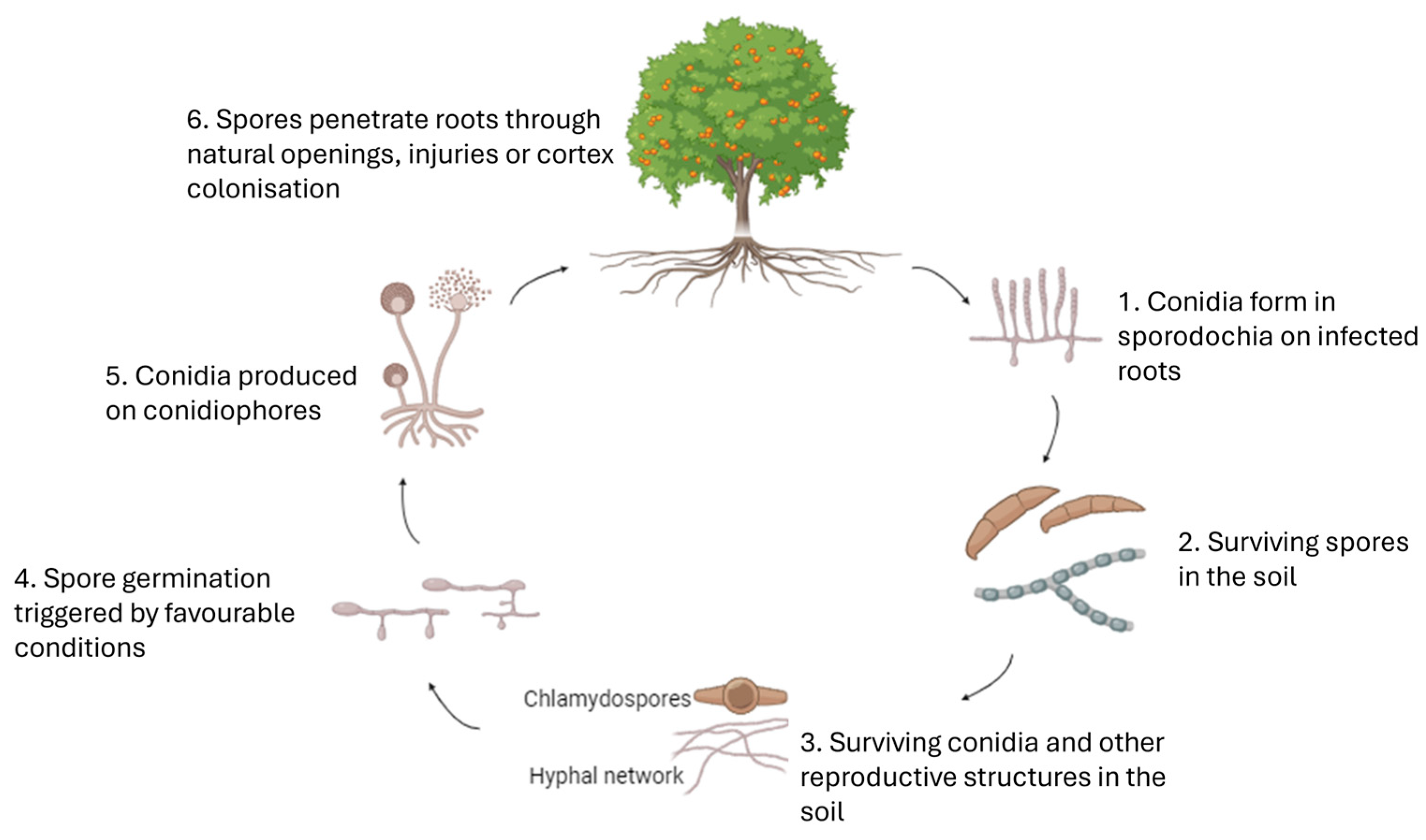
3. Etiology and Symptomatology of Fusarium spp. Associated with Citrus

3.1. Etiology of the Associated Diseases
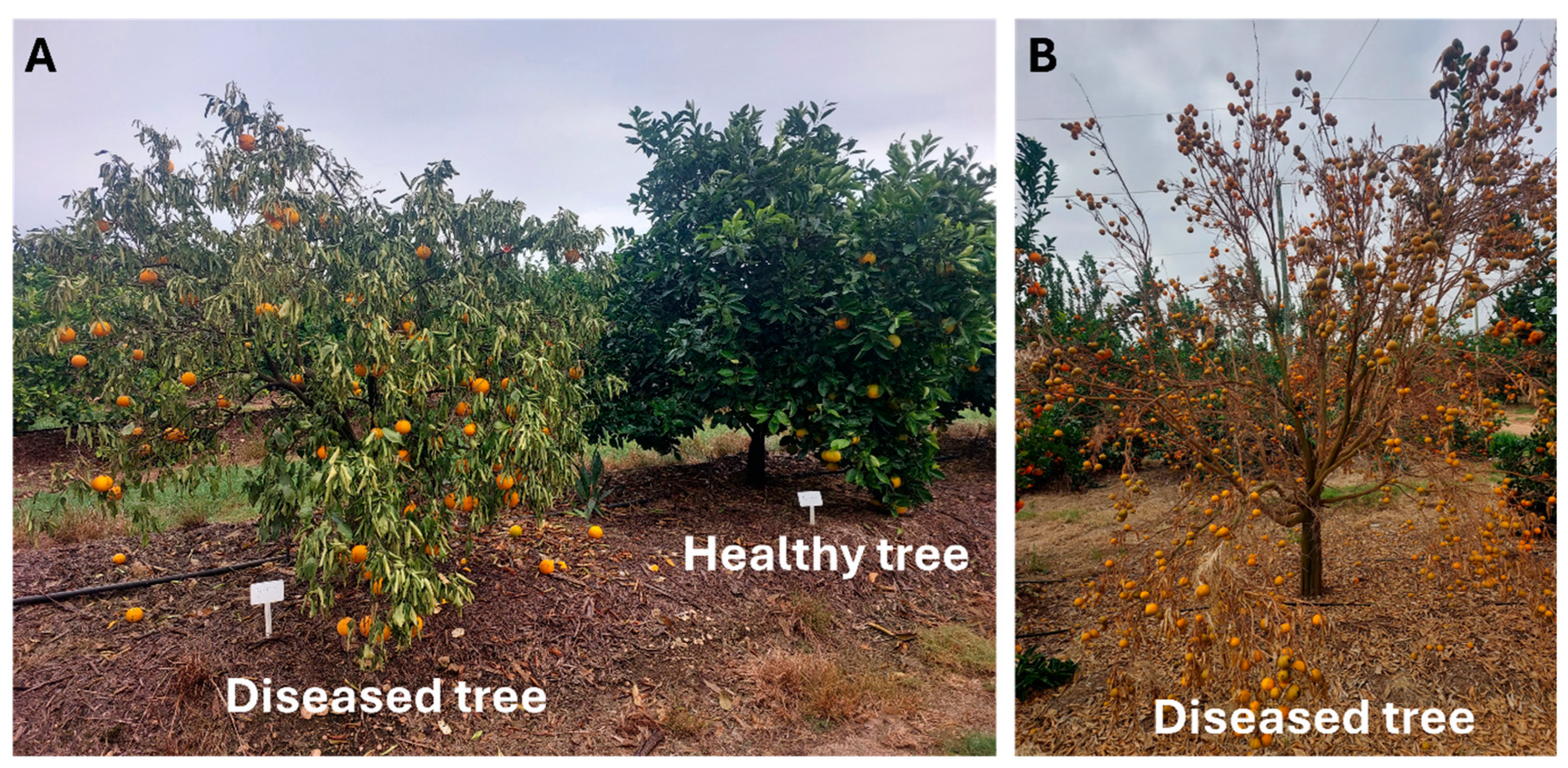
The infection process of Fusarium and Neocosmospora spp. in citrus can be broken down into several key stages, beginning with the persistence of spores in the soil (Figure 1). Fusarium spp. produce conidia, which are asexual spores, in specialized structures known as sporodochia, typically formed on infected roots or plant debris. These conidia, along with other spores such as chlamydospores, persist in the soil due to their thick-walled, durable nature, which makes them well adapted for survival in nonoptimal conditions, such as fluctuating moisture levels and temperature extremes. Once the environment becomes favourable, such as in the presence of moisture and suitable temperatures, the spores begin to germinate. During germination, the spores develop hyphal growth that forms an extensive mycelial network. This network spreads through the soil, facilitating the fungus’s ability to infect nearby plants or newly planted citrus trees. As the mycelium grows, specialized fungal structures called conidiophores are formed. These conidiophores produce new conidia, which are then released into the soil, where they are capable of infecting new roots. The spread of conidia through the soil continues the cycle of infection. When the hyphal network encounters citrus roots, the fungus adheres to the root structures and begins the infection process. The hyphae penetrate the root epidermis through natural openings, such as lenticels or root tips, or through injuries, using mechanical pressure and enzymatic activity to facilitate penetration. Once inside the roots, the fungus colonizes the plant by growing through the vascular tissue. This disruption of the plant’s vascular system impairs its ability to transport water and nutrients, leading to root rot, wilting, and other symptoms of decline. The spread of the fungus within the root system further exacerbates the damage, making it harder for the plant to recover, thus increasing the severity of the infection [17,64,65].
Several studies have highlighted the association between the presence of the citrus nematode Tylenchulus semipenetrans and the exacerbation of DRR in citrus rootstocks. This correlation is associated with a noticeable impact on the plant’s growth response, leading to a reduction in plant height [31,32]. The authors of these studies emphasized that the synergistic influence of N. solani and T. semipenetrans may contribute to the increased incidence of Fusarium root rot on citrus rootstocks that exhibit tolerance to Fusarium. Other pathogens, including nematodes such as T. semipenetrans [31] and specific fungi such as Neonectria macrodidyma Halleen, Schroers & Crous (Cylindrocarpon macrodidymum), isolated from symptomatic citrus trees in California, are believed to play a role in DRR disease progression [66]. Furthermore, it has been reported that inoculation of citrus trees with N. solani and P. nicotianae or P. citrophthora Leonian resulted in greater root rot compared to inoculation with P. nicotianae or P. citrophthora alone [67].

3.2. Symptomatology

Fusarium and Neocosmospora spp. are known to cause various diseases in citrus trees and fruits, each exhibiting similar yet distinct symptoms and severities. These species present substantial risks by causing diseases such as fruit rot, root rot, seedling damping-off, and plant wilt in numerous economically significant crops globally, encompassing citrus, vegetables, ornamentals, and field crops [68,69,70]. The infections are intricately linked to the interactions between the host and pathogens, as well as the presence of biotic and/or abiotic stresses, ultimately influencing disease incidence and expression [27]. They depend on plants for nutrients and have evolved mechanisms allowing them to colonize the plant tissues [71,72]. Fusarium wilt in citrus typically manifests as wilting and yellowing of leaves, eventually leading to their death, accompanied by a dark discolouration observable in the vascular tissue. Severe wilting attributed to F. oxysporum has been documented in C. sinensis ‘Washington Navel’ and C. clementina ‘Cassar’, and ‘Hernandina’ from commercial Tunisian orchards in the CapBon area, with noticeable discolouration of vascular tissue [52]. Additionally, wilting, chlorosis, and epinasty of young leaves, along with necrosis of rootlets, large roots, and crowns, have been observed in sour orange seedlings and orange trees (C. sinensis) var. ‘Valencia’ [52,73,74].
The progression of citrus DRR initiates with fungal penetration into the roots, leading to root rot, which then progresses into the xylem vessels, causing tree weakening. Typical symptoms include yellowing along the central vein of leaves, chlorosis, necrotic roots, root rot, purple wood discolouration, and dieback, ultimately culminating in tree subsidence, wilting, and eventual death (Figure 2) [11,24,27]. Investigations suggest that this disease is primarily caused by a complex of Fusarium spp., and N. solani being the most prevalent [27]. Infected trees often suffer crown loss due to desiccation, leaf fall, and fruit drying on branches [75]. Additionally, fibrous root rot is evident, correlated with reductions in crown size, defoliation, root cortex scaling, and, ultimately, tree death [76]. In some instances, hemiplegia along the longitudinal axis may occur in affected trees [30]. Citrus DRR is complex due to various biotic factors such as Phytophthora spp., citrus tristeza virus (CTV), T. semipenetrans, rodents, and insects, as well as abiotic stresses including drought, high temperatures, changes in soil pH, over-fertilization, root asphyxiation, and poor root aeration [77,78,79,80]. These factors collectively create opportune conditions for Fusarium and Neocosmospora spp., making it challenging to predict the development of citrus DRR. Further investigations are imperative to accurately identify the causative pathogens and understand the critical conditions and factors associated with citrus DRR occurrence.

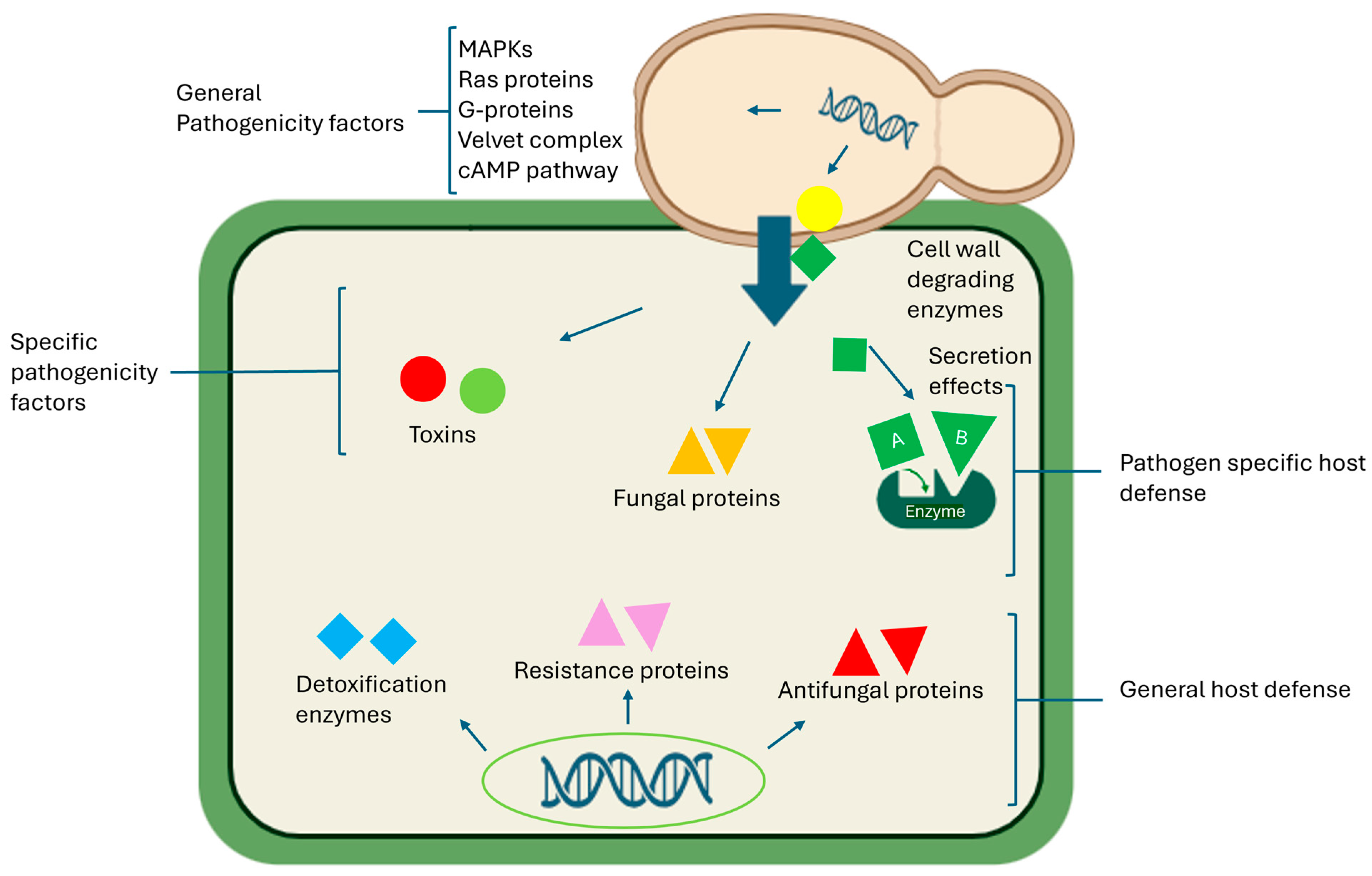
4. Fusarium spp.—Host Plant Interaction and Molecular Mechanisms

4.1. Host–Pathogen Interactions

The interactions between Fusarium spp. and host plants are intricate and multifaceted, often implying morphological and physiological alterations in both entities [71]. The Fusarium spp. pathogenic arsenal responsible for host plant invasion and disease includes host-specific toxins, effector proteins, and cell-wall-degrading enzymes. Fusarium spp. display high host specificity and effectively colonize and damage plant tissues through the production of host-specific toxins, enabling the pathogen to evade plant defence mechanisms (Figure 3) [81]. Upon encountering a host, Fusarium spp. trigger a complex network of interconnected signalling pathways. Many phytopathogenic Fusarium spp. produce toxins (Table 1) that induce plant disease by increasing host plant cell membrane permeability and electrolyte leakage, resulting in membrane damage, disrupted physiological processes, and eventual cell death [82,83]. These toxins, characterized by low molecular weight, manifest distinct symptoms in host plants such as chlorosis, growth inhibition, leaf spotting, necrosis, and wilting [84]. Additionally, they compromise the integrity of chloroplast inner membranes, leading to progressive basal lamella breakdown [81,85]. Moreover, these toxins can target host plant mitochondria, disrupting mitochondrial membrane structure and causing cristae swelling, vacuolization, and damage, ultimately impairing mitochondrial function [83]. Neocosmospora solani is recognized for its capacity to synthesize and release certain mycotoxins, contributing to its pathogenicity by inducing plasmolysis in vascular parenchyma cells at the root level and subsequent translocation to the leaves [86,87]. Throughout the infection process, the production of mycotoxins like naphthazarin has been documented, causing symptoms such as vein discolouration, leaf wilting, and the obstruction of vascular transport vessels [88,89].

4.2. Effector Proteins

Additionally, the process of infection is facilitated by secondary metabolites, including effector proteins, produced by Fusarium spp., which prompt the host plant to initiate a defence response known as effector-triggered immunity (ETI) or pathogen-associated molecular pattern (PAMP)-triggered immunity [90]. These effector proteins, such as Fg62 and Avr2, play a significant role in plant cells, thus influencing the host–phytopathogen interaction [91]. Successful infection relies on numerous convoluted biochemical pathways, including mitogen-activated protein (MAP) kinase signalling pathways [92], Ras proteins [93], G-protein signalling components [69,94,95], the velvet complex components [96], and cAMP pathways [93] (Figure 3). Several studies have investigated plant-phytopathogenic fungal effectors, particularly those with available whole-genome data, with F. oxysporum f. sp. lycopersici being a popular source of cloned phytopathogenic effectors [97,98]. Consequently, the virulence effects and transport molecular mechanisms of effector proteins remain largely unknown, given the infancy of research in this field. The specific effector proteins responsible for modifying plant metabolism to ensure nutrient availability for the infecting phytopathogen are yet to be identified, as are the plant signal transduction pathways that control effector protein gene expression [83]. However, comparative analysis may identify common motifs present in effectors of phytopathogenic fungi. Acquiring this knowledge and understanding would be crucial for elucidating the mechanisms involved in phytopathogen–plant host interactions, revealing the dynamic pathogenic mechanisms of Fusarium spp. and the host plant disease-resistance mechanisms [83].

4.3. Cell-Wall-Degrading Enzymes

Phytopathogenic fungi, including Fusarium spp., secrete cell-wall-degrading enzymes such as cutinases, cellulases, lipases, pectinases, and xylanases, which play a crucial role in successful penetration of the plant cell wall [99,100,101,102]. These enzymes facilitate the biochemical mechanisms involved in breaking down the cuticle and cell wall of the host plant during Fusarium spp. invasion, colonization, nutrient extraction, and proliferation [103,104]. The study of phytopathogenic cell-wall-degrading enzymes is currently a prominent research area, employing molecular biology and proteomics approaches. It has been documented that during infection, F. graminearum secretes cellulase, pectinase, and xylanase, which degrade host plant cell wall constituents and promote penetration and proliferation of the phytopathogen within the host tissue [105]. Fusarium spp. also produce β-galactosidases, which enhance the breakdown of lactose, producing galactose and glucose, thereby aiding in fruit softening [106]. High expression of β-galactosidase in the initial stages of fruit softening facilitates the degradation of cell wall galactosyl bonds, significantly reducing the integrity of the host plant’s cell wall [107]. Enzymes such as amylase, hemicellulase, phospholipase, and protease, produced during the infection process, have been reported to facilitate the degradation of important cell wall components such as starch, hemicellulose, lipids, and proteins, respectively [105]. The cellulase and pectinase enzymatic activity of N. solani has been reported to facilitate effective penetration across the entire root surface [108]. Moreover, F. oxysporum not only synthesizes cutinase and pectinase but also secretes enzymes like polygalacturonase, which degrade the cell wall to facilitate penetration [109].
It is important to note that cell-wall-degrading enzymes from phytopathogenic fungi, including Fusarium spp., are not the sole causative agents of infection and disease in host plants; coordinated secretion of phytohormones such as salicylic acid and jasmonic acid also plays a role in transducing the activation of the host plant’s defence systems against pathogen attack [110]. The interaction between Fusarium spp. and the host plant is biochemically complex, involving prompt expression of phytopathogen genes as well as activation of the plant’s enzymatic defence systems. This results in the formation of RNA interference, transcription of proteases and their inhibitors, biosynthesis of antifungal compounds, and production of reactive oxygen species (ROS) [111,112]. Collectively, these molecules drive a well-executed response to phytopathogen infections [83]. Therefore, effective management of Fusarium spp. diseases necessitates an in-depth understanding of the molecular mechanisms involved in the pathogenesis process. Resistant plants attempt to hinder the fungi’s expansion by synthesizing antifungal structures and compounds such as tyloses, gels, and gums to impede fungal growth and dissemination [71]. Citrus plants experience wilting because of xylem obstructions occurring either at the root or trunk levels. This results in the formation of a compact plug composed of interwoven filaments coated with an inert material at the intersection of xylem vessels [27]. DRR-affected citrus trees display an aggregation of unsaturated lipids, phospholipids, and choline at the plasmodesmata level. The onset of this accumulation is triggered by the progression of lipids through vessel pits from vessels into neighbouring parenchyma cells [28,113].

4.4. Induced Systemic Resistance (ISR) in Citrus

Induced Systemic Resistance (ISR) and Systemic Acquired Resistance (SAR) are essential for citrus plant defence against a wide range of pathogens. ISR is activated by beneficial microbial interactions or infection, enhancing the plant’s barriers without directly targeting the pathogens [114,115]. ISR triggers a salicylic-dependent signalling cascade, leading to systemic, broad-spectrum resistance. SAR, initiated by non-pathogenic stimuli such as mechanical wounding or herbivory, involves signalling molecules like salicylic acid (SA), jasmonic acid (JA), and ethylene, offering systemic protection against diverse pathogens [116,117]. These mechanisms can operate concurrently, providing cumulative pathogen protection [118]. Citrus plants can develop SAR through various treatments, including chemical elicitors, heat shock, or wounding, effectively managing diseases like citrus canker and Huanglongbing (HLB) [119,120]. SAR inducers, such as imidacloprid, thiamethoxam, and acibenzolar-S-methyl (ASM), have been shown to mitigate HLB symptoms in citrus trees [95,121]. Furthermore, the role of rootstock and rhizobacteria in inducing SAR against pests and pathogens highlights the importance of microbial interactions in plant defence [118,122,123].
Beneficial microbes, including plant growth-promoting bacteria and fungi, activate ISR, protecting against a broad spectrum of pathogens. Microbial strains such as Bacillus amyloliquefaciens and T. harzianum have been identified as key in activating defence pathways [124,125]. The application of beneficial fungi enhances defence against Fusarium root rot, with methods like root dipping and soil drenching proving effective [126,127,128,129]. Furthermore, the resistance of citrus rootstocks to Fusarium spp. and the application of fungal elicitors suggest methods to enhance citrus immunity [27,130]. A strain of Xanthomonas citri ssp. citri inducing a hypersensitive response in citrus indicates an alternative immunity mechanism against Fusarium spp. [131,132]. This examination underscores the multifaceted approach to defending citrus plants against Fusarium spp., combining microbial interactions and genetic resilience. By understanding and harnessing these natural defence mechanisms, new strategies for citrus disease management can be developed, reducing reliance on chemical control and enhancing sustainability in citrus agriculture.

4.5. Phytochemical Defence Mechanisms

Citrus plants harness phytochemicals as a multifaceted defence mechanism against diseases and herbivores, employing both direct and indirect strategies. These phytochemicals are crucial to the plant’s defence mechanism, which depends on genetic and environmental factors [133]. Notably, compounds like limonin, flavonoids, geranoxycumarine, and saponins have been pinpointed for their efficacy against Fusarium spp., which poses a significant threat to citrus crops [134]. These have been identified in citrus rind and extracts, and they possess antifungal potential, which underscores their value as natural fungicides, marking a pivotal step in the use of phytochemicals for plant protection [134,135,136]. More research into the phytochemistry of citrus fruits shows that they contain antifungal substances such as naringin, hesperidin, and especially limonin, which are very important for stopping the growth of bacteria and fungi [5,137,138,139]. While the precise mechanisms of their action remain under study, these discoveries hint at promising applications as natural fungicidal agents, offering a sustainable option for the citrus industry [136,140].
Enhancing the intricate defence network of citrus plants, specific phytochemicals such as citral activate defence pathways, including the jasmonic acid pathway, enhancing pathogen resistance. This involves the induction of plant hormone biosynthesis and signalling, which upregulates defence-related genes and enzymes, contributing to reduced postharvest fruit rot and highlighting the advanced strategies citrus plants employ against pathogens [141]. Furthermore, citrus essential oils, rich in linalool and limonene, demonstrate significant antimicrobial effects against Fusarium spp. [136]. These essential oils contain phenolic compounds, which exhibit bacteriostatic and fungistatic properties, causing structural damage to fungal cells and thus inhibiting their growth [142]. This antimicrobial prowess of phytochemicals exemplifies an effective defence against a wide array of pathogens.
The deep understanding of citrus defence mechanisms, fuelled by a synergy of genetic, environmental, and biological factors, showcases the antimicrobial strength of phytochemicals. Terpenoids, flavonoids, and alkaloids impede fungal pathogens by disrupting their enzyme systems and cell membrane integrity [143,144]. Recent studies have shown that essential oils from cloves effectively control blue mould in citrus by enhancing defence enzyme activities and stimulating phenylpropanoid metabolism, offering further evidence of the role of phytochemicals in disease control [128,141,142]. These insights encourage ongoing research into leveraging the innate defence capabilities of citrus plants to devise sustainable crop protection strategies. By reducing reliance on synthetic pesticides, this approach aims to promote a more sustainable agricultural ecosystem, demonstrating the potential of natural plant defences in modern agriculture [145,146,147,148].

4.6. Exploration of Novel Resistance Strategies

In the citrus industry, efforts are underway to devise innovative strategies for combating Fusarium spp., a significant threat to citrus health. One approach involves resistance breeding using rootstocks like Citrus sunki and Rough lemon, known for their inherent resistance to F. oxysporum [149]. Further research delves into the mechanisms of nonhost resistance (NHR), particularly in Citrus limon’s interactions with Xanthomonas campestris pv. campestris (Xcc), as investigated by Chiesa et al. [150]. This research highlights how NHR in C. limon, triggered by Xcc, results in sustained stomatal closure and involves the salicylic acid (SA) and abscisic acid (ABA) signalling pathways, underscoring the complex interplay of these pathways in plant defence mechanisms. Defence responses in C. limon against Xcc include cell wall reinforcement, phenylpropanoid pathway activation, and accumulation of callose and phenolic compounds, pointing to a robust set of mechanisms facilitating NHR. The regulation of the SA signalling pathway and the genes associated with it, alongside the engagement of ABA signalling pathway components, are critical for mounting an effective defence against pathogens like Xcc.
In parallel, studies to screen citrus genetic resources have identified genotypes with resistance to N. solani, contributing significantly to our understanding of resistance mechanisms in citrus [151]. This screening is particularly crucial as DRR caused by F. solani emerges as a destructive disease in citrus orchards. A pivotal study screening over 1400 citrus germplasm accessions has identified select genotypes such as ‘Fremont’ mandarin and ‘Lamas’ lemon that demonstrate enhanced resistance to N. solani [27]. These findings not only inform future research directions but also guide practical disease management applications, suggesting that these identified genotypes may offer promising avenues for further exploration of N. solani resistance. This underlines the importance of integrated pest management approaches, combining genetic resistance with an understanding of plant defence mechanisms to safeguard citrus orchards against pathogens and ensure the sustainability of citrus production globally.

5. Management Strategies for Fusarium spp.

Various methods and approaches are employed to manage soil-borne phytopathogens, including Fusarium spp. However, achieving desirable outcomes has proven challenging, primarily due to the intricate interactions between phytopathogens, rhizosphere microbiome and environmental conditions [152]. Currently, no identified cure exists for DDR; therefore, an effective strategy for addressing DRR involves an integrated management approach that combines the cultivation and use of resistant rootstock varieties with adequate sanitation practices, improved cultivation methods, and the utilization of biological control agents and chemical substances [153,154].

5.1. Cultural Practises

Prophylactic and sanitation practices serve as preventative measures aimed at eliminating stressors that render trees susceptible to pathogenic attacks. This method plays a crucial role in reducing the introduction and spread of pathogens. Continuous disease monitoring throughout the crop cycle is essential, and the removal of infected roots should be considered. Prudent irrigation methods tailored to the tree’s water needs and ensuring adequate drainage, especially in dense soils, are essential [80]. Rigorous disinfection of equipment, particularly when transitioning between orchards, and the judicious application of fertilizers are imperative for disease control. The recommended course of action for the complete removal of an infected tree is advisable only during the advanced stages of the disease, as suggested by [80]. Solarization is a recommended method, as the elevated temperatures generated can deactivate various soilborne pathogens, rendering Fusarium spp. inoculum inactive within a soil temperature range of 42 °C to 47 °C [155].

5.2. Chemical Control

Various chemicals have been utilized to manage DRR in citrus; however, there is currently no effective fungicide treatment available [153]. In efforts to mitigate disease incidence, fungicides have been studied against the Fusarium spp. complex in several investigations [156]. Fosetylaluminium, propineb, carbendazim, copper oxychloride, thiophanate-methyl, myclobutanil, penconazole, difenoconazole, and mancozeb have demonstrated inhibitory efficacy against N. solani mycelial growth in vitro [156]. Farmers combat soilborne pathogens by employing pre-planting soil disinfection techniques, often involving fumigants. Compounds such as Carbendazim, Copper oxychloride, Difenoconazole, Fosetyl-aluminium, Mandipropamid, Metiram, Myclobutanil, Penconazole, Propineb, and Thiophanate-methyl, when applied either in liquid or solid form, inhibit fungal growth, disrupt enzyme activity, or stimulate plant defences specifically targeting pathogens [157].

5.3. Phytochemical Control

Currently, there are no successful control measures capable of managing soilborne pathogens. Thus, the necessity of implementing an integrated management strategy arises to address this deficiency [80,158]. Control using phytochemicals is also one of the alternative strategies to chemical control. Among various plant extracts studied under laboratory conditions, including citrus (C. hystrix), neem (Azadirachta indica), garlic (Allium sativum), onion (Allium cepa), datura (Datura stramonium), calotropis (Calotropis procera), peppermint (Mentha piperita), fennel (Foeniculum vulgare), ginger (Zingiber officinale), and chilli (Capsicum annuum), higher doses of A. indica and C. procera extracts demonstrated the most significant inhibition of N. solani in vitro mycelial growth, while C. hystrix and C. annuum extracts were also effective in inhibiting it [157]. Furthermore, the application of commercially available biocides, specifically Plant Guard and Rhizo-N, has proven effective in managing citrus DRR. This effectiveness is manifested in the reduction of disease severity, decreased density of N. solani inoculum in the soil, and inhibited colonization of the root system [80]. Furthermore, the efficacy of two silicon-based products in controlling DRR disease in citrus was examined and shown to be a valuable tool in the management of the disease [159]. The application of soil water-soluble silicon emerged as a promising solution for disease management, preventing the deterioration of infected trees and maintaining fruit size, quality, and yield comparable to that of healthy plants.

5.4. Biological Control

The use of biological methodologies to manage plant diseases attributed to Fusarium spp. is recognized as a promising and environmentally sustainable approach. Numerous research studies have consistently highlighted the effectiveness of various biological control agents (BCAs) in mitigating soil-borne pathogens in controlled environments such as greenhouses and authentic field conditions. BCAs, recognized as promising alternatives for mitigating Fusarium-induced damage, have consistently demonstrated commendable efficacy. Species of the Bacillus genus exhibit antagonistic properties by producing antibiotics, lipopeptides, or volatile organic compounds (VOCs) that enhance plant resistance, with Bacillus subtilis reported as a biocontrol agent against the soil-borne fungus N. solani [160,161]. Furthermore, studying the antifungal activity of certain novel peptides isolated from Bacillus amyloliquefaciens resulted in the discovery of broad-spectrum antifungal activity against both N. solani and F. oxysporum [162]. Implementing strategic biological control measures with B. subtilis and Trichoderma spp. has shown favourable outcomes, leading to a decrease in infection and severity, consequently lowering the population density of N. solani in citrus soil [163]. The antagonistic potential of Trichoderma harzianum plays a significant role in controlling root and soil diseases caused by nematodes and soil-borne fungi such as F. oxysporum [164]. The assessment of biocontrol capabilities of various isolates of Pseudomonas fluorescens and B. subtilis against pathogens affecting rough lemon (Citrus × jambhiri Lush.) showed maximum disease control [165]. Under laboratory conditions, four P. fluorescens isolates and two B. subtilis isolates demonstrated efficacy in controlling P. nicotianae and other citrus phytopathogens, including Pythium sp., Fusarium sp., and Colletotrichum gloeosporioides [165]. Similarly, the inhibitory effects of B. subtilis on the mycelial growth of infectious Fusarium spp. were shown to be effective in controlling DRR in citrus in greenhouse experiments [153].
The capacity of Trichoderma spp. to control dry root rot caused by F. oxysporum was investigated, and the study showed a significant decrease in disease incidence in vitro [166]. The in vitro antagonistic activity of Trichoderma against F. oxysporum was tested on Potato Dextrose Agar medium, while an in vivo greenhouse trial was conducted on Japansche Citroen and Rough Lemon rootstock seedlings. Results indicated that three Trichoderma isolates inhibited F. oxysporum growth in vitro by over 60%. In glasshouse trials, certain Trichoderma isolates reduced the incidence of disease to as low as 3.33% [167]. Trichoderma spp. produce secondary metabolites and enzymes such as glucanases, chitinases, and proteases, which disrupt and degrade the cell wall of phytopathogens [168]. Many Trichoderma spp. exert their antagonistic activity by competing for nutrients and resources, thereby contrasting colonization by the pathogen and thus making the use of biopesticides a potential alternative to agrochemicals [168]. Although Trichoderma spp. are well-known as phytopathogen biocontrol agents, very few commercial bioactive products are currently labelled and widely marketed for use against citrus, with many studies focusing on BCA against P. digitatum and no other citrus pathogens, particularly Fusarium spp., highlighting a lack of research and representing a problem for citriculture.

5.5. Mycoviruses Infecting Fusarium spp.

Fusarium mycoviruses are ssRNA or double-stranded RNA (dsRNA) viruses with a high specificity for Fusarium spp. These mycoviruses thrive within Fusarium spp. cells, altering the phytopathogen’s biology. Research into the mycovirus mechanisms reveals a multifaceted scope concerning their ability to influence Fusarium spp. pathogenicity, virulence, and overall lifecycle. These mechanisms include antibiosis, where mycoviruses inhibit the growth of pathogenic fungi directly; nutritional competition, where they outcompete the pathogens for resources; and the activation of plant defences, which enhances the plant’s intrinsic resistance to fungal attacks [169,170]. Mycoviruses induce notable changes in Fusarium spp. phenotype, affecting growth, spore production, and secondary metabolite biosynthesis. Delving into the mechanisms behind these virus-induced changes suggests both molecular and phenotypic levels of interaction, which could serve as strategic targets to reduce pathogenicity and undesirable morphological transformations in the fungal host [171,172]. Mycovirus transmission methods such as protoplast fusion have been noted to induce hypovirulence in Fusarium spp., enhancing the biocontrol possibilities of mycoviruses [171,173,174]. Furthermore, advancements in RNA deep sequencing have expanded our understanding of mycovirus diversity within Fusarium, coupled with omics technologies that illuminate the complex interactions between mycoviruses and their fungal hosts. Viruses like FgV1 from the novel Fusariviridae family reduce F. graminearum virulence by impacting mycelial growth and mycotoxin production, while FgV-ch9 and FodV1 from the Chrysoviridae family are known to influence fungal host growth, morphology, and pathogenicity on crops such as wheat and maize [175,176]. Employing RNA-Seq for comprehensive transcriptome analysis has illuminated how mycoviruses alter transcriptional regulation and affect phytopathogen host proteins [177,178]. The FgHEX1 gene in F. graminearum is pivotal in asexual reproduction and virulence, also playing a role in viral RNA accumulation and host gene downregulation upon mycovirus infection [179].
Among mycoviruses, those infecting Fusarium spp. have garnered attention due to their potential use in biocontrol applications. These mycoviruses, often characterized by ssRNA or double-stranded RNA (dsRNA) genomes, are intricately classified into families by the International Committee for the Taxonomy of Viruses (ICTV), with the most important being Chrysoviridae, Totiviridae, Partitiviridae, and Reoviridae. They exhibit unique intracellular transmission modes, bypassing an extracellular phase and typically infecting hosts within specific vegetative compatibility groups [180,181]. Mycoviruses that reduce the virulence of pathogenic Fusarium spp. are particularly promising for developing biocontrol strategies given their potential to affect endophytic fungi and produce killer toxins [182,183,184]. Such mycoviruses, through their hypovirulence, offer a viable strategy for managing Fusarium infections, leveraging the unique propagation mechanisms that rely on the fungal host’s lifecycle to spread [171]. Important Fusarium spp. in agriculture have been found to host dsRNA mycoviruses like FusoV, which contains dsRNA segments capable of altering the pathogenicity of fungi [174]. This alteration often results in morphological changes and reduced virulence as these mycoviruses disseminate throughout the environment, presenting a compelling case for using mycoviruses in disease management strategies [185]. The discovery of various mycoviruses within Fusarium spp., including FgV1, FgV2, and the novel FgNSRV-1, highlights the potential of these viruses to induce phenotypic alterations in their hosts, potentially decreasing pathogenicity and offering a novel approach to controlling Fusarium-related diseases [175,186]. The identification of mycoviruses in F. virguliforme with reduced virulence and new dsRNA mycoviruses across different Fusarium spp. underscores the broader applicability of mycoviruses in citrus disease control strategies [187,188]. Furthermore, research into the interactions between F. graminearum and its associated mycoviruses has revealed significant alterations in the host’s gene expression patterns. Notably, genes like FgHex1 (FGSG_08737), a precursor protein crucial for asexual reproduction and virulence, and FGSG_10089, a glycosylphosphatidylinositol (GPI)-anchored protein involved in redox-sensing for virulence and cell wall integrity, were found to be upregulated in the presence of mycovirus infection [189]. Conversely, genes encoding putative metabolic enzymes such as enolase (FGSG_01346), saccharopine dehydrogenase (FGSG_10925), flavohemoglobin (FGSG_04458), mannitol dehydrogenase (FGSG_04826), and malate dehydrogenase (FGSG_02504) were downregulated [185]. Additionally, proteins such as peroxiredoxins (FGSG_03180 and FGSG_07536), which are thought to provide antioxidant protection within the cell, were also found to be downregulated in infected F. graminearum strains [190]. This complex interplay of gene expression changes underscores the impact of mycovirus infections on the metabolic and defensive mechanisms within F. graminearum.

6. Conclusions and Future Prospects

The utilization of molecular-based techniques has revolutionized the identification of Fusarium spp. associated with citrus, offering heightened sensitivity and reliability compared to conventional methods. Research indicates a complex involvement of various Fusarium and Neocosmospora spp. associated with DRR, with recent studies unveiling new species impacting citrus plantations globally. Despite inherent challenges in citrus breeding, the integration of molecular markers and gene pyramiding strategies holds substantial promise for bolstering disease resistance against pathogens such as Fusarium spp. Transcription factors (TFs) assume a pivotal role in regulating defence-related gene expression, presenting avenues for genetic engineering and marker-assisted selection (MAS) in cultivating disease-resistant citrus varieties. The orchestration of complex signal transduction networks and subsequent activation of defence-related genes underpin an efficacious immune response. Notably, prophylactic measures encompassing sanitation protocols and refined irrigation practices serve as cornerstones in curtailing pathogen introduction and dissemination. Furthermore, soil disinfection utilizes fumigants alongside the deployment of biological control agents like B. subtilis and Trichoderma spp. exhibit promising efficacy in reducing Fusarium spp. infections.
The Fusarium–citrus interaction from molecular and genetic perspectives remains relatively understudied. Advancements in this field of research hold significant potential to deepen our understanding and profoundly influence the cultivation of new germplasms exhibiting heightened tolerance to pathogenic Fusarium spp. Enhancements in research methodologies could pave the way for innovative approaches, such as the priming of seeds with beneficial microbes, phytohormones, and natural products, synthetic chemical inducers (chemical priming), as well as nano-emulsions and nanoparticles (nanomaterials priming), to effectively bolster crop resistance against Fusarium-linked diseases. Exploiting seed priming, particularly considering advancing knowledge on plant immune memory and priming methodologies, promises novel avenues for fortifying crop protection in future agricultural practices. Moreover, the integration of technologies such as genome-wide screening for defence-related quantitative trait loci (QTLs) in conjunction with CRISPR technology presents an effective strategy for advancing our understanding and fostering the development of resilient plant varieties.
In essence, the early establishment of advanced citrus defence research, coupled with the untapped genetic diversity within citrus accessions, innovative strides in analytics and genomics, and the dynamic interplay of environmental conditions, pathogens, and host genetic backgrounds, are poised to significantly enhance the efficacy and precision of global plant breeding endeavours in the foreseeable future. This trajectory ensures the sustainable growth and advancement of food production and safety on a global scale.

Funding

Department of Plant Pathology, Stellenboch University.

Acknowledgments

The authors would like to thank the Department of Plant Pathology, Stellenbosch University, for providing access to relevant resources that facilitated this review. We are also grateful to our colleagues for their insightful discussions and feedback during the preparation of this manuscript. Additionally, we acknowledge the support of Citrus Research International (CRI) for their financial assistance. Finally, we appreciate the constructive comments from the reviewers and editors, which helped improve the quality of this work.

Conflicts of Interest

The authors declare no conflict of interest.

References

  1. Hazarika, T.K. Citrus. In Fruit Nut Crops; Springer Nature: Singapore, 2023; pp. 1–44. [Google Scholar]
  2. Wu, G.A.; Terol, J.; Ibanez, V.; López-García, A.; Pérez-Román, E.; Borredá, C.; Domingo, C.; Tadeo, F.R.; Carbonell-Caballero, J.; Alonso, R.; et al. Genomics of the origin and evolution of Citrus. Nature 2018, 554, 311–316. [Google Scholar] [PubMed]
  3. Zhong, G.; Nicolosi, E. Citrus origin, diffusion, and economic importance. In Citrus Genome; Springer: Berlin, Germany, 2020; pp. 5–21. [Google Scholar]
  4. Ahmed, W.; Azmat, R. Citrus: An ancient fruit of promise for health benefits. In Citrus: Health Benefits and Production Technology; IntechOpen: London, UK, 2019; pp. 19–30. Available online: https://www.intechopen.com/chapters/64826 (accessed on 31 October 2024).
  5. Addi, M.; Elbouzidi, A.; Abid, M.; Tungmunnithum, D.; Elamrani, A.; Hano, C. An overview of bioactive flavonoids from citrus fruits. Appl. Sci. 2021, 12, 29. [Google Scholar] [CrossRef]
  6. Maqbool, Z.; Khalid, W.; Atiq, H.T.; Koraqi, H.; Javaid, Z.; Alhag, S.K.; Al-Shuraym, L.A.; Bader, D.M.D.; Almarzuq, M.; Afifi, M.; et al. Citrus waste as source of bioactive compounds: Extraction and utilization in health and food industry. Molecules 2023, 28, 1636. [Google Scholar] [CrossRef]
  7. Taghizadeh-Alisaraei, A.; Hosseini, S.H.; Ghobadian, B.; Motevali, A. Biofuel production from citrus wastes: A feasibility study in Iran. Renew. Sustain. Energy Rev. 2017, 69, 1100–1112. [Google Scholar]
  8. Varmie, E.B.; Thakur, M. Utilization of citrus processing waste: A review. Pharm. Innov. J. 2021, 10, 682–697. [Google Scholar]
  9. Suri, S.; Singh, A.; Nema, P.K. Current applications of citrus fruit processing waste: A scientific outlook. Appl. Food Res. 2022, 2, 100050. [Google Scholar]
  10. Gohel, N.M.; Prajapati, B.K.; Srivastava, J.N. Major diseases of citrus and their management. In Diseases of Horticultural Crops; Apple Academic Press: New York, NY, USA, 2022; pp. 155–167. [Google Scholar]
  11. Sandoval-Denis, M.; Guarnaccia, V.; Polizzi, G.; Crous, P.W. Symptomatic Citrus trees reveal a new pathogenic lineage in Fusarium and two new Neocosmospora species. Persoonia-Mol. Phylogeny Evol. Fungi 2018, 40, 1–25. [Google Scholar]
  12. Aoki, T.; O’Donnell, K.; Geiser, D.M. Systematics of key phytopathogenic Fusarium species: Current status and future challenges. J. General. Plant Pathol. 2014, 80, 189–201. [Google Scholar] [CrossRef]
  13. O’Donnell, K. Molecular phylogeny of Fusarium inferred from partial rna polymerase ii gene sequences. J. Plant Pathol. 2008, 90, 11–14. [Google Scholar]
  14. Lombard, L.; Van der Merwe, N.A.; Groenewald, J.Z.; Crous, P.W. Generic concepts in Nectriaceae. Stud. Mycol. 2015, 80, 189–245. [Google Scholar]
  15. O’Donnell, K.; Sutton, D.A.; Fothergill, A.; McCarthy, D.; Rinaldi, M.G.; Brandt, M.E.; Zhang, N.; Geiser, D.M. Molecular phylogenetic diversity, multilocus haplotype nomenclature, and in vitro antifungal resistance within the Fusarium solani species complex. J. Clin. Microbiol. 2008, 46, 2477–2490. [Google Scholar] [CrossRef]
  16. Chandra, N.S.; Wulff, E.G.; Udayashankar, A.C.; Nandini, B.P.; Niranjana, S.R.; Mortensen, C.N.; Prakash, H.S. Prospects of molecular markers in Fusarium species diversity. Appl. Microbiol. Biotechnol. 2011, 90, 1625–1639. [Google Scholar]
  17. Summerell, B.A. Resolving Fusarium: Current status of the genus. Annu. Rev. Phytopathol. 2019, 57, 323–339. [Google Scholar]
  18. O’Donnell, K.; McCormick, S.P.; Busman, M.; Proctor, R.H.; Ward, T.J.; Geiser, D.M. Whole-genome phylogenetics and chemotyping of the Fusarium genus reveal new insights into species complexes. Fungal Genet. Biol. 2013, 52, 20–31. [Google Scholar] [PubMed]
  19. Udomkun, P.; Wiredu, A.N.; Nagle, M.; Müller, J.; Vanlauwe, B.; Bandyopadhyay, R. Innovative technologies to manage aflatoxins in foods and feeds and the profitability of application–A review. Food Control 2017, 76, 127–138. [Google Scholar]
  20. Marrez, D.A.; Ayesh, A.M. Mycotoxins: The threat to food safety. Egypt. J. Chem. 2022, 65, 353–372. [Google Scholar] [CrossRef]
  21. Misihairabgwi, J.M.; Ishola, A.; Quaye, I.; Sulyok, M.; Krska, R. Diversity and fate of fungal metabolites during the preparation of oshikundu, a Namibian traditional fermented beverage. World Mycotoxin J. 2018, 11, 471–481. [Google Scholar] [CrossRef]
  22. Adeyeye, S.O.; Ashaolu, T.J.; Idowu-Adebayo, F. Mycotoxins: Food safety, consumer health and Africa’s food security. Polycycl. Aromat. Compd. 2022, 42, 5779–5795. [Google Scholar]
  23. Donkersley, P.; Silva, F.W.; Carvalho, C.M.; Al-Sadi, A.M.; Elliot, S.L. Biological, environmental and socioeconomic threats to citrus lime production. J. Plant Dis. Prot. 2018, 125, 339–356. [Google Scholar] [CrossRef]
  24. Liu, H.F.; Zhou, J.; Liao, J.; Yi, J.P.; Ma, D.F.; Deng, J.X. Grafted twig rot on Citrus sinensis caused by a member of the Fusarium solani species complex. Can. J. Plant Pathol. 2020, 42, 133–139. [Google Scholar]
  25. Ezrari, S.; Lahlali, R.; Radouane, N.; Tahiri, A.; Asfers, A.; Boughalleb-M’Hamdi, N.; Amiri, S.; Lazraq, A. Characterization of Fusarium species causing dry root rot disease of citrus trees in Morocco. J. Plant Dis. Prot. 2021, 128, 431–447. [Google Scholar]
  26. Umer, M.; Mubeen, M.; Ateeq, M.; Shad, M.A.; Atiq, M.N.; Kaleem, M.M.; Iqbal, S.; Shaikh, A.A.; Ullah, I.; Khan, M.; et al. Etiology, epidemiology and management of citrus black rot caused by Alternaria citri: An outlook. Plant Prot. 2021, 5, 105–115. [Google Scholar]
  27. Ezrari, S.; Radouane, N.; Tahiri, A.; El Housni, Z.; Mokrini, F.; Özer, G.; Lazraq, A.; Belabess, Z.; Amiri, S.; Lahlali, R. Dry root rot disease, an emerging threat to citrus industry worldwide under climate change: A review. Physiol. Mol. Plant Pathol. 2022, 117, 101753. [Google Scholar]
  28. Nemec, S. Microorganisms associated with healthy and sand hill-declined citrus roots. Plant Dis. Rep. 1975, 59, 210. [Google Scholar]
  29. Paguio, O.R. Citrus declinio in the State of Brazil: Occurrence and responses to blight diagnostic tests. Int. Organ. Citrus Virol. Conf. Proc. 1984, 9, 1957–2010. [Google Scholar]
  30. Malikoutsaki-Mathioudi, M.; Bourbos, V.A.; Skoudridakis, M.T. Dry root rot-a very serious citrus disease in Greece. Bull. OEPP 1987, 17, 335–340. [Google Scholar]
  31. Labuschagne, N.; Kotzé, J.M. Factors affecting feeder root rot of citrus caused by Fusarium solani. In Proceedings of the Sixth International Citrus Congress, Tel Aviv, Israel, 6–11 March 1988; Volume 14, pp. 839–845. [Google Scholar]
  32. El-Mohamedy, R.S.R.; Hasabo, S.A. Response of some citrus rootstocks to infection with Fusarium solani and citrus nematode Tylenchulus semipenetrans under greenhouse conditions. Egypt. J. Phytopathol. 2005, 33, 11–25. [Google Scholar]
  33. Chandran, M.R.; Kumar, M.R. Studies on cultural, morphological variability in isolates of Fusarium solani (Mart.) Sacc., incitant of dry root-rot of Citrus. Curr. Biot. 2012, 6, 152–162. [Google Scholar]
  34. Rehman, A.; Sahi, S.T.; Khan, M.A.; Mehboob, S. Fungi associated with bark, twigs and roots of declined shisham (Dalbergia sissoo roxb.) trees in Punjab, Pakistan. Pak. J. Phytopathol. 2012, 24, 152–158. [Google Scholar]
  35. Al-Sadi, A.M.; Al-Alawi, Z.A.; Deadman, M.L.; Patzelt, A. Etiology of four foliar and root diseases of wild plants in Oman. Can. J. Plant Pathol. 2014, 36, 517–522. [Google Scholar]
  36. Al-Karboli, M.H.; Kuthair, W.M. Isolation and pathogenicity of the fungus, Fusarium solani a causal of dry root rot on sour orange in Baghdad province. J. Agric. Technol. 2016, 12, 927–938. [Google Scholar]
  37. Liu, X.; Cao, A.; Wang, Q.; Yan, D.; Ouyang, C.; Fang, W. First report of small yellow ginger root rot caused by Fusarium solani in Jiangxi, China. Plant Dis. 2019, 103, 1415. [Google Scholar]
  38. Moosa, A.; Farzand, A.; Sahi, S.T.; Khan, S.A.; Abbas, M.F.; Abbas, A.; Binyamin, R. First report of post-harvest Fusarium rot caused by Fusarium oxysporum on Citrus reticulata Blanco cv. ‘Kinnow’ in Pakistan. J. Plant Pathol. 2020, 102, 945–946. [Google Scholar]
  39. Baker, R.A.; Tatum, J.H.; Nemec, S. Toxin production by Fusarium solani from fibrous roots of blight-diseased citrus. Phytopathology 1981, 71, 951–954. [Google Scholar]
  40. de HC Maciel, M.; do Amaral, A.C.T.; da Silva, T.D.; Bezerra, J.D.; de Souza-Motta, C.M.; da Costa, A.F.; Tiago, V.; de Oliveira, N.T. Evaluation of Mycotoxin Production and Phytopathogenicity of the Entomopathogenic Fungi Fusarium caatingaense and F. pernambucanum from Brazil. Curr. Microbiol. 2021, 78, 1218–1226. [Google Scholar]
  41. Gálvez, L.; Palmero, D. Fusarium dry rot of garlic bulbs caused by Fusarium proliferatum: A review. Horticulturae 2022, 8, 628. [Google Scholar] [CrossRef]
  42. Achar, P.N.; Sreenivasa, M.Y. Current perspectives of biocontrol agents for management of Fusarium verticillioides and its fumonisin in cereals—A review. J. Fungi 2021, 7, 776. [Google Scholar] [CrossRef]
  43. Gupta, R.C.; Doss, R.B.; Lall, R.; Srivastava, A.; Sinha, A. Trichothecenes and zearalenone. In Reproductive and Developmental Toxicology; Academic Press: Cambridge, MA, USA, 2022; pp. 1003–1016. [Google Scholar]
  44. Ma, X.; Zhai, L.; Jiang, Y.; Wang, Z.; He, L.; Song, F.; Wu, L. First report of Fusarium oxysporum and Fusarium solani causing root rot on trifoliate orange rootstock in China. Plant Dis. 2023, 107, 944. [Google Scholar]
  45. Guarnaccia, V.; Van Niekerk, J.; Crous, P.; Sandoval-Denis, M. Neocosmospora spassociated with dry root rot of citrus in South Africa. Phytopathol. Mediterr. 2021, 60, 79–100. [Google Scholar]
  46. Ortiz-Martínez, L.E.; Juárez-Rodríguez, L.; Robles-Yerena, L.; Leyva-Mir, S.G.; Camacho-Tapia, M. Fusarium sp., causal agent of vascular wilt in citrus and its sensitivity to fungicides. Rev. Mex. De Fitopatol. 2022, 40, 1–17. [Google Scholar] [CrossRef]
  47. Kurt, Ş.; Uysal, A.; Soylu, E.M.; Kara, M.; Soylu, S. Characterization and pathogenicity of Fusarium solani associated with dry root rot of citrus in the eastern Mediterranean region of Turkey. J. Gen. Plant Pathol. 2020, 86, 326–332. [Google Scholar] [CrossRef]
  48. Garzón-Nivia, M.A.; Mártiz Mártiz, J.; Moya-Elizondo, E.A.; Ruiz, B.; Cornejo, J.C.; Valdés-Gómez, H.A. Characterization and Identification of Neocosmospora solani and Fusarium oxysporum Causing Root Necrosis and Wilting of Orange Trees in Chile. Plants 2025, 14, 376. [Google Scholar] [CrossRef]
  49. Crous, P.W.; Wingfield, M.J. Fungi infecting woody plants: Emerging frontiers. Persoonia-Mol. Phylogeny Evol. Fungi 2018, 40, 1–3. [Google Scholar] [CrossRef]
  50. Hasan, M.F.; Islam, M.A.; Sikdar, B. First report on molecular identification of Fusarium species causing fruit rot of mandarin (Citrus reticulata) in Bangladesh. F1000Research 2020, 9, 1212. [Google Scholar] [CrossRef]
  51. Sukmawati, D.; Miarsyah, M. Pathogenic activity of Fusarium equiseti from plantation of citrus plants (Citrus nobilis) in the village Tegal Wangi, Jember Umbulsari, East Java, Indonesia. Asian J. Agric. Biol. 2017, 5, 202–213. [Google Scholar]
  52. Hannachi, I.; Rezgui, S.; Cherif, M. First report of mature citrus trees being affected by Fusarium wilt in Tunisia. Plant Dis. 2014, 98, 566. [Google Scholar] [CrossRef]
  53. Ezrari, S.; Radouane, N.; Tahiri, A.; Amiri, S.; Lazraq, A.; Lahlali, R. Environmental effects of temperature and water potential on mycelial growth of Neocosmospora solani and Fusarium spcausing dry root rot of citrus. Curr. Microbiol. 2021, 78, 3092–3103. [Google Scholar] [CrossRef]
  54. Sandoval-Denis, M.; Lombard, L.; Crous, W. Back to the roots: A reappraisal of Neocosmospora. Persoonia-Mol. Phylogeny Evol. Fungi 2019, 43, 90–185. [Google Scholar] [CrossRef]
  55. Aguayo, J.; Mostert, D.; Fourrier-Jeandel, C.; Cerf-Wendling, I.; Hostachy, B.; Viljoen, A.; Ioos, R. Development of a hydrolysis probe-based real-time assay for the detection of tropical strains of Fusarium oxysporum f. sp. cubense race 4. PLoS ONE 2017, 12, e0171767. [Google Scholar] [CrossRef]
  56. Mishra, K.; Fox, R.T.; Culham, A. Development of a PCR-based assay for rapid and reliable identification of pathogenic Fusaria. FEMS Microbiol. Lett. 2003, 218, 329–332. [Google Scholar] [CrossRef]
  57. Arif, M.; Chawla, S.; Zaidi, M.W.; Rayar, J.K.; Variar, M.; Singh, U.S. Development of specific primers for genus Fusarium and F. solani using rDNA sub-unit and transcription elongation factor (TEF-1α) gene. Afr. J. Biotechnol. 2012, 11, 444–447. [Google Scholar]
  58. Azadvar, M.; Alizadeh, H.; Safarnejad, M.R.; Najafinia, M.; Atellio Bianco, P. Etiology of quick decline disease of Citrus on Bakraee (Citrus sp.) rootstock in Southern Kerman. Iran. J. Plant Prot. Sci. 2019, 50, 87–97. [Google Scholar]
  59. Schoch, C.L.; Seifert, K.A.; Huhndorf, S.; Robert, V.; Spouge, J.L.; Levesque, C.A.; Chen, W.; Bolchacova, E.; Voigt, K. Nuclear ribosomal internal transcribed spacer (ITS) region as a universal DNA barcode marker for Fungi. Proc. Natl. Acad. Sci. USA 2012, 109, 6241–6246. [Google Scholar]
  60. Wurzbacher, C.; Larsson, E.; Bengtsson-Palme, J.; Van den Wyngaert, S.; Svantesson, S.; Kristiansson, E.; Kagami, M.; Nilsson, R.H. Introducing ribosomal tandem repeat barcoding for fungi. Mol. Ecol. Resour. 2019, 19, 118–127. [Google Scholar]
  61. Tedersoo, L.; Sánchez-Ramírez, S.; Koljalg, U.; Bahram, M.; Döring, M.; Schigel, D.; May, T.; Ryberg, M.; Abarenkov, K. High-level classification of the Fungi and a tool for evolutionary ecological analyses. Fungal Divers. 2018, 90, 135–159. [Google Scholar]
  62. Crous, P.W.; Wingfield, M.J.; Lombard, L.; Roets, F.; Swart, W.J.; Alvarado, P.; Carnegie, A.J.; Moreno, G.; Luangsaard, J.; Thangavel, R.; et al. Fungal Planet description sheets: 951–1041. Persoonia-Mol. Phylogeny Evol. Fungi 2019, 43, 223. [Google Scholar]
  63. Wang, Z.; Binder, M.; Schoch, C.L.; Johnston, R.; Spatafora, J.W.; Hibbett, D.S. New insights into fungal evolution from phylogenomic analysis of a taxonomically comprehensive dataset. Nat. Commun. 2022, 13, 1–13. [Google Scholar]
  64. Guan, Y.M.; Ma, Y.Y.; Jin, Q.; Wang, Q.X.; Liu, N.; Fu, Y.P.; Zhang, Y.Y.; Li, Y. Multi-locus phylogeny and taxonomy of the fungal complex associated with rusty root rot of Panax ginseng in China. Front. Microbiol. 2020, 11, 618942. [Google Scholar] [CrossRef]
  65. Swenie, R.A.; Looney, B.P.; Ke, Y.H.; Alejandro Rojas, J.; Cubeta, M.A.; Langer, G.J.; Vilgalys, R.; Matheny, B. PacBio high-throughput multi-locus sequencing reveals high genetic diversity in mushroom-forming fungi. Mol. Ecol. Resour. 2024, 24, e13885. [Google Scholar]
  66. Adesemoye, A.O.; Mayorquin, J.S.; Peacock, B.B.; Moreno, K.; Hajeri, S.; Yokomi, R.; Eskalen, A. Association of Neonectria macrodidyma with dry root rot of citrus in California. J. Plant Pathol. Microbiol. 2016, 8, 391–394. [Google Scholar]
  67. Hao, W.; Gray, M.A.; Förster, H.; Adaskaveg, J.E. Evaluation of new Oomycota fungicides for management of Phytophthora root rot of citrus in California. Plant Dis. 2019, 103, 619–628. [Google Scholar]
  68. Jackson, E.; Li, J.; Weerasinghe, T.; Li, X. The Ubiquitous Wilt-Inducing Pathogen Fusarium oxysporum—A Review of Genes Studied with Mutant Analysis. Pathogens 2024, 13, 823. [Google Scholar] [CrossRef]
  69. Ekwomadu, T.I.; Mwanza, M. Fusarium fungi pathogens, identification, adverse effects, disease management, and global food security: A review of the latest research. Agriculture 2023, 13, 1810. [Google Scholar] [CrossRef]
  70. Amby, D.B.; Thuy, T.T.T.; Ho, B.D.; Kosawang, C.; Son, T.B.; Jørgensen, H.J.L. First report of Fusarium lichenicola as a causal agent of fruit rot in pomelo (Citrus maxima). Plant Dis. 2015, 99, 1278. [Google Scholar]
  71. Dandurand, L.M.; Menge, J.A. Influence of Fusarium solani on citrus root rot caused by Phytophthora parasitica and Phytophthora citrophthora. Plant Soil 1992, 144, 13–21. [Google Scholar]
  72. Spina, S.; Coco, V.; Gentile, A.; Catara, A.; Cirvilleri, G. Association of Fusarium solani with rolABC and wild type Troyer Citrange. J. Plant Pathol. 2008, 90, 479–486. [Google Scholar]
  73. Parikh, L.; Kodati, S.; Eskelson, M.J.; Adesemoye, A.O. Identification and pathogenicity of Fusarium spin row crops in Nebraska. Crop Prot. 2018, 108, 120–127. [Google Scholar]
  74. Rivera-Jiménez, M.N.; Zavaleta-Mancera, H.A.; Rebollar-Alviter, A.; Aguilar-Rincón, V.H.; García-de-los-Santos, G.; Vaquera-Huerta, H.; Silva-Rojas, H.V. Phylogenetics and histology provide insight into damping-off infections of ‘Poblano’ pepper seedlings caused by Fusarium wilt in greenhouses. Mycol. Progress 2018, 17, 1237–1249. [Google Scholar]
  75. Joshi, R. A review of Fusarium oxysporum on its plant interaction and industrial use. J. Med. Plants Stud. 2018, 6, 112–115. [Google Scholar]
  76. Rana, A.; Sahgal, M.; Johri, B.N. Fusarium oxysporum: Genomics, diversity and plant–Host interaction. Dev. Fungal Biol. Appl. Mycol. 2017, 159–199. [Google Scholar] [CrossRef]
  77. Hannachi, I.; Poli, A.; Rezgui, S.; Prassad, R.D.; Cherif, M. Genetic and phenotypic differences of Fusarium oxysporum f. sp. citri isolated from sweet orange and tangerine. Eur. J. Plant Pathol. 2015, 142, 269–280. [Google Scholar]
  78. Parra-Cota, F.I.; García Pereyra, J.; Aviña Martínez, G.N.; Santos-Villalobos, S.D.L. First report of Fusarium wilt on Citrus sinensis var. Valencia in the Yaqui Valley, Mexico. Rev. Mex. Fitopatol. 2019, 37, 193–201. [Google Scholar]
  79. Kunta, M.; Salas, B.; Gonzales, M.; da Graça, J.V. First report on citrus dry rot in sour orange rootstock in Texas. J. Citrus Pathol. 2015, 2. [Google Scholar] [CrossRef]
  80. Usman, H.M.; Iffat, A.; Shakeel, Q.; Bhatti, A.M.; Karim, M.M.; Zafar, M.I.; Bajwa, R.T.; Rashid, I.; Kiptoo, J.J.; Shafique, T. Insights into Fusarium spp., Associated with Dry Root Rot of Citrus and its Management. Phytopathogenomics Dis. Control 2024, 3, 241–249. [Google Scholar]
  81. Rampersad, S.N. Pathogenomics and management of Fusarium diseases in plants. Pathogens 2020, 9, 340. [Google Scholar] [CrossRef]
  82. Labushagne, N.; van der Veote, P.A.; Kotze, J.M. Interaction between Fusarium solani and Tylenchulus semipenetrans on citrus roots. Phytophylactica 1989, 21, 29–34. [Google Scholar]
  83. Dandurand, L.M.; Menge, J.A. Influence of Fusarium solani on citrus root growth and population dynamics of Phytophthora parasitica and Phytophthora citrophthora. Phytopathology 1993, 83, 767–771. [Google Scholar]
  84. Adesemoye, A.; Eskalen, A.; Faber, B.; Bender, G.; O’connell, N.; Kallsen, C.; Shea, T. Current knowledge on Fusarium dry rot of citrus. Citrograph 2011, 2, 29–33. [Google Scholar]
  85. Srivastava, V.; Patra, K.; Pai, H.; Aguilar-Pontes, M.V.; Berasategui, A.; Kamble, A.; Di Pietro, A.; Redkar, A. Molecular dialogue during host manipulation by the vascular wilt fungus Fusarium oxysporum. Annu. Rev. Phytopathol. 2024, 62, 97–126. [Google Scholar]
  86. Lyu, X.; Shen, C.; Fu, Y.; Xie, J.; Jiang, D.; Li, G.; Cheng, J. Comparative genomic and transcriptional analyses of the carbohydrate-active enzymes and secretomes of phytopathogenic fungi reveal their significant roles during infection and development. Sci. Rep. 2015, 5, 15565. [Google Scholar]
  87. Peng, Y.; Li, S.J.; Yan, J.; Tang, Y.; Cheng, J.P.; Gao, A.J.; Yao, X.; Ruan, J.J.; Xu, B.L. Research progress on phytopathogenic fungi and their role as biocontrol agents. Front. Microbiol. 2021, 12, 670135. [Google Scholar]
  88. Jajić, I.; Dudaš, T.; Krstović, S.; Krska, R.; Sulyok, M.; Bagi, F.; Savić, Z.; Guljaš, D.; Stankov, A. Emerging Fusarium mycotoxins fusaproliferin, beauvericin, enniatins, and moniliformin in Serbian maize. Toxins 2019, 11, 357. [Google Scholar] [CrossRef] [PubMed]
  89. Kant, P.; Reinprecht, Y.; Martin, C.J.; Islam, R.; Pauls, K.P. Disease resistance. Compr. Biotechnol. 2019, 4, 789–805. [Google Scholar]
  90. Hou, S.; Liu, Z.; Shen, H.; Wu, D. Damage-associated molecular pattern-triggered immunity in plants. Front. Plant Sci. 2019, 10, 646. [Google Scholar]
  91. Shcherbakova, L.A.; Dzhavakhiya, V.G.; Duan, Y.; Zhang, J. Microbial proteins as elicitors of plant resistance to pathogens and their potential for eco-friendly crop protection in sustainable agriculture. Agric. Biol. 2023, 58, 789–820. [Google Scholar]
  92. Gutiérrez-Sánchez, A.; Plasencia, J.; Monribot-Villanueva, J.L.; Rodríguez-Haas, B.; Ruíz-May, E.; Guerrero-Analco, J.A.; Sánchez-Rangel, D. Virulence factors of the genus Fusarium with targets in plants. Microbiol. Res. 2023, 277, 127506. [Google Scholar] [CrossRef]
  93. Chen, A.; Ju, Z.; Wang, J.; Wang, J.; Wang, H.; Wu, J.; Yin, Y.; Zhao, Y.; Ma, Z.; Chen, Y. The RasGEF FgCdc25 regulates fungal development and virulence in Fusarium graminearum via cAMP and MAPK signalling pathways. Environ. Microbiol. 2020, 22, 5109–5124. [Google Scholar]
  94. Yang, Y.; Huang, P.; Ma, Y.; Jiang, R.; Jiang, C.; Wang, G. Insights into intracellular signaling network in Fusarium species. Int. J. Biol. Macromol. 2022, 222, 1007–1014. [Google Scholar]
  95. El-Defrawy, M.M.; Hesham, A.E.L. G-protein-coupled receptors in fungi. Fungal Biotechnol. Bioeng. 2020, 37–126. [Google Scholar] [CrossRef]
  96. Reddy, V.P.; Verma, S.; Sharma, D.; Thakur, A. Role of resistant-proteins in plant innate immunity—A review. Agric. Rev. 2019, 40, 12–20. [Google Scholar]
  97. Essarioui, A.; Mokrini, F.; Afechtal, M. Molecular interactions between tomato and its wilt pathogen Fusarium oxysporum f. sp. Lycopersici—A review. Rev. Marocaine Des Sci. Agron. Vétérinaires 2016, 4, 66–74. [Google Scholar]
  98. Li, X.; Ke, Z.; Xu, S.; Tang, W.; Liu, Z. The G-protein alpha subunit CgGa1 mediates growth, sporulation, penetration and pathogenicity in Colletotrichum gloeosporioides. Microb. Pathog. 2021, 161, 105254. [Google Scholar] [CrossRef] [PubMed]
  99. Gao, J.; Zhou, S.; Tang, W.; Wang, J.; Liu, H.; Zhang, Y.; Wang, L.; Li, X.; Liu, Z. The velvet proteins CsVosA and CsVelB coordinate growth, cell wall integrity, sporulation, conidial viability and pathogenicity in the rubber anthracnose fungus Colletotrichum siamense. Microbiol. Res. 2023, 268, 127290. [Google Scholar] [CrossRef]
  100. Stępień, Ł.; Lalak-Kańczugowska, J. Signaling pathways involved in virulence and stress response of plant-pathogenic Fusarium species. Fungal Biol. Rev. 2021, 35, 27–39. [Google Scholar]
  101. de Guillen, K.; Ortiz-Vallejo, D.; Gracy, J.; Fournier, E.; Kroj, T.; Padilla, A. Structure analysis uncovers a highly diverse but structurally conserved effector family in phytopathogenic fungi. PLoS Pathog. 2015, 11, e1005228. [Google Scholar]
  102. Tan, K.C.; Oliver, R.P. Regulation of proteinaceous effector expression in phytopathogenic fungi. PLoS Pathog. 2017, 13, e1006241. [Google Scholar]
  103. Rauwane, M.E.; Ogugua, U.V.; Kalu, C.M.; Ledwaba, L.K.; Woldesemayat, A.A.; Ntushelo, K. Pathogenicity and virulence factors of Fusarium graminearum including factors discovered using next generation sequencing technologies and proteomics. Microorganisms 2020, 8, 305. [Google Scholar] [CrossRef]
  104. Mishra, P.; Mishra, J.; Dwivedi, S.K.; Arora, N.K. Microbial enzymes in biocontrol of phytopathogens. In Microbial Enzymes: Roles and Applications in Industries; Springer: Berlin, Germany, 2020; pp. 259–285. [Google Scholar]
  105. Perincherry, L.; Urbaniak, M.; Pawłowicz, I.; Kotowska, K.; Waśkiewicz, A.; Stępień, Ł. Dynamics of Fusarium mycotoxins and lytic enzymes during pea plants’ infection. Int. J. Mol. Sci. 2021, 22, 9888. [Google Scholar] [CrossRef]
  106. Janusz, G.; Pawlik, A.; Sulej, J.; Świderska-Burek, U.; Jarosz-Wilkołazka, A.; Paszczyński, A. Lignin degradation: Microorganisms, enzymes involved, genomes analysis and evolution. FEMS Microbiol. Rev. 2017, 41, 941–962. [Google Scholar]
  107. Feng, L.; Zhao, G.; Sun, R.; Wang, J.; Sun, T.; Xing, S.; Lian, W.; Zhao, Y. Dynamic changes in cell wall-degrading enzymes and physio-biochemistry of ginseng in response to Fusarium oxysporum infection. Eur. J. Plant Pathol. 2023, 165, 569–578. [Google Scholar] [CrossRef]
  108. Bagherieh, B.; Taheri, P. Characterization, pathogenicity, and extracellular enzymes of Fusarium solani f. sp. phaseoli associated with common bean in Khorasan Razavi Province of Iran. J. Adv. Plant Prot. 2024, 1, 53–64. [Google Scholar]
  109. Elagamey, E.; Abdellatef, M.A.; Arafat, M.Y. Proteomic insights of chitosan mediated inhibition of Fusarium oxysporum f. sp. cucumerinum. J. Proteom. 2022, 260, 104560. [Google Scholar] [CrossRef]
  110. Majeed, G.; Noor, W.; Lone, R.; Agrawal, S.; Alaklabi, A.; Shah, M.A.; Kamili, A.N. Modulation of plant defenses by Jasmonic acid and salicylic acid in Capsicum annuum L. against Fusarium wilt in response to Fusarium oxysporum pathogen. Plant Stress 2024, 14, 100571. [Google Scholar] [CrossRef]
  111. Wang, C.; Zheng, Y.; Liu, Z.; Qian, Y.; Li, Y.; Yang, L.; Liu, S.; Liang, W.; Li, J. The secreted FolAsp aspartic protease facilitates the virulence of Fusarium oxysporum f. sp. lycopersici. Front. Microbiol. 2023, 14, 1103418. [Google Scholar]
  112. Fernandes, L.B.; Ghag, S.B. Molecular insights into the jasmonate signaling and associated defense responses against wilt caused by Fusarium Oxysporum. Plant Physiol. Biochem. 2022, 174, 22–34. [Google Scholar] [CrossRef] [PubMed]
  113. Nemec, S. Vessel blockage by myelin forms in citrus with and without rough-lemon decline symptoms. Can. J. Bot. 1975, 53, 102–108. [Google Scholar] [CrossRef]
  114. Hou, S.; Tsuda, K. Salicylic acid and jasmonic acid crosstalk in plant immunity. Essays Biochem. 2022, 66, 647–656. [Google Scholar]
  115. Appu, M.; Ramalingam, P.; Sathiyanarayanan, A.; Huang, J. An overview of plant defense-related enzymes responses to biotic stresses. Plant Gene 2021, 27, 100302. [Google Scholar] [CrossRef]
  116. Akbar, M.U.; Aqeel, M.; Shah, M.S.; Jeelani, G.; Iqbal, N.; Latif, A.; Elnour, R.O.; Hashem, M.; Alzoubi, O.M.; Habeeb, T.; et al. Molecular regulation of antioxidants and secondary metabolites act in conjunction to defend plants against pathogenic infection. S. Afr. J. Bot. 2023, 161, 247–257. [Google Scholar] [CrossRef]
  117. Iqbal, N.; Riyazuddin, R.; Nauman, M.; Czékus, Z.; Hayat, M.T.; Poór Ördög, A. Role of Plant Defence System in Crop Protection against Fusarium Pathogens. Fusarium-Recent Studies; IntechOpen: London, UK, 2024; p. 71. [Google Scholar]
  118. Kamle, M.; Borah, R.; Bora, H.; Jaiswal, A.K.; Singh, R.K.; Kumar, P. Systemic acquired resistance (SAR) and induced systemic resistance (ISR): Role and mechanism of action against phytopathogens. In Fungal Biotechnology and Bioengineering; Springer: Cham, Switzerland, 2020; pp. 457–470. [Google Scholar]
  119. Yu, Y.; Gui, Y.; Li, Z.; Jiang, C.; Guo, J.; Niu, D. Induced systemic resistance for improving plant immunity by beneficial microbes. Plants 2022, 11, 386. [Google Scholar] [CrossRef] [PubMed]
  120. Abdul Malik, N.A.; Kumar, I.S.; Nadarajah, K. Elicitor and receptor molecules: Orchestrators of plant defense and immunity. Int. J. Mol. Sci. 2020, 21, 963. [Google Scholar] [CrossRef] [PubMed]
  121. Maithani, D.; Singh, H.; Sharma, A. Stress alleviation in plants using SAR and ISR: Current views on stress signaling network. In Microbes and Signaling Biomolecules Against Plant Stress: Strategies of Plant-Microbe Relationships for Better Survival; Springer: Cham, Switzerland, 2021; pp. 7–36. [Google Scholar]
  122. Riera, N.; Wang, H.; Li, Y.; Li, J.; Pelz-Stelinski, K.; Wang, N. Induced Systemic Resistance against Citrus Canker Disease by Rhizobacteria. Phytopathology 2018, 108, 1038–1045. [Google Scholar] [PubMed]
  123. Kunwar, S.; Redondo, A.; Manker, D.; Lott, M.; Knobloch, T.; Brunet, S.; Dufour, J.; Batuman, O. Novel systemic acquired resistance (SAR) inducers for managing huanglongbing (citrus greening) and Citrus canker diseases. In Proceedings of the XXXI International Horticultural Congress (IHC2022): International Symposium on Sustainable Control of Pests and Diseases, Angers, France, 14–20 August 2022; Volume 1378, pp. 133–142. [Google Scholar]
  124. Ilham, B.; Noureddine, C.; Philippe, G.; Mohammed, E.G.; Brahim, E.; Sophie, A.; Muriel, M. Induced systemic resistance (ISR) in Arabidopsis thaliana by Bacillus amyloliquefaciens and Trichoderma harzianum used as seed treatments. Agriculture 2019, 9, 166. [Google Scholar] [CrossRef]
  125. Karuppiah, V.; Vallikkannu, M.; Li, T.; Chen, J. Simultaneous and sequential based co-fermentations of Trichoderma asperellum GDFS1009 and Bacillus amyloliquefaciens 1841: A strategy to enhance the gene expression and metabolites to improve the bio-control and plant growth promoting activity. Microb. Cell Factories 2019, 18, 1–16. [Google Scholar]
  126. Bagio, T.Z.; Canteri, M.G.; Barreto, T.P.; Júnior, R.P.L. Activation of systemic acquired resistance in citrus to control huanglongbing disease. Semin. Ciências Agrárias 2016, 37, 1757–1765. [Google Scholar]
  127. Frąckowiak, P.; Pospieszny, H.; Smiglak, M.; Obrępalska-Stęplowska, A. Assessment of the efficacy and mode of action of benzo (1, 2, 3)-thiadiazole-7-carbothioic acid s-methyl ester (bth) and its derivatives in plant protection against viral disease. Int. J. Mol. Sci. 2019, 20, 1598. [Google Scholar]
  128. Poveda, J.; Barquero, M.; González-Andrés, F. Insight into the microbiological control strategies against Botrytis cinerea using systemic plant resistance activation. Agronomy 2020, 10, 1822. [Google Scholar] [CrossRef]
  129. Zehra, A.; Raytekar, N.A.; Meena, M.; Swapnil, P. Efficiency of microbial bio-agents as elicitors in plant defense mechanism under biotic stress: A review. Curr. Res. Microb. Sci. 2021, 2, 100054. [Google Scholar]
  130. Llorens, E.; García-Agustín, P.; Lapeña, L. Advances in induced resistance by natural compounds: Towards new options for woody crop protection. Sci. Agric. 2017, 74, 90–100. [Google Scholar]
  131. Yan, Z.L.; Chen, J.P.; Nong, X.X.; Li, X.; Luo, H.Y.; Wei, L.L.; Ruan, J.H.; Guan, X.Y.; Lu, S. Isolation and identification of endophytic fungi from citrus cultivars and their inhibitory activity against Xanthomonas citri subscitri causing citrus canker. Sci. Press 2021, 41, 1196–1208. [Google Scholar]
  132. Wang, X.; Liang, L.; Shao, H.; Ye, X.; Yang, X.; Chen, X.; Shi, Y.; Zhang, L.; Xu, L.; Wang, J. Isolation of the novel strain Bacillus amyloliquefaciens F9 and identification of lipopeptide extract components responsible for activity against Xanthomonas citri subscitri. Plants 2022, 11, 457. [Google Scholar]
  133. Rabelo, R.S.; Dyer, L.A.; Yamaguchi, L.F.; Diniz, I.; Simbaña, W.; Kussano, A.J.M.; Kato, M.J.; Massad, T.J. Plasticity in plant defense and the role of phytochemical dissimilarity in limiting specialist herbivory. Front. Ecol. Evol. 2023, 11, 1175590. [Google Scholar]
  134. Okwu, D.E.; Awurum, A.N.; Okoronkwo, J.I. Phytochemical composition and in vitro antifungal activity screening of extracts from citrus plants against Fusarium oxysporum of okra plant (Hibiscus esculentus). Pest. Technol. 2007, 1, 145–148. [Google Scholar]
  135. Umeoka, N. Phytochemical screening and antifungal properties of the peel extract of citrus sinensis. J. Health Metab. Nutr. Stud. 2024, 3, 40–43. [Google Scholar]
  136. Oikeh, E.I.; Oviasogie, F.E.; Omoregie, E.S. Quantitative phytochemical analysis and antimicrobial activities of fresh and dry ethanol extracts of Citrus sinensis (L.) Osbeck (sweet Orange) peels. Clin. Phytoscience 2020, 6, 1–6. [Google Scholar]
  137. Alam, F.; Mohammadin, K.; Shafique, Z.; Amjad, S.T.; Asad, M.H.H.B. Citrus flavonoids as potential therapeutic agents: A review. Phytother. Res. 2022, 36, 1417–1441. [Google Scholar]
  138. Zahr, S.; Zahr, R.; El Hajj, R.; Khalil, M. Phytochemistry and biological activities of Citrus sinensis and Citrus limon: An update. J. Herbal. Med. 2023, 100737. [Google Scholar]
  139. M’hiri, N.; Ioannou, I.; Ghoul, M.; Mihoubi Boudhrioua, N. Phytochemical characteristics of citrus peel and effect of conventional and nonconventional processing on phenolic compounds: A review. Food Rev. Int. 2017, 33, 587–619. [Google Scholar]
  140. Bashir, L.U.; Abdulkadir, A.; Shah, M.M.; Hamisu, A.; Sharif, U.; Kamalu, A.A. Phytochemical screening and antifungal potentials of Citrus limon peels against Fusarium oxysporum and Rhizopus stolonifer causing rots in watermelon (Citrullus lanatus L.). J. Exp. Sci. 2020, 11, 1–5. [Google Scholar]
  141. Hernández, A.; Ruiz-Moyano, S.; Galván, A.I.; Merchán, A.V.; Nevado, F.P.; Aranda, E.; Serradilla, M.J.; de Guía Córdoba, M.; Martín, A. Anti-fungal activity of phenolic sweet orange peel extract for controlling fungi responsible for post-harvest fruit decay. Fungal Biol. 2021, 125, 143–152. [Google Scholar] [PubMed]
  142. Aumeeruddy-Elalfi, Z.; Gurib-Fakim, A.; Mahomoodally, M.F. Chemical composition, antimicrobial and antibiotic potentiating activity of essential oils from 10 tropical medicinal plants from Mauritius. J. Herbal. Med. 2016, 6, 88–95. [Google Scholar]
  143. Chen, Y.; Xing, M.; Chen, T.; Tian, S.; Li, B. Effects and mechanisms of plant bioactive compounds in preventing fungal spoilage and mycotoxin contamination in postharvest fruits: A review. Food Chem. 2023, 415, 135787. [Google Scholar]
  144. Zhou, X.; Zeng, M.; Huang, F.; Qin, G.; Song, Z.; Liu, F. The potential role of plant secondary metabolites on antifungal and immunomodulatory effect. Appl. Microbiol. Biotechnol. 2023, 107, 4471–4492. [Google Scholar] [PubMed]
  145. Duan, B.; Reymick, O.O.; Liu, Z.; Zhou, Y.; Wang, X.; Feng, Z.; Tao, N. Citral enhances disease resistance in postharvest citrus fruit through inducing jasmonic acid pathway and accumulating phenylpropanoid compounds. Postharvest Biol. Technol. 2024, 207, 112633. [Google Scholar]
  146. Tariq, S.; Wani, S.; Rasool, W.; Shafi, K.; Bhat, M.A.; Prabhakar, A.; Shalla, A.H.; Rather, M.A. A comprehensive review of the antibacterial, antifungal and antiviral potential of essential oils and their chemical constituents against drug-resistant microbial pathogens. Microb. Pathog. 2019, 134, 103580. [Google Scholar]
  147. Souto, A.L.; Sylvestre, M.; Tölke, E.D.; Tavares, J.F.; Barbosa-Filho, J.M.; Cebrián-Torrejón, G. Plant-derived pesticides as an alternative to pest management and sustainable agricultural production: Prospects, applications and challenges. Molecules 2021, 26, 4835. [Google Scholar] [CrossRef]
  148. Ngegba, P.M.; Cui, G.; Khalid, M.Z.; Zhong, G. Use of botanical pesticides in agriculture as an alternative to synthetic pesticides. Agriculture 2022, 12, 600. [Google Scholar] [CrossRef]
  149. Ahsen, M.A.; Naqvi, S.A.; Jaskani, M.J.; Waseem, M.; Khan, I.A.; Hussnain, K.; Mahmood, K.; Khan, M.M. Evaluation of exotic citrus rootstocks against Fusarium spp. J. Glob. Innov. Agric. Soc. Sci. 2019, 7, 151–156. [Google Scholar]
  150. Chiesa, M.A.; Roeschlin, R.A.; Favaro, M.A.; Uviedo, F.; Campos-Beneyto, L.; D’Andrea, R.; Gadea, J.; Marano, M.R. Plant responses underlying nonhost resistance of Citrus limon against Xanthomonas campestris pv. campestris. Mol. Plant Pathol. 2019, 20, 254–269. [Google Scholar]
  151. Krueger, R.R.; Bender, G.S. Screening a core collection of citrus genetic resources for resistance to Fusarium solani. Acta Hortic. 2015, 1065, 155–164. [Google Scholar]
  152. Wang, N.; Sundin, G.W.; Fuente, L.D.L.; Cubero, J.; Tatineni, S.; Brewer, M.T.; Zeng, Q.; Bock, C.H.; Cunniffe, N.J.; Wang, C.; et al. Key challenges in plant pathology in the next decade. Phytopathology 2024, 114, 837–842. [Google Scholar]
  153. Ezrari, S.; Mhidra, O.; Radouane, N.; Tahiri, A.; Polizzi, G.; Lazraq, A.; Lahlali, R. Potential role of rhizobacteria isolated from Citrus rhizosphere for biological control of Citrus dry root rot. Plants 2021, 10, 872. [Google Scholar] [CrossRef]
  154. Narayanasamy, P. Soilborne Microbial Plant Pathogens and Disease Management, Volume Two: Management of Crop Diseases; CRC Press: Boca Raton, FL, USA, 2019; Volume 2, pp. 1–10. [Google Scholar]
  155. Dubey, A.K.; Rao, K.K.; Kumar, S.; Tamta, M.; Dwivedi, S.K.; Kumar, R.; Mishra, J.S. Disease management in major field crops. In Conservation Agriculture for Climate Resilient Farming & Doubling Farmers’ Income; ICAR Research Complex for Eastern Region: Patna, India, 2019; p. 246. [Google Scholar]
  156. Pakdaman Sardrood, B.; Mohammadi Goltapeh, E. Effect of agricultural chemicals and organic amendments on biological control fungi. In Sustainable Agriculture Reviews 31: Biocontrol; Springer: Cham, Switzerland, 2018; pp. 217–359. [Google Scholar]
  157. Khanzada, M.A.; Tanveer, M.; Maitlo, S.A.; Hajano, J.; Ujjan, A.A.; Syed, R.N.; Lodhi, A.M.; Rajput, A.Q. Comparative efficacy of chemical fungicides, plant extracts and bio-control agents against Fusarium solani under laboratory conditions. Pak. J. Phytopathol. 2016, 28, 133–139. [Google Scholar]
  158. Marais, L.J. Efficacy of water-soluble silicon in managing Fusarium dry root rot of citrus. Acta Hortic. 2015, 1065, 993–1000. [Google Scholar]
  159. Laing, M.D.; Basdew, L. Sustainable Control of Pre-and Postharvest Diseases of Citrus Fruit in South Africa; University of KwaZulu-Natal: Durban, South Africa, 2018; pp. 34–36. [Google Scholar]
  160. Grahovac, J.; Pajčin, I.; Vlajkov, V. Bacillus VOCs in the context of biological control. Antibiotics 2023, 12, 581. [Google Scholar] [CrossRef]
  161. Chen, Y.H.; Lee, P.C.; Huang, T.P. Biological control of collar rot on passion fruits via induction of apoptosis in the collar rot pathogen by Bacillus subtilis. Phytopathology 2021, 111, 627–638. [Google Scholar]
  162. Kim, Y.G.; Kang, H.K.; Kwon, K.D.; Seo, C.H.; Lee, H.B.; Park, Y. Antagonistic activities of novel peptides from Bacillus amyloliquefaciens PT14 against Fusarium solani and Fusarium oxysporum. J. Agric. Food Chem. 2015, 63, 10380–10387. [Google Scholar]
  163. Hammam, M.M.; El-Mohamedy, R.S.; Abd-El-Kareem, F.; Abd-Elgawad, M.M. Evaluation of soil amended with bio-agents and compost alone or in combination for controlling citrus nematode Tylenchulus semipenetrans and Fusarium dry root rot on Volkamer lime under greenhouse conditions. Int. J. ChemTech Res. 2016, 9, 86–96. [Google Scholar]
  164. El-Mohamedy, R.S.; Hammam, M.M.; Abd-El-Kareem, F.; Abd-Elgawad, M.M. Biological soil treatment to control Fusarium solani and Tylenchulus semipenetrans on sour orange seedlings under greenhouse conditions. Int. J. ChemTech Res. 2016, 9, 73–85. [Google Scholar]
  165. Shinde, S.B.; Sadgir, M.D. Efficiency of Pseudomonas fluorescens and Bacillus subtilis against Phytophthora spin Citrus. Int. J. Plant Prot. 2016, 9, 15–20. [Google Scholar]
  166. Altinok, H.H.; Erdogan, O. Determination of the In vitro Effect of Trichoderma harzianum on Phytopathogenic Strains of Fusarium oxysporum. Not. Bot. Horti Agrobot. 2015, 43, 494–500. [Google Scholar]
  167. Dewi, O.K.M.; Abadi, A.L.; Widyaningsih, S. Fusarium Rot Biological Control of Citrus caused by Fusarium Oxysporum. Res. J. Life Sci. 2022, 9, 91–98. [Google Scholar] [CrossRef]
  168. Abbas, A.; Mubeen, M.; Zheng, H.; Sohail, M.A.; Shakeel, Q.; Solanki, M.K.; Iftikhar, Y.; Sharma, S.; Kashyap, B.K.; Hussain, S.; et al. Trichoderma spgenes involved in the biocontrol activity against Rhizoctonia solani. Front. Microbiol. 2022, 13, 884469. [Google Scholar]
  169. Juby, S.; Radhakrishnan, E.K.; Jayachandran, K. Exploring Mechanisms of Disease Suppression using Endophytic Fungi as Biocontrol Agents. In Biofungicides: Eco-Safety and Future Trends; CRC Press: Boca Raton, FL, USA, 2023; pp. 159–190. [Google Scholar]
  170. Palmieri, D.; Ianiri, G.; Del Grosso, C.; Barone, G.; De Curtis, F.; Castoria, R.; Lima, G. Advances and perspectives in the use of biocontrol agents against fungal plant diseases. Horticulturae 2022, 8, 577. [Google Scholar] [CrossRef]
  171. Pellan, L.; Dieye, C.A.T.; Durand, N.; Fontana, A.; Strub, C.; Schorr-Galindo, S. Biocontrol agents: Toolbox for the screening of weapons against mycotoxigenic Fusarium. J. Fungi 2021, 7, 446. [Google Scholar] [CrossRef]
  172. Sharma, M.; Guleria, S.; Singh, K.; Chauhan, A.; Kulshrestha, S. Mycovirus associated hypovirulence, a potential method for biological control of Fusarium species. Virusdisease 2018, 29, 134–140. [Google Scholar] [CrossRef]
  173. García-Pedrajas, M.D.; Cañizares, M.C.; Sarmiento-Villamil, J.L.; Jacquat, A.G.; Dambolena, J.S. Mycoviruses in biological control: From basic research to field implementation. Phytopathology 2019, 109, 1828–1839. [Google Scholar] [CrossRef] [PubMed]
  174. Muñoz-Adalia, E.J.; Fernández, M.M.; Diez, J.J. The use of mycoviruses in the control of forest diseases. Biocontrol Sci. Technol. 2016, 26, 577–604. [Google Scholar] [CrossRef]
  175. Li, P.; Bhattacharjee, P.; Wang, S.; Zhang, L.; Ahmed, I.; Guo, L. Mycoviruses in Fusarium species: An update. Front. Cell. Infect. Microbiol. 2019, 9, 257. [Google Scholar]
  176. Ćurković-Perica, M.; Ježić, M.; Rigling, D. Mycoviruses as antivirulence elements of fungal pathogens. In The Biological Role of a Virus; Springer International Publishing: Cham, Switzerland, 2022; pp. 209–249. [Google Scholar]
  177. Bocos-Asenjo, I.T.; Niño-Sánchez, J.; Ginésy, M.; Diez, J.J. New insights on the integrated management of plant diseases by RNA strategies: Mycoviruses and RNA interference. Int. J. Mol. Sci. 2022, 23, 9236. [Google Scholar] [CrossRef]
  178. Pedersen, C.; Marzano, S.Y.L. Mechanisms of primed defense: Plant immunity induced by endophytic colonization of a mycovirus-induced hypovirulent fungal pathogen. Mol. Plant-Microbe Interact. 2023, 36, 726–736. [Google Scholar] [PubMed]
  179. Wang, L.; He, H.; Wang, S.; Chen, X.; Qiu, D.; Kondo, H.; Guo, L. Evidence for a novel negative-stranded RNA mycovirus isolated from the plant pathogenic fungus Fusarium graminearum. Virology 2018, 518, 232–240. [Google Scholar] [PubMed]
  180. Umer, M.; Mubeen, M.; Shakeel, Q.; Ali, S.; Iftikhar, Y.; Bajwa, R.T.; Anwar, N.; Rao, M.J.; He, Y. Mycoviruses: Antagonistic potential, fungal pathogenesis, and their interaction with Rhizoctonia Solani. Microorganisms 2023, 11, 2515. [Google Scholar] [CrossRef]
  181. Urayama, S.I.; Zhao, Y.J.; Kuroki, M.; Chiba, Y.; Ninomiya, A.; Hagiwara, D. Greetings from virologists to mycologists: A review outlining viruses that live in fungi. Mycoscience 2024, 65, 1–11. [Google Scholar]
  182. Pandit, M.A.; Kumar, J.; Gulati, S.; Bhandari, N.; Mehta, P.; Katyal, R.; Rawat, C.D.; Mishra, V.; Kaur, J. Major biological control strategies for plant pathogens. Pathogens 2022, 11, 273. [Google Scholar] [CrossRef] [PubMed]
  183. Vero, S.; Garmendia, G.; Allori, E.; Sanz, J.M.; Gonda, M.; Alconada, T.; Cavello, I.; Dib, J.R.; Diaz, M.A.; Nally, C.; et al. Microbial biopesticides: Diversity, scope, and mechanisms involved in plant disease control. Diversity 2023, 15, 457. [Google Scholar] [CrossRef]
  184. Galli, M.; Sede, A.; Heinlein, M.; Kogel, K.H. A biocontrol perspective on mycoviruses in fungal pathogen management. J. Plant Dis. Prot. 2025, 132, 1–15. [Google Scholar]
  185. Khan, H.A.; Nerva, L.; Bhatti, M.F. The good, the bad and the cryptic: The multifaceted roles of mycoviruses and their potential applications for a sustainable agriculture. Virology 2023, 585, 259–269. [Google Scholar] [CrossRef]
  186. Guo, L. Mycoviruses in Fusarium Species: An Update. In Frontiers in Fungal Virus Research; Frontiers Media S.A.: Lausanne, Switzerland, 2020; Volume 9, p. 257. [Google Scholar]
  187. Ma, G.; Zhang, Y.; Ma, L.; Cui, K.; Zhang, B.; Jiang, H.; Qi, K.; Qi, J. Molecular and Biological Characterization of an Isolate of Fusarium graminearum dsRNA mycovirus 4 (FgV4) from a New Host Fusarium pseudograminearum. Microorganisms 2025, 13, 418. [Google Scholar] [CrossRef]
  188. Jacquat, A.G.; Theumer, M.G.; Cañizares, M.C.; Debat, H.J.; Iglesias, J.; García Pedrajas, M.D.; Dambolena, J.S. A survey of mycoviral infection in Fusarium spisolated from maize and sorghum in Argentina identifies the first mycovirus from Fusarium verticillioides. Viruses 2020, 12, 1161. [Google Scholar]
  189. Yu, J.; Kim, K.H. Exploration of the interactions between mycoviruses and Fusarium Graminearum. Adv. Virus Res. 2020, 106, 123–144. [Google Scholar] [PubMed]
  190. Lee, Y.; Son, H.; Shin, J.Y.; Choi, G.J.; Lee, Y.W. Genome-wide functional characterization of putative peroxidases in the head blight fungus Fusarium Graminearum. Mol. Plant Pathol. 2018, 19, 715–730. [Google Scholar] [PubMed]
Figure 1. Schematic representation of the progression of Fusarium spp. infecting Citrus spp. (1) Conidia form in sporodochia on infected roots, where they are produced as asexual spores. (2) Spores persist in the soil, surviving for extended periods, especially in organic matter. (3) Surviving conidia and other reproductive structures, such as chlamydospores and mycelium, act as inoculum sources in the soil. (4) Spore germination occurs when favourable conditions, such as moisture and the presence of roots, are met. (5) New conidia are produced on conidiophores and released into the soil to infect new roots. (6) Spores penetrate roots through natural openings or injuries, leading to cortex colonization and root rot.
Jof 11 00263 g001
Figure 2. Citrus decline symptoms in a South African orchard: (A) Comparison of healthy and diseased citrus trees. (B) Severely diseased tree with fruit production.
Jof 11 00263 g002
Figure 3. Fusarium pathogenicity and host defence mechanisms.
Jof 11 00263 g003
Disclaimer/Publisher’s Note: The statements, opinions and data contained in all publications are solely those of the individual author(s) and contributor(s) and not of MDPI and/or the editor(s). MDPI and/or the editor(s) disclaim responsibility for any injury to people or property resulting from any ideas, methods, instructions or products referred to in the content.

