Article

A Novel Effector Protein SsERP1 Inhibits Plant Ethylene Signaling to Promote Sclerotinia sclerotiorum Infection

1
Key Laboratory of Plant Functional Genomics of the Ministry of Education, Yangzhou University, Yangzhou 225009, China
2
Jiangsu Key Laboratory of Crop Genomics and Molecular Breeding, Yangzhou University, Yangzhou 225009, China
*
Authors to whom correspondence should be addressed.
J. Fungi 2021, 7(10), 825; https://doi.org/10.3390/jof7100825
Submission received: 2 September 2021 / Revised: 24 September 2021 / Accepted: 27 September 2021 / Published: 1 October 2021

Abstract

:
Sclerotinia sclerotiorum is one of the most devastating pathogens in Brassica napus and causes huge economic loss worldwide. Though around one hundred putative effectors have been predicted in Sclerotinia sclerotiorum genome, their functions are largely unknown. In this study, we cloned and characterized a novel effector, SsERP1 (ethylene pathway repressor protein 1), in Sclerotinia sclerotiorum. SsERP1 is a secretory protein highly expressed at the early stages of Sclerotinia sclerotiorum infection. Ectopic overexpression of SsERP1 in plant leaves promoted Sclerotinia sclerotiorum infection, and the knockout mutants of SsERP1 showed reduced pathogenicity but retained normal mycelial growth and sclerotium formation, suggesting that SsERP1 specifically contributes to the pathogenesis of Sclerotinia sclerotiorum. Transcriptome analysis indicated that SsERP1 promotes Sclerotinia sclerotiorum infection by inhibiting plant ethylene signaling pathway. Moreover, we showed that knocking down SsERP1 by in vitro synthesized double-strand RNAs was able to effectively inhibit Sclerotinia sclerotiorum infection, which verifies the function of SsERP1 in Sclerotinia sclerotiorum pathogenesis and further suggests a potential strategy for Sclerotinia disease control.

1. Introduction

Brassica napus is one of the most important oil crops and contributes a considerable proportion of the world’s edible vegetable oil [1,2]. However, the quality and yield of rapeseed are seriously threatened by Sclerotinia disease caused by Sclerotinia sclerotiorum, a typical necrotrophic plant pathogen with a broad host range [3]. In addition to B. napus, S. sclerotiorum infects many other agronomically important crops, such as soybean and sunflower [4]. So far, no genetic source of complete resistance to S. sclerotiorum has been reported within Brassica species [3,5], which hampered the creation of resistant cultivars by hybrid breeding. Therefore, deciphering the mechanisms of S. sclerotiorum pathogenesis is crucial for developing new effective and environment friendly methods to control Sclerotinia disease.
Effectors play important roles in plant–microbe interactions and are critical for the successful infection or transmission of many plant pathogens [6,7,8]. The action mechanisms of effectors have been extensively studied in biotrophic and host-specific necrotrophic plant pathogens [7,8,9]. These effectors adopted various strategies to overcome plant immune systems or manipulate plant-microbe-vector tritrophic interactions. For example, the Pseudomonas syringae effector HopA1 inactivates plant mitogen-activated protein kinases to repress pathogen-associated molecular pattern (PAMP) -induced plant immunity [10], and the Cucumber mosaic virus-encoded protein 2b not only suppresses plant RNA silencing machinery to facilitate infection but also attenuates plant jasmonate signaling to promote insect vector-mediated transmission [11]. However, compared with that in biotrophic and host-specific plant pathogens, the functions of effectors in broad host range necrotrophic plant pathogens are poorly understood [7,8].
Accumulating evidence suggests that, in addition to secreting toxins (for example, oxalic acid) and cell-wall-degrading enzymes, S. sclerotiorum and other broad host range necrotrophic plant pathogens also employ protein or RNA effectors to counteract host immune system for successful infection [4,12,13]. The recently released genome sequencing data of S. sclerotiorum provide unprecedented opportunities for comprehensive and in-depth investigation of its effectors [14,15]. Bioinformatic analysis predicted around one hundred putative effector-encoding genes in the genome of S. sclerotiorum [14,15,16]. However, apart from a few exceptions, most of these putative effectors have not been experimentally characterized [4,17].
In this study, we report a novel effector protein of S. sclerotiorum, designated as SsERP1 (ethylene pathway repressor protein 1). By ectopic overexpression, knockout mutant analysis, and spray-induced RNA silencing, we showed that SsERP1 significantly contributes to the pathogenicity of S. sclerotiorum. Transcriptome data further indicate that SsERP1 specifically inhibits plant ethylene signaling to promote S. sclerotiorum infection. Our work adds new insights into the mechanisms of effector-facilitated S. sclerotiorum pathogenesis.

2. Materials and Methods

2.1. Plant Growth Condition

Wild-type Arabidopsis thaliana (Col-0), Nicotiana benthamiana, and Brassica napus J9712 were grown in a greenhouse at 22–24 °C with a 16-h light/8-h dark cycle.

2.2. Yeast Secretion Assay

The function of the predicted signal peptide of SsERP1 was verified using the YTK12 yeast secretion assay as described by Yu Yang et al. [18]. The predicted signal peptide (SP) sequence was constructed into pSUC2 vector and transformed into yeast strain YTK12. The cloning primer sequences are shown in Table S3. Transformants were grown on YPDA (yeast extract 10 g/L, peptone 20 g/L, glucose 10 g/L, agar powder 20 g/L), CMD-W (amino acid-free nitrogen source YNB 6.7 g/L, Trp- deficient yeast extract 0.75 g/L, sucrose 20 g/L, glucose 1 g, agar powder 20 g/L), and YPRAA (yeast extract 10 g/L, peptone 20 g/L, raffinose 20 g/L, agar powder 20 g/L) medium to assess the secretion ability of SP. The YTK12 strain transformed with pSUC2-Mg87 or pSUC2-Avr1b was used as negative control and positive control, respectively [19].

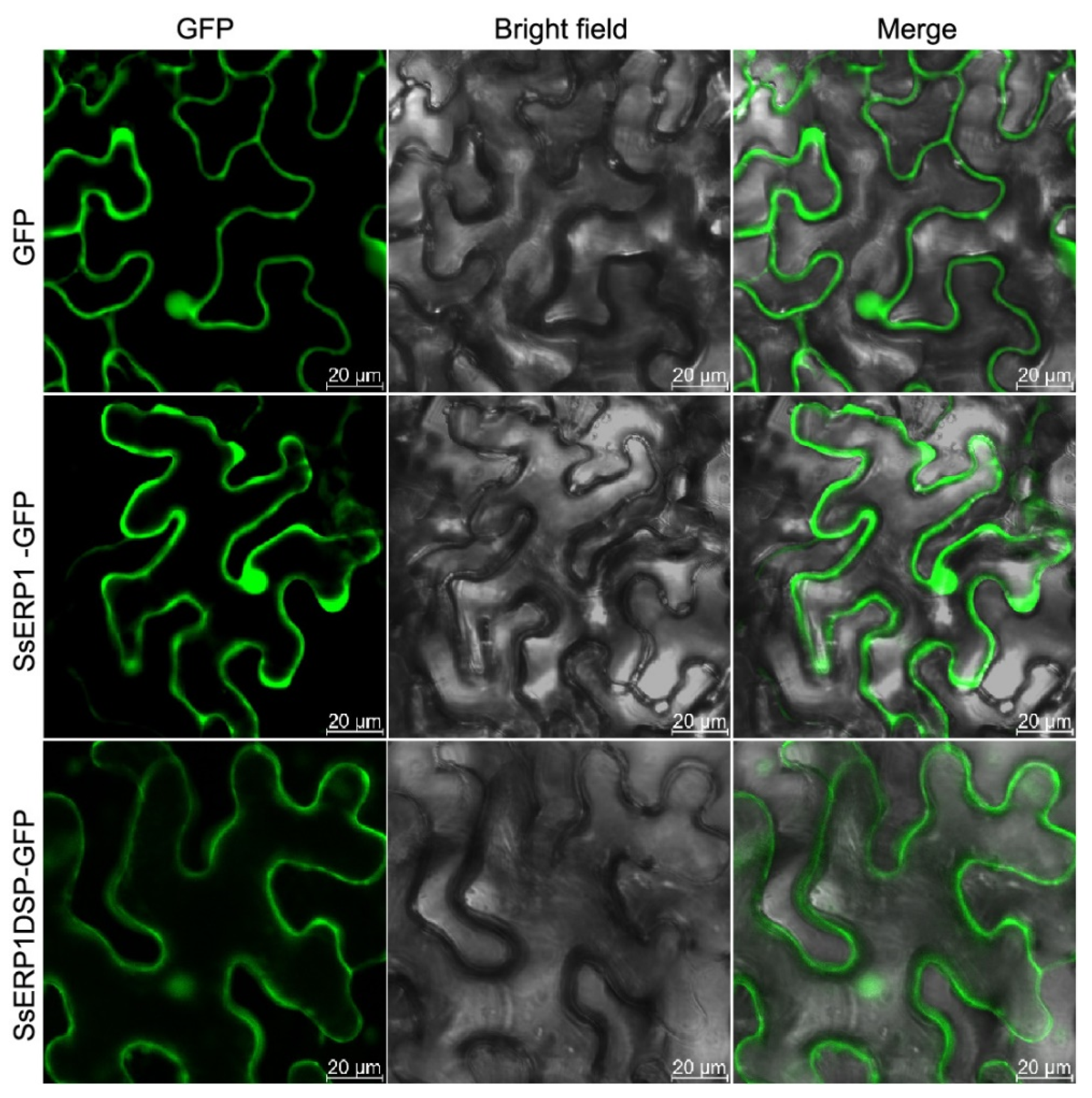
2.3. Subcellular Localization Analysis

To detect the subcellular localization of SsERP1, the coding sequences of full-length SsERP1 and the signal peptide-truncated SsERP1 (SsERP1DSP) were cloned into pJG186 vector. The cloning primer sequences are shown in Table S3. The resulting constructs (pJG186-SsERP1 and pJG186-SsERP1DSP) were transformed into Agrobacterium strain GV3101, and transiently expressed in N. benthamiana leaves by Agrobacterium infiltration. The subcellular localization of GFP-fused SsERP1 and SsERP1 were observed using a confocal microscope (LSM 880NLO) 4 days after infiltration.

2.4. The Effect of Ectopic SsERP1 Overexpression on S. sclerotiorum Pathogenicity

For transient expression of SsERP1, we used the Tobacco rattle virus (TRV)-based vectors pTRV1 and pTRV2 [20]. pTRV vectors were previously shown to be effective in gene overexpression, with an even higher overexpression level than 35S promoter-based vectors [21]. SsERP1 and SsERP1DSP were constructed into pTRV2 vector and then transformed into Agrobacterium strain GV3101. The cloning primer sequences are shown in Table S3. MES buffer (10 mM MgCl2, 10 mM MES, 200 μM acetosyringone, pH 5.7)-resuspended cultures of Agrobacterium strains harboring indicated pTRV2 constructs were mixed with that containing pTRV1 construct at a 1:1 ratio to make a final OD600 of 0.8. After incubation at room temperature in darkness for 2 h, Agrobacterium were injected into the leaves of N. benthamiana. The leaves were cut off and inoculated with S. sclerotiorum 48 h after Agrobacterium injection.

2.5. S. sclerotiorum Inoculation

The wild-type S. sclerotiorum strain used in this study is a field isolate and was maintained on PDA medium at 22 °C without light. For S. sclerotiorum inoculation with mycelium suspension, 6 fresh mycelium agar plugs with a diameter of 5 mm were inoculated into a 250 mL conical flask containing 150 mL PDB and incubated at 22 °C for 24 h at 150 rpm. The yielded mycelia balls were collected by filtering the overnight culture with sterilized gauzes and washed three times with ddH2O and PDB, respectively. The mycelia balls were smashed with, T18 digital (IAK) at 10,000 rpm for 15 min on ice. The resulting liquid mycelium suspension was adjusted to an OD600 of 2.0 using PDB, and 5 µL of the suspension was used for every single inoculation on plant leaves. For S. sclerotiorum inoculation with mycelium agar plugs, fresh mycelium agar plugs with a diameter of 5 mm were punched out from the plates and carefully inoculated onto plant leaves.

2.6. Transformation of S. sclerotiorum

The 796 bp upstream and 784 bp downstream flanking sequence of SsERP1 were inserted into the two multiple cloning sites at the left and right side of the hygromycin expression cassette in pLOB7, respectively [22]. The cloning primer sequences are shown in Table S3. The resulting pLOB7-SsERP1 construct was transformed into wild-type S. sclerotiorum by PEG-mediated protoplast transformation as described previously [21]. The transformants were selected on PDA medium containing 150 mg/L hygromycin three times, and homologous Sserp1 mutants were obtained by single ascospore isolation, which was further confirmed by PCR identification.

2.7. Transcriptome Analysis

Total RNAs were extracted from 0.5 g S. sclerotiorum-inoculated B. napus leaves. The quality of the RNAs was checked by electrophoresis (Figure S5). For each sample, 1 μg was taken for sequencing library preparation using the NEBNext®UltraTM RNA Library Prep Kit for Illumina® (NEB, Herts, UK). The clustering of the index-coded samples was performed on a cBot Cluster Generation System using TruSeq PE Cluster Kit v4-cBot-HS (Illumia, San Diego, CA, USA). After cluster generation, the prepared libraries were sequenced on an Illumina Hiseq x-ten platform at Biomarker Technologies (Beijing, China) and paired-end reads were generated. Differential expression analysis was performed using the DESeq R package (1.10.1). The resulting p values were adjusted using Benjamini and Hochberg’s approach to controlling the false discovery rate (FDR). The differently expressed genes (DEGs) were filtered out by setting the cut-off of FDR and fold-change at 0.01 and 2, respectively. KOBAS was used to perform KEGG enrichment of the differential expression genes.

2.8. Double-Stranded RNAs (dsRNAs) -Mediated Gene Knockdown

The target sequences were amplified (with T7 promoter) from S. sclerotiorum cDNA using primers shown in Table S5 (primer SsERP1A1 and primer SsERP1B1 for SsERP1A1, and primer SsERP1A2 and primer SsERP1B2 for SsERP1A2) to serve as the DNA templates for in vitro transcription. The dsRNAs were in vitro transcribed using T7 RNAi Transcription Kit (TR102-01, Vazyme Biotech, Nanjing, China) following the manufacturer’s instructions. For S. sclerotiorum inoculation, 5 μL dsRNA (900 ng/μL) was mixed with 1 mL liquid mycelium suspension (OD600 = 2.0), and then 10 μL liquid was pipetted onto plant leaves for each inoculation site.

2.9. Quantitative PCR (qPCR) Analysis

Total RNA was extracted from S. sclerotiorum or S. sclerotiorum-inoculated N. benthamiana leaves using Fungal Total RNA Isolation Kit (B518529, Sangon Biotech, Shanghai, China). DNase treatment and first-strand cDNA synthesis were conducted using HiScript 3 RT SuperMix for qPCR (+gDNA wiper) (R323-01, Vazyme Biotech, Nanjing, China). Quantitative real-time RCR was performed on Thermo Fisher Step One Plus instrument using Thermo Fisher PowerUp SYBR Green Master Mix (100031508, Thermo Fisher, Waltham, MA, USA). The following PCR program was used: 94 °C for 2 min, 40 cycles of 94 °C for 15 s, and 58 °C for 1 min. The internal reference genes for S. sclerotiorum, B.napus, and N. benthamiana were Tubulin, BnActin7, and L25, respectively. The relative gene expression levels were analyzed using the 2−ΔΔCT method [23]. The qPCR primer sequences are shown in Table S6. The specificity of the primers was checked by melting curve analysis (Figure S1).

2.10. The Statistical Analyses

The statistical analysis for lesion area, gene expression, mycelial growth, and acid production ability were performed using IBM SPSS 13.0 software. Statistical significance was tested using one-way ANOVA followed by Duncan’s test, with p < 0.05 being the threshold for statistical significance [24].

3. Results

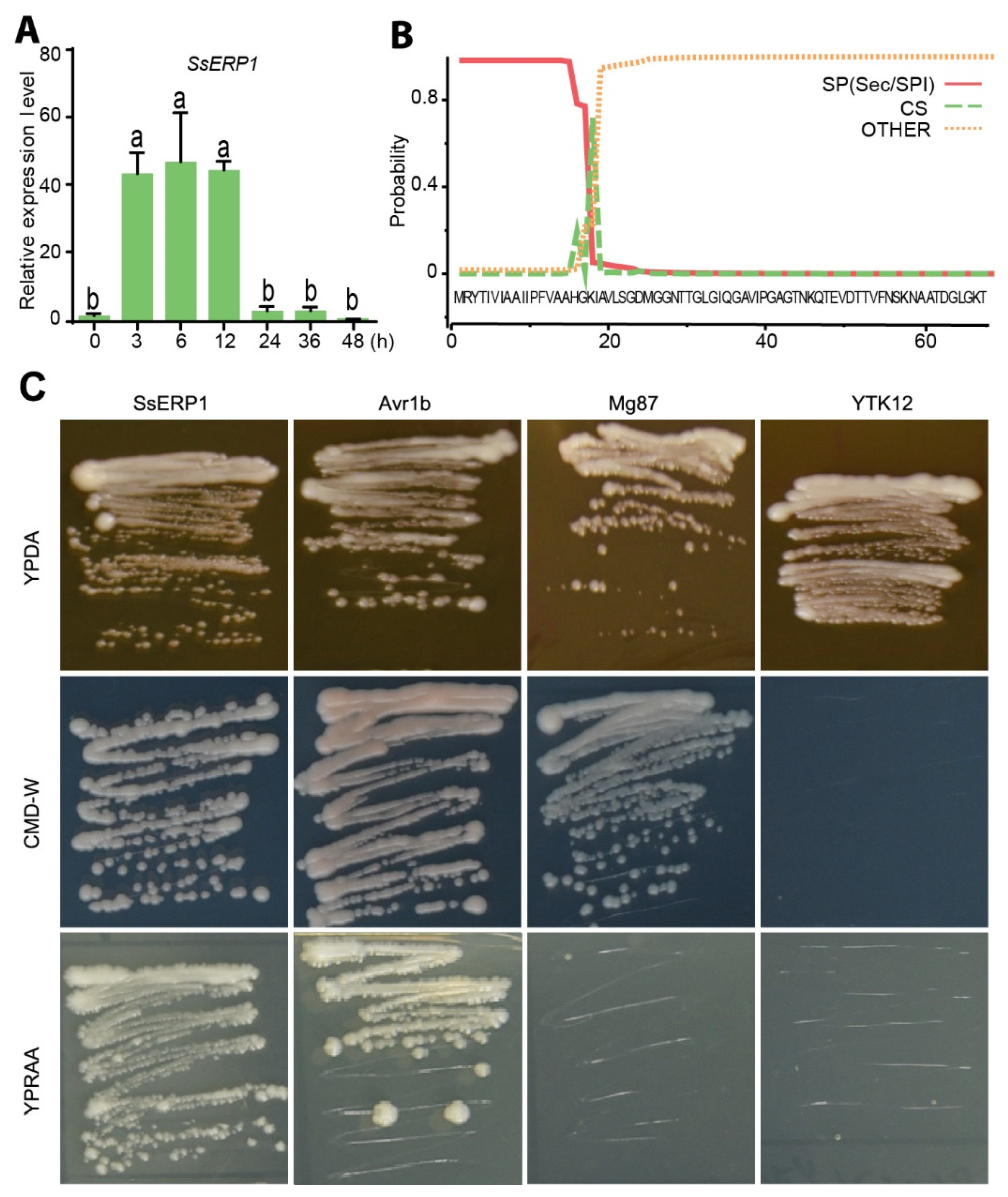
3.1. SsERP1 Is a Secretory Protein Highly Expressed in the Early Stage of S. sclerotiorum Infection

To identify the putative effectors of S. sclerotiorum, we carried out a bioinformatical screening using the following criteria: (1) contain secretory signal peptide, (2) have increased expression during S. sclerotiorum infection, and (3) do not harbor cell wall degrading enzyme-like domains, according to the data available in public databases or literatures [14,25]. As a result, we identified around one hundred effector candidates, including SS1G_11468 (hereinafter SsERP1) described in this study. Consistent with the published transcriptome data, our quantitative PCR results showed that the expression of SsERP1 was significantly induced at 3–12 h post S. sclerotiorum inoculation and decreased thereafter (Figure 1A), indicating that SsERP1 probably functions in the early stage of S. sclerotiorum infection.
Signal peptide analysis using SignalP revealed that SsERP1 contains a secretory signal peptide (SP) at its N terminal region (Figure 1B). In line with this, TMHMM 2.0 analysis found no transmembrane domain in SsERP1 (Figure S2). To verify the secretion property of SsERP1, we performed the yeast secretion assay. The predicted signal peptide of SsERP1 was constructed into the pSUC2 vector to fuse with the secretory peptide-truncated invertase gene SUC2. We also constructed the secretory signal of P. sojae Avr1b and the N-terminal of M. oryzae Mg87 into pSUC2 to serve as the positive and negative control, respectively. These plasmids were transformed into the yeast YTK12 strain, which is unable to grow on the YPRAA medium with raffinose as the sole carbon source because of lacking endogenous invertase. As shown in Figure 1C, the YTK12 strain transformed with the signal peptide sequences of SsERP1 and Avr1b, but not that transformed with the N-terminal of Mg87, regained the ability to grow on YPRAA medium, suggesting that the signal peptide sequences of SsERP1 (like that of Avr1b) was able to drive the secretion of SUC2 invertase, which degraded raffinose in YPRAA medium into simple sugars to support the growth of YTK12. This result indicates that the predicted signal peptide of SsERP1 is functional and SsERP1 is a secretory protein.
We next examined the subcellular localization of SsERP1 protein in plant cells. We fused the full-length (SsERP1) or signal peptide-truncated SsERP1 (SsERP1DSP) with GFP and transiently expressed them in tobacco leaves. Confocal microscopy showed that SsERP1, as well as SsERP1DSP, was distributed in both the nucleus and cytoplasm of plant cells (Figure 2).

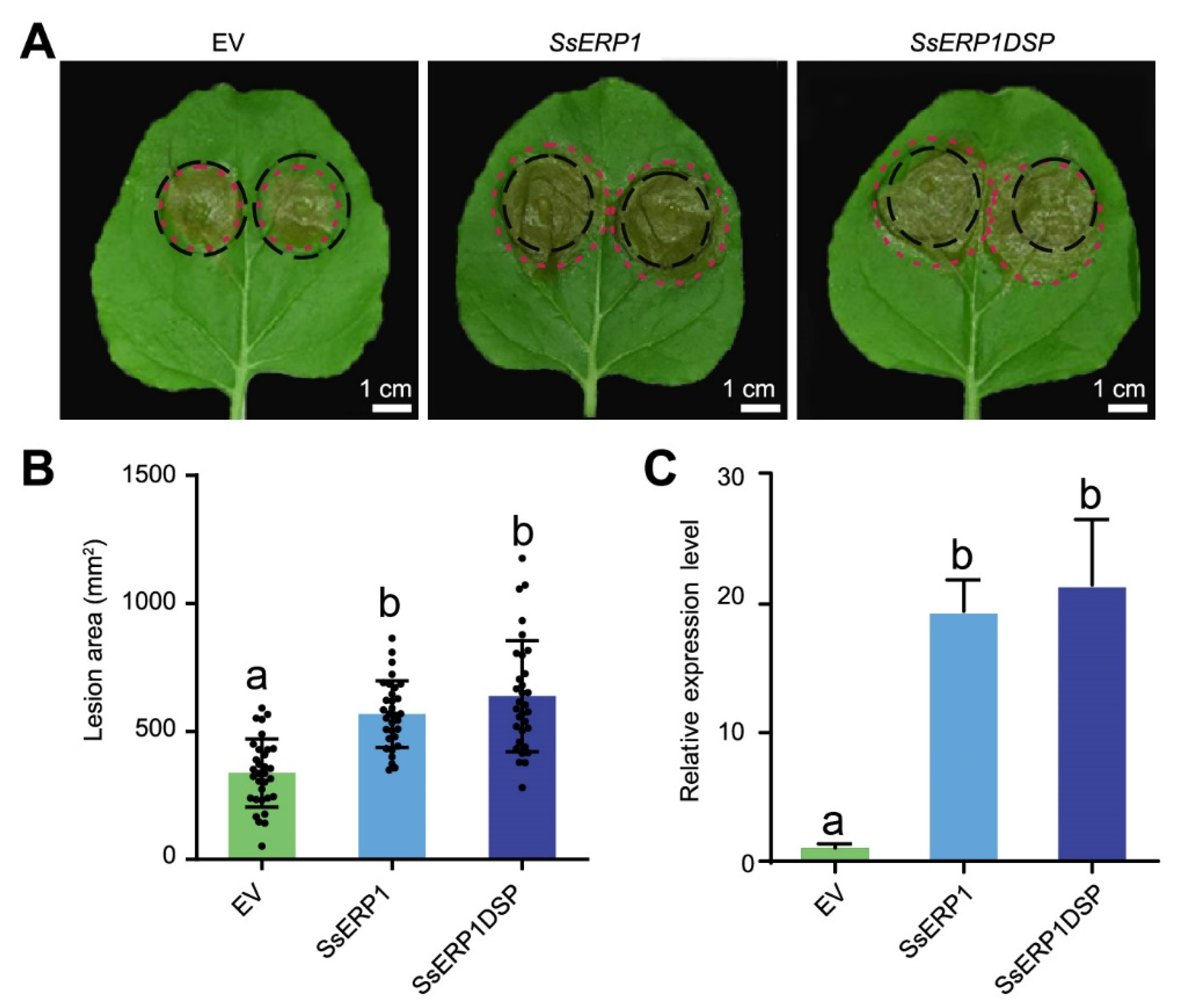
3.2. Overexpression of SsERP1 Promotes S. sclerotiorum Infection

Next, we tested the effect of SsERP1 on the pathogenicity of S. sclerotiorum. We constructed SsERP1 and SsERP1DSP into the pTRV2 vector, a virus backbone-based vector that was reported to be more effective than 35S promoter-based vectors in overexpressing effector genes [21]. We then transiently overexpressed SsERP1 in tobacco leaves by agroinfiltration and inoculated S. sclerotiorum 48 h after the infiltration. S. sclerotiorum caused obviously larger lesions on tobacco leaves expressing either SsERP1 or SsERP1DSP than on leaves expressing pTRV2 empty vector (control) (Figure 3A,B). Consistently, the expression of SsERP1 in tobacco leaves infiltrated with agrobacteria containing the pTRV2-SsERP1 or pTRV2-SsERP1DSP vectors was significantly higher than that in the leaves infiltrated with the pTRV2 empty vector 12 h after S. sclerotiorum inoculation (Figure 3C), which confirms that the enhanced pathogenicity of S. sclerotiorum resulted from the ectopic overexpression of SsERP1.

3.3. Knockout of SsERP1 Reduces the Pathogenicity of S. sclerotiorum

To verify the contribution of SsERP1 to the pathogenicity of S. sclerotiorum, we constructed SsERP1 knockout mutants by homologous recombination using PEG-mediated protoplast transformation (Figure S3A). Two homozygous SsERP1 knockout strains (Sserp1-1 and Sserp1-2) were obtained by single ascospore isolation. The complete deletion of SsERP1 gene in Sserp1-1 and Sserp1-2 were confirmed by PCR (Figure S3B).
First, we examined whether knocking out SsERP1 affects the growth and development of S. sclerotiorum. We inoculated the Sserp1 mutants and wild-type strain (WT) onto potato dextrose agar (PDA) and PBP (PDA supplemented with bromophenol blue, a pH indicator that will turn yellow when the pH becomes acidic). The mycelial growth rate of Sserp1 mutants on the PDA medium was not significantly different from that of WT (Figure 4A,C), indicating that SsERP1 is not required for the in vitro growth of S. sclerotiorum. When grown on PBP medium, Sserp1 mutants and WT generated a similar yellow color on their growth areas (Figure 4B,C), suggesting that the acid-producing ability of S. sclerotiorum is not obviously influenced by knocking out SsERP1 (Figure 4B,C). Since sclerotium plays an important role in the life cycle of S. sclerotiorum, we further examined the effect of knocking out SsERP1 on the sclerotium formation of S. sclerotiorum. As shown in Figure S4, the morphology and number of sclerotia formed by Sserp1-1 did not significantly differ from that of WT.
In contrast, we found that the lesion areas caused by the Sserp1 mutants were significantly smaller than those caused by WT strain when inoculated on tobacco or rapeseed leaves (Figure 4D,E). Together with the ectopic overexpression assays shown in Figure 3, these results suggest that SsERP1 specifically contributes to the pathogenicity of S. sclerotiorum, but not to its in vitro mycelial growth, acid production, and sclerotium formation.

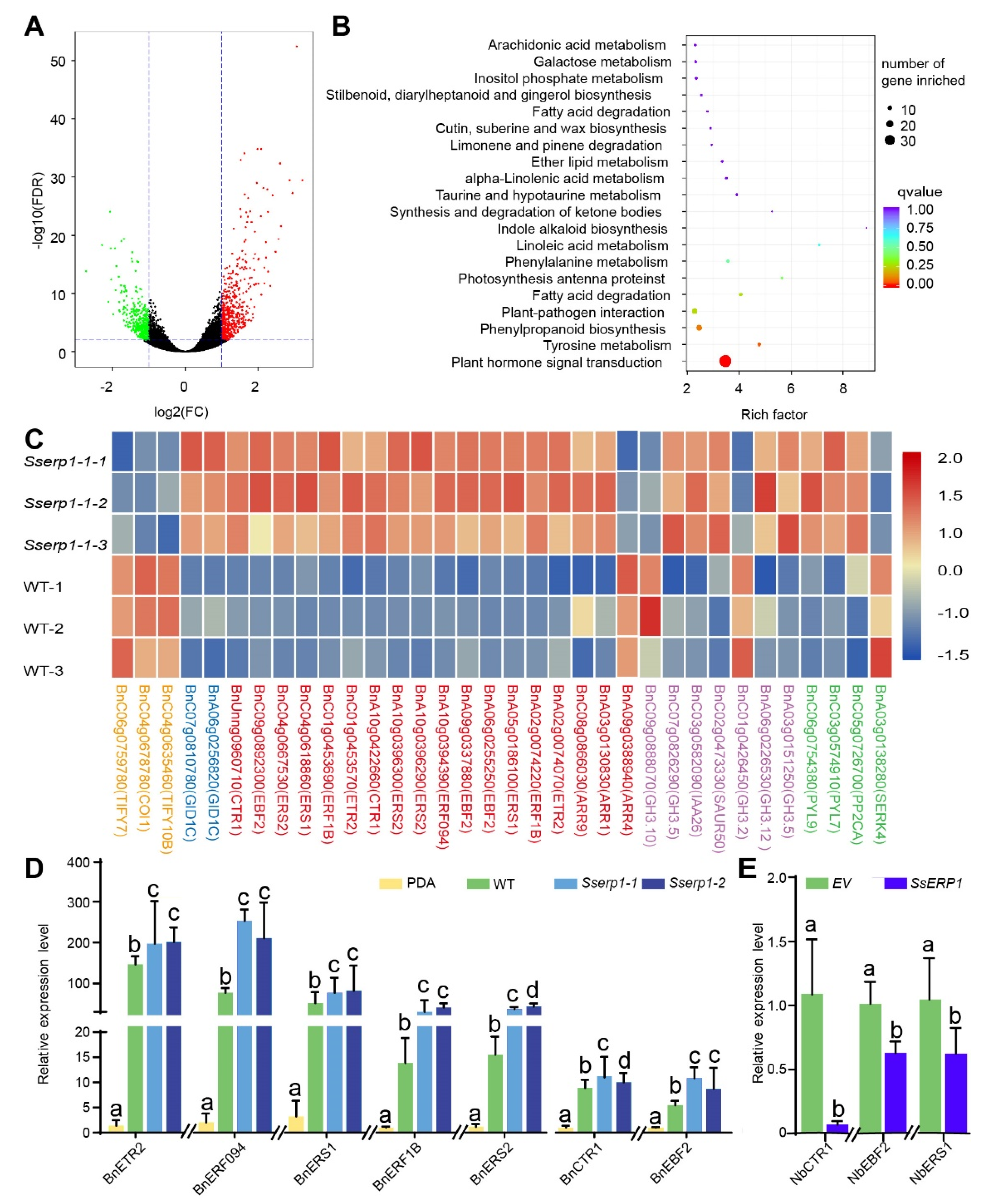
3.4. SsERP1 Suppresses Plant Ethylene Signaling

To gain insights into the molecular mechanisms of how SsERP1 promotes S. sclerotiorum infection, we carried out transcriptome analysis for B. napus plants infected with either Sserp1 mutant or the wild-type strains (Figure S5). As shown in Figure 5A, 522 genes were up-regulated and 386 were down-regulated in Sserp1 mutant-infected B. napus leaves compared with that infected with the wild-type strain. Subsequent KEGG pathway enrichment of these differentially expressed genes (DEGs) revealed the most significantly enriched pathway for plant hormone signaling transduction, in which 30 DEGs were included (Figure 5B,C). Strikingly, more than half (18) of these 30 DEGs are related to the ethylene signaling pathway, which is known to positively regulate plant defense against necrotrophic pathogens, including S. sclerotiorum (Figure 5C) [26,27,28]. In contrast to the ethylene signaling pathway, the expression of the ethylene biosynthetic genes (such as ACS and ACO) was not significantly different in Sserp1- and the WT strain-infected plants (Table S1). These results suggest that SsERP1 inhibits ethylene signaling transduction.
To verify the transcriptome data, we performed qPCR analysis. Consistent with our transcriptome data, all the ethylene responsive genes detected by qPCR showed higher expression levels in plants inoculated with Sserp1 mutants than those inoculated with the WT strain (Figure 5D), whereas no obvious trend of change was found for genes of jasmonate pathway, another phytohormone pathway required for full plant immunity against necrotrophic pathogens [29,30,31,32] (Figure S6). Moreover, we found that ectopic overexpression of SsERP1 also significantly attenuated the expression of ethylene responsive genes (NbCTR1, NbEBF2, and NbERS1) (Figure 5E), but showed significant impact on jasmonate responsive genes (NbAOS, NbOPR3, and NbPDF1.2) (Figure S6). These results confirm that SsERP1 specifically inhibits plant ethylene signaling.
Given the important role of the ethylene pathway in plant defense against necrotrophic pathogens [26,28], our results (Figure 3, Figure 4 and Figure 5) suggest that SsERP1 promotes S. sclerotiorum infection by repressing plant ethylene signaling.

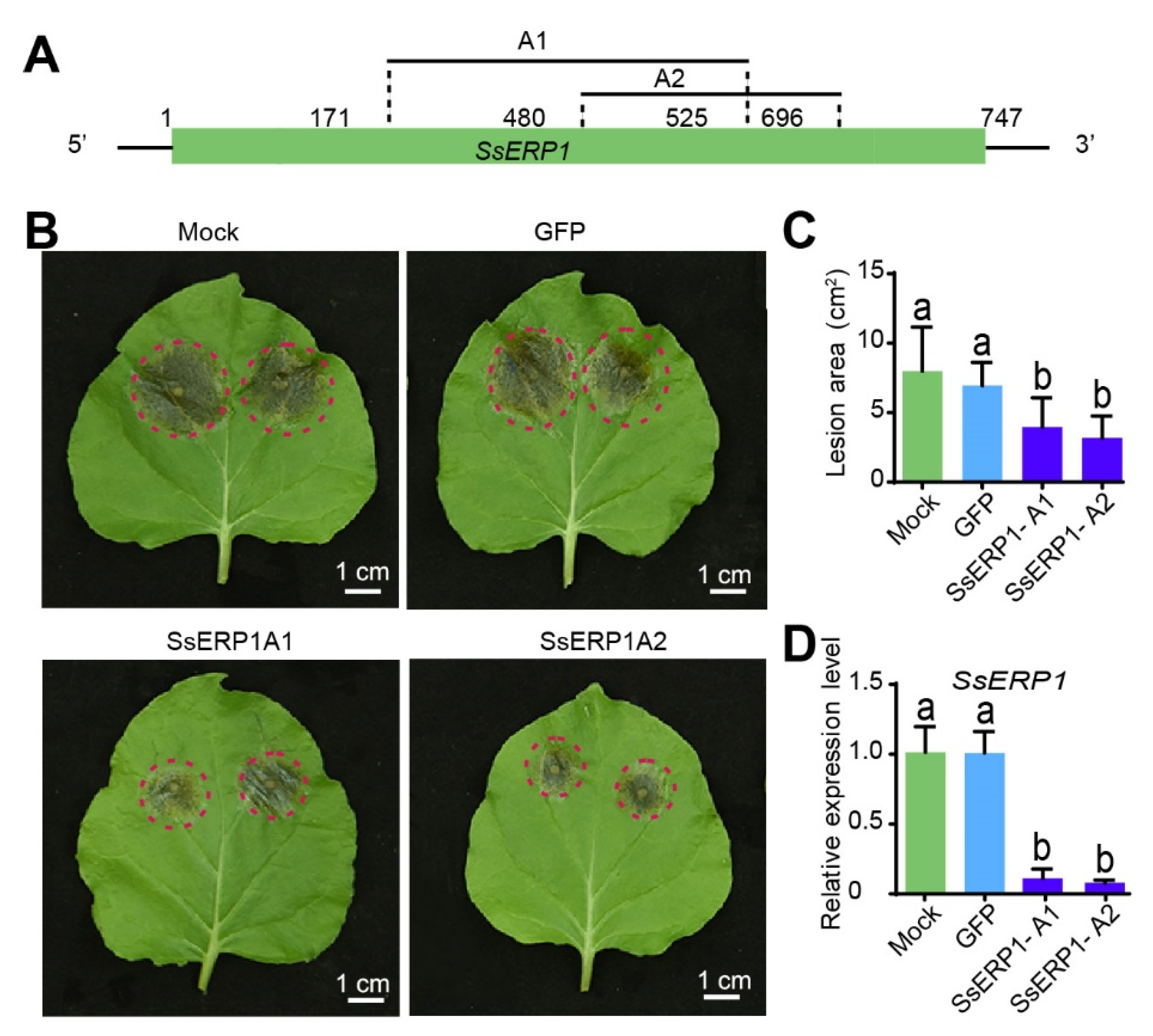
3.5. Silencing SsERP1 by Synthesized dsRNAs Inhibits S. sclerotiorum Infection

Previous studies have shown that knocking down virulence genes by in vitro synthesized double strand RNAs (dsRNA) can inhibit pathogen infection [33]. Thus, we tested whether silencing SsERP1 by dsRNA could attenuate S. sclerotiorum infection. We designed two dsRNAs (SsERP1A1 and SsERP1A2) targeting SsERP1 (Figure 6A). Target prediction using siFi21 software showed that SsERP1A1 and SsERP1A2 were able to specifically bind to SsERP1 without predictable off-target sites in either S. sclerotiorum or tobacco (Figure S7).
We synthesized the dsRNAs by in vitro transcription and then inoculated tobacco leaves with S. sclerotiorum pre-mixed with either SsERP1A1 or SsERP1A2 or pre-mixed with GFP-dsRNA (dsRNA targeting GFP) or an equal volume of water as controls. Quantitative PCR showed that the expression of SsERP1 in tobacco leaves inoculated with either SsERP1A1- or SsERP1A2-treated S. sclerotiorum was significantly lower than that in the control leaves (Figure 6D), demonstrating that SsERP1 was effectively silenced by the dsRNAs. Consistently, the lesions caused by S. sclerotiorum on tobacco leaves co-inoculated with SsERP1A1 or SsERP1A2 were smaller than those on the leaves co-inoculated with GFP-dsRNA or water (Figure 6B,C), suggesting that knocking down of SsERP1 by in vitro synthesized dsRNAs can inhibit S. sclerotiorum infection. These results provide additional support to our conclusion that SsERP1 contributes to S. sclerotiorum pathogenicity, and further suggest a potential strategy for controlling S. sclerotiorum disease by using in vitro synthesized dsRNAs that target SsERP1.

4. Discussion

Sclerotinia stem rot is one of the major diseases in B. napus. Considering the economic and environmental costs associated with fungicides, developing genetically resistant varieties is a more ideal way to cope with Sclerotinia disease. However, previous quantitative trait loci (QTL) mapping and genome-wide association studies (GWAS) indicate that the resistance of B. napus to S.sclerotiorum is a combined effect of many QTLs with small contributions [34,35,36,37], which obstructs the integration and application of these QTLs. The lack of highly resistant accessions within Brassica species also makes it challenging to identify novel genes that have major contribution to the trait. Given this predicament, it is critical to deepening our understanding of S. sclerotiorum pathogenesis, which would possibly provide a new prospective and inspire novel strategies for Sclerotinia disease management. Effectors are key weapons for diverse pathogens to overcoming plant immunity [6,11,38]. Compared with the extensive studies on effectors in specialized host-pathogen interactions, our understanding of effector-mediated interactions between plant and broad host range necrotrophic pathogens is limited [7,8]. Recent studies suggest that some broad host range necrotrophic fungi might also have a short biotrophic phase at the early infection stage, during which period they secrete effectors into plant cells to overcome host immunity and achieve successful colonization [14,15,39]. However, the functions of effectors encoded by S. sclerotiorum are largely unknown. In this study, we cloned and characterized a new S. sclerotiorum effector, SsERP1. By a series of biochemical and genetic assays, we demonstrated that SsERP1 is required for the full virulence of S. sclerotiorum.
Ethylene plays an important role in regulating plant defense against pathogens, including S. sclerotiorum [26,28]. A large number of ethylene pathway genes, including those involved in either ethylene biosynthesis or ethylene signaling transduction, are induced in plants upon S. sclerotiorum infection (Figure 5) [40,41]. Interestingly, our transcriptome data suggested that SsERP1 mainly repressed the induction of plant ethylene signaling transduction genes, but not the ethylene biosynthetic genes, during S. sclerotiorum infection, indicating that SsERP1 might target to some component(s) of ethylene signaling transduction processes to inhibit ethylene responses and facilitate S. sclerotiorum infection. However, we cannot exclude the possibility that SsERP1 inhibits ethylene biosynthetic genes at the post-transcriptional levels [42,43]. Identification of the direct target(s) of SsERP1 in plant cells is required to clearly elucidate how SsERP1 suppresses plant ethylene pathway.
Jasmonate pathway is also known to be required for full plant immunity against necrotrophic pathogens [44]. Previous studies showed that ethylene and jasmonate pathways synergistically regulate plant defense against necrotrophic pathogens, while antagonistically regulate plant development and defense against insects [29,30,31,32]. In our transcriptome data, we noted that three JA signaling pathway genes (TIFY7, COI1, and TIFY10B) were down-regulated in B. napus leaves inoculated with the Sserp1 strain compared to those inoculated with WT strain (Figure 5D and Figure S6). However, the expression of the vast majority of known jasmonate pathway genes was not significantly changed (Table S2 and Figure S6). For example, among the 42 JAZ genes expressed in oilseed rape, only TIFY7 (JAZ2, BnC04g0635460) and TIFY10B (JAZ9, BnC06g0759780) were differently expressed in Sserp1- and WT strain-infected plants (Table S2). In addition to JAZ genes, consistent with the transcriptome data, our qPCR analysis showed that the expression of other jasmonate pathway marker genes, such as MYC2, AOS, LOX3, and OPR3, was also not significantly affected by SsERP1 (Table S2 and Figure S6). These results suggest that SsERP1 specifically inhibits ethylene signaling pathway, while has no obvious impact on jasmonate pathway. Identifying the direct plant target of SsERP1 would provide insights into how SsERP1 specifically represses plant ethylene signaling pathway without perturbing jasmonate pathway. Given the critical role of jasmonate in plant defense against necrotrophic pathogens, we speculate that S. sclerotiorum might encode other effectors that target plant jasmonate pathway to enhance infection.
Spray-induced gene silencing (SIGS), where dsRNAs or small RNAs (sRNAs) are sprayed onto plant surface to silence the target genes of pathogens, has emerged as a new and powerful strategy for crop protection [45,46]. Recently, Mittes and colleagues developed a clay nanomaterial that could encapsulate dsRNA, which improved the stability of dsRNAs in the natural environment and prolonged their silencing activity [47]. The SIGS technology provides an alternative way to cope with Sclerotinia disease. In this study, we showed that silencing SsERP1 by the dsRNAs SsERP1A1 and SsERP1A2 effectively inhibited S. sclerotiorum infection (Figure 6). Our work validates the possible application of SIGS in Sclerotinia disease control and presents the effector gene SsERP1 as the potential target.

5. Conclusions

In this study, we cloned and characterized a novel effector of S. sclerotiorum, SsERP1 (ethylene pathway repressor protein 1), a secretory protein that is highly expressed in the early stages of S. sclerotiorum infection. Ectopic overexpression of SsERP1 in plant leaves promoted S. sclerotiorum infection, and knockout or RANi of SsERP1 expression reduced S. sclerotiorum pathogenicity without obvious impact on mycelial growth and sclerotium formation suggesting that SsERP1 specifically contributes to the pathogenicity of S. sclerotiorum. Transcriptome analysis further showed that SsERP1 promotes S. sclerotiorum infection by inhibiting plant ethylene signaling. Moreover, in vitro synthesized double-stranded RNA effectively suppressed SsERP1 expression and attenuated S. sclerotiorum infection, providing a potential new strategy for S. sclerotiorum control.

Supplementary Materials

The following are available online at www.mdpi.com/article/10.3390/jof7100825/s1. Figure S1: Melting curve analysis. Figure S2: SsERP1 does not contain transmembrane domains. Figure S3: Identification of SsERP1 knockout mutants. Figure S4: SsERP1 has no obvious impact on sclerotium formation. Figure S5: Quality check of RNA samples for transcriptome. Figure S6: sERP1 did not affect the expression of the majority of jasmonate pathway genes. Figure S7: Target site prediction for the SsERP1A1 and SsERP1A2 dsRNAs. Table S1: Expression of ethylene biosynthesis pathway genes in B.napus leaves inoculated with Sserp1 and WT strains, respectively. Table S2: Expression of JA pathway genes in B.napus leaves inoculated with Sserp1 and WT strains, respectively. Table S3: Primers for vector construction. Table S4: Primers for Sserp1 mutant identification. Table S5: Primers for dsRNA template amplification. Table S6: Primers for qPCR analysis.

Author Contributions

D.W. and Y.W. designed the research; H.F. and D.W. performed the experiments with the help of W.Y., J.N., W.Z. and J.W.; Y.W., D.W. and H.F. analyzed the data; D.W., H.F. wrote the manuscript with contributions of all the authors. All authors have read and agreed to the published version of the manuscript.

Funding

This research was supported by Natural Science Foundation of China (31901570, 31741097, 32172019), the Natural Science Foundation of Jiangsu Province (BE2018356, BK20180903), China Postdoctoral Science Foundation (BX201700205, 2018M630612), the Priority Academic Program Development of Jiangsu Higher Education Institutions, and the Project of Special Funding for Crop Science Discipline Development.

Institutional Review Board Statement

Not applicable.

Informed Consent Statement

Not applicable.

Data Availability Statement

Raw Ilumina sequencing data of Brassica nupas are available in the NCBI Sequence Reads Archive (SRA) under the accession number PRJNA759653.

Acknowledgments

We sincerely thank Daohong Jiang, Jiatao Xie, and Qili Zhu for the help with setting up experimental procedures and the gifts of materials.

Conflicts of Interest

The authors declare no conflict of interest.

Accession Numbers

SsERP1 (SS1G_11468), Tubulin (MH796665), BnERF094 (BnaC05g04210D), BnERF1B (BnaA01g23940D), BnETR2 (BnaCnng68360D), BnCTR1 (BnaCnng02610D), BnEBF2 (BnaA06g27640D), BnERS1 (BnaA05g00430D), BnERS2 (BnaA08g27820D), NbCTR1 (Niben101Scf06769g02003), NbEBF2 (Niben101Scf12609g01007), NbERS1 (Niben101Scf01430g02010), NbAOS (Niben101Scf05799g02010.1), NbOPR3 (NM_001325344.1), NbPDF1.2 (KP017278.1).

References

  1. Friedt, W.; Tu, J.; Fu, T. Academic and economic importance of Brassica napus rapeseed. In The Brassica Napus Genome; Springer: Berlin/Heidelberg, Germany, 2018; pp. 1–20. [Google Scholar]
  2. Zubair, M.; Maqbool, F.; Mehmood, I.; Muzammil, S.; Waseem, M.; Imran, M.; Nadeem, H.U.; Azeem, F.; Siddique, M.H. Rapeseed oil. In Green Sustainable Process for Chemical and Environmental Engineering and Science; Elsevier: Amsterdam, The Netherlands, 2021; pp. 41–55. [Google Scholar]
  3. Derbyshire, M.C.; Denton-Giles, M. The control of sclerotinia stem rot on oilseed rape (Brassica napus): Current practices and future opportunities. Plant Pathol. 2016, 65, 859–877. [Google Scholar] [CrossRef] [Green Version]
  4. Xu, L.; Li, G.; Jiang, D.; Chen, W. Sclerotinia sclerotiorum: An Evaluation of Virulence Theories. Annu. Rev. Phytopathol. 2018, 56, 311–338. [Google Scholar] [CrossRef] [PubMed]
  5. Neik, T.X.; Barbetti, M.J.; Batley, J. Current status and challenges in identifying disease resistance genes in Brassica napus. Front. Plant Sci. 2017, 8, 1788. [Google Scholar] [CrossRef] [PubMed]
  6. Dodds, P.N.; Rathjen, J.P. Plant immunity: Towards an integrated view of plant–pathogen interactions. Nat. Rev. Genet. 2010, 11, 539–548. [Google Scholar] [CrossRef] [PubMed]
  7. Giraldo, M.C.; Valent, B. Filamentous plant pathogen effectors in action. Nat. Rev. Microbiol. 2013, 11, 800–814. [Google Scholar] [CrossRef] [PubMed]
  8. Toruño, T.Y.; Stergiopoulos, I.; Coaker, G. Plant-pathogen effectors: Cellular probes interfering with plant defenses in spatial and temporal manners. Annu. Rev. Phytopathol. 2016, 54, 419–441. [Google Scholar] [CrossRef] [PubMed] [Green Version]
  9. Lo Presti, L.; Lanver, D.; Schweizer, G.; Tanaka, S.; Liang, L.; Tollot, M.; Zuccaro, A.; Reissmann, S.; Kahmann, R. Fungal effectors and plant susceptibility. Annu. Rev. Plant Biol. 2015, 66, 513–545. [Google Scholar] [CrossRef]
  10. Zhang, J.; Shao, F.; Li, Y.; Cui, H.; Chen, L.; Li, H.; Zou, Y.; Long, C.; Lan, L.; Chai, J. A Pseudomonas syringae effector inactivates MAPKs to suppress PAMP-induced immunity in plants. Cell Host Microbe 2007, 1, 175–185. [Google Scholar] [CrossRef] [Green Version]
  11. Wu, D.; Qi, T.; Li, W.-X.; Tian, H.; Gao, H.; Wang, J.; Ge, J.; Yao, R.; Ren, C.; Wang, X.-B. Viral effector protein manipulates host hormone signaling to attract insect vectors. Cell Res. 2017, 27, 402–415. [Google Scholar] [CrossRef]
  12. Cai, Q.; Qiao, L.; Wang, M.; He, B.; Lin, F.-M.; Palmquist, J.; Huang, S.-D.; Jin, H. Plants send small RNAs in extracellular vesicles to fungal pathogen to silence virulence genes. Science 2018, 360, 1126–1129. [Google Scholar] [CrossRef] [Green Version]
  13. Wang, X.; Jiang, N.; Liu, J.; Liu, W.; Wang, G.-L. The role of effectors and host immunity in plant–necrotrophic fungal interactions. Virulence 2014, 5, 722–732. [Google Scholar] [CrossRef] [PubMed] [Green Version]
  14. Guyon, K.; Balagué, C.; Roby, D.; Raffaele, S. Secretome analysis reveals effector candidates associated with broad host range necrotrophy in the fungal plant pathogen Sclerotinia sclerotiorum. BMC Genom. 2014, 15, 336. [Google Scholar] [CrossRef] [PubMed] [Green Version]
  15. Derbyshire, M.; Dentongiles, M.; Hegedus, D.; Seifbarghy, S.; Rollins, J.; Van, K.J.; Seidl, M.F.; Faino, L.; Mbengue, M.; Navaud, O. The complete genome sequence of the phytopathogenic fungus Sclerotinia sclerotiorum reveals insights into the genome architecture of broad host range pathogens. Genome Biol. Evol. 2017, 9, 593–618. [Google Scholar] [CrossRef]
  16. Heard, S.; Brown, N.A.; Hammond-Kosack, K. An interspecies comparative analysis of the predicted secretomes of the necrotrophic plant pathogens Sclerotinia sclerotiorum and Botrytis cinerea. PLoS ONE 2015, 10, e0130534. [Google Scholar] [CrossRef] [PubMed] [Green Version]
  17. Seifbarghi, S.; Borhan, M.H.; Wei, Y.; Ma, L.; Coutu, C.; Bekkaoui, D.; Hegedus, D.D. Receptor-Like Kinases BAK1 and SOBIR1 Are Required for Necrotizing Activity of a Novel Group of Sclerotinia sclerotiorum Necrosis-Inducing Effectors. Front. Plant Sci. 2020, 11, 1021. [Google Scholar] [CrossRef] [PubMed]
  18. Yu, Y.; Xiao, J.; Zhu, W.; Yang, Y.; Mei, J.; Bi, C.; Qian, W.; Qing, L.; Tan, W. Ss-Rhs1, a secretory Rhs repeat-containing protein, is required for the virulence of Sclerotinia sclerotiorum. Mol. Plant Pathol. 2017, 18, 1052–1061. [Google Scholar] [CrossRef]
  19. Dou, D.; Kale, S.D.; Wang, X.; Jiang, R.H.; Bruce, N.A.; Arredondo, F.D.; Zhang, X.; Tyler, B.M. RXLR-mediated entry of Phytophthora sojae effector Avr1b into soybean cells does not require pathogen-encoded machinery. Plant Cell 2008, 20, 1930–1947. [Google Scholar] [CrossRef] [Green Version]
  20. Liu, Y.; Schiff, M.; Marathe, R.; Dinesh-Kumar, S.P. ptrv2-Tobacco Rar1, EDS1 and NPR1/NIM1 like genes are required for N-mediated resistance to tobacco mosaic virus. Plant J. 2002, 30, 415–429. [Google Scholar] [CrossRef]
  21. Yang, G.; Tang, L.; Gong, Y.; Xie, J.; Fu, Y.; Jiang, D.; Li, G.; Collinge, D.; Chen, W.; Cheng, J. A cerato-platanin protein SsCP1 targets plant PR1 and contributes to virulence of Sclerotinia sclerotiorum. New Phytol. 2018, 217, 739–755. [Google Scholar] [CrossRef] [Green Version]
  22. M Patel, R.; N Heneghan, M.; van Kan, J.A.L.; M Bailey, A.; D Foster, G. The pOT and pLOB vector systems: Improving ease of transgene expression in Botrytis cinerea. J. Gen. Appl. Microbiol. 2008, 54, 367–376. [Google Scholar] [CrossRef] [Green Version]
  23. Livak, K.J.; Schmittgen, T.D. Analysis of relative gene expression data using real-time quantitative PCR and the 2−ΔΔCT method. Methods 2001, 25, 402–408. [Google Scholar] [CrossRef]
  24. Field, A. Discovering Statistics Using IBM SPSS Statistics; Sage: Newcastle, UK, 2013. [Google Scholar]
  25. Seifbarghi, S.; Borhan, M.H.; Wei, Y.; Coutu, C.; Robinson, S.J.; Hegedus, D.D. Changes in the Sclerotinia sclerotiorum transcriptome during infection of Brassica napus. Bmc. Genom. 2017, 18, 266. [Google Scholar] [CrossRef] [Green Version]
  26. Broekaert, W.F.; Delauré, S.L.; De Bolle, M.F.; Cammue, B.P. The role of ethylene in host-pathogen interactions. Annu. Rev. Phytopathol. 2006, 44, 393–416. [Google Scholar] [CrossRef] [PubMed]
  27. Guo, X.; Stotz, H.U. Defense against Sclerotinia sclerotiorum in Arabidopsis is dependent on jasmonic acid, salicylic acid, and ethylene signaling. Mol. Plant-Microbe Interact. 2007, 20, 1384–1395. [Google Scholar] [CrossRef] [Green Version]
  28. van Loon, L.C.; Geraats, B.P.; Linthorst, H.J. Ethylene as a modulator of disease resistance in plants. Trends Plant Sci. 2006, 11, 184–191. [Google Scholar] [CrossRef]
  29. Bürger, M.; Chory, J. Stressed out about hormones: How plants orchestrate immunity. Cell Host Microbe 2019, 26, 163–172. [Google Scholar] [CrossRef] [PubMed]
  30. Kazan, K.; Manners, J.M. JAZ repressors and the orchestration of phytohormone crosstalk. Trends Plant Sci. 2012, 17, 22–31. [Google Scholar] [CrossRef] [PubMed]
  31. Zhu, Z.; An, F.; Feng, Y.; Li, P.; Xue, L.; Mu, A.; Jiang, Z.; Kim, J.-M.; To, T.K.; Li, W. Derepression of ethylene-stabilized transcription factors (EIN3/EIL1) mediates jasmonate and ethylene signaling synergy in Arabidopsis. Proc. Natl. Acad. Sci. USA 2011, 108, 12539–12544. [Google Scholar] [CrossRef] [Green Version]
  32. Grant, M.R.; Jones, J.D. Hormone (dis) harmony moulds plant health and disease. Science 2009, 324, 750–752. [Google Scholar] [CrossRef]
  33. Höfle, L.; Biedenkopf, D.; Werner, B.; Shrestha, A.; Jelonek, L.; Koch, A. Study on the efficiency of dsRNAs with increasing length in RNA-based silencing of the Fusarium CYP51 genes. RNA Biol. 2020, 17, 463–473. [Google Scholar] [CrossRef]
  34. Li, J.; Zhao, Z.; Hayward, A.; Cheng, H.; Fu, D. Integration analysis of quantitative trait loci for resistance to Sclerotinia sclerotiorum in Brassica napus. Euphytica 2015, 205, 483–489. [Google Scholar] [CrossRef]
  35. Mei, J.; Shao, C.; Yang, R.; Feng, Y.; Gao, Y.; Ding, Y.; Li, J.; Qian, W. Introgression and pyramiding of genetic loci from wild Brassica oleracea into B. napus for improving Sclerotinia resistance of rapeseed. Theor. Appl. Genet. 2020, 133, 1313–1319. [Google Scholar] [CrossRef]
  36. Wu, J.; Chen, P.; Zhao, Q.; Cai, G.; Hu, Y.; Xiang, Y.; Yang, Q.; Wang, Y.; Zhou, Y. Co-location of QTL for Sclerotinia stem rot resistance and flowering time in Brassica napus. Crop J. 2019, 7, 227–237. [Google Scholar] [CrossRef]
  37. Zhang, F.; Huang, J.; Tang, M.; Cheng, X.; Liu, Y.; Tong, C.; Yu, J.; Sadia, T.; Dong, C.; Liu, L. Syntenic quantitative trait loci and genomic divergence for Sclerotinia resistance and flowering time in Brassica napus. J. Integr. Plant Biol. 2019, 61, 75–88. [Google Scholar] [CrossRef] [PubMed] [Green Version]
  38. Clark, K.; Franco, J.Y.; Schwizer, S.; Pang, Z.; Hawara, E.; Liebrand, T.W.; Pagliaccia, D.; Zeng, L.; Gurung, F.B.; Wang, P. An effector from the Huanglongbing-associated pathogen targets citrus proteases. Nat. Commun. 2018, 9, 1718. [Google Scholar] [CrossRef] [PubMed]
  39. Kabbage, M.; Yarden, O.; Dickman, M.B. Pathogenic attributes of Sclerotinia sclerotiorum: Switching from a biotrophic to necrotrophic lifestyle. Plant Sci. 2015, 233, 53–60. [Google Scholar] [CrossRef] [PubMed]
  40. Wu, J.; Zhao, Q.; Yang, Q.; Liu, H.; Li, Q.; Yi, X.; Cheng, Y.; Guo, L.; Fan, C.; Zhou, Y. Comparative transcriptomic analysis uncovers the complex genetic network for resistance to Sclerotinia sclerotiorum in Brassica napus. Sci. Rep. 2016, 6, 19007. [Google Scholar] [CrossRef] [Green Version]
  41. Zhao, J.; Buchwaldt, L.; Rimmer, S.R.; Sharpe, A.; McGregor, L.; Bekkaoui, D.; Hegedus, D. Patterns of differential gene expression in Brassica napus cultivars infected with Sclerotinia sclerotiorum. Mol. Plant Pathol. 2009, 10, 635–649. [Google Scholar] [CrossRef]
  42. Konishi, M.; Yanagisawa, S. Ethylene signaling in Arabidopsis involves feedback regulation via the elaborate control of EBF2 expression by EIN3. Plant J. 2008, 55, 821–831. [Google Scholar] [CrossRef] [PubMed]
  43. Booker, M.A.; DeLong, A. Producing the ethylene signal: Regulation and diversification of ethylene biosynthetic enzymes. Plant Physiol. 2015, 169, 42–50. [Google Scholar] [CrossRef] [Green Version]
  44. Wasternack, C. Action of jasmonates in plant stress responses and development—applied aspects. Biotechnol. Adv. 2014, 32, 31–39. [Google Scholar] [CrossRef] [PubMed]
  45. Wang, M.; Jin, H. Spray-induced gene silencing: A powerful innovative strategy for crop protection. Trends Microbiol. 2017, 25, 4–6. [Google Scholar] [CrossRef] [PubMed] [Green Version]
  46. Cai, Q.; He, B.; Weiberg, A.; Buck, A.H.; Jin, H. Small RNAs and extracellular vesicles: New mechanisms of cross-species communication and innovative tools for disease control. PLoS Pathog. 2019, 15, e1008090. [Google Scholar] [CrossRef] [Green Version]
  47. Mitter, N.; Worrall, E.A.; Robinson, K.E.; Li, P.; Jain, R.G. Clay nanosheets for topical delivery of RNAi for sustained protection against plant viruses. Nat. Plants 2017, 3, 16207. [Google Scholar] [CrossRef] [PubMed]
Figure 1. SsERP1 is a secretory protein that was highly expressed at the early stage of Sclerotinia sclerotiorum infection. (A), SsERP1 was highly expressed at the early stage of S. sclerotiorum infection. The relative expression of SsERP1 in S. sclerotiorum-infected tobacco leaves was detected by quantitative PCR (qPCR) at 3, 6, 12, 24, 36, and 48 h after inoculation. 0-h samples were collected immediately after inoculation. S. sclerotiorum Tubulin was used as an internal control. Data are shown as mean ± SD, n = 3. Different letters above the bars indicate statistically significant differences (p < 0.05, one-way ANOVA followed by Duncan’s test). (B), A signal peptide was predicted in SsERP1 by SignaIP-5.0. The red line denotes the probability of that the sequence contains a signal peptide. The green dotted line denotes the probability of that the sequence exists cleavage site. The red dotted line denotes the probability of that the sequence does not have a signal peptide. (C), The signal peptide of SsERP1 is functional in yeast secretion assay. YPDA medium was used to confirm normal growth of the yeast strains on complete medium, CMD-W medium to verify the successful transformation of indicated constructs, and YPRAA medium to assess the secretion ability of the transformed signal peptide. The YTK12 strain was unable to grow on CMD-W and YPRAA medium, Mg87 was unable to grow on the YPRAA medium, while SsERP1 and positive control Avr1b were able to grow on all these three types of medium.
Jof 07 00825 g001
Figure 2. Subcellular localization of SsERP1 and SsERP1DSP. GFP-fused full-length SsERP1 and signal peptide-removed SsERP1 (SsERP1DSP) were transiently expressed in tobacco leaves by Agrobacterium infiltration. GFP fluorescence was observed 4 days after infiltration.
Jof 07 00825 g002
Figure 3. Ectopic overexpression of SsERP1 promotes Sclerotinia sclerotiorum infection. (A), The representative phenotype of S. sclerotiorum-inoculated tobacco leaves. Tobacco leaves were inoculated with S. sclerotiorum mycelium suspension 48 h after infiltration of Agrobacterium containing pTRV2-SsERP1 (SsERP1), pTRV2-SsERP1DSP (SsERP1DSP), or pTRV2 empty vector (EV) as control. Photos were taken 48 h after S. sclerotiorum inoculation. Black circles indicate the leaf areas that infiltrated with indicated Agrobacterium and the margins of lesions caused by S. sclerotiorum were marked with red circles. (B), The lesion sizes caused by S. sclerotiorum in (A) were measured by ImageJ. Data are presented as mean ± SD, n = 16. Different letters above the bars indicate statistically significant differences (p < 0.05, one-way ANOVA followed by Duncan’s test). (C), qPCR analysis of SsERP1 expression levels. Tobacco leaves were inoculated with S. sclerotiorum mycelium suspension 48 h after infiltration of Agrobacterium containing pTRV2-SsERP1 (SsERP1), pTRV2-SsERP1DSP (SsERP1DSP), or pTRV2 empty vector (EV) as control. Tobacco leaves were collected for qPCR analysis 12 h after S. sclerotiorum inoculation. Data are presented as mean ± SD, n = 3. Different letters above the bars indicate statistically significant differences (p < 0.05, one-way ANOVA followed by Duncan’s test).
Jof 07 00825 g003
Figure 4. Knockout of SsERP1 reduces the pathogenicity of Sclerotinia sclerotiorum. (AC), Knockout of SsERP1 does not affect mycelial growth (A,C) or acid production ability (B,C) of S. sclerotiorum. Mycelium agar plugs of the wild-type strain (WT) and Sserp1 mutants were inoculated on PDA medium (A) or PBP (PDA medium containing bromophenol blue) medium (B) for assessing mycelial growth or acid production ability of S. sclerotiorum, respectively. Photos were taken 48 h after inoculation. Quantification of mycelium area on PDA and PBP plates as shown in (C). Data are presented as mean ± SD, n = 5. Different letters above the bars indicate statistically significant differences (p < 0.05, one-way ANOVA followed by Duncan’s test). (D,E), Knock-out mutants of Sserp1 showed reduced pathogenicity in both Nicotiana benthamiana (Nb) and Brassica napus (Bn). Mycelium agar plugs of the wild-type strain (WT) and Sserp1 mutants were inoculated on the left and right sides of the leaves, respectively. Photos were taken 36 h after inoculation, and the margins of lesions caused by S. sclerotiorum were marked with red circles (D). The lesion sizes were measured using ImageJ (E). Data are shown as mean ± SD, n = 9. Different letters above the bars indicate statistically significant differences (p < 0.05, one-way ANOVA followed by Duncan’s test).
Jof 07 00825 g004
Figure 5. Transcriptome analysis of rapeseed leaves infected with the wild-type strain (WT) and Sserp1 mutant strain. (A) Vol Plot of differentially expressed genes (DEGs) in Sserp1 mutant and wild-type strain (WT)-infected leaves. Rapeseed leaves were inoculated with mycelium agar plugs of Sserp1-1 or the wild-type strain, and leaves were collected for transcriptome analysis 24 h post inoculation. Up-regulated genes (n = 522), down-regulated genes (n = 386), and genes without significant change (n = 43,509) are shown as red, green, and black dots, respectively. (B), KEGG enrichment of the DEGs. The most significantly enriched pathway in this study in plants hormone signal transduction. (C), Heatmap of DEGs that enriched in the hormone signaling pathway. Genes involved in jasmonate, gibberellin, ethylene, auxin, or abscisic acid pathway are marked in orange, blue, red, purple, and green color, respectively. The heat map was drawn using Z-score normalized FPKM values of the DEGs by BMKCloud (www.biocloud.net, 20 August 2020). (D), quantitative PCR (qPCR) validation of ethylene-responsive DEGs. Rapeseed leaves were inoculated with mycelium agar plugs of Sserp1 mutants (Sserp1-1 and Sserp1-2) or the wild-type strain (WT), or mock-treated with PDA medium (PDA), and leaves were collected for qPCR analysis 24 h post inoculation. Data are shown as mean ± SD, n = 3. Different letters above the bars indicate statistically significant differences (p < 0.05, one-way ANOVA followed by Duncan’s test). (E), Ectopic overexpression of SsERP1 suppressed the expression of ethylene-responsive genes in tobacco. Tobacco leaves infiltrated with Agrobacterium containing pTRV2-SsERP1 (SsERP1), or pTRV2 empty vector (EV) as control, were collected for qPCR analysis 4 days after infiltration. Data are shown as mean ± SD, n = 3. Different letters above the bars indicate statistically significant differences (p < 0.05, one-way ANOVA followed by Duncan’s test).
Jof 07 00825 g005
Figure 6. Silencing SsERP1 by in vitro synthesized double-stranded RNAs (dsRNAs) inhibits Sclerotinia sclerotiorum infection. (A), Schematic diagram of SsERP1 gene structure. p, promoter; sp, signal peptide, t, terminator; 5′,5′ UTR; 3′, 3′ UTR. A1 (171–525 bp) and A2 (408–696 bp) denote the target positions of the two dsRNA. (BD), Knocking down SsERP1 by in vitro synthesized dsRNAs attenuates S. sclerotiorum infection. The dsRNAs that target SsERP1 (SsERP1A1 and SsERP1A2) or GFP (GFP), or an equal volume of RNase-free water were mixed with S. sclerotiorum mycelium suspensions and then inoculated on tobacco leaves. Photos were taken 2 days after inoculation. (B) The phenotype of inoculated tobacco leaves. The margins of lesions caused by S. sclerotiorum were marked with red circles. (C) Quantification of lesion sizes. The lesion areas were measured by ImageJ. Data are shown as mean ± SD, n = 20. Different letters above the bars indicate statistically significant differences (p < 0.05, one-way ANOVA followed by Duncan’s test). (D) Quantitative PCR (qPCR)analysis of SsERP1 expression levels. Leaves were collected for qPCR analysis two days after S. sclerotiorum inoculation. Data are shown as mean ± SD, n = 3. Different letters above the bars indicate statistically significant differences (p < 0.05, one-way ANOVA followed by Duncan’s test).
Jof 07 00825 g006
Publisher’s Note: MDPI stays neutral with regard to jurisdictional claims in published maps and institutional affiliations.

