Article

Effects of Laccaria bicolor on Gene Expression of Populus trichocarpa Root under Poplar Canker Stress

1
College of Forestry, Northwest A&F University, Xianyang 712100, China
2
State Key Laboratory of Conservation and Utilization of Subtropical Agro-Bioresources, Guangdong Laboratory for Lingnan Modern Agriculture, Guangdong Key Laboratory for Innovative Development and Utilization of Forest Plant Germplasm, College of Forestry and Landscape Architecture, South China Agricultural University, Guangzhou 510642, China
*
Author to whom correspondence should be addressed.
These authors contributed equally to this manuscript.
J. Fungi 2021, 7(12), 1024; https://doi.org/10.3390/jof7121024
Submission received: 26 September 2021 / Revised: 24 November 2021 / Accepted: 27 November 2021 / Published: 29 November 2021
(This article belongs to the Section Fungal Pathogenesis and Disease Control)

Abstract

:
Poplars can be harmed by poplar canker. Inoculation with mycorrhizal fungi can improve the resistance of poplars to canker, but the molecular mechanism is still unclear. In this study, an aseptic inoculation system of L. bicolorP. trichocarpaB. dothidea was constructed, and transcriptome analysis was performed to investigate regulation by L. bicolor of the expression of genes in the roots of P. trichocarpa during the onset of B. dothidea infection, and a total of 3022 differentially expressed genes (DEGs) were identified. Weighted correlation network analysis (WGCNA) was performed on these DEGs, and 661 genes’ expressions were considered to be affected by inoculation with L. bicolor and B. dothidea. Gene ontology (GO) and Kyoto Encyclopedia of Genes and Genomes (KEGG) enrichment analyses showed that these 661 DEGs were involved in multiple pathways such as signal transduction, reactive oxygen metabolism, and plant-pathogen interaction. Inoculation with L. bicolor changed the gene expression pattern of the roots, evidencing its involvement in the disease resistance response of P. trichocarpa. This research reveals the mechanism of L. bicolor in inducing resistance to canker of P. trichocarpa at the molecular level and provides a theoretical basis for the practical application of mycorrhizal fungi to improve plant disease resistance.

1. Introduction

Poplar canker, a disease caused by necrotrophic fungal pathogens, mainly damages the branches of poplars [1]. It is found worldwide, hindering the development of national forestry and causing economic losses to varying degrees [2,3,4,5]. At present, the prevention measures of poplar canker mainly include physical felling and chemical spraying, but these methods cause environmental pollution and economic losses. Therefore, a safe and effective method is needed to prevent or treat poplar canker.
Beneficial microorganisms present in soil have the potential to prevent plant diseases [6]. Mycorrhizal fungi are one kind of important beneficial symbiotic fungi found in soil. Ectomycorrhizal fungi (ECMF) [7] and arbuscular mycorrhizal fungi (AMF) [8] can establish a symbiotic relationship with Populus species. They not only enhance the absorption of nutrients and minerals by plants but also improve the ability of plants to resist disease, such as poplar canker [9,10,11,12,13,14]. Therefore, poplar canker can be controlled by inoculating poplars with mycorrhiza fungi. For example, Xerocomus chrysenteron has been used to control poplar canker in the field, and the control effect (((disease index of control groups—disease index of treatment groups)/disease index of control groups) × 100%) reached 54.5% [15].
The invasion of pathogenic fungi destroys cell membranes, causes membrane lipid peroxidation, produces reactive oxygen species (ROS) and malondialdehyde (MDA), and expands the damage range [16]. Plants can reduce these negative effects by regulating the activity of some defense enzymes, such as peroxidase (POD) and L-phenylalanine ammonia-lyase (PAL) [17]. The increase in POD activity can promote the oxidation of phenol to quinone, which is harmful to pathogenic fungi. PAL is one of the main enzymes of phenol metabolism, and it affects the synthesis of phenolic compounds [18,19]. Inoculation with Boletus luridus and Glomus mosseae has been shown to reduce the incidence of poplar canker; increase the activity of POD and PAL in the roots, stems, and leaves; and reduce the content of MDA [20]. Mycorrhizal fungi can change the enzyme activity in the stems and leaves of plants, which may be achieved by changing the expression of plant roots and then the gene expression of stems and leaves [21]. However, the effect of mycorrhizal fungi on host root gene expression under disease stress is limited [22,23].
In the process of the interaction between plants and pathogenic fungi, a series of signal transmissions occur in the plant to activate the plant’s defense system, including hormone signal transduction pathways and ROS signal transduction pathways [24,25,26,27,28]. Signal molecules participate in the connection between roots and stems. Mycorrhizal fungi participate in the disease-resistance process of stems and leaves and may rely on the transduction of signal molecules to change the activity of disease-resistant substances. Therefore, the regulatory role of mycorrhizal fungi in disease-resistant signal transduction pathways also requires detailed investigation.
Transcriptome sequencing analysis can reveal metabolic regulation mechanisms at the molecular level and has become an indispensable method for studying gene expression, RNA translation, and metabolism [29,30]. B. dothidea is one of the main pathogenic fungi of poplar canker in China. Populus trichocarpa and Laccaria bicolor are model organisms (representing poplars and mycorrhizal fungi, respectively). In this study, an aseptic inoculation system of L. bicolor–P. trichocarpa–B. dothidea was constructed and transcriptome analysis was performed to investigate the regulation of L. bicolor on the expression of genes in the roots of P. trichocarpa during the onset of B. dothidea. In this way, we can explore the gene expression patterns of mycorrhizal roots under disease stress.

2. Materials and Methods

2.1. Plant and Fungal Materials

Aseptic seedlings of P. trichocarpa were purchased from Nanjing Baisihe Biotechnology Co., Ltd. (Nanjing, China). They were grown on Woody Plant Medium (WPM) in glass culture bottles under a long-day photoperiod (16 h of light, 8 h of darkness) at 25 °C. The light intensity was 3000 lux [31,32].
L. bicolor S238N was provided by Professor Yahua Chen of Nanjing Agricultural College, which was grown on Potato Dextrose Agar (PDA) medium at 25 °C [33].
B. dothidea CXY001 was preserved at the Forest Disease Laboratory of the Forestry College of Northwest A&F University and activated on PDA medium at 28 °C [34].

2.2. L. bicolor–P. trichocarpa–B. dothidea Coculturing in Two Sandwich Culture Systems

The established method of L. bicolorP. trichocarpa in vitro culture system [32] was used with some modifications. Mycelium of L. bicolor was cultivated for 14 d on PDA medium, which contained 15 g·L−1 agar. Stem cuttings from in vitro P. trichocarpa (about 1 cm in length) were precultured on WPM medium containing 0.5 mg·L−1 indole-3-acetic acid for 14 d to synchronize rhizogenesis. One side of the 9 × 9 cm binary Petri dish had 7 mL low-sugar (3% glucose) WPM medium to cultivate the root and L. bicolor mycelium, while the stem and leaves were on the other side without culture medium. Cellophane containing L. bicolor mycelium was put on the medium before transferring the plant tissue, while cellophane with the blank medium served as control. Cultures were arranged vertically, and the lower part of the dish was covered with a small black plastic bag. Those poplars were cultured under a 16 h·d−1 light photoperiod at 25 °C for 3 weeks.
After 3 weeks of symbiosis between L. bicolor and P. trichocarpa, the stems were infected with B. dothidea in a sterile environment. The method of inoculation with B. dothidea was to make a wound on the stem and then inoculate B. dothidea cake under aseptic conditions, similar to the method of Li et al. [34]. Agar disks containing B. dothidea fungus (6 mm) and sterile PDA medium disks (6 and 10 mm, 10 mm medium disks were placed under the stem for support) were prepared for the follow-up experiment. The epidermis of the central section of the stem was scratched, and the wound was exposed to an agar disk containing B. dothidea mycelium. After being sealed again, those poplars were cultured for 72 h under the conditions mentioned above.
The treatments were (1) non-fungus control (NN); (2) inoculation with L. bicolor and B. dothidea (EB); (3) inoculation with B. dothidea but no L. bicolor (NB); and (4) inoculation with L. bicolor but no B. dothidea (EN). Each treatment was replicated three times (two plants’ roots were combined into one replicate). The samples’ roots were quick-frozen with liquid nitrogen and ground into powder in a pre-cooled mortar, then put into a pre-cooled cryotube and stored at −80 °C in a refrigerator for subsequent testing.

2.3. Estimation of Peroxidase (POD) and L-phenylalanine Ammonia-Lyase (PAL)

POD activity was determined as described by Fang and Kao [35] and calculated from the rise in absorbance at 470 nm. The activities of POD were expressed as μg·g−1·FW·min−1.
PAL activity was measured according to Sreelakshmi and Sharma [36]. The absorbance was measured at 290 nm. The activities of PAL were expressed as U·g−1·FW·h−1.

2.4. Content of Malondialdehyde (MDA)

MDA was assayed according to the method described by Kramer et al. [37]. The content of MDA was expressed as μmol·g−1·FW.

2.5. RNA Extraction, Transcriptome Sequencing, and Bioinformatics Analysis

Total RNA was extracted using the E.Z.N.A Plant RNA Kit R6827-01 (Omega Bio-Tek, Norcross, GA, USA). The RNA samples were accepted when the 260/280 ratio was 1.9–2.1 using a Nano Photometer® spectrophotometer (IMPLEN, CA, USA) and the RIN value (RNA integrity number) was >6.0 using an RNA Nano 6000 Assay Kit of the Bioanalyzer 2100 system (Agilent Technologies, Santa Clara, CA, USA). The clean reads after quality control were compared to the reference genome (https://ftp.ncbi.nlm.nih.gov/genomes/all/GCF/000/002/775/GCF_000002775.4_Pop_tri_v3/GCF_000002775.4_Pop_tri_v3_genomic.fna.gz, accessed: 10 December 2020) using Hisat2 (version 2.2.1) software [38]. The featureCounts tool in Subread (version 2.0.1) software [39] was used to count the number of reads covered from start to finish for each gene based on the location information of the gene alignment on the reference genome. Expression levels were estimated by transcripts per kilobase of exon model per million mapped reads (TPM). Differential expression analysis of control/treatment (two biological replicates per condition) was performed using the DESeq2 R package (version 1.32.0) [40] according to |log2 (Fold Change)| > 1 & padj < 0.05 for screening differentially expressed genes (DEGs). Significantly, DEGs were used for weighted correlation network analysis (WGCNA) using the WGCNA R package (version 1.69) [41], the soft thresholding powers value was 13, and the rest of the parameters were set according to the default parameters. Gene Ontology (GO) [42] and Kyoto Encyclopedia of Genes and Genomes (KEGG) [43] enrichment analysis of DEGs was implemented according to the default parameters by the clusterProfiler R package (version 4.0.0) [44]. The comparison of each treatment is expressed as control/treatment. The RNA-seq datasets using the Illumina-Solexa platform are available from the NCBI Sequence Read Archive database (SRA; http://www.ncbi.nlm.nih.gov/sra, accessed: 10 December 2020) under project number accession PRJNA683943.

2.6. The Quantitative Real-Time PCR (qRT-PCR)

Five genes (disease resistance protein RPM1, calmodulin-like protein 1, pentatricopeptide repeat-containing protein At3g18110, pathogenesis-related genes transcriptional activator PTI6, respiratory burst oxidase homolog protein B), mainly from plant–pathogen interaction-related pathways, were randomly selected and tested using quantitative real-time PCR (qRT-PCR) as described by Zhang et al. [45]. Two housekeeping genes (peptidyl-prolyl cis-trans isomerase 1, elongation factor 1-alpha-like) served as the reference genes [46,47]. All gene-specific primers in this study were designed using the NCBI Primer-BLAST (Table S1). The qRT-PCR reaction was conducted by the CF96X Real-time PCR system (Bio-Rad, Hercules, CA, USA). Each reaction mixture was 10 µL, containing 1 µL diluted cDNA template, 0.5 µL forward and reverse primers (10 mmol·L−1), 5 µL ChamQ SYBR qPCR Master Mix (Vazyme, Nanjing, China), and 3 µL sterilized ddH2O. The three-step qRT-PCR was run as follows: 3 min denaturation at 95 °C, 40 cycles of denaturation at 95 °C for 10 s, annealing at the annealing temperature (annealing temperature in Table S1) for 10 s, extension at 72 °C for 20 s, followed by heating from 65 to 95 °C at a rate of 0.5 °C every 5 s. All samples were amplified in duplicate from the same RNA preparation, and the mean value was considered. The relative expression of each target gene was calculated according to the 2△△Ct protocol [48].

2.7. Statistical Analysis

All experiments were repeated at least three times. All results are expressed as the mean ± standard error (SE) in tables and figures. Two-way analysis of variance (ANOVA) and Tukey’s tests using SPSS software (Version 26.0, SPSS Inc., Chicago, IL, USA) evaluated significant differences across all parameters.

3. Results

3.1. Enzyme Activity Analysis

As shown in Table 1, inoculation with L. bicolor significantly (p < 0.01) increased the activities of POD, PAL, and MDA content, suggesting that the infection with L. bicolor changed the reactive oxygen species content of the roots. Inoculation with B. dothidea also significantly (p < 0.05) increased the activities of POD and PAL and MDA content, indicating that disease stress affected gene expression. Inoculation with L. bicolor and B. dothidea extremely significantly (p < 0.01) reduced the MDA content of the roots, which indicated that L. bicolor could significantly reduce the effects of reactive oxygen species on the roots under disease stress.

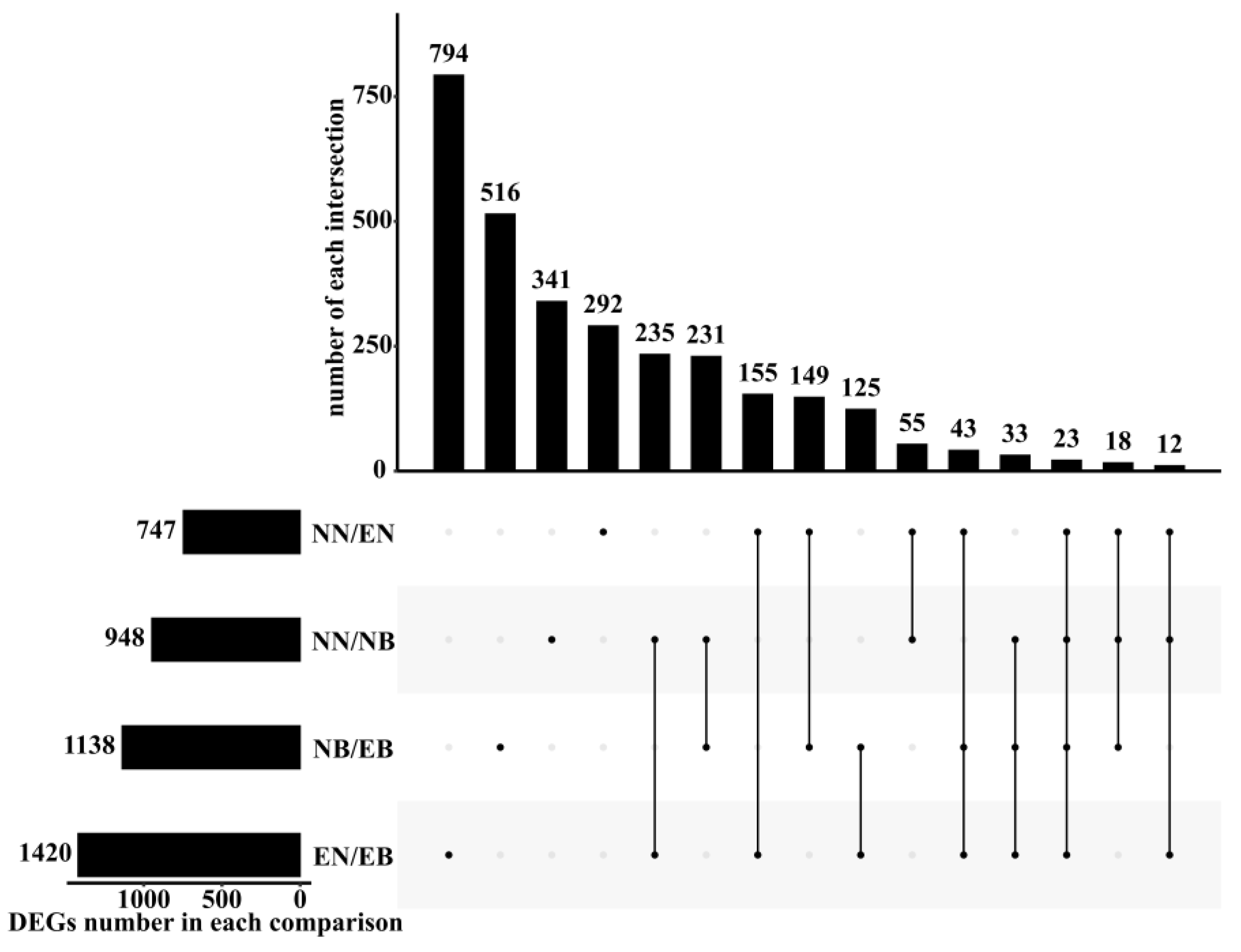
3.2. Analysis of Differentially Expressed Genes between Different Treatments

As shown in Table 2, the number of down-regulated DEGs was more than up-regulated DEGs in NN/EN. The infection with B. dothidea caused the disease resistance of the roots, and the number of up-regulated DEGs was more than that of the down-regulated DEGs. EN/EB had the greatest number of DEGs among the four comparison groups, indicating that maybe the disease resistance in the mycorrhizal P. trichocarpa was stronger. Compared with NN/EN, the number of up-regulated and down-regulated DEGs in EN/EB increased by 148% and 52%, respectively. In NB/EB, the number of up-regulated DEGs decreased by 66% as compared to down-regulated ones, which might be due to the root needing to maintain symbiosis during disease.
A total of 3022 DEGs were identified in the four groups (Figure 1). In the comparison group with and without L. bicolor (NN/NB, EN/EB), B. dothidea regulated a total of 303 DEGs. These might be the main genes in the roots of P. trichocarpa in response to B. dothidea infection. However, these genes did not exceed half of the total DEGs of NN/NB or EN/EB, showing the variable mechanism of mycorrhizal P. trichocarpa roots in response to B. dothidea infection.
As shown in Figure 2, 3022 DEGs were divided into different modules according to different expression patterns. In 13 modules, the expression patterns of 661 DEGs in the “MEorange”, “MEcyan”, “MEgrey”, “MEorangered4”, “MEsaddlebrown”, and “MEskyblue” modules were positively correlated with the inoculation of L. bicolor and B. dothidea (Figure 2b). It indicated that these genes might be regulated by L. bicolor and participate in the process of P. trichocarpa in response to infection with B. dothidea.

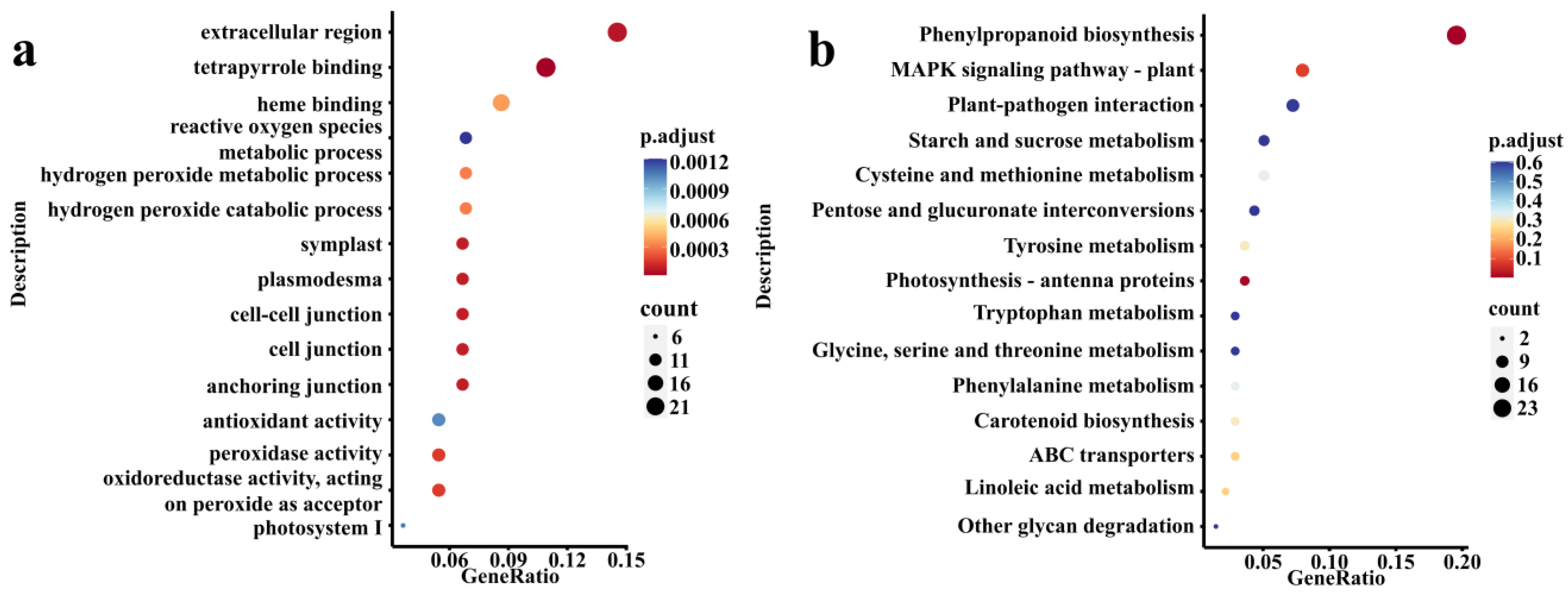
3.3. DEGs Enrichment Analysis

GO enrichment analysis showed that 661 DEGs were all enriched (p.adjust = 1) in 770 GO terms, and significantly enriched (p.adjust < 0.05) in 51 GO terms (Figure 3a). Most of these GO terms were related to reactive oxygen metabolism (“peroxidase activity”, “hydrogen peroxide metabolic process”, and “reactive oxygen species metabolic process”), hormones (“methyl salicylate esterase activity”, “abscisic acid binding”, and “methyl jasmonate esterase activity”), and other related GO terms. KEGG enrichment analysis showed that all 661 DEGs were enriched (p.adjust = 1) in 71 KEGG pathways, which were significantly enriched (p.adjust < 0.05) in the two metabolic pathways “Phenylpropanoid biosynthesis” and “Photosynthesis-antenna proteins” (Figure 3b). L. bicolor might participate in the process of P. trichocarpa in response to infection with B. dothidea by affecting the expression of genes in these pathways.

3.4. Analysis of Gene Expression Patterns Related to Signal Transduction Induced by L. bicolor

Figure 3b shows that the changes in gene expression in the roots of P. trichocarpa in response to disease stress under the conditions of inoculation and non-inoculation with L. bicolor were different, and this change was often regulated by signal molecules. In GO enrichment analysis, signal transduction-related DEGs were mainly enriched in “abscisic acid-activated signaling pathway”, “hormone-mediated signaling pathway”, “signaling receptor activity”, “auxin-activated signaling pathway”, “signaling receptor activator activity”, “calcium-mediated signaling”, “second-messenger-mediated signaling”, “signaling receptor binding”, and “intracellular signal transduction”. In KEGG enrichment analysis, signal transduction-related DEGs were enriched in “MAPK signaling pathway-plant” and “plant hormone signal transduction”. A total of 28 DEGs were enriched in these pathways (Figure 4). Among these DEGs, four were related to auxin. The expression of LOC7474608 (auxin-induced protein 22D), LOC7481201 (auxin-responsive protein SAUR32), and LOC7490981 (auxin-responsive protein IAA1) was down-regulated in NB but was up-regulated in EN, and the expression level was further increased in EB due to the influence of L. bicolor. LOC7470707 (abscisic acid receptor PYL2), LOC7472448 (abscisic acid receptor PYL4), LOC7487337 (abscisic acid receptor PYL4), LOC7488718 (abscisic acid receptor PYL4), and LOC7464619 (abscisic acid receptor PYL4) were related to abscisic acid. In contrast to auxin, the induced expression of L. bicolor was inhibited in EN, while LOC7472448, LOC7487337, LOC7488718, and LOC7464619 were increased in NB, while B. dothidea inhibited the expression of LOC7470707. In EB, L. bicolor could induce a further increase in the expression of these DEGs.
LOC18100289 (respiratory burst oxidase homolog protein E) and LOC18094446 (respiratory burst oxidase homolog protein A) belong to the family of respiratory burst oxidase homolog (Rboh) proteins. The single inoculation with L. bicolor inhibited the expression of LOC18094446 and increased the expression of LOC18100289. The opposite was true when inoculating with B. dothidea alone. In EB, L. bicolor would further increase the expression level of LOC18094446, and the expression level of LOC18094446 was also increased by the influence of B. dothidea, but the influence of L. bicolor was lower than the expression level when inoculated with B. dothidea alone. LOC7460408 (WRKY transcription factor 33) and LOC18100011 (WRKY transcription factor 24) belonged to the family of WRKY transcription factors. They were inhibited by L. bicolor in EN. The infestation of B. dothidea induces an increase in their expression, but their expression was lower than that in NB. Perhaps L. bicolor could help P. trichocarpa resist the infection with B. dothidea by regulating the expression of these genes.

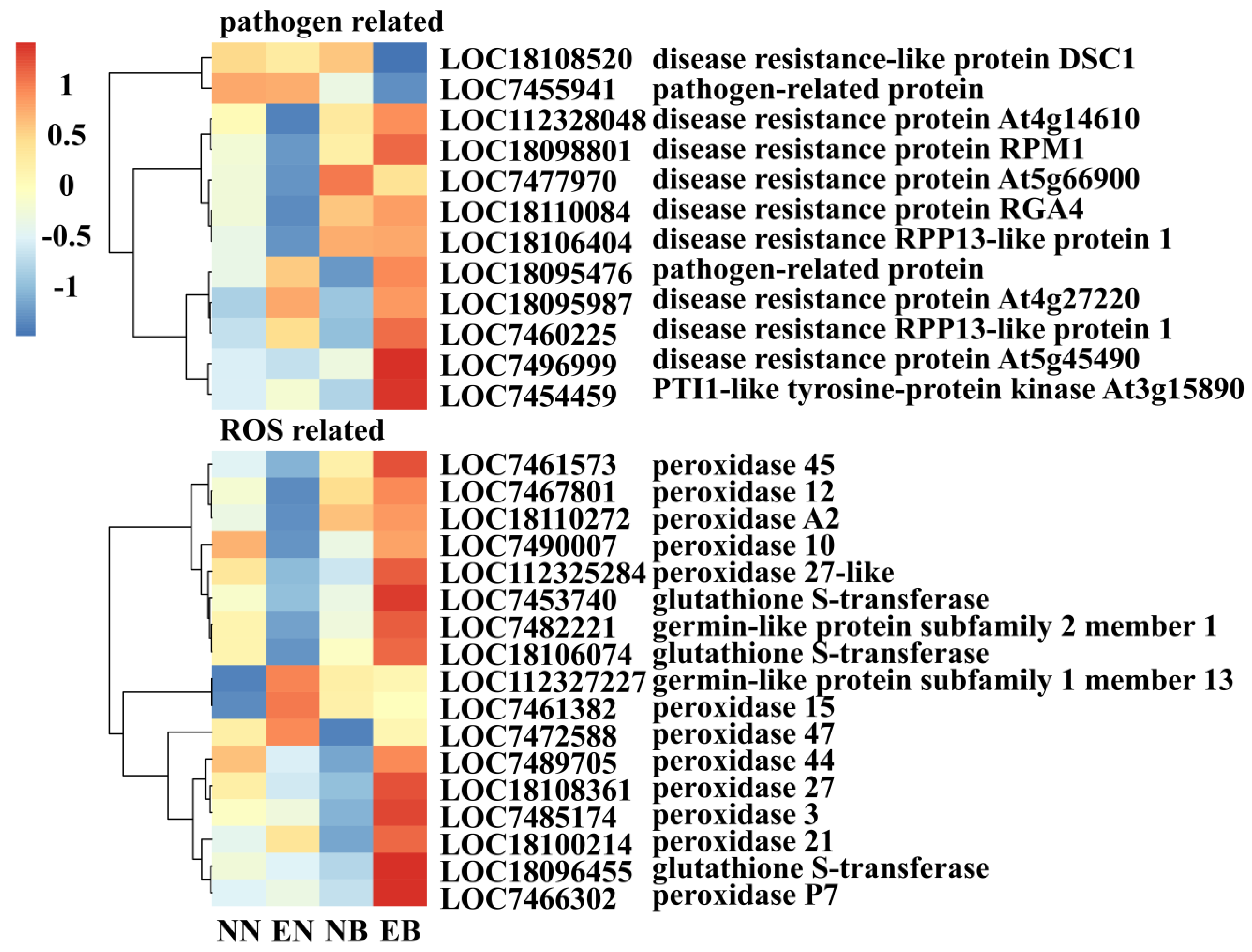
3.5. Analysis of the Expression Pattern of Disease Resistance-Related and Antioxidant-Related DEGs Induced by L. bicolor

Out of all 661 DEGs, a total of 12 disease resistance-related DEGs were found (Figure 5). Inoculation with L. bicolor inhibited the expression of LOC112328048 (disease resistance protein At4g14610), LOC18098801 (disease resistance protein RPM1), LOC7477970 (disease resistance protein At5g66900), LOC18110084 (putative disease resistance protein RGA4), and LOC18106404 (putative disease resistance RPP13-like protein 1), while single inoculation with B. dothidea increased the expression of these genes. In EB, the inoculation with L. bicolor could promote the expression of the remaining DEGs, except for LOC7477970. The expression of LOC18095476 (pathogen-related protein), LOC18095987 (probable disease resistance protein At4g27220), LOC7460225 (putative disease resistance RPP13-like protein 1), LOC7496999 (disease resistance protein At5g45490), and LOC7454459 (PTI1-like tyrosine-protein kinase At3g15890) were inhibited in NB, but under the influence of L. bicolor, the expression level increased. These results imply that L. bicolor changes the expression of disease-resistant genes, thereby protecting P. trichocarpa against infection with B. dothidea.
A total of 17 antioxidant enzyme-related DEGs were found, belonging to the POD family, germin-like protein (GLP) subfamily, and glutathione S-transferase (GST) family. Compared with NB, the expression level of 15 DEGs in EB was increased by the influence of L. bicolor. LOC112327227 (germin-like protein subfamily 1 member 13), LOC7461382 (peroxidase 15), and LOC7472588 (peroxidase 47) had the highest expression levels in EN, but inoculation with L. bicolor reduced the expression levels of these three DEGs.

3.6. The qRT-PCR Verification

LOC18098801 (disease resistance protein RPM1), LOC7483121 (calmodulin-like protein 1), LOC18106973 (pentatricopeptide repeat-containing protein At3g18110), LOC7494656 (pathogenesis-related genes transcriptional activator PTI6), and LOC18098678 (respiratory burst oxidase homolog protein B) were selected and analyzed by qRT-PCR to validate the RNA-Seq data. Their trends were similar to those of the transcriptome (Figure S1).

4. Discussion

In the process of resisting the infection of pathogenic fungi, plants have evolved a set of sophisticated and efficient defense mechanisms [49]. After the immunoreceptors on the surface of plant cells recognize the pathogenic fungus, they produce disease-resistant signals, which are transmitted to the whole body, changing the level of gene expression and producing anti-disease substances to inhibit or kill pathogenic fungi [50,51]. Inoculation with mycorrhizal fungi and pathogenic fungi will cause a defensive response in the non-infected parts [52]. The expression levels of genes change after the roots are infected by mycorrhizal fungi, which indirectly changes the gene expression level of stems and leaves [22,53]. This may be one of the important ways for mycorrhizal fungi to help the stems and leaves of the host resist the invasion of pathogens. The roots also foster disease resistance after pathogens invade the stems and leaves [54].
Similar to Zhan et al. [20], in our study, whether inoculated with L. bicolor or B. dothidea, the activity of disease-resistant enzymes (POD and PAL) in roots increased. After inoculation with the two fungi, the activities of POD and PAL in P. trichocarpa were significantly increased. Changes in enzymes activity are caused by a series of changes in gene expression. This proved to a certain extent the hypothesis put forward by this research; that is, L. bicolor changes the gene expression pattern of the roots of P. trichocarpa under disease stress and participates in the disease resistance of P. trichocarpa. With the help of WCGNA, a total of 661 DEGs were found to be affected by L. bicolor and B. dothidea. The different expression patterns of these DEGs under different treatments further confirmed the hypothesis of this study. Through the in-depth analysis of these DEGs, it was revealed that L. bicolor participates in the resistance of P. trichocarpa against B. dothidea infection by regulating the expression of root genes.
The transmission of disease resistance signals is one of the key steps in plant disease resistance response [55]. In this process, signal molecules bind to downstream receptors to regulate the expression of defense response genes. Disease resistance signal transduction pathways in plants include the hormone signal transduction pathway, the Ca2+ signal transduction pathway, and the ROS signal transduction pathway [56].
Auxin plays an important role in regulating the development of the plant root system and vascular system and establishing a symbiotic relationship between mycorrhizal fungi and roots [32]. ECMF changes the level of host plant auxin, thereby inducing lateral roots, restricting the growth of main roots, and making the roots grow horizontally while inhibiting host root hairs [57]. These strategies to change the morphological structure of the root system increased the infection point of mycelium and promoted the establishment of the ECMF symbiotic relationship [58,59]. It also directly or indirectly participates in the defense of plants against pathogens [60,61]. Auxin is generally considered to play a negative regulatory role in the process of plant disease resistance [62,63]. IAA treatment of rice will reduce the resistance of rice to Xanthomonas oryzae pv. oryzae [64]. In our study, infection with B. dothidea inhibited the expression of auxin-induced protein 22D (LOC7474608), auxin-responsive protein SAUR32 (LOC7481201), and auxin-responsive protein IAA1 (LOC7490981) in the roots. According to the idea that the increase in auxin content inhibits plant disease resistance, in the case of inoculation with B. dothidea, the roots of P. trichocarpa may inhibit the expression of these three genes to improve disease resistance. The results of the inoculation with L. bicolor were the opposite. Regardless of whether the disease occurred, the expression levels of these three genes were induced to increase. Mycorrhiza secretes trace hormones to regulate the growth and development of plants [65], which may be the reason for the increased expression of those three genes. At the same time, after the onset of the disease, the expression of genes further increased. Studies have found that biocontrol fungi and pathogenic fungi can increase the expression of some genes in the auxin pathway when they infect plants at the same time [63,66]. Therefore, the increase in the expression of these three genes may be one of the ways that L. bicolor participates in the resistance to B. dothidea infection with P. trichocarpa. The PYL (Pyrabactin-like) family is an ABA receptor that senses ABA changes in plants and plays an important role in the response to biotic and abiotic stresses [67]. Chen [68] found that overexpression of SiPYL4 in Arabidopsis thaliana can increase disease resistance to Macrophomina phaseolina and prolong survival time. Our results were similar to those. In NB, the expression of four DEGs out of five abscisic acid signal transduction DEGs were up-regulated. These four DEGs belonged to PYL4. However, in EB, the inoculation with L. bicolor further increased the expression of these four DEGs. This may be a way to improve the disease resistance of P. trichocarpa. In addition, inoculation with L. bicolor also increased the expression of PYL2, but the effect of PYL2 on disease stress is still unclear, and further research is needed.
The change of ROS content in plants is also an important aspect of inducing plant disease resistance [69,70,71,72,73]. When plants feel the stimulation of hormones and pathogens, the cells produce Ca2+ and combine with Rboh to activate NADPH (nicotinamide adenine dinucleotide phosphate) oxidase, catalyzing the production of a large amount of ROS to inhibit the growth of pathogens [68]. Yoshioka et al. [74] found that NbRbohA and NbRbohB were involved in the production of H2O2 and resistance to pathogenic oomycete (Phytophthora infestans) in tobacco disease resistance. Qin et al. [75] studied the changes in the expression of the Rboh family after citrus infection by B. dothidea and found that the expression of Rboh E in disease-resistant varieties was lower than that of susceptible varieties, indicating that it is involved in plant disease resistance. In our study, inoculation with B. dothidea up-regulated Rboh A and Rboh E, but the expression of these two DEGs in EB was higher than that in NB. It showed that L. bicolor increased the expression of these two genes and participated in the disease resistance of P. trichocarpa. At the same time, studies have shown that there are wound response elements in the promoter region of Rboh E. W-box interacts with members of the WRKY transcription factor family and plays a key role in biological stress [76,77]. Many studies have shown that most of the WRKY family genes are involved in plant disease resistance [78]. Although inoculation with L. bicolor increased the expression of Rboh E, the expression of WRKY33 and WRKY24 in EB were slightly lower than those in NB. It might be that L. bicolor slightly suppressed their expression to maintain a symbiotic relationship.
After a series of transductions, the disease-resistant signal finally acted on the direct disease-resistant protein and antioxidant enzyme synthesis gene to deal with the invasion of pathogenic fungi [49]. Inoculation with L. bicolor changed the expression of signal transduction pathway genes and finally acted on disease-resistant proteins and antioxidant enzyme genes, changing their expression levels. Both RPM1 and RPP13 are important members of the disease resistance network in plants, and they play an important role in identifying pathogens and regulating downstream disease resistance [79,80,81]. In our study, inoculation with L. bicolor mainly affected 12 genes related to direct disease resistance, among which inoculation with L. bicolor further increased the expression of RPM1 and RPP13. L. bicolor might participate in disease resistance through the regulation of direct disease resistance proteins.
The invasion of B. dothidea and L. bicolor will change the original structure of the plant, produce varying degrees of damage, and release ROS [20]. A small amount of ROS helps to activate the plant’s disease resistance response, but excessive ROS can damage cell membranes [82], so plants need to increase the activity of antioxidant enzymes to remove excess ROS. POD, GLP, and GST are important enzymes for removing ROS in plants [83]. Through transcriptome analysis, it was found that during the period of disease stress, L. bicolor induced an increase in the expression of POD, GST, and GLP genes. Perhaps the increase in the expression of these genes caused the increase in the activity of the corresponding protein, which reduced the ROS content in the plant, thereby alleviating the damage caused by the disease stress.

5. Conclusions

In this study, transcriptome analysis technology was used to explore the gene expression changes in the mycorrhizal P. trichocarpa roots under poplar canker stress. Our research showed that inoculation with L. bicolor changed the expression pattern of 661 genes in the roots. These genes were involved in many pathways such as signal transduction, reactive oxygen metabolism, and plant–pathogen interaction. The expression of these genes was changed due to inoculation with L. bicolor, which suggests that L. bicolor affects the disease resistance of P. trichocarpa. Our results not only provide a theoretical basis for revealing the molecular mechanism of mycorrhizal fungi improving the resistance of P. trichocarpa to poplar canker but also provide a theoretical basis for the development and application of biological agents.

Supplementary Materials

The following are available online at https://www.mdpi.com/article/10.3390/jof7121024/s1: Figure S1—The qRT-PCR verification of DEGs was identified by transcriptome analysis. Table S1—qRT-PCR primers used in this study. Table S2—Summary of transcriptome sequencing results. Table S3—Results of KEGG enrichment analysis on 661 DEGs obtained by WGCNA. Table S4—Results of GO enrichment analysis on 661 DEGs obtained by WGCNA.

Author Contributions

Conceptualization, F.D. and Y.W.; methodology and investigation, F.D.; data curation, F.D. and Y.W.; writing—original draft preparation, F.D.; writing—review and editing, Y.W. and M.T.; supervision and project administration, M.T.; funding acquisition, M.T. All authors have read and agreed to the published version of the manuscript.

Funding

This research was funded by the National Natural Science Foundation of China (32071639), the Laboratory of Lingnan Modern Agriculture Project (NZ2021025), and the National Key Research and Development Program of China (2018YFD0600203-3).

Institutional Review Board Statement

Not applicable.

Informed Consent Statement

Not applicable.

Data Availability Statement

The data that support the findings of this study are available on request from the corresponding author.

Conflicts of Interest

The authors declare no conflict of interest.

References

  1. Xing, J.; Li, P.; Zhang, Y.; Li, J.; Liu, Y.; Lachenbruch, B.; Su, X.; Zhao, J. Fungal pathogens of canker disease trigger canopy dieback in poplar saplings by inducing functional failure of the phloem and cambium and carbon starvation in the xylem. Physiol. Mol. Plant Pathol. 2020, 112, 101523. [Google Scholar] [CrossRef]
  2. Abelleira, A.; Moura, L.; Aguín, O.; Salinero, C. First Report of Lonsdalea populi Causing Bark Canker Disease on Poplar in Portugal. Plant Dis. 2019, 103, 2121. [Google Scholar] [CrossRef]
  3. Niemczyk, M.; Thomas, B.R. Growth parameters and resistance to Sphaerulina musiva-induced canker are more important than wood density for increasing genetic gain from selection of Populus spp. hybrids for northern climates. Ann. For. Sci. 2020, 77, 1–14. [Google Scholar] [CrossRef] [Green Version]
  4. Tabima, J.F.; Sondreli, K.L.; Kerio, S.; Feau, N.; Sakalidis, M.L.; Hamelin, R.C.; LeBoldus, J.M. Population Genomic Analyses Reveal Connectivity via Human-Mediated Transport across Populus Plantations in North America and an Undescribed Subpopulation of Sphaerulina musiva. Mol. Plant-Microbe Interact. 2020, 33, 189–199. [Google Scholar] [CrossRef]
  5. Zhong, Z.; Gao, Y. A brief report on the resistance of different poplar varieties to poplar vesicular canker. For. Sci. Technol. 1981, 1, 25–26. [Google Scholar] [CrossRef]
  6. Babu, S.; Bidyarani, N.; Chopra, P.; Monga, D.; Kumar, R.; Prasanna, R.; Kranthi, S.; Saxena, A.K. Evaluating microbe-plant interactions and varietal differences for enhancing biocontrol efficacy in root rot disease challenged cotton crop. Eur. J. Plant Pathol. 2015, 142, 345–362. [Google Scholar] [CrossRef]
  7. Bahram, M.; Põlme, S.; Kõljalg, U.; Tedersoo, L. A single European aspen (Populus tremula) tree individual may potentially harbour dozens of Cenococcum geophilum ITS genotypes and hundreds of species of ectomycorrhizal fungi. FEMS Microbiol. Ecol. 2011, 75, 313–320. [Google Scholar] [CrossRef] [Green Version]
  8. Wu, N.; Li, Z.; Wu, F.; Tang, M. Comparative photochemistry activity and antioxidant responses in male and female Populus cathayana cuttings inoculated with arbuscular mycorrhizal fungi under salt. Sci. Rep. 2016, 6, 37663. [Google Scholar] [CrossRef]
  9. Cui, J.Q.; Sun, H.B.; Sun, M.B.; Liang, R.T.; Jie, W.G.; Cai, B.Y. Effects of Funneliformis mosseae on Root Metabolites and Rhizosphere Soil Properties to Continuously-Cropped Soybean in the Potted-Experiments. Int. J. Mol. Sci. 2018, 19, 2160. [Google Scholar] [CrossRef] [Green Version]
  10. Leifheit, E.F.; Veresoglou, S.D.; Lehmann, A.; Morris, E.K.; Rillig, M.C. Multiple factors influence the role of arbuscular mycorrhizal fungi in soil aggregation—A meta-analysis. Plant Soil 2013, 374, 523–537. [Google Scholar] [CrossRef]
  11. Ortiz, N.; Armada, E.; Duque, E.; Roldan, A.; Azcon, R. Contribution of arbuscular mycorrhizal fungi and/or bacteria to enhancing plant drought tolerance under natural soil conditions: Effectiveness of autochthonous or allochthonous strains. J. Plant Physiol. 2015, 174, 87–96. [Google Scholar] [CrossRef]
  12. Ganugi, P.; Masoni, A.; Pietramellara, G.; Benedettelli, S. A Review of Studies from the Last Twenty Years on Plant–Arbuscular Mycorrhizal Fungi Associations and Their Uses for Wheat Crops. Agronomy 2019, 9, 840. [Google Scholar] [CrossRef] [Green Version]
  13. Liu, Y.; Feng, X.; Gao, P.; Li, Y.; Christensen, M.J.; Duan, T. Arbuscular mycorrhiza fungi increased the susceptibility of Astragalus adsurgens to powdery mildew caused by Erysiphe pisi. Mycology 2018, 9, 223–232. [Google Scholar] [CrossRef]
  14. Zhang, H.; Yu, H.; Tang, M. Prior contact of Pinus tabulaeformis with ectomycorrhizal fungi increases plant growth and survival from damping-off. New For. 2017, 48, 855–866. [Google Scholar] [CrossRef]
  15. Zhang, Y.; Ye, J.; Zhao, Y.; Ma, S. Fermentation conditions of Xerocomus chrysenteron and its control effect on poplar canker disease. J. For. Environ. 2016, 36, 397–403. [Google Scholar] [CrossRef]
  16. Lu, C.C.; Guo, N.; Yang, C.; Sun, H.B.; Cai, B.Y. Transcriptome and metabolite profiling reveals the effects of Funneliformis mosseae on the roots of continuously cropped soybeans. BMC Plant Biol. 2020, 20, 479. [Google Scholar] [CrossRef] [PubMed]
  17. Chen, Y.; Chen, Y.; Chen, Q.; Huang, X.; Huang, X. Cloning, Characterization and Expression of a Phenylalanine Ammonialyase Gene (M-PAL) from Plantain (Musa ABB cv. Dongguandajiao). J. Trop. Subtrop. Bot. 2007, 15, 421–427. [Google Scholar] [CrossRef]
  18. Li, D.; Chen, Z.; Nie, Y. Antifungal substances producted by a high-yielding mutant of Bs−916 and their effects inducing-resistance on rice plant. Acta Phytopathol. Sin. 2008, 38, 192–198. [Google Scholar] [CrossRef]
  19. Kilic-Ekici, O.; Yuen, G.Y. Induced resistance as a mechanism of biological control by Lysobacter enzymogenes strain C3. Phytopathology 2003, 93, 1103–1110. [Google Scholar] [CrossRef] [Green Version]
  20. Zhan, W.; Liu, H.; Tang, M. Physiological and Biochemical Mechanism of Mycorrhizal Fungi Improving the Resistance of Poplar to Canker Disease. Acta Bot. Boreali-Occident. Sin. 2010, 30, 2437–2443. [Google Scholar]
  21. Morcillo, R.J.; Zhao, A.; Tamayo-Navarrete, M.I.; García-Garrido, J.M.; Macho, A.P. Tomato Root Transformation Followed by Inoculation with Ralstonia Solanacearum for Straightforward Genetic Analysis of Bacterial Wilt Disease. J. Vis. Exp. 2020, 157, e60302. [Google Scholar] [CrossRef]
  22. Liu, J.; Maldonado-Mendoza, I.; Lopez-Meyer, M.; Cheung, F.; Town, C.D.; Harrison, M.J. Arbuscular mycorrhizal symbiosis is accompanied by local and systemic alterations in gene expression and an increase in disease resistance in the shoots. Plant J. 2007, 50, 529–544. [Google Scholar] [CrossRef]
  23. Campos-Soriano, L.; Garcia-Martinez, J.; Segundo, B.S. The arbuscular mycorrhizal symbiosis promotes the systemic induction of regulatory defence-related genes in rice leaves and confers resistance to pathogen infection. Mol. Plant Pathol. 2012, 13, 579–592. [Google Scholar] [CrossRef]
  24. Grant, M.; Lamb, C. Systemic immunity. Curr. Opin. Plant Biol. 2006, 9, 414–420. [Google Scholar] [CrossRef]
  25. Lee, S.; Rojas, C.M.; Ishiga, Y.; Pandey, S.; Mysore, K.S. Arabidopsis heterotrimeric G-proteins play a critical role in host and nonhost resistance against Pseudomonas syringae pathogens. PLoS ONE 2013, 8, e82445. [Google Scholar] [CrossRef] [Green Version]
  26. Bundo, M.; Coca, M. Enhancing blast disease resistance by overexpression of the calcium-dependent protein kinase OsCPK4 in rice. Plant Biotechnol. J. 2016, 14, 1357–1367. [Google Scholar] [CrossRef] [Green Version]
  27. Martos, G.G.; Teran Mdel, M.; Diaz Ricci, J.C. The defence elicitor AsES causes a rapid and transient membrane depolarization, a triphasic oxidative burst and the accumulation of nitric oxide. Plant Physiol. Biochem. 2015, 97, 443–450. [Google Scholar] [CrossRef] [PubMed]
  28. Zhang, Y.; Li, D.; Zhang, H.; Hong, Y.; Huang, L.; Liu, S.; Li, X.; Ouyang, Z.; Song, F. Tomato histone H2B monoubiquitination enzymes SlHUB1 and SlHUB2 contribute to disease resistance against Botrytis cinerea through modulating the balance between SA- and JA/ET-mediated signaling pathways. BMC Plant Biol. 2015, 15, 252. [Google Scholar] [CrossRef] [Green Version]
  29. Liao, W.; Ji, L.; Wang, J.; Chen, Z.; Ye, M.; Ma, H.; An, X. Identification of glutathione S-transferase genes responding to pathogen infestation in Populus tomentosa. Funct. Integr. Genom. 2014, 14, 517–529. [Google Scholar] [CrossRef] [PubMed]
  30. Zhao, J.; Yang, F.; Feng, J.; Wang, Y.; Lachenbruch, B.; Wang, J.; Wan, X. Genome-Wide Constitutively Expressed Gene Analysis and New Reference Gene Selection Based on Transcriptome Data: A Case Study from Poplar/Canker Disease Interaction. Front. Plant Sci. 2017, 8, 1876. [Google Scholar] [CrossRef] [PubMed] [Green Version]
  31. De Block, M. Factors Influencing the Tissue Culture and the Agrobacterium tumefaciens-Mediated Transformation of Hybrid Aspen and Poplar Clones. Plant Physiol. 1990, 93, 1110–1116. [Google Scholar] [CrossRef] [Green Version]
  32. Felten, J.; Kohler, A.; Morin, E.; Bhalerao, R.P.; Palme, K.; Martin, F.; Ditengou, F.A.; Legué, V.r. The Ectomycorrhizal Fungus Laccaria bicolor Stimulates Lateral Root Formation in Poplar and Arabidopsis through Auxin Transport and Signaling. Plant Physiol. 2009, 151, 1991–2005. [Google Scholar] [CrossRef] [Green Version]
  33. Wanwaen, S.; Youpensuk, S. Cultivation of Amanita princeps and Gyrodon suthepensis for Mycorrhizations with Castanopsis acuminatissima and their Effects on the Host Plants. Int. J. Agric. Biol. 2019, 22, 195–200. [Google Scholar] [CrossRef]
  34. Li, Y.; Feng, Y.; Lu, Q.; Yan, D.; Liu, Z.; Zhang, X. Comparative Proteomic Analysis of Plant–Pathogen Interactions in Resistant and Susceptible Poplar Ecotypes Infected with Botryosphaeria dothidea. Phytopathology 2019, 109, 2009–2021. [Google Scholar] [CrossRef] [PubMed]
  35. Fang, W.; Kao, C.H. Enhanced peroxidase activity in rice leaves in response to excess iron, copper and zinc. Plant Sci. 2000, 158, 71–76. [Google Scholar] [CrossRef]
  36. Sreelakshmi, Y.; Sharma, R. Differential regulation of phenylalanine ammonia lyase activity and protein level by light in tomato seedlings. Plant Physiol. Biochem. 2008, 46, 444–451. [Google Scholar] [CrossRef]
  37. Kramer, G.F.; Norman, H.A.; Krizek, D.T.; Mirecki, R.M. Influence of UV-B radiation on polyamines, lipid peroxidation and membrane lipids in cucumber. Phytochemistry 1991, 30, 2101–2108. [Google Scholar] [CrossRef]
  38. Mortazavi, A.; Williams, B.A.; McCue, K.; Schaeffer, L.; Wold, B. Mapping and quantifying mammalian transcriptomes by RNA-Seq. Nat. Methods 2008, 5, 621–628. [Google Scholar] [CrossRef] [PubMed]
  39. Liao, Y.; Smyth, G.K.; Shi, W. featureCounts: An efficient general purpose program for assigning sequence reads to genomic features. Bioinformatics 2014, 30, 923–930. [Google Scholar] [CrossRef] [PubMed] [Green Version]
  40. Love, M.I.; Huber, W.; Anders, S. Moderated estimation of fold change and dispersion for RNA-seq data with DESeq2. Genome Biol. 2014, 15, 550. [Google Scholar] [CrossRef] [Green Version]
  41. Langfelder, P.; Horvath, S. WGCNA: An R package for weighted correlation network analysis. BMC Bioinform. 2008, 9, 559. [Google Scholar] [CrossRef] [Green Version]
  42. Young, M.D.; Wakefield, M.J.; Smyth, G.K.; Oshlack, A. Gene ontology analysis for RNA-seq: Accounting for selection bias. Genome Biol. 2010, 11, R14. [Google Scholar] [CrossRef] [PubMed] [Green Version]
  43. Wixon, J.; Kell, D. The Kyoto Encyclopedia of Genes and Genomes—KEGG. Yeast 2000, 17, 48–55. [Google Scholar] [CrossRef]
  44. Wu, T.; Hu, E.; Xu, S.; Chen, M.; Guo, P.; Dai, Z.; Feng, T.; Zhou, L.; Tang, W.; Zhan, L.; et al. clusterProfiler 4.0: A universal enrichment tool for interpreting omics data. Innovation 2021, 2, 100141. [Google Scholar] [CrossRef] [PubMed]
  45. Zhang, X.Y.; Zhang, H.J.; Zhang, Y.X.; Liu, Y.Q.; Zhang, H.Q.; Tang, M. Arbuscular mycorrhizal fungi alter carbohydrate distribution and amino acid accumulation in Medicago truncatula under lead stress. Environ. Exp. Bot. 2020, 171, 103950. [Google Scholar] [CrossRef]
  46. Jiang, X. The Spatial and Temporal Expression of Thaumatin-Like Protein Coding Genes Induced by Tress Stem Canker Pathogen in Populus trichocarpa. Master’s Thesis, Hebei Agricultural University, Baoding, China, 2012. [Google Scholar]
  47. Su, X.; Fan, B.; Yuan, L.; Cui, X.; Lu, S. Selection and Validation of Reference Genes for Quantitative RT-PCR Analysis of Gene Expression in Populus trichocarpa. Chin. Bull. Bot. 2013, 48, 507–518. [Google Scholar] [CrossRef]
  48. Livak, K.J.; Schmittgen, T.D. Analysis of relative gene expression data using real-time quantitative PCR and the 2−ΔΔCT method. Methods 2001, 25, 402–408. [Google Scholar] [CrossRef]
  49. Andersen, E.J.; Ali, S.; Byamukama, E.; Yen, Y.; Nepal, M.P. Disease Resistance Mechanisms in Plants. Genes 2018, 9, 339. [Google Scholar] [CrossRef] [Green Version]
  50. Mekapogu, M.; Jung, J.A.; Kwon, O.K.; Ahn, M.S.; Song, H.Y.; Jang, S. Recent Progress in Enhancing Fungal Disease Resistance in Ornamental Plants. Int. J. Mol. Sci. 2021, 22, 7956. [Google Scholar] [CrossRef]
  51. Mogensen, T.H. Pathogen Recognition and Inflammatory Signaling in Innate Immune Defenses. Clin. Microbiol. Rev. 2009, 22, 240–273. [Google Scholar] [CrossRef] [Green Version]
  52. Bernaola, L.; Cosme, M.; Schneider, R.W.; Stout, M. Belowground Inoculation With Arbuscular Mycorrhizal Fungi Increases Local and Systemic Susceptibility of Rice Plants to Different Pest Organisms. Front. Plant Sci. 2018, 9, 747. [Google Scholar] [CrossRef]
  53. Zhang, R.Q.; Tang, M.; Chen, H.; Tian, Z.Q. Effects of ectomycorrhizal fungi on damping-off and induction of pathogenesis-related proteins in Pinus tabulaeformis seedlings inoculated with Amanita vaginata. For. Pathol. 2011, 41, 262–269. [Google Scholar] [CrossRef]
  54. Morales, J.; Kadota, Y.; Zipfel, C.; Molina, A.; Torres, M.A. The Arabidopsis NADPH oxidases RbohD and RbohF display differential expression patterns and contributions during plant immunity. J. Exp. Bot. 2016, 67, 1663–1676. [Google Scholar] [CrossRef] [Green Version]
  55. Emma, W.G.; Olusola, O.S.; Simeon, O.K. The molecular initiation and subsequent acquisition of disease resistance in plants. Afr. J. Biotechnol. 2003, 2, 26–32. [Google Scholar] [CrossRef] [Green Version]
  56. Ding, L.; Yang, G. Research Advances in the Mechanism and Signal Transduction of Plant Disease Resistance. Biotechnol. Bull. 2016, 32, 109–117. [Google Scholar] [CrossRef]
  57. Zaretsky, M.; Sitrit, Y.; Mills, D.; Roth-Bejerano, N.; Kagan-Zur, V. Differential expression of fungal genes at preinfection and mycorrhiza establishment between Terfezia boudieri isolates and Cistus incanus hairy root clones. New Phytol. 2006, 171, 837–846. [Google Scholar] [CrossRef]
  58. Sitrit, Y.; Roth-Bejerano, N.; Kagan-Zur, V.; Turgeman, T. Pre-symbiotic interactions between the desert truffle Terfezia boudieri and its host plant Helianthemum sessiliflorum. In Desert Truffles; Springer: Berlin/Heidelberg, Germany, 2014; Volume 38, pp. 81–92. [Google Scholar]
  59. Turgeman, T.; Lubinsky, O.; Roth-Bejerano, N.; Kagan-Zur, V.; Kapulnik, Y.; Koltai, H.; Zaady, E.; Ben-Shabat, S.; Guy, O.; Lewinsohn, E.; et al. The role of pre-symbiotic auxin signaling in ectendomycorrhiza formation between the desert truffle Terfezia boudieri and Helianthemum sessiliflorum. Mycorrhiza 2016, 26, 287–297. [Google Scholar] [CrossRef] [PubMed]
  60. Kieffer, M.; Neve, J.; Kepinski, S. Defining auxin response contexts in plant development. Curr. Opin. Plant Biol. 2010, 13, 12–20. [Google Scholar] [CrossRef]
  61. Swarup, R.; Peret, B. AUX/LAX family of auxin influx carriers—An overview. Front. Plant Sci. 2012, 3, 225. [Google Scholar] [CrossRef] [Green Version]
  62. Kazan, K.; Manners, J.M. Linking development to defense: Auxin in plant-pathogen interactions. Trends Plant Sci. 2009, 14, 373–382. [Google Scholar] [CrossRef]
  63. Zhong, T. Cloning and Resistance Mechanism of Genes for Grey Leaf Spot and Stalk Rot Resistance in Maize. Ph.D. Thesis, China Agricultural University, Beijing, China, 2019. [Google Scholar]
  64. Ding, X.; Cao, Y.; Huang, L.; Zhao, J.; Xu, C.; Li, X.; Wang, S. Activation of the indole-3-acetic acid-amido synthetase GH3-8 suppresses expansin expression and promotes salicylate- and jasmonate-independent basal immunity in rice. Plant Cell 2008, 20, 228–240. [Google Scholar] [CrossRef] [Green Version]
  65. Pons, S.; Fournier, S.; Chervin, C.; Becard, G.; Rochange, S.; Frei Dit Frey, N.; Puech Pages, V. Phytohormone production by the arbuscular mycorrhizal fungus Rhizophagus irregularis. PLoS ONE 2020, 15, e0240886. [Google Scholar] [CrossRef]
  66. Jiang, C. Transcriptome Analysis of the Auxin Key Genes in Populus davidiana × P. alba var. pyramidalis Response to Trichoderma asperellum and Alternaria alternata. Master’s Thesis, Northeast Forestry University, Harbin, China, 2017. [Google Scholar]
  67. Sah, S.K.; Reddy, K.R.; Li, J. Abscisic Acid and Abiotic Stress Tolerance in Crop Plants. Front. Plant Sci. 2016, 7, 571. [Google Scholar] [CrossRef] [Green Version]
  68. Chen, Y. Identification of Resistance of Sesame Varieties to Stem Rot and Functional Analysis of SiPYL4 and SiTLP genes. Master’s Thesis, Zhengzhou University, Zhengzhou, China, 2019. [Google Scholar]
  69. Miller, G.; Shulaev, V.; Mittler, R. Reactive oxygen signaling and abiotic stress. Physiol. Plant 2008, 133, 481–489. [Google Scholar] [CrossRef] [PubMed]
  70. Steffens, B.; Sauter, M. Epidermal cell death in rice is confined to cells with a distinct molecular identity and is mediated by ethylene and H2O2 through an autoamplified signal pathway. Plant Cell 2009, 21, 184–196. [Google Scholar] [CrossRef] [PubMed] [Green Version]
  71. Halliwell, B. Reactive species and antioxidants. Redox biology is a fundamental theme of aerobic life. Plant Physiol. 2006, 141, 312–322. [Google Scholar] [CrossRef] [PubMed] [Green Version]
  72. Gechev, T.S.; Van Breusegem, F.; Stone, J.M.; Denev, I.; Laloi, C. Reactive oxygen species as signals that modulate plant stress responses and programmed cell death. Bioessays 2006, 28, 1091–1101. [Google Scholar] [CrossRef]
  73. Neill, S.J.; Desikan, R.; Clarke, A.; Hurst, R.D.; Hancock, J.T. Hydrogen peroxide and nitric oxide as signalling molecules in plants. J. Exp. Bot. 2002, 53, 1237–1247. [Google Scholar] [CrossRef]
  74. Yoshioka, H.; Numata, N.; Nakajima, K.; Katou, S.; Kawakita, K.; Rowland, O.; Jones, J.D.; Doke, N. Nicotiana benthamiana gp91phox homologs NbrbohA and NbrbohB participate in H2O2 accumulation and resistance to Phytophthora infestans. Plant Cell 2003, 15, 706–718. [Google Scholar] [CrossRef] [PubMed] [Green Version]
  75. Qin, X.; Qi, J.; Dou, W.; Chen, S.; He, Y.; Li, Q. Identification of Rboh Family and the Response to Hormone and Citrus Bacterial Canker in Citrus. Sci. Agric. Sin. 2020, 53, 4189–4203. [Google Scholar] [CrossRef]
  76. Rushton, P.J.; Somssich, I.E.; Ringler, P.; Shen, Q.J. WRKY transcription factors. Trends Plant Sci. 2010, 15, 247–258. [Google Scholar] [CrossRef] [PubMed]
  77. Kaur, G.; Pati, P.K. Analysis of cis-acting regulatory elements of Respiratory burst oxidase homolog (Rboh) gene families in Arabidopsis and rice provides clues for their diverse functions. Comput. Biol. Chem. 2016, 62, 104–118. [Google Scholar] [CrossRef]
  78. Chen, F.; Hu, Y.; Vannozzi, A.; Wu, K.; Cai, H.; Qin, Y.; Mullis, A.; Lin, Z.; Zhang, L. The WRKY Transcription Factor Family in Model Plants and Crops. Crit. Rev. Plant Sci. 2018, 36, 311–335. [Google Scholar] [CrossRef]
  79. Bittner-Eddy, P.D.; Crute, I.R.; Holub, E.B.; Beynon, J.L. RPP13 is a simple locus in Arabidopsis thaliana for alleles that specify downy mildew resistance to different avirulence determinants in Peronospora parasitica. Plant J. 2000, 21, 177–188. [Google Scholar] [CrossRef] [PubMed]
  80. Cheng, J.; Fan, H.; Li, L.; Hu, B.; Liu, H.; Liu, Z. Genome-wide Identification and Expression Analyses of RPP13-like Genes in Barley. BioChip J. 2018, 12, 102–113. [Google Scholar] [CrossRef]
  81. Chao, J.; Jin, J.; Wang, D.; Han, R.; Zhu, R.; Zhu, Y.; Li, S. Cytological and transcriptional dynamics analysis of host plant revealed stage-specific biological processes related to compatible rice-Ustilaginoidea virens interaction. PLoS ONE 2014, 9, e91391. [Google Scholar] [CrossRef] [Green Version]
  82. Sharma, P.; Jha, A.B.; Dubey, R.S.; Pessarakli, M. Reactive Oxygen Species, Oxidative Damage, and Antioxidative Defense Mechanism in Plants under Stressful Conditions. J. Bot. 2012, 2012, 1–26. [Google Scholar] [CrossRef] [Green Version]
  83. Zhang, X.; Gao, H.; Liang, Y.; Cao, Y. Full-length transcriptome analysis of asparagus roots reveals the molecular mechanism of salt tolerance induced by arbuscular mycorrhizal fungi. Environ. Exp. Bot. 2021, 185, 104402. [Google Scholar] [CrossRef]
Figure 1. Upset diagram of differentially expressed genes. DEGs number in each comparison group represents the number of all DEGs in each comparison group; the number of each intersection represents the total number of DEGs in each comparison group; a point on the abscissa represents the number of unique DEGs in each comparison group; the line of multiple dots on the abscissa indicates the number of DEGs identified by the multiple comparison groups of the line.
Jof 07 01024 g001
Figure 2. (a) The correlation heat map of the genes between the modules. (b) The heat map of the correlation between each module and the trait. Pearson’s correlation coefficients between modules and fungal inoculation are shown, accompanied by the corresponding p value in brackets. From red to green, the correlation probability is from high to low. Each module is identified by color. (c) A heat map of the expression levels of 661 DEGs in the four groups. From red to blue, the expression level was from high to low.
Jof 07 01024 g002
Figure 3. (a) The scatter map of GO enrichment analysis on 661 DEGs obtained by WGCNA. Sorted by p.adjust (from 0 to 1), the top 15 GO enrichment results were selected to display. (b) The scatter map of KEGG enrichment analysis on 661 DEGs obtained by WGCNA. Sorted by p.adjust (from 0 to 1), the top 15 KEGG enrichment results were selected to display. Gene ratio: A score, the numerator is the number of genes enriched in this GO entry and the denominator is the number of input genes for enrichment analysis, which can be the genes obtained by differential expression analysis; count: enter the number of genes enriched to this GO entry in the genes for enrichment analysis; p.adjust: corrected p value. For the complete GO and KEGG enrichment results, please view Tables S3 and S4.
Jof 07 01024 g003
Figure 4. Heat map of DEGs related to signal transduction pathway.
Jof 07 01024 g004
Figure 5. Heat map of DEGs related to disease resistance and antioxidant.
Jof 07 01024 g005
Table 1. Enzyme activity under different treatments.
TreatmentPODPALMDA
NN1054.02 ± 325.66 c5678.41 ± 1428.19 c14.30 ± 3.90 d
EN3516.56 ± 484.89 ab16600.94 ± 779.80 ab35.49 ± 5.53 c
NB2330.89 ± 465.16 bc10399.50 ± 2842.16 bc128.66 ± 11.48 a
EB5252.68 ± 1370.74 a23236.47 ± 5403.70 a60.68 ± 8.61 b
L. bicolor******
B. dothidea****
L. bicolor & B. dothideansns**
NN: non-fungus control; EN: inoculation with L. bicolor but no B. dothidea; NB: inoculation with B. dothidea but no L. bicolor; EB: inoculation with L. bicolor and B. dothidea. FW: fresh weight. Data expressed as mean ± standard error (n = 3). Different lowercase letters indicate significant differences between the means by Tukey’s test (p < 0.05); “*” indicates that the interaction is significant (p < 0.05); “**” indicates that the interaction is extremely significant (p < 0.01); “ns” indicates no interaction (p ≥ 0.05). The activities of POD were expressed as μg·g−1·FW·min−1. The activities of PAL were expressed as U·g−1·FW·h−1. The content of MDA was expressed as μmol·g−1·FW.
Table 2. The number of differentially expressed genes (DEGs) in the four comparison groups.
Comparisons
(Control/Treatment)
All DEGsUp Regulated DEGsDown Regulated DEGs
NN/EN747297450
NN/NB948734214
EN/EB1420736684
NB/EB1138288850
Publisher’s Note: MDPI stays neutral with regard to jurisdictional claims in published maps and institutional affiliations.

