Article

Multilocus Phylogeography of the Tuber mesentericum Complex Unearths Three Highly Divergent Cryptic Species

1
Department of Life, Health and Environmental Sciences, University of L’Aquila, Via Vetoio Loc. Coppito, 67100 L’Aquila, Italy
2
School of Agricultural, Forestry, Food and Environmental Sciences, University of Basilicata, Viale dell’Ateneo Lucano, 10, 85100 Potenza, Italy
3
Apto. de Correos 6, Caldes de Malavella, 17455 Girona, Spain
*
Author to whom correspondence should be addressed.
J. Fungi 2021, 7(12), 1090; https://doi.org/10.3390/jof7121090
Submission received: 16 November 2021 / Revised: 13 December 2021 / Accepted: 15 December 2021 / Published: 17 December 2021
(This article belongs to the Special Issue Fungal Diversity in Europe)

Abstract

:
Tuber mesentericum is an edible European black truffle, apparently easy to recognize, but showing a high degree of genetic variability. In this study, we performed an integrative taxonomic assessment of the T. mesentericum complex, combining a multilocus phylogeographic approach with morphological analyses, and including authentic specimens of Vittadini, and Berkeley and Broome. We performed maximum likelihood phylogenetic analyses, based on single and concatenated gene datasets (ITS rDNA, β-tubulin, elongation factor 1-α), and including all available sequences from previous studies. Phylogenetic analyses consistently recovered three reciprocally monophyletic and well-supported clades: clade I, with a wide range across Europe; clade II, specimens collected mainly in the Iberian, Italian, and Balkan peninsulas; and clade III, specimens collected almost exclusively in central Italy. Genetic distance between clades ranged from 10.4% to 13.1% at the ITS region. We also designed new primer pairs specific for each phylogenetic lineage. Morphology of spores, asci, and peridium were investigated on specimens representing the three lineages. Macro- and micromorphological analyses of ascomata revealed only a few, but not diagnostic, differences between the three phylogenetic lineages, thus, confirming that they are morphologically cryptic. By studying authentic specimens of Vittadini, and Berkeley and Broome, it was possible to identify the three clades as T. mesentericum, Tuber bituminatum, and Tuber suave sp. nov., and to designate an epitype for T. mesentericum s.s. and a lectotype for T. bituminatum. Future investigations on volatile organic compound (VOC) composition are needed to define the aroma repertoires in this species complex.

1. Introduction

Truffles are hypogenous ascomata, mainly formed by fungi in the Ascomycota and Basidiomycota phyla. Those in the genus Tuber (Ascomycota, Pezizales), the so-called true truffles, live in mycorrhizal symbiosis with the roots of many trees, shrubs, and herbaceous plants [1]. Tuber species that produce ascomata with a pleasant smell and marketable size are considered edible and, therefore, of commercial interest. In some European countries, edible truffles have traditionally been considered one of the most appreciated and expensive foods, and their cultivation and consumption are now rapidly spreading worldwide [2].
Since the advent of molecular phylogenetics, many researchers have worked to revise the taxonomy within the genus Tuber [3,4,5,6]. Currently, about 120 Tuber species have been described and molecularly characterized from Asia, Europe, and Central and North America [7]. However, the last revision of the genus estimated from 180 to 220 species, grouped into 11 major clades [8]. A large portion of such species richness is due to the presence of cryptic lineages in many of these clades. Indeed, cryptic species have been found in Tuber anniae W. Colgan & Trappe [9], Tuber borchii Vittad. [10], Tuber brumale Vittad. [11], Tuber excavatum Vittad. [12], Tuber indicum Cooke & Massee [13,14], and Tuber rufum Picco [15]. However, the taxonomy of many of these species complexes has not been resolved.
The Aestivum clade contains the most morphologically diverse Tuber species [16]. In addition to truffles with a white (Tuber magnatum Picco) or brown (Tuber panniferum Tul. & C. Tul., Tuber pulchrosporum Konstantinidis, Tsampazis, Slavova, Nakkas, Polemis, Fryssouli & Zervakis) peridium, most species in the clade have black warty ascomata with alveolate spores (Tuber aestivum Vittad., Tuber mesentericum Vittad., Tuber sinoaestivum J.P. Zhang & P.G. Liu, Tuber malençonii Donadini, Riousset, G. Riousset & G. Chev.). Phylogenetic and phylogeographic studies in this clade have mainly focused on species with the highest economic values, such as T. magnatum and T. aestivum [17,18,19,20]. Within this clade, T. mesentericum is a truffle of some commercial interest, which is apparently easy to distinguish from other black truffles, based on its excavated base and a more or less strong bitumen-like smell, which has always been considered unpleasant by most authors [21].
Phylogenetic studies on T. mesentericum are scarce. Pacioni and Pomponi [22], based on allozyme polymorphisms, recognized four distinct clades within T. mesentericum, one of which was later associated with Tuber bellonae Quélet [23]. Sica et al. [24] analyzed rDNA internal transcribed spacer (ITS) sequences in 126 specimens, mainly from the Campania region (southern Italy), and found 34 distinct haplotypes, but did not reveal any significant morphological differences among the analyzed ascomata. Benucci et al. [25] and Marozzi et al. [26] uncovered three distinct clades within T. mesentericum, using ITS and elongation factor 1-α (EF1α) sequences, respectively. These lineages may well represent cryptic species, one of which is T. mesentericum sensu stricto. A proper taxonomic assessment of the T. mesentericum species complex has not been performed yet, and it is unclear whether phylogenetic lineages are also distinct by any morphological feature, or by what their geographic distributions are.
In this study, we performed a comprehensive phylogeographic assessment of the T. mesentericum species complex, based on multilocus data, and including specimens from all over Europe. The taxonomy of the lineages found was resolved through an integrative approach combining molecular data, morphological characters of peridium and spores, and analysis of voucher specimens of T. mesentericum determined by Vittadini, and the type specimens of T. bituminatum determined by Berkeley and Broome. The new species Tuber suave sp. nov. was also attributed to one of these lineages.

2. Materials and Methods

2.1. Specimens

A total of 51 voucher specimens identified as T. mesentericum and deposited in the herbaria of L’Aquila (AQUI), Eötvös Loránd (BPU), Uppsala (UPS), Duke (DUKE), Bologna (CMI-UNIBO) Universities, and the Civic Museum of Venice (MCVE) were sequenced and used in phylogenetic analyses (Table 1). Analyzed specimens also included one of Vittadini’s authentic specimens (K(M)190347), and three specimens designed as holotypes of T. bituminatum (K(M)30594, from the herbarium of C. E. Broome), preserved at the Herbarium of the Royal Botanic Gardens, Kew (Figure S1A, Table 1 and Table S1).

2.2. Molecular Analyses

2.2.1. DNA Extraction, Amplification, and Sequencing of Voucher Specimens

Each sample was ground in a TissueLyser II (Qiagen, Hilden, Germany) for 1 min at 30 Hz. Genomic DNA was isolated from ~20 mg of dried specimen, using the DNeasy Plant Mini Kit (Qiagen, Hilden, Germany), following the manufacturing protocol. DNA quality and concentration were assessed using a NanoDrop ND-1000 Spectrophotometer (Thermo Fisher Scientific, Madison, WI, USA). ITS regions, the β-tubulin (β-tub) gene, and elongation factor 1-α (EF-1-α) were amplified by polymerase chain reaction (PCR) for multigene analysis. Amplifications were performed using the primers ITS1F [27] and ITS4 [28], Bt2a/BT2b [29], and EFtFw/EFtBw [30] (primers specific for Ascomycetes), respectively. Amplifications were performed in 50 µL volume reactions containing 25 µL of BioMixTM (2×) (Bioline Reagents Ltd., London, UK), 200 nM of each primer, 10 µg of BSA (Bovine Serum Albumine, Roche Diagnostics GmbH, Mannheim, Germany), and ~10 µg of extracted DNA. The PCRs of the ITS regions were run with the following cycling protocol: initial denaturation at 94 °C for 2 min and 30 s; 25–30 cycles at 94 °C for 30 s, 55 °C for 30 s, and 72 °C for 45 s; and final extension at 72 °C for 7 min. Thermal protocols for amplification of β-tub and EF-1-α are reported in Paolocci et al. [30]. PCR products were purified using the QIAquick PCR Purification Kit (Qiagen, Milan, Italy) and sequenced by Eurofins Genomics Service (Ebersberg, Germany). Sequence reactions were carried out in both directions with the primer pairs ITS1F–ITS4, tubtubf/elytubr [31] (primers specific for β-tub of Tuber spp.), and EFtFw/EFtBw. Sequences were edited with BioEdit [32] and compared with those present in the GenBank (https://www.ncbi.nlm.nih.gov/BLAST/, accessed on 2 July 2021) database, using the BLASTn search [33]. Sequences generated in this study were deposited in the GenBank with the accession numbers: KP686239–KP686246, KT456261–KP456267, KX290123–KX290124, OL711588–OL711628, OL753348–OL753432 (Table 1).

2.2.2. ITS Amplification and Sequencing of Historical Voucher Specimens

Due to the low amount and bad quality of fungal material, the four historical voucher specimens from the Kew herbarium were amplified by a direct PCR approach, using newly designed primer pairs specific for each cryptic lineage, inferred from phylogenetic analyses.
Because of the high degree of DNA degradation of the historical voucher specimens, the lineage-specific primer pairs were targeted to short segments (under 380 bp) of the ITS regions. To this aim, all ITS sequences of the T. mesentericum complex considered in this study were aligned with BioEdit, and the most informative domains of ITS1 and ITS2 regions were identified to design forward and reverse primers, respectively (Figure S2). Primer selection was performed using Primer Express 3.0 (PE Applied Biosystems, Waltham, MA, USA) and Primer3 v 4.1.0 [34]. The specificity of each primer pair was preliminarily tested on in silico analyses, and then on DNAs extracted from ascomata of Tuber species genetically closely related to T. mesentericum (T. melanosporum, T. brumale, T. aestivum, T. indicum, and T. magnatum). The sequences of the lineage-specific primers designed in this study are reported in Table 2.
Small fragments of dry tissue (<1 mg) were transferred to 0.2 mL PCR tubes and rehydrated with 7 µL of ultrapure water, and then incubated at 65 °C for about 30 min before being submitted to a freeze–thaw process (1 min in liquid nitrogen followed by 1 min at 65 °C, three times) to facilitate tissue disruption and DNA release. Sample lysis was enhanced by using sterile glass micropestles specifically tailored for 0.2 mL tubes. The PCRs were carried out in 50 µL volume reactions containing 25 µL of BioMixTM (2×), 200 nM of each primer, and 40 µg of BSA. The PCRs for each ITS region were run with the following cycling protocol: initial denaturation at 95 °C for 6 min; 40 cycles at 94 °C for 45 s, 53 °C for 30 s, and 72 °C for 45 s; and final extension at 72 °C for 7 min. The PCR products were purified and sequenced, as described above.

2.2.3. Phylogenetic Analyses

DNA sequences were checked and assembled using Geneious R8 (Biomatters Ltd., Auckland, New Zealand). Tuber melanosporum and T. magnatum were used as outgroup taxa (Table S1). Multiple sequence alignments for the three loci were performed in MAFFT7 [35], using the E-INS-i iterative refinement algorithm.
Maximum likelihood (ML) phylogenetic analyses based on single-gene datasets (β-tub, EF-1-α, and ITS), as well as on the concatenated dataset β-tub + EF-1-α + ITS, were performed. The ITS dataset was significantly enlarged by adding all available sequences from previous studies (mainly from Sica et al. [24]) to maximize geographic coverage of samples. Maximum likelihood analyses were performed in raxmlGUI 1.5b2 [36], a graphical front end for RAxML 8.2.1 [37], with 100 independent ML searches, 100 bootstrap replicates, and 1000 rapid bootstrap replicates, and by applying the best-fit partitioning scheme and substitution models selected by PartitionFinder 2 [38]. For the concatenated dataset, a partitioned model with four partitions received the highest score under Akaike’s information criterion (ITS1 and ITS2: GTR + I + G; 5.8S: GTR + I + G; β-tub GTR + G; EF-1-α GTR + I + G).

2.3. Morphological Analyses

Microscopic characters of spores, asci, and peridium were examined on hand-made sections or squash preparations obtained from 16 specimen vouchers (5–6 for each cryptic lineage, Table 1). Each sample was rehydrated for 10 min in 20% KOH, rinsed with sterile water, and then soaked with 3% KOH, following the procedure described by Leonardi et al. [39]. Observations and measurements were made under a Zeiss AXIO imager2 microscope and images were captured by a Leica DFC320 camera.
Only fully mature spores in which the episporia were clearly distinguishable were considered for the analyses. The color of the spores was determined using the Rayner [40] (R) mycological color chart and the ColorHexa (H) color scale (https://www.colorhexa.com/color-names or https://www.w3schools.com/colors/colors_picker.asp, accessed on 10 November 2021), at 400× magnification with a 5000° K light source, without a filter. The measurements of the microscopic characters were carried out at 400× or 1000× magnification. The following spore characters were measured (Table 3, Figure S3): L1 and W1, and spore length and width, excluding episporium (cell lumen); L2 and W2, and spore length and width, including episporium; L3 and W3, and spore length and width, including episporium and exosporium (ornamentations); Q, length/width ratio (L1/W1, L2/W2, L3/W3); episporium thickness (spore wall); and exosporium thickness (height of ornamentations). L2 and W2 were also measured from one-, two-, three-, four-, five-, and six-spored asci, when present. In addition, dimensions of the asci, peridial elements (layers and cells of the exo- and endoperidium), and warts were also measured. Measurements were reported in the text as minimum and maximum values, while means ± standard deviations are reported in Table 3.
Differences in spore size (L2, W2, and L2/W2) among the cryptic lineages were evaluated by one-way repeated measure ANOVA, with repeated measures using the AoV function (measure~species + Error(ascoma)) in R [41]. Statistical analyses were carried out using log-transformed data to account for the effect of data skewness on the statistical tests.

3. Results

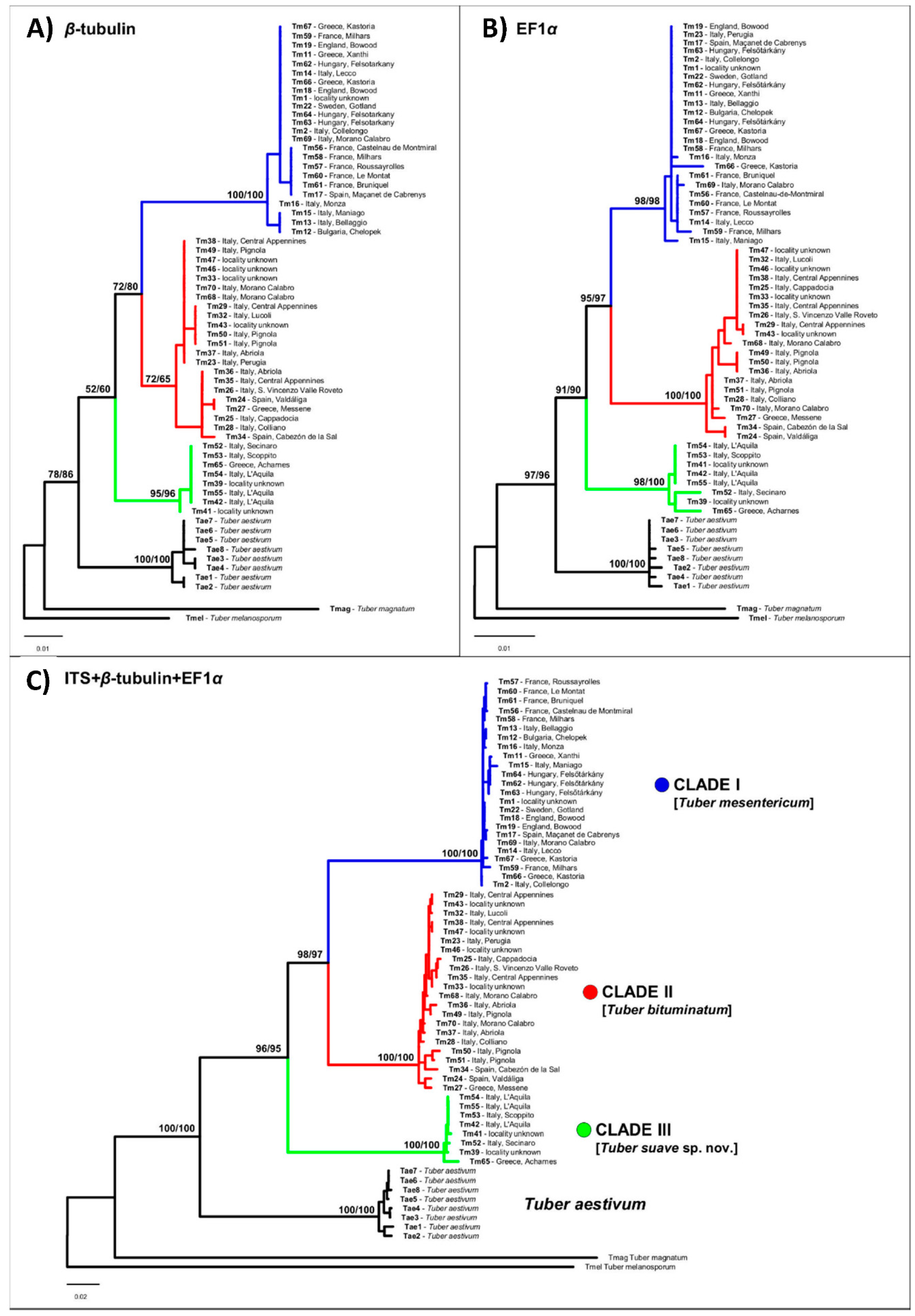
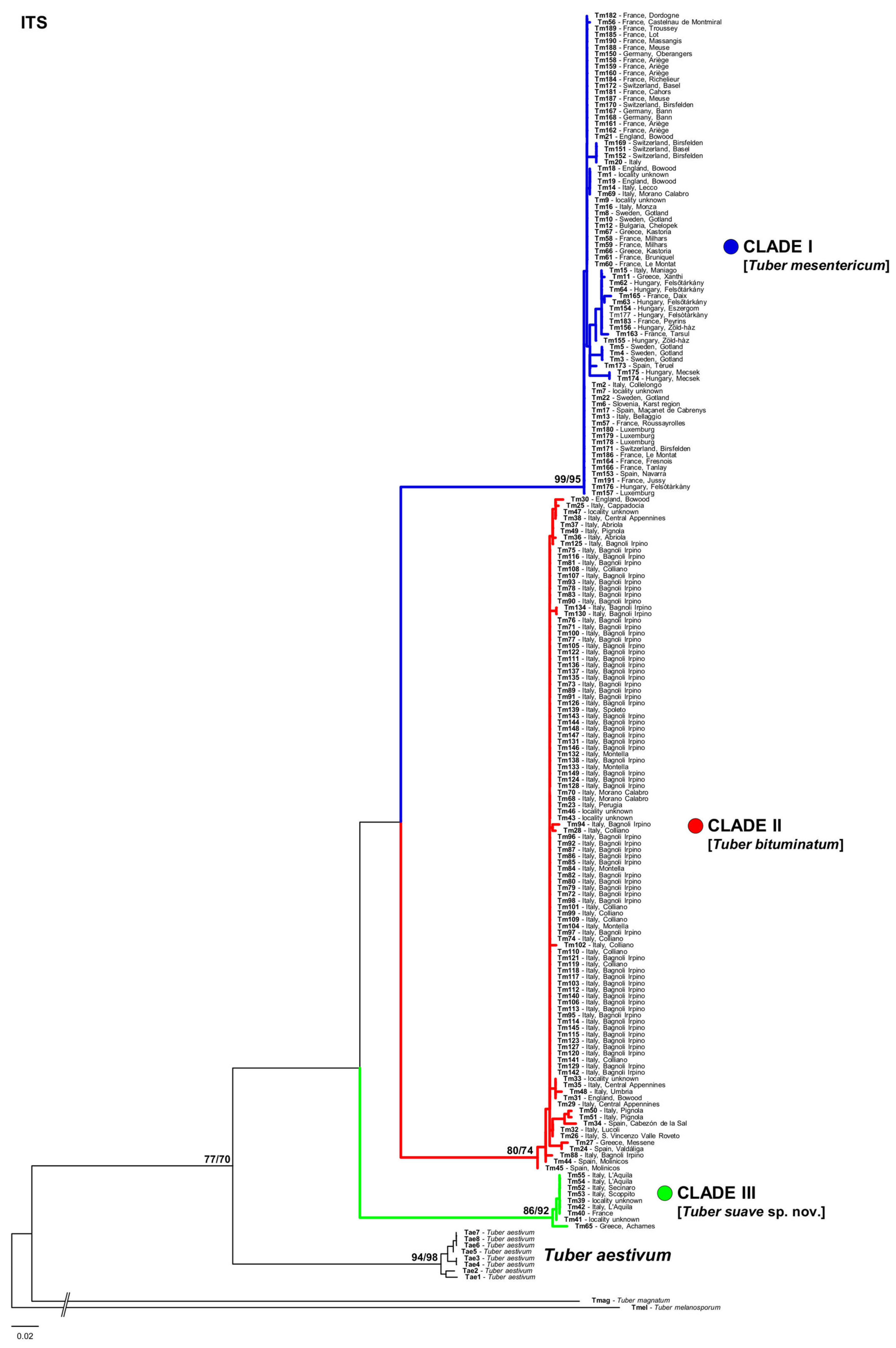
3.1. Phylogenetic Analyses

Phylogenetic analyses based on either single genes or on the concatenated dataset consistently recovered three reciprocally monophyletic clades (Figure 1 and Figure 2) that received maximum support (bootstrap support = 100) in the concatenated analysis: (i) one clade with the widest range across Europe (clade I); (ii) a second clade grouping specimens collected mainly in the Iberian, Italian, and Balkan peninsulas (clade II); and (iii) a third clade including specimens collected almost exclusively in central Italy and Greece (clade III). The ranges of the three clades were found to overlap in different European regions, particularly in western Europe, Italy, and Greece (Figure 3).
Genetic divergence among the three clades ranged from 0.097 to 0.123 (uncorrected p-distance) and from 0.104 to 0.131 (Kimura-2-parameter distance) at the ITS region; from 0.037 to 0.053 (uncorrected p-distance) and from 0.034 to 0.054 (Kimura-2-parameter distance) at the β-tubulin region; and from 0.026 to 0.034 (uncorrected p-distance) and from 0.027 to 0.032 (Kimura-2-parameter distance) at the EF-1-α region. Intra-clade divergence at the ITS region was 0.0047 for clade I, 0.0074 for clade II, and 0.0032 for clade III, using either p-distance or Kimura-2-parameter estimates.
The ITS sequence of T. mesentericum Vittadini’s authentic specimen and that of one specimen of T. bituminatum from the Kew herbarium were clustered into clade I (Figure 1 and Figure 2). ITS sequences of the other two T. bituminatum specimens were grouped with samples of clade II (Figure 1 and Figure 2). All of these sequences were generated with the species-specific primer pairs designed in this study (Table 2). The efficiency of these specific primers was also tested on the specimens used for phylogenetic analyses, and on the other 21 additional specimens deposited in the AQUI and MCVE herbaria (Table 1 and Table S1).

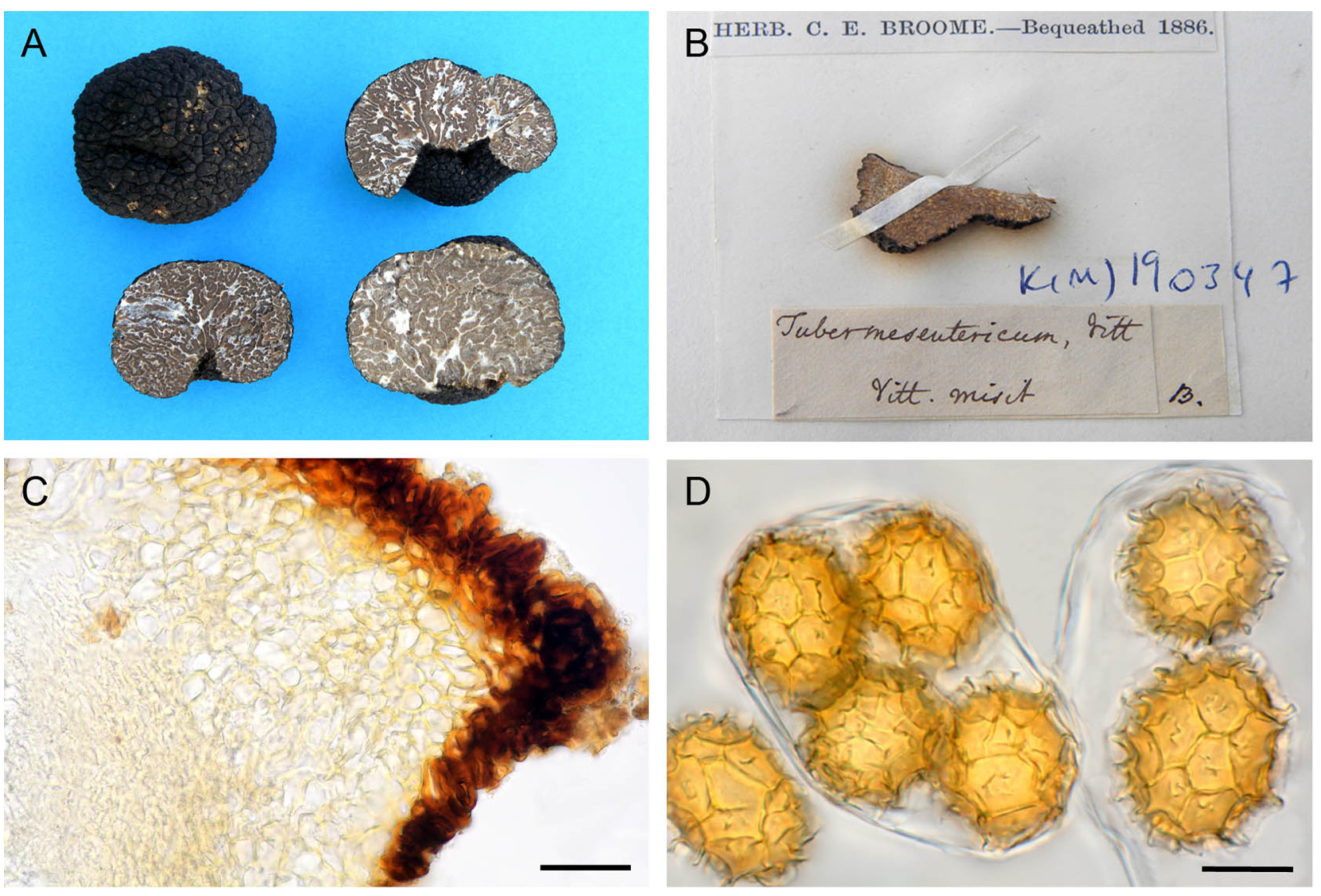
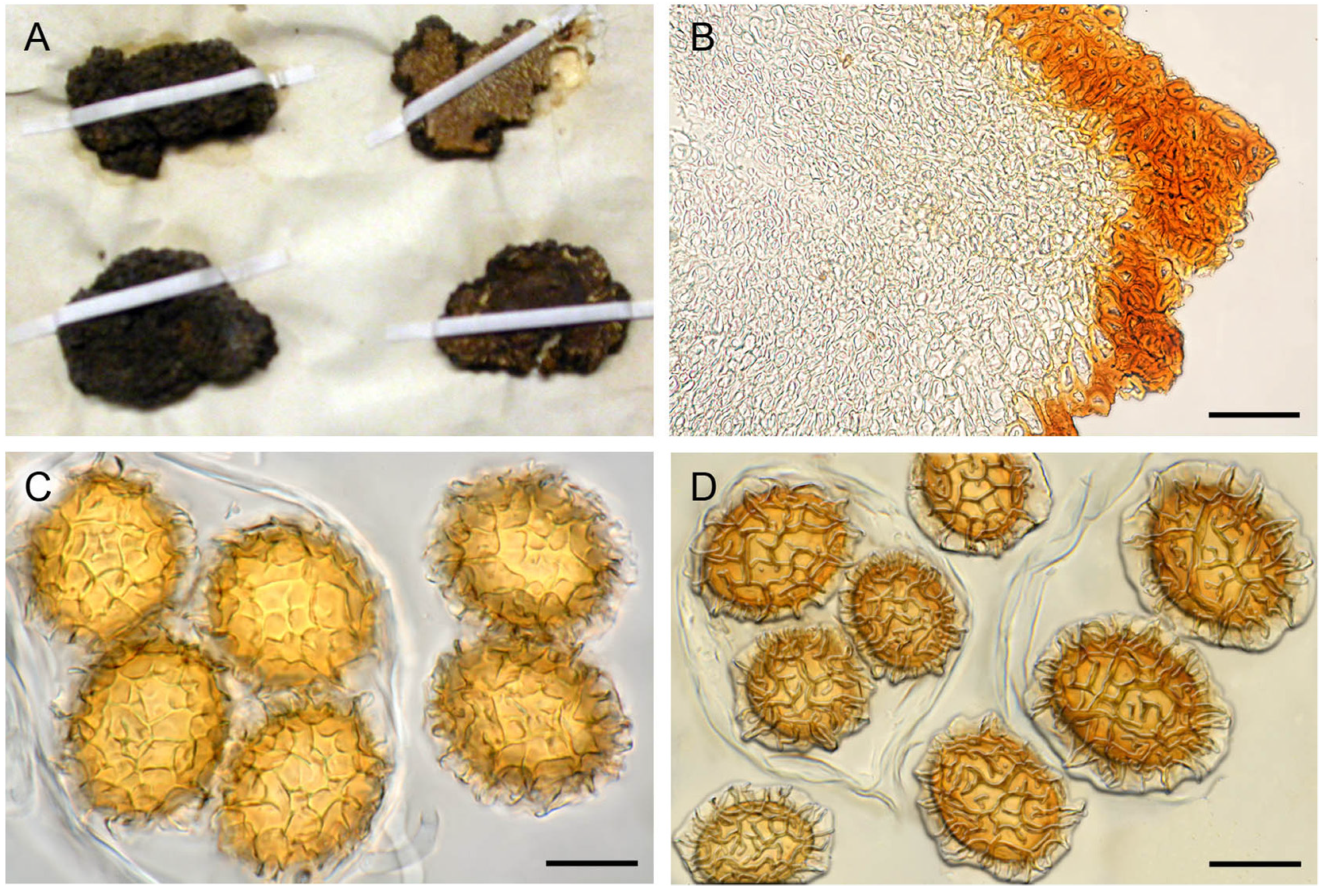
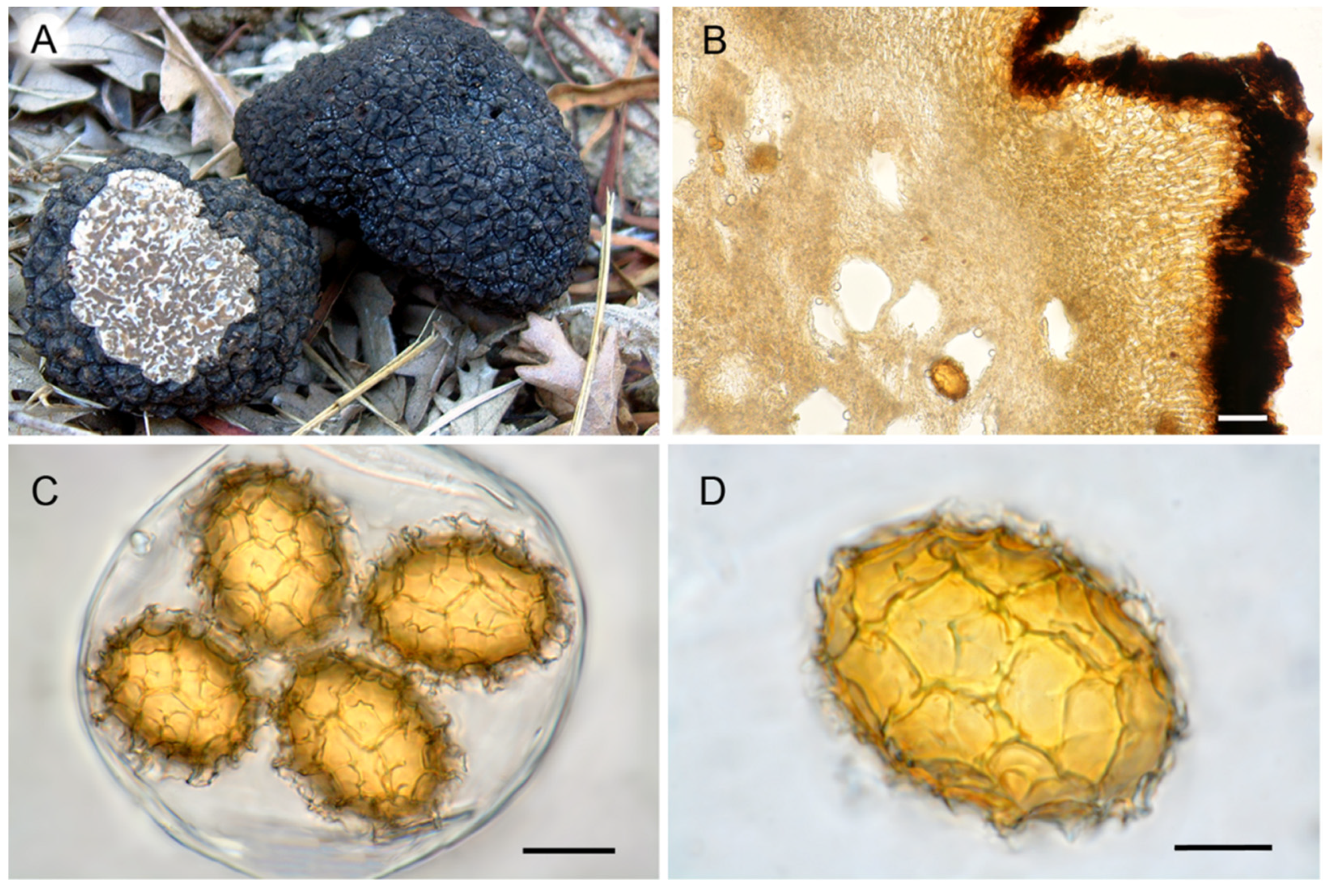
3.2. Ascoma Morphology

Micro- and macromorphological characters of ascomata showed a wide overlap among ascomata of different clades and have not proved useful for their taxonomic identification. Few differences were found, only in the shape of peridial cells constituting the center of warts and in the morphology of spore reticulum. Ascomata in clades I and II showed a pseudoparenchymatous peridium with globose–angular cells, whereas those in clade III showed a pseudoparenchyma composed of elongated cells that radiated out from the base of the warts (Figure 4C, Figure 5B and Figure 6B). The spores in clades I and III always showed regular and completely formed alveolae (Figure 4D and Figure 6C,D), whereas a variable percentage of those in clade II had labyrinthine pseudoreticula with incomplete and irregular alveolae (Figure 5C,D).
No statistical differences were found in any of the measured characters of the gleba hyphae, asci, or spores of the three clades. Nearly significant differences were found only for L2 (p = 0.051) and L2/W2 (p = 0.062) spore parameters when values were log-transformed before ANOVA (Figure S4, Table S2).

4. Discussion

The phylogeny of the T. mesentericum complex has been poorly investigated to date, and only recently has it attracted the interest of mycologists. So far, three studies have investigated the genetic diversity of T. mesentericum by applying a single-gene analysis [24,25,26], but none of them have resolved the taxonomic issues within this species complex. In this study, we performed an integrative taxonomic assessment of the T. mesentericum complex by combining a multilocus phylogeographic approach with in-depth morphological analyses, and including authentic specimens of Vittadini and Berkeley and Broome. This approach allowed us to resolve the main phylogenetic clades found within the T. mesentericum complex into three species and to designate an epitype for T. mesentericum s.s.
In agreement with previous studies by Benucci et al. [25] and Marozzi et al. [26], we found three distinct and well-supported lineages, both in single-locus and multi-locus phylogenetic analyses.
Phylogeographic data generated in this study combined with data from public repositories indicate that members of clade I are spread all over Europe, from Sweden to Italy and from Spain to Bulgaria, and are associated with many hardwood trees, including Quercus spp. (Q. cerris L., Q. robur L.), Fagus sylvatica L., Corylus avellana L., Ostrya carpinifolia Scop., but also with conifers, such as Picea spp. The range of clade II seems to be mainly limited to Mediterranean habitats, and their members show a similar host preference. Clade III is only distributed in central Italy and Greece, although a specimen from GenBank (JQ348414) had “France” as its general indication of origin, which should be verified. To date, ascomata of Clade III have been found only under thermophilic oaks.
Clades I and II seem to be sister species that diverged after clade III. While the average intraclade divergence at the ITS marker was <0.8%, the divergence between these clades was as high as 10% or 13%, values that are well above those observed among the currently accepted Tuber species. For example, interspecific divergence observed between closely related pairs of currently recognized species belonging to Rufum and Melanosporum clades are in the range of 4.7–7.4% [39]. Bonito et al. [42] suggested a threshold for Tuber species delimitation based on an interspecific divergence of ≥4%. However, interspecific divergence below 4% has been reported between closely related species of different clades, such as the Excavatum [12], Puberulum [9], and Gibbosum [43] clades.
Morphological and biometric characters of ascomata examined in this study do not allow an unambiguous identification of the clade to which they belong, thus confirming that these lineages represent cryptic species. Only two of the microscopic characters, the peridium (cell pattern of the peridium cells in the center of the single warts) and spores (reticulum integrity), showed some differences among clades, although they are not diagnostic and, therefore, of little taxonomic utility. ANOVA analyses based on spore sizes showed low differentiation among clades, with only spore length (L2) being marginally significant.
The mismatch between phylogenetic and morphological differentiation seems to be a common feature in the genus Tuber. Bonuso et al. [10] identified two genetically isolated groups in T. borchii, but no distinctive morphological features were found in support of this separation. Ascoma morphology has not been useful in solving the taxonomy of the T. indicum complex, where at least two cryptic species exist, according to different phylogenetic studies [13,14,44]. The morphological distinction between T. brumale and T. cryptobrumale is also challenging; Merényi et al. [11] verified that only a combination of different morphological characters would make it possible to differentiate most (95%) specimens belonging to these two pseudocryptic species. The issue of cryptic species seems even more complex in other Tuber lineages. Five different species complexes were identified by Healy et al. [15] in the Rufum clade, and as many cryptic species were found by Puliga et al. [12] for the European truffle T. excavatum. From a taxonomic point of view, the greatest challenge will be to define and name each cryptic species within these complexes.
The integrative approach used in this study has revealed itself to be particularly fruitful in resolving the taxonomy of the cryptic species complex of T. mesentericum. The genotyping of authentic specimens of Vittadini, and Berkeley and Broome from the Kew herbarium allowed us to identify clade I as T. mesentericum sensu stricto, clade II as T. bituminatum, and clade III as a new species named T. suave, for the pleasant smell that seems to be typical of its ascomata. The type of T. mesentericum, as well as the types of the other Tuber species described by Vittadini [45], are no longer available for genotyping. Vittadini’s private herbarium was never deposited in a public institution, and it was lost because of insect infestation [46]. Only a few authentic specimens determined as T. mesentericum by Vittadini are actually available at the herbaria of Kew, Padua, and Uppsala (Figure 4B and Figure S1). The specimen labelled as T. mesentericum (K(M)190347), and one of the three specimens labelled as T. bituminatum from the Kew herbarium (Figure 4B and Figure S1a), were successfully amplified by using the ITS primer pair newly designed for the members of clade I. On the contrary, the PCR amplifications of the specimens deposited in the Padua and Uppsala herbaria (Figure S1B,C) were unsuccessful, regardless of the lineage-specific primer pair designed in this study. This was probably due to their poor state of preservation, biological contamination, or the chemicals added to preserve the exsiccata. The iconography of Vittadini [45] (Table III, Figure XIX) constitutes an ambiguous lectotype because it only represents a detail of the gleba, and not the entire ascoma with the warty black peridium and the basal cavity, hence the epithet “mesentericum”. Therefore, we designated a specimen collected in Vittadini’s study area as an epitype of T. mesentericum s.s. This specimen has an ITS sequence identical (100% identity) to that of the authentic specimen voucher from the Kew herbarium (K(M)190347) (Figure 4B).
Tuber bituminatum was described by Berkeley and Broome [47], but was later considered a synonym of T. mesentericum or T. aestivum [48,49,50]. Our analyses demonstrated that two out of three specimens indicated by Berkeley and Broome as holotypes of T. bituminatum fall into clade II and, therefore, this species should be revaluated and exclusively attributed to the members of this lineage within the T. mesentericum complex. Recently, Crous et al. [51] described the new species Tuber alcaracense Ant. Rodr. & Morte, having ascomata similar to those of species of the T. mesentericum complex, but with a pleasant odour and lacking a basal cavity. A preliminary phylogenetic comparison of the two ITS sequences available for T. alcaracense (MN810046-7) with the ITS sequences generated in this study indicates that T. alcaracense is nested within the clade of T. bituminatum (clade II, results not shown). Therefore, most likely T. alcaracense is a synonym of T. bituminatum, as the latter name has priority. However, before drawing taxonomic conclusions in this regard, more in-depth morphological and phylogenetic analyses must be carried out.
As we observed for the Kew herbarium, it is possible that many collections labelled as T. mesentericum in private or public herbaria can also include T. bituminatum and/or T. suave specimens, due to the scarcity of morphological traits useful in distinguishing them. For example, the authentic specimen of T. mesentericum preserved in the PD herbarium (Milano 1841, leg. et det. C. Vittadini) for its labyrinthine spore ornamentation might belong to T. bituminatum.
The aroma of ascomata within the T. mesentericum complex still remains controversial. In our analysis, T. mesentericum s.s. generally had a smell of solvent, whereas the smell of T. suave ascomata was consistently pleasant, as also confirmed by Marozzi et al. [26] for the specimens of the same lineage. On the contrary, the smell of T. bituminatum was very variable: it was often unpleasant, mainly due to the production of 1-methoxy-3-methylbenzene (reported as 3-methyl-anisole by [52]), and sometimes it was very pleasant. Although studies on the volatile organic compound (VOC) composition of T. mesentericum have been carried out in the past [53], an extensive investigation on ascomata of this species complex should be planned in light of recent taxonomic insights.

5. Taxonomy

5.1. Tuber mesentericum Vittad.

MB# 180551
Lectotype: Vittadini C. 1831, Monographia Tuberacearum, Table III Figure XIX
Epitype: Italy, Lombardia, Parco Reale di Monza, 45°35′41″ N 9°16′26″ E, 175 m asl, O. carpinifolia, 5 October 2017, leg. Stefano Seghezzi, det. Giovanni Pacioni (AQUI 9717), (Figure 4A,C,D); MTB# 10004606.
GenBank: OL711593 (nrITS), OL753399 (nr β-tub), OL753354 (nr EF1-α).
Ascomata hypogeous, globose to slightly lobed, more or less hollowed at the base, 2–7 cm in diameter. Peridium blackish, covered with flat pyramidal warts. Warts 3–6 sided, up to 4 mm wide, 1.5–2.0 mm high, bearing 3–5 vertical grooves near the middle of the face, sometimes with rough and fine horizontal striae, apex open and depressed. Odour pleasant, sometimes with a faint solvent (similar to trichloroethylene) component, disappearing when the context is exposed. Peridium pseudoparenchymatous, 150–250 µm; outer layer (exoperidium) 50–90 µm thick, almost opaque, composed of angular, irregular cells 10–20 µm in diameter, with somewhat thickened (4–5 µm), dark red-brown walls and reduced cell lumen (“sclerenchymatous” type); inner layer (endoperidium) hyaline or pale yellow, 90–160 µm thick, composed of densely packed globose-angular cells, 8–28 × 8–20 µm in size, with thickened walls (2–4 µm) (“collenchymatous” type), merging with glebal tissue of interwoven hyphae. Gleba brown (R: from “88-hazel” to “63-sepia”; H: “#ad5c33-dark orange” or “#8b0000-dark red”, from “#5f2f00” to “#472300-very dark orange”), with whitish sterile veins, meandering or radiating from the cavity or base, composed of 3–6 µm wide, hyaline and interwoven hyphae.
Asci clavate–subglobose 1–6 spored, 80–100 µm, sometimes thick-walled when ripe, with a stalk 4–18 µm long, 6–9 µm wide, arising from a crozier 9–13 µm wide. Ascospores regularly reticulated, ellipsoid, rarely globose or elongated (Q2 = 1.00–1.69), straw (R: 46), pure yellow (R: 14), or yellow-orange (R: from 11-pale luteous to 12-luteous; H: from “#ffe066-light yellow” to “#ffae42-yellow orange”); spore lumen (L1 × W1) = 18–53 × 16–31 µm, episporium (L2 × W2) = 24–61 × 20–41 µm, including exosporium (L3 × W3) = 30–70 × 26–56 µm, spore wall thickness (episporium) 1.5–5.0 µm. Ascospores (L2 × W2) in 1-spored asci 33–61 × 22–48 µm, 2-spored asci 22–44 × 22–33 µm, 3-spored asci 23–38 × 18–30 µm, 4-spored asci 21–39 × 19–26 µm, 5-spored asci 23–34 × 20–28 µm, 6-spored asci 24–32 × 21–26. Exosporium consisting of a narrow to a wide reticulum of complete polygonal alveolae with 3–7 sides, 2–8 µm high, 1.5–10 µm wide; meshes from 2–4 to 7–11 and from 2–3 to 5–9 across spore length and width, respectively.
Habitat: beneath ectomycorrhizal hardwoods and conifers, especially beech on calcareous soils, mainly in autumn.
Additional specimens examined: Bulgaria: Vratsa, Chelopek, Carpinus orientalis Mill., 1 August 2017, leg. T. Nedelin, det. G. Pacioni (AQUI 10144); Loveč, Letnitsa, Carpinus betulus L., 16 August 2018, leg. T. Nedelin, det. G. Pacioni (AQUI 10233); France: Occitanie, Castelnau-de-Montmiral (Tarn), Pinus sylvestris L. and C. avellana, 15 October 2019, leg. L. Gerola (AQUI 10234); Roussayrolles (Tarn), Quercus pubescens Willd. and C. avellana, 15 October 2019, leg. L. Gerola (AQUI 10235); Milhars (Tarn), Bonan, Q. pubescens, C. avellana, and C. betulus, 15 October 2019, leg. L. Gerola (AQUI 10238, AQUI 10239); Bruniquel (Tarn-Garonne), Q. pubescens, C. avellana, and C. betulus, 16 October 2019, leg. L. Gerola (AQUI 10236); Le Montat (Lot), C. avellana, 16 October 2019, leg. L. Gerola (AQUI 10237); Great Britain: Wiltshire, Bowood, holotype of T. bituminatum Berk. and Broome (K(M)30594, specimen 01); Bowood, Q. robur and F. sylvatica, 27 March 2018, leg Y. Borukov, det. G. Pacioni (AQUI 10140, AQUI 10141); Greece: W. Macedonia, Kastoria, Populus sp., C. avellana, and C. betulus, 5 October 2013, leg. P. Kladopoulou, det. G. Pacioni (AQUI 10230, AQUI 10243); W. Thrace, Xanthi, F. sylvatica, 11 November 2007, leg. P. Kladopoulou, det. G. Pacioni (AQUI 6558); Hungary: Heves, Felsőtárkány, Mt. Bükk, F. sylvatica, October 2000, leg. I. Kiss, det. Z. Bratek (ZB 2077-BPU, ZB 2092-BPU, ZB 2228-BPU); Italy: “Vittadini misit” (K(M)190347); Abruzzo, L’Aquila (AQ), Vasto di Assergi, O. carpinifolia, 12 November 1988, det. G Pacioni (AQUI 6548); Lucoli (AQ), Campo Felice, F. sylvatica, 25 November 2012, leg. D. Marinucci, det. G. Pacioni (AQUI 8510, AQUI 8511); Roccaraso (AQ), Pietransieri, Q.cerris, and F. sylvatica, 11 November 2012, leg. P. Oddis, det. G. Pacioni (AQUI 7227); Collelongo (AQ), Prati di S. Elia, F. sylvatica, 21 November 2014, leg. M. Cassetta, det. G. Pacioni (AQUI 8990); Calabria, Morano Calabro (CS), Campiglioni, Bosco Donna Calda, 9 October 2019, det. A. Paz-Conde (AQUI 10326/IC9101901); Campania, Bagnoli Irpino (AV), 3 November 2011, det. G. Pacioni (AQUI 10324); Friuli-Venezia Giulia, Maniago (PN), O. carpinifolia, 16 June 2002, leg. and det. G. Zecchin (MCVE 24188); Montereale Valcellina (PN), 25 October 2009, leg. A. Dal Cin, det. E. Campo (MCVE 25329); Lombardia, Bellagio (CO), 3 October 2017, leg. A. Bincoletto, det. G. Pacioni (AQUI 10145); Lecco, 4 September 2017, leg. S. Seghezzi, det. G. Pacioni (AQUI 9716); Spain: Catalonia, Maçanet de Cabrenys (Girona), F. sylvatica, Quercus sp., and Picea sp., 13 March 2017, leg. A. Paz and C. Lavoise, (AQUI 9718/IC13031703); Cantabria, Tudanca, Reserva Nacional Nansa, F. sylvatica and C. avellana L., 28 September 2015, leg. A. Paz and C. Lavoise (AQUI 10138/IC28091526); Sweden: Gotland, Tingstäde, Q. robur and C. avellana, 7 October 2000, leg. C. Wedén (UPS F-118824-CW005/DUKE 0348880).
The ITS and EF1-α sequences of this species fall into clade I after Benucci et al. [25] and into subclade I, after Marozzi et al. [26], respectively.

5.2. Tuber bituminatum Berk. & Broome

MB# 196523
Lectotype: Great Britain, Whiltshire, Bowood, October 1847, (K(M)30594) specimens 02 and 03 (Figure 5A–D); MTB# 10004607
GenBank: OL711615 and OL711616 (nr ITS).
Ascomata hypogeous, globose, more or less hollowed at the base, 2–10 cm in diameter. Peridium blackish with flat pyramidal warts. Warts 4–6 sided, up to 3 mm wide, 1–1.5 mm high, with flat or center grooved faces, with rough horizontal striae, apex pointed, sometimes depressed. Odour from phenolic (hence the epithet “bituminatum”) to very pleasant. Peridium pseudoparenchymatous; exoperidium 80–120 µm thick, almost opaque, composed of angular, irregular cells 8–14 µm in diameter, with somewhat thickened (4–5 µm), dark-brown walls and reduced cell lumen (“sclerenchymatous” type); endoperidium hyaline or pale yellow, 100–150 µm thick, composed of tightly packed globose–angular cells, 11–32 × 8–14 µm in size, with thickened walls (up to 5 µm) (“collenchymatous” type), merging with glebal tissue of interwoven hyphae. Gleba brown (R: from “88-hazel” to “63-sepia”; H: from “#84329-dark red” to “#472300-very dark orange”), with whitish sterile veins, meandering or radiating from the cavity, composed of 2–6 µm wide, hyaline and interwoven hyphae.
Asci clavate–subglobose, 1–6 spored, 70–100 µm, sometimes thick-walled when ripe, with a stalk 8–30 µm, arising from a crozier 9–14 µm wide. Ascospores regularly and irregularly (“labyrinthine”) reticulated, ellipsoid, sometimes globose or fusiform (Q = 1.00–2.09), yellow (R: “14-pure yellow”; H: from “#ffd633-vivid yellow” to “#ffdb4d-light yellow”) or orange (R: from “12-luteous” to “47-amber”); cell lumen (L1 × W1) = 16–52 × 14–33 µm, episporium (L2 × W2) = 22–61 × 20–43 µm, including exosporium (L3 × W3) = 29–70 × 26–54 µm, spore wall thickness (episporium) 1.5–6.5 µm. Ascospores (L2 × W2) in 1-spored asci 36–61 × 30–38.5 µm, 2-spored asci 28–43 × 23–35 µm, 3-spored asci 25–38 × 22–32 µm, 4-spored asci 22–34 × 19–29 µm, 5-spored asci 27–34 × 22–26 µm, 6-spored asci 28–32 × 22–28 µm. Exosporium consisting of a mixture of 3–5 sided, complete and irregular alveolae whose main ridges are broken, branched, and interconnected with minor ridges, 2.0–9.5 µm high, 2.0–10 µm wide; meshes 4–5 and 3–4 across spore length and width, respectively.
Habitat: beneath ectomycorrhizal hardwoods, but also conifers, especially beech on calcareous soils, mainly in autumn.
Additional specimens examined: France: Aquitaine-Limousin-Poitou-Charentes, La Gripperie-Saint-Symphorien (Charente-Maritime), F. sylvatica, Quercus sp., and Picea sp., 14 November 2014, leg. P. Chautrand (AQUI 10136/IC14111417); Greece: Peloponnese, Messene, Mt. Taygetos, Q. pubescens, 10 March 2013, leg. P. Kladopoulou, det. G. Pacioni (AQUI 8579); Italy: Abruzzo, Lucoli (AQ), Campo Felice, F. sylvatica, 19 October 2010, D. Marinucci (AQUI 7097); Lucoli (AQ), Campo Felice, F. sylvatica, 8 November 2012, leg. D. Marinucci, det. G. Pacioni (AQUI 8417); Lucoli (AQ), Prato Capito, F. sylvatica, 27 November 2011 (AQUI 7162); L’Aquila (AQ), Arischia, Q. pubescens, 1 November 1976 (AQUI 3285); L’Aquila (AQ), Vasto di Assergi, C. avellana and O. carpinifolia, 13 September 1988 (AQUI 6505, AQUI 6509); Cappadocia (AQ), Camporotondo, F. sylvatica, 18 December 2016, leg. I. Apolloni, det. G. Pacioni (AQUI 9833); S. Vincenzo Valle Roveto (AQ), Morrea, F. sylvatica, 15 December 2014, leg. M. Cassetta, det. G. Pacioni (AQUI 9713); Gamberale (CH), Laghetto, F. sylvatica, 28 November 2016 (AQUI 9707); Palena (CH), Madonna dell’Altare, F. sylvatica and Q. cerris, 25 November 2016 (AQUI 9702); Palena (CH), Arsiccia, F. sylvatica and Q. cerris, 2 December 2016 (AQUI 9703); Palena (CH), Val di Terra, F. sylvatica, 4 December 2016 (AQUI 9704); Basilicata, Abriola (PT), Mt. Pierfaone, F. sylvatica, 11 November 2011, leg. and det. G.L. Rana (AQUI 8994, AQUI 8995); Pignola, La Sellata-Rifreddo, F. sylvatica, 11 November 2011, leg. and det. G.L. Rana (AQUI 8991, AQUI 8992, AQUI 8993); Calabria, Rossano Calabro (CS), Mediterranean maquis, 12 December 2000, leg. Di Leone (MCVE 16209); Morano Calabro (CS), Campiglioni, Bosco Donna Calda, 9 October 2019, leg. A. Paz-Conde (AQUI 10325, AQUI 10327); Campania, Colliano (SA), F. sylvatica, 10 October 2001, leg. and det. A. Zambonelli (CMI-UNIBO 1807); Umbria, Perugia (AQUI 8921) and central Apennines without indications (AQUI 7099; AQUI 8919; AQUI 8920); Spain: Cantabria, Comillas, Reserva Nacional Saja, F. sylvatica, 27 February 2015, leg. A. Paz and C. Lavoise (AQUI 10134/IC27021515); Cabezon de la Sal, Mt. Corona, Abies alba Mill., 15 October 2016, leg. A. Paz and C. Lavoise (AQUI 10137/IC15101604); Valdaliga, Bustriguado, A. alba, 15 October 2016, A. Paz and C. Lavoise (AQUI 10139/IC15101621).
The ITS and EF1-α sequences of this species fall into clade II after Benucci et al. [25] and into subclade II after Marozzi et al. [26], respectively.

5.3. Tuber suave Pacioni & M. Leonardi, sp. nov.

MB# 842206
Holotype: Italy, Abruzzo, L’Aquila, Secinaro, Q. pubescens, leg. Antonio Barbati, 11 November 2010 (AQUI 7131) (Figure 6A–D)
GenBank: OL711623 (nr ITS), OL753414 (β-tub), OL753375 (nr EF 1-α).
Etymology: from the Latin ‘suave’ (neuter), pleasant for its flavor.
Diagnosis: Ascoma with black and flat pyramidal warts, and flat or excavate base; peridium pseudoparenchymatous textura porrecta with parallelepiped cells that decrease in size from the inside out, while increasing the thickness and pigmentation of the walls. Ascospores yellowish, ellipsoid 22–60 × 18–40 µm without ornamentations, exosporium 2–7 µm high, alveolate with complete polygonal meshes with 4–7 sides. Smell and taste pleasant. Under thermophilic oaks in the Mediterranean area.
Ascomata hypogeous, globose, more or less hollowed at the base, 4–5 cm in diameter. Peridium black, covered with flat pyramidal warts. Warts 4–6 sided, up to 2–3 mm wide, 1.5–2 mm high, grooved faces, without horizontal striae, apex pointed or open, and depressed. Odour complex, intense, and pleasant. Peridium pseudoparenchymatous, 100–250 μm thick; exoperidium 40–100 μm thick, opaque, composed of elongated cells (outermost layers), with deep-brown walls (R: from “9-umber” to “63-Sepia”; H: from “#663300–nutmeg wood finish” to “#661a00–red oxide”), strongly thickened (3–5 μm) and reduced or absent cell lumen (“sclerenchymatous” type), 6–17 × 4.5–8 μm, followed by a palisade layer (55–100 μm thick) of elongated cells completely filling the wart, 12–40 × 6–14 μm, with the major axis perpendicular to the surface; endoperidium 50–150 μm thick, yellowish (R: “45–buff”; H “#ff6600–safety orange”, “#e65c00–persimon”, “#e287A3–burnt Sienna”), composed of a mixture of pseudoparenchymatous cells, 4–11 × 2–7 μm, with 2–3 μm thickened walls (“collenchymatous” type). Gleba deep brown (R: “63-Sepia”; H: “#5f2f00-baker’s chocolate” or “472300-very dark orange”), with white, meandering, rather wide sterile veins, composed of interwoven, thin-walled, 3–5 µm wide hyphae. Asci 1–5 spored, clavate–suglobose, 55–100 μm, sometimes thick-walled, with a stalk up to 30 µm long, 6–8 μm wide, arising from a crozier 9–12 μm wide.
Ascopores reticulated, ellipsoid, sometimes globose or slightly elongated (Q = 1.00–1.75); yellowish or burnt orange (R: from “47-amber” to “9-umber”; H “#e6b800-pure yellow”, from “#ffd119-vivid yellow” to “#cc5500-strong orange”); cell lumen (L1 × W1) = 16–52 × 14–33 µm, episporium (L2 × W2) 22–59 × 20–39 µm, including exosporium (L3 × W3) 29–70 × 26–54, spore wall thickness (episporium) 1.0–5.0 µm. Ascospores (L2 × W2) in 1-spored asci 46–60 × 32–40 µm, 2-spored asci 31–48 × 24–33 µm, 3-spored asci 24–44 × 22–33 µm, 4-spored asci 27–39 × 18–28 µm, 5-spored asci 28–34 × 22–27 µm. Exosporium consisting of a reticulum of complete polygonal alveolae with 4–6 sides, 2.0–7.0 µm high, 2.0–10 µm wide; meshes 3–6 and 2–3 across spore length and width, respectively.
Habitat: it seems to be a species associated with thermophilic oak species Q. pubescens, Q. ilex L. [26], and Quercus coccifera L., in central-south Italy and Greece.
Additional specimens examined: Greece: Attica, Acharnes, Mt. Parnitha, Q. coccifera and Pinus halepensis Mill., 12 July 2013, leg. P. Kladopoulou, det. G. Pacioni (AQUI 10229); Italy: Abruzzo, Scoppito (AQ), Q. pubescens, 1 February 1988, leg. C. Visca, det. G. Pacioni (AQUI 4882); L’Aquila (AQ), Colle di Sassa, Q. pubescens, 12 December 2012, leg. D. Marinucci, det. G. Pacioni (AQUI 8761, paratype); L’Aquila (AQ), Q. pubescens, 27 November 2011, leg. D. Marinucci, det. G. Pacioni (AQUI 7165); L’Aquila (AQ), Q. pubescens, 2 January 2012, leg D. Marinucci, det. G. Pacioni (AQUI 8514); L’Aquila (AQ), Q. pubescens, 3 December 2012, leg. D. Marinucci, det. G. Pacioni (AQUI 10323 paratype); central Apennines without indications (AQUI 292, AQUI 10240).
The ITS and EF1-α sequences of this species fall into clade III after Benucci et al. [25] and into subclade III after Marozzi et al. [26], respectively.

6. Conclusions

This study clarifies the taxonomy of a commercially important truffle group, and will be useful to support further studies on ecology, cultivation, and foodomics of these species. The specific primer pairs tested in this study can be used for rapid and easy identification of members of the three studied species. Finally, specific investigations on VOC composition are needed to define the aroma repertoires existing in the T. mesentericum complex and to commercially promote these species.

Supplementary Materials

The following are available online at https://www.mdpi.com/article/10.3390/jof7121090/s1, Figure S1: Historical voucher specimens from kew (A), Uppsala (B) and Padua (C) herbaria [54,55,56,57,58]; Figure S2: Localization of the three T. mesentericum clade-specific primer pairs on the ITS region alignment, Figure S3: Graphic representation of spore parameters measured in this study, Figure S4: Boxplots of sporal dimensions (A, L2-lenght; B, W2-width; C, L2/W2 ratio) in the three clades, Table S1: Metadata of the specimens analyzed in this study, Table S2: Results of one-way ANOVA with repeated measure for differences in spore size.

Author Contributions

Conceptualization, M.L., D.S. and G.P.; methodology, M.L. and D.S.; validation, M.I.; formal analysis, M.L. and D.S.; investigation, M.L. and G.P.; resources, G.L.R. and A.P.-C.; data curation, M.L., G.L.R. and G.P.; writing—original draft preparation, M.L. and G.L.R.; writing—review and editing, M.I. and D.S.; visualization, A.P.-C. and D.S.; supervision, G.P.; project administration, G.P.; funding acquisition, G.P. and M.I. All authors have read and agreed to the published version of the manuscript.

Funding

This research received no external funding.

Institutional Review Board Statement

Not applicable.

Informed Consent Statement

Not applicable.

Data Availability Statement

All data obtained and analyzed in this study have been included in this article and its Supplementary Materials.

Acknowledgments

We are grateful to Carmine Visca and Domenico Marinucci (L’Aquila University), Antonio Corsetto and Marco Diana (Potenza), Angelo Bincoletto (Milan), Yordan Borukov (UK), Laurent Genola (Toulouse), and Teodor Nedelin (Sofia) for fresh specimen harvest of T. mesentericum sl, Christina Wedén (Uppsala University), Bryn Dentinger and Begoña Aguirre-Hudson (Royal Botanic Gardens Kew), Connie Robertson (Duke Herbarium), Zoltán Bratek (BPU), Pavlina Klodopoulou (Athens), Alessandra Zambonelli (CMI-UNIBO) for the access to the respective herbaria for studying T. mesentericum sl voucher specimens; and Osvaldo Zarivi for his help in the setup of primers. We are strongly grateful to Stefano Seghezzi for providing the T. mesentericum epitype specimen from the Lombard areas frequented by Carlo Vittadini. We wish to express our gratitude to the late lamented R.G.W. Dennis (K) and A. Ceruti (TO), Rossella Marcucci (PD), and Åsa Kruys (UP) for allowing G.P. to study the original Vittadini collections. We thank Simone Fattorini (L’Aquila university) for advice on the statistical analyses of morphological data.

Conflicts of Interest

The authors declare no conflict of interest.

References

  1. Gryndler, M. True truffle host diversity. In True Truffle (Tuber spp.) in the World, Soil Biology 47; Zambonelli, A., Iotti, M., Murat, C., Eds.; Springer International Publishing: Cham, Switzerland, 2016; pp. 267–281. [Google Scholar]
  2. Zambonelli, A.; Iotti, M.; Hall, I.R. Current status of truffle cultivation: Recent results and future perspectives. Ital. J. Mycol. 2015, 44, 31–40. [Google Scholar]
  3. Percudani, R.; Trevisi, A.; Zambonelli, A.; Ottonello, S. Molecular phylogeny of truffles (Pezizales: Terfeziaceae, Tuberaceae) derived from nuclear rDNA sequence analysis. Mol. Phylog. Evol. 1999, 13, 169–180. [Google Scholar] [CrossRef] [PubMed]
  4. Wang, Y.; Tan, Z.M.; Zhang, D.C.; Murat, C.; Jeandroz, S.; Le Tacon, F. Phylogenetic relationships between Tuber pseudoexcavatum, a Chinese truffle, and other Tuber species based on parsimony and distance analysis of four different gene sequences. FEMS Microbiol. Lett. 2006, 259, 269–281. [Google Scholar] [CrossRef] [PubMed] [Green Version]
  5. Jeandroz, S.; Murat, C.; Wang, Y.; Bonfante, P.; Le Tacon, F. Molecular phylogeny and historical biogeography of the genus Tuber, the ‘true truffles’. J. Biogeogr. 2008, 35, 815–829. [Google Scholar] [CrossRef]
  6. Kinoshita, A.; Sasaki, H.; Nara, K. Phylogeny and diversity of Japanese truffles (Tuber spp.) inferred from sequences of four nuclear loci. Mycologia 2011, 103, 779–794. [Google Scholar] [CrossRef] [PubMed]
  7. Leonardi, M.; Iotti, M.; Pacioni, G.; Hall, I.R.; Zambonelli, A. Truffles: Biodiversity, ecological significances, and biotechnological applications. In Industrially Important Fungi for Sustainable Development; Abdel-Azeem, A.M., Yadav, A.N., Yadav, N., Usmani, Z., Eds.; Springer: Cham, Switzerland, 2021; pp. 107–146. [Google Scholar]
  8. Bonito, G.; Smith, M.E.; Nowak, M.; Healy, R.A.; Guevara, G.; Cázares, E.; Kinoshita, A.; Eduardo, R.; Nouhra, E.R.; Domínguez, L.S.; et al. Historical biogeography and diversification of truffles in the Tuberaceae and their newly identified southern hemisphere sister lineage. PLoS ONE 2013, 8, e52765. [Google Scholar] [CrossRef] [PubMed]
  9. Wang, X.H.; Benucci, G.M.N.; Xie, X.D.; Bonito, G.; Leisola, M.; Liu, P.G.; Shamekh, S. Morphological, mycorrhizal and molecular characterization of Finnish truffles belonging to the Tuber anniae species-complex. Fungal. Ecol. 2013, 6, 269–280. [Google Scholar] [CrossRef]
  10. Bonuso, E.; Zambonelli, A.; Bergemann, S.E.; Iotti, M.; Garbelotto, M. Multilocus phylogenetic and coalescent analyses identify two cryptic species in the Italian bianchetto truffle, Tuber borchii Vittad. Conserv. Genet. 2010, 11, 1453–1466. [Google Scholar] [CrossRef]
  11. Merényi, Z.; Varga, T.; Hubai, A.G.; Pitlik, P.; Erős, A.; Trappe, J.M.; Bratek, Z. Challenges in the delimitation of morphologically similar species: A case study of Tuber brumale agg. (Ascomycota, Pezizales). Mycol. Prog. 2017, 16, 613–624. [Google Scholar] [CrossRef]
  12. Puliga, F.; Illice, M.; Iotti, M.; Baldo, D.; Zambonelli, A. Tuber iranicum, sp. nov., a truffle species belonging to the Excavatum clade. Mycologia 2020, 112, 932–940. [Google Scholar] [CrossRef]
  13. Belfiori, B.; Riccioni, C.; Paolocci, F.; Rubini, A. Mating type locus of Chinese black truffles reveals heterothallism and the presence of cryptic species within the T. indicum species complex. PLoS ONE 2013, 8, e82353. [Google Scholar] [CrossRef] [PubMed] [Green Version]
  14. Qiao, P.; Tian, W.; Liu, P.; Yu, F.; Chen, J.; Deng, X.; Wan, S.; Wang, R.; Wang, Y.; Guo, H. Phylogeography and population genetic analyses reveal the speciation of the Tuber indicum complex. Fungal. Genet. Biol. 2018, 113, 14–23. [Google Scholar] [CrossRef] [PubMed]
  15. Healy, R.; Bonito, G.M.; Smith, M.E. A brief overview of the systematics, taxonomy, and ecology of the Tuber rufum clade. In True Truffle (Tuber spp.) in the World, Soil Biology 47; Zambonelli, A., Iotti, M., Murat, C., Eds.; Springer International Publishing: Cham, Switzerland, 2016; pp. 125–136. [Google Scholar]
  16. Bonito, G.M.; Smith, M.E. General systematic position of the truffles: Evolutionary theories. In True Truffle (Tuber spp.) in the World, Soil Biology 47; Zambonelli, A., Iotti, M., Murat, C., Eds.; Springer International Publishing: Cham, Switzerland, 2016; pp. 3–18. [Google Scholar]
  17. Molinier, V.; Peter, M.; Stobbe, U.; Egli, S. The Burgundy truffle (Tuber aestivum syn. uncinatum): A truffle species with a wide habitat range over Europe. In True Truffle (Tuber spp.) in the World, Soil Biology 47; Zambonelli, A., Iotti, M., Murat, C., Eds.; Springer International Publishing: Cham, Switzerland, 2016; pp. 33–47. [Google Scholar]
  18. Riccioni, C.; Rubini, A.; Belfiori, B.; Gregori, G.; Paolocci, F. Tuber magnatum: The special one. What makes it so different from the other Tuber spp.? In True Truffle (Tuber spp.) in the World, Soil Biology 47; Zambonelli, A., Iotti, M., Murat, C., Eds.; Springer International Publishing: Cham, Switzerland, 2016; pp. 87–103. [Google Scholar]
  19. Riccioni, C.; Rubini, A.; Türkoğlu, A.; Belfiori, B.; Paolocci, F. Ribosomal DNA polymorphisms reveal genetic structure and a phylogeographic pattern in the Burgundy truffle Tuber aestivum Vittad. Mycologia 2019, 111, 26–39. [Google Scholar] [CrossRef]
  20. Belfiori, B.; D’Angelo, V.; Riccioni, C.; Leonardi, M.; Paolocci, F.; Pacioni, G.; Rubini, A. Genetic structure and phylogeography of Tuber magnatum populations. Diversity 2020, 12, 44. [Google Scholar] [CrossRef] [Green Version]
  21. Hall, I.R.; Brown, G.T.; Zambonelli, A. Taming the Truffle: The History Lore and Science of the Ultimate Mushroom; Timber Press: Portland, Oregon, 2007. [Google Scholar]
  22. Pacioni, G.; Pomponi, G. Genotypic patterns of some Italian populations of the Tuber aestivum-T. mesentericum complex. Mycotaxon 1991, 42, 171–179. [Google Scholar]
  23. Pacioni, G.; Fantini, P. Tuber bellonae un tartufo mediterraneo del complesso Tuber aestivum-T. mesentericum. Micol. E Veg. Mediterr. 1997, 12, 15–20. [Google Scholar]
  24. Sica, M.; Gaudio, L.; Aceto, S. Genetic structure of Tuber mesentericum Vitt. based on polymorphisms at the ribosomal DNA ITS. Mycorrhiza 2007, 17, 405–414. [Google Scholar] [CrossRef] [Green Version]
  25. Benucci, G.M.N.; Gógán Csorbai, A.; Baciarelli Falini, L.; Marozzi, G.; Suriano, E.; Sitta, N.; Donnini, D. Taxonomy, biology and ecology of Tuber macrosporum Vittad. and Tuber mesentericum Vittad. In True Truffle (Tuber spp.) in the World, Soil Biology 47; Zambonelli, A., Iotti, M., Murat, C., Eds.; Springer International Publishing: Cham, Switzerland, 2016; pp. 69–86. [Google Scholar]
  26. Marozzi, G.; Benucci, G.M.N.; Suriano, E.; Sitta, N.; Raggi, L.; Lancioni, H.; Baciarelli Falini, L.; Albertini, E.; Donnini, D. Tuber mesentericum and Tuber aestivum truffles: New insights based on morphological and phylogenetic analyses. Diversity 2020, 12, 349. [Google Scholar] [CrossRef]
  27. Gardes, M.; Bruns, T.D. ITS primers with specificity for Basidiomycetes: Application to the identification of mycorrhizae and rust. Mol. Ecol. 1993, 2, 113–118. [Google Scholar] [CrossRef]
  28. White, T.J.; Bruns, T.; Lee, S.; Taylor, J. Amplification and direct sequencing of fungal ribosomal RNA genes for phylogenetics. In PCR Protocols: A Guide to Methods and Applications; Innis, M.A., Gelfand, D.H., Sninsky, J.J., White, T.J., Eds.; Academic Press: New York, NY, USA, 1990; pp. 315–322. [Google Scholar]
  29. Glass, N.L.; Donaldson, G.C. Development of primer sets designed for use with the PCR to amplify conserved genes from filamentous ascomycetes. Appl. Environ. Microb. 1995, 61, 1323–1330. [Google Scholar] [CrossRef] [PubMed] [Green Version]
  30. Paolocci, F.; Rubini, A.; Riccioni, C.; Topini, F.; Arcioni, S. Tuber aestivum and Tuber uncinatum: Two morphotypes or two species? FEMS Microbiol. Lett. 2004, 235, 109–115. [Google Scholar] [CrossRef]
  31. Zampieri, E.; Mello, A.; Bonfante, P.; Murat, C. PCR primers specific for the genus Tuber reveal the presence of several truffle species in a truffle-ground. FEMS Microbiol. Lett. 2009, 297, 67–72. [Google Scholar] [CrossRef] [PubMed] [Green Version]
  32. Hall, T.A. BioEdit: A user-friendly biological sequence alignment editor and analysis program for Windows 95/98/NT. Nucl. Acid. S 1999, 41, 95–98. [Google Scholar]
  33. Altschul, S.F.; Gish, W.; Miller, W.; Myers, E.W.; Lipman, D. Basic local alignment search tool. J. Mol. Biol. 1990, 215, 403–410. [Google Scholar] [CrossRef]
  34. Untergrasser, A.; Cutcutache, I.; Koressaar, T.; Ye, J.; Faircloth, B.C.; Remm, M.; Rozen, S.G. Primer3-new capabilities and interfaces. Nucleic Acids Res. 2012, 40, e115. [Google Scholar] [CrossRef] [PubMed] [Green Version]
  35. Katoh, K.; Standley, D.M. MAFFT multiple sequence alignment software version 7: Improvements in performance and usability. Mol. Biol. Evol. 2013, 30, 772–780. [Google Scholar] [CrossRef] [Green Version]
  36. Silvestro, D.; Michalak, I. raxmlGUI: A graphical front-end for RAxML. Org. Divers. Evol. 2012, 12, 335–337. [Google Scholar] [CrossRef]
  37. Stamatakis, A. RAxML version 8: A tool for phylogenetic analysis and post-analysis of large phylogenies. Bioinformatics 2014, 30, 1312–1313. [Google Scholar] [CrossRef]
  38. Lanfear, R.; Frandsen, P.B.; Wright, A.M.; Senfeld, T.; Calcott, B. PartitionFinder 2: New methods for selecting partitioned models of evolution for molecular and morphological phylogenetic analyses. Mol. Biol. Evol. 2016, 34, 772–773. [Google Scholar] [CrossRef] [Green Version]
  39. Leonardi, M.; Paz-Conde, A.; Guevara, G.; Salvi, D.; Pacioni, G. Two new species of Tuber previously reported as Tuber malacodermum. Mycologia 2019, 111, 676–689. [Google Scholar] [CrossRef]
  40. Rayner, R.W. A Mycological Colour Chart; Commonwealth Mycological Institute: Kew, UK, 1970. [Google Scholar]
  41. Development Core Team. R: A Language and Environment for Statistical Computing; R Foundation for Statistical Computing: Austria, Vienna, 2019. [Google Scholar]
  42. Bonito, G.M.; Gryganskyi, A.P.; Trappe, J.M.; Vilgalys, R. A global meta-analysis of Tuber ITS rDNA sequences: Species diversity, host associations and long-distance dispersal. Mol. Ecol. 2010, 19, 4994–5008. [Google Scholar] [CrossRef]
  43. Bonito, G.M.; Trappe, J.M.; Rawlinson, P.; Vilgalys, R. Improved resolution of major clades within Tuber and taxonomy of species within the Tuber gibbosum complex. Mycologia 2010, 102, 1042–1057. [Google Scholar] [CrossRef] [PubMed] [Green Version]
  44. Chen, J.; Guo, S.X.; Liu, P.G. Species recognition and cryptic species in the Tuber indicum complex. PLoS ONE 2011, 6, e14625. [Google Scholar] [CrossRef] [PubMed] [Green Version]
  45. Vittadini, C. Monographia Tuberacearum; Mediolani: Milan, Italy, 1831. [Google Scholar]
  46. Lazzari, G. Storia della Micologia Italiana; Saturnia: Trento, Italy, 1973. [Google Scholar]
  47. Berkeley, M.J.; Broome, C.E. Notices of British Hypogeous Fungi. Ann. Mag Nat. Hist 1851, 7, 176–186. [Google Scholar]
  48. Fischer, E. Ascomyceten. Tuberaceen und Hemiasceen. In Kryptogamen–Flora von Deutschland, Oesterreich und der Schweiz Band, Pilze V, Lief 57; Rabenhorst, L., Ed.; E. Kummer: Leipzig, Germany, 1897; pp. 1–64. [Google Scholar]
  49. Hawker, L.E. British hypogeous fungi. Philos. Trans. R. Soc. Lond. Ser. B Biol. Sci. 1954, 237, 429–546. [Google Scholar]
  50. Pegler, D.N.; Spooner, B.M.; Young, T.W.K. British Truffles. A Revision of British Hypogeous Fungi; Royal Botanic Gardens: Kew, UK, 1993. [Google Scholar]
  51. Rodríguez, A.; Navarro-Ródenas, A.; Arenas, F.; Guarnizo, A.L.; Morte, A. Fungal Planet 1107: Tuber alcaracense Ant. Rodr. & Morte, sp. nov. Persoonia 2020, 44, 301–459. [Google Scholar]
  52. Pacioni, G.; Bellina-Agostinone, C.; D’Antonio, M. On the odour of Tuber mesentericum. Mycol. Res. 1991, 94, 201–204. [Google Scholar] [CrossRef]
  53. Mauriello, G.; Marino, R.; D’Auria, M.; Cerone, G.; Rana, G.L. Determination of volatile organic compounds from truffles via SPME-GC-MS. J. Chromatogr. Sci. 2004, 42, 299–305. [Google Scholar] [CrossRef]
  54. Wedén, C.; Danell, E.; Tibell, L. Species recognition in the truffle genus Tuber– the synonyms Tuber aestivum and Tuber uncinatum. Environ. Microbiol. 2005, 7, 1535–1546. [Google Scholar] [CrossRef]
  55. Roux, C.; Séjalon-Delmas, N.; Martins, M.; Parguey-Leduc, A.; Dargent, R.; Bécard, G. Phylogenetic relationships between European and Chinese truffles based on parsimony and distance analysis of ITS sequences. FEMS Microbiol. Lett. 1999, 180, 147–155. [Google Scholar] [CrossRef]
  56. Stobbe, U.; Buntgen, U.; Sproll, L.; Tegel, W.; Egli, S.; Fink, S. Spatial distribution and ecological variation of re-discovered German truffle habitats. Fungal. Ecol. 2012, 5, 591–599. [Google Scholar] [CrossRef]
  57. Zhang, J.P.; Liu, P.G.; Chen, J. Tuber sinoaestivum sp. nov., and edible truffle from southwestern China. Mycotaxon 2012, 122, 73–82. [Google Scholar] [CrossRef]
  58. Paolocci, F.; Rubini, A.; Granetti, B.; Arcioni, S. Typing Tuber melanosporum and Chinese black truffle species by molecular markers. FEMS Microbiol. Lett. 1997, 153, 255–260. [Google Scholar] [CrossRef] [PubMed]
Figure 1. Phylogeny of T. mesentericum complex based on the single-loci datasets of β-tubulin (A) and elongation factor 1-α (B) and the concatenated dataset ITS + β-tub + EF-1-α (C). Bootstrap (BS) and rapid bootstrap (rBS) values (>70) are reported on nodes. Specimens are labelled by their geographic origin and ID, listed in Table 1 and Table S1.
Jof 07 01090 g001
Figure 2. Phylogeny of T. mesentericum complex, based on the ITS rDNA dataset. Bootstrap (BS) and rapid bootstrap (rBS) values (>70) are reported on nodes. Specimens are labelled by their geographic origin and ID, listed in Table 1 and Table S1.
Jof 07 01090 g002
Figure 3. Geographic distribution of main clades, based on specimens analyzed in this study. For each clade, the corresponding taxon and type locality are indicated.
Jof 07 01090 g003
Figure 4. T. mesentericum s.s. (A) Ascomata in section (neotype). (B) Fragment of the ascoma K(M)190347 from the Kew herbarium identified as T. mesentericum s.s. (C) Peridium structure. (D) Asci and ascospores. Bars = 20 µm.
Jof 07 01090 g004
Figure 5. T. bituminatum. (A) Ascomata in section (lectotypes). (B) Peridium structure. (C) Asci and ascospores with regular reticula. (D) Asci and ascospores with labyrinthine pseudoreticula. Bars = 20 µm.
Jof 07 01090 g005
Figure 6. T. suave sp. nov. (A) Ascoma in section (holotype). (B) Peridium structure. (C) Asci and ascospores. (D) Ascospore. Bars = 50 µm (B), 20 µm (C), 10 µm (D).
Jof 07 01090 g006
Table 1. Accession numbers and collection locality of the specimens analyzed in this study.
IDCladeLocalityITSβ-tubEF-1-α
Tm2IItaly—CollelongoKP686246KT456266OL753348
Tm11IGreece—XanthiOL711588OL753394OL753349
Tm12IBulgaria—ChelopekOL711589OL753395OL753350
Tm13IItaly—BellagioOL711590OL753396OL753351
Tm14IItaly—LeccoOL711591OL753397OL753352
Tm15IItaly—ManiagoOL711592OL753398OL753353
Tm16IItaly—MonzaOL711593OL753399OL753354
Tm17ISpain—Maçanet de CabrenysOL711594OL753400OL753355
Tm18IEngland—BowoodOL711595OL753401OL753356
Tm19IEngland—BowoodOL711596OL753402OL753357
Tm20IItalyKT456267--
Tm21IEngland—BowoodOL711597--
Tm22ISweden—GotlandAJ888043KT456265JX022595
Tm56IFrance—Castelnau-de-MontmiralOL711598OL753418OL753379
Tm57IFrance—RoussayrollesOL711599OL753419OL753380
Tm58IFrance—MilharsOL711600OL753420OL753381
Tm59IFrance—MilharsOL711601OL753421OL753382
Tm60IFrance—Le MontatOL711602OL753422OL753383
Tm61IFrance—BruniquelOL711603OL753423OL753384
Tm62IHungary—FelsőtárkányOL711604OL753424OL753385
Tm63IHungary—FelsőtárkányOL711605OL753425OL753386
Tm64IHungary—FelsőtárkányOL711606OL753426OL753387
Tm66IGreece—KastoriaOL711607OL753428OL753389
Tm67IGreece—KastoriaOL711608OL753429OL753390
Tm69IItaly—Morano CalabroOL711609OL753431OL753392
Tm23IIItaly—PerugiaKP686243KT456263OL753358
Tm24IISpain—ValdáligaOL711610OL753403OL753359
Tm25IIItaly—CappadociaOL711611OL753404OL753360
Tm26IIItaly—S. Vincenzo Valle RovetoOL711612OL753405OL753361
Tm27IIGreece—MesseneOL711613OL753406OL753362
Tm28IIItaly—CollianoOL711614OL753407OL753363
Tm29IIItaly—Central AppenninesKP686240KT456261OL753364
Tm30IIEngland—BowoodOL711615--
Tm31IIEngland—BowoodOL711616--
Tm32IIItaly—LucoliKP686239KT456260OL753365
Tm34IISpain—Cabezón de la SalOL711617OL753408OL753366
Tm35IIItaly—Central AppenninesKP686242KX290123OL753367
Tm36IIItaly—AbriolaKP686245KX290124OL753368
Tm37IIItaly—AbriolaKP686244OL753409OL753369
Tm38IIItaly—Central AppenninesKP686241KT456262OL753370
Tm68IIItaly—Morano CalabroOL711618OL753430OL753391
Tm49IIItaly—PignolaOL711619OL753411OL753372
Tm50IIItaly—PignolaOL711620OL753412OL753373
Tm51IIItaly—PignolaOL711621OL753413OL753374
Tm70IIItaly—Morano CalabroOL711622OL753432OL753393
Tm52IIIItaly—SecinaroOL711623OL753414OL753375
Tm53IIIItaly—ScoppitoOL711624OL753414OL753376
Tm54IIIItaly—L’AquilaOL711625OL753416OL753377
Tm55IIIItaly—L’AquilaOL711626OL753417OL753378
Tm65IIIGreece—AcharnesOL711627OL753427OL753388
Tm42IIIItaly—L’AquilaOL711628OL753410OL753371
Table 2. Sequences of the clade-specific primer pairs designed in this study.
CladeForward (5′→3′)Reverse (5′→3′)Amplicon Size (bp)
IATAACGAGAAGTCTGAACCCTGCTCTACGCTTATCACA357
IICGTGAACACACTTTGGACACTGCCTTACGCTAATTACAAC372
IIIACTTGGTAAACTGAAGCAGACCTTCACGCCGATTACAACG365
Table 3. Sporal dimensions of ascomata belonging to the three clades inferred in this study.
Clade IClade IIClade III
Min–MaxMean ± SDMin–MaxMean ± SDMin–MaxMean ± SD
L118–5327.84 ± 6.6416–5227.41 ± 5.9816–5227.64 ± 6.28
L224–6133.15 ± 7.1222–6133.06 ± 4.6622–5933.47 ± 5.91
L330–7041.71 ± 8.0629–7043.71 ± 7.2429–7043.24 ± 7.24
W116–3120.99 ± 3.4914–3321.41 ± 3.6914–3321.49 ± 3.69
W220–4126.84 ± 4.3820–4325.04 ± 3.6320–3927.11 ± 3.78
W326–5635.84 ± 5.6126–5437.13 ± 5.6226–5436.85 ± 5.62
Q1 (L1/W1)1.00–1.831.32 ± 0.191.00–2.211.28 ± 0.191.00–1.931.38 ± 0.16
Q2 (L2/W2)1.00–1.691.23 ± 0.141.00–2.091.23 ± 0.151.00–1.751.31 ± 0.16
Q3 (L3/W3)0.92–1.451.17 ± 0.120.95–1.671.17 ± 0.121.00–1.541.22 ± 0.12
L2–L13–105.43 ± 1.413–128.86 ± 2.412.0–10.04.98 ± 1.42
L3–L24–168.57 ± 2.334–1611.00 ± 2.314.0–14.08.02 ± 1.98
W2–W12–85.85 ± 1.414–139.71 ± 1.803.0–10.05.03 ± 1.32
W3–W25–158.74 ± 2.159–1910.14 ± 1.464.0–14.08.44 ± 2.06
Episp. Thickness 11.5–5.03.14 ± 0.951.5–6.52.93 ± 0.901.0–5.02.50 ± 0.68
Exosp. Height 22.0–8.04.07 ± 1.132.0–9.54.95 ± 1.312.0–7.04.11 ± 1.01
1 Episporium thickness parameters were obtained by halving L2–L1 and W2–W1 values. 2 Exosporium height parameters were obtained by halving L3–L2 and W3–W2 values.
Publisher’s Note: MDPI stays neutral with regard to jurisdictional claims in published maps and institutional affiliations.

