Article

Molecular and Environmental Triggering Factors of Pathogenicity of Fusarium oxysporum and F. solani Isolates Involved in the Coffee Corky-Root Disease

by
Roberto Gamboa-Becerra
1,
Daniel López-Lima
1,
Luc Villain
2,
Jean-Christophe Breitler
2,
Gloria Carrión
1,* and
Damaris Desgarennes
1,*
1
Red de Biodiversidad y Sistemática, Instituto de Ecología A.C. Carretera Antigua a Coatepec 351, El Haya, Xalapa, Veracruz 91073, Mexico
2
CIRAD, UMR DIADE, F-34394 Montpellier, France
*
Authors to whom correspondence should be addressed.
J. Fungi 2021, 7(4), 253; https://doi.org/10.3390/jof7040253
Submission received: 2 March 2021 / Revised: 12 March 2021 / Accepted: 17 March 2021 / Published: 27 March 2021
(This article belongs to the Special Issue Plant and Fungal Interactions)

Abstract

:
Coffee corky-root disease causes serious damages to coffee crop and is linked to combined infection of Fusarium spp. and root-knot nematodes Meloidogyne spp. In this study, 70 Fusarium isolates were collected from both roots of healthy coffee plants and with corky-root disease symptoms. A phylogenetic analysis, and the detection of pathogenicity SIX genes and toxigenicity Fum genes was performed for 59 F. oxysporum and 11 F. solani isolates. Based on the molecular characterization, seven F. oxysporum and three F. solani isolates were assessed for their pathogenicity on coffee seedlings under optimal watering and water stress miming root-knot nematode effect on plants. Our results revealed that a drastic increment of plant colonization capacity and pathogenicity on coffee plants of some Fusarium isolates was caused by water stress. The pathogenicity on coffee of F. solani linked to coffee corky-root disease and the presence of SIX genes in this species were demonstrated for the first time. Our study provides evidence for understanding the pathogenic basis of F. oxysporum and F. solani isolates on coffee and revealed the presence of SIX and Fum genes as one of their pathogenicity-related mechanisms. We also highlight the relevance of chlorophyll, a fluorescence as an early and high-throughput phenotyping tool in Fusarium pathogenicity studies on coffee.

1. Introduction

The Meloidogyne-based disease complexes (MDCs) involve the interaction of different root-knot nematodes (RKN) Meloidogyne spp. and phytopathogenic fungi, especially Fusarium spp. which causes severe damage to several important crops worldwide including coffee [1,2,3,4]. In addition, specific bacterial communities are also involved in these MDCs pathosystems and are likely to be responsible for more severe symptoms [3,5].
In coffee plants, the MDCs cause severe symptoms known as corky-root disease, which lead to necrosis and atrophy of the root system. This is the case with M. arabicida in Costa Rica, M. incognita mainly in Brazil or M. paranaensis in Brazil, Guatemala, Hawai, and Mexico [1,2,4,6,7,8]. As described by these authors, the inefficient functioning of roots with reduced uptake of water and nutrients (like drought symptoms in the plant canopy) is due to diverting water and nutrients to the growing female nematodes in infected roots. Consequently, coffee plants present significant damage including chlorosis, defoliation, necrosis of tip branches, and reduced production. Bertrand et al. (2000) [1] demonstrated that only the combination of Fusarium oxysporum with M. arabicida produced corky-root symptoms on Coffea arabica. Fusarium oxysporum alone was non-pathogenic, and M. arabicida alone caused simple galls and reduction in shoot height, but no corky-root symptoms. Hua et al. (2019) [9] reported that the inoculation of M. incognita together with F. oxysporum f. sp. niveum enhanced the susceptibility of several watermelon genotypes to Fusarium wilt (including genotypes resistant to F. oxysporum f. sp. niveum) and led to an early development of wilt symptoms and increased disease damage.
Loópez-Lima et al. (2020) [4] found that all the 27 F. oxysporum isolated from the coffee corky-roots were able to colonize the vascular system of roots and some the stem of coffee seedlings whether the inoculation was carried out on wounded or intact roots. However, none of these isolates caused wilting symptoms on the coffee plants in the absence of the nematode M. paranaensis. The question remains whether these fungi are latent pathogens, opportunists, or saprophytes in coffee roots while coffee plants are not infected by nematodes or submitted to another type of stress especially abiotic stress. Fusarium species are well known as capable of changing their trophic lifestyles, transitioning between asymptomatic biotropic to destructive necotrophic phase (biotrophy-necrotrophy switch) depending of the environmental conditions, including several biotic and abiotic stresses [10,11].
Fusarium genus consist of a group of soilborne non-plant-pathogenic and plant-pathogenic strains, the later causing vascular wilt and root diseases on a broad range of economically important crops [12]. Nowadays, more than 120 formae speciales (f. sp.) have been described according to their pathogenicity to just one or few host plant species [13,14]. In many cases, the host-specificity of phytopathogenic fungi depends on a repertoire of effector genes encoding for virulence factors such as small secreted proteins and enzymes participating in the synthesis of host-specific toxins that interfere with the host plant immunity [15,16]. Effector proteins are secreted in initial plant defense response to play a range of functions that include promotion of host colonization, masking of the presence of the pathogen, suppression of host defense responses, and transcriptional reprogramming of the host cell [14,17]. In Fusarium oxysporum species complex (FoSC), only one family of effectors has been identified, the Secreted In Xylem (SIX) genes, which are located in lineage-specific mobile pathogenicity chromosomes [16]. Fourteen SIX genes have been identified in F. oxysporum f. sp. lycopersici (Fol) and were initially thought to be exclusive to Fol. However, several homologous genes have been detected in other formae speciales such as betae, canariensis, cepae, ciceris, conglutinans, cubense, fragarie, lilii, medicaginis, melonis, niveum, passiflorae, pisi, radicis-cucumerinum, radices-lycopersici, raphani, vasinfectum, and zingiberi [12,13,17,18,19,20,21,22,23].
The elucidation of the molecular basis involved in the pathogenicity of Fusarium species complex associated with coffee corky-root disease, remains unexplored. The aim of this study was to characterize the genetic variability of Fusarium associated with coffee corky-root disease, through analysis of housekeeping, toxigenicity, and pathogenicity-related genes, and parallel to test their pathogenicity alone or combined with water stress which can mimic one of the major direct effect of RKN infection i.e., less water availability for the plant. The SIX-Fum genes profiles of Fusarium isolates were compared with their capacity to cause disease symptoms in coffee plants.

2. Materials and Methods

2.1. Coffee Corky-Roots Sampling

Samples of roots of healthy coffee plants and with corky-root disease symptoms were obtained from 13 coffee plantations (localities) belonging to nine municipalities of the main coffee cropping area of the state of Veracruz, Mexico. The sampled sites were selected based on previous information [4] registering coffee plants with symptoms of corky-root disease. In every plantation, roots of 3–5 coffee plants were sampled and brought together to obtain one composite bulk of every sampling site.

2.2. Obtention of Fusarium Isolates

The obtention of the Fusarium isolates from the 13 sampling sites was conducted by selecting roots of healthy coffee plants as well as roots with early symptoms of corky-root disease but without necrosis. Healthy and symptomatic plants were differentiated by visual inspection based in the absence (healthy coffee plants) or presence of the symptoms associated with coffee corky-root disease, which include main and secondary roots swelling with cracking aspect, visible nutritional deficiency symptoms of the plant, defoliation, chlorosis, necrosis of tip branches, and reduced production. In total, 70 Fusarium isolates were obtained, four isolates (CBF-244, CBF-254, CBF-263, and CBF-269) were obtained from roots of healthy coffee plants and 66 Fusarium isolates from roots of coffee plants with early symptoms of corky-root disease. Sampled roots were washed under tap water to remove excess soil and then surface disinfected by soaking them first in 70% ethanol (for 1 min), then in 3% NaCIO for 1 min, and in 96% ethanol for 30 s. Finally, root samples were washed three times with sterile distilled water. Longitudinal cuts were made on the root tissues and small pieces of at most 0.5 cm2 of the inner tissues were extracted and deposited on potato dextrose agar (PDA) Petri dishes containing 4 g/L potato infusion, 20 g/L dextrose, and 15 g/L agar, and chloramphenicol (1 mg mL−1). The plates were incubated at 25 °C during 4–7 days for the development of typical Fusarium mycelial growth and sub-cultured on PDA Petri dishes until obtaining pure cultures.

2.3. Molecular Characterization of Fusarium Isolates

2.3.1. DNA Extraction

Fusarium isolates were grown for four days at 25 °C in potato dextrose broth (PDB) containing 4 g/L potato infusion and 20 g/L dextrose. For each isolate, 50–100 mg of mycelium were placed in 1.5 mL tubes containing sterile glass microspheres (<0.5 mm Ø), then 500 µL of extraction buffer, and 2.5 µL RNAse A (10 mg mL−1) were added [24]. Tubes were vigorous vortexed and incubated for 40 min at 45 °C. Afterwards, 150 µL of potassium acetate (5 M) were added and incubated on ice for 15 min. Then, tubes were centrifuged at 12,000 rpm for 5 min and the supernatant transferred to a clean tube. DNA precipitation was made adding 500 µL of cold isopropanol and incubating at least for 2 h at −20 °C, and centrifuging at 12,000 rpm during 5 min. Supernatants were discarded, the DNA pellet was washed with 500 µL of cold ethanol, and centrifuged for 5 min. Finally, the pellet was resuspended in 50 µL of sterile miliQ water.

2.3.2. Phylogenetic Analysis

The translation elongation factor (EF-1α), RNA polymerase II second largest subunit (RPB2), and β-tubulin (TUB2) genes were selected to infer phylogenetic relationships between Fusarium isolates. Amplifications by PCR of the three housekeeping genes were conducted employing previously published primers [12,25,26]. Thermocycling conditions were as follows: one cycle of 3 min at 95 °C; 40 cycles of 1 min at 95 °C, 30 s for alignment (67, 59, and 61 °C for EF-1α, RPB2, and TUB2, respectively) and 1 min at 72 °C; finally, extension time of 10 min at 72 °C. PCR amplifications were performed in a Bio-Rad thermal cycler (T100TM) in 25 µL reactions containing 1 U of Taq DNA Pol (Qiagen), buffer 10× (MgCl2 15 mM), a final concentration of 0.5 µM of each primer, DNTPs mix (10 mM), 1 µL (~100 ng) of template DNA, and 19.3 µL of nuclease-free water. PCR products of 1269, 881, and 1500 bp were obtained for EF-1α, RPB2, and TUB2, respectively. The identification of the Fusarium isolates was performed in NCBI BLAST algorithm (https://blast.ncbi.nlm.nih.gov/Blast.cgi/ (accessed on 30 November 2020)) by comparison of EF-1α sequences against NCBI database. Amplicons were purified using the Wizard SV PCR and Gel Clean-Up kit (Promega) and sent for sequencing (Macrogen INC) using forward and reverse primers. The obtained sequences were edited, aligned by ClustalW, and concatenated using MEGA version 7.0.26 [27], the phylogenetic tree was generated through maximum likelihood method based on the best model Hasegawa–Kishino–Yano (HKY + G) using concatenated EF-1α, RPB2, and TUB2 data supported by 1000 bootstrap replicates. DNA Sequences of Fusarium genus deposited on Gene Bank were included as references. The tree was rooted through the outgroup Fusarium dimerum.

2.3.3. PCR Analysis of SIX Genes

Molecular pathogenicity characterization was made through the detection (presence/absence) of putative effector SIX1–SIX14 genes using primers previously reported [12,17,28,29]. All PCR amplifications for SIX1–SIX14 genes were conducted as mentioned for housekeeping genes. Thermocycling conditions were as follows: first, 3 min at 95 °C; 35 cycles of 1 min at 95 °C, 30 s for annealing (at 57 °C for SIX1, 60 °C for SIX2 and SIX 8, 59 °C for SIX3SIX 7, 67 °C for SIX 9, and 64 °C for SIX10–SIX14) and 1 min at 72 °C. Final extension of 10 min at 72 °C. Amplicons were visualized on 1.25% agarose gel stained with gel red.

2.3.4. Molecular Detection of Putative Toxigenic Fusarium Isolates

In order to identify potentially toxigenic Fusarium isolates, primers previously published [28,29] were employed by targeting Fum1 and Fum 13 genes for detection of fumonisin producing isolates. Reaction composition for amplifications were carried out as mentioned before, the PCR conditions were as follows: an initial denaturation at 95 °C for 3 min, 30 cycles of 95 °C for 1 min, 30 s min at 59 and 61 °C (for Fum 1 and Fum13, respectively), and 72 °C for 1 min, with a final extension of 10 min at 72 °C. PCR products were loaded and visualized onto 1.25% agarose gel containing GelRed Nucleic Acid Gel Stain (Biotium, Fremont, CA, USA).

2.4. Pathogenicity Testing in Coffee Seedlings

For pathogenicity test, seeds of C. arabica cv. Bourbon were germinated on sterile vermiculite at 28 °C in a growth chamber. After germination, the seedlings were transferred to individual tube-like pots with sterile vermiculite. Groups of 10 tube-like pots were placed in separate supporting containers to avoid contamination between treatments. Seedlings were grown at 25 °C with a 12 h photoperiod in a growth chamber. The selected Fusarium isolates were growth in PDB to obtain a solution of 1 × 10⁶ spores/mL. Then, 1 mL of that solution was inoculated in the stem base of each coffee seedling with three pairs of leaves (n = 10 plants/per treatment). The experiment was performed in two conditions: normal and under water stress. For the water-stress condition, the watering regime was changed, instead to water the seedlings to field capacity every other day (normal condition), they were watered every 6 days to get water stress. Peters ™ 9-45-15 water-soluble fertilizer was applied at a dose of 0.2 g per plant/week in the volume of water corresponding to each irrigation condition. Forty-five days after inoculation, coffee seedlings were removed from their containers and plant disease damage was assessed based on a symptom damage scale of 1–5 according to Reis and Boiteux (2007) [30]: 1 = plant free of symptoms, 2 = plant without wilting symptoms, but with light brown spots on the root, 3 = plants with vascular necrosis symptoms, wilting symptoms and slight yellowing, 4 = severe wilting associated with the presence of foliar necrosis and chlorosis, and 5 = dead plant. Aerial, root, and total fresh weights, plant height, and leaves number were measured.

2.5. Chlorophyll a Fluorescence Measurement in Coffee Seedlings

Chlorophyll a fluorescence was measured as an indicator of the physiological status as well as the photosynthetic stress level of the coffee seedlings in response to both biotic and abiotic stresses. Chl a fluorescence transient of 25 min dark-adapted attached coffee mature leaves (L3) were measured between 08:00 to 11:00 am with a Handy-PEA® chlorophyll fluorimeter (Handy-Plant Efficiency Analyser, Hansatech Instruments, King’s Lynn, Norfolk, UK). Measurements were performed 30 times on different seedlings per condition. Every measurement was performed on apparently healthy leaves, fully light-exposed. Chlorophyll a fluorescence transient was induced by 1 s illumination with an array of six light-emitting diodes providing a maximum light intensity of 3000 PAR. Fluorescent transients were analyzed using the JIP test developed by Strasser and Strasser (1995) [31], which evaluates the balance between total energy inflows and outflows and provides the probable distribution of light energy absorption (ABS) between the events: trapping (TR), electron transport (ET), and dissipation (DI). Some parameters were analyzed in more detail: the maximum quantum yield of photosystem II (FV/FM = TR/ABS), which expresses the trapping flux/absorption flux. This describes the performance of the light reaction; the energy flux transmitted per RC of PSII (TR/RC); the energy flux dissipated per RC (DI0/RC); the energy flux transported per RC (ET/RC) which represent the flux of electrons transported from QA to QB; and the “performance indices” (PIs) which combine information on the performance of PSII and efficiencies of specific electron transport reactions in the thylakoid membrane during the O-J-I-P rise [32]. These parameters provided information about specific and phenomenological fluxes, quantum yields, or “vitality” indexes, and permitted us to quantify the photosystems behavior in seedlings submitted to different treatments.

2.6. Re-Isolation and Identification of Fusarium Isolates by PCR Amplification

After 45 days of inoculation of Fusarium, isolates in coffee seedlings, tissue fragments of basal and apical zones of root and stem tissues were recovered. The tissue fragments were cut into small pieces and were disinfected as mentioned above, then they were placed on PDA plates to obtain mycelial growth. Finally, Fusarium isolates were re-isolated to confirm their presence by amplification of either EF-1α or RPB2 molecular markers. The obtained sequences were compared with the sequences previously determined in this study.

2.7. Statistical Data Analysis

Data analysis was performed in RStudio (Version 1.3.1073) by using the vegan, ComplexHeatmap, corrplot, scatterplot3d, FactoMineR, and factoextra R packages. An analysis of variance (one-way ANOVA) with Tukey’s post hoc test was used to compare control and treatments for the measured variables. Statistically significance differences were detected at p < 0.05. The three-dimensional non-metric multidimensional scaling (3D-NMDS) was generated based on the Jaccard dissimilarity matrix of binary data (presence/absence of SIX and Fum genes). A permutational multivariate analysis of variance (PERMANOVA) with 999 permutations was conducted to evaluate the influence of species, municipality, and locality on the SIX-Fum genes profile of the Fusarium isolates. Principal component analysis (PCA) was conducted to test the discriminant power of quantitative photosynthetic variables to differentiate between pathogenic and non-pathogenic Fusarium isolates. A correlation analysis was conducted between the disease damage and the measured variables. Spearman’s rank correlation was used for the mixed data: an ordinal variable (disease damage) and all the continuous variables measured. Spearman correlation coefficient s(SCC) are reported at p < 0.01.

3. Results

3.1. Identification of Fusarium Isolates

Seventy isolates were identified based on their morphology and their EF-1α DNA sequences (Table 1). The highest percentage of Fusarium isolates (84.3%) corresponded to Fusarium oxysporum, 17 of the isolate sequences presented high identity against formae speciales of F. oxysporum. Of these, 11 corresponded to F. oxysporum f. sp. vasinfectum, three matched with F. oxysporum f. sp. dianthi, two corresponded to F. oxysporum f. sp. phaseoli, and one belonged to F. oxysporum f. sp. cepae. Interestingly, 11 isolates (15.7%) of this Fusarium complex isolated from coffee seedlings with the corky root disease were identified as F. solani.

3.2. Phylogenetic Analysis of Fusarium Isolates

To determine the genetic diversity of the Fusarium isolates associated with coffee corky-root disease and their relationships, we generated separated phylogenetic trees based on the single sequences of EF-1α, RPB2, and TUB2, which recovered similar topologies and clade support (data not shown) compared to the combined sequence dataset, which is presented in Figure 1. In this figure, concatenated phylogenetic tree comprising EF-1α, RPB2, and TUB2 sequences showed the formation of two main clades separating F. solani from F. oxysporum. Isolates CBF-303 and CBF-267, which showed high EF-1α identity against F. oxysporum f. sp. phaseoli, and CBF-309 were clustered together with reference sequences of F. oxysporum f. sp. phaseoli and F. oxysporum f. sp. cepae. The majority of the F. oxysporum isolates were clustered in a large clade together with the closely related F. oxysporum f. sp. pisi, F. oxysporum f. sp. dianthi, and F. oxysporum f. sp. vasinfectum. The 11 isolates identified as F. solani were aligned in a separated clade with the reference sequence of F. solani NRRL52778 (JF740846.1)

3.3. Pathogenicity and Toxigenic Genes Characterization: SIX1–SIX14 and Fum Genes

In the Fusarium isolates CBF-244 (healthy plants isolate), CBF-253, CBF-301, and CBF-310 no SIX genes were detected. SIX10, SIX12, and SIX13 genes were absent in all Fusarium isolates. SIX11 gene was present only in CBF-263 isolate as it was also in the ATCC 417 strain. SIX1 and SIX8 were the most represented pathogenicity genes with a presence in 44.3% of total isolates, followed by SIX7 with 37.1%. Interestingly, the CBF-263 isolate from healthy coffee plants contained six SIX genes: SIX3, SIX4, SIX8, SIX9, SIX11, and SIX14 (Table 1).
Regarding toxigenicity genes, Fum1 and Fum13 were present always together in 27 of the studied Fusarium isolates (38.6%). One isolate, CBF-301, presented Fum1 and Fum13 genes but no SIX genes. Additionally, Fum1 and Fum13 were present in CBF-263 and CBF-269 isolates from healthy coffee plants (Table 1).
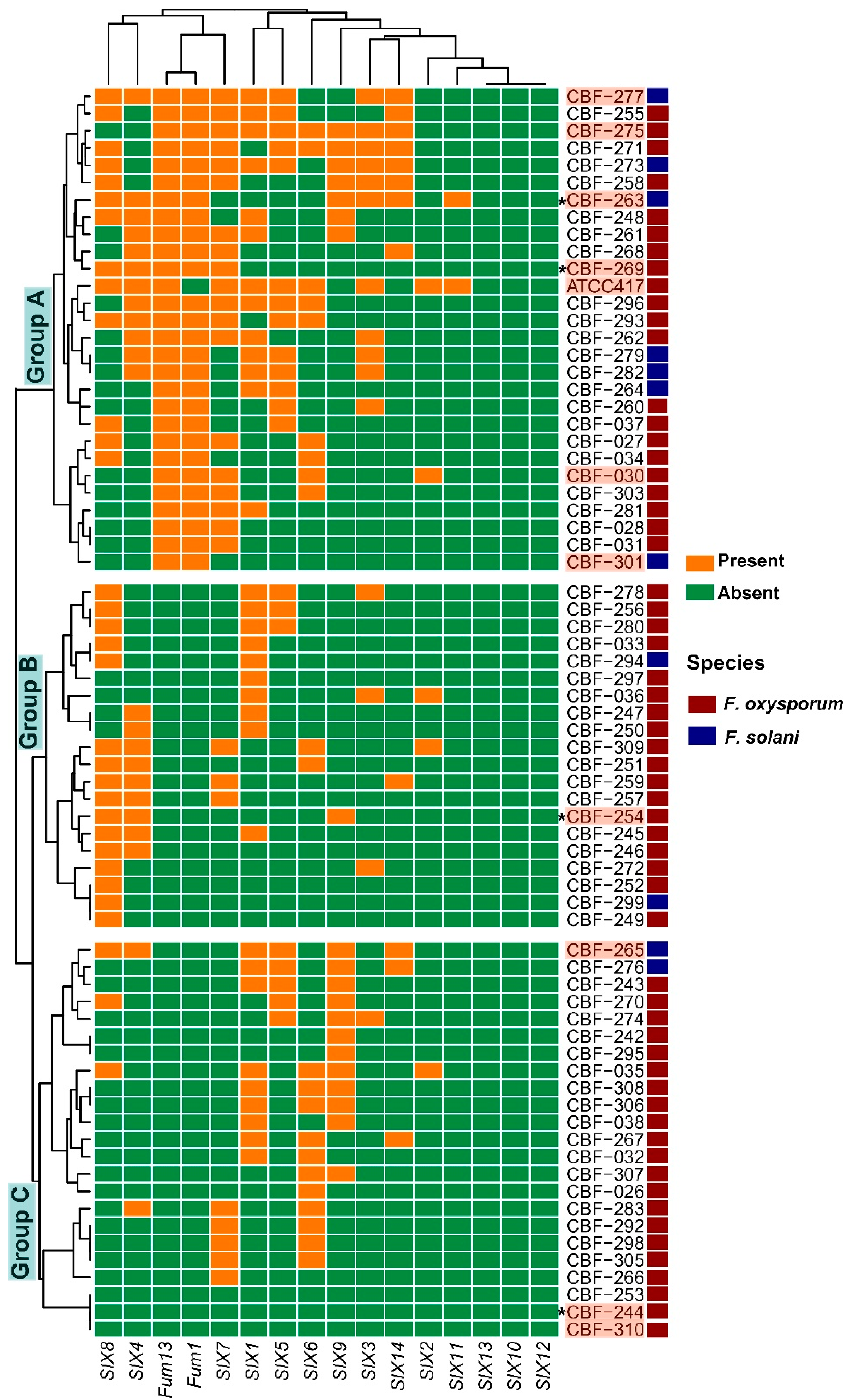
Clustering analysis of isolates based in SIX-Fum genes profiles (presence/absence) evidenced the formation of three big groups (Figure 2). All isolates belonging to group A, and only these ones, contained Fum1 and Fum13 genes. In group A, those isolates with the highest number of SIX genes were also found: CBF-263, CBF-271, CBF-273, CBF-275, and CBF-277 isolates. Additionally, seven of the eleven F. solani isolates (CBF-263, CBF-264, CBF- 273, CBF-277, CBF-279, and CBF-282) were clustered in group A. Group A included the ATCC417 strain. In addition to the absence of toxigenicity Fum genes, group B was defined by the absence of SIX11 gene and low frequencies of presence of SIX6, SIX7, SIX9, and SIX14 compared to isolates clustered in group A. The group C shared with the group B the absence of Fum and SIX11 genes, it was characterized by low frequencies of presence of SIX8 and SIX14. No correlation was found between the SIX genes repertoire and the relationships of the Fusarium isolates observed across the phylogenetic tree (Figure 1 and Figure 2).
We performed a three-dimensional non-metric multidimensional scaling (3D-NMDS) analysis based on the binary data (presence/absence of the SIX genes) by using Jaccard dissimilarity index to visualize the grouping of the Fusarium isolates. The stress value obtained for the overall dataset was 0.10, the 3D-NMDS plots were divided in three figures to avoid the overlapping of the multiple variables related to municipality and locality (Figure S1a–c). PERMANOVA analysis revealed that the SIX gene profiles of the Fusarium isolates were influenced by the species, i.e., F. oxysporum vs. F. solani (F = 3.58, R2 = 0.05, p < 0.002) as illustrated in the 3D-NMDS plot (Figure S1a). The SIX-Fum genes profiles of Fusarium isolates were also affected by the geographical origin factors, municipality (F = 2.29, R2 = 0.23, p < 0.001) and locality (F = 1.84, R2 = 0.27, p < 0.001), although the clustering of samples was not so clear (Figure S1b–c). Our results suggest that the species and geographical origin influence the SIX-Fum genes profiles of Fusarium isolates associated to coffee corky-root disease.

3.4. Relationship between Global Disease Damages and the SIX and Fum Gene Repertoires of Fusarium Isolates

Based on screening and clustering analysis of pathogenicity and toxigenicity genes, we selected 10 isolates of F. solani and F. oxysporum to test their pathogenicity in planta: (a) CBF-244 (isolated from healthy plants) and CBF-310, in which no SIX and no Fum genes were detected; (b) CBF-301 isolate, which contained Fum1 and Fum13 genes, but did not contain SIX genes; (c) CBF-254 and CBF-265 isolates which did not contain Fum genes but contained three SIX (SIX4, SIX8, and SIX9) and six SIX genes (SIX1, SIX4, SIX5, SIX8, SIX9, and SIX14), respectively; (d) CBF-269 (isolated from heathy coffee plants) containing three SIX genes (SIX4, SIX7, and SIX8) and the two Fum genes; (e) CBF-263, CBF-275, and CBF-277 which contained the two Fum genes and they are among the isolates with the largest number of SIX genes; (f) CBF-030 which was the only one in which was detected the combination of SIX2, SIX6, and SIX7 genes in addition to containing Fum1 and Fum13 toxigenicity genes; and (g) ATCC 417 (Fusarium oxysporum f. sp. lycopersici) was included to determine if despite its host specificity, it was able to elicit disease symptoms in coffee seedlings, considering its well-known pathogenicity level in tomato and its repertoire of SIX genes. In this study, the presence of SIX1SIX7 was confirmed for ATCC 417 and additionally, we detected amplification for SIX8 (being the only isolate containing this SIX gene), SIX11, and Fum13 genes (Table 1, Figure 2).
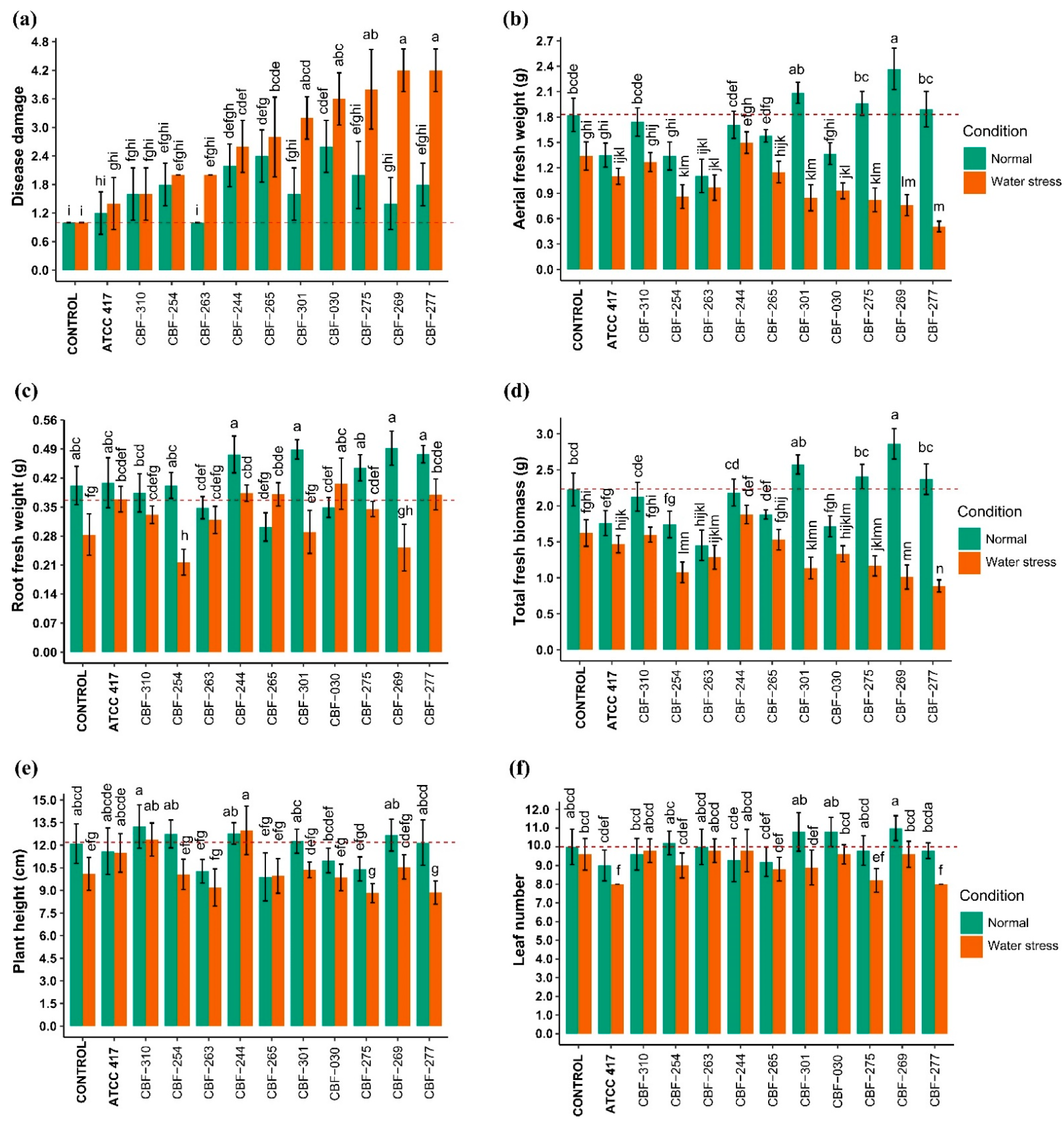
Our results indicated that the isolates CBF-310, CBF-254, CBF-263, and the strain ATCC 417 (Fusarium oxysporum f. sp. lycopersici) did not cause significant level of disease compared to the control, with or without water stress (Figure 3a). The analysis of variance with Tukey’s post hoc test showed that without water stress, three isolates, CBF-244, CBF-265, and CBF-030 caused higher disease damages compared to the control (p < 0.01). These three isolates also caused higher disease damages under water stress compared to the control although not significantly higher than under no-water-stress conditions (p < 0.01). The results obtained for the isolate CBF-244 were surprising because it was characterized by the absence of both SIX and Fum genes, whereas in the isolate CBF-265, Fum1 and Fum13 genes were absent, but it contained SIX1, SIX4, SIX5, SIX8, SIX9, and SIX14. Finally, though they did not cause significantly higher disease damages compared to the control in absence of water stress, four isolates caused significantly higher damages of disease under water stress: CBF-301 and especially CBF-275, CBF-269, and CBF-277, which showed the highest disease damage scores (4.2) (Figure 3a). Interestingly, all these Fusarium isolates were clustered in Group A of the SIX and Fum gene presence/absence heatmap (Figure 2) containing all and only the isolates carrying the Fum1 and Fum13 genes. Four of the isolates producing the most severe symptoms of disease in coffee seedlings, CBF-030, CBF-269, CBF-275, and CBF-277, were the only ones containing the SIX7 gene.

3.5. Effects of Fusarium Isolates Inoculation on Coffee Seedling Growth

Aerial (AFW), root (RFW), and total fresh weights (TFW), plant height (PH), and leaf number (LN) of coffee seedlings were measured in response to inoculation of selected Fusarium isolates under and without water-stress conditions.
We found that the isolates CF-244 and CBF-310 did not cause significant changes in any of the measured plant growth variables, with or without water stress (Figure 3b–f). Our results showed that the strain ATCC 417 and the isolates CBF-254, CBF-263, and CBF-030 decreased the AFW under no-water-stress conditions. The last three isolates also diminished AFW under water stress, but for CBF-254 and CBF-030, the AFW was significantly lower compared to the no-water-stress conditions (p < 0.01). The isolates CBF-301, CBF-275, CBF-269, and CBF-277 were not able to decrease the AFW under non-water-stress conditions, but under stress conditions they caused a reduction of up to 72% in AFW values as was the case of the isolate CBF-277 (Figure 3b). The RFW was only reduced by the isolate CBF-254 under stress conditions (Figure 3c). The isolates CBF-263 and CBF-030 were the only ones that declined the TFB values under no-water-stress conditions compared to the control, but no effect was observed for these isolates under water stress condition. On the contrary, although the isolates CBF-254, CBF-301, CBF-275, CBF-269, and CBF-277 did not cause significantly lower TFB values in absence of water stress, these isolates negatively affected the TFB under water stress (Figure 3d). The isolate CBF-265 was the only one that decreased the height of coffee seedlings under no-water-stress conditions (Figure 3e). Finally, we found that leaf number of coffee seedlings was only reduced under stress conditions by the strain ATCC 417 and the isolates CBF-275 and CB-277 (Figure 3f).

3.6. Effects of Fusarium Isolates Inoculation on Photosynthetic Activity of Coffee Seedlings

The ANOVA analysis with Tukey’s post hoc test demonstrated that the isolates CBF-254, CBF-263, and CBF-244 did not influence the variables PIabs, PItotal, and Fv/Fm either in no-water-stress or under stress conditions. Whereas on the contrary, a drastic decline (p < 0.01) of those three fluorescence parameters was caused by the isolates CBF-301, CBF-030, CBF-269, and CBF-277 under water stress conditions (Figure 4a–c).
The energy fluxes in the energy cascade in PSII for the events absorption (ABS), trapping (TR0), electron eransport (ET0), and dissipation (DI0), under water stress, were also affected by these Fusarium isolates, displaying a high level of energy absorption (ABS/RC), a lower level of trapping (TR0/RC) and electron transport (ET0/RC) combined with a high level of energy dissipation (DI0/RC) (data not shown). The isolates CBF-269 and CBF-277 also decreased the PIabs values in absence of water stress but those values were significantly lower under water stress conditions (Figure 4b). These results were in accordance with those observed for the global disease damages and the plant growth variables, since all those isolates (CBF-301, CBF-030, CBF-269, and CBF-277) were found to be among the most pathogenic isolates.
In accordance with the PIabs and PItotal results, the extreme decrease of Fv/Fm observed on coffee seedling inoculated with CBF-301 and CBF-277 and under water stress attest to irreversible physiological damage on these plants. A principal component analysis (PCA) was performed by employing the data of measurements of all photosynthetic variables. The first two principal components PC1 and PC2 explained 92.8% of the total variance (Table 2). The PC1 accounted for 80% of the variation and was correlated (r > 0.9) with most of the photosynthetic parameters except for Tr0/RC (r = −0.31). The clustering of Fusarium isolates was dependent of the photosynthetic status, which showed a clear discrimination of the five most pathogenic isolates: CBF-030, CBF-275, CBF-269, CBF-277, and CBF-301 (Figure 5).

3.7. Correlation between Disease Damage and the Phenotypic Variables Assessed in Coffee Seedlings

To investigate the relationship between disease damage and each of the phenotypic variables, a correlation analysis was conducted using Spearman’s rank correlation, with a significance at p < 0.01. Associations with Spearman correlation coefficient (SCC) values of 0.4–0.59 were considered as “moderate” correlations, 0.60–0.79 as “strong” correlations and values greater than 0.8 corresponded to “very strong” correlations. Photosynthetic activity indicators PIabs, PItotal, and Fv/Fm were the variables which showed the strongest correlation with the damge of the disease (Figure S2). Contrary to the plant growth variables, leaf number, plant height, and aerial fresh weight, for which negative correlations were observed (SCC: −0.41, −0.51 and −0.6, respectively) in relation to the disease damage.
The photosynthetic parameters were strong to very strong correlated with each other (SCC: 0.73 to 0.89). PIabs, PItotal, and Fv/Fm were positively correlated with AFW (SCC: 0.42, 0.52, and 0.49, respectively), whereas LN vs. Fv/Fm and PH vs. PItotal showed moderately positive correlations. Likewise, positive correlations were observed between all growth variables AFW, LN, PH, and RFW (Figure S2).

3.8. Vascular Colonization of Fusarium Isolates in Aerial and Root Tissues of Coffee Seedlings

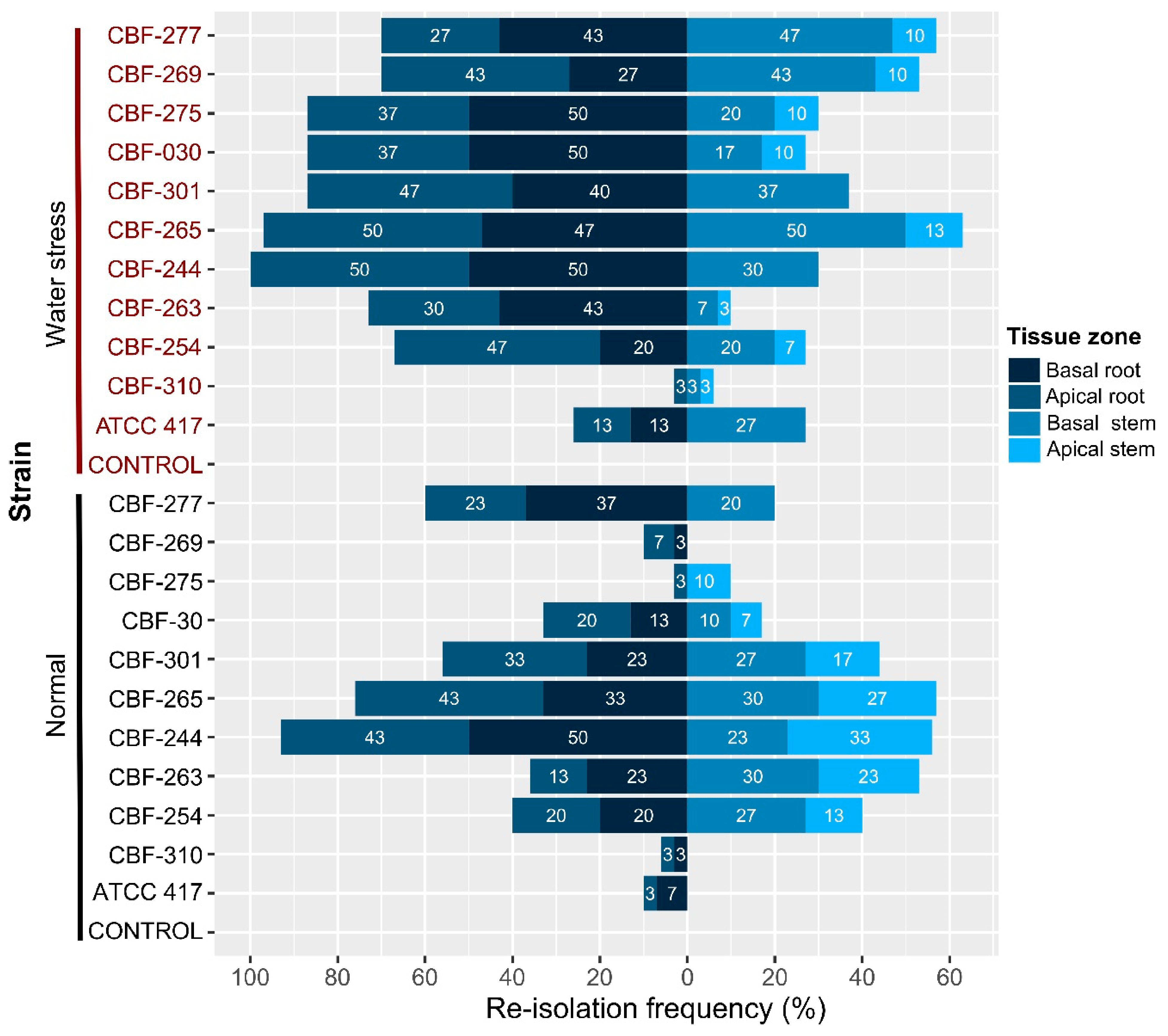
Most of the isolates inoculated in pathogenicity tests were re-isolated in percentages lower than 40% in root tissues under normal conditions (Figure 6). In general, the colonization by the Fusarium isolates was increased under water-stress conditions in both basal and apical zones of roots.
Interestingly, the isolates CBF-265 and CBF-244, which induced symptoms of disease in coffee seedlings without water stress, colonized in higher percentages (76 and 93%, respectively) the root tissue in absence of water stress. The CBF-030 isolate, which also caused disease symptoms under absence of water stress, colonized the root tissue in 33% but the percentages increased up to 87% under water stress condition.
Low colonization capacity of isolate CBF-310 was in accordance with the fact that it did not produce symptoms of disease either in normal or under water-stress conditions like the ATCC-417 strain. The isolates CBF-275, CBF-269, and CBF-277 characterized by producing the strongest negative effects on photosynthetic activity and growth of coffee seedlings under water stress, were also able to colonize both roots and stem tissues in high percentages under water-stress conditions (Figure 6). In general, our results indicated that: the percentages of colonization in root were higher compared to stem tissue; for some isolates, the colonization percentages were higher under water stress compared to normal conditions; and there was a global relationship between the observed disease damage and the colonization capacity of Fusarium isolates.

4. Discussion

4.1. Diversity of Fusarium Linked to Coffee Corky-Root Disease

Fusarium spp. have been reported as linked to coffee corky-root disease in association with the root-knot nematodes Meloydogine arabicida and M. paranaensis [1,7,8,33]. Among the diversity found in this studied Fusarium complex, 59 isolates belonged to F. oxysporum while only 11 isolates belonged to F. solani. These results are in accordance with the observations of López-Lima et al. (2020) [4] who found that F. oxysporum was the dominant fungal species in coffee corky-root samples. Molecular markers flanking the internal transcriber spacer (ITS) have been used to determine the identity of Fusarium isolates linked to coffee corky-root disease [4]. In this study, phylogenetic analysis using concatenated sequences of three different markers EF-1α, RPB2, and TUB2 allowed the identification of Fusarium isolates with high similarity against different formae speciales of F. oxysporum which corresponded to f. sp. vasinfectum, f. sp. dianthi, f. sp. phaseoli, and f. sp. cepae. Nevertheless, interestingly 45 of the 59 Fusarium isolates were grouped in a well-supported branch separated of the other formae speciales and F. oxysporum sequences included as references. Further research should focus on clarifying how the diversity of Fusarium isolates interact for the development of corky-root disease.

4.2. Isolates of F. oxysporum and F. solani Causing Disease Symptoms in Coffee Seedlings under Water Stress

In this study, we demonstrated that some F. oxysporum and F. solani isolates from coffee seedlings produced vascular wilting, chlorosis, tissue necrosis, and ultimately plant death, but without producing corky-root symptoms in absence of nematodes, and especially under water stress. Bertrand et al. (2000) [1] demonstrated that corky-root symptoms are exhibited only under the combined infection by the nematode M. arabicida and a Fusarium oxysporum isolate. The lack of pathogenicity of the studied Fusarium isolates in absence of nematodes or of an abiotic stress indicates that resistance to only M. paranaensis seems to be a good strategy for controlling the coffee corky-root disease. This has been confirmed by the observations in fields infected by M. paranaensis when planting susceptible Arabica cultivars grafted on C. canephora cv. Nemaya rootstock resistant to M. paranaensis [2].
It was found that two F. solani isolates were among the five isolates producing the strongest disease symptoms under water stress, which is a novel finding in the etiology of the coffee corky-root disease. There is only two reports from Kenya and Yemen in which F. solani was determined as a causal agent of a wilting disease but not of a corky-root disease in coffee [34,35].
Our results indicated that under water stress, some Fusarium isolates became very pathogenic on coffee seedlings, resulting in severe symptoms with wilting and a severely affected photosynthetic activity which results in a strongly affected plant growth. It was also observed that plant colonization by these Fusarium isolates increased under water stress. The underlying mechanisms leading to such severe physiological disruption of coffee plants under the combined action of some Fusarium and water stress have yet to be explained. However, we can hypothesize that pathogenic Fusarium highly reduce water flow in the plant by blocking the xylem vascular system, which has been demonstrated for many crops [36,37,38,39,40]. This work highlights that climate change that is increasing the frequency and intensity of drought periods in coffee crop [41], could lead to a greater impact of some Fusarium on coffee even in the absence of RKN as observed in Kenya and Yemen [34,35].
The synergy between abiotic and biotic stress-crop interactions has showed to induce changes in host physiology leading to changes on vegetative variables, deficit in nutritional status, increased susceptibility, as well as dysregulation in expression of photosynthetic genes [42,43,44,45,46]. Under abiotic and biotic stress, ROS are produced affecting photosynthetic electron transport, impairing the assembly and repair of PSII and affecting chloroplast development [47,48]. Depression of photosystems II and I performance has been observed either in the case of just wilting fungi infections [49,50,51].
In this study, the decreased efficiency of energy trapping in PSII reaction centers (Fv/Fm) observed for those isolates, decreased values of performance indices (PIs) combined with high level of photon flux absorption, low level of electron transport, and high level of energy dissipation indicated very low photosynthetic yield, probably irreversible photosystem damage and significant oxidative stress.
Chlorophyll a fluorescence and especially performance indices are used to estimate photosynthetic stress level, particularly in the case of abiotic stress and more rarely to evaluate biotic stress or a combination of both [52]. Toniutti et al. (2017) [53] demonstrated that parameters related to Photosystem II and photosynthetic electron transport chain components are powerful indicators of the physiological status of the coffee plants and predict infection intensity of Hemileia vastatrix in combination with abiotic stress.
In our case study, the use of quantitative data of the photosynthetic parameters was able to well discriminate between the non-pathogenic and the pathogenic Fusarium isolates, and especially the parameters Fv/Fm and PIabs,total proved to be the best indicators of the disease damage. Here, we demonstrated that chlorophyll a fluorescence could be used as a fast and precise assessment of the level of damage comparatively to growth evaluation.

4.3. The Repertoire of SIX-Fum Genes Is Associated with Pathogenicity of Fusarium Isolates

At present, there are no reports about the characterization of presence of the SIX genes and the Fum genes in Fusarium isolates of coffee plants. A significant characteristic of all Fusarium isolates from coffee seedlings, either healthy or with corky-root symptoms, was the absence of SIX10, SIX12, and SIX13 genes. Several formae speciales of Fusarium oxysporum including f. sp. lycopersici, f. sp. canariensis, f. sp. lini, f. sp. cepae, f. sp. pisi, f. sp. freesia, f. sp. dianthi, f. sp. cubense, and f. sp. narcissi have showed to contain SIX10, SIX12, and SIX13 genes [12,17,22,54,55].
Even though, we found that the SIX-Fum genes profiles of Fusarium isolates were influenced by the geographical origin factor, the clustering of samples was not so clear. That low correlation is not very surprising when we know that nursery coffee seedlings circulate widely between localities and even between regions. We demonstrated that the SIX-Fum gene profiles are influenced by species (F. oxysporum vs. F. solani). The main differences between these two species were the absence of SIX2 and SIX6 in F. solani compared to F. oxysporum, whereas SIX11 was present in at least one isolate of F. solani species but in F. oxysporum was not detected. The rest of the SIX genes except for SIX7 and SIX8 were overrepresented in the F. solani species in comparison with the percentages found in F. oxysporum isolates. It is well known that some Fusarium species different to the F. oxysporum species complex (FoSC) possess differential SIX gene profiles, this fact may be explained by the location of pathogenicity and toxin genes in mobile chromosomes which can be horizontally transferred, as it is the case of SIX and Fum genes [56,57]. These accessory chromosomes are involved in causing diseases by conferring advantages in specific environments, and virulence and pathogenic capacities on specific plant species [58].
We demonstrated an association between plant disease damages caused by the Fusarium isolates and their repertoire of SIX genes, as well as the presence of Fum 1 and Fum 13 genes. Fum1 gene encodes the crucial iterative polyketide synthase (PKS) which starts the synthesis of the backbone of the mycotoxin fumonisin, whereas a reductase is encoded by Fum13 gene [59]. Production of fumonisin has been reported for F. oxysporum, F. graminearum, F. culmorum, F. equiseti, F. semitectum, F. fujikuroi, F. poae, F. subglutinans, F. verticillioides, F. proliferatum, and F. solani [60,61,62,63]. In this study, we identified 20 F. oxysporum isolates and seven F. solani isolates as potential producers of fumonisin.
The isolate CBF-244 caused disease symptoms in coffee seedlings even though it contained no SIX and Fum genes, which means that its pathogenicity mechanisms do not involve the participation of these genes. Another interesting finding in this study was the presence of the SIX7 gene in the isolates CBF-030, CBF-275, CBF-269, and CBF-277 that were of the most pathogenic. The presence of SIX7 has been demonstrated in Fusarium oxysporum isolates from Welsh onion seedlings, tomato, banana, and sesame [17,20,64,65]. It is possible that SIX7 gene has an outstanding role in the pathogenicity showed in this study by Fusarium isolates associated with coffee corky-root disease.
In this study, we detected F. solani isolates containing at least one SIX gene. Recently, SIX genes have been identified in Fusarium species outside the FoSC including F. proliferatum, F. hostae, F. agapanthi [66], F. sacchari, and F. verticillioides [55]. However, until now, F. solani had not been reported as a species containing effector SIX genes. The F. solani isolates CBF-265 and CBF-277 which contained SIX genes were pathogenic for coffee seedlings. This fact suggests that the presence of SIX genes in F. solani isolates could be related to their pathogenic capacities. Our results support the idea that the potential of colonization and development of pathogenicity of the Fusarium complex in coffee seedlings might be tightly related to the profile of SIX genes. Nevertheless, the existence of different mechanisms related to pathogenicity should not be ruled out for these isolates, since even though the F. solani isolate CBF-301 does not contain SIX genes but does contain Fum genes, it was pathogenic for coffee seedlings.

4.4. Host-Specificity of Fusarium oxysporum f. sp. lycopersici Results in Low Vascular Colonization without Causing Disease Symptoms in Coffee Seedlings

The formae speciales of Fusarium oxysporum are defined based on host specificity, and the subcategorization into races is dependent of their pathogenicity to a specific group of the host cultivars [67]. The ATCC 417, a Fol strain with high degree of virulence on tomato [68], was included in this study to determine if despite its host specificity, it was able to cause symptoms of disease in coffee seedlings. As we expected, the pathogenicity test revealed a low frequency of colonization by the ATCC 417 accompanied by its inability to cause symptoms of disease in coffee seedlings. Fourteen SIX genes have been identified in Fol, SIX1, SIX3, and SIX5 directly contribute to virulence of Fol [38,54], whereas SIX1, SIX3, and SIX4, and interactions as SIX3SIX5 are recognized by the immune system and activate plant defense and resistance [69]. A specific SIX genes repertoire is associated with Fusarium isolates in the process of colonization and infection of a specific host [16,36]. In fact, host specificity of the phytopathogen Fusarium oxysporum f. sp. lycopersici is attributable to its lineage-specific mobile pathogenicity chromosomes in which are contained the secreted in xylem (SIX) genes [36].

5. Conclusions

Our investigation provides evidence: (i) about the association between the presence of SIX and Fum genes and the pathogenicity of Fusarium isolates linked to coffee corky-root disease. Future studies should be addressed to study the SIX genes transference intra- and inter-species. (ii) That some pathogenicity SIX genes and toxigenicity Fum genes seem to play a primordial role in the basic determinism of pathogenicity potential of Fusarium isolates on coffee, although in a complex and varied way for each of them depending on the isolate and independently of the species considered (F. oxysporum vs. F. solani). Transcriptomic studies quantifying the expression of these highlighted genes should allow to better understand this molecular basis of pathogenicity determinism in relation with the pathobiome environment. (iii) That Fusarium solani carries SIX and Fum genes, and because of its pathogenic abilities on coffee, appears as a potential pathogen contributing to the development of coffee corky-root disease. (iv) For the first time, the change in behavior and pathogenicity of F. oxysporum and F. solani isolates on coffee caused by water stress are demonstrated, which can be considered as mimicking the effect of root-knot-nematodes involved in corky-root disease. Efforts should focus in determining the function of the Fusarium SIX genes in coffee plant parasitism and its relationship with host specificity, as well as the mechanisms of other biotic and abiotic stresses activating the switch from hemi-biotrophic to necrotrophic phases in the pathogenic Fusarium species. Finally, crossed inoculations of the root-knot nematodes involved in the coffee corky-root disease, such as M. paranaensis or M. arabicida, with F. oxysporum and F. solani should be carried out to confirm the role of each of these causal agents in these pathogen complexes, and to better understand the interactions between them and with abiotic stresses in the etiology of this severe disease. (v) This work highlights the relevance of chlorophyll a fluorescence as an early and high-throughput phenotyping tool for plant pathogenicity studies on Fusarium.

Supplementary Materials

The following figures are available online at https://www.mdpi.com/article/10.3390/jof7040253/s1, Figure S1: Three dimensional non-metric multidimensional scaling (3D-NMDS) ordination of the Fusarium isolates associated to coffee corky-root disease, Figure S2: Spearman correlation plot between disease damage and physiological parameters of coffee seedlings.

Author Contributions

Conceptualization, R.G.-B., L.V., G.C. and D.D.; Data curation, J.-C.B.; Formal analysis, R.G.-B.; Funding acquisition, G.C. and D.D.; Methodology, R.G.-B., D.L.-L., L.V., J.-C.B. and D.D.; Writing—original draft, R.G.-B., L.V., J.-C.B. and D.D.; Writing—review & editing, R.G.-B., D.L.-L., L.V., J.-C.B., G.C. and D.D. All authors have read and agreed to the published version of the manuscript.

Funding

This research was funded by Instituto de Ecología A.C. (INECOL).

Institutional Review Board Statement

Not applicable.

Informed Consent Statement

Not applicable.

Acknowledgments

This study was financed by Instituto de Ecología A.C. (INECOL). We thank Magda Gómez Columna for her support in caring the coffee seedlings during the pathogenicity test, Diana López Ley and Nut Liahut Guin for their technical support in molecular analysis including fungal DNA isolation, PCR amplifications and purification.

Conflicts of Interest

The authors declare no conflict of interest.

References

  1. Bertrand, B.; Nunez, C.; Sarah, J.-L. Disease Complex in Coffee Involving Meloidogyne arabicida and Fusarium oxysporum. Plant Pathol. 2000, 49, 383–388. [Google Scholar] [CrossRef]
  2. Villain, L.; Lima-Salgada, S.M.; Trinh-Phap, Q. Plant Parasitic Nematodes in Subtropical and Tropical Agriculture. In Nematode Parasites of Coffee and Cocoa; Sikora, R.A., Coyne, D., Hallman, J., Timper, P., Eds.; CABI: Wallingford, UK, 2018; pp. 536–583. [Google Scholar]
  3. Lamelas, A.; Desgarennes, D.; López-Lima, D.; Villain, L.; Alonso-Sánchez, A.; Artacho, A.; Latorre, A.; Moya, A.; Carrión, G. The Bacterial Microbiome of Meloidogyne-Based Disease Complex in Coffee and Tomato. Front. Plant Sci. 2020, 11. [Google Scholar] [CrossRef]
  4. López-Lima, D.; Carrión, G.; Sánchez-Nava, P.; Desgarennes, D.; Villain, L. Fungal Diversity and Fusarium oxysporum Pathogenicity Associated with Coffee Corky-Root Disease in Mexico. Rev. Fac. Cienc. Agrar. UNCuyo 2020, 52, 276–292. [Google Scholar]
  5. Tian, B.-Y.; Cao, Y.; Zhang, K.-Q. Metagenomic Insights into Communities, Functions of Endophytes, and Their Associates with Infection by Root-Knot Nematode, Meloidogyne Incognita, in Tomato Roots. Sci. Rep. 2015, 5, 17087. [Google Scholar] [CrossRef] [Green Version]
  6. Carneiro, R.M.D.G.; Cofcewicz, E.T. Taxonomy of Coffee-Parasitic Root-Knot Nematodes, Meloidogyne spp. In Plant-Parasitic Nematodes of Coffee; Souza, R.M., Ed.; Springer: Dordrecht, The Netherlands, 2008; pp. 87–122. ISBN 978-1-4020-8720-2. [Google Scholar]
  7. Villain, L.; Sarah, J.L.; Hernández, A.; Bertrand, B.; Anthony, F.; Lashermes, P.; Charmetant, P.; Anzueto, F.; Figueroa, P.; Carneiro, R.M.D.G. Diversity of Root-Knot Nematodes Parasitizing Coffee in Central America. Nematropica 2013, 43, 194–206. [Google Scholar]
  8. Lopez-Lima, D.; Sánchez-Nava, P.; Carrion, G.; de los Monteros, A.E.; Villain, L. Corky-Root Symptoms for Coffee in Central Veracruz Are Linked to the Root-Knot Nematode Meloidogyne paranaensis, a New Report for Mexico. Eur. J. Plant Pathol. 2015, 141, 623–629. [Google Scholar] [CrossRef]
  9. Hua, G.K.H.; Timper, P.; Ji, P. Meloidogyne incognita Intensifies the Severity of Fusarium Wilt on Watermelon Caused by Fusarium oxysporum f. sp. niveum. Can. J. Plant Pathol. 2019, 41, 261–269. [Google Scholar] [CrossRef]
  10. Chowdhury, S.; Basu, A.; Kundu, S. Biotrophy-Necrotrophy Switch in Pathogen Evoke Differential Response in Resistant and Susceptible Sesame Involving Multiple Signaling Pathways at Different Phases. Sci. Rep. 2017, 7, 17251. [Google Scholar] [CrossRef] [PubMed] [Green Version]
  11. Rampersad, S.N. Pathogenomics and Management of Fusarium Diseases in Plants. Pathogens 2020, 9, 340. [Google Scholar] [CrossRef]
  12. Taylor, A.; Vágány, V.; Jackson, A.C.; Harrison, R.J.; Rainoni, A.; Clarkson, J.P. Identification of Pathogenicity-Related Genes in Fusarium oxysporum f. sp. cepae: Pathogenicity in Fusarium oxysporum f. sp. cepae. Mol. Plant Pathol. 2016, 17, 1032–1047. [Google Scholar] [CrossRef] [Green Version]
  13. Guo, L.; Han, L.; Yang, L.; Zeng, H.; Fan, D.; Zhu, Y.; Feng, Y.; Wang, G.; Peng, C.; Jiang, X.; et al. Genome and Transcriptome Analysis of the Fungal Pathogen Fusarium oxysporum f. sp. cubense Causing Banana Vascular Wilt Disease. PLoS ONE 2014, 9, e95543. [Google Scholar] [CrossRef]
  14. Czislowski, E.; Fraser-Smith, S.; Zander, M.; O’Neill, W.T.; Meldrum, R.A.; Tran-Nguyen, L.T.T.; Batley, J.; Aitken, E.A.B. Investigation of the Diversity of Effector Genes in the Banana Pathogen, Fusarium oxysporum f. sp. cubense, Reveals Evidence of Horizontal Gene Transfer: Effector Genes in Fusarium oxysporum f. sp. cubense. Mol. Plant Pathol. 2018, 19, 1155–1171. [Google Scholar] [CrossRef] [Green Version]
  15. Ma, L.; Houterman, P.M.; Gawehns, F.; Cao, L.; Sillo, F.; Richter, H.; Clavijo-Ortiz, M.J.; Schmidt, S.M.; Boeren, S.; Vervoort, J.; et al. The AVR2-SIX5 Gene Pair Is Required to Activate I-2 -Mediated Immunity in Tomato. New Phytol. 2015, 208, 507–518. [Google Scholar] [CrossRef]
  16. Li, J.; Fokkens, L.; Conneely, L.J.; Rep, M. Partial Pathogenicity Chromosomes in Fusarium oxysporum Are Sufficient to Cause Disease and Can Be Horizontally Transferred. Environ. Microbiol. 2020, 22, 4985–5004. [Google Scholar] [CrossRef] [PubMed]
  17. Lievens, B.; Houterman, P.M.; Rep, M. Effector Gene Screening Allows Unambiguous Identification of Fusarium oxysporum f. sp. lycopersici Races and Discrimination from other formae speciales. FEMS Microbiol. Lett. 2009, 300, 201–215. [Google Scholar] [CrossRef] [Green Version]
  18. Chakrabarti, A.; Rep, M.; Wang, B.; Ashton, A.; Dodds, P.; Ellis, J. Variation in Potential Effector Genes Distinguishing Australian and Non-Australian Isolates of the Cotton Wilt Pathogen Fusarium oxysporum f.sp. vasinfectum. Plant Pathol. 2011, 60, 232–243. [Google Scholar] [CrossRef]
  19. Thatcher, L.F.; Gardiner, D.M.; Kazan, K.; Manners, J.M. A Highly Conserved Effector in Fusarium oxysporum is Required for Full Virulence on Arabidopsis. Mol. Plant-Microbe Interact. 2011, 25, 180–190. [Google Scholar] [CrossRef] [Green Version]
  20. Meldrum, R.A.; Fraser-Smith, S.; Tran-Nguyen, L.T.T.; Daly, A.M.; Aitken, E.A.B. Presence of Putative Pathogenicity Genes in Isolates of Fusarium oxysporum f. sp. cubense from Australia. Australas. Plant Pathol. 2012, 41, 551–557. [Google Scholar] [CrossRef]
  21. Covey, P.A.; Kuwitzky, B.; Hanson, M.; Webb, K.M. Multilocus Analysis Using Putative Fungal Effectors to Describe a Population of Fusarium oxysporum from Sugar Beet. Phytopathology 2014, 104, 886–896. [Google Scholar] [CrossRef] [PubMed] [Green Version]
  22. Laurence, M.H.; Summerell, B.A.; Liew, E.C.Y. Fusarium oxysporum f. sp. canariensis: Evidence for Horizontal Gene Transfer of Putative Pathogenicity Genes. Plant Pathol. 2015, 64, 1068–1075. [Google Scholar] [CrossRef]
  23. Williams, A.H.; Sharma, M.; Thatcher, L.F.; Azam, S.; Hane, J.K.; Sperschneider, J.; Kidd, B.N.; Anderson, J.P.; Ghosh, R.; Garg, G.; et al. Comparative Genomics and Prediction of Conditionally Dispensable Sequences in Legume–Infecting Fusarium oxysporum formae speciales Facilitates Identification of Candidate Effectors. BMC Genom. 2016, 17, 191. [Google Scholar] [CrossRef] [Green Version]
  24. Nicholson, T.P.; Rudd, B.A.; Dawson, M.; Lazarus, C.M.; Simpson, T.J.; Cox, R.J. Design and Utility of Oligonucleotide Gene Probes for Fungal Polyketide Synthases. Chem. Biol. 2001, 8, 157–178. [Google Scholar] [CrossRef] [Green Version]
  25. O’Donnell, K.; Cigelnik, E. Two Divergent Intragenomic RDNA ITS2 Types within a Monophyletic Lineage of the Fungus Fusarium Are Nonorthologous. Mol. Phylogenet. Evol. 1997, 7, 103–116. [Google Scholar] [CrossRef] [PubMed]
  26. O’Donnell, K.; Sarver, B.A.J.; Brandt, M.; Chang, D.C.; Noble-Wang, J.; Park, B.J.; Sutton, D.A.; Benjamin, L.; Lindsley, M.; Padhye, A.; et al. Phylogenetic Diversity and Microsphere Array-Based Genotyping of Human Pathogenic Fusaria, Including Isolates from the Multistate Contact Lens-Associated U.S. Keratitis Outbreaks of 2005 and 2006. J. Clin. Microbiol. 2007, 45, 2235–2248. [Google Scholar] [CrossRef] [PubMed] [Green Version]
  27. Kumar, S.; Stecher, G.; Tamura, K. MEGA7: Molecular Evolutionary Genetics Analysis Version 7.0 for Bigger Datasets. Mol. Biol. Evol. 2016, 33, 1870–1874. [Google Scholar] [CrossRef] [Green Version]
  28. Ramana, M.V.; Balakrishna, K.; Murali, H.C.S.; Batra, H.V. Multiplex PCR-Based Strategy to Detect Contamination with Mycotoxigenic Fusarium Species in Rice and Fingermillet Collected from Southern India. J. Sci. Food Agric. 2011, 91, 1666–1673. [Google Scholar] [CrossRef]
  29. Ramana, M.V.; Nayaka, S.C.; Balakrishna, K.; Murali, H.S.; Batra, H.V. A Novel PCR–DNA Probe for the Detection of Fumonisin-Producing Fusarium Species from Major Food Crops Grown in Southern India. Mycology 2012, 3, 167–174. [Google Scholar] [CrossRef]
  30. Reis, A.; Boiteux, L.S. Outbreak of Fusarium oxysporum f. Sp. lycopersici Race 3 in Commercial Fresh-Market Tomato Fields in Rio de Janeiro State, Brazil. Hortic. Bras. 2007, 25, 451–454. [Google Scholar] [CrossRef] [Green Version]
  31. Strasser, B.J.; Strasser, R.J. Measuring Fast Fluorescence Transients to Address Environmental Questions: The JIP-Test. Photosynth. Light Biosphere 1995, 4869–4872. [Google Scholar] [CrossRef]
  32. Stirbet, A.; Lazár, D.; Kromdijk, J. Govindjee Chlorophyll a Fluorescence Induction: Can Just a One-Second Measurement Be Used to Quantify Abiotic Stress Responses? Photosynthetica 2018, 56, 86–104. [Google Scholar] [CrossRef]
  33. Bertrand, B.; Ramirez, G.; Topart, P.; Anthony, F. Resistance of Cultivated Coffee (Coffea arabica and C. canephora) Trees to Corky-Root Caused by Meloidogyne arabicida and Fusarium oxysporum, under Controlled and Field Conditions. Crop Prot. 2002, 21, 713–719. [Google Scholar] [CrossRef]
  34. Baker, C.J. Fusarium solani Associated with a Wilt of Coffea arabica in Kenya. East Afr. Agric. For. J. 1972, 38, 137–140. [Google Scholar] [CrossRef]
  35. Al-Areqi, A.H.N.A.; Chliyeh, M.; Touati, J.; Outcoumit, A.; Sghir, F.; Touhami, A.O.; Benkirane, R.; Douira, A. Effect of Endomycorrhizae on Decline of the Coffee Plants (Coffea arabica) Caused by Fusarium solani. IJAPBC 2015, 4, 397–404. [Google Scholar]
  36. Ma, L.-J.; van der Does, H.C.; Borkovich, K.A.; Coleman, J.J.; Daboussi, M.-J.; Di Pietro, A.; Dufresne, M.; Freitag, M.; Grabherr, M.; Henrissat, B.; et al. Comparative Genomics Reveals Mobile Pathogenicity Chromosomes in Fusarium. Nature 2010, 464, 367–373. [Google Scholar] [CrossRef] [PubMed]
  37. Yadeta, K.A.; Thomma, B.P.H.J. The Xylem as Battleground for Plant Hosts and Vascular Wilt Pathogens. Front. Plant Sci. 2013, 4, 97. [Google Scholar] [CrossRef] [Green Version]
  38. Houterman, P.M.; Ma, L.; van Ooijen, G.; de Vroomen, M.J.; Cornelissen, B.J.C.; Takken, F.L.W.; Rep, M. The Effector Protein Avr2 of the Xylem-Colonizing Fungus Fusarium oxysporum Activates the Tomato Resistance Protein I-2 Intracellularly. Plant J. Cell Mol. Biol. 2009, 58, 970–978. [Google Scholar] [CrossRef] [PubMed]
  39. Farahani-Kofoet, R.D.; Witzel, K.; Graefe, J.; Grosch, R.; Zrenner, R. Species-Specific Impact of Fusarium Infection on the Root and Shoot Characteristics of Asparagus. Pathogens 2020, 9, 509. [Google Scholar] [CrossRef] [PubMed]
  40. Warman, N.M.; Aitken, E.A.B. The Movement of Fusarium oxysporum f.sp. cubense (Sub-Tropical Race 4) in Susceptible Cultivars of Banana. Front. Plant Sci. 2018, 9, 1748. [Google Scholar] [CrossRef] [Green Version]
  41. Bertrand, R.; Riofrío-Dillon, G.; Lenoir, J.; Drapier, J.; de Ruffray, P.; Gégout, J.-C.; Loreau, M. Ecological Constraints Increase the Climatic Debt in Forests. Nat. Commun. 2016, 7, 12643. [Google Scholar] [CrossRef] [Green Version]
  42. Bilgin, D.D.; Zavala, J.A.; Zhu, J.; Clough, S.J.; Ort, D.R.; De Lucia, E.H. Biotic Stress Globally Downregulates Photosynthesis Genes. Plant Cell Environ. 2010, 33, 1597–1613. [Google Scholar] [CrossRef] [Green Version]
  43. Prasch, C.M.; Sonnewald, U. Simultaneous Application of Heat, Drought, and Virus to Arabidopsis Plants Reveals Significant Shifts in Signaling Networks. Plant Physiol. 2013, 162, 1849–1866. [Google Scholar] [CrossRef]
  44. Rojas, C.M.; Senthil-Kumar, M.; Tzin, V.; Mysore, K. Regulation of Primary Plant Metabolism during Plant-Pathogen Interactions and Its Contribution to Plant Defense. Front. Plant Sci. 2014, 5, 17. [Google Scholar] [CrossRef] [PubMed] [Green Version]
  45. Pandey, P.; Sinha, R.; Mysore, K.S.; Senthil-Kumar, M. Impact of Concurrent Drought Stress and Pathogen Infection on Plants. In Combined Stresses in Plants: Physiological, Molecular, and Biochemical Aspects; Mahalingam, R., Ed.; Springer International Publishing: Cham, Switzerland, 2015; pp. 203–222. ISBN 978-3-319-07899-1. [Google Scholar]
  46. Sinha, R.; Irulappan, V.; Mohan-Raju, B.; Suganthi, A.; Senthil-Kumar, M. Impact of Drought Stress on Simultaneously Occurring Pathogen Infection in Field-Grown Chickpea. Sci. Rep. 2019, 9. [Google Scholar] [CrossRef] [PubMed] [Green Version]
  47. De Torres Zabala, M.; Littlejohn, G.; Jayaraman, S.; Studholme, D.; Bailey, T.; Lawson, T.; Tillich, M.; Licht, D.; Bölter, B.; Delfino, L.; et al. Chloroplasts Play a Central Role in Plant Defence and Are Targeted by Pathogen Effectors. Nat. Plants 2015, 1, 15074. [Google Scholar] [CrossRef]
  48. Qi, J.; Song, C.-P.; Wang, B.; Zhou, J.; Kangasjärvi, J.; Zhu, J.-K.; Gong, Z. Reactive Oxygen Species Signaling and Stomatal Movement in Plant Responses to Drought Stress and Pathogen Attack. J. Integr. Plant Biol. 2018, 60, 805–826. [Google Scholar] [CrossRef] [Green Version]
  49. Pshibytko, N.L.; Zenevich, L.A.; Kabashnikova, L.F. Changes in the Photosynthetic Apparatus during Fusarium Wilt of Tomato. Russ. J. Plant Physiol. 2006, 53, 25–31. [Google Scholar] [CrossRef]
  50. Prokopová, J.; Spundová, M.; Sedlárová, M.; Husicková, A.; Novotný, R.; Dolezal, K.; Naus, J.; Lebeda, A. Photosynthetic Responses of Lettuce to Downy Mildew Infection and Cytokinin Treatment. Plant Physiol. Biochem. PPB 2010, 48, 716–723. [Google Scholar] [CrossRef] [PubMed]
  51. Yan, K.; Han, G.; Ren, C.; Zhao, S.; Wu, X.; Bian, T. Fusarium solani Infection Depressed Photosystem Performance by Inducing Foliage Wilting in Apple Seedlings. Front. Plant Sci. 2018, 9. [Google Scholar] [CrossRef] [Green Version]
  52. Jedmowski, C.; Bayramov, S.; Brüggemann, W. Comparative Analysis of Drought Stress Effects on Photosynthesis of Eurasian and North African Genotypes of Wild Barley. Photosynthetica 2014, 52, 564–573. [Google Scholar] [CrossRef]
  53. Toniutti, L.; Breitler, J.-C.; Etienne, H.; Campa, C.; Doulbeau, S.; Urban, L.; Lambot, C.; Pinilla, J.-C.H.; Bertrand, B. Influence of Environmental Conditions and Genetic Background of Arabica Coffee (C. arabica L) on Leaf Rust (Hemileia vastatrix) Pathogenesis. Front. Plant Sci. 2017, 8, 2025. [Google Scholar] [CrossRef] [PubMed] [Green Version]
  54. Rep, M.; van der Does, H.C.; Meijer, M.; van Wijk, R.; Houterman, P.M.; Dekker, H.L.; de Koster, C.G.; Cornelissen, B.J.C. A Small, Cysteine-Rich Protein Secreted by Fusarium oxysporum during Colonization of Xylem Vessels Is Required for I-3-Mediated Resistance in Tomato. Mol. Microbiol. 2004, 53, 1373–1383. [Google Scholar] [CrossRef]
  55. Maldonado-Bonilla, L.D.; Calderón-Oropeza, M.A.; Villarruel-Ordaz, J.L.; Sánchez-Espinosa, A.C. Identification of Novel Potential Causal Agents of Fusarium Wilt of Musa sp. AAB in Southern Mexico. J. Plant Pathol. Microbiol. 2019, 10. [Google Scholar] [CrossRef] [Green Version]
  56. Vlaardingerbroek, I.; Beerens, B.; Rose, L.; Fokkens, L.; Cornelissen, B.J.C.; Rep, M. Exchange of Core Chromosomes and Horizontal Transfer of Lineage-Specific Chromosomes in Fusarium oxysporum: Chromosome Transfer and Exchange in F. oxysporum. Environ. Microbiol. 2016, 18, 3702–3713. [Google Scholar] [CrossRef] [PubMed]
  57. Borah, N.; Albarouki, E.; Schirawski, J. Comparative Methods for Molecular Determination of Host-Specificity Factors in Plant-Pathogenic Fungi. Int. J. Mol. Sci. 2018, 19, 863. [Google Scholar] [CrossRef] [PubMed] [Green Version]
  58. Bertazzoni, S.; Williams, A.H.; Jones, D.A.; Syme, R.A.; Tan, K.-C.; Hane, J.K. Accessories Make the Outfit: Accessory Chromosomes and Other Dispensable DNA Regions in Plant-Pathogenic Fungi. Mol. Plant Microbe Interact. MPMI 2018, 31, 779–788. [Google Scholar] [CrossRef] [Green Version]
  59. Brown, D.W.; Cheung, F.; Proctor, R.H.; Butchko, R.A.E.; Zheng, L.; Lee, Y.; Utterback, T.; Smith, S.; Feldblyum, T.; Glenn, A.E.; et al. Comparative Analysis of 87,000 Expressed Sequence Tags from the Fumonisin-Producing Fungus Fusarium verticillioides. Fungal Genet. Biol. 2005, 42, 848–861. [Google Scholar] [CrossRef] [Green Version]
  60. Proctor, R.H.; Busman, M.; Seo, J.-A.; Lee, Y.W.; Plattner, R.D. A Fumonisin Biosynthetic Gene Cluster in Fusarium oxysporum Strain O-1890 and the Genetic Basis for B versus C Fumonisin Production. Fungal Genet. Biol. 2008, 45, 1016–1026. [Google Scholar] [CrossRef]
  61. Stępień, Ł.; Koczyk, G.; Waśkiewicz, A. FUM Cluster Divergence in Fumonisins-Producing Fusarium Species. Fungal Biol. 2011, 115, 112–123. [Google Scholar] [CrossRef] [PubMed]
  62. Irzykowska, L.; Bocianowski, J.; Waśkiewicz, A.; Weber, Z.; Karolewski, Z.; Goliński, P.; Kostecki, M.; Irzykowski, W. Genetic Variation of Fusarium oxysporum Isolates Forming Fumonisin B(1) and Moniliformin. J. Appl. Genet. 2012, 53, 237–247. [Google Scholar] [CrossRef] [Green Version]
  63. Abd-Elsalam, K.A. Polyphasic Approach for Detecting Toxigenic Fusarium Species Collected from Imported Grain and Seed Commodities. Plant Pathol. Quar. 2016, 6, 81–99. [Google Scholar] [CrossRef]
  64. Sasaki, K.; Nakahara, K.; Tanaka, S.; Shigyo, M.; Ito, S. Genetic and Pathogenic Variability of Fusarium oxysporum f. sp. cepae Isolated from Onion and Welsh Onion in Japan. Phytopathology 2015, 105, 525–532. [Google Scholar] [CrossRef] [PubMed] [Green Version]
  65. Duan, Y.; Qu, W.; Chang, S.; Li, C.; Xu, F.; Ju, M.; Zhao, R.; Wang, H.; Zhang, H.; Miao, H. Identification of Pathogenicity Groups and Pathogenic Molecular Characterization of Fusarium oxysporum f. sp. sesami in China. Phytopathology 2020, 110. [Google Scholar] [CrossRef]
  66. Van Dam, P.; Rep, M. The Distribution of Miniature Impala Elements and SIX Genes in the Fusarium Genus Is Suggestive of Horizontal Gene Transfer. J. Mol. Evol. 2017, 85, 14–25. [Google Scholar] [CrossRef] [PubMed]
  67. Kashiwa, T.; Kozaki, T.; Ishii, K.; Turgeon, B.G.; Teraoka, T.; Komatsu, K.; Arie, T. Sequencing of Individual Chromosomes of Plant Pathogenic Fusarium oxysporum. Fungal Genet. Biol. 2017, 98, 46–51. [Google Scholar] [CrossRef] [PubMed]
  68. Suleman, P.; Tohamy, A.M.; Saleh, A.A.; Madkour, M.A.; Straney, D.C. Variation in Sensitivity to Tomatine and Rishitin among Isolates Of Fusarium oxysporum f. sp. lycopersici, and Strains Not Pathogenic on Tomato. Physiol. Mol. Plant Pathol. 1996, 48, 131–144. [Google Scholar] [CrossRef]
  69. Takken, F.; Rep, M. The Arms Race between Tomato and Fusarium oxysporum. Mol. Plant Pathol. 2010, 11, 309–314. [Google Scholar] [CrossRef]
Figure 1. Phylogenetic tree of Fusarium isolates associated to coffee corky-root disease. The maximum likelihood tree was inferred from the concatenated EF-1α, RPB2, and TUB2 genes sequence data set of the 70 isolates, based on the best model Hasegawa–Kishino–Yano (HKY + G), bootstrap values (n = 1000). The bar indicates 0.05 substitution per site. The tree is rooted through the outgroup Fusarium dimerum (NRRL36140). Reference sequences from Fusarium-ID database and GenBank included in the analysis are highlighted in bold letters.
Jof 07 00253 g001
Figure 2. Binary heatmap of SIX and Fum genes (presence/absence) identified in the Fusarium isolates. The binary distance matrix was used to perform a hierarchical clustering with ward.D2 method. Presence, orange; absence, green. The Fusarium isolates used for pathogenicity test are highlighted. Fusarium specie are indicated by red (F. oxysporum) and blue squares (F. solani). The asterisks (*) indicate Fusarium isolates from healthy coffee seedlings.
Jof 07 00253 g002
Figure 3. Effects of Fusarium isolates on disease damage and agronomical parameters of coffee seedlings. (a) Disease damage; (b) aerial fresh weight; (c) root fresh weight; (d) total fresh weight; (e) plant height; (f) leaf number. Data points represent mean ± SD (n = 5). Treatments sharing one or more letters are not significantly different (p < 0.05). Red dotted lines indicate control average.
Jof 07 00253 g003
Figure 4. Effects of Fusarium isolates on photosynthetic activity indicators. (a) PIabs; (b) PItotal; (c) Fv/Fm. Data points represent mean ± SD of at least twenty measurements of ten coffee plants. Treatments sharing one or more letters indicate are not significantly different (p < 0.05). Red dotted lines indicate control average.
Jof 07 00253 g004
Figure 5. Principal component analysis (PCA) of Fusarium isolates based in photosynthetic parameters. PCA analysis was generated with the quantitative data of measurements PItotal, PIabs, phi(Eo), Fv/Fm, ABS/RC, Dio/RC, TRo/RC, Eto/CSo, and ψ0/Vj. The first and second PC together account for 92.8% of discriminant power.
Jof 07 00253 g005
Figure 6. Re-isolation frequency of Fusarium isolates from roots and stem tissues after 45 days of post-inoculation. Fusarium isolates were reisolated of basal and apical zones of both root and stem tissues. The identification of re-isolates was determined by amplification and sequencing of either EF-1α or RPB2 gene. Sequences were compared against sequences obtained previously for all the isolates.
Jof 07 00253 g006
Table 1. PCR screening targeting pathogenicity SIX1-SIX14 genes, and two toxigenicity genes (Fum1 and Fum13) for detection of putative fumonisin producing Fusarium isolates.
SIX Genes
Fusarium SpeciesIsolate CodeAccessionMunicipalityLocality1234567891011121314Fum1Fum13
F. oxysporumCBF-297CP053267.1AtzalanChachalacas+---------------
F. oxysporum f. sp. vasinfectumCBF-298KT323848.1 -----++---------
F. solaniCBF-299JF740846.1 -------+--------
F. oxysporumCBF-032KX822794.1 Napoala+----+----------
F. oxysporumCBF-033KP964880.1 +------+--------
F. oxysporumCBF-296KP964859.1 +--++++-------++
F. oxysporumCBF-027KP964859.1CosautlánLa Lagunilla-----+++------++
F. oxysporumCBF-038KP964880.1 +-------+-------
F. oxysporumCBF-242KP964900.1 --------+-------
F. oxysporumCBF-243KP964880.1 +---+---+-------
F. oxysporumCBF-244 *KP964880.1 ----------------
F. oxysporumCBF-308CP053267.1 +----+--+-------
F. oxysporumCBF-309KP964859.1 -+-+-+++--------
F. oxysporumCBF-245KP964880.1Ixhuatlán del caféOcotitlán+--+---+--------
F. oxysporumCBF-246KP964878.1 ---+---+--------
F. oxysporumCBF-026KP964880.1 Moctezuma-----+----------
F. oxysporumCBF-247KP964880.1 +--+------------
F. oxysporumCBF-248KP964900.1 +--+---++-----++
F. oxysporum f. sp. vasinfectumCBF-249KT323856.1 -------+--------
F. oxysporumCBF-310KP964880.1 ------- --------
F. oxysporum f. sp. vasinfectumCBF-035KT323856.1 Nevería++---+-++-------
F. oxysporumCBF-250KP964900.1 +--+------------
F. oxysporumCBF-251KP964880.1 ---+-+-+--------
F. oxysporumCBF-252KP964880.1 -------+--------
F. oxysporumCBF-253KP964900.1 ----------------
F. oxysporum f. sp. vasinfectumCBF-254 *KT323838.1 ---+---++-------
F. oxysporumCBF-305KP964880.1 -----++---------
F. oxysporum f. sp. vasinfectumCBF-306KT323856.1 +----+--+-------
F. oxysporumCBF-307CP053267.1 -----+--+-------
F. oxysporumCBF-255CP053267.1Emiliano ZapataPacho Nuevo+---+-++-----+++
F. oxysporumCBF-256KP964880.1 +---+--+--------
F. oxysporum f. sp. cepaeCBF-257KP964904.1 ---+--++--------
F. oxysporum f. sp. vasinfectumCBF-258KT323846.1 --+---+++----+++
F. oxysporumCBF-028KP964880.1JilotepecPaso San Juan------+-------++
F. oxysporumCBF-030CP053267.1 -+---++-------++
F. oxysporumCBF-031KP964880.1 ------+-------++
F. oxysporum f. sp. dianthiCBF-259LT841231.1 ---+--++-----+--
F. oxysporum f. sp. vasinfectumCBF-260KT323848.1 --+-+---------++
F. oxysporum f. sp. dianthiCBF-261LT841231.1 +--+--+-+-----++
F. oxysporumCBF-262KP964878.1 +-++--+-------++
F. solaniCBF-263 *JF740784.1 --++---++-+--+++
F. oxysporumCBF-036KP964880.1SochiapaSochiapa+++-------------
F. oxysporumCBF-037KP964859.1 ----+--+------++
F. solaniCBF-264JF740846.1 +---+---------++
F. solaniCBF-265JF740846.1 +--++--++----+--
F. oxysporumCBF-266KP964880.1 ------+---------
F. oxysporum f. sp. phaseoli CBF-267KP964890.1 +----+-------+--
F. oxysporumCBF-268KP964880.1 ---+--+------+++
F. oxysporumCBF-269 *KP964859.1 ---+--++------++
F. oxysporum f. sp. dianthiCBF-292LT841231.1 -----++---------
F. oxysporumCBF-293KP964859.1 ---+++++------++
F. solaniCBF-294JF740846.1 +------+--------
F. oxysporum f. sp. vasinfectumCBF-295KT323856.1 --------+-------
F. oxysporum f. sp. vasinfectumCBF-270KT323869.1TotutlaTotutla----+--++-------
F. oxysporum f. sp. vasinfectumCBF-271KT323869.1 --+-+++++----+++
F. oxysporumCBF-272KP964859.1 --+----+--------
F. solaniCBF-273JF740784.1CoatepecTuzamapan+-+-+-+++----+++
F. oxysporumCBF-274KP964880.1 --+-+---+-------
F. oxysporumCBF-275KP964880.1 +-+-+++-+----+++
F. solaniCBF-276JF740846.1 +---+---+----+--
F. solaniCBF-277JF740846.1 +-+++-++-----+++
F. oxysporum f. sp. vasinfectumCBF-278KT323848.1 +-+-+--+--------
F. solaniCBF-279JF740727.1 +-+++---------++
F. oxysporumCBF-280KP964878.1 +---+--+--------
F. oxysporumCBF-281KP964880.1 Zimpizahua+-----+-------++
F. solaniCBF-282JF740846.1 +-+++---------++
F. oxysporumCBF-283KP964878.1 ---+-++---------
F. oxysporumCBF-034KP964859.1Yecuatla La Victoria-----+-+------++
F. solaniCBF-301JF740786.1 --------------++
F. oxysporum f. sp. phaseoli CBF-303KP964890.1 -----++-------++
F. oxysporum f. sp. lycopersiciATCC 417 ++++++++--+----+
+, Presence of SIX gene; -, absence of SIX gene; *, Fusarium isolates from healthy coffee seedlings.
Table 2. Correlations between variables and dimensions, eigenvalues, and percentage of variance for the first four dimensions resulting from the PCA analysis on photosynthetic variables.
PC1PC2PC3PC4
PItotal−0.900.210.31−0.15
PIabs−0.940.26−0.01−0.06
Fv/Fm−0.93−0.13−0.310.01
ABS/RC0.980.110.050.12
Di0/RC0.940.260.060.18
Tr0/RC−0.31−0.930.170.10
Et0/RC−0.940.09−0.040.29
phi(Eo)−1.00−0.01−0.01−0.02
ψ0/Vj−0.910.280.130.20
Eigenvalues7.201.160.250.21
Variance (%)79.9512.892.732.35
Publisher’s Note: MDPI stays neutral with regard to jurisdictional claims in published maps and institutional affiliations.