Article

Production and Activity of Cristazarin in the Lichen-Forming Fungus Cladonia metacorallifera

1
Korean Lichen Research Institute, Sunchon National University, Sunchoeon 57922, Korea
2
Department of Plant Medicine, Sunchon National University, Suncheon 57922, Korea
3
National Institute of Biological Resources, Incheon 22689, Korea
*
Authors to whom correspondence should be addressed.
These authors contributed equally to this work.
J. Fungi 2021, 7(8), 601; https://doi.org/10.3390/jof7080601
Submission received: 30 June 2021 / Revised: 20 July 2021 / Accepted: 21 July 2021 / Published: 26 July 2021
(This article belongs to the Special Issue Application of Lichen-Forming Fungi for Industrial Use)

Abstract

:
Lichens are a natural source of bioactive compounds. Cladonia metacorallifera var. reagens KoLRI002260 is a rare lichen known to produce phenolic compounds, such as rhodocladonic, thamnolic, and didymic acids. However, these metabolites have not been detected in isolated mycobionts. We investigated the effects of six carbon sources on metabolite biosynthesis in the C. metacorallifera mycobiont. Red pigments appeared only in Lilly and Barnett’s media with fructose at 15 °C after 3 weeks of culture and decreased after 6 weeks. We purified these red pigments using preparative-scale high performance liquid chromatography and analyzed them via nuclear magnetic resonance. Results indicated that 1% fructose-induced cristazarin and 6-methylcristazarin production under light conditions. In total, 27 out of 30 putative polyketide synthase genes were differentially expressed after 3 weeks of culture, implying that these genes may be required for cristazarin production in C. metacorallifera. Moreover, the white collar genes Cmwc-1 and Cmwc-2 were highly upregulated at all times under light conditions, indicating a possible correlation between cristazarin production and gene expression. The cancer cell lines AGS, CT26, and B16F1 were sensitive to cristazarin, with IC50 values of 18.2, 26.1, and 30.9 μg/mL, respectively, which highlights the value of cristazarin. Overall, our results suggest that 1% fructose under light conditions is required for cristazarin production by C. metacorallifera mycobionts, and cristazarin could be a good bioactive compound.

1. Introduction

Lichens are symbiotic organisms composed of a lichen-forming fungus (the mycobiont) and an alga, a cyanobacterium, or both (the photobiont). Lichens produce various characteristic secondary metabolites, such as depsides, depsidones, dibenzofurans, pulvinates, chromones, and quinones. Notably, these metabolites, which have been detected in extracts of lichen thalli, are also produced when isolated mycobionts are cultured without their algal partners [1,2,3,4]. However, some mycobiont isolates cannot produce the metabolites that are detected in their symbiotic lichen form [5,6]. While the precise causal factors are unknown, it is expected that differences in nutrient conditions, especially carbon sources, account for changes in the polyketide biosynthesis pathway, which lead to differences in secondary metabolite profiles [7]. The secondary metabolite productions in various lichens with different carbohydrate carbon sources have been compared [8,9,10,11]. Lichen mycobionts have been reported to produce secondary metabolites in culture media supplemented with 10–20% sucrose [9,10,11,12], mannitol, or ribitol [8].
The red compounds cristazarin (3-ethyl-2-hydroxy-7-methoxynaphthazarin) and 6-methylcristazarin are derived from naphthazarin (5,8-dihydroxy-1,4-naphthoquinone; Figure 1) and produced by the isolated Cladonia cristatella mycobiont [6]. These compounds have been identified in solid or liquid cultures of the C. cristatella mycobiont [6,12], but not in the lichen thalli of C. cristatella nor in other lichens to date. Furthermore, cristazarin displays potent biological properties, including antibacterial and antitumor activities [7], and hence may be a potential antibacterial and antitumor agent. The red compounds cristazarin (3-ethyl-2-hydroxy-7-methoxynaphthazarin) and 6-methylcristazarin are derived from naphthazarin (5,8-dihydroxy-1,4-naphthoquinone; Figure 1) and produced by the isolated Cladonia cristatella mycobiont [6]. These compounds have been identified in solid or liquid cultures of the C. cristatella mycobiont [6,12], but not in the lichen thalli of C. cristatella nor in other lichens to date. Furthermore, cristazarin displays potent biological properties, including antibacterial and antitumor activities [7], and hence may be a potential antibacterial and antitumor agent
The majority of secondary metabolites in fungi and lichens are aromatic polyketides, such as cristazarin [13]. Polyketide metabolites constitute a structurally diverse family of natural compounds. They are produced by the successive condensation of acyl coenzyme A (CoA) subunits in a head-to-tail manner via the polyketide pathway [14,15], and they are synthesized by polyketide synthases (PKSs). In recent years, several advanced techniques, including whole-genome sequencing and bioinformatics, have offered new opportunities to identify putative PKS genes via known domains such as the keto-synthase (KS), acyltransferase (AT), and acyl carrier (AC), and to predict the putative chemical structures of their corresponding secondary metabolites without genetic nor chemical analyses [16,17,18,19,20,21,22,23,24]. Moreover, the analysis of gene expression provides a comprehensive understanding of gene regulation under specific conditions.
Light is a major environmental signal that influences various biological activities. In fungi, light is a source of information, such as the induction or inhibition of morphogenesis, reproduction, phototrophy, circadian clock resetting, and secondary metabolism [25,26,27,28,29,30]. Furthermore, the production of a number of secondary metabolites in fungi is induced by light [27,28,30,31,32]. The photoreceptors, including white collar photoreceptors, have been identified and characterized [27,28,30,31,32]. The white collar genes wc-1 and wc-2 have been reported to be involved in several biological processes, such as the production of secondary metabolites [27,28,30,31,32]. In lichen, numerous secondary metabolites have been reported [14,33]. However, little is known about the correlation between secondary metabolites and white collar genes in lichens because of limitations in the functional analysis of lichen mycobionts.
In a previous study, we have sequenced, assembled, and analyzed the genome of the lichen-forming fungus C. metacorallifera [34], which is a species closely related to C. cristatella [35]. The genome sequence of C. metacorallifera contains approximately 11,361 genes across 36.7 Mb, and a total of 30 putative PKS genes that code for KS, AT, and AC domains have been identified via bioinformatics [34]. To date, none of these genes have been analyzed structurally nor functionally, except for the CmPKS1 gene [36]. Moreover, the lichen thalli of C. metacorallifera are known to produce rhodocladonic, thamnolic, and didymic acids. However, these metabolites have not been detected in the isolated C. metacorallifera mycobiont.
The objective of this study was to investigate whether the isolated C. metacorallifera mycobiont can produce secondary metabolites from different carbon sources under appropriate conditions. We discovered that the C. metacorallifera mycobiont produces cristazarin on a medium supplemented with 1% fructose. We demonstrated that fructose and light induced the production of secondary metabolites, namely cristazarin and 6-methylcristazarin, in this fungus. We also highlighted that purified cristazarin suppressed the growth of several cancer cell lines. Thus, the cultured C. metacorallifera mycobiont may be a good biological material to reveal the biosynthetic switching mechanism of cristazarin at the molecular level. Finally, we hypothesized that the mycobiont C. metacorallifera alone could produce cristazarin rather than some lichen substances without the partner algae Coccomyxa sp.

2. Materials and Methods

2.1. Fungal Isolate

Cladonia metacorallifera var. reagens KoLRI002260 was obtained from the Korean Lichen and Allied Bioresource Center (KOLABIC) of the Korean Lichen Research Institute (KoLRI) at Sunchon National University, Korea [34].

2.2. Media and Culture Conditions

The fungal isolate was grown at 15 °C under dark conditions on malt-yeast (MY) agar medium (i.e., 15 g malt extract broth [Difco] and 15 g/L agar) or Lilly and Barnett’s (LB) liquid medium (i.e., 10 g glucose; 2 g asparagine; 1 g KH2PO4; 0.5 g MgSO4·7H2O; 0.2 mg Fe(NO3)3·9H2O; 0.2 mg ZnSO4·7H2O; 0.1 mg MnSO4·4H2O; 0.1 mg thiamine; and 5 μg/L biotin) [37]. DNA and RNA were isolated from mycelia, which were grown on a liquid LB medium for 14 days. LB medium without a carbon source was used as the basal medium. Solid media were prepared using 1.5% agar prior to autoclaving. A 1% (w/v) carbon source, namely glucose, fructose, sucrose, ribitol, mannitol, or sorbitol, was added to the LB medium without glucose and sterilized via autoclaving.
To induce the production of secondary metabolites by the C. metacorallifera mycobiont, 2-month-old mycelial plugs (3 mm in diameter) freshly grown on MY agar medium were gently crushed with 1 mL sterilized distilled water using a sterile mortar and a pestle. A total of 100 μL of the homogenized fungal suspension was dropped on a sterilized 0.45 μm pore cellulose nitrate membrane (47 mm diameter; Whatman, Cytiva, Marlborough, MA, USA), which was overlaid on a LB medium without a carbon source or with one of the 6 different carbon sources. The fungus on the plate was grown at 15 °C under fluorescent light.
As for liquid cultures, 30 mL LB medium with fructose was prepared in a 100 mL volumetric flask, and 100 μL of the homogenized fungal suspension was transferred to the medium. The inoculated liquid was incubated at 15 °C under fluorescent light (6500 k, 18 wattage) in an orbital shaker (150 rpm) for 1–6 weeks. All samples were harvested from 3 replicates of 3 biological repeats, immediately frozen in liquid nitrogen, and stored at −80 °C until further processing.

2.3. Analysis of Secondary Metabolites Using Thin Layer Chromatography (TLC) and High Performance Liquid Chromatography (HPLC)

Secondary metabolites were analyzed via thin-layer chromatography (TLC) and high-performance liquid chromatography (HPLC). Prior to the TLC analysis, lichen thalli and cultured isolated fungal mycelia were soaked in 1 mL acetone for 5 min, and 50 µL of the concentrated extract was spotted around 2 mm in diameter on Silica gel 60 F254 pre-coated plates (Merck KGaA, Darmstadt, Germany) using a microcap several times. Solvent A (toluene:dioxin:acetic acid 180:45:5 [v/v/v]), as per Culberson’s improved standardized method [38], was used in this study. The results were examined and marked in daylight and under UV light at 254 nm and 350 nm. As for the HPLC analysis, lichen thalli and freeze-dried fungal mycelia were soaked in 1 mL acetone overnight. To obtain metabolites from fungal cultures in liquid media, cell-free fluid was collected after removing the mycelium via centrifugation (3000 rpm during 10 min) at room temperature. The suspension was treated with an equal volume of ethyl acetate, and the upper phase of the extract was evaporated and dissolved in 1 mL acetone. Acetone extracts were subjected to HPLC analysis (LC-20A, Shimadzu, Kyoto, Japan) on a reversed-phase column (150 mm × 3.9 mm I.D.; YMC-Pack ODS-A, YMC, Kyoto, Japan) containing a fully endcapped C18 material (particle size: 5 μm; pore size: 12 nm). The injection volume of samples used was 10 µL. Before subsequent injection, elution was performed at a flow rate of 1 mL/min under the following conditions: a column temperature of 40 °C and a solvent system consisting of methanol:water:phosphoric acid (80:20:1 [v/v/v]). Analyses were monitored using a photodiode array detector (SPD-M20A, Shimadzu) with a range of 190–800 nm throughout the HPLC run. The observed peaks were scanned between 190 nm and 400 nm. The sample injection volume was of 10 μL. The standards that were used were obtained acetone extracts from the following sources: rhodocladonic acid (retention time [tR] = 2.6 min) and didymic acid (tR = 10.7 min) isolated from the lichen thallus of Cladonia macilenta, thamnolic acid (tR = 3.3 min) from the lichen thallus of Thamnolia vermicularis, and cristazarin (tR = 3.4 min) and 6-methylcristazarin (tR = 8.1 min) from liquid cultures of C. cristatella Tuck.

2.4. Purification of Cristazarin Peak (tR = 3.4 min) by Prep-HPLC

To confirm the presence of cristazarin based on the observed HPLC peak (tR = 3.4 min), putative cristazarin was purified using preparative HPLC (prep-HPLC). A total of 50 μL acetone extract was injected at a time. Following the purification of the 3.4 min tR peak (putative cristazarin), the purified metabolite was concentrated after ethyl acetate extraction and then subjected to a nuclear magnetic resonance (NMR) analysis.

2.5. NMR Spectroscopy Analysis

The chemical structures of the isolated compounds were determined by 1- and 2-dimensional NMR spectroscopies. 1H and 13C NMR spectra were measured in CD3OD (Cambridge Isotope Laboratories, Tewksbury, MA, USA) with a Bruker AMX-500 spectrometer (Bruker, Billerica, MA, USA) at 500 MHz for 1H NMR spectra and at 125 MHz for 13C NMR spectra. Chemical shifts were calculated using tetramethylsilane as an internal standard. 1H and 13C NMR assignments were supported by 1H−1H correlation spectroscopy, heteronuclear multiple-quantum coherence, and heteronuclear multiple-bond correlation experiments.

2.6. Genome Information and PKS Gene Annotation

Genome sequencing information was obtained from NCBI (accession number AXCT02000000) [34]. The annotation of conserved domains was conducted by scanning proteins using Pfam and NCBI CDD. The PKS and non-ribosomal peptide synthetase genes were identified by scanning the online database SBSPKS [16].

2.7. Analysis of Transcript Levels

Quantitative real-time PCR (qRT-PCR) was performed to measure transcript levels. Total RNA samples and first-strand cDNA were prepared as previously described [39]. qRT-PCR was conducted in a Hard-Shell 96-well semi-skirted PCR plate (Bio-Rad, Hercules, CA, USA), using a Chromo4 Real-Time PCR Detector (Bio-Rad). Each well contained 5 μL 2 × SYBR Green RT-PCR Reaction Mix (Bio-Rad), 2 μL cDNA (12.5 ng/μL), and 15 pmol of each primer (Table 1). All the reactions were performed with 3 biological replicates, using 3 combined RNA samples extracted from independent fungal materials. A β-tubulin gene was included in the assays as an internal control for normalization (Table 1). All amplification curves were analyzed with a normalized reporter threshold of 0.25 to obtain the threshold cycle (Ct) values. The comparative ΔΔCt method was used to evaluate the relative quantities of each amplified product in the samples. Fold changes were calculated as 2ΔΔCt _ENREF_53 [40].

2.8. 3-(4,5-Dimethylthiazol-2-yl)-2,5-diphenyltetrazolium Bromide (MTT) Assay

The human cancer cell lines AGS(gastric cancer), B16F1(melanoma), RV1(Prostate cancer), A549(lung cancer), U251(glioblastoma), and the mouse line CT26(mouse colon cancer), were cultured in Roswell Park Memorial Institute (RPMI) 1640 medium/Dulbecco’s Modified Eagle Medium (DMEM) (Gen Depot, Barker, TX, USA) supplemented with 10% fetal bovine serum and 1% penicillin-streptomycin solution in an incubator with a humidified 5% CO2 atmosphere at 37 °C. HaCaT Cell (Human immortalized keratinocytes) was also used as a normal cell line. Cells were obtained from the Korean Cell Line Bank (Seoul, Korea). The treatment of cristazarin was repeatedly assessed at 8 different concentrations: 0.8, 1.6, 3.1, 6.3, 12.5, 25, 50, and 100 μg/mL. Cells (26,104 cells/well) were seeded in a 96-well plate, grown overnight, and then treated for 48 h. Once the treatment was completed, the cultures were supplemented with MTT. After incubation with MTT at 37 °C, cells were lysed with lysis buffer containing 50% dimethyl sulfoxide and 20% sodium dodecyl sulfate, and absorbance was measured at 570 nm using a microplate reader (VERSAmax, Molecular Devices, San Jose, CA, USA). The percentage of viable cells was calculated using the following formula:
C e l l   v i a b i l i t y ( % ) = ( O D e / O D c ) 100
where CV is the cell viability (%), ODe is the optical density of the experimental samples, and ODc is the optical density of the control. The half-maximal inhibitory concentration (IC50) values were calculated using SPSS software. All experiments were repeated at least 2 times.

3. Results

3.1. Characterization of C. metacorallifera

C. metacorallifera formed a fruticose and red-fruited lichen (Figure 2a). In a previous study, we have reported the presence of C. metacorallifera in South Korea and identified the fungus via its molecular biological characteristics using an isolated mycobiont (Figure 2b) [34]. The thalli of C. metacorallifera contained rhodocladonic, didymic, and thamnolic acids (Figure 2c). There are two chemotypes of C. metacorallifera, which are referred to as the varieties metacorallifera (i.e., usnic, didymic, and squamatic acids) and reagens Asahina (i.e., usnic, didymic, and thamnolic acids) [41]. Based on the chemotype reported herein, C. metacorallifera KoLRI002260 was identified as var. reagens. However, no noticeable metabolites were detected from the isolated C. metacorallifera mycobiont that cultured on MY agar medium.

3.2. Effects of the Carbon Source on the Production of Secondary Metabolites

Many lichen mycobionts have displayed increased levels of secondary metabolites in environments with additional or specific carbon sources [12,42]. The cultured C. metacorallifera mycobiont showed a different pigmentation on each of the six media, except for the control, which contained no carbon source (Figure 3a).
The acetone extracts contained various pigments (Figure 3b). Specifically, the extract from the LB medium supplemented with fructose as a carbon source included a red pigment (Figure 3b). The HPLC analysis revealed that the extracts did not display any notable peaks, except for the extracts from the fructose medium. Two peaks appeared at a tR of 3.4 min and 8.1 min, respectively (Figure 3c). Based on the UV spectrum and the tR [6], the peaks were identified as cristazarin (i.e., 3.4 min) and 6-methylcristazarin (i.e., 8.1 min).

3.3. Identification of Secondary Metabolites via NMR

The 13C NMR spectra were assigned by comparison with the spectra of cristazarin [6]: C1 (δ 176.99), C2 (155.83), C3 (128.44), C4 (181.97), C4a (105.02), C5 (166.69), C6 (109.19), C7 (158.51), C8 (158.61), C8a (111.70), C9 (17.10), C10 (12.99), and C11 (57.14) positions (Table 2). The 1H NMR spectra were identical to those reported for cristazarin (Table 2) [6]. These data clearly indicated that the mycobiont C. metacorallifera can produce cristazarin on LB medium supplemented with 1% fructose.

3.4. Production of Cristazarin in a Liquid-Based System

To develop optimum conditions for the mass production of cristazarin under a liquid-based system, we introduced C. metacorallifera in a liquid LB medium and incubated the culture at four different temperatures, namely 10 °C, 15 °C, 20 °C, and 25 °C, and with or without fructose.
After 4 weeks of growth in an LB liquid culture, the color of the experimental culture with 1% fructose changed from white to red only at 15 °C under light conditions (Figure 4a,b). In contrast, the color of the experimental cultures without fructose did not change (Figure 4a,b). We observed various indeterminate forms of crystal structure on intercellular hyphae, except in the control (Figure 4c,d).
We also assessed whether the additional production of cristazarin was in accordance with the increased growth period. We observed that the red pigments gradually increased (Figure 4e), and the HPLC analysis indicated that the production of cristazarin increased only under light conditions after 3 weeks but then decreased after 6 weeks (Figure 4f). These results indicated that C. metacorallifera requires fructose as a carbon source, as well as specific light and temperature conditions, to produce cristazarin.

3.5. Expression Analysis of 30 Putative PKS Genes in C. metacorallifera

The expression of putative PKS genes (Table 1; Figure 5a) under conditions that generated the production of cristazarin by C. metacorallifera (i.e., liquid LB medium supplemented with 1% fructose, for 1–6 weeks) was analyzed to reveal a possible correlation between the expression of PKS genes in C. metacorallifera and cristazarin production.
To select the most stable reference gene under the conditions that were assessed, we evaluated six candidate genes: the β-tubulin, α-tubulin, elongation factor 1β, ubiquitin extension protein, actin-2, and glyceraldehyde-3-phosphate dehydrogenase genes. Using the GeNorm software [43], we evaluated which genes were the most stable. As β-tubulin gene transcripts were present at a similar intensity across all the conditions that were evaluated (data not shown), we used the expression of β-tubulin gene as a reference gene and the expression of C. metacorallifera under non-treated fructose medium as a control.
The results indicated that most of the PKS genes were differentially expressed under induction medium conditions for 1–6 weeks (Figure 5b), except for three CmPKS genes (i.e., CmPKS8, CmPKS24, and CmPKS25). A total of 25 genes were classified into three groups according to their gene expression pattern (Figure 5b). Among these groups, group I-2, which included CmPKS3, CmPKS4, CmPKS9, and CmPKS15, was highly upregulated after 3 weeks of culture. This implies that PKS genes may be required for the production of cristazarin in C. metacorallifera. In addition, group II was also highly upregulated from weeks 1 to 3, which suggests that these genes may be required for the production of cristazarin at a relatively early stage.

3.6. Expression Analysis of White Collar-1 and White Collar-2 in C. metacorallifera

Previous studies have revealed that the transcription factors white collar-1 (wc-1) and white collar-2 (wc-2) genes are essential for most of the light-mediated processes in several fungal species [27,28,29,30,31,32]. We observed that the production of cristazarin in light differed markedly from that under dark conditions (Figure 4e,f). That is, the mycobiont of C. metacorallifera needed time and light to produce cristazarin. Furthermore, wc-1 and wc-2 orthologs in C. metacorallifera were identified according to the InterPro classification [44] and named Cmwc-1 and Cmwc-2.
To assess the potential correlation between the expression of the white collar genes and the production of cristazarin in C. metacorallifera, we measured the expression of the Cmwc-1 and Cmwc-2 genes under cristazarin-induced conditions during weeks 1 to 6. Cristazarin was the most abundant after 5 weeks of culture, and it gradually decreased thereafter (Figure 4f). These results indicated that both Cmwc-1 and Cmwc-2 genes may be involved in the production of cristazarin in C. metacorallifera.

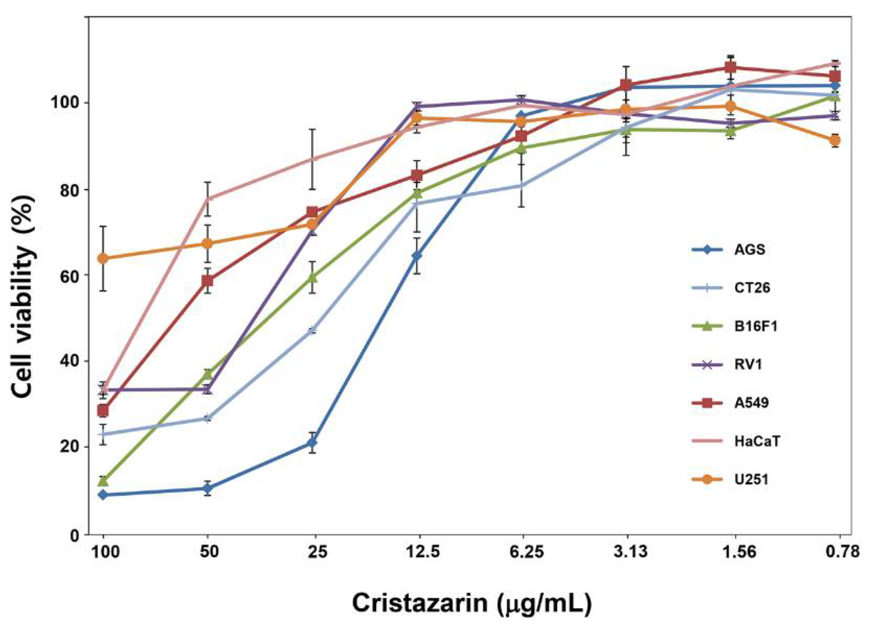
3.7. Biological Activity of Cristazarin Produced by C. metacorallifera

In order to investigate the biological activity of cristazarin produced by C. metacorallifera, we determined the cytotoxicity of cristazarin in six cancer cell lines and one non-cancer cell line, namely HaCaT (human keratinocytes), by exposing these cells to different concentrations of cristazarin (concentration range: 0.78–100 μg/mL). The IC50 of cristazarin ranged from 18.17 μg/mL to >100 μg/mL in the six cancer cell lines (Figure 6). The AGS, CT26, and B16F1 cell lines were sensitive to cristazarin, with IC50 values of 18.2 μg/mL, 26.1 μg/mL, and 30.9 μg/mL, respectively. RV1 and A594 cell lines were less sensitive, with IC50 values of 48.3 μg/mL and 55.5 μg/mL, respectively. Finally, the U251 cell line was the most resistant (i.e., IC50 = 141.42 μg/mL).

4. Discussion

Lichen photobionts are green algae or cyanobacteria that produce different types of carbohydrates via photosynthesis. Green algae produce polyols, such as ribitol in Trebouxia spp., Myrmecia spp., and Coccomyxa spp., sorbitol in Hyalococcus spp., and erythritol in Trentepohlia spp. [45]. Cyanobacteria produce glucose in Nostoc spp., Scytonema spp., and Rhizonema spp. [46,47]. The algal photobiont of C. metacorallifera is Coccomyxa viridis, which produces ribitol. Ribitol is a crystalline sugar alcohol (C5H12O5) that is formed by the reduction of ribose and converted into mannitol by the mycobiont via the pentose phosphate pathway [48,49,50]. Converted carbohydrates are rapidly and irreversibly metabolized into various forms and consumed for growth, reproduction, and other metabolic activities or diverted into secondary metabolites [51]. In natural habitats, C. metacorallifera produces unique phenolic substances such as didymic, squamatic, barbatic, rhodocladonic, and usnic acids, which are synthesized via the polyketide pathway by an acetyl-CoA carboxylase that produces malonyl-CoA [14,52].
According to Yamamoto, Matsubara, Kinoshita, Kinoshita, Koyama, Takahashi, Ahmadjiam, Kurokawa and Yoshimura [6], cristazarin and 6-methylcristazarin are produced by C. cristatella mycobionts in MY extract medium without an additional carbon source [6]. However, in our study, a red pigment was produced by C. metacorallifera mycobionts only in the fructose-supplemented medium under fluorescent light conditions, and this substance was revealed to be composed of cristazarin and 6-methylcristazarin. It is reported that some lichen substances have been detected only in symbiosis, but others have produced their compounds cultured only in cultured mycobionts [6]. Interestingly, cristazarin has not been detected in lichen thalli to date. Thus, it is necessary to understand the conversion of the carbon source, based on photobiont carbohydrates, to determine secondary metabolite production by mycobionts.
The mycobiont of C. metacorallifera produced cristazarin only in the medium with 1% fructose, but not in the media supplemented with glucose, sucrose, ribitol, mannitol, or sorbitol. Specifically, unlike the three kinds of sugar alcohols, pale red pigments were observed in media supplemented with glucose and sucrose. These sugar alcohols and glucose are commonly present in the thalli of lichens that have green algae as photobionts. Thus, sugar alcohols and glucose, which are already familiar with symbiotic mycobionts, can be difficult to become stressors or inducers that express genes related to secondary metabolites. The secondary metabolite production of lichen mycobionts is influenced by the carbon source [8,51]. Conversely, fructose is a monosaccharide that is not produced by symbiotic algae in lichens. Regardless of the production of cristazarin, the most important compounds for the classification of carbohydrates depending on the presence or absence of red pigments are monosaccharides and sugar alcohols. Among the three carbohydrates that produce red pigments, the second classification factor is the difference in carbohydrate structure. Structurally, glucose is an aldose and fructose is a ketose.
One of the main differences between aldoses and ketoses is that the carbonyl group in aldoses is present at the end of the carbon chain, while that in ketoses is present in the middle of the carbon chain. Moreover, the three sugar alcohols are also derived from the reduction of an aldose (sorbitol and mannitol can be reduced in both aldose and ketose). Among the carbohydrates used in the present study, fructose was the only ketohexose with six carbon sources and the only carbon source that stimulated the production of cristazarin. Based on these results, it is expected that cristazarin will also be produced from sucrose, which is a mixture of fructose and glucose, and actually pale red pigment was shown. A previous study on C. cristatella has also highlighted that sucrose promotes pigment production more than sugar alcohols do, and 4% is the optimal concentration in an LB medium [12].
Lichens produce many phenolic compounds, such as depsides, depsidones, dibenzofurans, pulvinates, chromones, and quinones. Furthermore, without symbiotic photobionts, mycobionts produce anthraquinones and cristazarin [2,6]. Some carbohydrates are converted via polyketide pathways to form diverse polyketides, which are produced by the successive condensation of acetyl-CoA with malonyl-CoA by PKS [36,52]. The most abundant classes of phenolic compounds, namely depsides and depsidones, are composed of orcinol (orsellinic and resorcyclic acids) or β-orcinol (β-orsellinic acid) type subunits [52,53]. Presumably, the mechanism by which red pigments are produced in a medium containing glucose, fructose, and sucrose may be described via the example of Seliwanoff’s test, which distinguishes between aldose and ketose sugars. The reagents in this test consist of resorcinol and concentrated hydrochloric acid and rely on the principle that aldose and ketose generate light pink and deep red colors, respectively, through acid hydrolysis and resorcinol condensation reactions [54]. Similarly, in our study, the red pigments, or cristazarin, can be produced by the reaction of furfural with orcinol subunits biosynthesized via the PKS pathway. Consequently, glucose (i.e., aldose) and sucrose (i.e., aldose and ketose) may generate a pink color lighter than that produced with fructose.
Many lichen compounds are derived from fatty acids. Acetyl-CoA carboxylase assembles structurally diverse products from simple acyl-CoA substrates via a catalytic cycle that involves reduction and dehydration [52,55]. Kozaki and Sasaki [56] have assumed that, under redox regulation, acetyl-CoA carboxylase is activated by light, and this is consistent with the formation of cristazarin under light conditions that was observed in our study. Therefore, it is presumed that fructose, which is a ketohexose carbohydrate, acts as a substrate via the same pathway and contributes to the production of diverse and specific secondary metabolites such as cristazarin. In the PKS gene analysis, CmPKS3, CmPKS4, CmPKS9, and CmPKS15, which belong to group I-2, were highly expressed. This group presumably includes the PKS domain that synthesizes single aromatic ring polyketides, such as orcinol (orsellinic acid). In addition to group I-2, most of the other gene groups were also expressed after 3 weeks of culture, a time which corresponds with the appearance of red pigments. Furthermore, MTT assay of cristazarin clearly showed the potential anticancer drug (Figure 6).

5. Conclusions

Overall, we showed that C. metacorallifera mycobionts produce special metabolites in media supplemented with 1% fructose. Thus, cultured C. metacorallifera mycobionts might represent a good biological material to assess the biosynthetic switching mechanism of cristazarin at the molecular level. As cristazarin has shown antibacterial activity against Bacillus subtilis (10 μg/mL) and Staphylococcus aureus (20 μg/mL), as well as antitumor activity (100 μM) [7], it could be a promising candidate for the development of antitumor and antibacterial agents. Therefore, further research on cell metastasis using cristazarin should be conducted.

Author Contributions

Conceptualization, S.K., J.-S.H., and S.-Y.P.; methodology, M.-H.J., C.-H.P., and J.A.K.; validation, M.-H.J., C.-H.P., and J.A.K.; investigation, M.-H.J., C.-H.P., J.A.K. and E.D.C.; resources, S.K. and J.-S.H.; data curation, S.-Y.P.; writing—original draft preparation, M.-H.J., C.-H.P., J.-S.H. and S.-Y.P.; writing—review and editing, S.-Y.P.; visualization, S.-Y.P.; supervision, J.-S.H.; funding acquisition, S.K., J.-S.H., and S.-Y.P. All authors have read and agreed to the published version of the manuscript.

Funding

This work was supported by grants from the National Research Foundation of Korea (NRF-2017R1D1A1B0435888) and by grant from the National Institute of Biological Resources, funded by the Ministry of Environment of the Republic of Korea (projects NIBR201503101 and 202124101).

Conflicts of Interest

The authors declare no conflict of interest.

References

  1. Castle, H.; Kubsch, F. The production of usnic, didymic, and rhodocladonic acids by the fungal component of the lichen Cladonia cristatella. Arch. Biochem. 1949, 23, 158–160. [Google Scholar]
  2. Ejiri, H.; Sankawa, U. Graciliformin and its acetates in Cladonia graciliformis. Phytochemistry 1975, 14, 277–279. [Google Scholar] [CrossRef]
  3. Nakano, H.; Komiya, T.; Shibata, S. Anthraquinones of the lichens of Xanthoria and Caloplaca and their cultivated mycobionts. Phytochemistry 1972, 11, 3505–3508. [Google Scholar] [CrossRef]
  4. Yoshimura, I.; Kurokawa, T.; Kinoshita, K.; Yamamoto, Y.; Miyawaki, H. Lichen substances in cultured lichens. J. Hatt. Bot. Lab. 1994, 76, 249–261. [Google Scholar] [CrossRef]
  5. Kinoshita, Y.; Yamamoto, Y.; Yoshimura, I.; Kurokawa, T.; Yamada, Y. Production of usnic acid in cultured Usnea hirta. Lichenol 1993, 53, 137–145. [Google Scholar] [CrossRef]
  6. Yamamoto, Y.; Matsubara, H.; Kinoshita, Y.; Kinoshita, K.; Koyama, K.; Takahashi, K.; Ahmadjiam, V.; Kurokawa, T.; Yoshimura, I. Naphthazarin derivatives from cultures of the lichen Cladonia cristatella. Phytochemistry 1996, 43, 1239–1242. [Google Scholar] [CrossRef]
  7. Yamamoto, Y.; Kinoshita, Y.; Matsubara, K.; Kinoshita, K.; Koyama, K.; Takahashi, K.; Kurokawa, T.; Yoshimura, I. Screening of biological activities and isolation of biological-active compounds from lichens. Recent Res. Devel. Phytochem. 1998, 2, 23–34. [Google Scholar]
  8. Brunauer, G.; Hager, A.; Grube, M.; Turk, R.; Stocker-Worgotter, E. Alterations in secondary metabolism of aposymbiotically grown mycobionts of Xanthoria elegans and cultured resynthesis stages. Plant Physiol. Biochem. 2007, 45, 146–151. [Google Scholar] [CrossRef]
  9. Hamada, N. Induction of the production of lichen substances by non-metabolites. Bryologist 1996, 99, 68–70. [Google Scholar] [CrossRef]
  10. Hamada, N.; Miyagawa, H. Secondary metabolites from isolated lichen mycobionts cultured under different osmotic conditions. Lichenologist 1995, 27, 201–205. [Google Scholar] [CrossRef]
  11. Hamada, N.; Miyagawa, H.; Miyawaki, H.; Inoue, M. Lichen substances in mycobionts of crustose lichens cultured on media with extra sucrose. Bryologist 1996, 99, 71–74. [Google Scholar] [CrossRef]
  12. Yamamoto, Y.; Kinoshita, Y.; Kurokawa, T.; Yoshimura, I.; Ahmadjiam, V.; Yamada, Y. Cell growth and pigment production in suspension cultures of a mycobiont isolated from the lichen Cladonia cristatella. Can. J. Bot. 1995, 73, 590–594. [Google Scholar] [CrossRef]
  13. Stocker-Worgotter, E. Metabolic diversity of lichen-forming ascomycetous fungi: Culturing, polyketide and shikimate metabolite production, and PKS genes. Nat. Prod. Rep. 2008, 25, 188–200. [Google Scholar] [CrossRef] [PubMed]
  14. Fahselt, D. Secondary biochemistry of lichens. Symbiosis 1994, 16, 134–140. [Google Scholar]
  15. O’Hagan, D. The Polyketide Metabolites; Ellis Horwood: Chichester, UK, 1991. [Google Scholar]
  16. Anand, S.; Prasad, M.V.; Yadav, G.; Kumar, N.; Shehara, J.; Ansari, M.Z.; Mohanty, D. SBSPKS: Structure based sequence analysis of polyketide synthases. Nucleic Acids Res. 2010, 38, W487–W496. [Google Scholar] [CrossRef] [PubMed] [Green Version]
  17. Ansari, M.Z.; Yadav, G.; Gokhale, R.S.; Mohanty, D. NRPS-PKS: A knowledge-based resource for analysis of NRPS/PKS megasynthases. Nucleic Acids Res. 2004, 32, W405–W413. [Google Scholar] [CrossRef]
  18. Bachmann, B.O.; Ravel, J. Chapter 8. Methods for in silico prediction of microbial polyketide and nonribosomal peptide biosynthetic pathways from DNA sequence data. Methods Enzymol. 2009, 458, 181–217. [Google Scholar] [CrossRef]
  19. Jenke-Kodama, H.; Dittmann, E. Bioinformatic perspectives on NRPS/PKS megasynthases: Advances and challenges. Nat. Prod. Rep. 2009, 26, 874–883. [Google Scholar] [CrossRef]
  20. Li, M.H.; Ung, P.M.; Zajkowski, J.; Garneau-Tsodikova, S.; Sherman, D.H. Automated genome mining for natural products. BMC Bioinform. 2009, 10, 185. [Google Scholar] [CrossRef] [Green Version]
  21. Starcevic, A.; Zucko, J.; Simunkovic, J.; Long, P.F.; Cullum, J.; Hranueli, D. ClustScan: An integrated program package for the semi-automatic annotation of modular biosynthetic gene clusters and in silico prediction of novel chemical structures. Nucleic Acids Res. 2008, 36, 6882–6892. [Google Scholar] [CrossRef] [Green Version]
  22. Tae, H.; Kong, E.B.; Park, K. ASMPKS: An analysis system for modular polyketide synthases. BMC Bioinform. 2007, 8, 327. [Google Scholar] [CrossRef] [Green Version]
  23. Weber, T.; Rausch, C.; Lopez, P.; Hoof, I.; Gaykova, V.; Huson, D.H.; Wohlleben, W. CLUSEAN: A computer-based framework for the automated analysis of bacterial secondary metabolite biosynthetic gene clusters. J. Biotechnol. 2009, 140, 13–17. [Google Scholar] [CrossRef]
  24. Yadav, G.; Gokhale, R.S.; Mohanty, D. Computational approach for prediction of domain organization and substrate specificity of modular polyketide synthases. J. Mol. Biol. 2003, 328, 335–363. [Google Scholar] [CrossRef]
  25. Avalos, J.; Estrada, A.F. Regulation by light in Fusarium. Fungal Genet. Biol. 2010, 47, 930–938. [Google Scholar] [CrossRef]
  26. Bayram, O.; Braus, G.H.; Fischer, R.; Rodriguez-Romero, J. Spotlight on Aspergillus nidulans photosensory systems. Fungal Genet. Biol. 2010, 47, 900–908. [Google Scholar] [CrossRef]
  27. Estrada, A.F.; Avalos, J. The White Collar protein WcoA of Fusarium fujikuroi is not essential for photocarotenogenesis, but is involved in the regulation of secondary metabolism and conidiation. Fungal Genet. Biol. 2008, 45, 705–718. [Google Scholar] [CrossRef] [PubMed]
  28. Fuller, K.K.; Ringelberg, C.S.; Loros, J.J.; Dunlap, J.C. The fungal pathogen Aspergillus fumigatus regulates growth, metabolism, and stress resistance in response to light. mBio 2013, 4, e00142-13. [Google Scholar] [CrossRef] [PubMed] [Green Version]
  29. Kim, H.; Ridenour, J.B.; Dunkle, L.D.; Bluhm, B.H. Regulation of stomatal tropism and infection by light in Cercospora zeae-maydis: Evidence for coordinated host/pathogen responses to photoperiod? PLoS Pathog. 2011, 7, e1002113. [Google Scholar] [CrossRef]
  30. Kim, H.; Son, H.; Lee, Y.W. Effects of light on secondary metabolism and fungal development of Fusarium graminearum. J. Appl. Microbiol. 2014, 116, 380–389. [Google Scholar] [CrossRef] [PubMed]
  31. Hitzenhammer, E.; Buschl, C.; Sulyok, M.; Schuhmacher, R.; Kluger, B.; Wischnitzki, E.; Schmoll, M. YPR2 is a regulator of light modulated carbon and secondary metabolism in Trichoderma reesei. BMC Genome 2019, 20, 211. [Google Scholar] [CrossRef] [PubMed] [Green Version]
  32. Kramer, G.J.; Pimentel-Elardo, S.; Nodwell, J.R. Dual-PKS cluster for biosynthesis of a light-induced secondary metabolite found from genome sequencing of Hyphodiscus hymeniophilus fungus. ChemBio Chem. 2020, 21, 2116–2120. [Google Scholar] [CrossRef]
  33. BeGora, M.D.; Fahselt, D. Usnic acid and atranorin concentrations in lichens in relation to bands of UV irradiance. Bryologist 2001, 104, 1687–1692. [Google Scholar] [CrossRef]
  34. Park, S.Y.; Choi, J.; Lee, G.W.; Kim, J.A.; Oh, S.O.; Jeong, M.H.; Yu, N.H.; Kim, S.; Lee, Y.H.; Hur, J.S. Draft genome sequence of lichen-forming fungus Cladonia metacorallifera strain KoLRI002260. Genome Announc. 2014, 2, e01065-13. [Google Scholar] [CrossRef] [PubMed] [Green Version]
  35. Stenroos, S.; Hyvonen, J.; Myllys, L.; Thell, A.; Ahti, T. Phylogeny of the genus Cladonia s.lat. (Cladoniaceae, Ascomycetes) inferred from molecular, morphological, and chemical data. Cladistics 2002, 18, 237–278. [Google Scholar] [CrossRef]
  36. Kim, J.A.; Hong, S.G.; Cheong, Y.H.; Koh, Y.J.; Hur, J.S. A new reducing polyketide synthase gene from the lichen-forming fungus Cladonia metacorallifera. Mycologia 2012, 104, 362–370. [Google Scholar] [CrossRef]
  37. Lilly, V.G.; Barnett, H.L. Physiology of the Fungi; McGraw-Hill, Inc.: New York, NY, USA, 1951. [Google Scholar]
  38. Culberson, C.F. Improved conditions and new data for the identification of lichen products by a standardized thin-layer chromatographic method. J. Chromatogr. 1972, 72, 113–125. [Google Scholar] [CrossRef]
  39. Park, S.Y.; Choi, J.; Lim, S.E.; Lee, G.W.; Park, J.; Kim, Y.; Kong, S.; Kim, S.R.; Rho, H.S.; Jeon, J.; et al. Global expression profiling of transcription factor genes provides new insights into pathogenicity and stress responses in the rice blast fungus. PLoS Pathog. 2013, 9, e1003350. [Google Scholar] [CrossRef] [PubMed] [Green Version]
  40. Livak, K.J.; Schmittgen, T.D. Analysis of relative gene expression data using real-time quantitative PCR and the 2(-Delta Delta C(T)) Method. Methods 2001, 25, 402–408. [Google Scholar] [CrossRef]
  41. Stenroos, S. Taxonomy of the Cladonia coccifera group II. Ann. Bot. Fenn. 1989, 26, 307–317. [Google Scholar]
  42. Honegger, R.; Kutasi, V. Anthraquinone Production in Three Aposymbiotically Cultured Teloschistalean Lichen Mycobionts: The Role of the Carbon Source; Institut National de la Recherche Agronomique: Paris, France, 1990. [Google Scholar]
  43. Schlotter, Y.M.; Veenhof, E.Z.; Brinkhof, B.; Rutten, V.P.; Spee, B.; Willemse, T.; Penning, L.C. A GeNorm algorithm-based selection of reference genes for quantitative real-time PCR in skin biopsies of healthy dogs and dogs with atopic dermatitis. Vet. Immunol. Immunopathol. 2009, 129, 115–118. [Google Scholar] [CrossRef] [Green Version]
  44. Jones, P.; Binns, D.; Chang, H.Y.; Fraser, M.; Li, W.; McAnulla, C.; McWilliam, H.; Maslen, J.; Mitchell, A.; Nuka, G.; et al. InterProScan 5: Genome-scale protein function classification. Bioinformatics 2014, 30, 1236–1240. [Google Scholar] [CrossRef] [Green Version]
  45. Palmqvist, K.; Dahlman, L.; Jonsson, A.; Nash, T.H. The carbon economy of lichens. In Lichen Biology, 2nd ed.; Nash, T.H., III, Ed.; Cambridge University Press: Cambridge, UK, 2008; pp. 182–215. [Google Scholar]
  46. Wastlhuber, R.; Loos, E. Differences between cultured and freshly isolated cyanobiont from Peltigera-is there symbiosis-specific regulation of a glucose carrier? Lichenologist 2007, 28, 67–78. [Google Scholar] [CrossRef]
  47. Cornejo, C.; Nelson, P.R.; Stepanchikova, I.; Himelbrant, D.; JØRgensen, P.-M.; Scheidegger, C. Contrasting pattern of photobiont diversity in the Atlantic and Pacific populations of Erioderma pedicellatum (Pannariaceae). Lichenologist 2016, 48, 275–291. [Google Scholar] [CrossRef]
  48. Gustavs, L.; Schiefelbein, U.; Tatyana Darienko, T.; Proschold, T. Symbioses of the green algal genera Coccomyxa and Elliptochloris (Trebouxiophyceae, Chlorophyta). In Algal and Cyanobacteria Symbioses; Grobe, M., Seckbach, J., Mugga, L., Eds.; World Scientific Publishing Company: Singapore, 2017; pp. 169–208. [Google Scholar] [CrossRef]
  49. Kono, M.; Tanabe, H.; Ohmura, Y.; Satta, Y.; Terai, Y. Physical contact and carbon transfer between a lichen-forming Trebouxia alga and a novel Alphaproteobacterium. Microbiology 2017, 163, 678–691. [Google Scholar] [CrossRef] [PubMed]
  50. Lines, C.E.M.; Ratcliffe, R.G.; Rees, T.A.V.; Southon, T.E. A 13C NMR study of photosynthate transport and metabolism in the Lichen Xanthoria calcicola Oxner. New Phytol. 1989, 111, 447–456. [Google Scholar] [CrossRef]
  51. Elshobary, M.E.; Osman, M.E.; Abo-Shady, A.M.; Komatsu, E.; Perreault, H.; Sorensen, J.; Piercey-Normore, M.D. Algal carbohydrates affect polyketide synthesis of the lichen-forming fungus Cladonia rangiferina. Mycologia 2016, 108, 646–656. [Google Scholar] [CrossRef] [Green Version]
  52. Elix, J.A.; Stocker-Wörgötter, E. Biochemistry and secondary metabolites. In Lichen Biology, 2nd ed.; Nash, H., III, Ed.; Cambridge University Press: Cambridge, UK, 2008; pp. 104–133. [Google Scholar]
  53. Boustie, J.; Grube, M. Lichens-a promising source of bioactive secondary metabolites. Plant Genet. Resour. 2005, 3, 273–287. [Google Scholar] [CrossRef] [Green Version]
  54. Seliwanoff, T. Notiz über eine Fruchtzuckerreaction. Ber. Dtsch. Chem. Ges. 1887, 20, 181–182. [Google Scholar] [CrossRef] [Green Version]
  55. Raharjo, T.J.; Chang, W.-T.; Choi, Y.H.; Peltenburg-Looman, A.M.G.; Verpoorte, R. Olivetol as product of a polyketide synthase in Cannabis sativa L. Plant Sci. 2004, 166, 381–385. [Google Scholar] [CrossRef]
  56. Kozaki, A.; Sasaki, Y. Light-dependent changes in redox status of the plastidic acetyl-CoA carboxylase and its regulatory component. Biochem. J. 1999, 339 Pt 3, 541–546. [Google Scholar] [CrossRef]
Figure 1. Chemical structure of naphthazarin, cristazarin, and 6-methylcristazarin.
Jof 07 00601 g001
Figure 2. Characteristics of the lichen Cladonia metacorallifera. (a) Morphology of C. metacorallifera; (b) isolated C. metacorallifera mycobiont; (c) high performance liquid chromatography (HPLC) analysis of acetone extracts from C. metacorallifera thalli. Compounds were eluted with Me-OH during 40 min and monitored at 254 nm. 1 (retention time [tR] = 2 min): acetone; 2 (tR = 2.6 min): rhodocladonic acid; 3 (tR = 3.3 min): thamnolic acid; and 4 (tR = 10.7 min): didymic acid.
Jof 07 00601 g002
Figure 3. Characteristics of the mycobiont of C. metacorallifera. (a) Growth of isolated C. metacorallifera mycobionts on Lilly and Barnett’s (LB) media without a carbon source or with one of six different carbon sources after three months; (b) acetone extracts from the grown mycelia under each carbon condition; (c) HPLC analysis of the acetone extracts. Compounds were eluted with Me-OH for 40 min, monitored at 254 nm. Acetone was eluted at tR = 2 min as a blank control. 5 (tR = 3.4 min): cristazarin; and 6 (tR = 8.1 min): 6-methylcristazarin.
Jof 07 00601 g003
Figure 4. C. metacorallifera in liquid culture, and detection of its production of cristazarin 1–6 weeks after inoculation. (a) C. metacorallifera after 2 months of cultivation in liquid LB media without fructose or with 1% fructose at different temperatures; (b) liquid LB cultures of C. metacorallifera without (left; control) and with (right) 1% fructose at 15 °C; (c) microscopic observation of the control displayed in Figure 4b; (d) microscopic observation of the culture with fructose displayed in Figure 4b, with the presence of a red indeterminate crystal; (e) acetone extraction of total substance from C. metacorallifera in liquid induction media under light or dark conditions; (f) production of cristazarin by C. metacorallifera under light or dark conditions (HPLC analysis).
Jof 07 00601 g004
Figure 5. Domain structure and expression profiles of polyketide synthase (PKS) genes; (a) the protein domain structures of 30 putative PKS genes were obtained from the InterPro protein database (http://www.ebi.ac.kr/interpro, accessed on 20 July 2021); (b) heat map of the expression patterns of 27 PKS genes; (c) quantitative RT-PCR analysis of the transcripts from the genes Cmwc-1 and Cmwc-2 of C. metacorallifera cultured in liquid LB media with 1% fructose during 1–6 weeks.
Jof 07 00601 g005
Figure 6. Cytotoxic activity of cristazarin in six different cancer cell lines (AGS, CT26, B16F1, RV1, A549, and U251) and one normal cell line (HaCaT). Cells were counted 48 h after exposure to cristazarin. IC50 values were calculated using SPSS. All treatments were triplicated; standard errors are showed.
Jof 07 00601 g006
Table 1. Primers used for quantitative real-time PCR.
Target Genes *Forward Primer (5’ → 3’)Reverse Primer (5’ → 3’)
β-tubulinTGAGGCACTTTACGACATCTGGAAGTGAAGACGAGGGAAAGG
α-tubulinTCGTCTCTTCAATCACTGCCGAATTGGATTCATGTGCAGCC
EF1βTCTACAGGGCAAGTTCAATCGGCAAAGTAGAGGATCAGGAGTG
UEP1CCTCCATCGCATTACCCTTAGCTTCCCTGCGAAAATCAAACG
Actin2CACAGCAACCCTATACTCCTTCCTATTCTCACTATCACACCATCCC
GAPDHAAGACGCTGAATGGGATATGGTCTTATGCAAGCTTAGCCCTC
CmPKS1GCTGTTTTTGCGGGCATGGACATACGGACGGCTTGATGT
CmPKS2ACCAGTTCGGATCACTTCCGTAGCAATATCTGTTCG
CmPKS3CAAGGCTTCCACTTCTCACGGAAGATGTGTAACCTC
CmPKS4TGCGATTCACGCTCCATATGACATATCTCTGGCACG
CmPKS5GTCGAACGTATCATTCATGATCGATATGAGTGTGCA
CmPKS6TCGAAGAGGTGCAAGCAAGTTGACCAGCTGTCGAAG
CmPKS7TATCGAGTACACCAGTGCGCACAGTGTTCTGATCGT
CmPKS8GTCTCATTAGCTATGTACACGGACCAGCTCTCTTGG
CmPKS9AGAGTGCAGCAGAGCTGTCGTAGTGTCTCTTGGTGC
CmPKS10GCTCCGCAAGAACAGCAATGGAATGCGGCAGTGATC
CmPKS11ACTGATGCTTCATGTCGATGAGCCGTTGCACCAACA
CmPKS12CAGGTCTTGGAGACTACTGGTCGACATTCCTGTCTT
CmPKS13ACTAGACAGGCTCCAGAGTTCATTGTGTCGAAGGTC
CmPKS14TCAGCATACTAACACTGCTCAGACACCTGAAGACCT
CmPKS15AGGTACATAATCCAGAGAAGTATACCATTCGCATCG
CmPKS16ATTGGCCTCTTGAGCGCTGATGTACGTATCCGGATA
CmPKS17ATGGCTGAAGAGGCGGATGCGCGACCAATTGAATCA
CmPKS18ATGGAGATGGCGATACGATGAGAGTACCAGCCGCAT
CmPKS19TGTGGATGTTGCCTGTCAGACAGCATCTTCGAGACG
CmPKS20GAGGAGAAGTGGCATCTAGCATTCTCAAGTCCTTCA
CmPKS21TGGCTTCTGATTATACCACGGCTAACGTTCGGAGACGATG
CmPKS22ATGGAGCTCTGCATGGTATCATCGGCATTGTGAATC
CmPKS23CAGCATACCTGCCGAGAGACACCGCATTCTCAAGT
CmPKS24ACACATAATGAAGACATCGTCGATAATGTTCTGGAG
CmPKS25TCATCGGCACAGTGCACAGTATAGCAATGCGATATC
CmPKS26ACATCGTGGAGCAGAAGGTGTGATGCCAGCGTCTTCT
CmPKS27CACAGTGGTACGAAGACACGATGTAGCATGAGGTAT
CmPKS28GCGACGTAGATGGATATGGATGATTGGTTGCTGGAC
CmPKS29GGCGAGACGATATTGATCCATCTTGGCCAGTTGAACCG
CmPKS30TGTTAGACAAGCTCACTTTGAATATGATGCTATCGT
Cmwc1TGCTAATTGCCATACCCGCCGTAGCTGAATTGTGTGAG
Cmwc2TCGCTTCTTCTACCGCTTCTTGGTTAGGCGCTCATT
* EF1β: elongation factor1-β; UEP1: ubiquitin extension protein gene; GAPDA: glyceraldehydes-3-phosphate dehydrogenase.
Table 2. NMR data.
Table 2. NMR data.
C No.13C a1H (Number of Protons, Multiplicity) b (j-Hz) c
1176.99
2155.83
3128.74
4181.97
4a105.02
5166.69
6109.196.62 (1H, s)
7158.51
8158.61
8a111.70
917.102.61 (2H, q, 7.5)
1012.991.11 (3H, t, 7.5)
1157.143.93 (3H, s)
a 125 MHz.; b multiplet; q: quadruplet, s: singlet, t: triplet; c 500 MHz.
Publisher’s Note: MDPI stays neutral with regard to jurisdictional claims in published maps and institutional affiliations.

