Article

Monitoring of Host Suitability and Defense-Related Genes in Wheat to Bipolaris sorokiniana

1
Department of Plant Protection, Faculty of Agriculture, Bolu Abant Izzet Baysal University, Bolu 14030, Turkey
2
Department of Plant Protection, Faculty of Agriculture, Ankara University, Ankara 06110, Turkey
3
Bahri Dagdas International Agricultural Research Institute, Konya 42050, Turkey
4
Phytopathology Unit, Department of Plant Protection, Ecole Nationale d’Agriculture de Meknès, BPS 40, Meknes 50001, Morocco
5
Biotechnology Research Unit, Laboratory of Nematology, Regional Center of Agricultural Research, National Institute of Agronomic Research (INRA), Rabat 10060, Morocco
6
Wheat Health, Genetics and Quality Research Unit, United States Department of Agriculture, Agricultural Research Service, Washington State University, Pullman, WA 99164, USA
7
International Maize and Wheat Improvement Centre (CIMMYT), P.O. Box 39, Emek, Ankara 06170, Turkey
*
Authors to whom correspondence should be addressed.
J. Fungi 2022, 8(2), 149; https://doi.org/10.3390/jof8020149
Submission received: 3 January 2022 / Revised: 23 January 2022 / Accepted: 28 January 2022 / Published: 31 January 2022

Abstract

:
Spot blotch caused by Bipolaris sorokiniana is a destructive disease of wheat worldwide. This study investigated the aggressiveness of B. sorokiniana isolates from different wheat-growing areas of Bolu province in Turkey on the cultivar Seri-82. Host susceptibility of 55 wheat cultivars was evaluated against the most aggressive isolate. Our results indicated that the cultivars Anafarta and Koç-2015 were the most resistant. A specific and sensitive qPCR assay was developed for detecting the pathogen in plant tissues and evaluating wheat plants with different resistance levels. Three primer sets, BsGAPDHF/BsGAPDHR, BsITSF/BsITSR, and BsSSUF/BsSSUR, were designed based on glyceraldehyde-3-phosphate dehydrogenase, internal transcribed spacers, and 18S rRNA loci of B. sorokiniana with detection limits of 1, 0.1, and 0.1 pg of pathogen DNA, respectively. The qPCR assay was highly sensitive and did not amplify DNA from the other closely related fungal species and host plants. The protocol differentiated wheat plants with varying degrees of resistance. The assay developed a useful tool for the quantification of the pathogen in the early stages of infection and may provide a significant contribution to a more efficient selection of wheat genotypes in breeding studies. In the present study, expression levels of PR proteins, phenylalanine ammonia-lyase, catalase, ascorbate peroxidase, and superoxide dismutase enzymes were upregulated in Anafarta (resistant) and Nenehatun (susceptible) cultivars at different post-infection time points, but more induced in the susceptible cultivar. The results showed considerable variation in the expression levels and timing of defense genes in both cultivars.

1. Introduction

Wheat (Triticum spp.) is among the most important food sources for human food and the most widely grown crop in the world. Many biotic factors continuously challenge wheat production worldwide [1,2]. Among the fungal diseases, spot blotch and common root rot caused by Bipolaris sorokiniana (Sacc. in Sorokin) Shoemaker (teleomorph: Cochliobolus sativus (S. Ito and Kuribayashi) Drechsler ex Dastur) is a significant disease that occurs widely in wheat-growing areas worldwide and affects leaf, head, stem, and root tissue of wheat [3]. The pathogen survives from season to season in infected seed, soil, or plant debris and causes economically important losses of up to 100% in yield and quality of wheat under favorable conditions [2,3]. In Turkey, the disease occurs widely and causes serious damage in wheat-growing areas as well as in the rest of the world [2,4,5].
The use of resistant varieties is considered the most beneficial and eco-friendly for managing spot blotch disease [6]. However, genetic and pathogenic variation within pathogen populations complicated the studies of resistance breeding [7,8]. Thus, more detailed studies are needed to increase control methods’ effectiveness and better understand the host–pathogen relationship. Assessment of host response to different pathogen populations is indispensable to reveal plant–pathogen interaction. Several studies have been performed to estimate the disease severity of B. sorokiniana by using different disease assessment methods [9,10,11] to reveal resistance sources. These screening methods are affected by environmental conditions plants growth stage and assessing experts, which cause difficulties in the appropriate assessment of host resistance. Therefore, more reliable and repeatable screening techniques are needed to evaluate disease reactions in plants. Additionally, fast and reliable identification of fungi responsible for plant diseases at an early stage of disease development is important to improve disease management practices. Many techniques have been used routinely for disease detection and pathogen identification [12,13]. Molecular techniques have become a useful approach to study the phylogenetic relationship among plant pathogens and conserved sequences of internal transcribed spacers (ITS), β-tubulin, 28S rRNA gene (LSU), 18S rRNA gene (SSU), RNA polymerase second largest subunit (RPB2), translation elongation factor 1-alpha (EF1-α), and glyceraldehyde-3-phosphate dehydrogenase (GAPDH) provided informative sites for the specific detection of numerous fungal diseases [14,15,16]. Different approaches based on PCR were used for routine detection of spot blotch disease [17,18,19,20]. However, the development of more novel disease recognition and assessment techniques is needed to improve the efficiency of plant disease management practices.
Plants have developed sophisticated mechanisms to recognize attack by pathogens and activate an effective natural immune response. The understanding of signal transduction genes that play a key role in the resistance mechanisms of plants is essential for the development of novel disease management strategies associated with host resistance. Plant–pathogen interaction is known to induce defense responses including the generation of reactive oxygen species, phytoalexin biosynthesis, cell wall cross-linking, synthesis of defense enzymes, and the accumulation of pathogenesis-related (PR) proteins [21,22,23]. PR proteins involved in defense mechanism during plant–pathogen interaction have been widely recognized and clustered into 17 groups linked to amino acid sequences, serological characteristics, and enzymatic activities [24,25,26]. Thus far, many studies have been carried out to reveal the relationship between PR proteins and stress defense mechanisms of plants on the basis of gene expression [26,27,28,29]. The overexpression of PR genes in wheat plants resulted in increased resistance to several pathogens, i.e., Fusarium graminearum (FHB), Puccinia spp., and Blumeria graminis [30,31,32,33]. Similarly, the role of phenylalanine ammonia-lyase (PAL) involving in the production of defense-related compounds and guaiacol peroxidase (POD), catalase (CAT), ascorbate peroxidase (APX), and superoxide dismutase (SOD) enzymes associated with reactive oxygen species (ROS) detoxification has been indicated in a number of plant–pathogen interactions [34,35,36].
Therefore, this study aimed to: (i) obtain and characterize B. sorokiniana isolates from wheat fields in Bolu province, (ii) develop a qPCR assay for fast and reliable detection of pathogen infection in plant tissues at the early stages and monitoring of wheat genotypes with different resistance levels, to reveal disease reaction of common Turkish wheat cultivars as candidate plants for breeding studies, and (iii) evaluate the transcription level of several PR proteins (PR1, PR2, PR3, PR5, and PR10) and defense enzymes (PAL, CAT, SOD, and APX) that play a role in defense response of wheat plants with different resistance levels to spot blotch disease caused by B. sorokiniana.

2. Materials and Methods

2.1. Fungal Material

The fungal isolates were recovered from diseased leaf and root samples collected from different wheat fields located in Bolu province, Turkey. Symptomatic tissues showing leaf spot, root, and crown rot were surface sterilized in a 1% sodium hypochlorite solution for 2 min, rinsed twice with sterile distilled water, and placed in Petri dishes containing 1/5 strength potato dextrose agar (PDA) medium amended with 100 mg/L streptomycin sulphate and 25 mg/L chloramphenicol to inhibit bacterial growth. The Petri dishes were incubated for 3 days at 23 ± 1 °C in the dark. The growing cultures were examined on a light microscope (DM1000, Leica Microsystems, Wetzlar, Germany) and transferred to a new PDA medium. The isolates derived from hyphal tips were preserved on filter paper at 4 °C. Morphological identification of the fungal isolates was performed according to the criteria of Sivanesen [37]. The other fungal pathogens associated with wheat plant in previous studies, Fusarium culmorum (Wm.G. Sm.) Sacc., Fusarium graminearum Schwabe, Fusarium pseudograminearum Aoki and O’Donnell, Fusarium oxysporum Schltdl, Fusarium acuminatum Ellis ve Everh., Fusarium equiseti (Corda) Sacc., Fusarium sambucinum Fuckel, Fusarium avenaceum (Fr.) Sacc., Alternaria alternata (Fr.) Keissl., Pyrenophora teres Drechsler, Pyrenophora tritici-repentis (Died.) Drechsler, ZymoSeptoria tritici (Desm.) Quaedvlieg and Crous, Macrophomina phaseolina (Tassi) Goid, and Rhizoctonia solani J.G. Kühn, were included in the study to control the specificity of the designed primers.

2.2. DNA Extraction

DNA isolation from approximately 100 mg of frozen leaf tissue or mycelial mat was performed using DNeasy Plant Mini Kit (Qiagen, Hilden, Germany) or DNeasy Blood & Tissue Kit (Qiagen, Hilden, Germany) according to the manufacturer’s protocol. The concentration and quality of DNA was measured using a DS-11 FX+ spectrophotometer (Denovix Inc., Wilmington, DE, USA) and diluted to 10 ng/μL with ultrapure ddH2O. To quantify the amount of pathogen, a standard calibration curve was obtained using a 10-fold serial dilution of fungal DNA with healthy plant DNA. DNA concentrations in the calibration curve ranged from 10 ng to 0.01 pg/μL and were used in qPCR reactions as an internal control of DNA quantification.

2.3. DNA Sequencing and Phylogeny

Morphological identification of the pathogen isolates was also confirmed by DNA sequencing of the rDNA ITS and GAPDH loci with the primer pairs ITS1/ITS4 [38] and gpd1/gpd2 [39], respectively. Amplification was performed in a total volume of 50 μL containing 1× PCR buffer, 2 mM MgCl2, 0.4 μM each primer, 0.2 mM dNTPs, and 1.25 U Taq DNA polymerase (New England Biolabs, Beverly, MA, USA). The PCR amplifications consisted of an initial denaturation of one cycle for 3 min at 95 °C, followed by 35 cycles of 1 min at 95 °C, 1 min at 52 °C, and 1 min at 72 °C, while the final extension step was carried out at 72 °C for 10 min. PCR products were visualized on 1.4% agarose gel using 1× TAE buffer and sequenced in both directions using the same primers by Macrogen company (Seoul, Korea). The sequences obtained were examined by BLAST analysis using the NCBI website (http://blast.ncbi.nlm.nih.gov/Blast.cgi, accessed on 10 December 2021). Sequences were read and edited with MegAlign module of DNASTAR software version 7.1.0 (DNASTAR Inc., Madison, WI, USA), and deposited in the NCBI GenBank nucleotide database (https://www.ncbi.nlm.nih.gov/genbank/, accessed on 10 December 2021).
For phylogenetic reconstruction, the sequences of ITS and GAPDH were compared with published Bipolaris and Curvularia spp. sequences. Preliminary sequence alignments of two individual loci (ITS and GAPDH) were generated using MAFFT v. 7.490 [40] (http://mafft.cbrc.jp/alignment/server/index.html, accessed on 10 December 2021) with the default parameters. Phylogenetic analysis based on the combined alignment of the two loci of the isolates from the current study and those of reference isolates from GenBank were reconstructed using Maximum-Likelihood (ML) analysis with MEGA 7 [41]. The bootstrap re-sampling analysis for 1000 replicates was used to estimate the confidence of tree topologies [42]. Phylogenetic trees were rooted to Pyrenophora tritici-repentis (ITS: AF071348; GAPDH: AF081370) and visualized with MEGA 7.

2.4. Pathogenicity Tests of Isolates and Host Susceptabilty of Wheat Cultivars

All isolates from different fields were subjected to a preliminary pathogenicity test on the susceptible wheat cultivar Seri-82. To prepare the inoculum, the isolates were grown on PDA medium in the dark at 23 °C for 7 days. Mycelial plugs from the edge of the cultures were transferred into the polyester bags containing 200 g of sterilized wheat seeds and incubated at 23 °C for 15 days in the dark. To ensure homogeneity, the bags were turned upside down every two days. The inoculum was harvested by mixing the inoculated wheat seeds with 200 mL of distilled water on a magnetic shaker and filtering through sterile filter paper. Spore concentration was adjusted to a final concentration of 1 × 10⁴ conidia/mL by diluting in sterile distilled water.
Wheat seeds were surface sterilized with a 1% sodium hypochlorite solution for 2 min, rinsed with distilled water and sown in pots (17 cm in length and 11 cm in diameter) containing sterilized compost. Three pots for each isolate were grown at 23 °C under a 12-h photoperiod. Fifteen days after sowing, plants were sprayed with spore suspensions until run-off. Plants were immediately covered with a transparent plastic bag to maintain humidity and maintained at 20 °C for 18 h. The inoculated plants were incubated at 23 °C for 12 days. Disease severity of wheat plants was evaluated using a 1–9 scale as per Fetch and Steffenson [11]. The infection response was classified into three general categories of low, intermediate, and high host-parasite compatibility. The infection response 1, 2 and 3 were considered indicative of low compatibility; 4 and 5 were intermediate compatibility; and 6, 7, 8, and 9 were high compatibility. The data obtained from disease scores were also analyzed using Statistical Analysis System computer software (SAS Version 9.0; SAS Institute Inc.; Cary, NC, USA). Means values were separated according to Tukey’s HSD method (Honestly Significant Difference (HSD) test.
Using the most aggressive isolates in a population derived from specific regions or provinces is desirable to ensure the most effective line screening in wheat and barley breeding programs. The most aggressive isolate was used to evaluate the disease reaction of 55 wheat cultivars that are widely cultivated in Turkey (Table 1). The disease severity data were subjected to analysis with Levene’s homogeneity of variance test and then one-way ANOVA, followed by Tukey HSD test (p ≤ 0.05) contained in the SAS software.

2.5. Primer Design

The reference sequences of the ITS, GAPDH, and SSU region of B. sorokiniana and closely related fungi were retrieved from GenBank and aligned for the conserved region together with the obtained sequences in this study by using MEGA 7 software. Species-specific primer sets were designed by using Primer3 ([43], https://bioinfo.ut.ee/primer3-0.4.0/, accessed on 10 December 2021) and UNAFold software (https://eu.idtdna.com/unafold/, accessed on 10 December 2021). The specificity of the primers was also confirmed by NCBI-BLAST analysis for searching the primer sequences.

2.6. Detection of Bipolaris sorokiniana Infection in Wheat Tissues

The reaction of the resistant (Altay, Koç-2015, and Anafarta) and susceptible (Kırik, Damla, and Nenehatun) wheat cultivars to spot blotch disease was also evaluated by qPCR assay with the designed primer sets. The inoculation was performed as mentioned above. The second leaves of wheat plants from each pot were collected at 3, 5, and 8 days after inoculation, washed with sterile water, placed into 50 mL falcon tubes, and immediately frozen in liquid nitrogen. DNA extraction was carried out as described above.
Real-time PCR assay was performed using a BioRad CFX96 Real-time PCR system (Bio-Rad Laboratories Inc., Hercules, CA, USA). PCR mixtures consisted of 2 μL diluted cDNA, 0.6 μM primer, 1 × iTaq Universal SYBR Green Supermix (Bio-Rad Laboratories Inc., Hercules, CA, USA) in 20 μL volume. The PCR amplifications consisted of an initial denaturation of one cycle for 3 min at 95 °C, 45 cycles of 10 s at 95 °C, and 30 s at 60 °C, followed by denaturation for melting curve analysis. The amount of pathogen DNA in plant tissues was quantified using the standard curves constructed with known concentrations of pathogen DNA, from 10 ng to 0.01 pg.

2.7. Expression Analysis of Defense-Related Genes in Wheat to the Pathogen

The transcript level of defense genes was evaluated in Anafarta (resistant) and Nenehatun (susceptible) wheat cultivars. The second leaves of inoculated plants were collected at 12, 24, 48, 72, and 96 h post-inoculation (hpi). The samples were immediately frozen in liquid nitrogen and stored until used for RNA isolation. Mock inoculations were performed only with distilled water. Three pots were used for each inoculation point and leaves from 10 plants for each replicate were pooled.
RNA extraction was performed from 100 mg of leave tissue using NucleoZOL (Macherey-Nagel, Düren, Germany) RNA isolation buffer following the manufacturer’s specifications. To eliminate residual DNA, the samples were treated with 1 U of DNase I (Thermo Fisher Scientific, Waltham, MA, USA) according to the protocol recommended by the manufacturer. RNA quality was evaluated using the DS-11 FX+ spectrophotometer. After the purification process, cDNA synthesis was performed from the mRNA using an iScript™ cDNA synthesis kit (Bio-Rad Laboratories Inc., Hercules, CA, USA) according to the manufacturer’s instructions. For gene expression analysis with qRT-PCR, all cDNA samples were diluted 10-fold with sterile ultrapure water.
The qRT-PCR reaction comprised of 2 μL cDNA, 0.5 μM primer, 1 × Sso Advanced SYBR Green Supermix (Bio-Rad Laboratories Inc., Hercules, CA, USA) in 10 μL volume. The cycling condition was 3 min at 95 °C, 45 cycles of 10 s at 95 °C, and 10 s at 60 °C. After the ending of the PCR assay, a denaturation curve from 65 to 95 °C was performed. The housekeeping gene β-tubulin was used as an internal control for relative quantification in all analyses (Table 2). Three biological replicates and two technical replicates were analyzed for each gene. The expression level of the gene was determined according to the 2 −ΔΔCT method of Livak and Schmittgen [44] using Ct-values of β-tubulin gene for normalization. The fold changes were subjected to ANOVA analysis or Student’s t-test analysis using Minitab 17 statistical software for Windows (Minitab, Inc.: State College, PA, USA, www.minitab.com, accessed on 2 January 2022).

3. Results

3.1. The Identification of Pathogen Isolates

Bipolaris sorokiniana occurred commonly in wheat fields located in Bolu province and was successfully isolated from all samples suggesting typical spot blotch symptoms (Figure 1). Based on morphological and cultural characteristics, the fungal identification was also confirmed molecularly by DNA sequencing of ITS and GAPDH gene region of ten isolates representing each different field. BLAST analysis of the 524 and 594 bp amplicons revealed 100% identity with ITS and GAPDH sequences of B. sorokiniana CBS 110.14, respectively. The resulting sequences were deposited in GenBank under accession numbers: MT271240-MT271249 for ITS and MW248907-MW248916 for GAPDH. The phylogenetic tree based on the ML method showed the isolates collected in this study, clustering with the reference isolates of B. sorokiniana derived from GenBank (Figure 2).

3.2. Aggressiveness of the Pathogen Isolates and Disease Reaction of Wheat

Ten representative isolates from each field were subjected to the preliminary pathogenicity test on the susceptible cultivar Seri-82. All isolates caused typical spot blotch lesions on wheat leaves and disease index ranged from 3.5 to 5.5. Levene’s test indicated that there were statistically significant differences in the variance between all isolates (HSD = 0.856, p ≤ 0.05).
The most aggressive isolate TR-Cs-3 was selected to further evaluate disease reaction of 55 wheat cultivars, widely cultivated in Turkey. The results indicated that there were significant differences among the responses of wheat cultivars to the pathogen. The cultivars Anafarta and Koç-2015 were the most resistant with disease index of 1.63, while the cultivar Kırik was found to be the most susceptible cultivar with disease index of 7.97, followed by the cultivars Lancer and Doğu-88. Disease index of the other cultivars varied from 2.17 to 7.5.

3.3. Designing Species-Specific Primers

Conserved sequences of ITS, GAPDH, and SSU genes were selected as targets for determining the causal agent. Three primer sets, BsGAPDHF/BsGAPDHR, BsITSF/BsITSR, and BsSSUF/BsSSUR, were designed based on the sequence alignment of B. sorokiniana and the other fungi associated with wheat diseases (Table 2). The primer pairs BsGAPDHF/BsGAPDHR amplified a single product of 143 bp in size, while BsITSF/BsITSR and BsSSUF/BsSSUR primers produced a 108 and 130 bp amplicon from DNA samples of B. sorokiniana isolates, respectively. No cross-amplification occurred in DNA samples from the other fungi and plant tissues with these primer pairs. Detection limits of the primers were evaluated by using standard regression lines constructed from seven dilution series ranging from 10 ng to 0.01 pg (Figure 3). The detection sensitivity for BsGAPDHF/BsGAPDHR, BsITSF/BsITSR, and BsSSUF/BsSSUR primers was found as 1, 0.1, and 0.1 pg of pathogen DNA, while Cq values of the dilution series ranged from 18.54 to 33.69, 16.12 to 32.55, 15.36 to 31.12, respectively. The efficiency of qRT-PCR was 86.5% (GAPDH: R2 = 0.994, slope = −3.693, y-int = 22.212), 103.0% (ITS: R2 = 0.994, slope = −3.251, y-int = 19.933), and 110.2% (SSU: R2 = 0.980, slope = −3.100, y-int = 19.323). The melting curve analysis produced a single peak with dissociation temperature of approximately 85 °C, 83 °C, and 76.5 °C for BsGAPDHF/BsGAPDHR, BsITSF/BsITSR, and BsSSUF/BsSSUR primers, confirming the specificity of the PCR reaction, respectively (Figure 3).

3.4. Detection of Bipolaris sorokiniana Infection in Wheat Tissues

The efficiency of primer pairs was also evaluated on the resistant (Altay, Koç-2015, and Anafarta) and susceptible (Kırik, Damla, and Nenehatun) wheat cultivars on the third, fifth, and eighth days after inoculation. All primer sets ensured the detection of fungal DNA in the early stages of pathogen infection. The obtained results indicated that there were significant differences among Cq values of the resistant and susceptible cultivars based on Tukey HSD test (p ≤ 0.01) (Figure 4). The mean DNA values detected in the qPCR assay with BsGAPDHF/BsGAPDHR, BsITSF/BsITSR, and BsSSUF/BsSSUR primers were 0.69, 0.13, and 0.74 in resistant cultivars on the third day after inoculation, while the mean values in susceptible cultivars were 2.34, 0.72, and 2.07, respectively. The amount of pathogen DNA increased gradually in the later days after inoculation. Additionally, the quantity of pathogen DNA in susceptible cultivars was higher than that in resistant cultivars for all primer sets on the fifth and eighth day after inoculation. No significant difference was observed in the amount of pathogen at different time points between resistant cultivars except for the fifth day with BsITSF/BsITSR primers, while the pathogen amounts in susceptible cultivars were variable for all days with all primers.

3.5. Differential Expression of Defense-Related Genes in Wheat Cultivars

Expression levels of PR1, PR2, PR3, PR5, PR10, PAL, CAT, SOD, and APX genes were evaluated in the resistant cultivar ‘Anafarta’ and the susceptible cultivar ‘Nenehatun’ at different time points. The PR1 gene was significantly upregulated in the defense response of both cultivars to the pathogen. The expression elevation was considerably higher in the susceptible cultivar Nenehatun compared to the resistant cultivar Anafarta (Figure 5 and Figure 6). The transcript level in Nenehatun elevated dramatically in the first 12 hpi and the maximum level was observed at 72 hpi with 14.03 of log2 fold change. The transcript level of PR1 in the resistant cultivar was gradually upregulated from 12 to 72 hpi. The highest expression in the resistant cultivar at 96 hpi reached 9.85 log2 fold. The PR2 level increased 3.55 and 8.4-log2 fold in Anafarta and Nenehatun at 12 hpi, respectively. The transcript level in both cultivars showed no significant differences at 24 and 96 hpi, while the expression enhancements at 12, 48, and 72 hpi were statistically different. The maximum activity in the resistant cultivar was observed at 48 hpi with 9.58 of log2 fold change. The highest level of PR2 expression was reached in Nenehatun at 72 hpi. PR3 transcript induced strongly in both cultivars but showed no significant difference between the cultivars except for 12 hpi. The highest expression of PR3 gene in Nenehatun and Anafarta cultivars at 72 hpi reached 12.74 and 12.52-log2 fold, respectively. The transcript level of PR5 was significantly upregulated in the susceptible cultivar than in the resistant cultivar at all inoculation point. PR5 level showed a similar fluctuation ranging from 11.15 to 13.95 log2 fold in the susceptible cultivar for all time points, while the resistant cultivar, Anafarta, showed a weak elevation at 12 hpi and reached a peak of 8.53-log2 fold at 72 hpi. PR10 transcript showed an expression profile similar to PR5 regulation in both cultivars. The expression of PR10 was significantly higher in Nenehatun than Anafarta at all time points. The highest expression of this gene in both cultivars at 96 hpi reached 16.25 and 10.4-log2 fold, respectively.
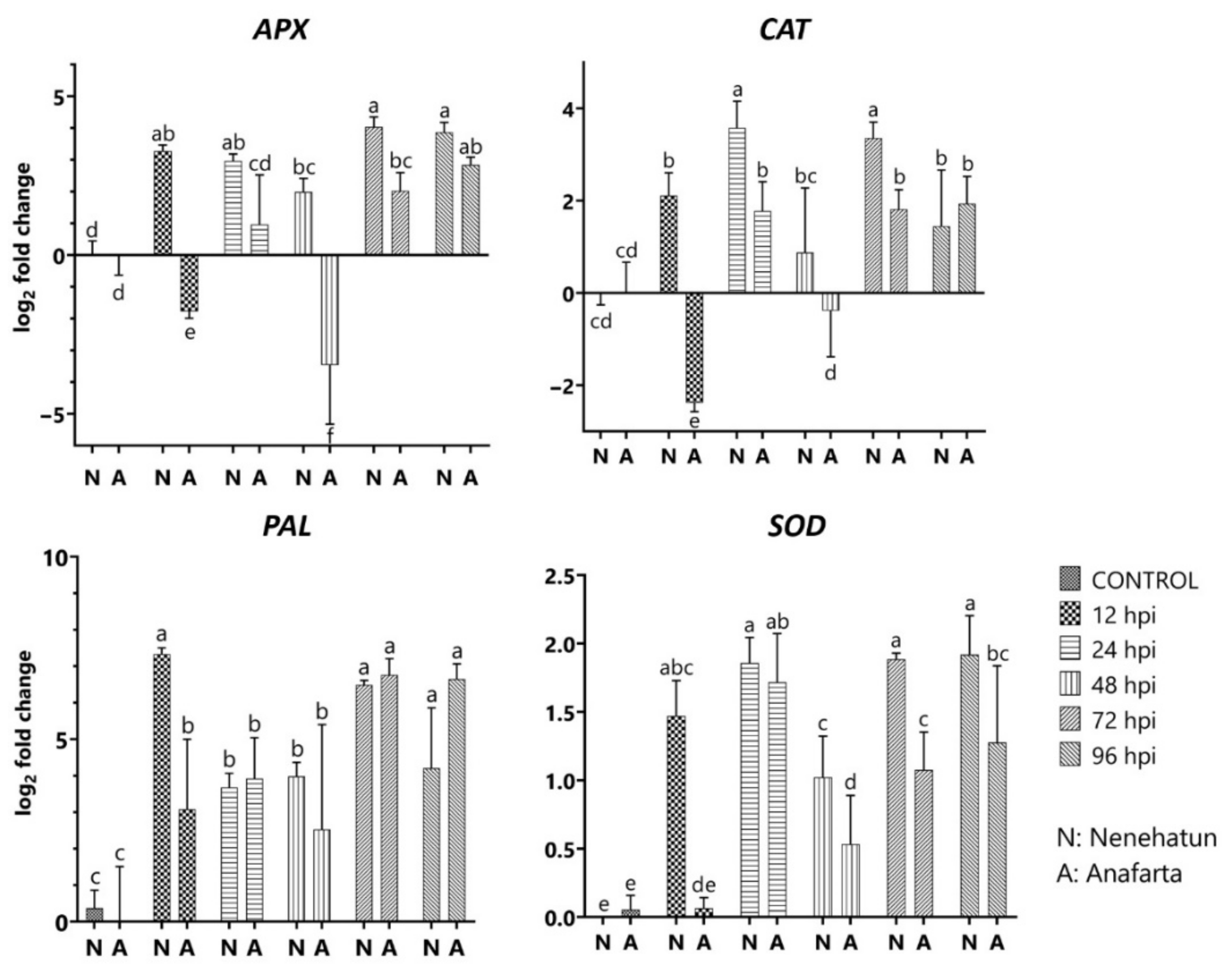
The transcript level of PAL attenuated rapidly and reached the maximum level in the susceptible cultivar at 12 hpi with 7.33 of log2 fold change, followed by a decrease until 48 hpi (Figure 6). The resistant cultivar showed a similar expression profile, ranging from 2.53 to 3.92-log2 fold until 48 hpi and strong induction of PAL gene with 6.76-log2 fold at 72 hpi. SOD activity in both cultivars remained below the baseline level at 12 and 48 hpi and the transcript level slightly increased at 24 hpi. Similarly, a slight induction was observed in the susceptible cultivar at 72 and 96 hpi, but not in the resistant cultivar. The expression enhancement of CAT activity in the susceptible cultivar continued until 24 hpi, followed by a decrease at 48 hpi, while CAT level increased again at 72 hpi about 3.36-fold. The transcript in the resistant cultivar was downregulated at 12 hpi. The highest expression of this gene in the resistant cultivar reached a peak of 1.94-log2 fold at 96 hpi but was not statistically different from that in the susceptible cultivar. APX activity induced 3.28-log2 fold in susceptible cultivar at 12 hpi, attenuated until 48 hpi, and reached the maximum level with 4.03-log2 fold at 72 hpi. The expression level in the resistant cultivar reduced −1.77 and −3.46-log2 fold at 12 and 48 hpi. The transcript was upregulated again at 72 hpi and reached 2.84-log2 fold at 96 hpi.

4. Discussion

Spot blotch, caused by B. sorokiniana, is a major disease of wheat and barley worldwide. The pathogen occurs widely in growing areas and causes important economic yield losses in cereals even to triticale that is adapted to harsh conditions [47]. In this study, the pathogen isolates from wheat fields in Bolu province were identified based on morphological characteristics and sequence analysis and evaluated for their aggressiveness. All isolates caused typical symptoms of spot blotch on inoculated wheat cultivar Seri-82 plants and indicated significant pathogenic variation, ranging from 3.3 to 5.5 of disease index. Similar results were observed in a previous study by Özer et al. [48], who reported that the disease severity of 96 isolates from the winter wheat-growing regions of Azerbaijan ranged from 1.58 to 3.60. Kang et al. [49] observed significant variation in the pathogenicity of 262 B. sorokiniana isolates from wheat samples in 97 locations of China. Similarly, variation in the aggressiveness of B. sorokiniana isolates was reported by several authors [8,50]. However, no considerable difference was found among the pathogenicity of B. sorokiniana isolates from Mexico and India [51,52]. In the current study, pathogenicity tests indicated that there were significant differences in the reactions of the 55 wheat cultivars to the TR-Cs-3isolate. The most resistant response was detected in the cultivar Anafarta and Koç-2015, while the cultivar Kırik showed the most susceptible reaction. Cultivation of these resistant cultivars in growing areas where the pathogen is common can be considered beneficial in improving the efficiency of disease management methods. Additionally, these may be potentially useful as genitor plants for breeding studies. Evaluating 625 wheat lines from a breeding program in India, Adlakha et al. [10] found 16 resistant lines. In China, resistant cultivars consisted of a low percentage among 21 wheat cultivars tested to the pathogen [49]. Testing 99 modern European winter wheat cultivars and breeding lines to four B. sorokiniana isolates revealed that the tested material had a low level of resistance [53]. These studies, which indicated the presence of limited resistance to the pathogen worldwide, increased the potential importance of resistance sources determined in this study.
Bipolaris sorokiniana exhibits a complex structure in wheat plants with other pathogens, such as Pyrenophora tritici-repentis, Stagonospora nodorum, Blumeria graminis, and Septoria tritici, which are a challenge to detect the main pathogen responsible for the disease in the early stages [54]. Classical diagnostic methods based on visual evaluation as well as microscopic and cultural methods provide limited benefit in the identification of fungal pathogens. Molecular methods have several advantages over the classical methods used to identify plant pathogens [14,16,55]. Various PCR-based methods have been developed to diagnose spot blotch disease of wheat [17,18,19]. In this study, a qPCR method was developed for detecting the pathogen in wheat tissue at the early stages of the infection process and for evaluating plant genotypes with different levels of resistance. Phylogenetically alignments of ITS, GAPDH, and SSU genes provided informative sites for separating B. sorokiniana and closely related fungi species from each other. Similarly, these conserved genes have widely been used in the taxonomic classification of Bipolaris species [15]. qPCR assay developed was highly sensitive and provided the determination of pathogen DNA up to 0.1 pg. The designed primer did not amplify DNA extracts from healthy plant tissue and other fungi species. Species-specific PCR developed by Matusinsky et al. [17] allowed detection of 0.001 ng of pathogen DNA, while a multiplex PCR (mPCR) system established for detecting wheat pathogens detected 100 pg for B. sorokiniana [56]. Real-time PCR assay described by Orina Aleksandra et al. [20] confirmed the presence of B. sorokiniana in 100% of the barley and oats samples and in 56% of wheat samples. The utility of the method was also demonstrated for confirming the presence of B. sorokiniana in plant samples at the early stages of infection and monitoring of resistant and susceptible plants as an optional method to visual scoring of disease severity. Our results showed a close relationship between the assessments of disease severity and the quantities of pathogen DNA in wheat cultivars. DNA quantities were observed at higher levels in susceptible genotypes than in resistant genotypes, suggesting this assay may possess an important potential for the discrimination of resistant and susceptible wheat genotypes. These results coincided with those of Bayraktar et al. [57], who observed a strong relationship between disease resistance and DNA quantification in resistant and susceptible chickpea cultivars to A. rabiei infection. Similarly, Daniëls et al. [58] designed a real-time PCR assay to evaluate host resistance to Venturia inaequalis. The results revealed a significant correlation between the resistance levels and DNA quantities of apple cultivars. Researchers reported that qPCR assay was the more robust and sensitive tool to assess resistance level of apple cultivars over the classical method. Leiminger et al. [59] evaluated the resistance of potato plants to Alternaria solani and A. alternata with classical and molecular methods. They found a significant correlation between the amount of pathogen DNA and the ratio of necrotic areas caused by A. solani.
The potential roles of PRs, PAL, CAT, SOD, and APX genes in defense response of wheat to B. sorokiniana were also investigated in both resistant and susceptible wheat cultivars. All the defense-related genes studied were upregulated in both cultivars except for the expression of CAT and APX genes at 12 and 48 hpi and showed considerable variation in their expression levels and timing. The upregulation was partially observed at higher levels in the susceptible cultivar than in the resistant cultivar. PR proteins, individually or in combination, have been widely reported to impair or uplift the level of defense response in plants to a wide range of pathogens [26]. PR1 genes play a key role in disease formation as a result of host–pathogen interactions [29,60]. Our results showed the upregulation of PR1 gene in defense response of both wheat cultivars, but more induced in the susceptible cultivar. Overexpression of PR1 gene was detected in defense response of wheat to Erysiphe graminis, ParaStagonospora nodorum, and Puccinia triticina [33,61,62]. Soltanloo et al. [63] reported higher expression of PR1 transcript in the resistant wheat genotypes compared to susceptible genotype upon infection with F. graminearum. Previous work carried out by Muhae-Ud-Din et al. [64] showed that the expression level of PR1.1 transcript in defense response of wheat to Tilletia controversa was higher in the resistant cultivar than in the susceptible cultivar, while the expression of PR1.2 was less expressed in the resistant cultivar than in the susceptible cultivar. PR1 gene showed no or a slight induction in both resistant and susceptible wheat genotypes to yellow dwarf virus (YDV) aphid-transmitted and Hessian fly infestation, while a strong induction was observed to FHB-resistant cultivars Ning 7840 and KS24-1 and susceptible cultivar Len [65]. Additionally, the expression level of PR1 gene in the susceptible cultivar Len was higher than that in the resistant cultivar Ning 7840, in agreement with the results of this study. PR2 proteins (β-1,3-glucanases) that are directly involved in defense mechanisms by hydrolyzing the cell walls of fungal pathogens have been characterized from a wide range of species [27]. The upregulation of PR2 gene was observed in wheat resistant to P. triticina and F. graminearum [66,67]. In this study, PR2 gene contributed to resistance in both cultivars and reached the maximum level earlier in the resistant cultivar. Similarly, the increased expression of PR1 and PR2 was associated with enhanced resistance to rust pathogens in wheat [68]. However, Wu et al. [65] detected no significant change in the expression of PR2 gene in both susceptible and resistant cultivars of wheat to F. graminearum. Our results also showed the potential involvement of PR3, PR5, and PR10, strongly inducing in both cultivars resistance pathways to B. sorokiniana. In wheat, PR3, which are chitinases, has been reported to be upregulated in response to fungal pathogens, Puccinia spp., Blumeria graminis [69], while overexpression of PR5 gene referred to as thaumatin-like proteins provided to enhance wheat resistance to leaf rust [70]. The results obtained in the present study coincided with those of Zhang et al. [68], who reported that PR1, PR2, and PR5 in wheat resistance to rust pathogens had a more significant role than PR3 and PR10 and the expression profiles of PR gene were different in response to different rust species or races of the same species. PR10 was upregulated to TaCAD12 transcript involving resistance response to sharp eyespot disease in wheat [28].
Expression of defense-related enzymes (PAL, CAT, SOD, and APX) is known to play a crucial role in the host resistance to pathogens. These enzymes are either directly or indirectly involved in plant defense pathways, such as the production of reactive oxygen species and secondary metabolites, and hypersensitive reactions. Our data presented the upregulation in gene expression levels of PAL and SOD in both cultivars to pathogen infection, but partially in the levels of CAT and APX in the resistant cultivar. Overexpression of PAL was detected in a resistant cultivar of soybean to F. solani f. sp. glycine, but not in the susceptible cultivar [71]. The silencing of PAL in wheat reduced aphid and nematode resistance [72], while PAL-RNAi in Brachypodium increased susceptibilities to the fungal pathogens F. culmorum and Magnaporthe oryzae [73]. However, the suppression of some PAL genes had no significant effect on plant resistance to F. graminearum [35]. Similar kinds of results that we observed were also reported by Christensen et al. [74], who indicated that overexpression of TaGLP4 and HvGLP4 with SOD activity enhanced resistance against B. graminis in wheat and barley, whereas transient silencing reduced basal resistance in both cereals. Our results are consistent with the results of Debona et al. [75], who detected the increased activities of SOD, POX, APX, and GST in both resistant and susceptible wheat cultivars infected with Pyricularia oryzae compared with noninoculated plants. Similarly, the antioxidant enzyme activities of the POD, CAT, and APX in resistant wheat genotypes were more efficient than susceptible genotypes to Magnaporthe oryzae [36]. Spanic et al. [34] observed differences in the antioxidant response of wheat varieties with different levels of resistance, namely an enhancement in APX and polyphenol oxidase (PPO) activity in FHB-resistant variety Vulkan in the early stages after infection, and a higher activity of POD and H2O2 in the moderately resistant variety Kraljica. Susceptible variety Golubica responded with enhanced POD activity to the pathogen.
In summary, we examined expression profiles of PRs, PAL, CAT, SOD, and APX genes in the resistance response of wheat to B. sorokiniana. Significant differences were observed depending on the timing and magnitude of these genes in wheat plants with different resistance levels. However, further investigation is necessary on other defense genes that play a role in plant defense mechanisms to gain more insight into the interaction of wheat pathogen.

Author Contributions

Conceptualization, G.Ö.; methodology, G.Ö., H.B. and M.A.; formal analysis, M.A., M.İ., F.Ö., R.L. and F.M.; writing, original draft, G.Ö. and H.B.; writing, review and editing, G.Ö., H.B., T.P. and A.A.D. All authors have read and agreed to the published version of the manuscript.

Funding

This study was supported by The Scientific and Technological Research Council of Turkey (TUBITAK), grant TOVAG 119O956.

Institutional Review Board Statement

Not applicable.

Informed Consent Statement

Not applicable.

Data Availability Statement

All relevant data generated or analyzed during this study are included in this manuscript.

Acknowledgments

This research is produced from a part of the master thesis of Mehtap Alkan. We thank Mehmet Z. Yeken and Emrah Güler for the helpful suggestion about the statistical analysis. The authors also would like to thank the CRP-WHEAT, CIMMYT for supporting this study.

Conflicts of Interest

The authors declare no conflict of interest.

References

  1. Singh, D.P. (Ed.) Management of Wheat and Barley Diseases; CRC Press: Boca Raton, FL, USA, 2017. [Google Scholar]
  2. Al-Sadi, A.M. Bipolaris sorokiniana-induced black point, common root rot, and spot blotch diseases of wheat: A review. Front. Cell. Infect. Microbiol. 2021, 11, 118. [Google Scholar] [CrossRef] [PubMed]
  3. Kumar, S.; Kumar, N.; Prajapati, S.; Maurya, S. Review on spot blotch of wheat: An emerging threat to wheat basket in changing climate. J. Pharmacogn. Phytochem. 2020, 9, 1985–1997. [Google Scholar]
  4. Tunali, B.; Nicol, J.M.; Hodson, D.; Uçkun, Z.; Büyük, O.; Erdurmuş, D.; Bağci, S.A. Root and crown rot fungi associated with spring, facultative, and winter wheat in Turkey. Plant Dis. 2008, 92, 1299–1306. [Google Scholar] [CrossRef] [PubMed] [Green Version]
  5. Özer, G.; Paulitz, T.C.; Imren, M.; Alkan, M.; Muminjanov, H.; Dababat, A.A. Identity and pathogenicity of fungi associated with crown and root rot of dryland winter wheat in Azerbaijan. Plant Dis. 2020, 104, 2149–2157. [Google Scholar] [CrossRef]
  6. Gupta, P.K.; Chand, R.; Vasistha, N.K.; Pandey, S.P.; Kumar, U.; Mishra, V.K.; Joshi, A.K. Spot blotch disease of wheat: The current status of research on genetics and breeding. Plant Pathol. 2018, 67, 508–531. [Google Scholar] [CrossRef]
  7. Ghazvini, H.; Tekauz, A. Molecular diversity in the barley pathogen Bipolaris sorokiniana (Cochliobolus sativus). Australas. Plant Pathol. 2012, 41, 283–293. [Google Scholar] [CrossRef]
  8. Al-Sadi, A.M. Variation in resistance to spot blotch and the aggressiveness of Bipolaris sorokiniana on barley and wheat cultivars. J. Plant Pathol. 2016, 98, 97–103. [Google Scholar] [CrossRef]
  9. James, W.C. Assessment of plant diseases and losses. Annu. Rev. Phytopathol. 1974, 12, 27–48. [Google Scholar] [CrossRef]
  10. Adlakha, K.L.; Wilcoxson, R.D.; Raychaudhuri, S.P. Resistance of wheat to leaf spot caused by Bipolaris sorokiniana. Plant Dis. 1984, 68, 320–321. [Google Scholar] [CrossRef] [Green Version]
  11. Fetch, T.G., Jr.; Steffenson, B.J. Rating scales for assessing infection responses of barley infected with Cochliobolus sativus. Plant Dis. 1999, 83, 213–217. [Google Scholar] [CrossRef] [Green Version]
  12. Fang, Y.; Ramasamy, R.P. Current and prospective methods for plant disease detection. Biosensors 2015, 5, 537–561. [Google Scholar] [CrossRef] [PubMed] [Green Version]
  13. Tsedaley, B. A review on disease detection, pathogen identification and population genetics in fungi. J. Biol. Agric. Healthc. 2015, 5, 6–20. [Google Scholar]
  14. Brunner, K.; Mach, R.L. Quantitative detection of fungi by molecular methods: A case study on Fusarium. In Molecular Identification of Fungi; Gherbawy, Y., Voigt, K., Eds.; Springer: Berlin/Heidelberg, Germany, 2010; pp. 179–193. [Google Scholar] [CrossRef]
  15. Manamgoda, D.S.; Cai, L.; McKenzie, E.H.; Crous, P.W.; Madrid, H.; Chukeatirote, E.; Shivas, R.G.; Tan, Y.P.; Hyde, K.D. A phylogenetic and taxonomic re-evaluation of the Bipolaris-Cochliobolus-Curvularia complex. Fungal Divers. 2012, 56, 131–144. [Google Scholar] [CrossRef]
  16. Raja, H.A.; Miller, A.N.; Pearce, C.J.; Oberlies, N.H. Fungal identification using molecular tools: A primer for the natural products research community. J. Nat. Prod. 2017, 80, 756–770. [Google Scholar] [CrossRef] [PubMed]
  17. Matusinsky, P.; Frei, P.; Mikolasova, R.; Svacinova, I.; Tvaruzek, L.; Spitzer, T. Species–specific detection of Bipolaris sorokiniana from wheat and barley tissues. Crop Prot. 2010, 29, 1325–1330. [Google Scholar] [CrossRef]
  18. Aggarwal, R.; Gupta, S.; Banerjee, S.; Singh, V.B. Development of a SCAR marker for detection of Bipolaris sorokiniana causing spot blotch of wheat. Can. J. Microbiol. 2011, 57, 934–942. [Google Scholar] [CrossRef] [PubMed]
  19. Fajolu, O.L.; Wadl, P.A.; Vu, A.L.; Gwinn, K.D.; Scheffler, B.E.; Trigiano, R.N.; Ownley, B.H. Development and characterization of simple sequence repeats for Bipolaris sorokiniana and cross transferability to related species. Mycologia 2013, 105, 1164–1173. [Google Scholar] [CrossRef] [PubMed]
  20. Orina Aleksandra, S.; Orina, A.S.; Gavrilova, O.P.; Gagkaeva, T.Y. Adaptation of the quantitative PCR method for the detection of the main representatives of cereal grain mycobiota. Microbiol. Indep. Res. J. 2018, 5, 78–83. [Google Scholar] [CrossRef]
  21. Van Loon, L.C.; Van Strien, E.A. The families of pathogenesis–related proteins, their activities, and comparative analysis of PR1 type proteins. Physiol. Mol. Plant Pathol. 1999, 55, 85–97. [Google Scholar] [CrossRef]
  22. Thakur, M.; Sohal, B.S. Role of elicitors in inducing resistance in plants against pathogen infection: A review. Int. Sch. Res. Notices 2013, 2013, 762412. [Google Scholar] [CrossRef] [Green Version]
  23. Dubey, N.; Singh, K. Role of NBS–LRR proteins in plant defense. In Molecular Aspects of Plant–Pathogen Interaction; Springer: Singapore, 2018; pp. 115–138. [Google Scholar] [CrossRef]
  24. Van Loon, L.C. Induced resistance in plants and the role of pathogenesis-related proteins. Eur. J. Plant Pathol. 1997, 103, 753–765. [Google Scholar] [CrossRef]
  25. Sels, J.; Mathys, J.; De Coninck, B.M.; Cammue, B.P.; De Bolle, M.F. Plant pathogenesis-related (PR) proteins: A focus on PR peptides. Plant Physiol. Biochem. 2008, 46, 941–950. [Google Scholar] [CrossRef] [PubMed]
  26. Ali, S.; Ganai, B.A.; Kamili, A.N.; Bhat, A.A.; Mir, Z.A.; Bhat, J.A.; Grover, A. Pathogenesis-related proteins and peptides as promising tools for engineering plants with multiple stress tolerance. Microbiol. Res. 2018, 212, 29–37. [Google Scholar] [CrossRef]
  27. Balasubramanian, V.; Vashisht, D.; Cletus, J.; Sakthivel, N. Plant β-1, 3-glucanases: Their biological functions and transgenic expression against phytopathogenic fungi. Biotechnol. Lett. 2012, 34, 1983–1990. [Google Scholar] [CrossRef] [PubMed]
  28. Rong, W.; Luo, M.; Shan, T.; Wei, X.; Du, L.; Xu, H.; Zhang, Z. A wheat cinnamyl alcohol dehydrogenase TaCAD12 contributes to host resistance to the sharp eyespot disease. Front. Plant Sci. 2016, 7, 1723. [Google Scholar] [CrossRef] [Green Version]
  29. Lincoln, J.E.; Sanchez, J.P.; Zumstein, K.; Gilchrist, D.G. Plant and animal PR1 family members inhibit programmed cell death and suppress bacterial pathogens in plant tissues. Mol. Plant Pathol. 2018, 19, 2111–2123. [Google Scholar] [CrossRef] [Green Version]
  30. Mackintosh, C.A.; Lewis, J.; Radmer, L.E.; Shin, S.; Heinen, S.J.; Smith, L.A.; Muehlbauer, G.J. Overexpression of defense response genes in transgenic wheat enhances resistance to Fusarium head blight. Plant Cell Rep. 2007, 26, 479–488. [Google Scholar] [CrossRef] [Green Version]
  31. Eissa, H.F.; Hassanien, S.E.; Ramadan, A.M.; El–Shamy, M.M.; Saleh, O.M.; Shokry, A.M.; Bahieldin, A. Developing transgenic wheat to encounter rusts and powdery mildew by overexpressing barley chi26 gene for fungal resistance. Plant Methods 2017, 13, 41. [Google Scholar] [CrossRef]
  32. Zhang, Y.; Yan, H.; Wei, X.; Zhang, J.; Wang, H.; Liu, D. Expression analysis and functional characterization of a pathogen–induced thaumatin–like gene in wheat conferring enhanced resistance to Puccinia triticina. J. Plant Interact. 2017, 12, 332–339. [Google Scholar] [CrossRef] [Green Version]
  33. Wang, F.; Yuan, S.; Wu, W.; Yang, Y.; Cui, Z.; Wang, H.; Liu, D. TaTLP1 interacts with TaPR1 to contribute to wheat defense responses to leaf rust fungus. PLoS Genet. 2020, 16, e1008713. [Google Scholar] [CrossRef]
  34. Spanic, V.; Vuletic, M.V.; Abicic, I.; Marcek, T. Early response of wheat antioxidant system with special reference to Fusarium head blight stress. Plant Physiol. Biochem. 2017, 115, 34–43. [Google Scholar] [CrossRef]
  35. Hao, Q.; Wang, W.; Han, X.; Wu, J.; Lyu, B.; Chen, F.; Fu, D. Isochorismate-based salicylic acid biosynthesis confers basal resistance to Fusarium graminearum in barley. Mol. Plant Pathol. 2018, 19, 1995–2010. [Google Scholar] [CrossRef] [PubMed] [Green Version]
  36. Biswas, C.S.; Hannan, A.; Monsur, A.; Sagor, G.H.M. Screening and biochemical characterization of wheat cultivars resistance to Magnaporthe oryzae pv. triticum (MoT). J. Plant Stress Physiol. 2020, 6, 1–6. [Google Scholar] [CrossRef] [Green Version]
  37. Sivanesan, A. Graminicolous Species of Bipolaris, Curvularia, Drechslera, Exserohilum and Their Teleomorphs; Mycological Papers No. 158; C.A.B. International: Wallingford, UK, 1987; 261p. [Google Scholar]
  38. White, T.J.; Bruns, T.; Lee, S.; Taylor, J.W. Amplification and direct sequencing of fungal ribosomal RNA genes for phylogenetics. In PCR Protocols: A Guide to Methods and Applications; Innis, M.A., Gelfand, D.H., Sninsky, J.J., White, T.J., Eds.; Academic Press: Cambridge, MA, USA, 1990; Volume 18, pp. 315–322. [Google Scholar] [CrossRef]
  39. Berbee, M.L.; Pirseyedi, M.; Hubbard, S. Cochliobolus phylogenetics and the origin of known, highly virulent pathogens, inferred from ITS and glyceraldehyde-3-phosphate dehydrogenase gene sequences. Mycologia 1999, 91, 964–977. [Google Scholar] [CrossRef]
  40. Katoh, K.; Standley, D.M. MAFFT multiple sequence alignment software version 7: Improvements in performance and usability. Mol. Biol. Evol. 2013, 30, 772–780. [Google Scholar] [CrossRef] [PubMed] [Green Version]
  41. Kumar, S.; Stecher, G.; Tamura, K. MEGA7: Molecular evolutionary genetics analysis version 7.0 for bigger datasets. Mol. Biol. Evol. 2016, 33, 1870–1874. [Google Scholar] [CrossRef] [Green Version]
  42. Felsenstein, J. Confidence limits on phylogenies: An approach using the bootstrap. Evolution 1985, 39, 783–791. [Google Scholar] [CrossRef]
  43. Untergasser, A.; Cutcutache, I.; Koressaar, T.; Ye, J.; Faircloth, B.C.; Remm, M.; Rozen, S.G. Primer3-new capabilities and interfaces. Nucleic Acids Res. 2012, 40, e115. [Google Scholar] [CrossRef] [Green Version]
  44. Livak, K.J.; Schmittgen, T.D. Analysis of relative gene expression data using real–time quantitative PCR and the 2−ΔΔCT method. Methods 2001, 25, 402–408. [Google Scholar] [CrossRef]
  45. Desmond, O.J.; Edgar, C.I.; Manners, J.M.; Maclean, D.J.; Schenk, P.M.; Kazan, K. Methyl jasmonate induced gene expression in wheat delays symptom development by the crown rot pathogen Fusarium pseudograminearum. Physiol. Mol. Plant Pathol. 2005, 67, 171–179. [Google Scholar] [CrossRef]
  46. Zhang, H.; Wang, C.; Cheng, Y.; Wang, X.; Li, F.; Han, Q.; Kang, Z. Histological and molecular studies of the non–host interaction between wheat and Uromyces fabae. Planta 2011, 234, 979–991. [Google Scholar] [CrossRef] [PubMed]
  47. Özer, G.; İmren, M.; Özdemir, F.; Morgounov, A.; Dababat, A.A. First report of common root rot on triticale caused by Bipolaris sorokiniana in Kazakhstan. Plant Dis. 2020, 104, 2735. [Google Scholar] [CrossRef]
  48. Özer, G.; Imren, M.; Alkan, M.; Paulitz, T.C.; Bayraktar, H.; Palacıoğlu, G.; Dababat, A.A. Molecular and pathogenic characterization of Cochliobolus anamorphs associated with common root rot of wheat in Azerbaijan. Phytopathol. Mediterr. 2020, 59, 147–158. [Google Scholar] [CrossRef]
  49. Kang, R.; Hu, Y.; Wang, L.; Xie, S.; Li, Y.; Yuan, H.; Li, H. Pathogenicity variation and DNA polymorphism of Bipolaris sorokiniana infecting winter wheat in the Huanghuai floodplain of China. Plant Pathol. 2021, 70, 87–99. [Google Scholar] [CrossRef]
  50. Sultana, S.; Adhikary, S.K.; Islam, M.M.; Rahman, S.M.M. Evaluation of pathogenic variability based on leaf blotch disease development components of Bipolaris sorokiniana in Triticum aestivum and agroclimatic origin. Plant Pathol. J. 2018, 34, 93. [Google Scholar] [CrossRef]
  51. Duveiller, E.; Garcia Altamirano, I. Pathogenicity of Bipolaris sorokiniana isolates from wheat roots, leaves and grains in Mexico. Plant Pathol. 2000, 49, 235–242. [Google Scholar] [CrossRef]
  52. Jaiswal, S.K.; Prasad, L.C.; Sharma, S.; Kumar, S.; Prasad, R.; Pandey, S.P.; Joshi, A.K. Identification of molecular marker and aggressiveness for different groups of Bipolaris sorokiniana isolates causing spot blotch disease in wheat (Triticum aestivum L.). Curr. Microbiol. 2007, 55, 135–141. [Google Scholar] [CrossRef]
  53. Liatukas, Ž.; Ruzgas, V. Resistance of European winter wheat cultivars to spot blotch at juvenile growth stages. Phytopathol. Mediterr. 2011, 50, 350–358. [Google Scholar] [CrossRef]
  54. Bockus, W.W.; Bowden, R.L.; Hunger, R.M.; Morril, W.L.; Murray, T.D.; Smiley, R.W. Diseases caused by fungi and fungus–like organisms. In Compendium Wheat Diseaseand Pests; Bockus, W.W., Bowden, R.L., Hunger, R.M., Morrill, W.L., Murray, T.D., Smiley, R.W., Eds.; APS Press: St. Paul, MN, USA, 2010; pp. 15–86. [Google Scholar] [CrossRef]
  55. Özer, G.; Bayraktar, H. Development of conventional and real-time PCR assays to detect Alternaria burnsii in cumin seed. Gesunde Pflanz. 2019, 71, 205–212. [Google Scholar] [CrossRef]
  56. Sun, X.; Zhang, L.; Meng, C.; Zhang, D.; Xu, N.; Yu, J. Establishment and application of a multiplex PCR assay for detection of Rhizoctonia cerealis, Bipolaris sorokiniana, and Fusarium spp. in winter wheat. J. Plant Pathol. 2020, 102, 19–27. [Google Scholar] [CrossRef]
  57. Bayraktar, H.; Özer, G.; Aydoğan, A.; Palacıoğlu, G. Determination of Ascochyta blight disease in chickpea using real–time PCR. J. Plant Dis. Prot. 2016, 123, 109–117. [Google Scholar] [CrossRef]
  58. Daniëls, B.; Dreesen, R.; Davey, M.W.; Keulemans, J. Real–time PCR as a promising tool to monitor growth of Venturia spp. in scab–susceptible and–resistant apple leaves. Eur. J. Plant Pathol. 2012, 134, 821–833. [Google Scholar] [CrossRef]
  59. Leiminger, J.; Bäßler, E.; Knappe, C.; Bahnweg, G.; Hausladen, H. Quantification of disease progression of Alternaria spp. on potato using real-time PCR. Eur. J. Plant Pathol. 2015, 141, 295–309. [Google Scholar] [CrossRef]
  60. Zhu, F.; Xu, M.; Wang, S.; Jia, S.; Zhang, P.; Lin, H.; Xi, D. Prokaryotic expression of pathogenesis related protein 1 gene from Nicotiana benthamiana: Antifungal activity and preparation of its polyclonal antibody. Biotechnol. Lett. 2012, 34, 919–924. [Google Scholar] [CrossRef] [PubMed]
  61. Molina, A.; Görlach, J.; Volrath, S.; Ryals, J. Wheat genes encoding two types of PR1 proteins are pathogen inducible, but do not respond to activators of systemic acquired resistance. Mol. Plant-Microbe Interact. 1999, 12, 53–58. [Google Scholar] [CrossRef] [PubMed] [Green Version]
  62. Sung, Y.C.; Outram, M.A.; Breen, S.; Wang, C.; Dagvadorj, B.; Winterberg, B.; Solomon, P.S. PR1-mediated defense via C-terminal peptide release is targeted by a fungal pathogen effector. New Phytol. 2021, 229, 3467–3480. [Google Scholar] [CrossRef]
  63. Soltanloo, H.; Khorzoghi, E.G.; Ramezanpour, S.; Arabi, M.K.; Pahlavani, M.H. The expression profile of ‘Chi-1, Glu-2, Glu-3 and PR1. 2’ genes in scab–resistant and susceptible wheat cultivars during enfection by ‘Fusarium graminearum’. Plant Omics 2010, 3, 162–166. [Google Scholar]
  64. Muhae-Ud-Din, G.; Chen, D.; Liu, T.; Chen, W.; Gao, L. Characterization of the wheat cultivars against Tilletia controversa Kühn, causal agent of wheat dwarf bunt. Sci. Rep. 2020, 10, 9029. [Google Scholar] [CrossRef]
  65. Wu, S.W.; Wang, H.W.; Yang, Z.D.; Kong, L.R. Expression comparisons of pathogenesis–related (PR) genes in wheat in response to infection/infestation by Fusarium, Yellow dwarf virus (YDV) aphid–transmitted and hessian fly. J. Integr. Agric. 2014, 13, 926–936. [Google Scholar] [CrossRef]
  66. Li, W.L.; Faris, J.D.; Muthukrishnan, S.; Liu, D.J.; Chen, P.D.; Gill, B.S. Isolation and characterization of novel cDNA clones of acidic chitinases and β-1, 3-glucanases from wheat spikes infected by Fusarium graminearum. Theor. Appl. Genet. 2001, 102, 353–362. [Google Scholar] [CrossRef]
  67. Casassola, A.; Brammer, S.P.; Chaves, M.S.; Martinelli, J.A.; Stefanato, F.; Boyd, L.A. Changes in gene expression profiles as they relate to the adult plant leaf rust resistance in the wheat cv. Toropi. Physiol. Mol. Plant Pathol. 2015, 89, 49–54. [Google Scholar] [CrossRef]
  68. Zhang, H.; Qiu, Y.; Yuan, C.; Chen, X.; Huang, L. Fine–tuning of PR genes in wheat responding to different Puccinia rust species. J. Plant Physiol. Pathol. 2018, 6, 2. [Google Scholar] [CrossRef]
  69. Campbell, J.; Zhang, H.; Giroux, M.J.; Feiz, L.; Jin, Y.; Wang, M.; Huang, L. A mutagenesis–derived broad–spectrum disease resistance locus in wheat. Theor. Appl. Genet. 2012, 125, 391–404. [Google Scholar] [CrossRef] [PubMed] [Green Version]
  70. Li, X.; Zhang, Y.; Zhang, W.; Zhang, J.; Wang, H.; Liu, D. Expression profiles of pathogenesis–related gene, TaLr35PR1, as it relate to Lr35–mediated adult plant leaf rust resistance. Plant Mol. Biol. Rep. 2016, 34, 1127–1135. [Google Scholar] [CrossRef]
  71. Iqbal, M.J.; Yaegashi, S.; Ahsan, R.; Shopinski, K.L.; Lightfoot, D.A. Root response to Fusarium solani f. sp. glycines: Temporal accumulation of transcripts in partially resistant and susceptible soybean. Theor. Appl. Genet. 2005, 110, 1429–1438. [Google Scholar] [CrossRef] [PubMed]
  72. Zhang, H.; Huang, Q.; Yi, L.; Song, X.; Li, L.; Deng, G.; Long, H. PAL-mediated SA biosynthesis pathway contributes to nematode resistance in wheat. Plant J. 2021, 107, 698–712. [Google Scholar] [CrossRef]
  73. Cass, C.L.; Peraldi, A.; Dowd, P.F.; Mottiar, Y.; Santoro, N.; Karlen, S.D.; Sedbrook, J.C. Effects of Phenylalanine Ammonia Lyase (PAL) knockdown on cell wall composition, biomass digestibility, and biotic and abiotic stress responses in Brachypodium. J. Exp. Bot. 2015, 66, 4317–4335. [Google Scholar] [CrossRef] [Green Version]
  74. Christensen, A.B.; Thordal–Christensen, H.; Zimmermann, G.; Gjetting, T.; Lyngkjær, M.F.; Dudler, R.; Schweizer, P. The germinlike protein GLP4 exhibits superoxide dismutase activity and is an important component of quantitative resistance in wheat and barley. Mol. Plant-Microbe Interact. 2004, 17, 109–117. [Google Scholar] [CrossRef] [Green Version]
  75. Debona, D.; Rodrigues, F.Á.; Rios, J.A.; Nascimento, K.J.T. Biochemical changes in the leaves of wheat plants infected by Pyricularia oryzae. Phytopathology 2012, 102, 1121–1129. [Google Scholar] [CrossRef] [Green Version]
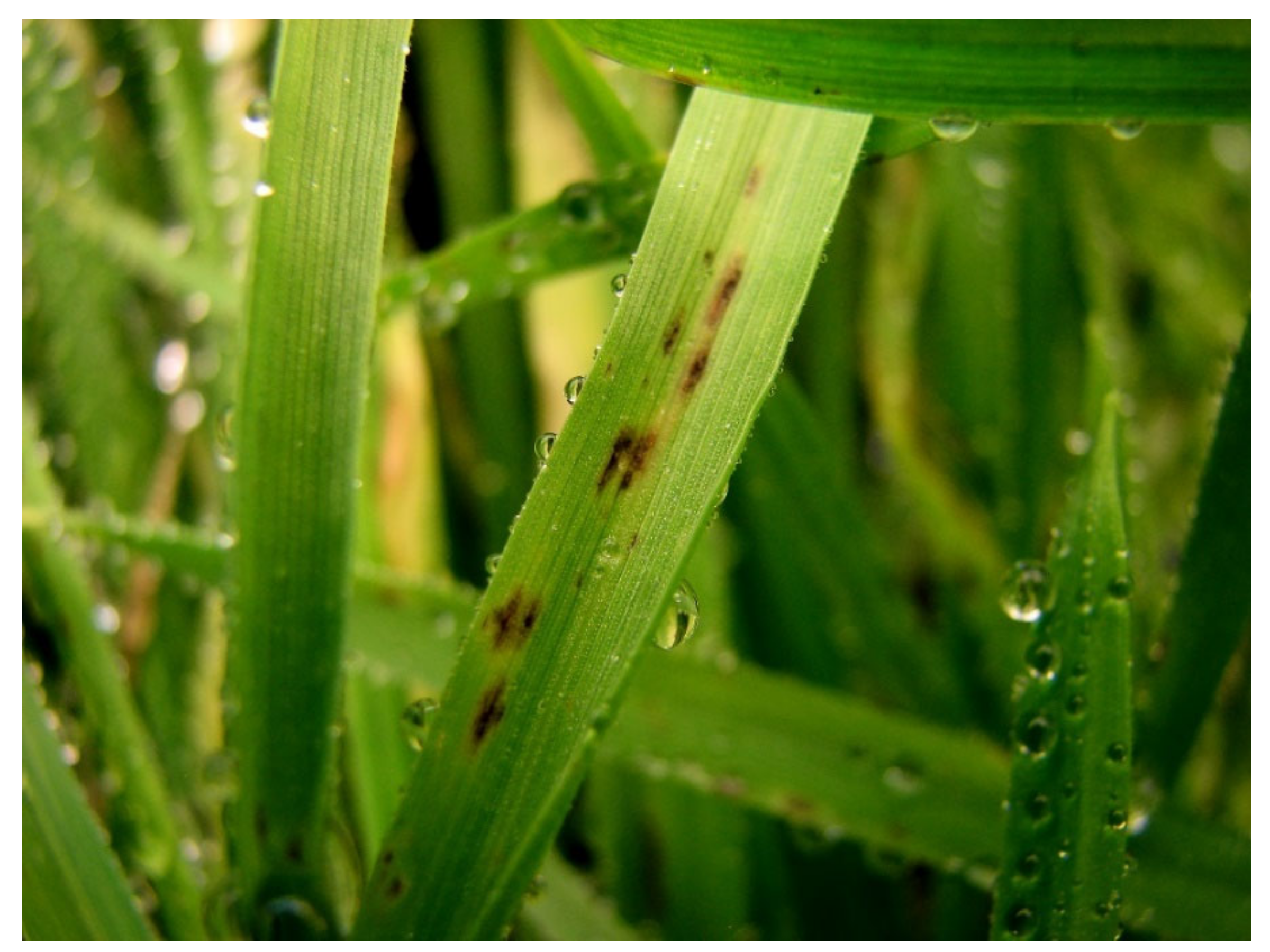
Figure 1. Typical spot blotch symptom caused by Bipolaris sorokiniana on wheat plants.
Jof 08 00149 g001
Figure 2. The Maximum Likelihood tree was generated using the ITS/GAPDH sequences of fungal isolates from this study (bold) and reference isolates derived from GenBank. The percentage of replicate trees in which the associated taxa clustered together in the bootstrap test (1000 replicates) is shown next to the branches.
Jof 08 00149 g002
Figure 3. Real-time amplification profiles and melting curves for a 10-fold dilution series of Bipolaris sorokiniana genomic DNA, starting from 10 ng in 20 ng DNA of a healthy wheat plant using primers BsGAPDHF/BsGAPDHR (a), BsITSF/BsITSR (b), and BsSSUF/BsSSUR (c) designed in this study.
Jof 08 00149 g003
Figure 4. Quantification of Bipolaris sorokiniana infection in the resistant (ALT: Altay, ANF: Anafarta, KOÇ: Koç-2015) and susceptible (DAM: Damla, KIR: Kırik, NEN: Nenehatun) wheat cultivars using primers of BsGAPDHF/BsGAPDHR, BsITSF/BsITSR, and BsSSUF/BsSSUR on the third, fifth, and eighth days after inoculation. Each error bar is constructed using one standard error from the mean. Levels connected by the same letter on each line are not significantly different DNA amounts based on Tukey’s HSD.
Jof 08 00149 g004
Figure 5. Expression profiles of the genes PR1, PR2, PR3, PR5, and PR10 in susceptible (Nenehatun) and resistant (Anafarta) wheat cultivars at different time points post-inoculation of Bipolaris sorokiniana. Each column represents an average of three replicates, and error bars represent the standard error of means. Bars with different letters are significantly different from each other based on Student’s t-test analysis.
Jof 08 00149 g005
Figure 6. Expression profiles of the genes APX, CAT, PAL, and SOD in the susceptible (Nenehatun) and resistant (Anafarta) wheat cultivars at different time points post-inoculation of Bipolaris sorokiniana. Each column represents an average of three replicates, and error bars represent the standard error of means. Means indicated with different letters are significantly different from each other based on Student’s t-test analysis.
Jof 08 00149 g006
Table 1. List of wheat cultivars used in this study and their reactions to the most aggressive Bipolaris sorokiniana isolate TR-Cs-3.
Wheat CultivarSourceWheat TypeDisease Severity *
KırikEast Anatolian Agricultural Research InstituteWinter7.97 a
LancerEast Anatolian Agricultural Research InstituteWinter7.50 a
Doğu-88East Anatolian Agricultural Research InstituteWinter6.27 b
NenehatunEast Anatolian Agricultural Research InstituteWinter6.13 b
DamlaTrakya Agricultural Research InstituteWinter6.03 b
Palandöken-97East Anatolian Agricultural Research InstituteWinter5.43 bc
Karasu-90East Anatolian Agricultural Research InstituteWinter5.30 bd
Ceyhan-99Eastern Mediterranean Agricultural Research InstituteSpring5.20 be
AyyıldızEast Anatolian Agricultural Research InstituteWinter4.87 cf
Es-26Transitional Zone Agricultural Research InstituteWinter4.60 cg
SarıbaşakEastern Mediterranean Agricultural Research InstituteSpring4.60 cg
Soyer-02Transitional Zone Agricultural Research InstituteWinter4.47 ch
MüfitbeyTransitional Zone Agricultural Research InstituteWinter4.47 ch
Bezostaya-1Transitional Zone Agricultural Research InstituteWinter4.40
Seri-2013Eastern Mediterranean Agricultural Research InstituteSpring4.40
NevzatbeyBlack Sea Agricultural Research InstituteWinter4.40
Adana-99Eastern Mediterranean Agricultural Research InstituteSpring4.37
KöprüTrakya Agricultural Research InstituteWinter4.33
EkinoksEastern Mediterranean Agricultural Research InstituteSpring4.27 dj
İzgi-2001Transitional Zone Agricultural Research InstituteWinter4.26 dj
YükselTrakya Agricultural Research InstituteWinter4.13 ek
BereketTrakya Agricultural Research InstituteWinter4.13 ek
GökkanEastern Mediterranean Agricultural Research InstituteSpring4.03 fk
YunusTransitional Zone Agricultural Research InstituteWinter3.93 fl
AlturnaEast Anatolian Agricultural Research InstituteWinter3.90 fm
Gerek-79Transitional Zone Agricultural Research InstituteWinter3.90 fm
AldaneTrakya Agricultural Research InstituteWinter3.87 fm
Sönmez-2001Transitional Zone Agricultural Research InstituteWinter3.87 fm
CandaşEastern Mediterranean Agricultural Research InstituteSpring3.83 fn
AlparslanEast Anatolian Agricultural Research InstituteWinter3.83 fn
Harmankaya-99Transitional Zone Agricultural Research InstituteWinter3.77 fn
Sultan-95Transitional Zone Agricultural Research InstituteWinter3.77 fn
SakinBlack Sea Agricultural Research InstituteWinter3.73 fn
KirveBlack Sea Agricultural Research InstituteSpring3.60 go
NacibeyTransitional Zone Agricultural Research InstituteWinter3.60 go
ÖzcanBlack Sea Agricultural Research InstituteWinter3.53 go
Canik-2003Black Sea Agricultural Research InstituteWinter3.50 go
OsmaniyemEastern Mediterranean Agricultural Research InstituteSpring3.47 go
Çetinel-2000Transitional Zone Agricultural Research InstituteWinter3.37 hp
DaphanEast Anatolian Agricultural Research InstituteWinter3.30 ıq
TekirdağTrakya Agricultural Research InstituteWinter3.30 ıq
SabanTrakya Agricultural Research InstituteWinter3.30 ıq
PehlivanTrakya Agricultural Research InstituteWinter3.13 jo
AltındaneBlack Sea Agricultural Research InstituteSpring3.03 kq
MesutTransitional Zone Agricultural Research InstituteWinter2.87 lq
Alpu-2001Transitional Zone Agricultural Research InstituteWinter2.87 lq
SelimiyeTrakya Agricultural Research InstituteWinter2.80 lq
AltayTransitional Zone Agricultural Research InstituteWinter2.77 mr
YıldırımEast Anatolian Agricultural Research InstituteWinter2.77 mr
AltınbaşakEastern Mediterranean Agricultural Research InstituteSpring2.70 mr
AbideTrakya Agricultural Research InstituteWinter2.53 or
GeliboluTrakya Agricultural Research InstituteWinter2.27 pr
YakamozEastern Mediterranean Agricultural Research InstituteSpring2.17 qr
Koç-2015Bati Akdeniz Agricultural Research InstituteSpring1.63 r
AnafartaTrakya Agricultural Research InstituteWinter1.63 r
* The values with the same letters are not significantly different from each other based on the HSD (p = 0.05).
Table 2. Sequences of primers used for the detection of Bipolaris sorokiniana and the evaluation of the expression level of defense-related genes in wheat.
Target GenesPrimer NamesSequence (5′–3′)References
ITSBsITSFTTCTGGGAGACTCGCCTTA
BsITSRGTCTTGATGGATTACCGTCCTT
GAPDHBS_F01CCATTCACGCATATTAAAGCTGin this study
BS_R01CTCTGGTGAAAGGTTCTGGATT
SSUBsSSUFGCGAAGGCAAACCTCTATGTA
BsSSURCGTCCCTCAACGTCAGTTATAG
β-tubulinβ-tubulin_FGCCATGTTCAGGAGGAAGG[45]
β-tubulin_RCTCGGTGAACTCCATCTCGT
PR1TaPR1_FGAGAATGCAGACGCCCAAGC
TaPR1_RCTGGAGCTTGCAGTCGTTGATC
PR2TaPR2_FAGGATGTTGCTTCCATGTTTGCCG
TaPR2_RAAGTAGATGCGCATGCCGTTGATG
PR3TaPR3_FTACTGCTTCAAGGACCAGATAGAC
TaPR3_RCACCAGGTTCGGGTTGTTTA
PR5TaPR5_FCAAGCAGTGGTATCAACGCAGAG
TaPR5_RGTGAAGCCACAGTTGTTCTTGATGTT
PR10TaPR10_FTTAAACCAGCACGAGAAACATCAG
TaPR10_RATCCTCCCTCGATTATTCTCACG
Phenylalanine ammonia-lyaseTaPAL_FCGTCAAGAGCTGTGTGAAGATGG[46]
TaPAL_RGGTAGTTGGAGCTGCAAGGGTC
CatalaseTaCAT_FTGCCTGTGTTTTTTATCCGAGA
TaCAT_RCTGCTGATTAAGGTGTAGGTGTTGA
Superoxide dismutaseTaSOD_FCGATAGCCAGATTCCTTTGACT
TaSOD_RGAAACCAGCGACCTACAACG
Ascorbate-peroxidaseTaAPX_FGGTTTGAGTGACCAGGACATTG
TaAPX_RGCATCCTCATCCGCAGCAT
Publisher’s Note: MDPI stays neutral with regard to jurisdictional claims in published maps and institutional affiliations.