Article

Transcriptome Analysis Revealed That Hydrogen Peroxide-Regulated Oxidative Phosphorylation Plays an Important Role in the Formation of Pleurotus ostreatus Cap Color

1
College of Food Science and Engineering, Shanxi Agricultural University, Taigu 030801, China
2
Shanxi Key Laboratory of Edible Fungi for Loess Plateau, Taigu 030801, China
3
Shanxi Research Center for Engineering Technology of Edible Fungi, Taigu 030801, China
*
Authors to whom correspondence should be addressed.
These authors contributed equally to this work.
J. Fungi 2023, 9(8), 823; https://doi.org/10.3390/jof9080823
Submission received: 14 June 2023 / Revised: 29 July 2023 / Accepted: 31 July 2023 / Published: 3 August 2023
(This article belongs to the Section Fungal Cell Biology, Metabolism and Physiology)

Abstract

:
Pleurotus ostreatus is widely cultivated in China. H2O2, as a signaling molecule, can regulate the formation of cap color, but its regulatory pathway is still unclear, severely inhibiting the breeding of dark-colored strains. In this study, 614 DEGs specifically regulated by H2O2 were identified by RNA-seq analysis. GO-enrichment analysis shows that DEGs can be significantly enriched in multiple pathways related to ATP synthesis, mainly including proton-transporting ATP synthesis complex, coupling factor F(o), ATP biosynthetic process, nucleoside triphosphate metabolic processes, ATP metabolic process, purine nucleoside triphosphate biosynthetic and metabolic processes, and purine ribonuclease triphosphate biosynthetic metabolic processes. Further KEGG analysis revealed that 23 DEGs were involved in cap color formation through the oxidative phosphorylation pathway. They were enriched in Complexes I, III, IV, and V in the respiratory chain. Further addition of exogenous uncoupling agents and ATP synthase inhibitors clarifies the important role of ATP synthesis in color formation. In summary, H2O2 may upregulate the expression of complex-encoding genes in the respiratory chain and promote ATP synthesis, thereby affecting the formation of cap color. The results of this study lay the foundation for the breeding of dark-colored strains of P. ostreatus and provide a basis for the color-formation mechanism of edible fungi.

1. Introduction

Fungal melanin is a high-molecular weight pigment with multiple biological functions. At present, with increasing attention being paid to food safety, natural pigments have received widespread attention. Studies have shown that fungal melanin is a potential antibacterial agent [1]. For example, Schizophyllum commune melanin has significant antibacterial activity against multidrug-resistant pathogens [2]. Streptomyces glaucescens melanin has in vitro anticancer activity against skin cancer cells. Furthermore, melanin is a highly effective radiation protective agent, and its radiation protection mechanism may be related to regulating survival-promoting signals, preventing oxidative stress, and regulate immunity. In addition, natural melanin also has anti-inflammatory activity and hypoglycemic and antihyperlipidemic effects [3]. It also has potential as a functional food [4]. Meanwhile, melanin may be useful as a potent agent for the prevention and management of high-fat diet-induced hyperlipidemia [5] with significant immunoregulatory activities, and it might be a promising source of immunoregulators in the healthcare field [6].
Recently, as an important source of natural pigments, research on the color-related properties of edible mushrooms has gradually begun. At present, various edible mushroom pigments have been extracted and identified. For example, carotenoids from Cordyceps militaris [7], red pigments from Lactarius lilacinus [8], and melanins from Auricularia heimuer [9] have all been studied and reported. Due to people’s desire for health, edible mushrooms are popular due to their functional biomolecules and biological activities. Therefore, research on the function of edible mushroom melanin has also been gradually carried out. For example, A. heimuer melanin has a significant effect on the treatment of alcoholic liver injury in vitro and in vivo, which may be an effective strategy to alleviate alcohol-induced liver injury [10]. The application of omics technology in the field of edible mushroom research has promoted in-depth research on the melanin synthesis pathway of edible mushrooms. For example, 26 genes were selected from the Agaricus bisporus H97 genome to participate in melanin synthesis through omics analysis [11]. Cytochrome p450 plays an important role in the formation of the cap color of Hypsizygus marmoreus. The L-DOPA pathway is the main pathway of melanin synthesis in the cap according to transcriptome analysis [12].
P. ostreatus, as a typical heterothallism edible fungus, is widely cultivated all over the world [13,14] and has the potential to be a model organism of edible mushrooms. In addition, P. ostreatus is rich in nutrients and contains various bioactive substances. It has various functions, such as antitumor, antioxidant, anti-inflammatory, and antiviral activities [15]. With the development of the big health industry, “black food” is favored by consumers because it contains mang anthocyanidins, proteins, vitamins, and other substances [16]. The color depth of the mushroom cap has become an important factor affecting consumers’ choices. From the perspective of the consumer market, the breeding of dark-colored varieties is an inevitable choice to improve industrial efficiency [17]. Cap color is an important agronomic trait that varies greatly among different varieties. Second, the color difference of the fruiting body cap will also be affected by various environmental factors. Current research shows that melanin mainly exists in three different forms: true melanin, brown melanin, and allomelanin [18,19]. In Pleurotus spp., the change in the color of the fruiting body cap is caused by the different relative ratios of true melanin and brown melanin [20]. However, the molecular mechanism is still unknown, which seriously restricts the breeding of dark-colored varieties of edible mushrooms.
H2O2, as a signaling molecule, can participate in various intracellular metabolic processes. Research has shown that H2O2 can participate in plant growth and morphogenesis, including seed germination, root gravitropism, and secondary wall differentiation [21,22,23]. Moreover, H2O2 is an important signaling molecule that can mediate stomatal closure and gene expression in the abscisic acid response, as well as UV-B-induced isoflavone accumulation [24,25]. In addition, H2O2 also plays an important role in the synthesis of melanin. Previous studies have shown that when H2O2 is present, cytochrome c can oxidize catecholamines and their cysteine derivatives, ultimately producing melanin. In contrast, when H2O2 is lacking, melanin cannot be formed [26]. This indicates the importance of H2O2 in the melanin-synthesis pathway. In addition, studies have shown that H2O2 can activate melanogenesis-related proteins, including cAMP responsive element-binding proteins, microphthalmia-related transcription factors, tyrosinase, and phenylalanine hydroxylase, thereby promoting melanogenesis [27,28]. Furthermore, H2O2 can increase the expression of ATP5B, intracellular ATP, and cAMP levels, thereby increasing the expression of melanin-promoting proteins and cellular melanogenesis [29]. In fungi, H2O2 can also serve as a signaling molecule to regulate fungal growth and development [30]. However, whether H2O2 can affect fungal color formation has not been reported.
At present, the mature construction of genetic transformation systems in edible mushrooms promotes the study of gene function and regulatory mechanisms. For example, research has been conducted on the regulatory mechanism of ganoderic acid biosynthesis in G. lucidum [31], the response mechanism to heat stress in Pleurotus ostreatus [32,33,34], and the growth and development mechanism of Flammulina filiformis [35,36]. This establishes a foundation for in-depth exploration of the molecular mechanism of color formation in edible mushrooms. Our previous research found that H2O2 can affect the expression of key genes in the melanin synthesis pathway, thereby affecting the formation of cap color [37]. In this study, RNA-seq-based transcriptome analysis was applied to further investigate the molecular mechanism by which H2O2 affects the formation of cap color. Our results revealed a global transcriptional response and further illustrated that the oxidative phosphorylation pathway may play a key role in the formation of cap color.

2. Materials and Methods

2.1. Strain

The strain CCMSSC 00389 of P. ostreatus was provided by the China Center for Mushroom Spawn Standards and Control and stored in the germplasm resource bank of Shanxi Agricultural University. The genome of strain CCMSSC 00389 has been published and can be obtained from DDBJ/EMBL/GenBank under the registration number MAYC0000000 [38].

2.2. Experiment with the Addition of Exogenous H2O2 or N,N′-dimethylthiourea (DMTU)

In this study, mushroom culture materials were prepared, bottled (200 g per bottle), and sterilized at 126 °C for 2 h for mushroom production. According to previous research reports [33], cultivation bottles were grouped and treated with an exogenous addition of H2O2 and DMTU. The bottles of different groups were processed once a day, and the changes in cap color were observed.

2.3. RNA Extraction, cDNA Library Construction, and RNA-seq

To understand the molecular mechanism through which H2O2 acts as a signaling molecule affecting the formation of cap color, young fruiting body samples of different treatment groups (control, 25 mM H2O2, and 50 mM DMTU) were established for RNA-seq. In this study, the total RNA of young fruiting bodies was extracted using TRIzol® Reagent (Invitrogen, Carlsbad, CA, USA), and contaminating DNA was removed using DNase I (TaKaRa, Kyoto, Japan). Then, 1 μg of total RNA samples was processed using the TruSeqTM RNA sample preparation kit from Illumina (San Diego, CA, USA) to construct the RNA-seq transcriptome library. The RNA-seq library was sequenced with the Illumina NovaSeq 6000 sequencer. The sequencing data were deposited into the Sequence Read Archive of the National Center for Biotechnology Information (NCBI) with the accession number SUB13511634.

2.4. Analysis of DEGs

The fragments per kilobase of exon per million mapped reads (FRKM) method was used to normalize the expression levels of genes between the control, H2O2, and DMTU groups. The gene abundances were quantified using the expression quantification software RSEM. Then, DEGSeq2 was used to compare the different gene expression levels between different sample groups and screen for DEGs.

2.5. Bioinformatics Analysis

To further explore the regulatory pathways influenced by H2O2, Gene Ontology (GO) enrichment analysis was conducted on DEGs using the software Goatools to obtain GO functions that can be enriched by DEGs. Second, enrichment analysis of DEGs was conducted through the Kyoto Encyclopedia of Genes and Genomes (KEGG) database (http://www.genome.jp/kegg/, accessed on 28 December 2022) to obtain the regulatory pathways in which DEGs were enriched. Then, the connection between DEGs was analyzed based on the correlation between gene expressions.

2.6. Quantitative Real-Time PCR (qPCR)

Total RNA was extracted from different samples using the E.Z.N.A. plant RNA Kit (Omega Bio-Tek, Norcross, GA, USA) and subsequently converted to cDNA. In addition, the expression of DEGs in different samples was detected through qPCR by a ChamQTM SYBR qPCR Master Mix Kit (Vazyme, Nanjing, China). In this study, the β-actin gene and β-tubulin gene were used as references, and the relative expression of the gene was calculated according to the 2−ΔΔCT method [34]. The primers used in this study for qPCR are shown in Supplementary Table S1.

2.7. Experiment with the Addition of Exogenous Oligomycin A or Valinomycin

Based on RNA-seq analysis, we further explored the factors that affect the formation of fungal cap color by spraying exogenous ATP synthase inhibitors (Oligomycin A) and uncoupling agents (valinomycin). According to the previous method, the cultivation bottles were prepared and incubated and then cultured at 25 °C in the dark. After the mycelium grows fully in the culture bottle, the bottles were transferred to the mushroom production box for production experiments. After the formation of young fruiting bodies, the bottles were divided into five groups with 10 bottles in each group. Considering that both Oligomycin A and valinomycin are insoluble in water, 10% ethanol was used for dissolution in this experiment. Therefore, 10% ethanol was established as the control group, and the other two groups were sprayed with 25 mM or 50 mM valinomycin externally. The last two groups were sprayed with 25 mM or 50 mM Oligomycin A externally.

2.8. Data Analysis

The experimental data were analyzed by one-way ANOVA according to Duncan’s test by SPSS software. GraphPad Prism 6 and photoshop software were used for figure analysis.

3. Results

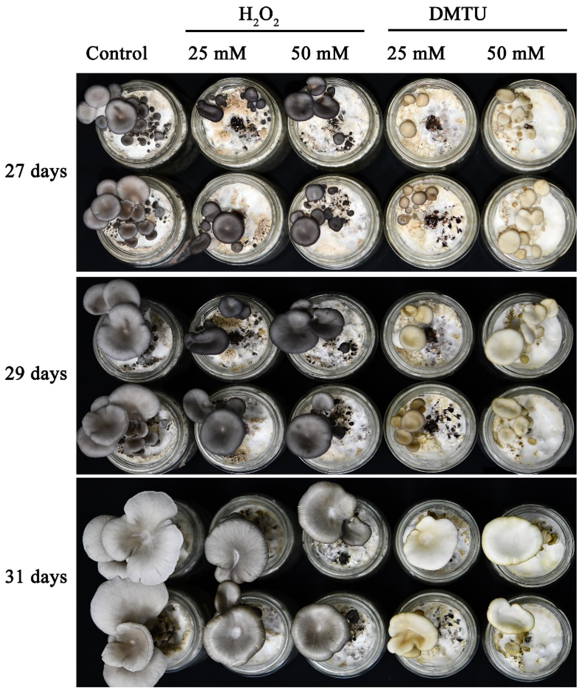
3.1. H2O2 Regulates Cap Color Formation in P. ostreatus

To determine whether H2O2 regulates cap color formation in P. ostreatus, we treated the P. ostreatus cap with DMTU, which is a highly permeable molecule and an H2O2 scavenger that can reduce body damage in various biological systems. Figure 1 shows that exogenous spraying of H2O2 and DMTU after the formation of the primordia can significantly affect the formation of the cap color of P. ostreatus. In the mushroom production experiment, the results at 27 d and 29 d showed a visible darkening of the cap color after the external application of H2O2. In contrast, the addition of exogenous DMTU causes the cap color to turn white. This indicates that H2O2 may play a crucial role in the formation of the cap color of P. ostreatus.

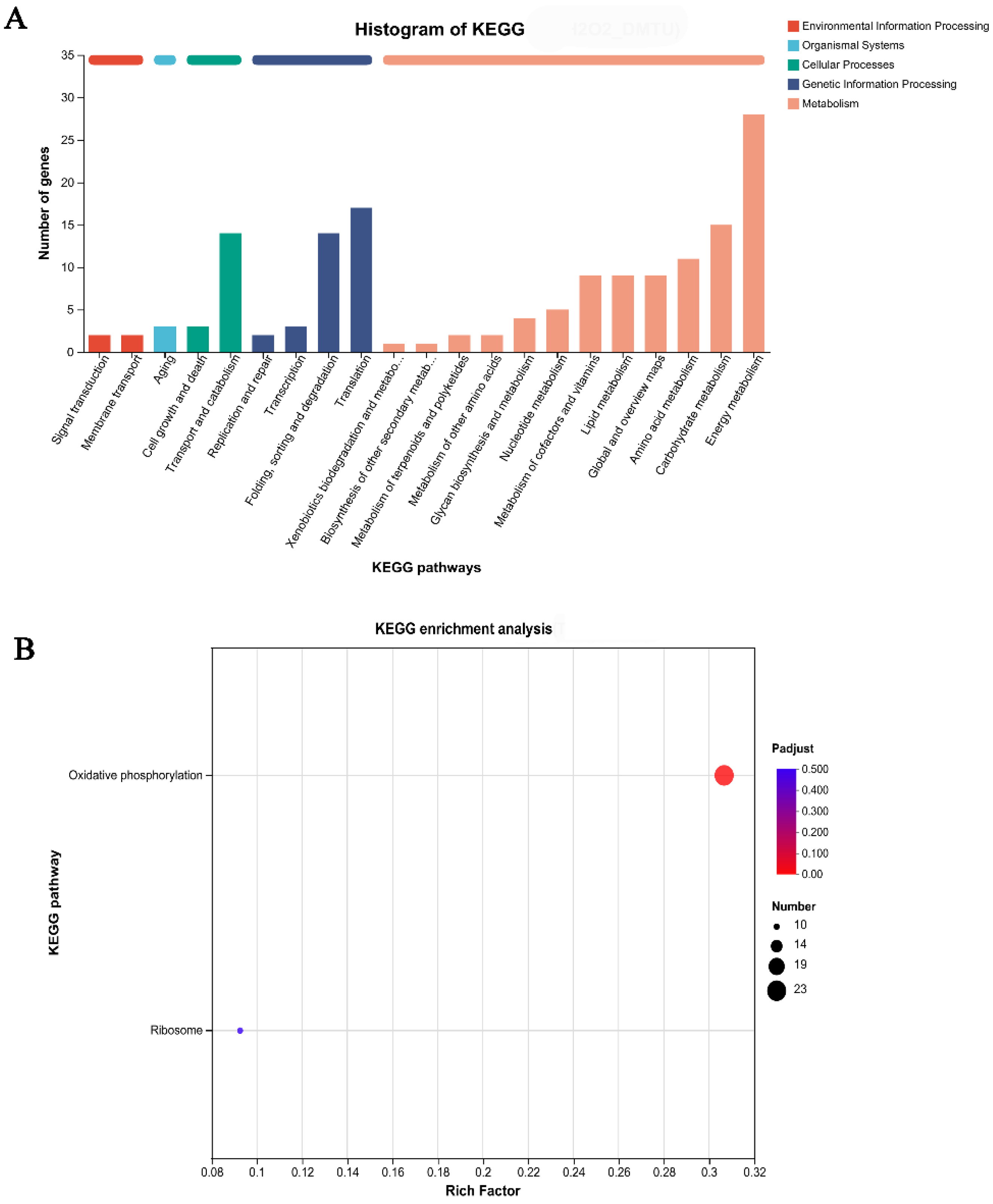
3.2. The Oxidative Phosphorylation Pathway Plays a Key Role in Cap Color Formation

To identify the global transcriptomic changes in response to H2O2 or DMTU treatment, RNA-seq analysis was conducted. Treatment with exogenous DMTU hindered the formation of the cap color. The results showed that after the external application of DMTU, 1242 downregulated differentially expressed genes (DEGs) and 179 upregulated DEGs were identified from the control versus DMTU comparison (Figure 2). Figure 2 shows that there were 986 downregulated DEGs and 405 upregulated DEGs in the H2O2 experimental group compared with the DMTU experimental group. Among them, 314 upregulated DEGs and 300 downregulated DEGs were specifically regulated by H2O2 and DMTU. Therefore, the functions of 614 DEGs identified from the H2O2 versus DMTU comparison were examined to elucidate the possible mechanism through which H2O2-signaling molecules regulate the formation of the cap color.
To understand the functions of the DEGs identified from the H2O2 versus DMTU comparison, GO pathway enrichment analysis was performed (Figure 3A). The results showed that DEGs were significantly enriched in 18 pathways, mainly concentrated in the following pathways: proton-transporting ATP synthase complex, coupling factor F(o), ATP biosynthetic process, nucleoside triphosphate metabolic processes, ATP metabolic process, purine nucleoside triphosphate biosynthetic and metabolic processes, and purine ribonucleoside triphosphate biosynthetic metabolic processes. These pathways are closely related to ATP biosynthesis, so it is speculated that ATP synthesis and metabolism may be related to the formation of cap color. In addition, some important pathways were also significantly enriched in the following: inorganic ion transmembrane transport, ion transport, cation transmembrane transport, inorganic cation transmembrane transport, and proton transmembrane transporter activity. These pathways are closely related to the transport of ions in the mitochondrial respiratory chain. Furthermore, DEGs were significantly enriched in pathways such as the inner mitochondrial membrane protein complex and mitochondrial protein-containing complex (Figure 3A). Figure 3B shows that 20 key DEGs were further screened through a GO Enrichment Chord Chart. The results showed that 20 key genes can be found in multiple pathways, such as aerobic respiration, cellular respiration, aerobic electron transport chain, aerobic respiratory electron transport chain, and electron transport chain.
In the KEGG annotation analysis, the DEGs identified from the H2O2 versus DMTU comparison were classified into five categories: “Environmental Information Processing”, “organismal systems”, “molecular function”, “cellular processes”, “genetic information processing”, and “metabolism”. Within the five categories, 28 DEGs were collected and enriched in the energy metabolism pathway (Figure 4A). To further determine the functions of DEGs, a KEGG function enrichment analysis was performed. The results showed that the DEGs were significantly enriched in one metabolic pathway: oxidative phosphorylation. In addition, 23 DEGs were enriched in this metabolic pathway. More interestingly, after comparison, it is found that the differential genes in energy metabolism in Figure 4A and oxidative phosphorylation in Figure 4B are the same. Twenty-three DEGs are shown in Table 1.
In conclusion, oxidative phosphorylation and energy metabolism may be very important metabolic pathways in the formation of P. ostreatus cap color and can be regulated by the signaling molecule H2O2.

3.3. H2O2 Regulates Cap Color Formation by Affecting the Respiratory Chain

To further analyze the molecular mechanism by which H2O2 regulates the formation of the cap color, the functions of 23 DEGs were further analyzed. The results are shown in Table 1. The results showed that all 23 DEGs encoded subunits of complexes in the respiratory chain. In addition, the expression pattern of the 23 DEGs after the addition of H2O2 or DMTU was further analyzed with a heatmap. As shown in Figure 5, 23 DEGs were significantly upregulated under the action of exogenous H2O2. In contrast, DEGs were significantly downregulated after DMTU treatment. This further indicates that H2O2 may affect the formation of the cap color by upregulating the expression of key genes in the respiratory chain.
Furthermore, the pathway ID is map00190. As shown in Figure 6, 23 differentially expressed genes were enriched in Complexes I, III, IV, and V in the respiratory chain. Figure 7 shows that two DEGs (g400, g8839) participate in the encoding of Complex I; three DEGs participate in the encoding of Complex III; six DEGs participate in the encoding of Complex IV; and 12 DEGs participate in the encoding of Complex V. Figure 7 shows that there was a close connection between the 23 DEGs, with a high correlation between each gene.
To further verify the reliability of the transcriptome data, the relative gene expression of DEGs in each complex of the respiratory chain was detected. As shown in Figure 8A, in Complex I, compared with the control group, the relative expression levels of g8849 and g400 increased by 1.30- and 1.34-fold, respectively, in the exogenous H2O2 group and decreased by 40.9% and 62.81% in the DMTU group. In Complex III, compared to the control, the relative expression levels of g2860, g13251, and g2546 in the H2O2 group were significantly increased by 1.57-, 1.40-, and 1.19-fold, respectively. In the DMTU group, the expression levels of g2860 and g13251 were downregulated by 43.54% and 39.31%, respectively, with g2546 showing no significant changes compared to the control (Figure 8B). Figure 8C shows that six DEGs were involved in the encoding of Complex IV. Compared with the control group, the expression levels of all six DEGs in the H2O2 group were significantly upregulated. In the H2O2 scavenging experimental group (DMTU), all six DEGs were significantly downregulated. This indicated that the H2O2-signaling molecule can regulate the expression of the encoding genes of Complex IV subunits. Figure 8D shows that 12 DEGs were involved in the encoding of Complex V (ATP synthase). Under the regulation of the signal molecule H2O2, only the relative expression level of g12225 showed no significant difference compared to the control, and the expression levels of 11 DEGs were significantly upregulated. However, after treatment with exogenous DMTU (H2O2 scavenger), the relative expression levels of 12 DEGs were significantly inhibited. The above results indicated that H2O2 plays an important regulatory role in the encoding of ATP synthase.
In summary, the signaling molecule H2O2 may participate in the formation of cap color by upregulating the expression of genes encoding Complexes I, III, IV, and V in the respiratory chain.

3.4. ATP Synthesis Affects the Formation of P. ostreatus Cap Color

Subsequently, valinomycin is a type of uncoupling agent that can dissipate the energy generated by electron transfer in the respiratory chain in the form of heat. To explore the factors that affect color formation by spraying exogenous valinomycin. The results showed that the addition of exogenous valinomycin resulted in a lighter cap color compared to the control group, indicating that ATP synthesis was hindered and affected by the formation of cap color (Figure 9). Furthermore, we determined the important role of ATP in the formation of cap color by adding exogenous Oligomycin A (ATP synthase inhibitor). The results showed that after the addition of exogenous Oligomycin A, the color of the fruiting body cap became lighter, further indicating that ATP plays an important role in the formation of cap color (Figure 9).

4. Discussion

H2O2 is a key signaling molecule in cell proliferation, development, tissue differentiation, and environmental responses in various organisms. Under stress, H2O2, as the main component of ROS, can cause cell damage. For example, under heat stress, the H2O2 content of P. ostreatus mycelia significantly increases, causing damage to the cell wall and significantly inhibiting the growth rate of the mycelia [33,39]. On the other hand, as a signaling molecule, H2O2 also plays an important role in the growth and development of mushrooms. For example, in G. lucidum, H2O2 can regulate the synthesis of the active substance triterpenoid compounds [30]. In F. filiformis, ROS (O2/H2O2) redistribution mediated by NADPH oxidase and MnSODs is linked to the gradient elongation of the stipe [40]. Our team’s previous research found that H2O2 can be significantly influenced by NO regulation to significantly affect the formation rate of P. ostreatus primordia [32]. Currently, increasing evidence suggests that H2O2 can act as a signaling molecule to upregulate the expression of many genes activated under environmental stress [41]. In plants, H2O2, as a central redox-signaling molecule in physiological oxidative stress, can induce the expression of 113 genes during oxidative stress, which are involved in cellular rescue and defense processes [42]. The growth and development of edible mushrooms is a very complex process and can also be subjected to various environmental stresses. For example, P. ostreatus is subjected to temperature and light stress during the mushroom production process. Our previous research found that H2O2 is distributed differently in different parts of the P. ostreatus fruiting body and significantly accumulates in the cap [37]. In this study, the results further found that when H2O2 in the cap is removed by its scavenger (DMTU), the cap color turns white, indicating the key role of H2O2 in the formation of cap color. Previous studies have shown that the biological function of the browning of F. filiformis could act as a protective mechanism response to the damage of cell integrity, and the enzymatic dopa melanin pathway could contribute to the browning mechanism [43]. In addition, the browning of F. filiformis may be caused by DNA damage, proteolysis, and oxidative photosynthesis process, which together lead to the damage of cell integrity and then trigger the biosynthesis of melanin [44]. Therefore, it is speculated that the melanin in the cap of P. ostreatus may also be a protective mechanism against environmental stress. Further analysis by RNA-seq showed that a link between oxidative phosphorylation and the formation of the P. ostreatus cap color existed. Our results are similar to those of previous studies, suggesting that oxidative phosphorylation may be involved in the synthesis of melanin.
The application of omics technology in the study of edible mushrooms provides a scientific basis for exploring regulatory mechanisms. In animals and plants, research on oxidative phosphorylation and color is also in preliminary progress. The formation of red color is an obvious sign of the beginning of strawberry fruit ripening. Transcriptome analysis showed that a series of metabolic changes occurred in the green-white-red phase. DEGs change from variable to less in this process, and oxidative phosphorylation plays an important role in regulating the ripening process [45]. Previous studies have shown that oxidative phosphorylation has a significant effect on the color of black-cut beef [46]. Moreover, efficient energy production and metabolism pathways (i.e., oxidative phosphorylation and the citric acid cycle) may promote the expression of key genes and proteins in the carotenoid selective transport system of the silkworm [47]. In Crassostrea gigas with orange and black shells, RNA high-throughput sequencing data showed that DEGs could be significantly enriched in “oxidative phosphorylation” related to ATP synthesis, suggesting that they might participate in pigment deposition [48]. In this study, 614 DEGs specifically regulated by H2O2 were preliminarily screened by RNA-seq, and only the oxidative phosphorylation pathway was significantly enriched by KEGG-enrichment analysis. This study preliminarily clarified the possible regulatory pathway of H2O2 as a signaling molecule in P. ostreatus cap color formation.
Oxidative phosphorylation plays a key role in the growth and development of organisms: it is the fundamental source of carbon skeletons and the driving force of biochemical reactions. In this study, further analysis revealed that 23 DEGs in the oxidative phosphate pathway were enriched in Complexes I, III, IV, and V of the respiratory chain. Therefore, H2O2 may affect the formation of fungal cap color by regulating the respiratory chain. The addition of exogenous inhibitors is a commonly used experimental method in many studies. In this study, it was found that the addition of respiratory chain uncoupling agents resulted in a lighter cap color. Further evidence suggests that ATP synthesis may play a crucial role in the formation of cap color in P. ostreatus. Moreover, the addition of exogenous ATP synthase inhibitors further demonstrated the importance of ATP synthesis. Previous studies have shown that the postharvest browning of A. bisporus is closely related to energy metabolism [49]. It is speculated that ATP may play an important role in color formation.
H2O2, as a signaling molecule, may have complex and multiple signal transduction mechanisms in regulating the color synthesis process of the P. ostreatus cap. At present, our study showed that H2O2 may regulate the formation of cap color by promoting ATP synthesis. In the future, it is necessary to further explore the molecular mechanism involved in the downstream stages of the H2O2 pathway and provide a better understanding of the color formation mechanism of mushrooms. Superoxide dismutase and catalase are important enzymes regulating H2O2 content. Respiratory chain Complexes I and III are the main sites of H2O2 production. Therefore, in future work, it will be necessary to explore the functions of these key genes and further clarify the molecular mechanism by which H2O2 regulates cap color formation.

5. Conclusions

In conclusion, the color of the P. ostreatus cap is an important agricultural trait, and its formation process is regulated by multiple genes with complex regulatory mechanisms. H2O2 plays an important regulatory role in P. ostreatus cap color formation. In this study, RNA-seq was used to comprehensively understand the molecular basis and regulatory mechanism by which H2O2 regulates cap color formation. The study showed that H2O2 may affect cap color formation by regulating the metabolic pathway of oxidative phosphorylation. The 23 DEGs encoding Complexes I, III, IV, and V subunits in the respiratory chain can be significantly regulated by H2O2. Furthermore, ATP synthesis was found to be the key factor affecting the formation of cap color.

Supplementary Materials

The following supporting information can be downloaded at: https://www.mdpi.com/article/10.3390/jof9080823/s1, Table S1: Primers used in this study.

Author Contributions

Conceptualization, L.H., M.C. and J.M.; Funding acquisition, L.H.; Methodology, L.H., K.Y., S.D. and L.G.; Supervision, J.M.; Validation, K.Y.; Writing-original draft, L.H.; Writing–review & editing, L.H., J.L. and S.W. All authors have read and agreed to the published version of the manuscript.

Funding

The study was funded by the Shanxi Provincial basic Research and Development Project (202103021223159), Shanxi Province Work Award Fund Research Project (SXBYK2022029), and the Doctoral Science Foundation of Shanxi Agricultural University (2021BQ90).

Institutional Review Board Statement

Not applicable.

Informed Consent Statement

Not applicable.

Data Availability Statement

The data of all results in this study are included in the manuscript and Supplementary Materials. If necessary, the data can be obtained by contacting the corresponding author.

Acknowledgments

We thank the Chinese Academy of Agricultural Sciences for the experimental strains (CCMSSC 00389), Beihang University Kaimin Li for his help in RNA-seq analysis, Shanxi Research Center for Engineering Technology of Edible Fungi for the experimental platform, and all members of the team for their help.

Conflicts of Interest

The authors declare no conflict of interest.

References

  1. Xu, C.; Li, J.L.; Yang, L.Q.; Shi, F.; Ye, M. Antibacterial activity and a membrane damage mechanism of Lachnum YM30 melanin against Vibrio parahaemolyticus and Staphylococcus aureus. Food Control 2017, 73, 1445–1451. [Google Scholar] [CrossRef]
  2. Arun, G.; Eyini, M.; Gunasekaran, P. Characterization and biological activities of extracellular melanin produced by Schizophyllum commune (Fries). Indian J. Exp. Biol. 2015, 53, 380–387. [Google Scholar] [PubMed]
  3. Liu, X.; Hou, R.L.; Wang, D.T.; Mai, M.X.; Wu, X.P.; Zheng, M.F.; Fu, J. Comprehensive utilization of edible mushroom Auricularia auricula waste residue-extraction, physicochemical properties of melanin and its antioxidant activity. Food Sci. Nutr. 2019, 7, 3774–3783. [Google Scholar] [CrossRef] [PubMed] [Green Version]
  4. Yang, X.T.; Tang, C.H.; Zhao, Q.Y.; Jia, Y.X.; Qin, Y.C.; Junmin, Z. Melanin: A promising source of functional food ingredient. J. Funct. Foods 2023, 105, 105574. [Google Scholar] [CrossRef]
  5. Shi, F.; Li, J.L.; Yang, L.Q.; Hou, G.H.; Ye, M. Hypolipidemic effect and protection ability of liver-kidney functions of melanin from Lachnum YM226 in high-fat diet fed mice. Food Funct. 2018, 9, 880–889. [Google Scholar] [CrossRef]
  6. Li, L.; Shi, F.; Li, J.L.; Huang, Q.L.; Xu, C.; Yang, L.Q.; Yang, Q.H.; Shaikh, F.; Ye, M. Immunoregulatory effect assessment of a novel melanin and its carboxymethyl derivative. Bioorg. Med. Chem. Lett. 2017, 27, 1831–1834. [Google Scholar] [CrossRef]
  7. Dong, J.Z.; Wang, S.H.; Ai, X.R.; Yao, L.; Sun, Z.W.; Lei, C.; Wang, Y.; Wang, Q. Composition and characterization of cordyxanthins from Cordyceps militaris fruit bodies. J. Funct. Foods 2013, 5, 1450–1455. [Google Scholar] [CrossRef]
  8. Spiteller, P.; Arnold, N.; Spiteller, M.; Steglich, W. Lilacinone, a red aminobenzoquinone pigment from Lactarius lilacinus. Nat. Prod. 2023, 66, 1402–1403. [Google Scholar] [CrossRef]
  9. Sun, S.J.; Zhang, X.J.; Sun, S.W.; Zhang, L.Y.; Shan, S.K.; Zhu, H. Production of natural melanin by Auricularia auricula and study on its molecular structure. Food Chem. 2016, 190, 801–807. [Google Scholar] [CrossRef]
  10. Hou, R.L.; Liu, X.; Wu, X.P.; Zheng, M.F.; Fu, J.S. Therapeutic effect of natural melanin from edible fungus Auricularia auricula on alcohol-induced liver damage in vitro and in vivo. Food Sci. Hum. Well. 2021, 10, 514–522. [Google Scholar] [CrossRef]
  11. Weijn, A.; Bastiaan-Net, S.; Wichers, H.J.; Mes, J.J. Melanin biosynthesis pathway in Agaricus bisporus mushrooms. Fungal Genet. Biol. 2013, 55, 42–53. [Google Scholar] [CrossRef] [PubMed]
  12. Wang, G.; Chen, L.F.; Tang, W.Q.; Wang, Y.Y.; Zhang, Q.; Wang, H.B.; Zhou, X.; Wu, H.F.; Guo, L.; Dou, M.J.; et al. Identifying a melanogenesis-related candidate gene by a high-quality genome assembly and population diversity analysis in Hypsizygus marmoreus. J. Genet. Genom. 2021, 48, 75–87. [Google Scholar] [CrossRef] [PubMed]
  13. Khan, M.A.; Tania, M. Nutritional and medicinal importance of Pleurotus mushrooms: An overview. Food Rev. Int. 2012, 28, 313–329. [Google Scholar] [CrossRef]
  14. Kües, U.; Liu, Y. Fruiting body production in basidiomycetes. Appl. Microbiol. Biotechnol. 2000, 54, 141–152. [Google Scholar] [CrossRef]
  15. Barbosa, J.R.; Freitas, M.M.S.; Oliveira, L.C.; Martins, L.H.S.; Almada-Vilhena, A.O.; Oliveira, R.M.; Julio, C.P.; Davi, D.S.B.B.; Raul, N.C.J. Obtaining extracts rich in antioxidant polysaccharides from the edible mushroom Pleurotus ostreatus using binary system with hot water and supercritical CO2. Food Chem. 2020, 330, 127173. [Google Scholar] [CrossRef]
  16. Nguyen, D.H.H.; Elramady, H.; Llanaj, X.; Törős, G.; Hajdú, P.; Prokisch, J. Chemical composition and health attributes of agri-foods: A scientific overview on black foods. Sustainability 2023, 15, 3852. [Google Scholar] [CrossRef]
  17. Qing, Q.R.; Xiao, J.Y.; Yao, H. Opportunities, challenges and countermeasures of edible fungi industry development under the development background of big health industry. Edible Fungi China 2020, 39, 94–96. [Google Scholar] [CrossRef]
  18. Toledo, A.V.; Franco, M.E.E.; Lopez, S.M.Y.; Troncozo, M.I.; Saparrat, M.C.N.; Balatti, P.A. Melanins in fungi: Types, localization and putative biological roles. Physiol. Mol. Plant P 2017, 99, 2–6. [Google Scholar] [CrossRef]
  19. Singh, S.; Nimse, S.B.; Mathew, D.E.; Dhimmar, A.; Sahastrabudhe, H.; Gajjar, A. Microbial melanin: Recent advances in biosynthesis, extraction, characterization, and applications. Biotechnol. Adv. 2021, 53, 107773. [Google Scholar] [CrossRef]
  20. Zhang, Y.; Wu, X.; Huang, C.; Zhang, Z.; Gao, W. Isolation and identification of pigments from oyster mushrooms with black, yellow and pink caps. Food Chem. 2022, 372, 131171. [Google Scholar] [CrossRef]
  21. Guaraldo, M.M.D.S.; Pereira, T.M.; Santos, H.O.D.; Oliveira, T.L.D.; Pereira, W.V.S.; Pinho, E.V.D.R.V. Priming with sodium nitroprusside and hydrogen peroxide increases cotton seed tolerance to salinity and water deficit during seed germination and seedling development. Environ. Exp. Bot. 2023, 209, 1005294. [Google Scholar] [CrossRef]
  22. Joo, J.H.; Bae, Y.S.; Lee, J.S. Role of auxin-induced reactive oxygen species in root gravitropism. Plant Physiol. 2001, 126, 1055–1060. [Google Scholar] [CrossRef] [Green Version]
  23. Potikha, T.S.; Collins, C.C.; Johnson, D.I.; Delmer, D.P.; Levine, A. The involvement of hydrogen peroxide in the differentiation of secondary walls in cotton fibers. Plant Physiol. 1999, 119, 849–858. [Google Scholar] [CrossRef] [PubMed] [Green Version]
  24. Ma, M.; Xu, W.L.; Wang, P.; Gu, Z.X.; Zhang, H.Z.; Yang, R. UV-B-triggered H2O2 production mediates isoflavones synthesis in germinated soybean. Food Chem. X 2022, 14, 100331. [Google Scholar] [CrossRef]
  25. Pei, Z.M.; Murata, Y.; Benning, G.; Thomine, S.; Klusener, B.; Allen, G.J.; Grill, E.; Schroeder, J.I. Calcium channels activated by hydrogen peroxide mediate abscisic acid signalling in guard cells. Nature 2022, 406, 731–734. [Google Scholar] [CrossRef]
  26. Rosei, M.A.; Blarzino, C.; Coccia, R.; Foppoli, C.; Cini, C. Production of melanin pigments by cytochrome c/H2O2 system. Int. J. Biochem. Cell Biol. 1998, 30, 457–463. [Google Scholar] [CrossRef] [PubMed]
  27. Kemmerling, U.; Munoz, P.; Muller, M.; Sanchez, G.; Aylwin, M.L.; Klann, E.; Carrasco, M.A.; Hidalgo, C. Calcium release by ryanodine receptors mediates hydrogen peroxide-induced activation of ERK and CREB phosphorylation in N2a cells and hippocampal neurons. Cell Calcium 2007, 41, 491–502. [Google Scholar] [CrossRef] [PubMed]
  28. Schallreuter, K.U.; Wazir, U.; Kothari, S.; Gibbons, N.C.J.; Moore, J.; Wood, J.M. Human phenylalanine hydroxylase is activated by H2O2: A novel mechanism for increasing the L-tyrosine supply for melanogenesis in melanocytes. Biochem. Biophys. Res. Commun. 2004, 32, 88–92. [Google Scholar] [CrossRef]
  29. Kim, H.E.; Lee, S.G. Induction of ATP synthase beta by H2O2 induces melanogenesis by activating PAH and cAMP/CREB/MITF signaling in melanoma cells. Int. J. Biochem. Cell Biol. 2013, 45, 1217–1222. [Google Scholar] [CrossRef] [PubMed]
  30. Mu, D.S.; Li, C.Y.; Zhang, X.C.; Li, X.B.; Shi, L.; Ren, A.; Zhao, M.W. Functions of the nicotinamide adenine dinucleotide phosphate oxidase family in Ganoderma lucidum: An essential role in ganoderic acid biosynthesis regulation, hyphal branching, fruiting body development, oxidative-stress resistance, and ganoderic acid biosynthesis regulation. Environ. Microbiol. 2014, 16, 1709–1728. [Google Scholar] [CrossRef]
  31. Shi, D.K.; Zhu, J.; Sun, Z.H.; Zhang, G.; Liu, R.; Zhang, T.J.; Wang, S.L.; Ren, A.; Zhao, M.W. Alternative oxidase impacts ganoderic acid biosynthesis by regulating intracellular ROS levels in Ganoderma lucidum. Microbiology 2017, 163, 1466–1476. [Google Scholar] [CrossRef]
  32. Hou, L.D.; Huang, C.Y.; Wu, X.L.; Zhang, J.X.; Zhao, M.R. Nitric oxide negatively regulates the rapid formation of Pleurotus ostreatus primordia by inhibiting the mitochondrial aco gene. J. Fungi 2022, 8, 1055. [Google Scholar] [CrossRef] [PubMed]
  33. Hou, L.D.; Wang, L.N.; Wu, X.L.; Gao, W.; Zhang, J.X.; Huang, C.Y. Expression patterns of two pal genes of Pleurotus ostreatus across developmental stages and under heat stress. BMC Microbiol. 2019, 19, 231. [Google Scholar] [CrossRef] [PubMed] [Green Version]
  34. Hou, L.D.; Zhao, M.R.; Huang, C.Y.; He, Q.; Zhang, L.J.; Zhang, J.X. Alternative oxidase gene induced by nitric oxide is involved in the regulation of ROS and enhances the resistance of Pleurotus ostreatus to heat stress. Microb. Cell Fact. 2021, 20, 137. [Google Scholar] [CrossRef]
  35. Wu, T.J.; Hu, C.C.; Xie, B.G.; Wei, S.L.; Zhang, L.; Zhu, Z.X.; Zhang, Z.Y.; Li, S.J. A putative transcription factor LFC1 negatively regulates development and yield of winter mushroom. Appl. Microbiol. Biotechnol. 2022, 104, 5827–5844. [Google Scholar] [CrossRef] [PubMed]
  36. Wu, T.J.; Hu, C.C.; Xie, B.G.; Zhang, L.; Yan, S.; Wang, W.; Tao, Y.; Li, S. A single transcription factor (PDD1) determines development and yield of winter mushroom (Flammulina velutipes). Appl. Environ. Microbiol. 2019, 85, e01735-19. [Google Scholar] [CrossRef]
  37. Hou, L.D.; Yan, K.X.; Dong, S.; Liu, X.Y.; Chang, M.C.; Meng, J.L. Preliminary study on the mechanism of signal molecule H2O2 regulating the formation of Pleurotus ostreatus cap color. Acta Hortic. Sinica. 2023, 50, 1243–1254. [Google Scholar] [CrossRef]
  38. Qu, J.B.; Zhao, M.R.; Hsiang, T.; Feng, X.X.; Zhang, J.X.; Huang, C.Y. Identification and characterization of small noncoding RNAs in genome sequences of the edible fungus Pleurotus ostreatus. Biomed. Res. Int. 2016, 2016, 2503023. [Google Scholar] [CrossRef] [PubMed] [Green Version]
  39. Song, C.; Chen, Q.; Wu, X.L.; Zhang, J.X.; Huang, C.Y. Heat stress induces apoptotic-like cell death in two Pleurotus species. Curr. Microbiol. 2014, 69, 611–616. [Google Scholar] [CrossRef]
  40. Yan, J.J.; Chekanova, J.; Liu, Y.Y.; Gan, B.C.; Long, Y.; Han, X.; Tong, Z.J.; Miao, J.; Lian, L.D.; Xie, B.G.; et al. Reactive oxygen species distribution involved in stipe gradient elongation in the mushroom Flammulina filiformis. Cells 2022, 11, 1896. [Google Scholar] [CrossRef]
  41. Desikan, R.; Mackerness, S.A.H.; Hancock, J.T.; Neill, S.J. Regulation of the Arabidopsis transcriptome by oxidative stress. Plant Physiol. 2001, 127, 159–172. [Google Scholar] [CrossRef] [PubMed] [Green Version]
  42. Sies, H. Hydrogen peroxide as a central redox signaling molecule in physiological oxidative stress: Oxidative eustress. Redox Biol. 2017, 11, 613–619. [Google Scholar] [CrossRef] [PubMed]
  43. Fu, Y.; Yu, Y.; Tan, H.; Wang, B.; Peng, W.H.; Sun, Q. Metabolomics reveals dopa melanin involved in the enzymatic browning of the yellow cultivars of East Asian golden needle mushroom (Flammulina filiformis). Food Chem. 2022, 370, 131295. [Google Scholar] [CrossRef]
  44. Fu, Y.; Tan, H.; Wang, B.; Peng, W.H.; Sun, Q.; Yu, Y. Integrated multi-omic analyses on yellow Flammulina filiformis cultivar reveal postharvest oxidative damage responses. Postharvest Biol. Tec. 2023, 195, 112111. [Google Scholar] [CrossRef]
  45. Wang, Q.H.; Zhao, C.; Zhang, M.; Li, Y.Z.; Shen, Y.Y.; Guo, J.X. Transcriptome analysis around the onset of strawberry fruit ripening uncovers an important role of oxidative phosphorylation in ripening. Sci. Rep. 2017, 7, 41477. [Google Scholar] [CrossRef] [PubMed] [Green Version]
  46. Wu, S.; Luo, X.; Yang, X.Y.; Hopkins, D.L.; Mao, Y.; Zhang, Y.M. Understanding the development of color and color stability of dark cutting beef based on mitochondrial proteomics. Meat Sci. 2020, 163, 108046. [Google Scholar] [CrossRef]
  47. Yuan, Y.; Xiao, R.; Ge, Q.; Taha, R.H.; Chen, K.Q. Complementary transcriptomic and proteomic analysis of bombyx mori middle silk glands reveals a predominant ribosome-biogenesis regulating network during silkworm yellow-cocoon color formation. J. Asia Pac. Entomol. 2021, 24, 260–270. [Google Scholar] [CrossRef]
  48. Li, Z.Z.; Li, Q.; Liu, S.K.; Han, Z.Q.; Kong, L.F.; Yu, H. Integrated analysis of coding genes and non-coding rnas associated with shell color in the pacific oyster (Crassostrea gigas). Mar. Biotechnol. 2021, 23, 417–429. [Google Scholar] [CrossRef]
  49. Li, L.; Sun, H.; Kitazawa, H.; Wang, X.Y. Effects of a high O2 dynamic-controlled atmosphere technology on the browning of postharvest white mushroom (Agaricus bisporus) in relation to energy metabolism. Food Sci. Technol. Int. 2017, 23, 385–395. [Google Scholar] [CrossRef]
Figure 1. H2O2 regulates cap color formation in P. ostreatus.
Jof 09 00823 g001
Figure 2. Number of upregulated and downregulated DEGs among the control, H2O2, and DMTU groups.
Jof 09 00823 g002
Figure 3. Significantly enriched pathways and GO enrichment chord chart for the DEGs. (A) GO enrichment analysis of DEGs from the H2O2 vs. DMTU comparison. (B) GO enrichment chord chart of DEGs from the H2O2 vs. DMTU comparison.
Jof 09 00823 g003
Figure 4. Enriched KEGG pathways of identified DEGs. (A) Enrichment of function; (B) Enrichment of metabolic pathways.
Jof 09 00823 g004
Figure 5. Heatmap of oxidative phosphorylation consisting of the 23 DEGs.
Jof 09 00823 g005
Figure 6. Regulatory effect of H2O2 on the oxidative phosphorylation metabolic pathway. The red rectangle represents the 23 DEGs of interest and their encoded subunits in the oxidative phosphorylation metabolic pathway.
Jof 09 00823 g006
Figure 7. Correlation between the expression of 23 DEGs. Each node in the graph represents a gene, and the connections between nodes represent a correlation in gene expression. The larger the node, the number of genes with expression correlation with other genes.
Jof 09 00823 g007
Figure 8. H2O2 regulates the expression of complex subunit-encoding genes in the respiratory chain. (A) Validation of DEGs by qPCR in Complex I; (B) Validation of DEGs by qPCR Complex III; (C) Validation of DEGs by qPCR Complex IV. (D) Validation of DEGs by qPCR Complex V. Different letters indicate significant differences for the comparison of samples (p < 0.05 according to Duncan’s test).
Jof 09 00823 g008
Figure 9. ATP synthesis affects the formation of cap color.
Jof 09 00823 g009
Table 1. The functions of 23 DEGs in oxidative phosphorylation.
Gene IDGene Function
g10165ATP4 subunit B of the stator stalk of mitochondrial F1F0 ATP synthase
g2546Cytochrome bc1 complex subunit 7
g7640Cytochrome c oxidase subunit 6
g8839NdufA4 NADH dehydrogenase 1 ɑ subcomplex
g10989V-type proton ATPase 16 kDa proteolipid subunit 2
g11401ATP17 subunit F of the F0 sector of mitochondrial F1F0 ATP synthase
g1602ATP3 γ subunit of the F1 sector of mitochondrial F1F0 ATP synthase
g11242ATP synthase F1 β subunit
g12225ATP synthase E chain-domain-containing protein
g12829ATP20 subunit g of the mitochondrial F1F0 ATP synthase
g3195Hypothetical protein
g9081Cytochrome c oxidase assembly protein cox15
g9180ATP synthase F1 ɑ subunit
g12052Cytochrome c oxidase subunit vib
g13251Ubiquinol-cytochrome c reductase complex subunit 8
g2860Ubiquinol-cytochrome-c reductase subunit 6
g12390ATP synthase subunit 5
g12499Cytochrome c oxidase subunit 4
g8845ATP synthase d subunit
g2230Cytochrome c oxidase copper chaperone
g400Acyl carrier protein
g8493ATP18 subunit j of the mitochondrial F1F0 ATP synthase
g8872Hypothetical protein
Disclaimer/Publisher’s Note: The statements, opinions and data contained in all publications are solely those of the individual author(s) and contributor(s) and not of MDPI and/or the editor(s). MDPI and/or the editor(s) disclaim responsibility for any injury to people or property resulting from any ideas, methods, instructions or products referred to in the content.

