


Abstract
Melanoma remains one of the most aggressive forms of skin cancer, characterized by high metastatic potential and risks of tissue trauma and post-surgical recurrence, with limited therapeutic options currently available. While conventional monotherapies often fail to provide comprehensive and efficient treatment, nanoparticle-functionalized hydrogel platforms have emerged as promising multifunctional systems for localized combination cancer therapy, capable of integrating multiple therapeutic modalities, such as chemodynamic therapy (CDT), photodynamic therapy (PDT), and photothermal therapy (PTT), tissue recovery support, antibacterial activity, and image-guided treatment into a single biomaterial. This review examines stimulus-responsive hydrogels based on the anionic polysaccharides carboxymethylcellulose (CMC) and hyaluronic acid (HA), conjugated with nanoparticles for melanoma theranostic applications. It evaluates their design, synergistic mechanisms, and therapeutic potential. It considers how combining CMC and HA overcomes the limitations of individual polymers, such as poor mechanical integrity and rapid degradation, and enhances key functionalities, particularly cell targeting and specificity. It also analyzes how incorporating functional nanoparticles can enable on-demand drug release, diagnostic imaging, immune microenvironment remodeling, and combined phototherapy (PTT/PDT). While HA-based nanotheranostic systems are widely documented, the potential of CMC in melanoma treatment remains largely untapped, despite its proven success in tissue engineering and wound management. Most significantly, the literature currently lacks any bio-nanohybrid platform that integrates CMC and HA, driven by functional hybrid nanoparticles, for targeted melanoma therapy. By identifying this critical gap, this review proposes new research avenues for designing these unified, multifunctional platforms as a highly promising strategy for cutaneous melanoma management.
1. Introduction
Skin cancer is a type of cancer characterized by abnormal proliferation of skin cells due to DNA mutations or genetic defects. This neoplasia is broadly classified into two major categories: melanoma and non-melanoma skin cancers, with incidence rates varying across different continents [1]. Although melanoma accounts for only approximately 1% of cases of skin cancer, it is considered the most aggressive and lethal form, being responsible for approximately 80% of skin cancer-related deaths worldwide, largely due to its high metastatic potential [2,3,4]. Furthermore, these rates are projected to increase in the coming years, underscoring the need to develop novel strategies for early diagnosis and more effective therapeutic approaches [1,3].
Conventional dressings currently available for skin cancer patients still present several limitations, including the absence of active immunomodulatory properties and the lack of localized and targeted therapeutic delivery, with the vast majority being unable to simultaneously achieve tumor ablation and tissue regeneration [5,6]. Therefore, there is a strong clinical need for advanced transdermal systems and bifunctional biomaterials that combine antitumor activity with wound-healing and antimicrobial properties. Such a device would enable the eradication of resistant melanoma cells while supporting tissue regeneration and neovascularization [7]. In this context, hydrogels have attracted attention as therapeutic platforms for skin cancer treatment due to their unique chemical structures and properties, including high biocompatibility and bioactivity, form adaptability, injectability, and self-healing [8,9].
Notably, no single polymeric precursor has been reported to simultaneously satisfy all the properties required for an effective skin cancer platform. This is because adequate mechanical robustness, controllable degradation kinetics, tunable stimulus-responsiveness, and biological activity rarely coexist within a single macromolecule. Thus, deliberately mixing two or more precursors has become a central strategy in hydrogel design, allowing the shortcomings of one component to be compensated by the strengths of another. This approach enables rational engineering of platforms with tailored, application-specific activity, achieving multifunctional responses and improving overall performance across a range of medical applications, including tumor therapy and tissue regeneration [8,10,11,12]. Over the last decade, a growing number of studies have explored synergistic combinations of polysaccharides, such as Hyaluronic Acid (HA) and Carboxymethylcellulose (CMC), in hybrid networks aiming to overcome the limitations of individual polymers. In such an approach, one can combine, for example, the bioactivity of HA with the physical stability and enzymatic durability of CMC [13].
In skin cancer treatment, nanotechnology has also been extensively investigated, enabling innovative strategies to improve therapeutic outcomes and enhance treatment efficacy. Examples include advancements in chemotherapy, targeted drug delivery, photodynamic therapy (PDT), photothermal therapy (PTT), and immunotherapy [14,15,16]. In this context, hydrogel-based nanoplatforms have emerged as promising biomaterials for cancer therapy and tissue engineering applications [12].
Despite this growing body of evidence, knowledge regarding CMC- and HA-based hydrogels remains fragmented. Existing studies typically examine each polymer in isolation or explore their combination for applications entirely unrelated to skin cancer. Accordingly, this review synthesizes the state of the art in nanostructured, stimulus-responsive hydrogels utilizing these polysaccharides for melanoma theranostics. While recent notable reviews—such as those by Jin et al. [6], Zhang et al. [17], Alavarse et al. [18], and Wu et al. [12]—have discussed hydrogels for melanoma, stimuli-responsive platforms, or polysaccharide crosslinking chemistry, they treat these subjects independently. Herein, we unify these concepts to rationalize the integration of CMC and HA into a single bio-nanohybrid platform. We evaluate the specific functions, strengths, and limitations of each polymer, justifying their pairing based on targeted suitability rather than outright superiority over other polysaccharide hybrids. Crucially, aiming to guide the development of multifunctional theranostic platforms, we also intend to translate this synthesis into a practical design framework, supported by a crosslinking decision matrix and a rationale for nanoparticle integrations. To our knowledge, no existing review addresses this comprehensive intersection.
2. Literature Search Strategy and Scope
This work was conducted as a narrative review with a well-defined scope. Rather than an exhaustive quantitative synthesis of the field, this work aims to provide a critical, integrative overview of CMC- and HA-based stimuli-responsive hydrogels biofunctionalized with nanoparticles for melanoma theranostics. The literature search covered major electronic databases and indexing platforms, including PubMed/MEDLINE, Web of Science, Scopus, and Google Scholar as a supplementary source. Search terms varied by section and included hydrogel, nanogel, scaffold, carboxymethylcellulose, hyaluronic acid, melanoma, skin cancer, wound healing, theranostic, and nanoparticles. To confirm the research gap, we ran a targeted search combining polysaccharide-based hydrogels with nanoparticles and melanoma, but no studies met these criteria. We also screened the reference lists of retrieved articles to identify additional relevant studies.
Eligible sources included peer-reviewed original research articles and review articles published in English. The search excluded editorials, conference abstracts, and non-peer-reviewed materials. We generally excluded studies published before 2018, with preference given to works published within the last five years. Given the limited number of studies in this context that specifically address melanoma, we included selected works on wound healing and other cancer types to provide comparative context and support the rationale for the CMC-HA hybrid design. Because the studies in this review range from material synthesis and physicochemical characterization to preclinical biological assays, each with distinct methodologies and outcome metrics, we did not apply formal quality or risk-of-bias assessments. Instead, studies were selected based on their relevance to the scope, methodological transparency, and inclusion of experimental validation, with their limitations discussed throughout the text.
3. Clinical Context: Melanoma Treatment and the Post-Surgical Wound
Skin cancer represents a growing threat to global health systems, with case numbers projected to keep rising over the next two decades [1]. Among its forms, melanoma arises from the dysregulated proliferation of melanocytes, the pigment-producing cells responsible for skin color and for shielding the skin from harmful UV radiation [19]. Its development is driven by the interplay between genetic predisposition and environmental factors, most notably excessive exposure to sunlight and artificial UV radiation [20]. Such a growing burden is already evident in numbers, as in 2022 alone, melanoma accounted for approximately 330,000 new cases and around 60,000 deaths worldwide [21].
Despite advances in melanoma treatment, significant therapeutic challenges remain. Surgery prevails as the primary treatment strategy for malignant melanoma; however, it inevitably causes tissue damage and may impair skin function, while also carrying risks of infection and tumor recurrence [22]. Conventional therapeutic approaches, including chemotherapy and radiotherapy, often have limited efficacy due to the aggressive and metastatic nature of melanoma and are frequently associated with adverse effects [23]. Notably, many anticancer drugs exhibit poor solubility, limited specificity, and systemic toxicity, which can further restrict their therapeutic effectiveness [24]. These challenges may be further exacerbated by immune dysfunction, chronic inflammation, and recurrent infections in cancer patients, potentially contributing to disease progression [25,26].
In modern oncology, two major systemic treatment approaches for advanced or metastatic melanoma are immunotherapy and targeted therapy. Immunotherapies consist of monoclonal antibodies administered via intravenous infusion to reactivate T lymphocytes. In contrast, targeted therapies involve small organic molecules—conventional synthetic chemical drugs such as BRAF inhibitors (dabrafenib, vemurafenib, encorafenib) and MEK inhibitors (trametinib, cobimetinib, binimetinib), formulated for oral administration [2]. Both approaches have transformed the management of advanced melanoma over the past decade. However, despite their clinical success, they both face acquired resistance, which can arise through genetic, epigenetic, signaling, or tumor-microenvironmental mechanisms, ultimately leading to disease progression and treatment failure [6]. Furthermore, these approaches can cause clinically significant adverse effects that affect normal tissues and organs, which may limit treatment tolerability. Immunotherapy, for example, can trigger immune-related adverse events, including colitis, hepatitis, and pneumonitis [20]. Consequently, innovative therapies are urgently needed to evade drug resistance, reduce off-target toxicity, and preserve potent antitumor efficacy against melanoma [27,28].
Although surgical excision is a cornerstone of skin cancer treatment, post-surgical care is equally critical for patient recovery and quality of life [29]. As Jakovija and Chtanova [30] note, the skin serves not only as a physical barrier, but also as an essential immunological defense site. Consequently, restoring this barrier through wound healing is vital; however, the process remains highly complex due to metabolic disruptions within the wound microenvironment [31]. Thus, proper treatment of post-surgical wounds is a fundamental aspect of skin cancer management, especially in patients with systemic conditions that compromise the healing process [26].
Conventional wound dressings for cancer patients present major limitations, such as adhesion-induced tissue damage and poor penetration of topical chemotherapy into the tumor stroma [29]. Furthermore, many UV-responsive hydrogels rely on poorly water-soluble photoinitiators that generate cytotoxic free radicals, damaging healthy tissue and restricting biomedical use [29]. Therefore, advanced hydrogel platforms can serve as complementary, locally acting adjuvant tools. While systemic agents target disseminated and metastatic disease, hydrogel matrices implanted in the surgical bed could provide targeted, sustained local delivery to address residual tumor cells at surgical margins, modulate postoperative inflammation, and support tissue repair. In this context, such platforms may offer a synergistic multimodal approach that could reduce the risk of local tumor recurrence while promoting postoperative wound healing [6,12].
4. Nanoparticle-Functionalized Hydrogels for Melanoma Theranostics
Defined as structures with at least one dimension between 1 and 100 nm, nanoparticles (NPs) exhibit a high surface-to-volume ratio and unique physicochemical properties [32]. They are engineered from organic constituents (lipids, polymers, liposomes) or inorganic elements (gold, silver, iron oxide, quantum dots, silica) and are widely utilized as theranostic platforms in oncology [33]. To overcome the shortcomings of conventional treatments, combination therapy has emerged as a promising strategy. By employing two or more modalities (e.g., chemotherapy, PDT, PTT, and immunotherapy) with complementary mechanisms, this approach achieves therapeutic outcomes unachievable by single-modality treatments [1,34,35].
Figure 1 outlines the primary conventional and systemic treatments for melanoma—including chemotherapy and immunotherapy—alongside nanotechnology-based solutions designed to overcome challenges such as drug resistance and off-target toxicity. Additionally, it summarizes major skin cancer types and primary risk factors, providing a comprehensive overview of disease pathophysiology, risk profiles, and the evolving therapeutic landscape [1].
Figure 1.
Types and causes of skin cancer with the main treatment and nanoparticle approaches. Reprinted with permission of [1] under the terms of the Creative Commons Attribution (CC BY 4.0) license.
Driven by unique physicochemical and optical properties, NPs are promising tools in cancer therapy and can be engineered for targeted cellular interactions [22,36]. As depicted in Figure 2, NPs leverage internal stimuli (redox, pH) or external triggers (light, heat, ultrasound, magnetic fields) to alter the tumor microenvironment [37], enabling on-demand drug release and boosting photothermal or photodynamic treatments [37]. Moreover, they support simultaneous diagnostic imaging and therapy, creating an integrated theranostic platform.
Figure 2.
External and internal stimuli in nanotherapeutics for cancer theranostics. Reprinted with permission of [38] under the terms of the Creative Commons Attribution (CC BY 4.0) license.
The high mortality rate of melanoma is primarily driven by its strong metastatic potential and rapid dissemination, making early diagnosis critical for successful patient outcomes. Traditional diagnostic tools often suffer from imprecision, high costs [24], and a strong reliance on examiner expertise. Moreover, definitive confirmation still requires invasive biopsy and histopathological analysis—a workflow plagued by interobserver variability, where specialist panels reclassify up to 17% of initial diagnoses. This diagnostic challenge also leads to a high rate of unnecessary excisions, with some centers performing up to 287 biopsies to identify a single melanoma case [39].
Nanotheranostics have emerged at the forefront of melanoma management by integrating diagnostic imaging and multiple therapeutic agents into a single nanoscale platform [17]. Owing to their high surface-area-to-volume ratio and tunable surface chemistry, these nanostructures can overcome formidable skin barriers to actively target melanoma cells [33,40,41]. Once localized within the tumor stroma, these nanocarriers achieve high accumulation and retention, enhancing cancer cell destruction while mitigating systemic toxicity and side effects in healthy tissues [32,42].
NPs can serve as contrast agents, near-infrared fluorescence sources for deep-tissue imaging, or active targeting agents for diseased cells [14,43,44]. Moreover, they act as versatile nanocarriers for drug delivery, mRNA therapeutics, immunotherapy, tissue regeneration, and cutaneous wound healing [45,46,47]. By contrast, biomaterial scaffolds can provide temporary mechanical support and mass transport, delivering physical and chemical cues with spatiotemporal precision to promote cell adhesion, proliferation, and functional modulation [48]. Consequently, research has increasingly focused on developing cell-inspired biomaterials equipped with drug-loading capacity, stimuli-responsive release kinetics, and cell-membrane camouflage [49].
Among biomaterials, hydrogels stand out as one of the most extensively studied and versatile classes, categorized by origin (natural, synthetic, or hybrid) and crosslinking mechanism (physical or chemical networks) [50]. Their defining feature is a hydrophilic polymeric network capable of absorbing and retaining vast amounts of water or biological fluids while maintaining structural integrity. Because crosslinking type and synthesis conditions govern their final properties [18], hydrogels can be tailored to closely mimic the extracellular matrix (ECM) and support cellular interactions within a biocompatible, mechanically tunable environment [50], making them ideal base materials for scaffold construction. Beyond structural adaptability, they have garnered extensive biomedical interest for traits such as injectability and self-healing capacity, the latter driven by reversible chemical bonds that allow the material to withstand physiological mechanical stress and spontaneously repair internal damage [9]. These features are also central to minimizing systemic toxicity while maximizing local therapeutic efficacy [33,51]. Furthermore, their porous architecture facilitates the diffusion of bioactive molecules, enabling sustained local drug release and fostering a favorable microenvironment for tissue regeneration [50].
5. Anionic Polysaccharides as Building Blocks: CMC and HA
5.1. Polysaccharides as Hydrogel Building Blocks
Polysaccharides are an important class of macromolecules composed of repeating carbohydrate units. They are among the most widely used building blocks in hydrogel design because of properties such as bifunctionality, therapeutic selectivity, self-assembly capacity, and stimuli responsiveness [26,52,53]. Their molecular chains carry several readily modifiable functional groups that form intra- and intermolecular interactions, such as hydrogen bonding, hydrophobic effects, and electrostatic interactions, which drive chain entanglement and ultimately give rise to the three-dimensional crosslinked networks discussed above [54]. These same chemical groups also allow polysaccharides to be functionalized at different sites, tailoring their structure to produce hydrogels with improved physicochemical and biological properties [55].
Categorizing polysaccharides according to their ionizable functional groups is fundamentally important: neutral polysaccharides are characterized by abundant hydroxyl groups (-OH); cationic polysaccharides incorporate amino groups (-NH2) subject to protonation-induced positive charges; and anionic polysaccharides contain acidic functionalities, notably carboxyl groups (-COOH), which ionize to form negative charges [56]. The high density of these anionic groups facilitates robust hydration and water retention, establishing hydrogel networks that support cell viability, growth, and tissue repair. Moreover, their susceptibility to enzymatic degradation into biocompatible, non-toxic metabolites underlines their high utility in long-term biomedical interventions [57].
5.2. Carboxymethylcellulose and Hyaluronic Acid
Carboxymethylcellulose is an anionic, semi-processed biopolymer derived from cellulose by partially substituting hydroxyl groups (-OH) on its glucopyranose rings with carboxymethyl groups (-CH2-COOH) [58]. It is a non-toxic, biodegradable, water-soluble, environmentally friendly, low-cost compound. In addition, this polysaccharide adheres effectively to wound sites, absorbs exudates (fluids), and maintains a favorable healing environment; it is also used as a multifunctional organic shell to cap ligands and stabilize inorganic nanoparticles [38]. Previous research has shown that CMC can provide a biocompatible surface for developing hydrogels for biomedical applications, such as skin substitutes, drug delivery systems, and bioimaging platforms for treating various diseases, including cancer [38]. Figure 3A represents the chemical structure of CMC.
Figure 3.
Chemical structure of (A) CMC and (B) Hyaluronic Acid. Adapted and reprinted with permission [13] under the terms of the Creative Commons Attribution (CC BY 4.0) license.
Hyaluronic acid is a natural, linear, anionic mucopolysaccharide that serves as a major constituent of the native extracellular matrix, linking tissues and the dermis. Involved in diverse biological processes, HA exhibits molar-mass-dependent properties that regulate angiogenesis, tissue regeneration, and inflammatory responses [55]. As depicted in Figure 3B, HA features versatile reactive functional groups—including hydroxyl, carboxyl, and acetyl groups—that enable chemical modification, alongside intrinsic biocompatibility, biodegradability, and non-immunogenicity [59]. Figure 4 provides a comprehensive overview of the chemical versatility of CMC and HA for theranostic platform development, highlighting key reaction sites derived from literature-reported strategies for chemical modification, functionalization, and crosslinking. Specifically, the figure illustrates how these reactive moieties, predominantly carboxyl and hydroxyl groups, are systematically exploited to engineer advanced multifunctional architectures.
Figure 4.
(A) Diverse bioapplications of carboxymethylcellulose (CMC), exploiting the reactivity of its hydroxyl and carboxyl groups toward conjugation with: Ag nanoparticles for antimicrobial mem-branes [25]; amino acid peptides for increased cellular uptake of nanoconjugates [60]; iron oxide magnetic nanoparticles for hyperthermia-based cancer treatment [61]; RGD for polysaccharide-ride-drug conjugates [62]; folic acid for targeted cancer theranostics [63]; AIS/ZAIS quantum dots for bioimaging [60,64]; Au nanoparticles for biosensing [14]; and doxorubicin (DOX) for antitumor membranes for skin cancer treatment [65,66]. (B) Bioconjugation strategies for hyaluronic acid (HA), exploiting the reactivity of its functional groups toward: deacetylation for exposure of free active amino sites [55]; conjugation with metallic nanoparticles for nanotheranostics [33]; DOX for skin cancer treatment [67]; RGD peptides for bioactive signaling [62]; fatty acid conjugation for amphiphilic micelle self-assembly [68]; cationic nanovesicles for sustained release [69]; camptothecin for skin cancer treatment [67]; methacrylic anhydride for photocrosslinkable properties [70]; and antibodies for immunotherapy and targeting [71]. (C) A highlight of potential modification sites of a CMC and HA repeating units with different aims (Created in BioRender. Miranda, D. (2026) https://BioRender.com/sszbyfe (accessed on 2 September 2026).
5.3. Hybrid Systems with CMC and HA for Melanoma Management
Researchers have widely investigated combining CMC and HA with other natural or synthetic polymers to form hybrid hydrogel networks, offering significant physicochemical, mechanical, and biological benefits for melanoma management, including tissue regeneration, wound care, and tumor cell elimination [5,72,73,74,75,76,77,78]. Research on hybrid hydrogels for cutaneous wound treatment increasingly focuses on blending CMC and HA with complementary polymers to overcome individual shortcomings and achieve synergistic performance [79].
To address the intrinsic limitations of CMC alone, Jeong et al. [74] investigated a wound-healing hydrogel composed of gelatin and oxidized carboxymethylcellulose (OCMC) loaded with fucoidan, a sulfated marine polysaccharide. In this design, gelatin served as an ECM-mimetic protein base, while fucoidan acted as a bioactive therapeutic agent, inhibiting nitric oxide (NO) production and stimulating de novo collagen synthesis by fibroblasts [74]. To counteract the poor mechanical strength and thermal instability of pure gelatin, the authors incorporated CMC. By oxidizing CMC to generate reactive aldehyde groups, they facilitated spontaneous condensation with gelatin’s free amine groups, establishing a stable three-dimensional network without toxic synthetic crosslinkers. Because these imine bonds are reversible, the resulting network balanced structural integrity with dynamic self-recovery, enabling the gel to adapt to mechanical stresses within the wound bed. In a murine full-thickness model, this optimized formulation accelerated wound closure by the third day and achieved complete re-epithelialization within 14 days [74]. Nonetheless, fucoidan-based formulations present certain challenges: their high swelling capacity can lead to an initial burst release of bioactive cargo, and structural variations across seaweed species can complicate standardization and batch-to-batch reproducibility [74]. Ultimately, these mechanical adaptations and self-healing properties, combined with effective exudate management, position CMC-based hydrogels as safe and promising wound-care options for melanoma patients. Furthermore, this same CMC–fucoidan combination has been explored for minimally invasive adjuvant melanoma therapy as a dissolvable microneedle patch, where CMC provides the requisite mechanical support while fucoidan acts as the primary antitumor agent [76].
On the other hand, despite its exceptional biocompatibility and water-retention capacity, HA forms hydrogels that often exhibit mechanical weakness, high solubility, and rapid or uncontrolled degradation in physiological environments [78]. As a native macromolecule, HA is highly susceptible to enzymatic degradation by hyaluronidases, which can further compromise its structural stability [55,69]. To address such drawback, Zhao et al. [72] crosslinked thiolated gelatin with dopamine-modified HA and loaded the resulting bioabsorbable scaffold with doxorubicin-carrying, reduction-sensitive polymeric nanomicelles: once implanted in the post-resection bed, the porous matrix absorbs the wound exudate and physically traps the residual B16F10 cells that escaped surgery, which are then eliminated through RGD-mediated endocytosis of the nanocarriers, while hyaluronic acid contributes to tissue repair by regulating cell proliferation, adhesion and differentiation; the platform was nevertheless designed for antitumor therapy alone, without functions such as bleeding control or skin reconstruction. Schmid et al. [75] followed the opposite rationale and used a printable alginate/HA/gelatin bioink, in which hyaluronic acid provides CD44 binding and gelatin provides integrin binding, to biofabricate vascularized metastatic melanoma models for disease modeling and drug screening rather than a therapeutic product. Other combinations associate phototherapy with tissue regeneration [5,73,77] and are analyzed individually in Section 7.
Taken together, these studies show that combining CMC or HA with a second polymer improves the physicochemical and mechanical properties and responsive behavior of the gels. Read comparatively, however, they also reveal a consistent division of labor directly relevant to the design proposed here: in each of these hybrids, one polysaccharide carries the tumor- or tissue-directed function, while the partner supplies structure. Gelatin mimics the dermal extracellular matrix but is mechanically weak and unstable at physiological temperature, and in both systems in which it appears it had to be rescued by a polysaccharide—by oxidized CMC, whose aldehyde groups condense with its free amines, in the dressing of Jeong et al. [74], and by dopamine-modified HA in the bioabsorbable scaffold of Zhao et al. [72]. Schmid et al. [75] state the division explicitly, assigning CD44 binding to HA and integrin binding to gelatin within an alginate-based printable network.
Two key properties account for this recurrence and distinguish the CMC–HA pair from other hybrid combinations reported for melanoma. First, cellular targeting is intrinsic to the matrix rather than artificially grafted: HA is the endogenous ligand for CD44, a receptor heavily overexpressed in melanoma. Thus, an HA-based network displays the recognition motif natively at a density dictated by formulation rather than a separate conjugation step. Conversely, integrin binding is not selective for melanoma, and neither chitosan nor alginate offers a comparable receptor-level interaction. Second, electrostatic compatibility with the nanoparticle phase is uniquely favorable. Both CMC and HA are polyanions across physiological and mildly acidic pH ranges; the identical carboxylate groups engaging CD44 also cap and anchor metal, metal-oxide, and semiconductor nanoparticles. Thus, network crosslinking and nanoparticle stabilization rely on a single, unified chemistry. In contrast, polycation–polyanion pairs (e.g., chitosan–alginate) form networks through the electrostatic complexation of those precise functional groups, rendering them unavailable to stabilize anionic nanoparticles.
Importantly, this rationale is founded on functional suitability rather than absolute superiority, and trade-offs must be acknowledged. Quaternized chitosan, for instance, contributes intrinsic antimicrobial activity that neither CMC nor HA possesses (as seen in the self-healing dressing in Section 6.1), meaning a CMC–HA platform must derive its antimicrobial behavior from the nanoparticle phase. Similarly, gelatin supplies cell-adhesive sequences and protease-susceptible degradation sites, while HA is far from an inert scaffold, as its molecular weight and degree of chemical modification govern both CD44 engagement and angiogenic responses (Section 8.1 and Section 8.2). The CMC–HA combination is not an inherently superior polymer, but a unique pairing among those reviewed in which receptor-level targeting, anionic nanoparticle compatibility, enzymatically regulated degradation, and established wound-dressing performance all derive from the same two macromolecules. To our knowledge, no existing system unites both polysaccharides into a single nanoparticle-functionalized network for melanoma. It is this convergence of functions—rather than the standalone novelty of the polymer combination—that motivates the design framework developed in Section 8. To avoid redundancy, Table 1 (Section 7.3) compares the composition, crosslinking chemistry, therapeutic modality, validation stage, outcomes, biosafety evidence, and limitations of these systems.
6. HA and CMC-Stimuli-Responsive Hydrogels in Cancer Management and Wound Healing
The inherent stimulus responsiveness of polysaccharides such as HA and CMC becomes a major practical advantage upon forming a hydrogel network. These “smart” platforms can sense physical and chemical changes in their surrounding environment—such as variations in pH, temperature, or enzyme concentration—and respond accordingly by releasing therapeutic agents at controlled concentrations directly to pathological sites while minimizing exposure to healthy tissues [80,81].
Hydrogels can be strategically engineered to respond to internal cues, such as ionic strength, reactive oxygen species (ROS), or specific enzymes, or external triggers, to mention magnetic fields, electric fields, ultrasound, or temperature [82]. Researchers achieve this responsiveness by incorporating functional compounds, introducing dynamic or cleavable crosslinks tailored to biological conditions, or integrating functional nanoparticles sensitive to external stimuli like magnetic fields, light, or heat. These triggers induce structural alterations—such as conformational shifts, phase transitions, swelling changes, or permeability adjustments, thereby enabling precise, on-demand drug delivery [53,83,84].
Among these design strategies, the choice of crosslinking approach is particularly influential, as it dictates stimuli-responsive behavior while governing broader structural and biological performance. Salma-Ancane et al. [85] highlight this dual role by synthesizing hyaluronic acid and ε-polylysine (ε-PL) hydrogels via distinct physical and chemical methods. While both hydrogel types exhibited rapid swelling, long-term stability, and comparable stiffness, their properties diverged based on the crosslinking mechanism. Physical crosslinking relied on electrostatic interactions between anionic HA and cationic ε-PL, whereas chemical crosslinking explored EDC/NHS coupling. Chemically crosslinked networks featured lower crosslinking density and higher porosity, enabling significantly greater liquid absorption and swelling. Furthermore, these chemically crosslinked hydrogels showed reduced antibacterial activity because fewer free amine groups were available to interact with bacterial cell walls, yet they exhibited superior cytocompatibility [85]. Most of all, this study underscores the importance of tailoring the crosslinking method to the intended application.
6.1. CMC and HA Stimuli-Responsive Hydrogels in Wound Healing
The tunability of polysaccharides such as HA and CMC has driven their application across diverse biomedical fields, particularly in developing smart, stimulus-responsive hydrogels for advanced wound management. For instance, Yang et al. [86] engineered a self-healing, antibacterial hydrogel composed of aldehyde-modified hyaluronic acid and fungal-derived quaternized chitosan (QAC), utilizing 3,3′-dithiobis(propionyl hydrazide) (DTP) as a crosslinking agent to form both dynamic chemical bonds and physical interactions. Driven by fast reaction kinetics and reversible networks, including dynamic covalent bonds (hydrazone and/or disulfide linkages) and electrostatic interactions, the hydrogels exhibited rapid gelation and excellent self-healing capabilities [86]. This dynamic nature allows the material to self-repair: while these bonds break under mechanical stress, they spontaneously reform when damaged surfaces re-contact, restoring structural integrity and function. While cytocompatible and promising for wound dressings, the system suffered from limited mechanical strength due to its dynamic crosslinks, a defect compounded by QAC, which reduced the stress–strain at break. The authors also disclosed that QAC imparted type-dependent antibacterial efficacy, achieving complete inhibition (~100%) against Gram-negative E. coli but lower performance (75.9%) against Gram-positive S. aureus. Consequently, optimizing such platforms will require replacing or supplementing QAC with alternative broad-spectrum antimicrobial agents—such as antimicrobial peptides or inorganic nanoparticles—to boost bactericidal activity without compromising mechanical performance [86].
Seeking to improve wound treatment while maintaining effective antibacterial activity, Liu and colleagues [87] developed a multifunctional hydrogel termed HTFC, which incorporates HA and calcium peroxide nanoparticles. These nanoparticles not only boost antibacterial efficacy against both Gram-positive and Gram-negative strains via chemodynamic therapy, but also mitigate hypoxia—critical for supporting cell proliferation and angiogenesis within the wound bed—while enhancing overall hydrogel stability. Overall, this work highlights nanoparticle incorporation as a powerful multifunctional strategy for treating infected wounds, enabling simultaneous bactericidal activity, microenvironmental modulation, and accelerated healing. However, as with other calcium peroxide-based systems, this therapeutic benefit relies on tightly controlling H2O2 and ROS generation rates, since excessive release can damage healthy tissue [87].
In contrast to Yang’s reliance on dynamic, reversible bonds, B. Huang et al. [88] developed a system centered on covalent crosslinking to ensure structural integrity and mechanical stability. The authors synthesized an HA-based hydrogel for wound repair, designed to combat bacterial infections through PDT via ROS generation by combining methacrylated hyaluronic acid (HAMA), a modified porphyrin photosensitizer (TPP), and dopamine methacrylamide (DMA). In this formulation, functionalization introduced methacrylate groups into HA and dopamine, providing chemical anchoring points for subsequent crosslinking while imparting structural stability and strong tissue adhesion [88]. Furthermore, the hydrogel produced significantly more ROS upon laser irradiation than an equivalent solution of TPP nanoparticles encapsulated in an amphiphilic copolymer. The authors attributed this difference to self-quenching of porphyrins within the standalone nanoparticles, demonstrating that hydrogel integration prevents aggregation and enhances efficiency [88]. Ultimately, this approach yielded a smart, synergistic wound dressing capable of eliminating bacterial infections, adhering firmly to damaged skin, and providing robust biological support for tissue regeneration [88].
6.2. CMC and HA Stimuli-Responsive Hydrogels in Cancer Management
Several studies have explored incorporating nanoparticles, functionalization, and advanced crosslinking strategies to develop more effective therapeutic approaches and improve outcomes in cancer management. For instance, Ali and collaborators [89] developed near-infrared (NIR)-responsive carboxymethyl cellulose hydrogels containing thioketal linkages for on-demand drug delivery in cancer therapy. This crosslinker serves two critical functions: it confers ROS-responsiveness and features tetrazine end groups that react with polymer precursors chemically modified with complementary reactive groups. Crosslinking proceeds via an ultrafast Inverse Electron Demand Diels-Alder (IEDDA) click reaction, which generates nitrogen (N2) gas as a by-product. This outgassing creates porosity and microbubbles within the network, enhancing drug loading and release. Furthermore, NIR light acts as a remote trigger to release doxorubicin (DOX) and boost antitumor efficacy through chemo-photothermal therapy, facilitated by the encapsulated photosensitizer indocyanine green (ICG), which converts light into localized heat to disrupt the hydrogel matrix. The choice of crosslinker was fundamental to the platform’s success, enabling precise on-demand responsiveness. Through chemo-photothermal combination therapy and in vitro testing, the hydrogel demonstrated high efficacy, safety, and highly controlled release kinetics [89]. Nevertheless, comprehensive in vivo studies remain essential to fully establish its translational potential.
Shi et al. [54] developed an injectable hyaluronic acid-based hydrogel loaded with DOX for breast cancer treatment and metastasis prevention. The HA chains were first modified by conjugating methacrylohydrazide (MHB) molecules via amidation, yielding unsaturated double bonds and amide linkages. The modified precursor was then crosslinked with a sulfhydryl-based agent via a Michael addition reaction to form the hydrogel network, into which DOX was dissolved to create the therapeutic matrix. By tailoring HA modification and selecting an appropriate crosslinking mechanism, the authors engineered a biomaterial responsive to thermal stimuli and pH-dependent swelling, enabling refined control over release kinetics and reduced systemic toxicity [54]. Although the platform showed promising results with isolated DOX delivery, its potential could be expanded by incorporating synergistic drug combinations—an approach particularly relevant for metastatic breast cancer. Additionally, future investigations could explore integrating functional nanoparticles into the hydrogel network to boost therapeutic efficacy and introduce multi-modal functionalities.
Zheng et al. [64] developed a multifunctional CMC-based nanohydrogel incorporating DNA-templated CdTeSe quantum dots (QDs) for tumor theranostics, with Figure 5 schematically illustrating its design, synthesis, and mechanism of action. In this architecture, CMC serves as the structural scaffold, while the QDs function as fluorescent probes for diagnostic imaging and monitoring. Rather than relying solely on gradual framework degradation, the nanohydrogel achieves on-demand, intelligent drug release via a dual-stimulus disassembly mechanism triggered by elevated levels of glutathione (GSH) and specific microRNAs (miRNAs) within the tumor microenvironment, ensuring targeted doxorubicin delivery [64]. Whilst the system successfully protected cargo from degradation, enabled active tumor targeting, and provided intelligent release kinetics, heavy metal toxicity remains a significant barrier to clinical translation despite low short-term cellular toxicity. Furthermore, the authors reported challenges in tracking the nanohydrogel in vivo via intravenous fluorescence imaging, likely attributable to insufficient QD concentration. Consequently, this work underscores the critical need for alternative nanoparticle formulations featuring reduced toxicity and enhanced fluorescence efficiency to enable precise oncological diagnostics [64].
Figure 5.
Multifunctional carboxymethylcellulose (CMC) nanohydrogel structured with DNA-templated CdTeSe quantum dots (QDs) for tumor theranostics, combining fluorescence imaging with dual-stimulus (GSH/miRNA) triggered doxorubicin release. Reprinted with permission of [64] under the terms of the Creative Commons Attribution-NonCommercial (CC BY-NC 3.0) license.
Recent literature has investigated a wide range of nanomaterials for cancer diagnosis and treatment, including conventional metallic and magnetic nanoparticles as well as semiconductor quantum dots. However, the clinical translation of nanotheranostic platforms depends not only on imaging and therapeutic performance but also on biological safety. Achieving an optimal balance among high optical brightness, therapeutic efficacy, and biocompatibility remains a critical challenge in developing next-generation nanotheranostics, underscoring the need for continued research [90,91].
Wang et al. [92] engineered an injectable hydrogel containing all-trans retinoic acid (ATRA) and gold nanoparticles designed to prevent breast cancer recurrence and metastasis by directly targeting tumor stem cells. Specifically, the researchers developed ATRA-loaded HA micelles to induce the differentiation of cancer stem cells (CSCs) into regular cancer cells, rendering them far more vulnerable to elimination via localized hyperthermia generated by the gold nanoparticles under laser stimulation. In this formulation, HA was chemically modified by conjugating it with oleic acid via cystamine linkages to form stable micelles that gradually release and bind to CD44 receptors on tumor cells. Thus, hyaluronic acid plays a pivotal role by enabling highly selective targeting of cancer stem cells overexpressing the CD44 receptor. While this targeting approach yielded highly promising therapeutic outcomes, treating deep-seated tumors with PTT remains challenging in clinical settings due to the limited tissue penetration depth of infrared light [33,93].
In oncology, polysaccharide-based systems have been extensively validated across various cancer models, with previous studies independently demonstrating advances in sustained drug delivery, tumor control, diagnostics, and combination therapies [55,67,94]. Despite this broad body of work, CMC- and HA-based systems investigated specifically for melanoma nanotheranostics remain scarce. While the individual properties of CMC and HA hydrogels—such as intrinsic tumor targeting, anticancer and antibacterial activities, and high nanoparticle-loading capacity—are highly promising, a significant research gap persists regarding their direct application to melanoma. Consequently, the following sections examine the limited yet growing body of research dedicated to treating this aggressive cutaneous neoplasm, which is frequently complicated by ulceration, high metastatic potential, and severe infection risks.
7. CMC and HA Stimuli-Responsive Hydrogels in Melanoma Management
Treating melanoma is inherently challenging due to complex biological, genetic, and anatomical factors, establishing it as one of the most aggressive and treatment-resistant malignancies known. Key factors driving this complexity include a high propensity for rapid metastasis, extreme genetic heterogeneity, intrinsic and acquired treatment resistance, an immunosuppressive tumor microenvironment, and clinical hurdles such as the persistence of residual tumor cells [2,20,95]. Consequently, developing alternative therapeutic interventions remains essential to bypass resistance mechanisms, minimize adverse side effects, and maintain robust therapeutic control [27,28]. In this context, smart hydrogels represent a highly promising strategy for cutaneous cancers like melanoma, as they overcome conventional limitations through advanced targeting, enhanced safety profiles, and multi-functional performance [55,69].
7.1. Nanoparticle-Functionalized CMC Hydrogels for Melanoma Management
Despite the widespread use of nanoparticle-functionalized hydrogels in wound healing and oncology, literature specifically combining CMC with nanoparticles for melanoma treatment remains notably scarce, with few studies directly validating CMC-based nanostructured hydrogels against melanoma cell lines. In a pioneering study, Capanema et al. [25] designed and synthesized hybrid hydrogels composed of CMC conjugated to DOX alongside silver nanoparticles (AgNPs), which serve as dual antitumor and antimicrobial agents by inducing oxidative stress and apoptosis. The AgNPs act synergistically with doxorubicin, substantially amplifying human melanoma cell death (A375 line). While the pristine polymeric hydrogel exhibited no inherent cytotoxicity, the drug-loaded hybrid formulations demonstrated a protective release profile. Given that skin cancer patients frequently present with compromised immunity and ulcerated, infection-prone lesions, the inclusion of AgNPs provided vital antimicrobial protection [25]. Crucially, carboxymethylcellulose functioned far beyond a passive structural matrix: it played an indispensable, multifunctional role by promoting the in situ reduction of silver ions, acting as a capping agent via carboxylate groups to stabilize the AgNPs, facilitating doxorubicin entrapment and controlled release, and ensuring overall biocompatibility. Furthermore, modulating the density of CMC carboxylate groups (comparing degrees of substitution of 0.8 versus 1.2) enabled precise tuning of network swelling and release kinetics. Although the hydrogel demonstrated excellent physicochemical and biological properties in vitro, the authors emphasized the necessity for robust in vivo validation to account for the structural and functional complexity of human skin [25].
Priya and collaborators [63] developed an innovative nanostructured CMC-based polymeric gel for targeted topical melanoma treatment. The system features a smart core–shell architecture in which the core consists of a self-assembled carboxymethyl cellulose and casein (CA) nanogel, while the shell comprises an ultrathin outer coating built from alternating layers of positively charged casein and folic acid = as a negatively charged ligand. The outermost folic acid layer acts as a targeting motif, as melanoma cells (MEL-39 line) overexpress folate receptors that selectively recognize and internalize the nanogel. CMC plays a vital role in structural cohesion and physical stability: as an anionic polysaccharide, it engages in direct electrostatic interactions with cationic casein to form a highly integrated, three-dimensional porous network that drastically improves the aqueous solubility of curcumin. Curcumin is a highly promising natural anticancer compound whose clinical utility is otherwise severely restricted by extremely low water solubility and rapid in vivo degradation. The authors designed this nanoscale formulation to overcome skin barriers, enhance tissue permeability, facilitate cellular endocytosis, and stabilize curcumin, yielding a smart-release system significantly more effective at inducing melanoma cell death (MEL-39 line) than the free drug. Nevertheless, this platform lacks in vivo evaluation in complex living organisms, and its long-term storage stability remains unassessed [63].
Beyond these systems, literature describing nanostructured or nanoparticle-incorporated CMC hydrogels specifically for melanoma treatment remains exceptionally sparse. Nevertheless, studies by the same research group as Capanema et al. [25] describe localized therapeutic platforms—specifically skin patches and dressings—composed of CMC crosslinked with citric acid and loaded with doxorubicin [65,66]. Incorporating DOX into these CMC matrices successfully mitigated the severe, uncontrolled systemic toxicity associated with the free chemotherapeutic agent, while ensuring sustained drug delivery and cellular internalization via active endocytosis. Although these earlier iterations did not incorporate nanoparticles, both formulations yielded highly promising outcomes against melanoma, laying a solid foundation for advancing nanostructured platforms in this field.
The study by Silva et al. [76] describes the development of dissolvable polymeric microneedle patches composed of CMC and fucoidan—a bioactive polysaccharide derived from brown algae that acts as the active agent against tumor cells—for the minimally invasive, adjuvant treatment of melanoma. Fucoidan exerted concentration-dependent cytotoxic and antiproliferative effects on A375 melanoma cells through apoptosis induction, cell cycle arrest, and the inhibition of tumor migration. Meanwhile, CMC formed the body and rigid shafts of the needles, providing mechanical strength comfortably above the minimum force of 0.15 N required to penetrate the stratum corneum, while dissolving completely within approximately 12 min to ensure the rapid release of fucoidan into deeper skin layers. Cell viability was reduced by approximately 83% in conventional two-dimensional cultures and by about 56% in a bioprinted three-dimensional tumor model after 48 h, establishing the biomaterial as a promising strategy to prevent local recurrence following surgical excision [76]. The lower sensitivity observed in the three-dimensional model reflects the greater architectural and physiological complexity of architectures that faithfully mimic the tumor microenvironment. Nonetheless, the researchers emphasize that in-depth evaluations of the patch’s biocompatibility and systemic toxicity remain essential. Furthermore, because fucoidan alone may be insufficient against highly aggressive melanomas, the authors suggest investigating co-administration of fucoidan with complementary chemotherapeutic drugs or bioactive compounds within a unified microneedle platform [76].
CMC hydrogels functionalized with metal oxides have demonstrated potent cytotoxicity against skin cancer cells [78]. In research conducted by Enoch and Somasundaram [78], a CMC-based drug-delivery platform was engineered with exceptional water-retention capacity and biodegradability, incorporating Carbopol as a co-polymer to confer structural stability and eliminate the need for potentially toxic synthetic crosslinkers. Furthermore, the authors utilized a phytotherapeutic active ingredient (Justicia adhatoda) alongside manganese oxide nanoparticles, which aided in gel stabilization, modulated release kinetics, and served as active agents against bacterial pathogens and tumor cells. In summary, the hydrogel achieved controlled drug delivery, antimicrobial efficacy, and targeted anticancer activity. Nevertheless, the study did not address the in vivo behavior or long-term toxicity profile of the manganese oxide nanoparticles—an essential consideration given the known potential for off-target oxidative stress associated with metal-oxide nanomaterials [78].
A study by Behbood and colleagues [68] described a sophisticated platform for the topical treatment of skin cancer, consisting of self-assembling, nanostructured polymeric micelles based on deoxycholic acid incorporated into a CMC hydrogel support. The system was designed for dual drug delivery: releasing the chemotherapeutic agent 5-fluorouracil (5-FU) encapsulated within the micelles, alongside the anti-inflammatory steroid dexamethasone incorporated directly into the CMC matrix to mitigate 5-FU-induced skin irritation. Here, CMC served as a structural delivery vehicle, providing a protective coating effect and acting as a pH-sensitive release trigger. The system demonstrated pH-modulated drug release driven by microenvironmental acidity and exhibited potent in vitro anticancer efficacy against human epidermoid carcinoma cells (A431). However, the researchers emphasize that further in vivo investigations are required to fully evaluate actual skin permeation, local pharmacokinetics, and therapeutic efficacy. Concurrently, CMC systems incorporating NIR-responsive gold nanorods, developed by Chang et al. [92], have yielded promising results in colon cancer treatment, demonstrating high anticancer efficacy. Similarly, Ali et al. [89] reported a smart CMC hydrogel that uses indocyanine green—an organic compound that absorbs NIR light to generate heat and reactive oxygen species—effectively combining photothermal and therapeutic modalities for cervical cancer management.
7.2. Nanoparticle-Functionalized HA Hydrogels for Melanoma Management
A promising histidine-functionalized HA nanogel (HA-His) was developed in a study led by Pan et al. [96] aiming at melanoma theranostics. In this work, the system was self-assembled in a single step using manganese ions as reversible crosslinkers and loaded with DOX alongside the photosensitizer Chlorin e6 (Ce6). Beyond forming the three-dimensional physical matrix that houses the therapeutic cargo, HA is displayed on the outer surface of the nanogel, enabling active tumor targeting via specific binding to overexpressed CD44 receptors. Together, these features empower the platform to integrate active targeting, responsive drug release, and laser-activated PDT. Further, manganese-ion-enabled magnetic resonance imaging (MRI) provides diagnostic capability, enabling in vivo tracking of nanogel distribution and confirming high accumulation within tumor tissue [96]. However, the study encountered limitations: the generation rate of singlet oxygen (a reactive oxygen species) was somewhat lower than that of free Ce6 in solution, attributable to partial aggregation of Ce6 molecules upon encapsulation within the polymer matrix. Furthermore, future investigations must validate whether larger organisms can safely eliminate or tolerate this nanostructure over extended periods.
Like Pan et al. [96], other researchers have utilized the photosensitizer Chlorin e6 (Ce6) to engineer hydrogels for melanoma treatment, each employing a distinct strategy [5,97]. Specifically, Zhang et al. [97] modified cerium oxide (ceria) nanoparticles with HA and covalently linked Ce6 to construct a nanostructured phototherapeutic complex capable of sustained oxygen generation. Figure 6 illustrates the general synthesis scheme—encompassing nanoparticle formation, oxidation state transitions, and thermosensitive gelation—alongside the system’s underlying mechanism of action. Upon administration at the tumor site, calcium peroxide progressively reacts with infiltrated water to generate hydrogen peroxide, which the nanoceria (HCeC) subsequently catalyzes to release molecular oxygen, alleviating tumor hypoxia. Once the microenvironment is reoxygenated and loaded with targeted Ce6, 660 nm laser irradiation excites the photosensitizer to generate highly cytotoxic ROS and singlet oxygen for melanoma eradication. In this architecture, HA functions as both a surface stabilizer and a targeting ligand, preventing nanoparticle aggregation and ensuring excellent aqueous dispersion. Consistent with Pan et al.’s platform [96], HA further promotes selective cellular uptake via overexpressed CD44 receptors on tumor cells, ensuring high therapeutic specificity.
Figure 6.
(A) Hyaluronic acid (HA)-modified cerium oxide (ceria) nanoparticles covalently linked to the photosensitizer chlorin e6 (Ce6), incorporated into an injectable HA hydrogel for sustained oxygen-generating phototherapy of melanoma. (B,C) The tumors of mice in each group were photographed (#1: PBS; #2: Gel-CaO2; #3: HCeC; #4: Gel-HCeC-CaO2; #5: HCeC+Laser; #6: Gel-HCeC-CaO2+Laser; #7: HCeC+Laser*2; #8: Gel-HCeC-CaO2+Laser*2). Reprinted with permission of [97], distributed under the terms of the Creative Commons Attribution (CC BY 4.0) license.
Zhang et al. [5] developed an innovative HA-based hydrogel microneedle system designed for simultaneous melanoma ablation and skin regeneration. In this architecture, Ce6 is chemically conjugated to a multifunctional nanomaterial comprising carbon nanotubes, calcium peroxide, and a tannic acid-Fe complex (CCa@TF/Ce6) to form microneedle tips that penetrate the epidermis and deliver the therapeutic payload. This CCa@TF/Ce6 nanocomposite drives tumor destruction by synergizing immune activation, PTT, and PDT. Concurrently, the microneedle base—composed of HA and polyvinyl alcohol (PVA)—remains on the skin surface to facilitate post-treatment tissue repair. Demonstrating high efficacy and a favorable safety profile, the system achieved near-complete tumor suppression in melanoma xenograft models [5]. Furthermore, it successfully overcame a primary limitation of PDT—insufficient singlet oxygen production under hypoxic conditions—by employing a self-oxygenating strategy: the incorporated calcium peroxide and catalytic complex generate in situ oxygen to alleviate hypoxia and fuel Ce6-mediated ROS production [5].
Similarly addressing the obstacle of tumor hypoxia, S. Chen et al. [80] and Xu et al. [77] engineered NIR-responsive hydrogels that generate ROS for melanoma eradication, albeit through distinct mechanisms. To prevent local recurrence and metastasis while accelerating the closure of large surgical defects, S. Chen et al. [80] designed an in situ sprayable calcium alginate matrix. This hydrogel encapsulates photosynthetic cyanobacteria alongside a targeted nanodrug featuring an ICG photosensitizer and an HA coating for CD44-selective cellular uptake. As illustrated in Figure 7, the system is applied to the postsurgical bed and activated via a dual-light irradiation sequence: a 635 nm red laser first stimulates photosynthetic oxygenation by the cyanobacteria to reverse the hypoxic microenvironment, followed by an 808 nm NIR laser to activate ICG and trigger phototoxicity. While this comprehensive approach demonstrated exceptional in vitro and in vivo efficacy—achieving complete tumor eradication without recurrence and accelerating neovascularized skin regeneration—its clinical translation may be hindered by the operational complexity of requiring sequential dual-laser irradiation [80].
Figure 7.
Schematic illustration of the sprayable HIL@Z/P/H therapeutic hydrogel encapsulating a tumor-targeted nanodrug (HIL@Z) and photosynthetic cyanobacteria (PCC 7942) for postsurgical melanoma treatment. (a) Preparation of the HIL@Z nanodrug. (b) In situ formation and mechanism of action of the sprayed hydrogel: photosynthetically generated O2 relieves the hypoxic tumor microenvironment and fuels intracellular cascade reactions (ROS, NO, RNS) that prevent local recurrence and distant metastasis of residual tumor cells, while the sustained O2 supply and PCC 7942-secreted extracellular vesicles jointly promote angiogenesis and accelerate wound healing. Reprinted with permission of [80], distributed under the terms of the Creative Commons Attribution (CC BY 4.0) license.
Offering a simpler, single-nanomaterial alternative to the multicomponent oxygen-generating strategies discussed previously, Xu et al. [77] developed an injectable hydrogel for synergistic photothermal and oxidative therapy utilizing saffron-derived carbon dots (achieving a high photothermal conversion efficiency of 31.9%). While S. Chen et al. [80] addressed PDT oxygen limitations via active cyanobacterial photosynthesis, Xu et al. [77] relied on a distinct, secondary oxidative mechanism: near-infrared (808 nm) heating physically disrupts melanoma cell mitochondria, inducing electron leakage and a subsequent surge in intracellular ROS. Consequently, laser irradiation triggers pronounced cytotoxicity in B16F10 melanoma cells without requiring exogenous oxygen sources.
Moving beyond direct physical tumor ablation, HA-based hydrogels have also emerged as potent immunomodulatory platforms capable of reversing the immunosuppressive melanoma microenvironment, as highlighted by H. Chen et al. [98] and Hu et al. [71]. Specifically, H. Chen et al. [98] engineered an injectable HA hydrogel to overcome the inherent tendency of nanohydroxyapatite (nHAp) to aggregate and precipitate. In this system, surface HA acts as an active targeting ligand to stimulate cellular endocytosis. Following internalization, nHAp degrades within lysosomes, triggering a massive cytoplasmic calcium release that destabilizes mitochondria to induce apoptosis, while simultaneously accumulating in the endoplasmic reticulum to halt tumor cell protein synthesis. Crucially, this nHAp-loaded matrix drives the repolarization of tumor-associated macrophages from a pro-tumor M2 phenotype (CD206+) to an anti-tumor, pro-inflammatory M1 phenotype (iNOS+). By actively dismantling the immunosuppressive microenvironment, the platform achieved a melanoma growth inhibition rate of 78.6% [98]. However, whether this local immune reprogramming provides durable systemic protection against recurrence and metastasis, or merely represents a transient effect requiring repeated administration, remains to be established.
Advancing localized immunotherapy, Hu et al. [71] engineered an innovative, biocompatible HA hydrogel that functions as an in situ “immune cell factory” to prevent post-surgical melanoma recurrence. Implanted at the resection site, the hydrogel acts as a sustained reservoir for the local delivery of CAR-T cells (targeting the melanoma antigen CSPG4), IL-15 nanoparticles (to promote T-cell proliferation), and anti-PD-L1 antibody-conjugated platelets, which are activated by post-surgical inflammation to block immune-exhaustion pathways. Remarkably, this platform not only eradicated local residual tumors but also induced a robust abscopal effect, inhibiting the growth of distant, untreated tumors via the systemic recirculation of the locally primed CAR-T cells [71].
To overcome acquired drug resistance, Alfei et al. [69] developed an HA-based hydrogel for the topical treatment of metastatic cutaneous melanoma utilizing a novel triphenylphosphonium-based molecule termed BPPB. In this architecture, BPPB acts dually as the active chemotherapeutic agent and the network crosslinker, featuring highly pH-responsive release kinetics. The formulation relies entirely on non-covalent, physical crosslinking—a deliberate design choice that enables mild fabrication conditions and eliminates the need for potentially toxic chemical catalysts. Mechanistically, BPPB targets mitochondria to drive an extra-genomic mechanism of action that bypasses conventional drug resistance. Because mitochondria possess independent DNA distinct from the highly mutable nuclear DNA of cancer cells, their destruction triggers apoptosis via direct physicochemical damage rather than genomic pathways. Ultimately, this light-independent, ROS-mediated therapy demonstrated potent, dose- and time-dependent cytotoxicity with high selectivity against melanoma cells—specifically the MeOV and MeTRAV lines, which harbor distinct BRAF mutations—without risking the selection of resistant tumor clones [69].
7.3. Comparative Assessment of Efficacy, Safety and Translational Readiness
Taken together, while most studies on nanoparticle-functionalized CMC and HA hydrogels for melanoma demonstrate compelling proof-of-concept efficacy in vitro or in early-stage animal models, critical translational questions—such as long-term pharmacokinetics, systemic biodistribution, and manufacturing scalability—remain largely unaddressed. For instance, metal-oxide and metal-ion systems, such as the MnO2 nanoparticles employed by Enoch and Somasundaram [78], lack long-term in vivo toxicity evaluations, raising significant concerns given their known potential for off-target oxidative damage. As a precedent, cadmium-based quantum dots undergo core degradation that releases cytotoxic Cd2+ ions, generates membrane-damaging ROS, and exhibits size-dependent renal and splenic clearance, prompting regulatory phase-outs in other application domains [99]. Manganese-based systems warrant similar caution: recent repeated-dose toxicity data reveal that manganese oxide nanoparticles can induce measurable hepatic and renal histopathological alterations at doses as low as 100 mg/kg [100]. This reinforces the necessity for dose-ranging, long-term clearance studies before advancing manganese crosslinkers or fillers toward clinical hybrid platforms. Likewise, while immunomodulatory platforms—such as the macrophage-repolarizing nHAp hydrogel [98] and the CAR-T/platelet reservoir [71]—exhibit striking short-term efficacy, it remains unclear whether these effects are durable beyond the study window, or if their complex, cell-based manufacturing processes can be standardized for routine clinical application.
Table 1 summarizes the melanoma-specific systems discussed in Section 7.1 and Section 7.2 to facilitate direct comparisons of their efficacy, safety, and translational readiness. Two distinct asymmetries emerge from this analysis. First, the level of evidence differs markedly between the two polymers: all CMC-based systems remain at the in vitro stage (utilizing A375, MEL-39, or 3D bioprinted models) [25,63,76], with in vivo testing limited strictly to a chorioallantoic membrane irritation assay [25]. By contrast, seven of the eight HA-based systems were validated in animal models [5,71,77,80,96,97,98], leaving only the BPPB-loaded formulation restricted to cell culture [69]. Second, most in vivo studies relied on subcutaneous B16F10 tumors; only two [71,80] used post-resection models, which more closely reflect the clinical scenario targeted by post-surgical dressings.
Furthermore, reported efficacy data remain highly heterogeneous, complicating direct comparisons. Endpoints vary widely, encompassing tumor inhibition rates (e.g., 89.16% for the irradiated HA-His nanogel [96] and 78.6% for the nHAp hydrogel [98]), relative tumor volume reductions (~42% for the nanoceria system [97]), animal survival (80% at 42 days for the cyanobacteria-loaded hydrogel [80]), and cell viability reductions (~83% in 2D culture versus 56% in 3D models [76]). Moreover, these outcomes were achieved using disparate doses, irradiation parameters, initial tumor volumes, and follow-up periods. Rather than artificially ranking these systems, Table 1 makes this heterogeneity explicit and outlines the minimum parameter set that future studies on CMC–HA hybrids should report to enable standardized comparisons: nanoparticle type and dose, stimulus and irradiation conditions, tumor model and baseline volume, quantitative endpoints, and follow-up duration.
Table 1.
Nanoparticle-functionalized and hybrid CMC- and HA-based systems evaluated against melanoma.
A final observation concerns the administration routes and the structural role of the polymers. Although broadly categorized as hydrogels for melanoma, several platforms are not applied locally to the tumor bed. Both the HA-His nanogel [96] and the nanoceria system [97] were administered intravenously; in the latter, the structural network is actually a poloxamer thermogel, with HA serving solely as a nanoparticle targeting ligand—a role it also plays in the calcium alginate matrix of the sprayable system [80]. Consequently, the number of studies applying a structural HA network directly to a melanoma wound bed is smaller than the literature initially suggests, underscoring the specific research gap addressed in this review. Regarding safety, available evidence is uniformly short-term, relying on body weight, hemolysis assays, and end-of-treatment histology [5,77,96,97,98], with no extended biodistribution data and a complete absence of toxicity endpoints for the CAR-T reservoir [71]. Ultimately, in terms of translational readiness, the simplest platforms—comprising a single nanomaterial dispersed within an injectable network [77,98]—are closest to scalable manufacturing, whereas the systems achieving the most robust responses rely on operationally complex dual-laser protocols with live bacteria [80] or highly customized, patient-specific cell engineering [71].
8. CMC–HA–Nanoparticle Hybrid Networks for Melanoma: A Design Framework
8.1. Existing CMC–HA Hybrid Networks in Biomedical Applications
To date, the literature lacks any reports detailing the development of HA-CMC hybrid networks specifically as nanoplatforms for melanoma treatment. Nevertheless, this polymer combination has been successfully harnessed for various other biomedical applications, including dermal fillers and aesthetic medicine [13,101], advanced wound healing [8,70,102], dura mater substitutes and anti-adhesion barriers in neurosurgery [103], and guided bone regeneration [104].
By manipulating the physicochemical properties of the hydrogel via network modifications and diverse gelation strategies, encompassing physical, chemical, and photo-induced approaches, these biomaterials can be precisely tailored to specific clinical requirements [18]. The chosen crosslinking strategy represents the fundamental mechanism governing the transition from a simple polymer blend to a structurally coherent three-dimensional matrix. Crosslinking agents drive this structural formation by establishing critical intermolecular interactions—whether via covalent bonds or non-covalent forces—thereby securing the architectural stability of the polymer network [18]. By strategically selecting these crosslinking mechanisms, researchers can carefully modulate essential properties such as matrix stiffness, elasticity, degradation kinetics, and biocompatibility, while enabling highly desirable clinical features like in situ injectability.
For instance, to formulate long-lasting dermal fillers, researchers frequently employ robust chemical crosslinkers like 1,4-butanediol diglycidyl ether (BDDE) to retard thermal and enzymatic degradation in vivo [13], or alternatively, utilize stable physical complexation with keratin to circumvent the need for synthetic chemical additives [101]. Conversely, for in situ applications, such as conformable wound dressings or neurosurgical interventions, visible- or UV-light-induced photocrosslinking utilizing photopolymerizable chemical groups is often preferred. This approach allows the precursor solution to be applied as a conformable liquid that rapidly solidifies to flawlessly replicate the geometry of irregular tissue defects [70,103]. Furthermore, for dynamic wound-healing applications, studies have utilized reversible crosslinking modalities (such as hemiacetal or dual-network crosslinking) that impart intrinsic self-healing properties and the mechanical flexibility required to accommodate the continuous movement of injured skin [8,102]. Table 2 summarizes these foundational studies exploring CMC-HA hybrid networks, detailing their respective biomedical applications and targeted crosslinking methodologies.
The studies summarized in Table 2 demonstrate the versatility of these hybrid hydrogels and confirm their synergistic advantages across non-oncological biomedical applications. Specifically, HA contributes CD44 receptor recognition, high water retention, and rapid soft-tissue regeneration—particularly in its low-molecular-weight formulations [70,104]. Conversely, CMC compensates for the inherently low thermal stability and mechanical strength of pure HA. This combination yields a robust polymeric network that withstands autoclave sterilization without compromising its physical or rheological integrity, while retaining the thickening, fluid-absorption, and moisture-retention capacities essential for clinical wound dressings [13,79,104,105].
Importantly, the biological influence of HA molecular weight is highly endpoint-specific and is not abolished by the crosslinking process. For instance, while low-molecular-weight HA actively promoted bone formation in guided-regeneration models [104], high-molecular-weight HA—but not its low-molecular-weight counterpart—significantly enhanced endothelial cell viability, tube formation, and scaffold vascularization following subcutaneous implantation. This angiogenic effect is largely attributed to CD44 receptor clustering, a biological mechanism that remains functionally intact even when high-molecular-weight HA is crosslinked into microporous annealed particle scaffolds [106].
Table 2.
Key studies utilizing CMC–HA hybrid networks in the biomedical area.
8.2. Crosslinking Selection for a Melanoma-Targeted CMC–HA Hybrid Hydrogel
Translating the complementary relationship of CMC, HA, and nanoparticles into a functional hybrid network requires the strategic selection of a crosslinking architecture. Ultimately, this choice dictates whether the CMC–HA–nanoparticle platform behaves as a mechanically robust, long-term implant or a rapidly degrading, readily injectable system [108]. Drawing upon the crosslinking mechanisms discussed in Section 8.1 and their outcomes (Table 2), a dual-network design emerges as a highly promising theoretical approach for a melanoma-targeted CMC–HA hybrid. In this design, the CMC backbone is chemically crosslinked to provide the primary mechanical scaffold and the sterilization resistance necessary for a post-surgical dressing. This structural foundation can be achieved via citric acid-mediated esterification or EDC/NHS-mediated amidation. In the latter, EDC activates CMC carboxyl groups to form a reactive O-acylisourea intermediate, which NHS stabilizes into a reactive ester that bonds with a primary amine, which must be added to the system by a third molecule, to establish the covalent network [18].
Complementing a robust CMC scaffold, a secondary HA sub-network can be formed in situ under physiological conditions. This HA network could be covalent yet cytocompatible (e.g., via thiol–(meth)acrylate Michael addition yielding thioether bonds [54]), dynamic-covalent (hydrazone bonds), or purely physical (polyelectrolyte complexation or metal–ligand coordination [55,85]). Crucially, these mild in situ methods confer injectability, rapid gelation at body temperature, and stimuli-responsive drug release without compromising HA’s receptor-mediated targeting. Because the carboxylate groups on HA are essential for both CD44 recognition and nanoparticle capping, any chemical modification at these sites proportionally reduces the fraction available for active targeting.
This competition for carboxylate groups necessitates a careful selection of crosslinking chemistries based on their specific consumption profiles. Approaches such as esterification with citric acid or the photocrosslinking of methacrylated polysaccharides react exclusively through the polymer’s hydroxyl groups. Consequently, they leave the carboxylate domains intact, preserving both CD44-binding capacity and nanoparticle-capping sites. Purely ionic or physical interactions offer similar benefits by avoiding covalent bonds entirely. While etherification with BDDE also targets hydroxyls, its requisite strongly alkaline conditions are generally incompatible with pre-formed nanoparticles and sensitive therapeutic payloads; thus, while carboxylates are preserved, overall payload capacity is severely restricted.
At the opposite extreme, carbodiimide (EDC/NHS) amidation directly consumes the precise carboxylates required for CD44 recognition and nanoparticle stabilization. For this reason, we propose restricting this specific chemistry entirely to the structural CMC backbone. Quantitatively, HA modification degrees exceeding ~40% significantly interfere with CD44 binding [106,109], establishing a strict upper limit for functionalization. To optimally preserve active targeting and receptor-mediated signaling, biomaterial designs must aim for much lower modification levels (e.g., ~10% to 15%). Intermediate approaches include periodate oxidation (which opens sugar rings to generate aldehydes for dynamic networks) and targeted carboxylate thiolation (the precursor to thiol-Michael and disulfide crosslinking). In these cases, a low degree of modification suffices for gelation while preserving the majority of binding sites; notably, in thiolated systems, the modified polymer inherently aids in dispersing and targeting the nanoparticles [98]. Conversely, grafting catechol groups onto HA [70] consumes carboxylates and requires careful evaluation against this same targeting trade-off. Therefore, a CMC–HA hybrid engineered for active CD44 targeting should concentrate its covalent crosslinking on the CMC network, keeping HA modification strictly to the minimum required for gelation.
Table 3 compares the crosslinking chemistries available for CMC–HA networks across several key parameters: synthesis conditions, injectability, mechanical strength, degradation kinetics, stimuli-responsiveness, and their specific impact on the carboxylates critical for CD44 recognition and nanoparticle capping. Furthermore, it outlines sterilization constraints, regulatory considerations, and the proposed structural role for each chemistry within the hybrid platform.
Table 3.
Decision matrix linking crosslinking chemistry to the requirements of a melanoma-targeted CMC–HA platform.
8.3. Nanoparticle Selection
Regarding nanoparticle selection, the safety profiles discussed throughout Section 7.1 and Section 7.2 clearly favor gold nanoparticles/nanorods, carbon-based dots, and iron- or cerium-oxide nanostructures as the most viable initial candidates for a hybrid CMC–HA platform. These specific nanomaterials successfully integrate photothermal, photodynamic, or oxygen-generating functionalities with comparatively well-characterized in vivo clearance behaviors. Consequently, they represent a far more realistic translational route than cadmium-based quantum dots or high-dose manganese oxide, which suffer from severe toxicity and translational limitations (as detailed in Section 7.3) [99,100].
For hypoxia management specifically, the choice between single- and multi-component oxygen-generating strategies also affects hybrid gel stability [110,111]. A single-component source such as calcium peroxide offers mechanistic simplicity but generates H2O2/O2 via an uncontrolled, burst-type hydrolysis reaction, producing alkaline byproducts that can shift the local microenvironment by several pH units [112]; this pH shift could interfere with ionic and hydrogen-bonding interactions that stabilize a CMC-HA network. Multi-component oxygen generators that pair a peroxide source with a catalytic or enzymatic regulator—for example, catalase supplementation or a protective polymeric barrier that limits water access to the peroxide core—have already been shown to reduce this burst effect and sustain more stable, titratable O2 release in other hydrogel systems [112,113], which may better preserve hybrid gel integrity over the days-to-weeks timescale required for a post-surgical wound bed. Nonetheless, this specific trade-off has not yet been tested in a CMC-HA hybrid and represents a concrete open question for future formulation studies.
Table 4 systematically compares the diverse classes of nanoparticles utilized across the reviewed systems. This comparison evaluates their primary theranostic functions, reported experimental performance, imaging capabilities, biodegradation pathways, clearance mechanisms, and primary toxicity concerns. Furthermore, it outlines their current stage of development and proposes a clear prioritization framework for integrating these specific nanoparticle classes into a CMC–HA platform targeted for melanoma therapy.
Table 4.
Comparison of nanoparticle classes reported for melanoma theranostics in the studies reviewed.
9. Translational and Regulatory Considerations
Beyond materials design, translating a multi-component CMC–HA–nanoparticle platform requires navigating a regulatory pathway significantly more complex than those established for single-polymer hydrogels. Because these systems integrate a biopolymer matrix, inorganic or metal-based nanoparticles, and often an active chemotherapeutic or photosensitizing payload, they typically classify as combination products. Consequently, they demand a coordinated regulatory review of their device, drug, and potentially biologic constituents, precluding a straightforward single-pathway approval [118,119].
The specific regulatory jurisdiction cannot be inferred merely from the dosage form. Instead, it is dictated by the platform’s composition, intended use, and—most critically—its primary mode of action (PMOA). Regulators establish this determination through formal procedures rather than leaving it to developer presumption. In the United States, combination product definitions and lead-center assignments are governed by 21 CFR Part 3. If the PMOA is ambiguous, developers must submit a Request for Designation to the Office of Combination Products [120]. Therefore, several FDA pathways are possible: a device-led review, where a novel dressing lacking a predicate proceeds via Premarket Approval or the De Novo route, or a drug-led review, where the payload provides the principal therapeutic action.
In the European Union, classification follows Annex VIII of Regulation (EU) 2017/745. Devices incorporating nanomaterials are classified based on their potential for internal exposure. A device incorporating an ancillary medicinal substance automatically falls into the highest risk class. Conversely, if the principal intended action is pharmacological, immunological, or metabolic, the system is regulated as a medicinal product, requiring the device component to be assessed by a notified body under Article 117 [121]. These frameworks are presented as potential scenarios rather than a determined route; for conceptual platforms, the definitive pathway requires early engagement with competent authorities. Ultimately, these regulatory realities are highlighted because they must directly inform the initial design choices discussed in Section 8.
For the nanoparticle components specifically, regulatory bodies increasingly demand long-term biodistribution, clearance, and degradation data that extend far beyond the short-term in vitro and early in vivo endpoints typical of current literature. This requirement is particularly stringent for non-biodegradable or heavy-metal-based nanomaterials [99,119]. Addressing this translational gap early in the design phase—for example, by prioritizing nanoparticles with established clearance pathways alongside biopolymer crosslinkers already utilized in approved devices—will substantially bridge the gap between current proof-of-concept platforms and clinically viable CMC–HA nanotheranostics.
Finally, advancing these hydrogels toward clinical translation necessitates comprehensive biological evaluation in strict accordance with the ISO 10993 series for medical devices, including testing for cytotoxicity, irritation, sensitization, and hemocompatibility [122]. Future studies must systematically establish critical translational parameters, including the compatibility of the chosen crosslinking chemistry with the nanoparticle formulation, the hydrogel’s resilience to standard sterilization protocols, and its long-term storage stability [123,124].
As summarized in Figure 8, conventional melanoma management remains hindered by factors such as systemic toxicity, incomplete tumor clearance, and the risk of secondary infection, particularly within the physiologically complex skin microenvironment. The proposed hybrid hydrogel addresses these limitations synergistically: CMC provides structural integrity and fluid-handling capacity, while HA mimics the extracellular matrix and enables CD44-mediated biological recognition. Crucially, the core innovation of this platform lies in its integration with functional hybrid nanoparticles. This combination allows for the seamless consolidation of diagnostics and multiple therapeutic modalities into a single wound-dressing platform, offering a multifunctional alternative for localized melanoma management and forming the foundational strategy proposed in this article.
Figure 8.
Graphical summary of the current challenges in melanoma skin cancer management (systemic toxicity, risk of secondary infection, residual tumor cells, tissue damage and metastatic potential associated with conventional surgery and therapy) and of the CMC/HA hybrid hydrogel-based approach discussed throughout this section. The synergistic combination of CMC (improved structural integrity and fluid management) and HA (extracellular-matrix mimicry and CD44-mediated biological recognition) forms a wound-dressing hydrogel network capable of stimuli-responsive, nanoparticle-mediated multiple therapeutic modalities—including diagnostic imaging, antibacterial activity, photothermal, photodynamic, and chemodynamic therapy—to support tissue recovery. Created in BioRender. Miranda, D. (2026) https://BioRender.com/23ywg85 (accessed on 2 September 2026).
10. Conclusions and Future Aspects
This review surveys recent advances in the development of hybrid CMC–HA hydrogels for melanoma applications, highlighting the challenges and remaining gaps in transitioning these biomaterials into effective nanostructured systems. While combining carboxymethyl cellulose and hyaluronic acid into a single network synergistically merges mechanical robustness with targeted cell biology, the true potential of these platforms lies in their integration with hybrid nanoparticles as the core innovation. Although individual nanoparticles (e.g., QDs, AgNPs, and AuNPs) show promise in treating skin diseases, hybrid networks integrating both these specific polysaccharides and functional nanoparticles for melanoma management remain unexplored, representing a critical gap in the current literature.
Future studies should prioritize nanoparticle systems with proven safety and biocompatibility profiles over heavy-metal-based alternatives, which are limited by toxicity and a lack of data regarding long-term biological interactions. Future work must also evaluate which cross-linking strategies (e.g., physical, chemical, photo-induced, or hybrid) best balance mechanical stability with the injectability and rapid gelation required for post-surgical melanoma wound beds. Given the broad design space of these platforms, systematic studies are essential to optimize critical formulation parameters—such as the polymer-to-crosslinker ratio, nanoparticle type, and surface functionalization—to achieve the desired biological performance.
It is also important to note that most of the individual CMC- and HA-based systems reviewed here remain limited to in vitro validation. Advancing hybrid CMC–HA nanoplatforms will require robust in vivo studies to substantiate their translational potential for human skin, which possesses unique physiological characteristics. Specifically, detailed studies are needed to determine biodistribution, long-term safety, and therapeutic efficacy in highly representative melanoma models.
Ultimately, this review aims to guide future research on hybrid hydrogel–nanoparticle architectures as bifunctional post-surgical dressings. By eliminating residual melanoma cells through combined nanotherapies while simultaneously promoting tissue repair and preventing opportunistic infections, these platforms pave the way for multifunctional, clinically relevant approaches to melanoma treatment.
Author Contributions
Conceptualization, D.V.M., A.A.P.M. and H.S.M.; investigation, D.V.M. and I.C.C.; writing—original draft preparation, D.V.M.; writing—review and editing, I.C.C., A.A.P.M. and H.S.M.; supervision, I.C.C. and H.S.M.; project administration, H.S.M. and I.C.C.; funding acquisition, H.S.M. and I.C.C. All authors have read and agreed to the published version of the manuscript.
Funding
This research was funded in part by the Coordenação de Aperfeiçoamento de Pessoal de Nível Superior—Brasil (CAPES), grant numbers [PROEX-2018-2025; PROINFRA-2010–2014]; by the Fundação de Amparo à Pesquisa do Estado de Minas Gerais (FAPEMIG), grant numbers [APQ-01179-26, APQ-00291-18; APQ-01838-22]; and by the Conselho Nacional de Desenvolvimento Científico e Tecnológico (CNPq), grant numbers [UNIVERSAL-407686/2023-1; INCT-APS-46/2024; PDS-102412/2024-3; PQ1A-307720/2023-3]; by FINEP grant numbers [SOS/Equipamentos/2018—01.19.0032.00; CTINFRA/PROINFRA 2008/2010/2011/2018].
Institutional Review Board Statement
Not applicable.
Informed Consent Statement
Not applicable.
Data Availability Statement
No new data were created or analyzed in this study. Data sharing is not applicable to this article, as this is a literature review with no primary/original datasets generated.
Acknowledgments
The authors acknowledge the Federal University of Minas Gerais (UFMG), Coordenação de Aperfeiçoamento de Pessoal de Nível Superior—Brasil (CAPES), Conselho Nacional de Desenvolvimento Científico e Tecnológico (CNPq), and Fundação de Amparo à Pesquisa do Estado de Minas Gerais (FAPEMIG) for institutional and infrastructural support. The authors declare that no generative artificial intelligence (AI) technologies were used in preparing this document. The authors confirm that commercial software was exclusively utilized to enhance the readability and English language of the work, as well as its formatting and structure features (Grammarly Premium®, web-based version, accessed in September 2026, comprising English Grammar Checker, Correctness, Clarity, Polishing Text, and Plagiarism/Similarity Checker; QuillBot© for Word®, free version, with Plagiarism Checker/Paraphrase tools).
Conflicts of Interest
The authors declare no conflicts of interest.
References
- Hasan, N.; Nadaf, A.; Imran, M.; Jiba, U.; Sheikh, A.; Almalki, W.H.; Almujri, S.S.; Mohammed, Y.H.; Kesharwani, P.; Ahmad, F.J. Skin cancer: Understanding the journey of transformation from conventional to advanced treatment approaches. Mol. Cancer 2023, 22, 168. [Google Scholar] [CrossRef] [Scilit] [PubMed]
- Caraviello, C.; Nazzaro, G.; Tavoletti, G.; Boggio, F.; Denaro, N.; Murgia, G.; Passoni, E.; Benzecry Mancin, V.; Marzano, A.V. Melanoma Skin Cancer: A Comprehensive Review of Current Knowledge. Cancers 2025, 17, 2920. [Google Scholar] [CrossRef] [Scilit] [PubMed]
- Centeno, P.P.; Pavet, V.; Marais, R. The journey from melanocytes to melanoma. Nat. Rev. Cancer 2023, 23, 372–390. [Google Scholar] [CrossRef] [Scilit] [PubMed]
- Chatzilakou, E.; Hu, Y.; Jiang, N.; Yetisen, A.K. Biosensors for melanoma skin cancer diagnostics. Biosens. Bioelectron. 2024, 250, 116045. [Google Scholar] [CrossRef] [Scilit] [PubMed]
- Zhang, Z.; Zhang, Z.; Zeng, W.; Li, Y.; Zhu, C. A hyaluronic acid-based dual-functional hydrogel microneedle system for sequential melanoma ablation and skin regeneration. Int. J. Biol. Macromol. 2024, 283, 138039. [Google Scholar] [CrossRef] [Scilit] [PubMed]
- Jin, Q.; Lin, S.Q.; Xue, X.J.; Wang, L.L.; Jiang, M.Y.; Wang, J.; Wu, S.F.; Hu, Y.Y. New Strategies in Comprehensive Melanoma Treatment: Applications, Potential, and Challenges of Hydrogels. Int. J. Nanomed. 2025, 20, 15313–15342. [Google Scholar] [CrossRef] [Scilit] [PubMed]
- Farasatkia, A.; Maeso, L.; Gharibi, H.; Dolatshahi-Pirouz, A.; Stojanovic, G.M.; Edmundo Antezana, P.; Jeong, J.H.; Federico Desimone, M.; Orive, G.; Kharaziha, M. Design of nanosystems for melanoma treatment. Int. J. Pharm. 2024, 665, 124701. [Google Scholar] [CrossRef] [Scilit] [PubMed]
- Wu, J.; Gao, J.; You, P.; Li, X.; Wang, Y.; Shi, X.; Li, Z.; Li, J.; Qiao, F.; Wang, S. A drug-free hyaluronic acid and modified carboxymethyl cellulose double cross-linked multifunctional hydrogel wound dressing. J. Mech. Behav. Biomed. Mater. 2026, 181, 107508. [Google Scholar] [CrossRef] [Scilit] [PubMed]
- Jain, N.; Kejariwal, M.; Chowdhury, F.I.; Noor, I.M.; Savilov, S.; Yahya, M.Z.A.; Diantoro, M.; Singh, P.K. Synthesis, charac-terization and application of hydrogel for cancer treatment. Chem. Phys. Impact 2024, 9, 100737. [Google Scholar] [CrossRef] [Scilit]
- Omidian, H.; Wilson, R.L. Enhancing Hydrogels with Quantum Dots. J. Compos. Sci. 2024, 8, 203. [Google Scholar] [CrossRef] [Scilit]
- He, W.; Zhang, Y.; Qu, Y.; Liu, M.; Li, G.; Pan, L.; Xu, X.; Shi, G.; Hao, Q.; Liu, F.; et al. Research progress on hydro-gel-based drug therapy in melanoma immunotherapy. BMB Rep. 2024, 57, 71–78. [Google Scholar] [CrossRef] [Scilit] [PubMed]
- Wu, Y.; Zheng, W.; Li, X.; Wu, S.; Zhou, L.; Zhang, D.; Chen, Z. Hydrogel-based delivery systems for cutaneous melanoma therapy: From chemical design and crosslinking strategies to structure–activity relationships. RSC Adv. 2026, 16, 14259–14293. [Google Scholar] [CrossRef] [Scilit] [PubMed]
- Della Sala, F.; di Gennaro, M.; Makvandi, P.; Borzacchiello, A. A Covalently Cross-Linked Hyaluronic Acid/Carboxymethyl Cellulose Composite Hydrogel as a Potential Filler for Soft Tissue Augmentation. Gels 2024, 10, 67. [Google Scholar] [CrossRef] [Scilit] [PubMed]
- Anik, M.I.; Mahmud, N.; Al Masud, A.; Hasan, M. Gold nanoparticles (GNPs) in biomedical and clinical applications: A review. Nano Sel. 2022, 3, 792–828. [Google Scholar] [CrossRef] [Scilit]
- Ding, C.; Huang, Y.; Shen, Z.; Chen, X. Synthesis and Bioapplications of Ag2S Quantum Dots with Near-Infrared Fluorescence. Adv. Mater. 2021, 33, 2007768. [Google Scholar] [CrossRef] [Scilit] [PubMed]
- Nieves, L.M.; Mossburg, K.; Hsu, J.C.; Maidment, A.D.A.; Cormode, D.P. Silver chalcogenide nanoparticles: A review of their biomedical applications. Nanoscale 2021, 13, 19306–19323. [Google Scholar] [CrossRef] [Scilit] [PubMed]
- Zhang, Y.; Liu, X.; Wu, G. Advances in mechanisms and challenges in clinical translation of synergistic nanomaterial-based therapies for melanoma. Front. Cell Dev. Biol. 2025, 13, 1648379. [Google Scholar] [CrossRef] [Scilit] [PubMed]
- Alavarse, A.C.; Frachini, E.C.G.; da Silva, R.L.C.G.; Lima, V.H.; Shavandi, A.; Petri, D.F.S. Crosslinkers for polysaccharides and proteins: Synthesis conditions, mechanisms, and crosslinking efficiency, a review. Int. J. Biol. Macromol. 2022, 202, 558–596. [Google Scholar] [CrossRef] [Scilit] [PubMed]
- Roky, A.H.; Islam, M.M.; Ahasan, A.M.F.; Mostaq, M.S.; Mahmud, M.Z.; Amin, M.N.; Mahmud, M.A. Overview of skin cancer types and prevalence rates across continents. Cancer Pathog. Ther. 2025, 3, 89–100. [Google Scholar] [CrossRef] [Scilit] [PubMed]
- Tasdogan, A.; Sullivan, R.J.; Katalinic, A.; Lebbe, C.; Whitaker, D.; Puig, S.; van de Poll-Franse, L.V.; Massi, D.; Schadendorf, D. Cutaneous melanoma. Nat. Rev. Dis. Primers 2025, 11, 23. [Google Scholar] [CrossRef] [Scilit] [PubMed]
- Bray, F.; Laversanne, M.; Sung, H.; Ferlay, J.; Siegel, R.L.; Soerjomataram, I.; Jemal, A. Global cancer statistics 2022: GLO-BOCAN estimates of incidence and mortality worldwide for 36 cancers in 185 countries. CA Cancer J. Clin. 2024, 74, 229–263. [Google Scholar] [CrossRef] [Scilit] [PubMed]
- Zhang, S.; Zhang, X.; Gao, H.; Zhang, X.; Sun, L.; Huang, Y.; Zhang, J.; Ding, B. Cell Membrane-Coated Biomimetic Nano-particles in Cancer Treatment. Pharmaceutics 2024, 16, 531. [Google Scholar] [CrossRef] [Scilit] [PubMed]
- Wang, X.; Ma, S.; Zhu, S.; Zhu, L.; Guo, W. Advances in Immunotherapy and Targeted Therapy of Malignant Melanoma. Biomedicines 2025, 13, 225. [Google Scholar] [CrossRef] [Scilit] [PubMed]
- Naik, K.; Chaudhary, S.; Ye, L.; Parmar, A.S. A Strategic Review on Carbon Quantum Dots for Cancer-Diagnostics and Treatment. Front. Bioeng. Biotechnol. 2022, 10, 882100. [Google Scholar] [CrossRef] [Scilit] [PubMed]
- Capanema, N.S.V.; Carvalho, I.C.; Mansur, A.A.P.; Carvalho, S.M.; Lage, A.P.; Mansur, H.S. Hybrid Hydrogel Composed of Carboxymethylcellulose-Silver Nanoparticles-Doxorubicin for Anticancer and Antibacterial Therapies against Melanoma Skin Cancer Cells. ACS Appl. Nano Mater. 2019, 2, 7393–7408. [Google Scholar] [CrossRef] [Scilit]
- Hou, Y.; Tian, Y.; Tian, J.; Shi, J.; Zhao, H.; Hu, J.; Zhang, Y. Peptide-Based Double-Network Hydrogels for Melanoma Treatment and Wound Healing Promotion. ACS Appl. Mater. Interfaces 2023, 15, 29927–29938. [Google Scholar] [CrossRef] [Scilit] [PubMed]
- Maia, V.S.C.; Berzaghi, R.; Arruda, D.C.; Machado, F.C.; Loureiro, L.L.; Melo, P.M.S.; Morais, A.S.; Budu, A.; Travassos, L.R. PLP2-derived peptide Rb4 triggers PARP-1-mediated necrotic death in murine melanoma cells. Sci. Rep. 2022, 12, 2890. [Google Scholar] [CrossRef] [Scilit] [PubMed]
- Feng, C.; Yu, A.; Wang, Z.; Wang, K.; Chen, J.; Wu, Y.; Deng, T.; Chen, H.; Hou, Y.; Ma, S.; et al. A novel PDPN antagonist peptide CY12-RP2 inhibits melanoma growth via Wnt/β-catenin and modulates the immune cells. J. Exp. Clin. Cancer Res. 2024, 43, 9. [Google Scholar] [CrossRef] [Scilit] [PubMed]
- Ma, H.; Zhou, Q.; Chang, J.; Wu, C. Grape Seed-Inspired Smart Hydrogel Scaffolds for Melanoma Therapy and Wound Healing. ACS Nano 2019, 13, 4302–4311. [Google Scholar] [CrossRef] [Scilit] [PubMed]
- Jakovija, A.; Chtanova, T. Skin immunity in wound healing and cancer. Front. Immunol. 2023, 14, 1060258. [Google Scholar] [CrossRef] [Scilit] [PubMed]
- Deng, X.; Gould, M.; Ali, M.A. A review of current advancements for wound healing: Biomaterial applications and medical devices. J. Biomed. Mater. Res. B Appl. Biomater. 2022, 110, 2542–2573. [Google Scholar] [CrossRef] [Scilit] [PubMed]
- Ghazal, H.; Waqar, A.; Yaseen, F.; Shahid, M.; Sultana, M.; Tariq, M.; Bashir, M.K.; Tahseen, H.; Raza, T.; Ahmad, F. Role of nanoparticles in enhancing chemotherapy efficacy for cancer treatment. Next Mater. 2024, 2, 100128. [Google Scholar] [CrossRef] [Scilit]
- Dastgheib, Z.S.; Abolmaali, S.S.; Farahavar, G.; Salmanpour, M.; Tamaddon, A.M. Gold nanostructures in melanoma: Ad-vances in treatment, diagnosis, and theranostic applications. Heliyon 2024, 10, e35655. [Google Scholar] [CrossRef] [Scilit] [PubMed]
- Kuryk, L.; Bertinato, L.; Staniszewska, M.; Pancer, K.; Wieczorek, M.; Salmaso, S.; Caliceti, P.; Garofalo, M. From conventional therapies to immunotherapy: Melanoma treatment in review. Cancers 2020, 12, 3057. [Google Scholar] [CrossRef] [Scilit] [PubMed]
- Baranwal, J.; Barse, B.; Di Petrillo, A.; Gatto, G.; Pilia, L.; Kumar, A. Nanoparticles in Cancer Diagnosis and Treatment. Materials 2023, 16, 5354. [Google Scholar] [CrossRef] [Scilit] [PubMed]
- Carvalho, I.C.; Mansur, A.A.P.; Carvalho, S.M.; Mansur, H.S. Nanotheranostics through mitochondria-targeted delivery with fluorescent peptidomimetic nanohybrids for apoptosis induction of brain cancer cells. Nanotheranostics 2021, 5, 213–239. [Google Scholar] [CrossRef] [Scilit] [PubMed]
- Mohapatra, A.; Uthaman, S.; Park, I.K. External and Internal Stimuli-Responsive Metallic Nanotherapeutics for Enhanced Anticancer Therapy. Front. Mol. Biosci. 2021, 7, 597634. [Google Scholar] [CrossRef] [Scilit] [PubMed]
- Pham, S.H.; Choi, Y.; Choi, J. Stimuli-responsive nanomaterials for application in antitumor therapy and drug delivery. Pharmaceutics 2020, 12, 630. [Google Scholar] [CrossRef] [Scilit] [PubMed]
- Horton, L.; Fakhoury, J.W.; Manwar, R.; Rajabi-Estarabadi, A.; Turk, D.; O’Leary, S.; Fotouhi, A.; Daveluy, S.; Jain, M.; Nouri, K.; et al. Review of Non-Invasive Imaging Technologies for Cutaneous Melanoma. Biosensors 2025, 15, 297. [Google Scholar] [CrossRef] [Scilit] [PubMed]
- Hartmann, T.; Perron, R.; Razavi, M. Utilization of Nanoparticles, Nanodevices, and Nanotechnology in the Treatment Course of Cutaneous Melanoma. Adv. Ther. 2022, 5, 2100208. [Google Scholar] [CrossRef] [Scilit]
- Xu, M.; Li, S. Nano-drug delivery system targeting tumor microenvironment: A prospective strategy for melanoma treatment. Cancer Lett. 2023, 574, 216397. [Google Scholar] [CrossRef] [Scilit] [PubMed]
- Carvalho, S.M.; Mansur, A.A.P.; da Silveira, I.B.; Pires, T.F.S.; Victória, H.F.V.; Krambrock, K.; Leite, M.F.; Mansur, H.S. Nanozymes with Peroxidase-like Activity for Ferroptosis-Driven Biocatalytic Nanotherapeutics of Glioblastoma Cancer: 2D and 3D Spheroids Models. Pharmaceutics 2023, 15, 1702. [Google Scholar] [CrossRef] [Scilit] [PubMed]
- Elumalai, K.; Srinivasan, S.; Shanmugam, A. Review of the efficacy of nanoparticle-based drug delivery systems for cancer treatment. Biomed. Technol. 2024, 5, 109–122. [Google Scholar] [CrossRef] [Scilit]
- Kamyab, H.; Khalili, E.; Yuzir, A.; Taheri, M.M.; Zambrano, A.K.; Rajendran, S. Emerging nanoparticle-based strategies for advanced cancer imaging and diagnosis. Int. J. Pharm. 2025, 683, 126046. [Google Scholar] [CrossRef] [Scilit] [PubMed]
- Trucillo, P. Biomaterials for Drug Delivery and Human Applications. Materials 2024, 17, 456. [Google Scholar] [CrossRef] [Scilit] [PubMed]
- Nazir, S.; Umar Aslam Khan, M.; Shamsan Al-Arjan, W.; Izwan Abd Razak, S.; Javed, A.; Rafiq Abdul Kadir, M. Nanocom-posite hydrogels for melanoma skin cancer care and treatment: In-vitro drug delivery, drug release kinetics and anti-cancer activities. Arab. J. Chem. 2021, 14, 103120. [Google Scholar] [CrossRef] [Scilit]
- Schäfer, M.E.A.; Keller, F.; Schumacher, J.; Haas, H.; Vascotto, F.; Sahin, U.; Hafner, M.; Rudolf, R. 3D Melanoma Cocultures as Improved Models for Nanoparticle-Mediated Delivery of RNA to Tumors. Cells 2022, 11, 1026. [Google Scholar] [CrossRef] [Scilit] [PubMed]
- Oleksy, M.; Dynarowicz, K.; Aebisher, D. Advances in Biodegradable Polymers and Biomaterials for Medical Applications—A Review. Molecules 2023, 28, 6213. [Google Scholar] [CrossRef] [Scilit] [PubMed]
- Wang, M.; Li, H.; Luo, Y.; Chen, J.; Tang, Z.; Wei, Y.; Yang, Q.; Xiao, W.; You, W.; Feng, M.; et al. Biomimetic nano dressing in wound healing: Design strategies and application. Burns Trauma 2025, 13, tkaf038. [Google Scholar] [CrossRef] [Scilit] [PubMed]
- Segneanu, A.-E.; Bejenaru, L.E.; Bejenaru, C.; Blendea, A.; Mogoşanu, G.D.; Biţă, A.; Boia, E.R. Advancements in Hydrogels: A Comprehensive Review of Natural and Synthetic Innovations for Biomedical Applications. Polymers 2025, 17, 2026. [Google Scholar] [CrossRef] [Scilit] [PubMed]
- Liu, M.; Jin, J.; Zhong, X.; Liu, L.; Tang, C.; Cai, L. Polysaccharide hydrogels for skin wound healing. Heliyon 2024, 10, e35014. [Google Scholar] [CrossRef] [Scilit] [PubMed]
- Faivre, J.; Pigweh, A.I.; Iehl, J.; Maffert, P.; Goekjian, P.; Bourdon, F. Crosslinking hyaluronic acid soft-tissue fillers: Current status and perspectives from an industrial point of view. Expert Rev. Med. Devices 2021, 18, 1175–1187. [Google Scholar] [CrossRef] [Scilit] [PubMed]
- Patra, P.; Upadhyay, T.K.; Alshammari, N.; Saeed, M.; Kesari, K.K. Alginate-Chitosan Biodegradable and Biocompatible Based Hydrogel for Breast Cancer Immunotherapy and Diagnosis: A Comprehensive Review. ACS Appl. Bio Mater. 2024, 7, 3515–3534. [Google Scholar] [CrossRef] [Scilit] [PubMed]
- Shi, Y.; Zhu, H.; Xu, S.; Zhao, J.; Wang, Y.; Pan, X.; Zhao, B.; Sun, Z.; Yin, Y.; Xu, L.; et al. Injectable doxorubicin-loaded hyaluronic acid-based hydrogel for locoregional therapy and inhibiting metastasis of breast cancer. Colloids Surf. B Biointerfaces 2025, 247, 114433. [Google Scholar] [CrossRef] [Scilit] [PubMed]
- Luo, Z.; Wang, Y.; Li, J.; Wang, J.; Yu, Y.; Zhao, Y. Tailoring Hyaluronic Acid Hydrogels for Biomedical Applications. Adv. Funct. Mater. 2023, 33, 2306554. [Google Scholar] [CrossRef] [Scilit]
- Nosrati, H.; Khodaei, M.; Alizadeh, Z.; Banitalebi-Dehkordi, M. Cationic, anionic and neutral polysaccharides for skin tissue engineering and wound healing applications. Int. J. Biol. Macromol. 2021, 192, 298–322. [Google Scholar] [CrossRef] [Scilit] [PubMed]
- Malliappan, S.P.; Yetisgin, A.A.; Sahin, S.B.; Demir, E.; Cetinel, S. Bone tissue engineering: Anionic polysaccharides as promising scaffolds. Carbohydr. Polym. 2022, 283, 119142. [Google Scholar] [CrossRef] [Scilit] [PubMed]
- Mansur, A.A.P.; Carvalho, S.M.; Brito, R.M.d.M.; Capanema, N.S.V.; Duval, I.d.B.; Cardozo, M.E.; Rihs, J.B.R.; Lemos, G.G.M.; Lima, L.C.D.; dos Reys, M.P.; et al. Arginine-Biofunctionalized Ternary Hydrogel Scaffolds of Carboxymethyl Cellulose–Chitosan–Polyvinyl Alcohol to Deliver Cell Therapy for Wound Healing. Gels 2024, 10, 679. [Google Scholar] [CrossRef] [Scilit] [PubMed]
- Ding, Y.W.; Wang, Z.Y.; Ren, Z.W.; Zhang, X.W.; Wei, D.X. Advances in modified hyaluronic acid-based hydrogels for skin wound healing. Biomater. Sci. 2022, 10, 3393–3409. [Google Scholar] [CrossRef] [Scilit] [PubMed]
- Carvalho, I.C.; Mansur, A.A.P.; Carvalho, S.M.; Florentino, R.M.; Mansur, H.S. L-cysteine and poly-L-arginine grafted car-boxymethyl cellulose/Ag-In-S quantum dot fluorescent nanohybrids for in vitro bioimaging of brain cancer cells. Int. J. Biol. Macromol. 2019, 133, 739–753. [Google Scholar] [CrossRef] [Scilit] [PubMed]
- Szwed, M.; Marczak, A. Application of Nanoparticles for Magnetic Hyperthermia for Cancer Treatment—The Current State of Knowledge. Cancers 2024, 16, 1156. [Google Scholar] [CrossRef] [Scilit] [PubMed]
- Yin, L.; Li, X.; Wang, R.; Zeng, Y.; Zeng, Z.; Xie, T. Recent Research Progress of RGD Peptide-Modified Nanodrug Delivery Systems in Tumor Therapy. Int. J. Pept. Res. Ther. 2023, 29, 53. [Google Scholar] [CrossRef] [Scilit]
- Priya, P.; Mohan Raj, R.; Vasanthakumar, V.; Raj, V. Curcumin-loaded layer-by-layer folic acid and casein coated carbox-ymethyl cellulose/casein nanogels for treatment of skin cancer. Arab. J. Chem. 2020, 13, 694–708. [Google Scholar] [CrossRef] [Scilit]
- Zheng, Y.; He, S.; Jin, P.; Gao, Y.; Di, Y.; Gao, L.; Wang, J. Construction of multifunctional carboxymethyl cellulose nanohy-drogel carriers based on near-infrared DNA-templated quantum dots for tumor theranostics. RSC Adv. 2022, 12, 31869–31877. [Google Scholar] [CrossRef] [Scilit] [PubMed]
- Carvalho, S.M.; Mansur, A.A.P.; Capanema, N.S.V.; Carvalho, I.C.; Chagas, P.; de Oliveira, L.C.A.; Mansur, H.S. Synthesis and in vitro assessment of anticancer hydrogels composed by carboxymethylcellulose-doxorubicin as potential transdermal delivery systems for treatment of skin cancer. J. Mol. Liq. 2018, 266, 425–440. [Google Scholar] [CrossRef] [Scilit]
- Capanema, N.S.V.; Mansur, A.A.P.; Carvalho, S.M.; Carvalho, I.C.; Chagas, P.; de Oliveira, L.C.A.; Mansur, H.S. Bioengi-neered carboxymethyl cellulose-doxorubicin prodrug hydrogels for topical chemotherapy of melanoma skin cancer. Carbohydr. Polym. 2018, 195, 401–412. [Google Scholar] [CrossRef] [Scilit] [PubMed]
- Krishnan, V.; Peng, K.; Sarode, A.; Prakash, S.; Zhao, Z.; Filippov, S.K.; Todorova, K.; Sell, B.R.; Lujano, O.; Bakre, S.; et al. Hyaluronic acid conjugates for topical treatment of skin cancer lesions. Sci. Adv. 2021, 7, eabe6627. [Google Scholar] [CrossRef] [Scilit] [PubMed]
- Behbood, L.; Rezvanfar, A.; Pourmanoucheri, Z.; Ranjbar, S.; Jalal, T.J.; Hosseinzadeh, L.; Rasekhian, M. Synergistic topical cancer therapy using dual drug delivery of dexamethasone and 5-fluorouracil via deoxycholic acid micelle-carboxymethyl cellulose hydrogel composites. Int. J. Biol. Macromol. 2025, 300, 139513. [Google Scholar] [CrossRef] [Scilit] [PubMed]
- Alfei, S.; Zuccari, G.; Athanassopoulos, C.M.; Domenicotti, C.; Marengo, B. Strongly ROS-Correlated, Time-Dependent, and Selective Antiproliferative Effects of Synthesized Nano Vesicles on BRAF Mutant Melanoma Cells and Their Hyaluronic Ac-id-Based Hydrogel Formulation. Int. J. Mol. Sci. 2024, 25, 10071. [Google Scholar] [CrossRef] [Scilit] [PubMed]
- Patrojanasophon, P.; Chidchai, P.; Opanasopit, P.; Charoenying, T.; Pamornpathomkul, B.; Pengnam, S.; Chaksmithanont, P.; Pornpitchanarong, C. Injectable photo-crosslinked hyaluronic acid/carboxymethyl cellulose hydrogel for the sustained deliv-ery of rhFGF2 and accelerated wound healing. Eur. J. Pharm. Sci. 2026, 222, 107559. [Google Scholar] [CrossRef] [Scilit] [PubMed]
- Hu, Q.; Li, H.; Archibong, E.; Chen, Q.; Ruan, H.; Ahn, S.; Dukhovlinova, E.; Kang, Y.; Wen, D.; Dotti, G.; et al. Inhibition of post-surgery tumour recurrence via a hydrogel releasing CAR-T cells and anti-PDL1-conjugated platelets. Nat. Biomed. Eng. 2021, 5, 1038–1047. [Google Scholar] [CrossRef] [Scilit] [PubMed]
- Zhao, M.; Xu, R.; Yang, Y.; Tong, L.; Liang, J.; Jiang, Q.; Fan, Y.; Zhang, X.; Sun, Y. Bioabsorbable nano-micelle hybridized hydrogel scaffold prevents postoperative melanoma recurrence. J. Control. Release 2023, 356, 219–231. [Google Scholar] [CrossRef] [Scilit] [PubMed]
- Li, Z.; Ma, X. Multifunctional Nanocomposite Hydrogel with Enhanced Chemodynamic Therapy and Starvation Therapy for Inhibiting Postoperative Tumor Recurrence. Int. J. Mol. Sci. 2024, 25, 11465. [Google Scholar] [CrossRef] [Scilit] [PubMed]
- Jeong, J.W.; Park, D.J.; Kim, S.C.; Kang, H.W.; Lee, B.; Kim, H.W.; Kim, Y.M.; Linh, N.V.; Jung, W.K. Wound healing effect of fucoidan-loaded gelatin/oxidized carboxymethyl cellulose hydrogel. Int. J. Biol. Macromol. 2025, 286, 138254. [Google Scholar] [CrossRef] [Scilit] [PubMed]
- Schmid, R.; Schmidt, S.K.; Detsch, R.; Horder, H.; Blunk, T.; Schrüfer, S.; Schubert, D.W.; Fischer, L.; Thievessen, I.; Heltmann-Meyer, S.; et al. A New Printable Alginate/Hyaluronic Acid/Gelatin Hydrogel Suitable for Biofabrication of In Vitro and In Vivo Metastatic Melanoma Models. Adv. Funct. Mater. 2022, 32, 2107993. [Google Scholar] [CrossRef] [Scilit]
- Silva, A.C.Q.; Teixeira, M.C.; Jesus, A.; Costa, P.C.; Almeida, I.F.; Dias-Pereira, P.; Correia-Sá, I.; Oliveira, H.; Silvestre, A.J.D.; Vilela, C.; et al. Carboxymethylcellulose-fucoidan dissolvable microneedle patches for minimally invasive melanoma treatment: Demonstration on a 3D bioprinted A375 cell line model. Int. J. Biol. Macromol. 2025, 319, 145320. [Google Scholar] [CrossRef] [Scilit] [PubMed]
- Xu, W.; Wang, C.; Wang, Y.; Li, S.; Liu, D.; Cen, Y.; Liu, X.; Peng, J.; Zhang, Z.; Zhao, J. Saffron-derived carbon dot-embedded hyaluronic acid hydrogels for synergistic photothermal and ROS-mediated therapy of melanoma. Diam. Relat. Mater. 2026, 163, 113336. [Google Scholar] [CrossRef] [Scilit]
- Enoch, K.; Somasundaram, A.A. Development of Mn3O4 nanoparticles and Justicia adhatoda extract incorporated Carbox-ymethyl cellulose/Carbopol hydrogels: Rheological and in vitro bioactivity for sustained drug delivery system. Int. J. Biol. Macromol. 2025, 307, 141713. [Google Scholar] [CrossRef] [Scilit] [PubMed]
- Capanema, N.S.V.; Mansur, A.A.P.; Carvalho, I.C.; Carvalho, S.M.; Mansur, H.S. Bioengineered Water-Responsive Car-boxymethyl Cellulose/Poly(vinyl alcohol) Hydrogel Hybrids for Wound Dressing and Skin Tissue Engineering Applications. Gels 2023, 9, 166. [Google Scholar] [CrossRef] [Scilit] [PubMed]
- Chen, S.; Luo, Y.; He, Y.; Li, M.; Liu, Y.; Zhou, X.; Hou, J.; Zhou, S. In-situ-sprayed therapeutic hydrogel for oxygen-actuated Janus regulation of postsurgical tumor recurrence/metastasis and wound healing. Nat. Commun. 2024, 15, 814. [Google Scholar] [CrossRef] [Scilit] [PubMed]
- Liang, X.; Huang, C.; Liu, H.; Chen, H.; Shou, J.; Cheng, H.; Liu, G. Natural hydrogel dressings in wound care: Design, advances, and perspectives. Chin. Chem. Lett. 2024, 35, 109442. [Google Scholar] [CrossRef] [Scilit]
- Li, D.; Liang, R.; Wang, Y.; Zhou, Y.; Cai, W. Preparation of silk fibroin-derived hydrogels and applications in skin regenera-tion. Health Sci. Rep. 2024, 7, e2295. [Google Scholar] [CrossRef] [Scilit] [PubMed]
- Gong, J.; Hou, L.; Ching, Y.C.; Ching, K.Y.; Hai, N.D.; Chuah, C.H. A review of recent advances of cellulose-based intelli-gent-responsive hydrogels as vehicles for controllable drug delivery system. Int. J. Biol. Macromol. 2024, 264, 130525. [Google Scholar] [CrossRef] [Scilit] [PubMed]
- Alipournazari, P.; Pourmadadi, M.; Abdouss, M.; Rahdar, A.; Pandey, S. Enhanced delivery of doxorubicin for breast cancer treatment using pH-sensitive starch/PVA/g-C3N4 hydrogel. Int. J. Biol. Macromol. 2024, 265, 130901. [Google Scholar] [CrossRef] [Scilit] [PubMed]
- Salma-Ancane, K.; Sceglovs, A.; Tracuma, E.; Wychowaniec, J.K.; Aunina, K.; Ramata-Stunda, A.; Nikolajeva, V.; Loca, D. Effect of crosslinking strategy on the biological, antibacterial and physicochemical performance of hyaluronic acid and ɛ-polylysine based hydrogels. Int. J. Biol. Macromol. 2022, 208, 995–1008. [Google Scholar] [CrossRef] [Scilit] [PubMed]
- Yang, K.; Yang, J.; Chen, R.; Dong, Q.; Yang, H.; Gu, S.; Zhou, Y. Antibacterial hyaluronic acid hydrogels with enhanced self-healing properties via multiple dynamic bond crosslinking. Int. J. Biol. Macromol. 2024, 256, 128320. [Google Scholar] [CrossRef] [Scilit] [PubMed]
- Liu, T.; Cao, H.C.; Wang, R.; Yang, Q.; Wei, S.; Pan, P.; Shi, H. Polyphenol-hyaluronic acid-based hydrogel remodels the wound microenvironment and eliminates bacterial infection for accelerating wound healing. Int. J. Biol. Macromol. 2024, 280, 135931. [Google Scholar] [CrossRef] [Scilit] [PubMed]
- Huang, B.; Hu, D.; Dong, A.; Tian, J.; Zhang, W. Highly Antibacterial and Adhesive Hyaluronic Acid Hydrogel for Wound Repair. Biomacromolecules 2022, 23, 4766–4777. [Google Scholar] [CrossRef] [Scilit] [PubMed]
- Ali, I.; Rizwan, A.; Vu, T.T.; Jo, S.H.; Oh, C.W.; Kim, Y.H.; Park, S.H.; Lim, K.T. NIR-responsive carboxymethyl-cellulose hydrogels containing thioketal-linkages for on-demand drug delivery system. Int. J. Biol. Macromol. 2024, 260, 129549. [Google Scholar] [CrossRef] [Scilit] [PubMed]
- Lankoff, A.M.; Czerwińska, M.; Kruszewski, M. Advances in Nanotheranostic Systems for Concurrent Cancer Imaging and Therapy: An Overview of the Last 5 Years. Molecules 2024, 29, 5985. [Google Scholar] [CrossRef] [Scilit] [PubMed]
- Gupta, D.; Roy, P.; Sharma, R.; Kasana, R.; Rathore, P.; Gupta, T.K. Recent nanotheranostic approaches in cancer research. Clin. Exp. Med. 2024, 24, 8. [Google Scholar] [CrossRef] [Scilit] [PubMed]
- Wang, T.; Yu, Y.; Wang, B.; Jiang, T.; Meng, X.; Zhao, X. Photothermal hyaluronic acid composite hydrogel targeting cancer stem cells for inhibiting recurrence and metastasis of breast cancer. Int. J. Biol. Macromol. 2023, 252, 126358. [Google Scholar] [CrossRef] [Scilit] [PubMed]
- Lopes, J.; Rodrigues, C.M.; Godinho-Santos, A.; Coelho, J.M.P.; Cabaço, L.C.; Barral, D.C.; Faísca, P.; Catarino, J.; Nunes, D.; Fortunato, E.; et al. Combination of gold nanoparticles with near-infrared light as an alternative approach for melanoma management. Int. J. Pharm. 2025, 668, 124952. [Google Scholar] [CrossRef] [Scilit] [PubMed]
- Ahrens, T.; Assmann, V.; Fieber, C.; Termeer, C.C.; Herrlich, P.; Hofmann, M.; Simon, J.C. CD44 is the principal mediator of hyaluronic-acid-induced melanoma cell proliferation. J. Investig. Dermatol. 2001, 116, 93–101. [Google Scholar] [CrossRef] [Scilit] [PubMed]
- Turner, L.M.; Terhaar, H.; Jiminez, V.; Anderson, B.J.; Grant, E.; Yusuf, N. Tumor Microenvironmental Dynamics in Shaping Resistance to Therapeutic Interventions in Melanoma: A Narrative Review. Pharmaceuticals 2025, 18, 1082. [Google Scholar] [CrossRef] [Scilit] [PubMed]
- Pan, Y.T.; Ding, Y.F.; Han, Z.H.; Yuwen, L.; Ye, Z.; Mok, G.S.P.; Li, S.; Wang, L.H. Hyaluronic acid-based nanogels derived from multicomponent self-assembly for imaging-guided chemo-photodynamic cancer therapy. Carbohydr. Polym. 2021, 268, 118257. [Google Scholar] [CrossRef] [Scilit] [PubMed]
- Zhang, L.; Liu, X.; Mao, Y.; Rong, S.; Chen, Y.; Qi, Y.; Cai, Z.; Li, H. Inhibition of melanoma using a nanoceria-based prolonged oxygen-generating phototherapy hydrogel. Front. Oncol. 2023, 13, 1126094. [Google Scholar] [CrossRef] [Scilit] [PubMed]
- Chen, H.; Zhao, M.; Liu, J.; Xu, R.; Zou, Y.; Wang, P.; Tong, L.; Fan, Y.; Zhang, X.; Liang, J.; et al. Hyaluronated nanohy-droxyapatite responsively released from injectable hydrogels for targeted therapy of melanoma. Nanoscale 2024, 16, 11762–11773. [Google Scholar] [CrossRef] [Scilit] [PubMed]
- Campalani, C.; Monbaliu, J.-C.M. Towards sustainable quantum dots: Regulatory framework, toxicity and emerging strate-gies. Mater. Sci. Eng. R Rep. 2025, 163, 100940. [Google Scholar] [CrossRef] [Scilit]
- Raiturker, A.P.; Fernandes, D.; Naik, S.; Velho-Pereira, S.; Ganguly, A.; D’Costa, A.H. Assessment of in vitro and in vivo toxicity induced by manganese dioxide submicron particles (MnO2-SMPs). BioNanoScience 2025, 15, 447. [Google Scholar] [CrossRef] [Scilit]
- Garcés Cremé, D.L.; Patiño-Herrera, R.; Hidalgo Márquez, G.; Sánchez-Balderas, G.; Gonzalez-Calderon, J.A.; Pérez, E. Stable formulation of dermal fillers based on carboxymethylcellulose, hyaluronic acid, and keratin: An advanced approach to reduce signs of aging. Materialia 2024, 34, 102069. [Google Scholar] [CrossRef] [Scilit]
- Mohebi, Z.; Enayati, M.H.; Ghasemi, M.; Rostamabadi, H. Self-healing hydrogels from hyaluronic acid and dialdehyde car-boxymethyl cellulose for accelerated wound healing. J. Colloid Interface Sci. 2025, 700, 138522. [Google Scholar] [CrossRef] [Scilit] [PubMed]
- Huang, Y.C.; Liu, Z.H.; Kuo, C.Y.; Chen, J.P. Photo-Crosslinked Hyaluronic Acid/Carboxymethyl Cellulose Composite Hydrogel as a Dural Substitute to Prevent Post-Surgical Adhesion. Int. J. Mol. Sci. 2022, 23, 6177. [Google Scholar] [CrossRef] [Scilit] [PubMed]
- Lin, C.Y.; Kuo, P.J.; Lin, Y.H.; Lin, C.Y.; Lin, J.C.Y.; Chiu, H.C.; Hung, T.F.; Lin, H.Y.; Huang, H.M. Fabrication of Low-Molecular-Weight Hyaluronic Acid–Carboxymethyl Cellulose Hybrid to Promote Bone Growth in Guided Bone Re-generation Surgery: An Animal Study. Polymers 2022, 14, 3211. [Google Scholar] [CrossRef] [Scilit] [PubMed]
- Kim, S.Y.; Lee, J.K.; Jung, S.W.; Lee, K.W.; Song, S.Y. Injectable Human Acellular Adipose Matrix with Crosslinked Hyaluronic Acid and Carboxymethyl Cellulose Gels for Soft Tissue Augmentation. Tissue Eng. Regen. Med. 2025, 22, 647–660. [Google Scholar] [CrossRef] [Scilit] [PubMed]
- Karam, J.; Singer, B.J.; Miwa, H.; Chen, L.H.; Maran, K.; Hasani, M.; Garza, S.; Onyekwere, B.; Yeh, H.C.; Li, S.; et al. Molecular weight of hyaluronic acid crosslinked into biomaterial scaffolds affects angiogenic potential. Acta Biomater. 2023, 169, 228–242. [Google Scholar] [CrossRef] [Scilit] [PubMed]
- Chen, C.H.; Chang, C.J.; Kuo, C.Y.; Luo, G.J.; Dash, B.S.; Govindaraju, D.T.; Chen, J.P. Chondroitin sulfate/hyaluronic ac-id/carboxymethylcellulose macroporous cryogels for controlled delivery of TGF-β1 and IGF-1 to induce chondrogenic differentiation of adipose-derived stem cells in cartilage tissue engineering. Int. J. Biol. Macromol. 2025, 317, 144756. [Google Scholar] [CrossRef] [Scilit] [PubMed]
- Sabarees, G.; Jebaraj, Y.S.; Ezhilarasan, E.; Ragul, Y.D. Next-generation injectable hydrogels: Advanced crosslinking strategies, multi-stimuli responsiveness, and translational advances for precision regenerative medicine. Nano TransMed 2026, 5, 100109. [Google Scholar] [CrossRef] [Scilit]
- Kwon, M.Y.; Wang, C.; Galarraga, J.H.; Puré, E.; Han, L.; Burdick, J.A. Influence of hyaluronic acid modification on CD44 binding towards the design of hydrogel biomaterials. Biomaterials 2019, 222, 119451. [Google Scholar] [CrossRef] [Scilit] [PubMed]
- Jiang, S.; Zheng, Y.; Xia, H.; Liu, Z.; Rao, S.; Wang, Y.; Sun, H.; Lu, X.; Xie, C. Oxygen-releasing hydrogels for tissue regen-eration. Adv. NanoBiomed Res. 2024, 4, 2300133. [Google Scholar] [CrossRef] [Scilit]
- He, Y.; Chang, Q.; Lu, F. Oxygen-releasing biomaterials for chronic wounds breathing: From theoretical mechanism to ap-plication prospect. Mater. Today Bio 2023, 20, 100687. [Google Scholar] [CrossRef] [Scilit] [PubMed]
- Suvarnapathaki, S.; Nguyen, M.A.; Goulopoulos, A.A.; Lantigua, D.; Camci-Unal, G. Engineering calcium peroxide based oxygen generating scaffolds for tissue survival. Biomater. Sci. 2021, 9, 2519–2532. [Google Scholar] [CrossRef] [Scilit] [PubMed]
- Yan, X.; Chen, Q. Self-oxygenating hydrogel: Regulation of postsurgical tumor recurrence/metastasis and wound healing. MedComm–Oncology 2024, 3, e81. [Google Scholar] [CrossRef] [Scilit]
- Hossain, A.; Rayhan, M.T.; Mobarak, M.H.; Rimon, M.I.H.; Hossain, N.; Islam, S.; Al Kafi, S.M.A. Advances and significances of gold nanoparticles in cancer treatment: A comprehensive review. Results Chem. 2024, 8, 101559. [Google Scholar] [CrossRef] [Scilit]
- Chang, L.; Liu, X.; Zhu, J.; Rao, Y.; Chen, D.; Wang, Y.; Zhao, Y.; Qin, J. Cellulose-based thermo-responsive hydrogel with NIR photothermal enhanced DOX released property for anti-tumor chemotherapy. Colloids Surf. B Biointerfaces 2022, 218, 112747. [Google Scholar] [CrossRef] [Scilit] [PubMed]
- Mansur, A.A.P.; Carvalho, S.M.; Lobato, Z.I.P.; Leite, M.F.; Krambrock, K.; Mansur, H.S. Bioengineering stimuli-responsive organic-inorganic nanoarchitetures based on carboxymethylcellulose-poly-L-lysine nanoplexes: Unlocking the potential for bioimaging and multimodal chemodynamic-magnetothermal therapy of brain cancer cells. Int. J. Biol. Macromol. 2025, 290, 138985. [Google Scholar] [CrossRef] [Scilit] [PubMed]
- Piao, Z.; Yang, D.; Cui, Z.; He, H.; Mei, S.; Lu, H.; Fu, Z.; Wang, L.; Zhang, W.; Guo, R. Recent Advances in Metal Chalco-genide Quantum Dots: From Material Design to Biomedical Applications. Adv. Funct. Mater. 2022, 32, 2207662. [Google Scholar] [CrossRef] [Scilit]
- Rodríguez-Gómez, F.D.; Monferrer, D.; Penon, O.; Rivera-Gil, P. Regulatory pathways and guidelines for nanotechnolo-gy-enabled health products: A comparative review of EU and US frameworks. Front. Med. 2025, 12, 1544393. [Google Scholar] [CrossRef] [Scilit] [PubMed]
- Guo, J.; Weng, J.; Zhu, Q.; Zhou, F.; Chen, Q.; Gu, X.; Zhou, W. A review of recent FDA-approved biologic-device combination products. J. Pharm. Sci. 2024, 113, 866–879. [Google Scholar] [CrossRef] [Scilit] [PubMed]
- Code of Federal Regulations, Title 21, Part 3—Product Jurisdiction; U.S. Food and Drug Administration: Silver Spring, MD, USA. Available online: https://www.ecfr.gov/current/title-21/chapter-I/subchapter-A/part-3 (accessed on 17 September 2026).
- Regulation (EU) 2017/745 of the European Parliament and of the Council of 5 April 2017 on medical devices, amending Directive 2001/83/EC, Regulation (EC) No 178/2002 and Regulation (EC) No 1223/2009 and repealing Council Directives 90/385/EEC and 93/42/EEC. Off. J. Eur. Union 2017, L 117, 1–175.
- ISO 10993-5:2009; Biological Evaluation of Medical Devices. International Organization for Standardization: Geneva, Switzerland, 2009.
- Ma, M.-X.; Gu, S.-L.; Fang, J.-A.; Yin, G.-W. Hydrogels for Local Treatment of Cutaneous Malignancies: Preclinical Progress and Translational Prospects. Dermatol. Ther. 2026, 2026, 1039315. [Google Scholar] [CrossRef] [Scilit]
- Slavkova, M.; Tzankov, B.; Popova, T.; Voycheva, C. Gel Formulations for Topical Treatment of Skin Cancer: A Review. Gels 2023, 9, 352. [Google Scholar] [CrossRef] [Scilit] [PubMed] [PubMed Central]
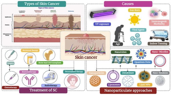
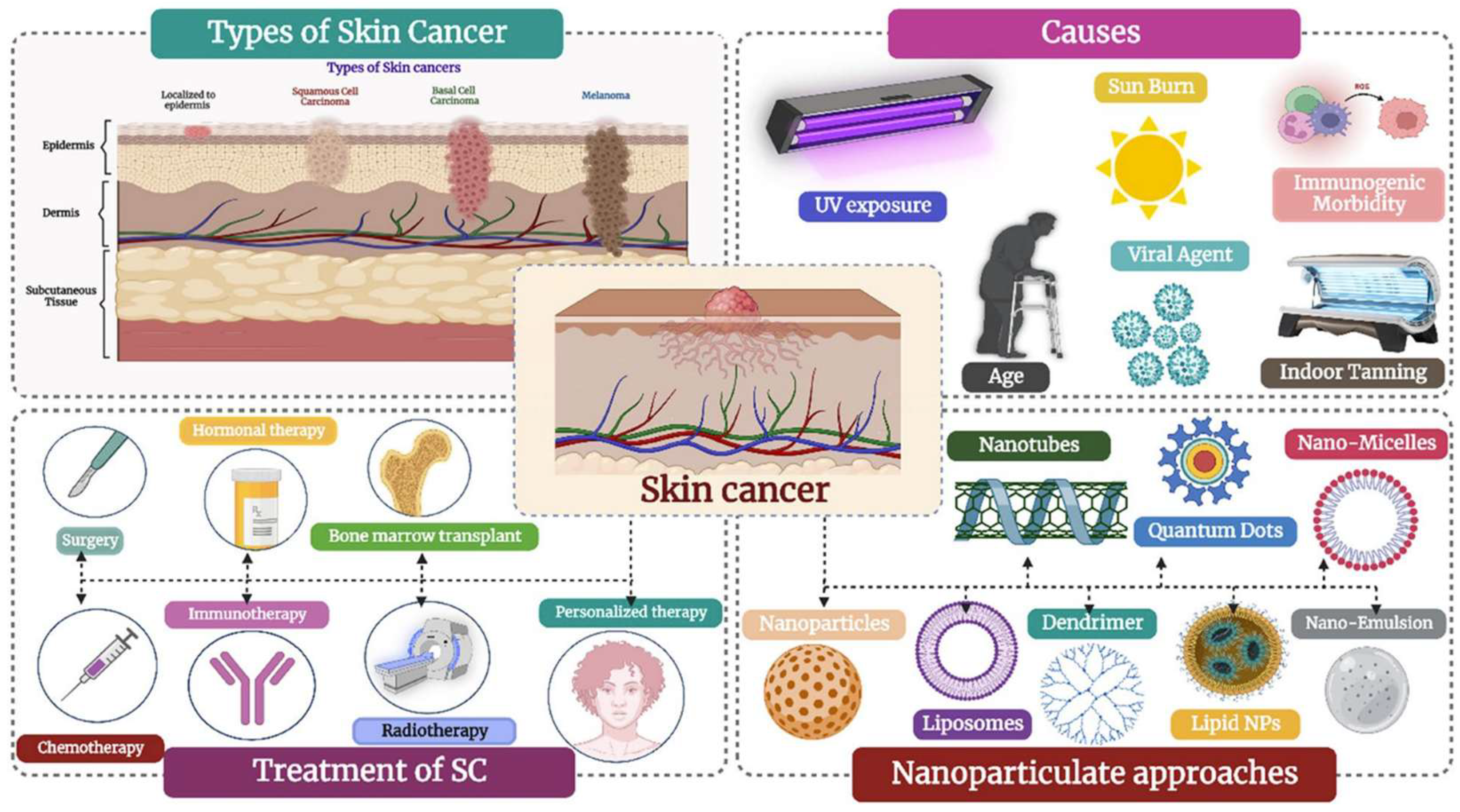
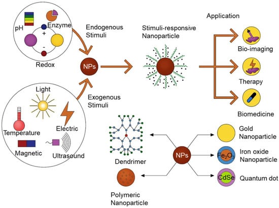
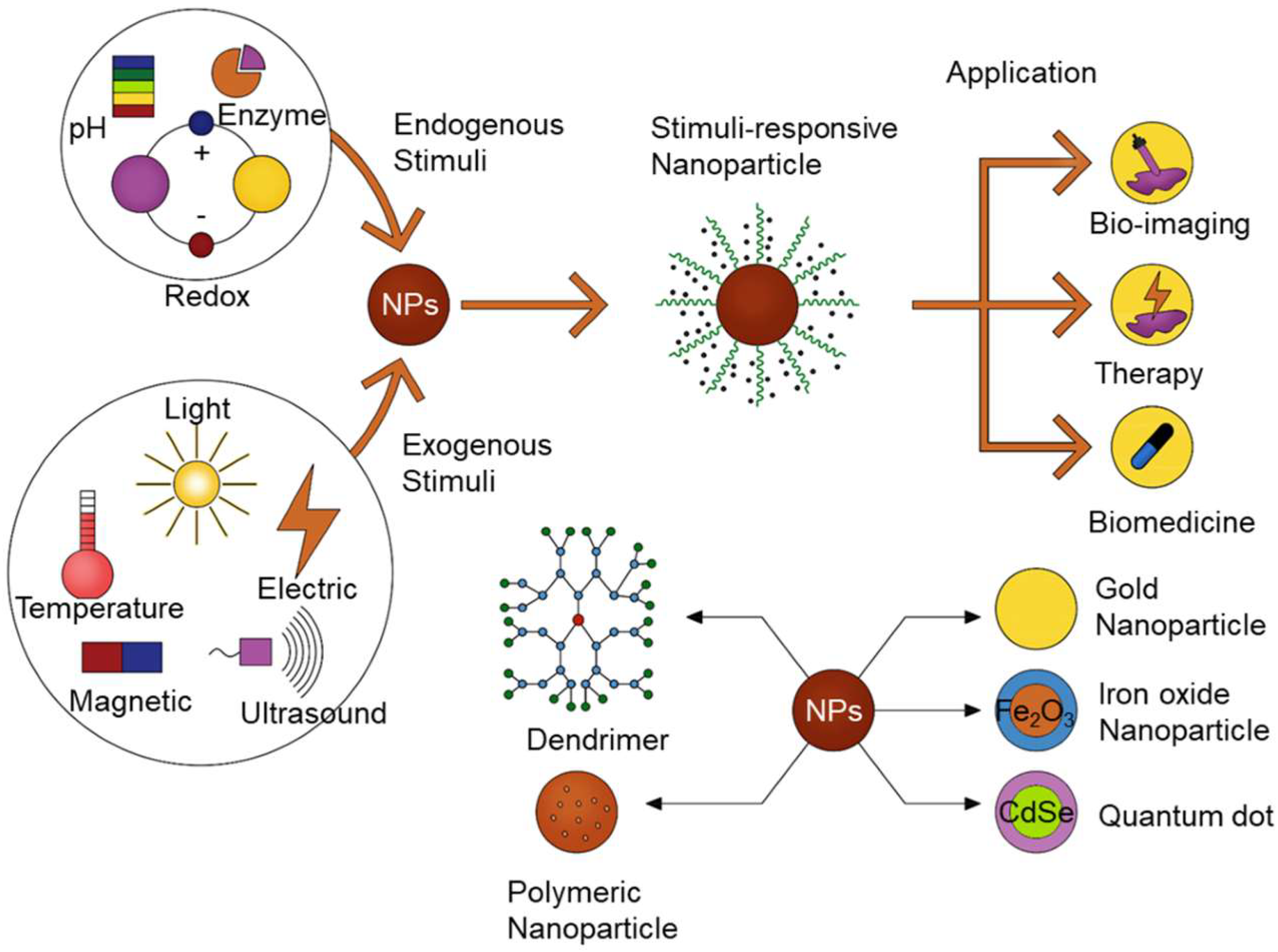
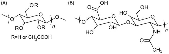
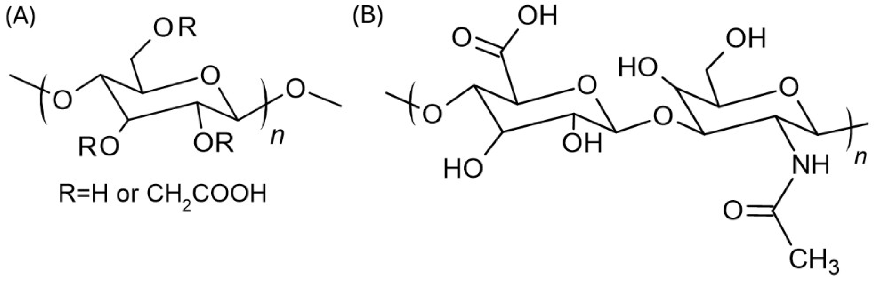
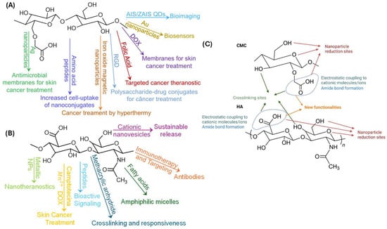
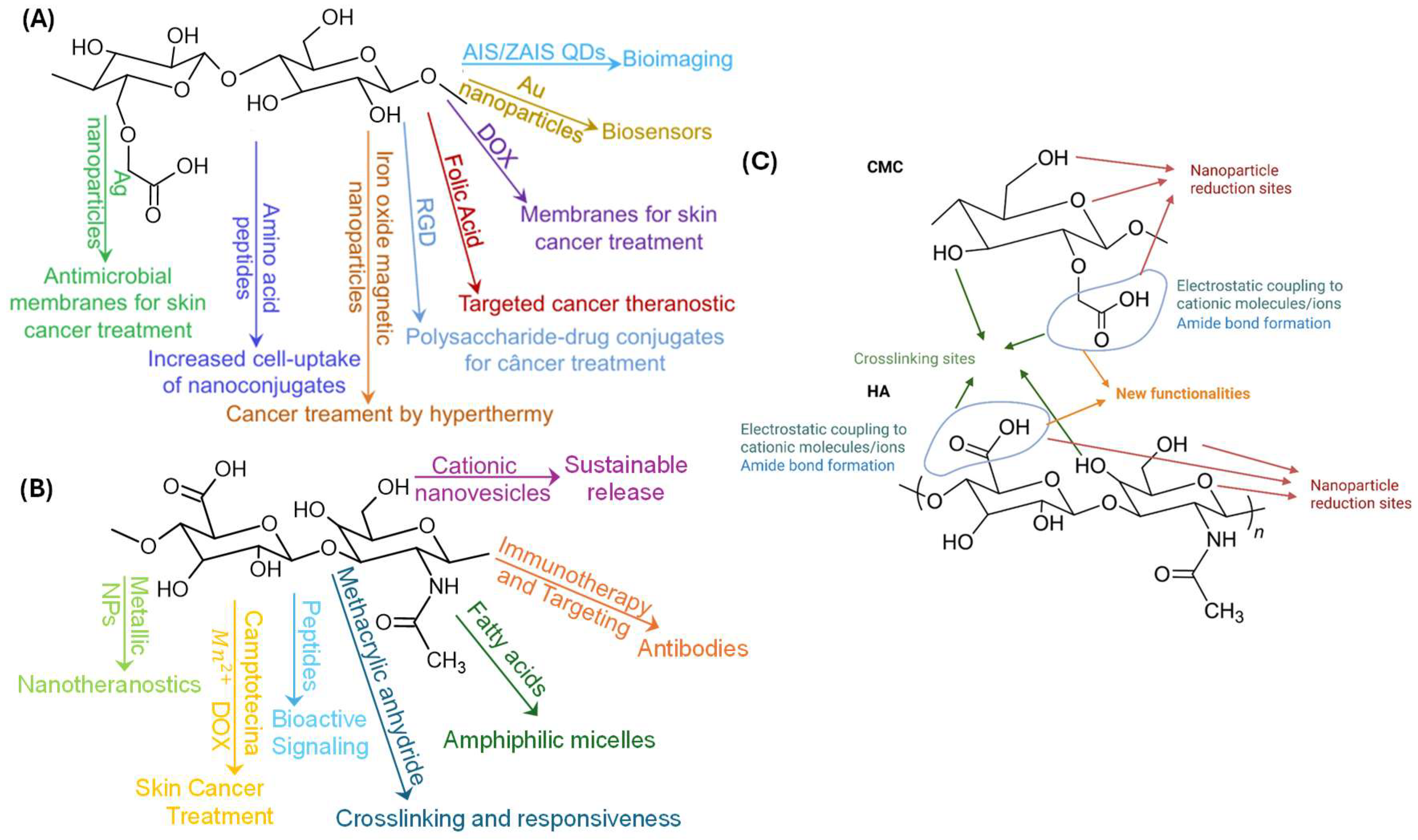
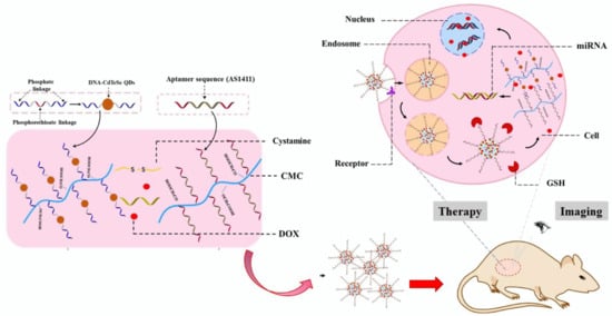
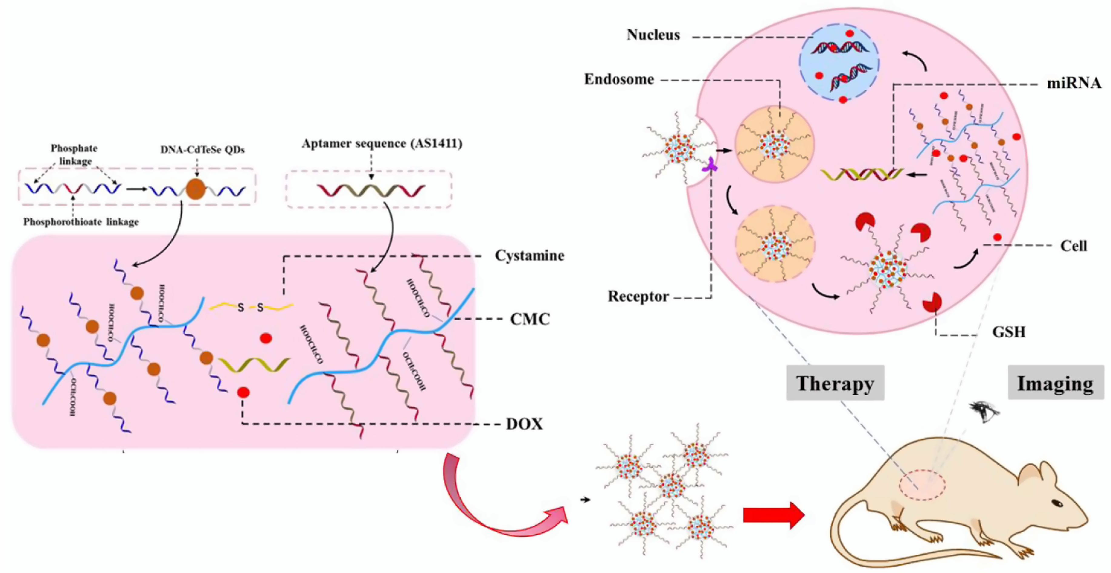
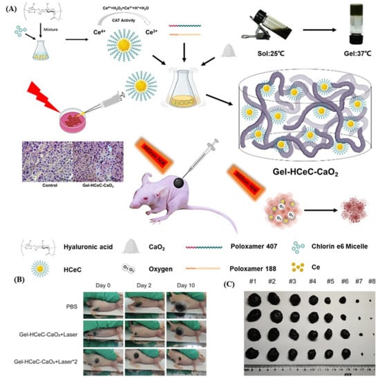
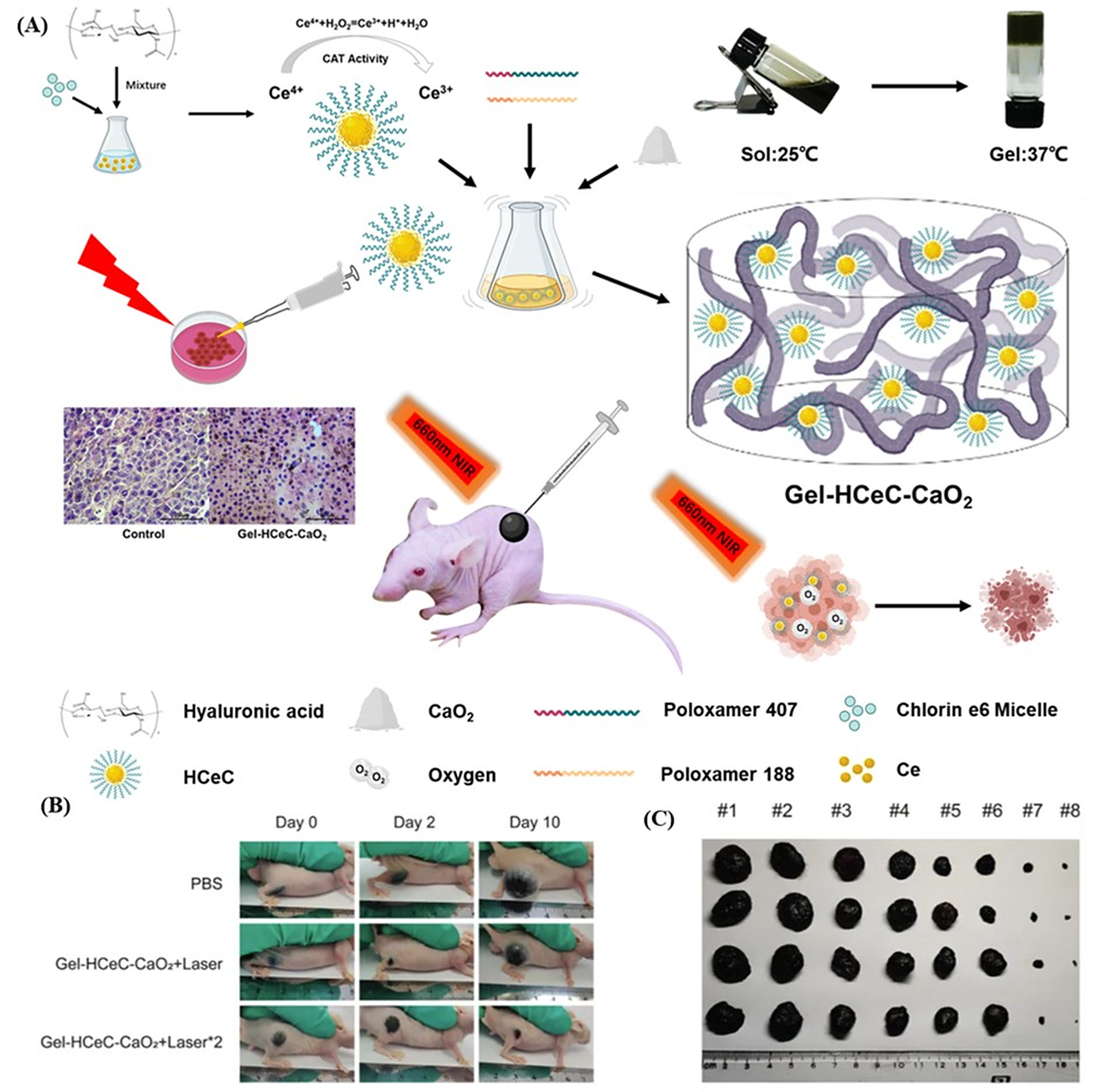
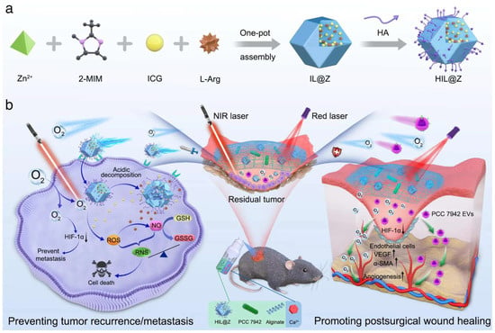
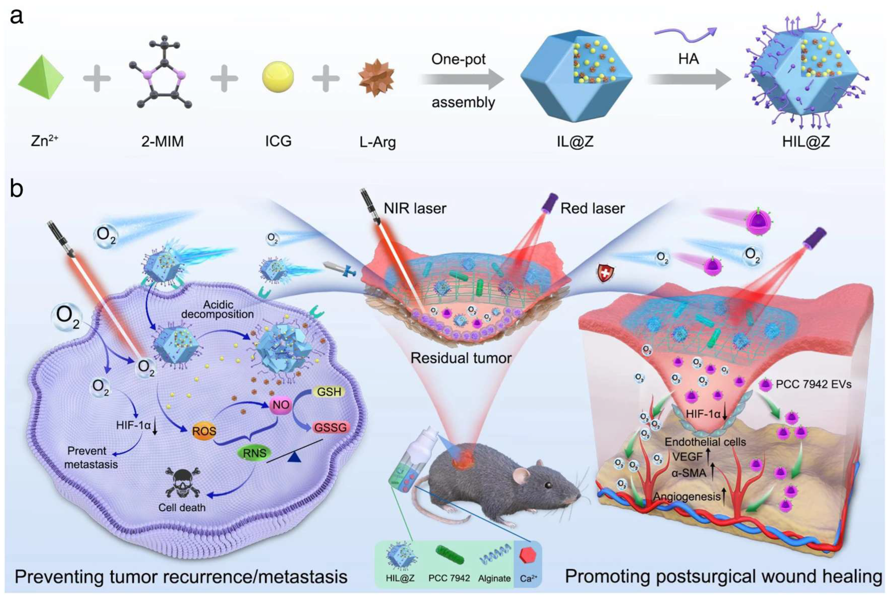
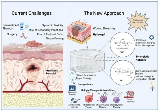
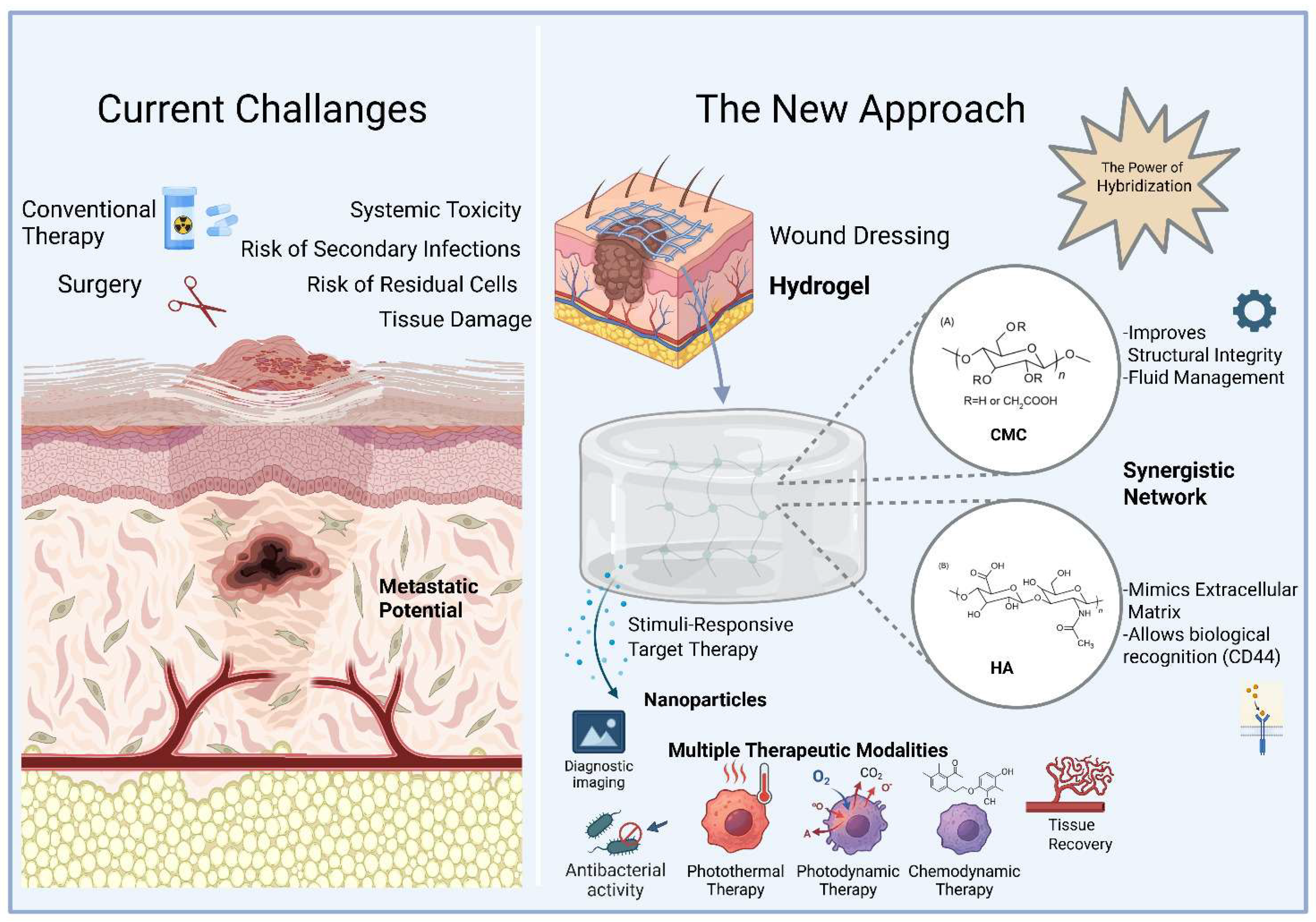

Disclaimer/Publisher’s Note: The statements, opinions and data contained in all publications are solely those of the individual author(s) and contributor(s) and not of MDPI and/or the editor(s). MDPI and/or the editor(s) disclaim responsibility for any injury to people or property resulting from any ideas, methods, instructions or products referred to in the content. |
© 2026 by the authors. Licensee MDPI, Basel, Switzerland. This article is an open access article distributed under the terms and conditions of the Creative Commons Attribution (CC BY) license.