Article

Production Optimization, Partial Characterization, and Gluten-Digesting Ability of the Acidic Protease from Clavispora lusitaniae PC3

by
Scheherazed Dakhmouche Djekrif
1,2,*,
Amel Ait Kaki El Hadef El Okki
3,
Leila Bennamoun
2,
Abdelhak Djekrif
2,
Tahar Nouadri
2 and
Louisa Gillmann
4
1
Teachers Training School El Katiba Assia Djebar Constantine, Department of Natural Sciences, University Town Ali Mendjeli, Constantine 25000, Algeria
2
Laboratory of Microbiological Engineering and Applications, Faculty of Natural Sciences, Department of Biochemistry & Cellular and Molecular Biology Constantine 1, BP 325, Route Ain El Bey, Constantine 25017, Algeria
3
Institute of Nutrition and Food and of Agri-Food Technologies, Constantine 1 University, Constantine 25000, Algeria
4
Laboratoire des Sciences Biotechnologie et Agro-Alimentaire–Groupe d’Etude des Interactions Hôte-Pathogène (LASBA-GEIHP), Université d’Angers, 49933 Angers, France
*
Author to whom correspondence should be addressed.
Fermentation 2024, 10(3), 139; https://doi.org/10.3390/fermentation10030139
Submission received: 5 January 2024 / Revised: 12 February 2024 / Accepted: 26 February 2024 / Published: 29 February 2024
(This article belongs to the Section Industrial Fermentation)

Abstract

:
Protease-producing yeasts were isolated from potato wastes and screened for protease production on skim milk agar plates. The best producer of protease isolate was identified as Clavispora lusitaniae. The strain showed higher enzyme production using tomato pomace and bread waste mix as a solid fermentation substrate. The optimized conditions improved enzyme activity and showed a maximal production of 33,450 ± 503 IU/g compared with the initial activity of 11,205.78 ± 360 without medium optimization. A threefold increase in protease activity after medium optimization proved the reliability of using the PBD and CCD design. A 19.76-fold purified enzyme and a yield of 32.94% were obtained after purification. The protease showed maximum activity at pH 4 and 60 °C and was resistant to Tween 20, Tween 80, SDS, and β-mercaptoethanol, Ca2+, and Mg2+ stimulated it. The protease activity was strongly inhibited in the presence of urea, and EDTA. The results revealed Clavispora lusitaniae protease’s ability to degrade wheat seeds and flour gluten by 98.7% and 97% respectively under pH 4 for 24 h at 40 °C. According to this study, this enzyme could be a potential candidate for the food industry, particularly for treating wheat seed and flour to reduce the immunogenicity of gluten.

1. Introduction

Proteases (E.C. 3.4.21-24.x) comprise a significant group of industrial enzymes and represent 60% of the enzyme market, estimated to be $7 billion in 2023 [1,2,3]. They hydrolyze proteins into small polypeptides or amino acids [4]. Protease enzymes degrade proteins at different sites [5]. Proteases within clans and families can be classified as asparagine proteases, aspartic proteases, cysteine proteases, glutamic proteases, mixed proteases, metalloproteases, threonine proteases, serine proteases, or unknown proteases based on these phylogenetic relationships and mechanisms of action [6,7].
A variety of enzymes are used in place of chemicals, with microbial enzymes—particularly proteases—being used most frequently within industrial sectors for the production of food (particularly in cheese making). They are also used in detergents; paper and waste management strategies [7,8,9]; the pharmaceutical, textile, feather bioconversion, and leather industries [10,11]; silver recovery and silver extraction from cast-off X-ray films [12,13]; and medical sectors [1,14].
Microbial proteases are the leading catalysts in use, with a sharp rise in global demand in recent decades [10] due to their low cost of production, extracellular nature, availability, and stability. Furthermore, given that the estimated cost of the growth medium is 30–40% of the industrial enzyme production cost [15], protease production necessitates lower-cost media, which can be obtained by utilizing easily accessible and reasonably priced agro-industrial wastes. The composition of tomato pomace and bread represents a significant source of protein [16]. In addition, bread waste contains a significant amount of starch, which is easily hydrolyzed into monomeric sugars. This makes it possible to use this waste in several areas, notably animal feed and the microbial production of enzymes such as protease.
The optimization of the nutritional and physicochemical conditions of the culture medium has been realized via the application of statistical designs. Plackett–Burman designs [17] are used for the selection of factors with a significant effect on proteolytic production, and Box–Wilson designs [18] are applied to determine the optima of the factors selected during the first design.
Molds and bacteria have historically been used in the production of industrial en-zymes. Among the protease-producing yeasts there are Wickerhamomyces anomalus 227, Metschnikovia pulcherrima 446, Candida spp., Yarrowia lipolytica, Rhototorula mucilaginosa KKU-M12C and Cryptococcus albidus KKU-M13C [15]. In recent years, the production of proteases from yeast has increased in demand [19]. As there are not many highly proteolytic yeast species [20], it would be interesting to isolate new protease-producing yeast strains. However, only a few studies have been performed on the production of enzymes by the yeast Clavispora lusitaniae. This yeast has shown its ability to produce phytase [21], amylopullulanase [22], and alpha amylase [23].
Because of its viscoelasticity, gluten is essential to the baking and food processing industries. Conversely, wheat proteins—particularly gluten—have a negative impact on those who are genetically susceptible to conditions such as celiac disease (CD), wheat allergy, gluten intolerance, and asthma in bakers.
Currently, CD patients strictly stick on to life-long gluten-free diet. An alternative investigation is in progress to reduce the immunostimulatory celiac toxic peptide sequence in wheat prolamins through the use of modern technologies such as proline cleaving specific proteolytic enzymes, sourdough technology, chemical deamination, and enzymatic modification gluten [24]. It would be interesting to investigate whether the acidic protease produced by Clavispora lusitaniae has hydrolyzing capacity.
The aim of this research is to study the production, purification, characterization, and application of yeast protease isolated from potato waste. The strain Clavispora lusitaniae isolated from potato peels was used for protease production on a low-cost medium. Protease purification and characterization were carried out after production optimization was achieved using the statistical design of experiments through the Plackett–Burman design and RSM. Additionally, the enzyme’s ability to break down gluten was tested.

2. Materials and Methods

2.1. Isolation and Identification of Protease-Producing Strain

2.1.1. Sampling and Yeast Strains Isolation

Potato wastes (PW) were collected from waste dumping sites in restaurants. Ten grams of PW was put into a sterile 250 mL flask Erlenmeyer containing 90 mL of sterile Yeast Malt (YM) liquid medium (glucose 2%, malt extract 1% and yeast extract 1%). After 48 h of incubation in a shaker at 28 °C and 150 rpm, the presence of yeast cells was examined using light microscopy. After several dilutions with sterile distilled water, 0.1 mL of the culture was spread out on the surface of the YMA medium supplemented with 0.01% chlortetracycline (Sigma-Aldrish chemical Co., Ltd., St. Louis, MI, USA), and 0.01% chloramphenicol (Sigma-Aldrish chemical Co., Ltd.) to prevent bacterial growth.
After incubation at 28 °C for 5–7 days, strain colonies were periodically checked; the representative colonies of each morphological type were purified, and maintained on YPGA: yeast extract 1%, peptone 1%, glucose 2%, and agar 2% stored at 4 °C. The ability of isolated strains to produce protease was studied on Skim Milk Agar (SMA) medium: 1% skim milk, 2% agar. The inoculated plates were incubated at 30 °C for 4 to 5 days. Yeasts showing a clear zone around the colonies were selected and, considered protease-positive [7,25].
The yeast colonies were purified and conserved on YMA, and YPGA media at 4 °C. They were also, stored in cryo-beads at −80 °C, for long storage.

2.1.2. Characterization and Identification of Strain PC3

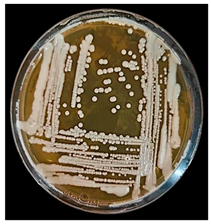
Morphological studies of yeast isolates were performed using YM agar and YM broth incubated at 28 °C for 72 h.
For aspects in liquid medium, the pure strains are inoculated on liquid YM medium and incubated for 3 days at 28 °C. During this incubation we check for the presence of a cloud or not, a veil, a ring, dusty granular deposit and the formation of gas.
For aspects in solid media, the pure strains were subcultured on YM agar using the exhaustion streak method. The boxes were incubated for 3 days at 28 °C then left in the light and at room temperature to encourage the possible appearance of pigment.
The colonies were observed using a binocular magnifying glass, their shape (regular or irregular outline, convex or concave), their appearance (shiny or matte)

Microscopic Examination

It allows in the fresh state (×40 magnification and/or ×100 magnification) the study of the shape of the cells and their size, as well as the different organizations of the yeasts. This study was carried out on young cultures in liquid YM and YMA medium.
The vegetative reproduction mode was observed on a slide in the fresh state or fixed by staining with methylene blue. For sexual reproduction, MacClary’s acetate agar sporulation medium was used: 0.1% glucose, 0.18% potassium chloride, 0.82% sodium acetate, 0.25% yeast extract and 1.5% agar. The tubes containing the inoculated inclined media were incubated for 1 week to 1 month, at 28 °C. A slide spread was made from these sporulation agar tubes. These slides were then observed under a microscope in a fresh state or stained with methylene blue.
The filamentation test was carried out on R.A.T medium. (Rice Agar Tween): 20 g of unpolished rice were simmed in 1 L of water for 45 min. After Filtration, water was addede to restore the volume to 1 L. 20 g of agar were added and dissolved. Medium was sterilized in an autoclave at 121 °C for 15 min.—Physiological and biochemical characteristics of the yeast isolates were determined according to the API test (API 32C AIX; BioMerieux S.A., Marcyl’Etoile, France).
The inoculated gallery covered with a lid was incubated at 30 °C for 48 h.
Visual readings were taken after 24 and 48 h of incubation. The possible presence of a disorder was noted after comparison with the control (O). A cloudier well than the control indicates a positive reaction and fermentation of the compound.
The results obtained from API 32C were interpreted using the APIWEB download provided by the manufacturer.
The selected yeast strain was submitted for identification via molecular approach (sequencing of the D1/D2 domain of 26S rRNA gene). DNA extraction was carried out according to Bennamoun et al. [16]. DNA was first amplified as a template by the PCR method using the primers V9G (50-TGCGTTGATTACGTCCCTGC-30) and RLR3R (50-GGTCCGTGTTTCAAGAC-30; Sigma-Aldrich Co.). A 600–650 bp region was sequenced by the forward primer (50-GCATATCAATAAGCGGAGGAAAAG-30) and the reverse primer NL4 (50-GGTCCGTGTTTCAAGACGG-30; Sigma-Aldrich Co.). The PCR products were sequenced using a commercial sequencing facility (Macrogen, Amsterdam, The Netherlands). The sequences obtained were compared with those included in the GenBank database (Blast freeware from http://www.ncbi.nlm.nih.gov/BLAST (accessed on 11 April 2022) [16].

2.2. Protease Production

2.2.1. Substrates Preparation

Tomato pomace (TP) was obtained from the tomato-processing plant Ben Azzouz (Skikda, Algeria). It was dried in the open air (25–30 °C, 4 days). The bread waste (BW) was collected from restaurants, in Constantine, Algeria, and was cut into pieces, and dried at room temperature for 3 days [26]. The substrates were incubated in an oven at 70 °C until constant weight. The dried bread waste and tomato pomace were milled with the Pulverisette 14-rotor mill Fritsch (Alsace- Lorraine, France). The milled TP and BW were sieved (2 mm), and preserved in hermetically sealed boxes.
Both wastes were characterized for the following parameters: Total Sugars (TS) by Dubois et al.’s method, [27], Ash dosage was determined according to AFNOR [28], determination of fat using the Wisman method [29] and Proteins by Total Kjeldahl Nitrogen (TKN) methode [30]. Starch determination was carried out using the iodo-iodide reagent method [31]
The composition of tomato pomace was as follows: 22.4% total sugars, 21.2% protein, 3% lipids and 6.1% minerals and that of bread was 48.46% total sugars, 46.3% starch, 8.4% proteins, 0.28% lipids and 1.3% minerals.
The phenolic composition of the vegetative parts of the tomato stem-root-leaf has not been characterized. However, the leaves contain significant amounts of total polyphenols.
Polyphenols have also been identified among the volatile compounds of tomato.
Moreover, the tomato waste used in this work is composed only of pulp and skin, which shows no interactive effect between phenolic compounds and folin ciocalteu [16].

2.2.2. Fermentation

Clavispora lusitaniae PC3 protease was produced by solid fermentation and liquid fermentation in the presence of dried and milled TW or BW or a mixture of both.
For solid-state fermentation (SSF), 05 Five grams of tomato waste, leftover bread, and a mixture of both (2.5 g tomato waste and 2.5 g waste bread) were introduced, separately, into 250 mL Erlenmeyer flasks (3 tests were carried out for each waste) then, a quantity of distilled water was added to obtain moisture level of 60% [19]. After sterilization, the microbial inoculum of 105 cells/mL was used, and contents must be mixed well using a sterile rod (the operation was carried out aseptically) and then incubated for 48 h at 30 °C.
After incubation, fermented substrate was mixed with 50 mL of Tween 80 solution (0.1%) for 10 min and centrifuged at 10,000× g at 4 °C for 20 min. The supernatant was filtered using Whatman paper n°1, and the filtrate was used as a crude enzyme.
For submerged fermentation (SmF), the flour from tomato waste and bread waste and a mixture of both was dispersed in distilled water at a concentration of 5%. Media were divided at a rate of 40 mL per 250 mL Erlenmeyer flasks and were autoclaved at 121 °C for 20 min. The inoculated sterile media (105 cells/g) were incubated in a shaker incubator at 30 °C for 48 h at 150 rpm. After incubation, the culture media were centrifuged at 10,000 rpm at 4 °C for 20 min, and the supernatant represents the enzymatic extract.

2.3. Production Optimization

Protease production is influenced by various production parameters, including nutritional and environmental parameters. For this, the composition of the culture medium was optimized according to statistical methods using two experimental designs.
Screening of significant factors: Plakett and Burman’s design (PBD) [17] was applied to determine factors with a significant positive or negative effect on protease production, using a minimum number of experiments [32]. For this, a matrix of 12 experiments was used to evaluate the effect of the three dummy variables (D, H, and K) and eight reel variables (factors) studied. Each factor was studied at two levels high (+1) and low (−1) [33,34] namely, A: Temperature, B: Moisture, C: Inoculum, E: Glucose, F: «Corn steep liquor»: G: (NH4) SO4, I: NaCl, and J :Time fermentation (Table 1).

Optimum Determination of Selected Factors

The Central composite design (CCD) of Box-Wilson [18] under RSM is an effective experimental tool allowing the study of the interactions of the effective factors and the selection of the optimal conditions of the enzyme production [35].
The three most significant variables (factors): Humidity (B), inoculum (C) and fermentation time (J) selected by Plackett and Burman’s design were optimized by the CCD under RSM. Each factor in the design was studied at five levels (Table 2). Statistical analysis was performed using Minitab software version 19.

2.4. Analytical Methods

2.4.1. Protease Activity Assay

The proteolytic activity in enzymatic extract of Clavispora lusitaniae PC3 was measured according to method described by Benkahoul et al. [32] with minor modifications using casein as substrate. The mixture containing 0.625 mL of 2.5% casein as the substrate and 0.25 mL of enzymatic extract and 0.375 mL of buffer was prepared. It was incubated at 40 °C for 30 min, and then 2.5 mL of trichloroacetic acid (TCA) 4% were added to the reaction mixture to terminate the enzyme reaction. After 10 min, the reaction mixture was filtrate and to 0.5 mL of filtrate, 2.5 mL of Na2CO3 2% and 0.25 mL of 25% Folin Ciocaltreu reagent (before use, 25 mL of reagent diluted with 100 mL of distilled water) were added and the mixture was incubated at room temperature. After 30 min, the protease activity was read at 750 nm in a spectrophotometer (UV/VIS) using a tyrosine standard curve as a reference. One unit of protease activity was defined as the amount of enzyme required to liberate 1 µmole tyrosine/min.

2.4.2. Protein Assay

Total protein content was determined by Lowry et al. [36] using bovine serum albumin as reference.

2.5. Purification of Protease

Clavispora lusitaniae PC3 protease was partially purified by ammonium sulfate precipitation and gel filtration chromatography. Crude enzyme was precipitated by ammonium sulfate (60% saturation) and kept overnight at 4 °C. The precipitate was collected by centrifugation at 10,000 rpm for 30 min, dissolved in 0.1 mol·L−1 phosphate buffer (pH 5), and then dialyzed overnight at 4 °C against the same buffer to eliminate residual ammonium sulfate. The concentrated dialysate was loaded in the Sephadex G-100 column equilibrated with the same phosphate buffer (pH5). Fractions (1.5 mL) were collected and examined, for protein content and protease activity.

2.6. Electrophoresis SDS PAGE

The molecular weight of the pure protein was estimated by SDS-PAGE on 10% homogeneous polyacrylamide gel [37].

2.7. Biochemical Characterization of Clavispora lusitaniae PC3 Protease

2.7.1. Effect of Temperature on Protease Activity and Stability

The optimal temperature for the enzyme activity was determined at temperatures of 30 to 80 °C.
Enzyme samples were taken at various times (0, 10, 20, 30, 40, 50, and 60 min) after incubation at various temperatures (50, 60, 70, 80, and 90 °C) in order to evaluate thermostatability. Following this period of incubation, the test tubes containing the enzymes were placed in an ice bath to chill them down, and the enzyme activity was measured.

2.7.2. Effects of pH on Protease Activity

The effect of pH on the enzyme activity was determined by incubating the purified enzyme between pH 3 and 10 using the standard assay condition. The buffers used were 0.2 mol·L−1 citrate-sodium buffer (pH 3 to pH 7), 0.1 mol·L−1 glycine-NaOH buffer (pH 8–10).

2.7.3. Effects of Metal Ions and Chemicals on Enzyme Activity

The effect of ions on protease activity was studied in the presence of 5 mM salts such as CuSO4, FeSO4, KCl, MnSO4, ZnSO4, MgSO4, NaCl, and CaCl2. EDTA, urea, and 5 mM SDS, β-marcaptoethanol, tween 80, tween 20 and 1 µg/mL of pepstatin were also tested for enzymatic activity. After incubating the enzyme with each metal ion or reagent at 60 °C for 10 min, the activities are measured, and compared with that of the control incubated, under the same conditions without effectors [22]. All assays were repeated three times, and the values presented are averages of the three experiments.

2.7.4. Protease Kinetic Parameters

Using a range of casein concentrations of 1, 2, 3, 4, 5, 10 and 15 mg/mL, the kinetic characteris-tics of the partially purified enzyme were determined under conventional test condi-tions. Tennalli et al. [38] determined the kinetic rate constants Km and Vmax using the Lineweaver Burk plot.

2.8. Gluten Digestion

2.8.1. Proteolytic Degradation of Gluten

The wheat flour (25 g/flask) was placed in conical flasks along Clavispora lusitaniae PC3 protease (7200 ± 216 IU) at pH 4.0 for hydrolysis. Following the procedure by Luoto et al. [39] with slight adjustments, the flasks were incubated in a water bath, shaken at 40 °C for 8, 16, and 24 h. As a control, distilled water replaced the enzymatic extract without incubation. Additionally, 25 g of wheat seeds were mixed with 50 mL of enzymatic extract, incubated at 40 °C for 8, 16, and 24 h. After drying, the mixture was crushed into flour, combined with 12 to 13 mL of distilled water. The control involved crushing wheat seeds without enzymatic treatment, and 25 g of resulting flour was mixed with 12 to 15 mL of distilled water.

2.8.2. Gluten Determination

The dosage of gluten was based on its solubility in salt water and on its agglomeration during mixing under a stream of water [40].
After the addition of the enzymatic extract and distilled water to 25 g of wheat flour. Form a homogeneous dough that, after two to three minutes of kneading, should not adhere to the walls of the container. After resting for 15 min, this dough was kneaded by hand to transform it into a homogeneous piece of dough that stretches perfectly. This dough was kneaded under a stream of water by compressing it slightly, or by placing it above a sieve intended to retain the gluten fragments that are entrained, to recover them (the starch is therefore eliminated while the gluten gradually welds itself to itself). When the gluten forms a homogeneous mass, the water flow was increased to carry out the washing until transparent washing water is obtained. The gluten was then squeezed out by compressing it strongly several times between the palms of the hands, which have been carefully wiped until it adheres to the hand. Finally, the wet gluten was quickly weighed.
Dry gluten was obtained by drying wet gluten at a temperature of 102 °C, until a constant weight is obtained, after approximately 18 h.

Results Expression

Wet gluten weight (WG) is expressed in grams per 100 g of flour by:
WG = 100(Mw/25)
Dry gluten (DG) is expressed in grams per 100 g of flour by:
DG = 100(Md/25)
Mw: mass in g of wet gluten; Md: mass in g of dry gluten.

3. Results and Discussion

3.1. Isolation and Identification of Yeast Isolate

Proteases are a group of enzymes that have wide applications potentials in various industries. Due to the increased demand for protease utilization for specific properties, researchers are interested in novel protease sources.
Among 70 isolated strains from potato wastes (PW), 10 showed their ability to hydrolyze proteins and were considered. One strain (PC3) was selected for further studies based on the maximum zone of clearance (Table 3).
The isolated strain seemed to be a yeast according to its cultural and biochemical characteristics. The results of the macroscopic and microscopic characteristics are summarized in Table 3. Microscopic observation reveals a structure similar to that of yeasts and the mode of reproduction is by Budding (unipolar and bipolar). Also, Clavispora lusitaniae PC3 is capable of producing pseudomycelium.
Since the yeasts are chemoheterotrophs, the most common substrates utilized by yeast are carbon compounds. Some yeasts have the ability to use a wide range of carbon com pounds unlike others, which assimilate less. Given the importance of this criterion in the physiology of yeasts, we used the API 32C gallery for the identification of the selected PC3 strain. The results are interpreted using the APIWEB download provided by the manufacturer (summarized in Table 3). Analysis of the 26S rRNA gene D1/D2 region sequencing revealed that the isolate was Clavispora lusitaniae. The identified strain was submitted to GenBank with accession number PP057739 (Table 3).
The nine potential strains selected (Figure 1) were also identified as Candida diddensiae D26, Candida parapsilosis P1, C. parapsilosis P2, Clavispora lusitaniae M3, Clavispora lusitaniae PC3, Debaromyces hansenii M1, Rhodotorula mucilaginosa P8, C. parapsilosis P4, C. parapsilosis P6, C. parapsilosis P7.
Different yeast species were reported to produce protease such as Rhodotorula mucilaginose [41], Pichia anomala CO-1 [42], Candida humicola [43], Yarrowia lipolytica [19,44], Candida albicans [45], Wickerhamomyces anomalus and Metschnikovia pulcherrima [46], Pichia membranifaciens [47], and Candida tropicalis [20].

3.2. Fermentation and Substrate Selection

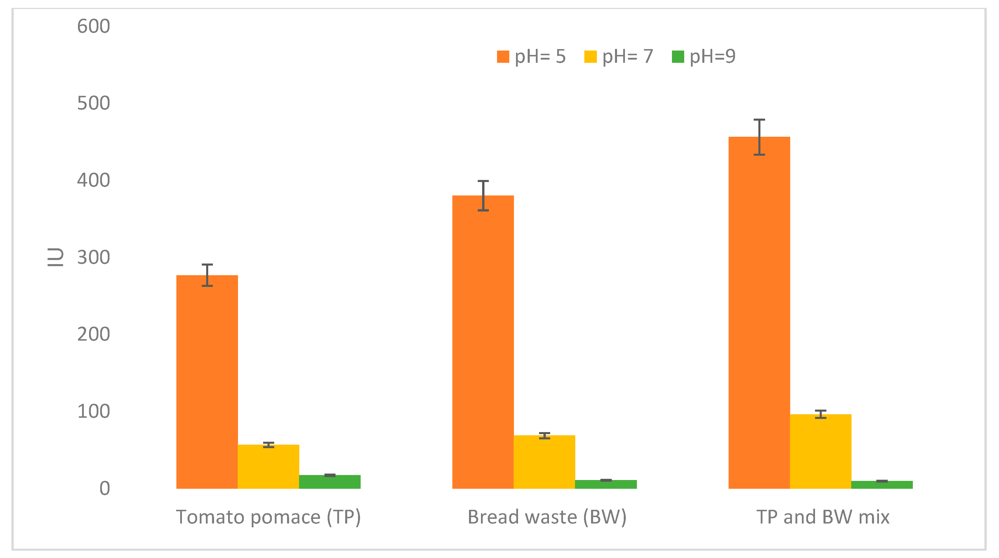
Clavispora lusitaniae PC3 protease production was studied in SmF and SSF on three media: tomato pomace (TP), bread waste (BW), and the TP and BW mix. The protease activity was assayed at different pH.
From Figure 1 and Figure 2, it appears that Clavispora lusitaniae isolated from potato peels shows protease activities when it is grown in solid fermentation and liquid fermentation of different substrates studied.
In SMF, in the presence of tomato pomace, bread waste, and a mixture of the two substrates, the synthesis of acidic protease was the most dominant followed by less important neutral protease activity and a minor quantity of alkaline protease (Figure 2). Nevertheless, the best production of acidic protease (456.2 IU) was obtained via the mixture of TP and BW.
In SSF, we noticed that Clavispora lusitaniae PC3 cultivated on different substrates (tomato pomace, bread waste, and a mixture of both substrates) mainly produced acidic protease.
However, acidic protease production was the highest (11,205.78 ± 88.5 IU/g) for the TP and BW mix compared to that produced separately in both media TP and BW (Figure 3). It appears that the TP and BW mix used constitute a good substrate for protease production by Clavispora lusitaniae PC3 in SmF because an interesting result may be obtained without any nutriment supplementation, owing to its sufficient biochemical composition, particularly regarding proteins.
This observation is in agreement with the experimental results obtained by Hisham et al. [15] who found that among 23 strains isolated from different natural sources collected from Abha region, Kingdom of Saudi Arabia, five successful yeasts for protease production were selected. At pH6 H. uvarum KKU-M19c produced neutral protease, whereas the other four yeast isolates, R. mucilaginosa KKU-M12c, C. albidus KKU-M13c, P. membranifaciens KKU-M18c, and C. californica KKU-M20c, produced acidic proteases at pH 4 and neutral proteases at pH 6. However, none of the five yeasts produced alkaline proteases.
According to the results (Figure 3), the non-existence of alkaline protease production can be explained by the fact that the pH of the medium is not favorable. The TP, BW, and TP-BW mix media are acid (pH 4.2, pH 5.4, and pH 4.7, respectively). The synthesis of protease depends on the pH of the medium as it plays a role in regulating its yield and affects the type of protease produced [48]. Similar results were also obtained by Rucha et al. [49] after their study focused on C. albicans protease production and found that the strain produced acidic protease activity, but not neutral or alkaline protease activity when grown in an acidic medium. During his study, Kim [42] showed that when the yeast Pichia anomala CO-1 was grown in an acidic medium (pH 4) at 30 °C, it secreted an extracellular neutral protease with an optimum pH of 7.
According to the results obtained with Y. lipolytica and C. olea, it was suggested that pH seems to be a major factor in regulation and neutral or alkaline proteases may not be produced in an acidic environment [20]. The presence of a low level of neutral protease from Clavispora lusitaniae is probably due to the change in the pH of the medium during fermentation. Aspartic peptidases are predominantly active in an acidic medium; however some also demonstrate activity at a neutral pH, according to James [50], whereas carboxypeptidases catalyze reactions in the pH range of 6 to 9 [19,51,52].
The diversity of protease production in Clavispora lusitaniea PC3 grown in different media can be explained by the adaptation of the strain to the culture medium, particle size, composition, and the pattern of degradation of the substrate [20].
It also appears that the combination of tomato pomace and bread waste is the best for both types of fermentation, allowing for a more important enzyme yield than each of the substrates used separately. This agrees with the results of Sharma et al. [14] who found that a mixture of two or three different substrates gives greater enzyme yields than use of the substrates individually [14]. The reduction in the cost of the culture medium through the use of food industry wastes has previously been investigated. Various agricultural or industrial wastes such as sugar cane bagasse, grape waste, wheat or barley straw, pineapple waste, orange peel waste, wheat bran, and rice bran can be used as substrates and carriers for the production of microbial protease by SSF [9,41,53].
Clavispora lusitaniae PC3 showed a good production of acidic protease. This type of enzyme is an active proteolytic enzyme present everywhere in acidic environments. Additionally, it was revealed that yeast proteases are extracellular. Currently, interest in using their aspartic proteases in different industrial applications is increasing [46]. Several studies have shown that many non-saccharomyces yeasts produce extracellular acidic proteases and the rate of protease produced can vary significantly between strains [20,46].
Yarrowia lipolytica was studied for extracellular protease production. The yeast secreted the alkaline and acidic proteases and less neutral protease production. Whereas most Y. lipoIytica strains only produce an alkaline protease and no neutral protease, it was shown that strain 37-1 of Y. lipoIytica produces a neutral protease and no alkaline protease. Another study reported that Candida oleu (Y. lipolytica) grown in a glucose BSA-based medium produced a single acidic protease, a single alkaline protease, and no neutral protease [54].
Murao et al. [55] reported that Rhodotorula glutinis is a producer of extracellular acidic proteases and found the enzyme to be stable between pH 2.4 and 6.5 after a 20 h incubation at 37 °C. The yeasts Wickerhamomyces anomalus and Metschnikovia pulcherrima also produce acidic proteases [46].
The strain is very efficient in the production of acidic protease (Aspartic protease) in the presence of the three media for both types of fermentation. Compared to SmF, SSF has several advantages such as simplicity, low cost, higher yield of enzymes, concentrated nature of the solid substrate, reduction in contamination due to the low moisture content, and increased volumetric productivity [3], as well as the possibility of using several agro-industrial wastes as a relatively cheap substrate [2]. Moreover, SSF is especially appropriate for fungal enzyme production [3].
For this, we will proceed with the production of acidic protease from Clavispora lusitaniae in solid fermentation.

3.3. Optimization of Protease Production from Clavispora lusitaniae PC3

3.3.1. Screening of Significant Factors

The Plackett–Burman design (PBD) [17] was used for screening factors that affect protease synthesis. The protease activities of the PB experimental design for 12 assays are shown in Table 4.
The statistical analysis of the results is shown in Table S1.
The effect was calculated by changing the response as the factor changes from its lower (−1) level to its higher (+1) level using Student’s t-test. The p-value of each factor was also evaluated. Factors with p-values less than 0.05 (p-value < 0.05) were considered significant factors for protease production [28,38]
Protease production was influenced by factors with a significant positive or negative effect (p ≤ 0.05) (Table 3), namely B: humidity (p = 0.049), C: inoculum (p = 0.026), and J: incubation time (p = 0.004). The reduced polynomial equation of protease production (Y) is as follows:
Y = 16,040 − 1919 Moisture − 4909 inoculum + 10,025 incubation period
The Fisher of the model F-value = 15 with p = 0.024 (≤0.05) is very significant, allowing us to conclude that the model was adequate and that the production of the protease was well explained by humidity, inoculum, and incubation period. This is also supported by the value of R2 (coefficient of determination) of the model, at 0.9756 (a value > 0.75 indicates aptness of the model) [56]. This means that 97.56% of the protease production was influenced by the selected factors (Table S2).

3.3.2. Determination of the Significant Factors’ Optimum

RMS is a statistical method used to determine the optimum of significant factors in order to develop the best growth medium for Clavispora lusitaniae PC3 and protease production. The results of the optimization experiments are presented in Table 5 and were analyzed using Minitab software version 17.
The p-value and F-value in the ANOVA (Table S3) were determined, and the lesser the p-value was, the greater the importance of the corresponding coefficient was [35,38].
The Table S3 indicates a highly significant Fisher value of 52.40 with p = 0.000, which means that the chosen model was adequate for the production of the enzyme.
The results of the statistical analysis of the CCD showed that proteolytic activity could be presented by the following regression equation:
Proteolytic activity = 32,584 + 5177 C1 − 1339 C2 − 3557 C3 − 5548 C12 − 8291 C22 − 6841 C32 + 2477 C1C2 + 3361 C1C3 + 2712 C2C3
The analysis of variance produced a highly significant Fisher value of 52.40 with p = 0, confirming a strong relationship between the production of the enzyme and the three selected factors and demonstrating that the model was significant and adequate for the enzymatic production [15].
According to Adetunji and Olaniran [57], the lack of fit determines the model’s incapacity to accurately represent data at points that are excluded from the regression. The study’s lack of fit value (p = 0.323) was not statistically significant, which is favorable since it indicates that the model equation might accurately predict the protease production for any interaction between the variable values.
The value of the coefficient of determination (R2) being 0.9792 shows that 97.92% of the variation in acidic protease production can be explained by the three factors studied. The adjusted R2 was 0.9605, which has a close relationship with R2. The predicted R2 was 0.9194, indicating a close resemblance with R2 and adjusted R2. This reveals the goodness of the model and the factors optimized [38,57].
Protease production by solid-state fermentation (SSF) was evaluated with a TP and BW mix as a substrate for Clavispora lusitaniae PC3. It has been reported that enzyme production is strongly influenced by different culture conditions (physical factors) such as moisture content, inoculum concentration and fermentation time.
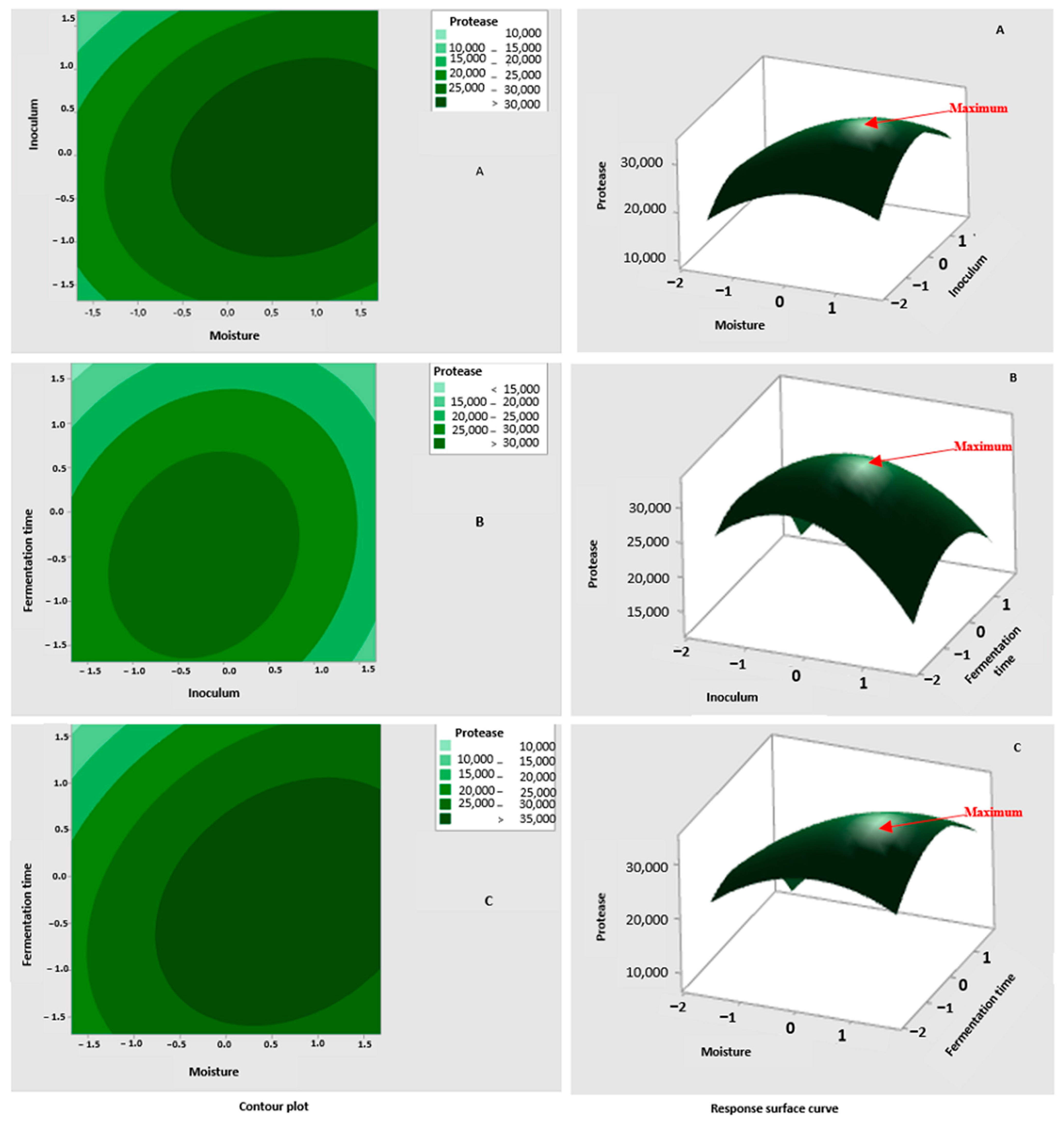
The iso-response plot of protease production was generated employing Equation (2) using Minitab software version 17. Figure 4 represents the 2D and 3D regression equations.
Each contour curve represents an infinite number of combinations of two test variables, with the third held at its respective zero level. Ellipses were obtained when there was a perfect interaction between independent variables [57]. Contour plots visually interpret the interaction between the two variables and make it easier to locate optimal experimental conditions. The dark areas provide information on the conditions that optimize the production of the enzyme.

3.3.3. Moisture

For maximum enzyme productivity, the optimum moisture content is required [9]. Low humidity has been reported to reduce the solubility of substrate nutrients and the degree of swelling. However, the increase in the rate of moisture leads to a decrease in the porosity of the substrate and thus limits the transfer of oxygen. A maximum yield of protease from Cl. lusitaniae PC3 was obtained when the humidity was 76.63%. A moisture content of 50% appeared to be the best for the production of protease from Candida utilis [58], Penicillium sp., and Aspergillus oryzae NRRL 2217 [14]. A humidity rate of 60% was obtained for maximum protease production in Penicillium godlewskii SBSS 25 [59], and for growth and protease production in Y. lipolytica [19]. The moisture of 65% was used for Rhizopus oryzae [60], 80% for T. thalpophilus PEE 14 [14], 120% for Beauveria felina [61], 90–170% for B. circulans, and 100% for B. subtilis [14].

3.3.4. Inoculum Concentration

The production of enzymes depends on the size of the inoculum, which plays an important role in the rate of fermentation [62]. The value of the inoculum rate of 8.22 × 10⁶ cells/g is within the range of values (106 to 108 cells/mL) provided by the literature [63]. This inoculum rate is also close to 2 × 10⁷ cells/g 2.5 × 10⁷ cells/g used for protease production in Y. lipolytica [19] and Candida utilis [58]. In other fungi, the inoculum of 105 spores /g was used for the production of the protease of Aspergillus oryzae MTCC 5341 [64] and Rhizopus oryzae [60].
The decrease in enzyme production with increasing inoculum size could have been due to the rapid initial growth of the microorganism, increased competition for insufficient nutrients in the medium, and reduced dissolved oxygen. On the other hand, a low amount of inoculum leads to a reduced production of proteases in microorganisms, this may be due to a deficit of microbial cells, insufficient to better consume the fermentation medium [65]. There is no precise rate of microbial inoculum suitable for the production of an enzyme.
Media composition and culture conditions are important for microbial growth and enzyme production. To achieve a high production of proteases, it is crucial to outline optimal conditions of growth and induction. It should be noted that there is no common medium suitable for all producer microorganisms; each strain has unique specific conditions for the maximum production of an enzyme [66].

3.3.5. Fermentation Time

Enzyme production is greatly affected by the incubation period, which varies from 24 h to 9 days depending on the type of microorganism, the concentration of the inoculum, the pH, and the temperature. The maximum production of the acidic protease of Clavispora lusitaniae PC3 was obtained after 56.5 h of incubation. It has been reported that a very prolonged incubation period leads to low enzyme activity, possibly due to reaching the decline phase and death of microorganisms, depletion of nutrients in the medium, or release of toxic metabolites or inhibitors [65].
In fungi, the incubation time for maximum protease production in SSF is 48 h for A. oryzae NRRL 2217, and 72 h for A. niger MTCC 281, Penicillium sp., A. flavus, and A. terreus [14]. Other studies found that better protease production was obtained at 96 h for P. godlewskii SBSS 25 [59], and 120 h fermentation period under SSF in M. circinelloides, A. oryzae MTCC 5341, and Aspergillus spp. [67], and 168 h for Beauveria felina [61].
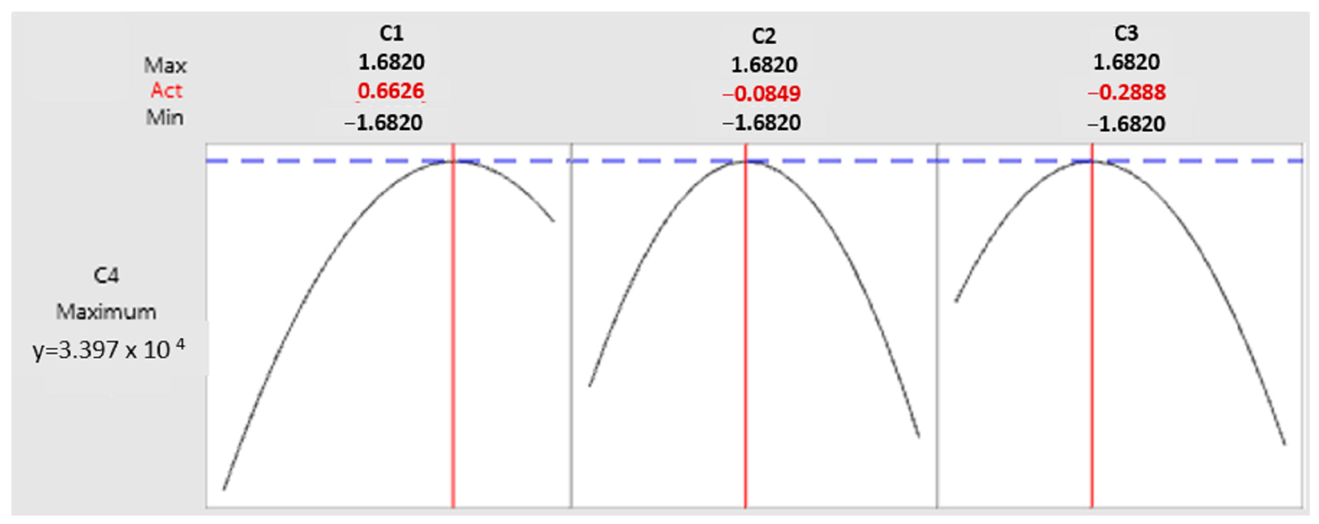
The predicted value of maximum protease production was 33,970 IU/g (Figure 3). Under optimal fermentation conditions of moisture (76.63%), inoculum (8.22 × 10⁶ cell/g), and fermentation time (56.5 h), the maximum protease production of 33,450 ± 503 IU/g was obtained. This result is in close agreement with the prediction of the statistical model, and this was found to be 3-fold higher than protease yield recorded in the unoptimized medium (11,205.78 ± 360 IU/g) (Figure 5). It was concluded that the statistical plans were effective for the optimization of the culture medium for acidic protease production from Clavispora lusitaniae PC3.
Culture medium optimization produced an increase of about three times in protease activity when compared with the activity of the initial production medium (33,450 ± 503 IU/g, 11,205.75 ± 360 IU/g, respectively).
In biotechnology, there is a growing recommendation for the use of statistical experimental designs, and several scientists achieved the optimization of protease production from microbial sources using the statistical designs [32,34,57].
In this study, PBD and CCD with RSM were shown to be efficient for optimizing enzyme production. The cost of production media was always the main problem due to the enzyme’s commercial interest [56]. Protease production by Cl. lusitaniae PC3 in the initial medium was deficient compared with the optimized medium. Therefore, we conclude that optimization of the initial culture medium showed that it is possible to increase yeast’s protease production and reduce the cost of the culture medium using the food wastes.

3.4. Purification of Protease

Clavispora lusitaniae PC3 produces a protease activity of 31,589.2 IU. The use of ammonium sulfate increased the specific activity of acid protease by 552.4 U/mg with a recovery of 92.67% (Table 6).
After dialysis, gel filtration chromatography is applied. Purification of protease from Clavispora lusitaniae PC3 led to a recovery of 32.94% protease with a purification fold of 19.76 (Table 6).

3.5. Electrophoresis SDS PAGE

In contrast to fungal aspartic proteases, which have molecular weights between 30 and 45 kDa [68], The molecular weight of the purified enzyme was found to be approximately 55 kDa (Figure 6), which is higher than the molecular weights reported for Aspergillus parasiticus (36 kDa) [69], Rhodotorula mucilagenosa (34.5 kDa) [70], Candida humicola (36 kDa) [43], Rhizopus oryzae MTCC 3690 (34 kDa) [70], R. oryzae NBRC 4749 (34 kDa) [71], and Aspergillus hennebergii HX08 (33 kDa) [72]. These findings imply that Clavispora lusitaniae PC3’s isolated protease is a unique one.

3.6. Protease Characteristics

3.6.1. Effect of pH on Protease Activity

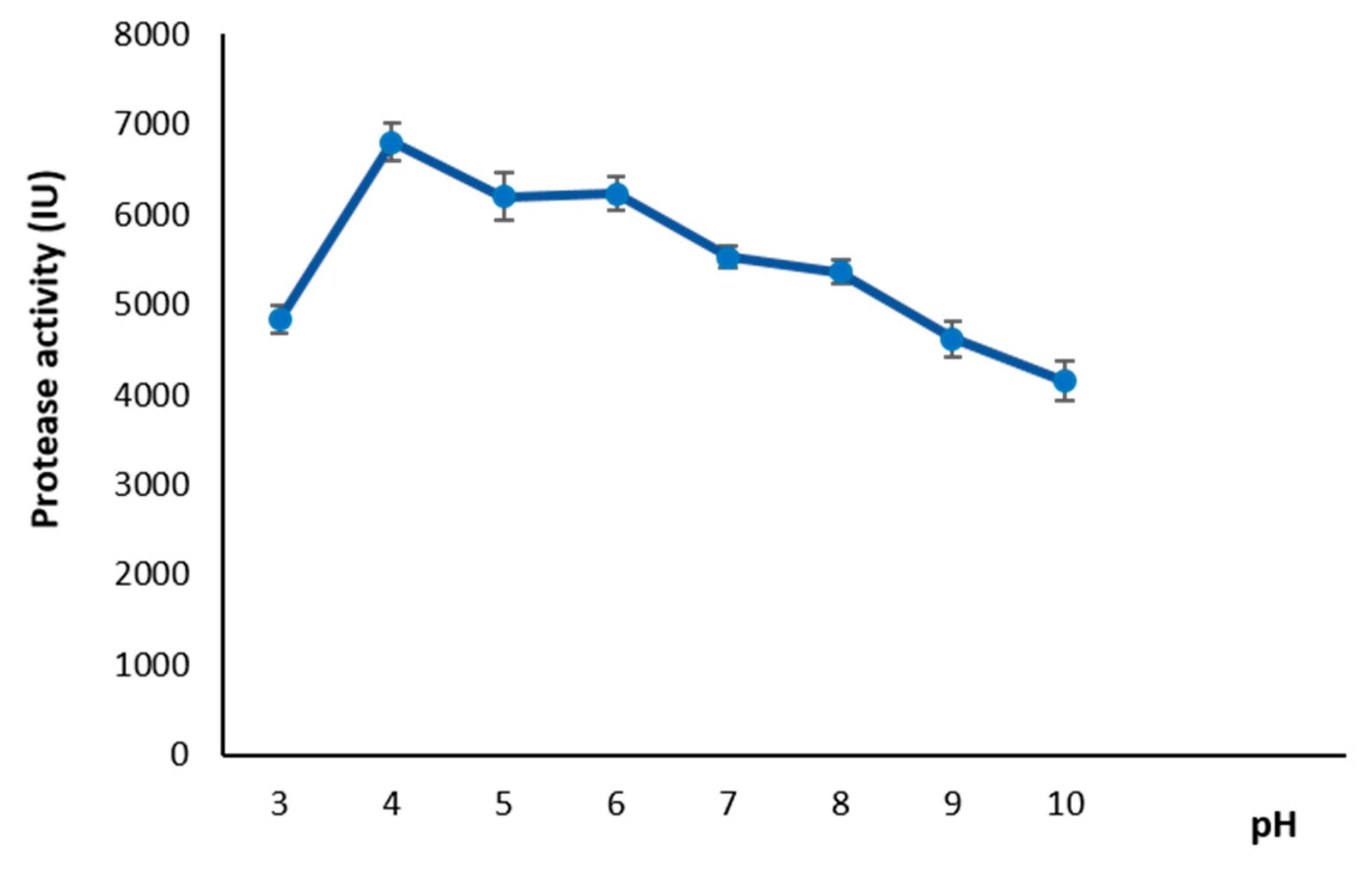
Analysis of the experimental results of the protease activity using the ANOVA method revealed that the pH affects the protease activity (F = 58.13; p = 0.000). The study showed a wide range of activity from pH 3 to 10, with a maximum at pH 4 (Figure 7). These results suggested that the Cavispora lusitaniae PC3’s enzyme was an acidic protease, which is expected for proteases produced by fungi [73]. Considerable activity was observed at pH 6 (95, 6%). Beyond this pH value, a decline in activity was observed (Figure 7).
The extracellular acidic protease produced by Rhodotorula glutinis, Candida parapsilosis, Candida tropicalis, and Candida humicola, Rhodotorula mucilaginosa L7 and Rhodotorula oryzicola had a pH optimum of 2.8 to 3.5, 4.3, 3.4 to 3.8, 4, 5 and 6.51, respectively [43,74,75].
Furthermore, it was revealed that in addition to producing an alkaline serine protease, C. albicans [43,76], Candida olea [77], Saccharomycopsis lipolytica [78], and Rhodotorula glutinis [41,79] secrete acidic proteases with a molecular mass ranging from 30 to 45 kDa and an optimal pH range of 2.5 to 3.9. Similarly, Schlander et al. [46] showed that the pH optimum of W. anomalus 227 and M. pulcherrima 446 was 3 and 4, respectively.
Srividya [80] reported that an acidic environment (pH 5) was ideal for the fungal protease activity of Aspergillus sp. The pH optimum of Aspergillus niger protease is 3–4 [24,81,82].
Acidic proteases are aspartic proteases that are mostly active at an acidic medium, which is between pH 3.8 and pH 5.6. Their ideal pH is between 3 and 4 [11,83].
The principal producers of acidic proteases are fungi, as opposed to bacteria, which mostly generate alkaline proteases. The most frequently documented genera of acidic proteases are Aspergillus, Penicillium, Endothia, Mucor, etc. [84]. Numerous reports have indicated that certain yeasts and molds secrete new acidic proteases [11,85].

3.6.2. Effect of Temperature on Protease Activity and Stability

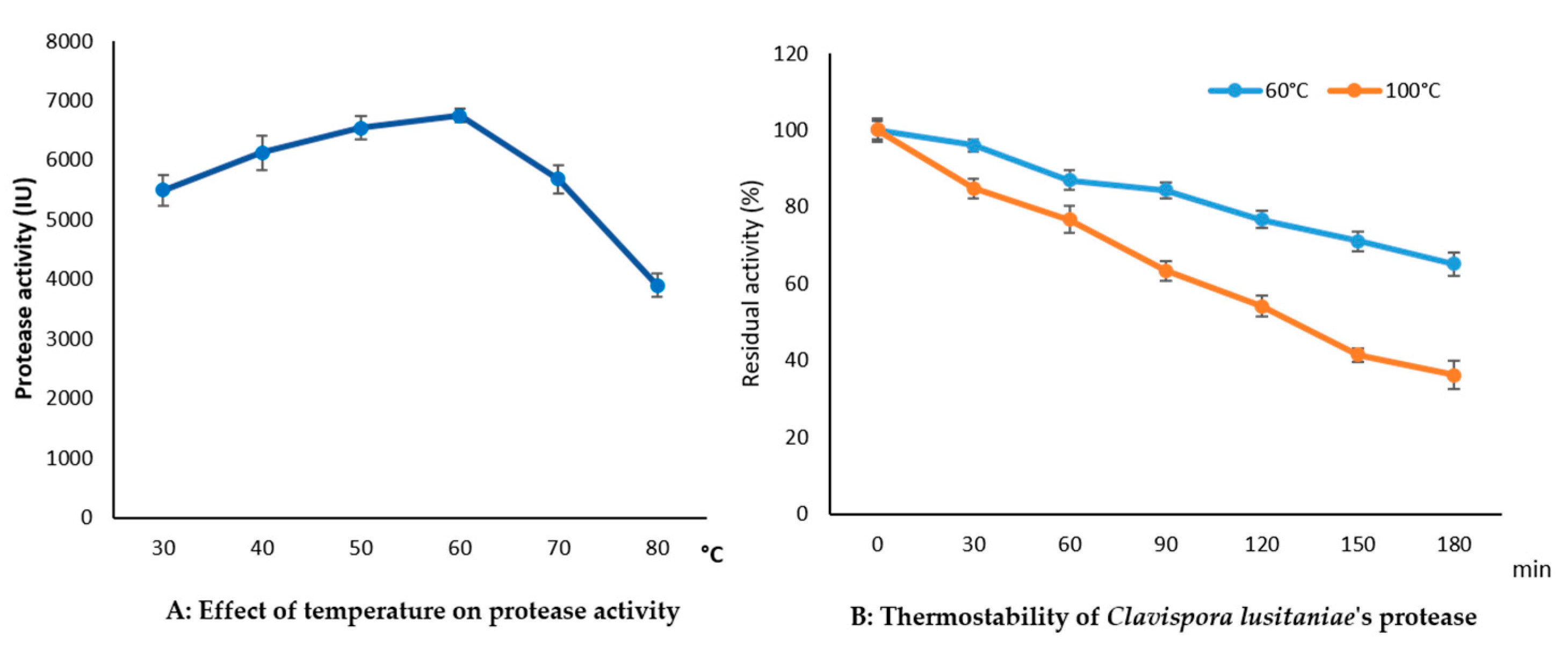
Temperature is one of the major factors affecting enzyme activity. The influence of temperature was examined via the reaction of the enzyme at temperatures ranging from 30 °C to 80 °C. The profile of protease activities as a function of temperature presents a somewhat broad aspect (Figure 8A). Enzyme activity highly depends on the temperature (highly significant difference with F = 12.6 (p 0.001). Clavispora lusitaniae PC3 protease showed maximum activity at 60 °C. However, considerable activity was observed at 30 °C (81.5%) and more than 90% of the maximal activity between 40 °C and 50 °C, while at 70 °C and 80 °C, the enzyme activity was 84.2% and 58%, respectively (Figure 8A).
A similar result was detected for protease produced by Rhodotorula glutinis, with maximum activity at 60 °C, and the enzyme preserved 80% of its maximal activity after incubation at 70 °C [79]. Moreover, Rhodotorula oryzicola protease reached its maximum at 63.04 °C [75].
Proteases with optimal temperatures between 50 and 60 °C have also been reported to be produced by a variety of cold-adapted organisms [86,87,88]; however, the mechanisms underlying these enzymes’ thermal stability remain undetermined [74].
This temperature is quite different from the optimal values reported for protease from Rhodotorula mucilaginosa L7, with an optimal temperature of 50 °C [74]. Aguilar and Sato [89] showed that acidic protease (Aspartic proteases) activity has an optimum pH ranging between 3 and 5, and an optimum temperature of 40 to 55 °C. Clavispora lusitaniae PC3 protease is an acidic protease with an optimum pH of 4 and an optimum temperature of 60 °C. No study on the protease of the species Clavispora lusitaniae has been reported in the literature. However, the study by Nakamura et al. [21] revealed that Clavispora lusitaniae produced a phytase having an optimal temperature of 70 °C. Amylopullulanase from Clavispora lusitaniae ABS7 isolated from arid-zone wheat has an optimal temperature of 75–80 °C [22].
It appears that the acidic protease of Clavispora lusitaniae PC3 presents a best thermostability, retaining 65% of its initial activity after an incubation of 180 min at 60 °C (Figure 8B).
At 100 °C for 120 min and 180 min, the enzyme maintains 54% and 36% respectively of its activity. Its half-life (t1/2) at 100 °C is 130 min.
High temperatures affect the ionizable functional groups that are involved in substrate contact and enzymatic activity’s dissociation state, which causes the protease to become denaturated and ultimately inactivated [75]. It is probable that the Clavispora lusitaniae protease has a mechanism that is effective in absorbing heat from the surrounding environment. This mechanism helps to minimize temperature increases inside the enzyme microenvironment and preserves enzyme activation.

3.6.3. Effect of Metal Ions and Additives on Protease Activity

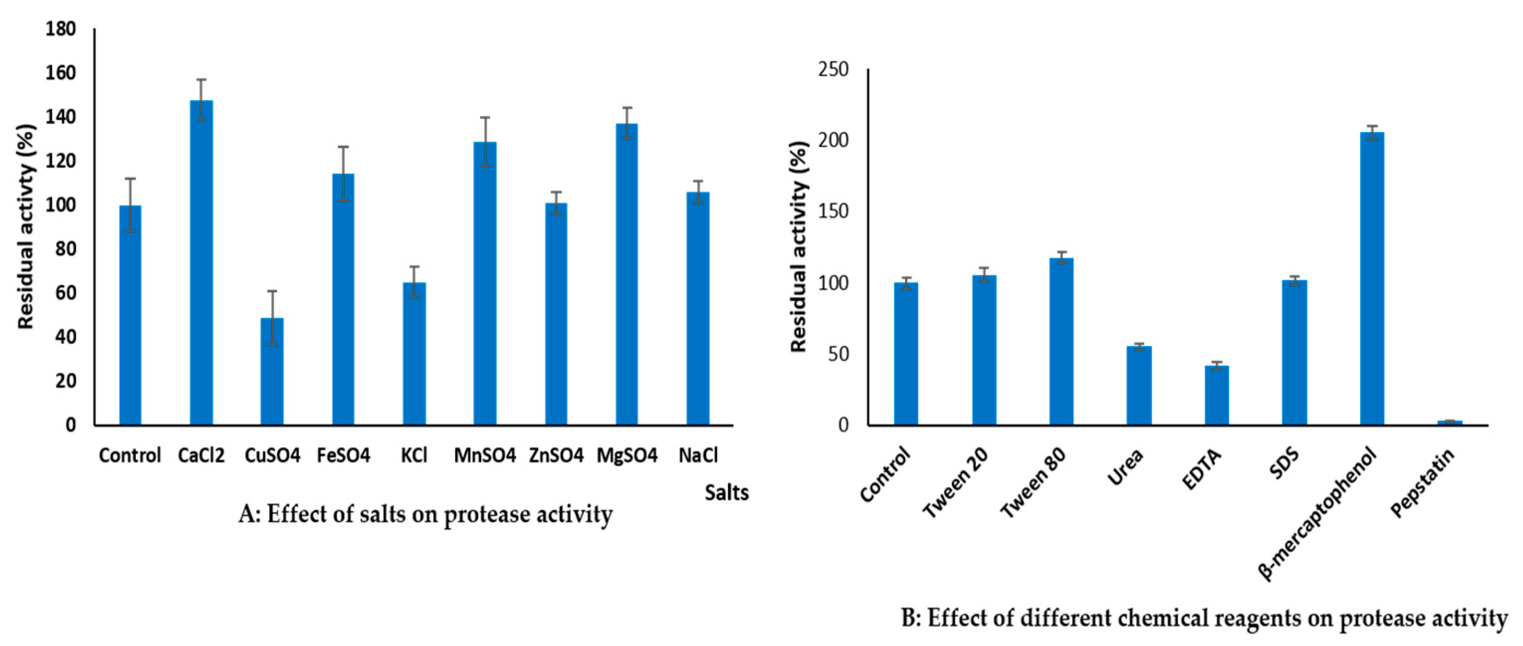
According to an assessment of the effects of different metal ions on enzyme activity, Clavispora lusitaniae PC3 protease activity was stimulated by Ca2+, Mg2+, Mn2+, and Fe2+ to 47.67%, 37.16%, 28.5, and 14.15%, respectively (Figure 9A), indicating that these ions had a functional role in the molecular structure of the enzyme [90]. Previous research investigations have mentioned that Ca2+ and Mg2+ ions play a role in maximizing the production of enzymes [9] and in stabilizing ternary structures of enzymes [68]. The best enzyme activity and stability were obtained with Ca2+, with an increase in activity suggesting that metal ions had compatibility with the enzyme. The same observation was made by Srividya [80]. However, enzyme activity was significantly reduced by the addition of Cu2+ and K+ compared to the control (Figure 9A), and was stable against Zn2+ and Na+.
From Figure 9B, it appears that mercaptoethanol significantly increased protease activity (2-fold), followed by tween 80 (17.7%). The enzyme was inhibited by urea and EDTA, and remained stable in the presence of tween 20 and SDS. It is widely recognized that urea breaks intramolecular hydrogen bonds to act as a denaturant of proteins. The removal of bonds that support the tertiary structure of enzyme molecules is assumed to be the cause of the loss of catalytic activity in the presence of urea. Usually, urea is a competitive, reversible enzyme inhibitor [91]. EDTA does not inhibe the enzyme directly; it chelates calcium, which inhibits the enzymes that are dependent on calcium. it prevents the substrate from binding with the calcium or zinc present in the active site. EDTA inhibited the activity of the enzyme very rapidly when added to reaction mixtures,. This inhibition could not be reversed by the addition of divalent ions or by the removal of EDTA by dialysis or gel filtration. Also, preincubation of the enzyme with EDTA caused an irreversible inactivation [92]. Ray et al. [43] were shown that SDS irreversibly inactivated the protease of the yeast Candida humicola, no activity band was observed even after the removal of SDS.
EDTA inhibits protease activity, with a 58.12% loss of its residual activity. This result indicates that the protease is a metalloenzyme and the activity is CaCl2-dependent. Although they are not necessary for their catalytic functions, calcium ions act as inducers and stabilizers of many enzymes against thermal denaturation and self-digestion, and protect them from conformational changes [73]. Different ions have shown a positive influence on the protease activity of different bacterial strains, e.g., Ca2+, Mg2+, and Mn2+ on the enzyme of B. circulans [14] and Aspergillus braziliensis [73]. Mn2+ enhanced Pseudomonas thermaerum GW1 protease activity 5-fold, while Cu2+, Mg2+, and Ca2+ activated it moderately [93]. The presence of CaCl2 enhanced the alkaline protease activity of A. niger by 105.3% [94].

3.6.4. Protease Kinetic Studies

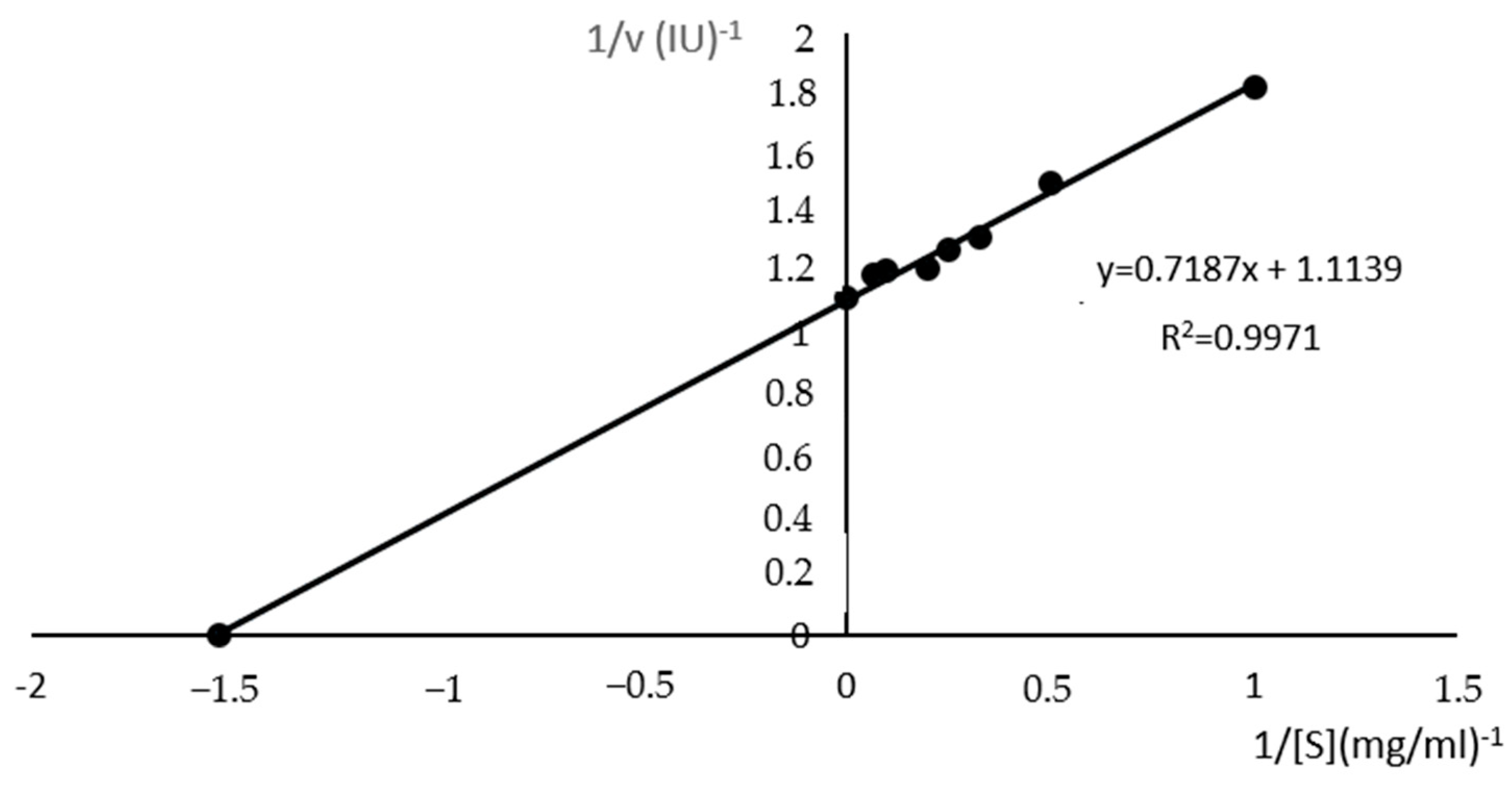
The effect of casein concentrations on the enzyme activity was evaluated using different concentrations of casein. The Lineweaver Burk plot were used to determine the value of Km and Vmax (Figure 10). The Km was 0.645 mg/mL and Vmax was 8977.46 IU. A low Km value of the protease suggests a better affinity for the substrate. Regarding the maximum indicative speed on the state of the catalytic activity of an enzyme, it is desirable that it be as high as possible. The maximal enzymatic rate of a reaction at which the enzyme is saturated with substrate is indicated by Vmax, and the affinity of the enzyme for its substrate is given by Km [38].

3.7. Action of Clavispora lusitaniae Protease on Gluten

The food and beverage industries depend heavily on aspartic proteases (EC 3.4.23), also known as acidic proteases. They are a subfamily of endopeptidases that have been isolated from diverse sources such as fungi, bacteria, plants, and animals. Microbial-origin proteases are being employed in place of animal-origin enzymes, such as rennin, in the process of clotting milk for the making of cheese [84]. Likewise, acidic proteases are employed in bakeries to enhance dough properties. Acidic proteases are also used to remove the haze from juices and beer [83].
The aspartic protease of Clavispora lusitaniae PC3 exhibits the best activity at a low pH (pH 4) and temperature of 60 °C, suggesting that the enzyme is active in an acidic medium and suitable for the food industry and beverage industry [67].
Because gliadin, one of the toxic protein fractions of gluten, is responsible for the development of celiac disease due to the presence of celiac disease eliciting epitopes in gluten [95], gluten proteins play an active role in celiac disease and their ingestion leads to damage to the villi of the small intestine. Therefore, enzymatic treatments of wheat gliadins appear to be an alternative method for reducing celiac activity.
The action of Clavispora lusitaniae PC3 protease on gluten was studied on wheat seeds and flour. The results (Figure 11) show a reduction in gluten over time and reach 97% for wheat seeds and 98.7% for wheat flour after 24 h of incubation. Our results corroborate those of Luoto et al. [39], who revealed that the protease treatment of native wheat, barley, and rye malt allowed for the reduction in prolamins by 99.95%, 99.17%, and 99.95%, respectively. After 48 h of incubation, Walter et al. [96] noted a reduction of 99% in the gluten content of wheat bran. Heredia-Sandoval et al. [97] also obtained a reduction in gluten content of 98% in wheat flour at 8 h of incubation.
In the study by Luoto et al. [39], Aspergillus niger prolyl endoprotease was required to reduce the gluten in germinated wheat products. The proteolytic activity of the strain Clavispora lusitaniae PC3 presented an interesting capacity to degrade gluten and can be called glutenase.

4. Conclusions

The present study selected Clavispora lusitaniae PC3 isolated from potato wastes for its ability to produce protease. It achieved the best production of acidic protease in SSF on a tomato pomace and bread waste mix (11,205.78 ± 360 IU/g). However, the production of enzymes remains very expensive due to the high cost of the raw material, which represents between 40% and 60% of the production cost. In this study, the use of waste as raw material for the production of enzymes. will reduce the cost of this process due to their presence in very large quantities, their carbon richness and their elimination of its own adverse effects on the environment. The optimization of the protease production was carried out by the Plackett–Burman plan for the selection of factors with a significant effect on protease production. Box and Wilson’s plan made it possible to determine their optima, namely a humidity of 76.63%, inoculum of 8.22 × 10⁶, and fermentation time of 56.5 h, and the production increased 3-fold (33,450 ± 503 IU/g). The enzyme was 19.76-fold purified, with a yield of 32.94%. Its maximum activity was obtained at pH 4 and 60 °C. This acidic protease was activated by CaCl2 and inhibited by EDTA. This leads to the conclusion that it is probably a Ca2+-dependent metalloenzyme.
As per our knowledge, acidic protease production by Clavispora lusitaniae species has not been reported. Thus, according to this study, Clavispora lusitaniae PC3 is a very promising strain for biotechnological application. Its acidic protease was produced in a low-cost medium, providing a novel and effective alternative for producing a higher-value product. These yeast extracellular enzymes have the potential to stabilize beer and wine. The use of acidic proteases is important to prevent haze formation in wine. This study showed the possibility of using this enzyme as a natural alternative to chemicals in food processing industries, particularly to produce low-gluten wheat and thus reduce gluten in cereal products for celiac patients as a step forward for clinical studies to treat celiac disease.

Supplementary Materials

The following supporting information can be downloaded at: https://www.mdpi.com/article/10.3390/fermentation10030139/s1, Table S1. Statistical analysis of the Plackett–Burman design (PBD). Table S2. ANOVA of PBD for Clavispora lusitaniae protease production. Table S3. ANOVA for response surface quadratic model.

Author Contributions

S.D.D. performed the experiments and wrote the paper; A.A.K.E.H.E.O. helped in the statistical analysis and data interpretation, L.B., helped in data interpretation; A.D. helped in wrote the paper and creation of figures; T.N. contributed to the laboratory, reagents, and materials and L.G. supervised the study. All authors have read and agreed to the published version of the manuscript.

Funding

This research received no external funding.

Acknowledgments

The authors are thankful to the Algerian Ministry of Higher Education and Scientific Research for funding this work. The author Scheherazed Dakhmouche-Djekrif is grateful to all the people for their helpful comments on the manuscript.

Conflicts of Interest

The authors declare no conflict of interest.

References

  1. Nadeem, F.; Mehmood, T.; Naveed, M.; Shamas, S.; Saman, T.; Anwar, Z. Protease Production from Cheotomium globusum Through Central Composite Design Using Agricultural Wastes and Its Immobilization for Industrial Exploitation. Waste Biomass Valori. 2019, 11, 6529–6539. [Google Scholar] [CrossRef]
  2. Gimenes, N.C.; Silveira, E.; Tambourgi, E.B. An Overview of Proteases: Production, Downstream Processes and Industrial Applications. Sep. Purif. Rev. 2021, 50, 223–243. [Google Scholar] [CrossRef]
  3. Alahmad Aljammas, H.; Yazji, S.; Azizieh, A. Optimization of protease production from Rhizomucor miehei Rm4 isolate under solid-state fermentation. J. Genet. Eng. Biotechnol. 2022, 20, 82. [Google Scholar] [CrossRef]
  4. Bhavikatti, J.S.; Bodducharl, S.M.; Kamagond, R.S.; Desai, S.V.; Shet, A.R. Statistical optimisation of protease production using a freshwater bacterium Chryseobacterium cucumeris SARJS-2 for multiple industrial applications. 3 Biotech 2020, 10, 279. [Google Scholar] [CrossRef]
  5. Marathe, S.K.; Vashistht, M.A.; Prashanth, A.; Praveen, N.; Chakraborty, S.; Nair, S.S. Isolation, partial purification, biochemical characterization and detergent compatibility of alkaline protease produced by Bacillus subtilis, Alcaligenes faecalis and Pseudomonas aeruginosa obtained from sea water samples. J. Genet. Eng. Biotechnol. 2018, 16, 39–46. [Google Scholar] [CrossRef]
  6. Rawlings, N.D.; Barrett, A.J.; Thomas, P.D.; Huang, X.; Bateman, A.; Finn, R.D. The MEROPS database of proteolytic enzymes, their substrates and inhibitors in 2017 and a comparison with peptidases in the Panther database. Nucleic Acids Res. 2017, 46, D624–D632. [Google Scholar] [CrossRef] [PubMed]
  7. Song, P.; Zhang, X.; Wang, S.; Xu, W.; Wang, F.; Fu, R.; Wei, F. Microbial proteases and their applications. Front. Microbiol. 2023, 14, 1236368. [Google Scholar] [CrossRef] [PubMed]
  8. Aruna, V.; Chandrakala, V.; Angajala, G.; Nagarajan, E.R. Proteases: An overview on its recent industrial developments and current scenario in the revolution of biocatalysis. Mater. Today Proc. 2023, 92, 565–573. [Google Scholar] [CrossRef]
  9. Balachandran, C.; Vishali, A.; Arun Nagendran, N.; Baskar, K.; Hashem, A.; Abd_Allah, E.F. Optimization of protease production from Bacillus halodurans under solid state fermentation using agrowastes. Saudi J. Biol. Sci. 2021, 28, 4263–4269. [Google Scholar] [CrossRef]
  10. Adetunji, A.I.; Olaitan, M.O.; Erasmus, M.; Olaniran, A.O. Microbial proteases: A next generation green catalyst for industrial, environmental and biomedical sustainability. Food Mater. Res. 2023, 3, 12. [Google Scholar] [CrossRef]
  11. Solanki, P.; Putatunda, C.; Kumar, A.; Bhatia, R.; Walia, A. Microbial proteases: Ubiquitous enzymes with innumerable uses. 3 Biotech 2021, 11, 428. [Google Scholar] [CrossRef]
  12. Hashmi, S.; Iqbal, S.; Ahmed, I.; Janjua, H.A. Production, Optimization, and Partial Purification of Alkali- Thermotolerant Proteases from Newly Isolated Bacillus subtilis S1 and Bacillus amyloliquefaciens KSM12. Processes 2022, 10, 1050. [Google Scholar] [CrossRef]
  13. Wahab, W.A.A.; Ahmed, S.A. Response surface methodology for production, characterization and application of solvent, salt and alkali-tolerant alkaline protease from isolated fungal strain Aspergillus niger WA 2017. Int. J. Biol. Macromol. 2018, 115, 447–458. [Google Scholar] [CrossRef] [PubMed]
  14. Sharma, K.M.; Kumar, R.; Panwar, S.; Kumar, A. Microbial alkaline proteases: Optimization of production parameters and their properties. J. Genet. Eng. Biotechnol. 2017, 15, 115–126. [Google Scholar] [CrossRef] [PubMed]
  15. Hesham, A.E.L.; Alrumman, S.A.; Al-Dayel, M.A.; Salah, H.A. Screening and genetic identification of acidic and neutral protease-producing yeasts strains by 26S rRNA gene sequencing. Cytol. Genet. 2017, 51, 221–229. [Google Scholar] [CrossRef]
  16. Bennamoun, L.; Hiligsmann, S.; Dakhmouche, S.; Ait-Kaki, A.; Labbani, F.-Z.K.; Nouadri, T.; Meraihi, Z.; Turchetti, B.; Buzzini, P.; Thonart, P. Production and Properties of a Thermostable, pH—Stable Exo-Polygalacturonase Using Aureobasidium pullulans Isolated from Saharan Soil of Algeria Grown on Tomato Pomace. Foods 2016, 5, 72. [Google Scholar] [CrossRef] [PubMed]
  17. Plackett, R.L.; Burman, J.P. The design of optimum multifactorial experiments. Biometrika 1946, 33, 305–325. [Google Scholar] [CrossRef]
  18. Box, G.E.P.; Wilson, K.B. On the Experimental Attainment of Optimum Conditions. J. R. Stat. Soc. Ser. B (Methodol.) 1951, 13, 1–38. [Google Scholar] [CrossRef]
  19. López-Trujillo, J.; Mellado-Bosque, M.; Ascacio-Valdés, J.A.; Prado-Barragán, L.A.; Hernández-Herrera, J.A.; Aguilera-Carbó, A.F. Temperature and pH optimization for protease production fermented by Yarrowia lipolytica from Agro-Industrial Waste. Fermentation 2023, 9, 819. [Google Scholar] [CrossRef]
  20. Ogrydziak, D.M. Yeast Extracellular Proteases. Crit. Rev. Biotechnol. 1993, 13, 1–55. [Google Scholar] [CrossRef]
  21. Nakamura, Y.; Fukuhara, H.; Sano, K. Secreted phytase activities of yeasts. Biosci. Biotechnol. Biochem. 2000, 64, 841–844. [Google Scholar] [CrossRef] [PubMed]
  22. Dakhmouche Djekrif, S.; Bennamoun, L.; Labbani, F.-Z.K.; Ait Kaki, A.; Nouadri, T.; Pauss, A.; Meraihi, Z.; Gillmann, L. An Alkalothermophilic Amylopullulanase from the Yeast Clavispora lusitaniae ABS7: Purification, Characterization and Potential Application in Laundry Detergent. Catalysts 2021, 11, 1438. [Google Scholar] [CrossRef]
  23. Ranjan, K.; Ahmad, L.M.; Sahay, S. Detergent compatible cold-active alkaline amylases from Clavispora lusitaniae CB13. J. Microbiol. Biotechnol. Food Sci. 2016, 5, 306–310. [Google Scholar] [CrossRef]
  24. Mohan Kumar, B.V.; Sarabhai, S.; Prabhasankar, P. Targeted degradation of gluten proteins in wheat flour by prolyl endoprotease and its utilization in low immunogenic pasta for gluten sensitivity population. J. Cereal Sci. 2019, 87, 59–67. [Google Scholar] [CrossRef]
  25. Shaikh, I.A.; Turakani, B.; Malpani, J.; Goudar, S.V.; Mahnashi, M.H.; Hamed Al-Serwi, R.; Ghoneim, M.M.; El-Sherbiny, M.; Mannasaheb, B.A.; Alsaikhan, F.; et al. Extracellular protease production, optimization, and partial purification from Bacillus nakamurai PL4 and its applications. J. King Saud. Univ. Sci. 2023, 35, 102429. [Google Scholar] [CrossRef]
  26. Svensson, S.E.; Bucuricova, L.; Ferreira, J.A.; Souza Filho, P.F.; Taherzadeh, M.J.; Zamani, A. Valorization of Bread Waste to a Fiber and Protein-Rich Fungal Biomass. Fermentation 2021, 7, 91. [Google Scholar] [CrossRef]
  27. Dubois, M.; Gills, K.A.; Hamilton, J.K.; Rebers, P.A.; Smith, F. Colorimetric method for determination of sugar and related substances. Anal. Chem. 1956, 28, 350–356. [Google Scholar] [CrossRef]
  28. AFNOR. Collection of French Standards. Milks and Dairy Products. Methods of Analysis; AFNOR: Paris, France, 1980; pp. 33–54, 212–215. [Google Scholar]
  29. Lecoq, R. Manual of Analysis and Usual Expertise; Doin: Paris, France, 1965; Volume 1, pp. 200–203. [Google Scholar]
  30. AFNOR (Agence Française de Normalisation). Quality Control of Food Products, Collection of French Standards, 3rd ed.; AFNOR: Paris, France, 1991. [Google Scholar]
  31. Smith, B.W.; Roe, J.H. A photometric method for the determination of amylase in blood and urine with use of the starch iodine cola. J. Biol. Chem. 1949, 7, 53–59. [Google Scholar] [CrossRef]
  32. Benkahoul, M.; Bramki, A.; Belmessikh, A.; Mechakra-Maza, A. Statistical optimization of an acid protease production by a local Aspergillus niger MH109542 using a medium based on decommissioned dates. Asia Pac. J. Mol. Biol. Biotechnol. 2020, 28, 68–76. [Google Scholar] [CrossRef]
  33. Djekrif-Dakhmouche, S.; Gheribi-Aoulmi, Z.; Meraihi, Z.; Bennamoun, L. Application of a statistical design to the optimization of culture medium for α-amylase production by Aspergillus niger ATCC 16404 grown on orange waste powder. J. Food Eng. 2006, 73, 190–197. [Google Scholar] [CrossRef]
  34. Shrestha, S.; Chio, C.; Khatiwada, J.R.; Kognou, A.L.M.; Qin, W. Optimization of multiple enzymes production by fermentation using lipid-producing Bacillus sp. Front. Microbiol. 2022, 13, 1049692. [Google Scholar] [CrossRef]
  35. Shafique, T.; Shafique, J.; Zahid, S.; Kazi, M.; Alnemer, O.; Ahmad, A. Screening, selection and development of Bacillus subtilis apr-IBL04 for hyper production of macromolecule alkaline protease. Saudi J. Biol. Sci. 2021, 28, 1494–1501. [Google Scholar] [CrossRef]
  36. Lowry, O.H.; Rosebrough, N.J.; Farr, A.L.; Randall, R.J. Protein measurement with the folin phenol reagent. J. Biol. Chem. 1951, 193, 265–275. [Google Scholar] [CrossRef]
  37. Laemmli, U.K. Cleavage of structural proteins during the assembly of the head of bacteriophage T4. Nature 1970, 227, 680–685. [Google Scholar] [CrossRef]
  38. Tennalli, G.B.; Garawadmath, S.; Sequeira, L.; Murudi, S.; Patil, V.; Divate, M.N.; Hungund, B.S. Media optimization for the production of alkaline protease by Bacillus cereus PW3A using response surface methodology. J. Appl. Biol. Biotechnol. 2022, 10, 17–26. [Google Scholar] [CrossRef]
  39. Luoto, S.; Jiang, Z.; Brinck, O.; Sontag-Strohm, T.; Kanerva, P.; Bruins, M.; Edens, L.; Salovaara, H.; Loponen, J. Malt hydrolysates for gluten-free applications: Autolytic and proline endopeptidase assisted removal of prolamins from wheat, barley and rye. J. Cereal Sci. 2012, 56, 504–509. [Google Scholar] [CrossRef]
  40. Sayaslan, A. Wet-milling of wheat flour: Industrial processes and small scale test methods. LWT Food Sci. Technol. 2004, 37, 499–515. [Google Scholar] [CrossRef]
  41. Lario, L.D.; Pillaca-Pullo, O.S.; Sette, L.D.; Converti, A.; Casati, P.; Spampinato, C.; Pessoa, A. Optimization of protease production and sequence analysis of the purifed enzyme from the cold adapted yeast Rhodotorula mucilaginosa CBMAI 1528. Biotechnol. Rep. 2020, 28, e00546. [Google Scholar] [CrossRef]
  42. Kim, J.Y. Isolation of the Protease-producing Yeast Pichia anomala CO-1 and Characterization of Its Extracellular Neutral Protease. J. Life Sci. 2019, 29, 1126–1135. [Google Scholar] [CrossRef]
  43. Ray, M.K.; Uma Devi, K.; Seshu Kumar, G.; Shivaji, S. Extracellular Protease from the Antarctic Yeast Candida humicola. Appl. Environ. Microbiol. 1992, 58, 1918–1923. [Google Scholar] [CrossRef] [PubMed]
  44. Gonzalez-Lopez, C.I.; Szabo, R.; Blanchin-Roland, S.; Gaillardin, C. Genetic control of extracellular protease synthesis in the yeast Yarrowia lipolytica. Genetics 2002, 160, 417–427. [Google Scholar] [CrossRef]
  45. Theron, L.W.; Divol, B. Microbial aspartic proteases: Current and potential applications in industry. Appl. Microbiol. Biotechnol. 2014, 98, 8853–8868. [Google Scholar] [CrossRef]
  46. Schlander, M.; Distler, U.; Tenzer, S.; Thines, E.; Claus, H. Purification and Properties of Yeast Proteases Secreted by Wickerhamomyces anomalus 227 and Metschnikovia pulcherrima 446 during Growth in a White Grape Juice. Fermentation 2016, 3, 2. [Google Scholar] [CrossRef]
  47. Fernández, M.; Úbeda, J.F.; Briones, A.I. Typing of non-Saccharomyces yeasts with enzymatic activities of interest in wine-making. Int. J. Food. Microbiol. 2000, 59, 29–36. [Google Scholar] [CrossRef]
  48. Abdelal, A.T.H.; Kennedy, E.H.; Ahearn, G. Purification and characterization of a neutral protease from Saccharomycopsis lipolytica. J. Bacteriol. 1977, 130, 1125–1129. [Google Scholar] [CrossRef]
  49. Ruchel, R.; Tegeler, R.; Trost, M. A comparison of secretory proteinases from different strains of Candida albicans. Sabouraudia 1982, 20, 233–244. [Google Scholar] [CrossRef]
  50. James, M.N. Catalytic pathway of aspartic peptidases. In Handbook of Proteolytic Enzymes, 2nd ed.; Beynon, R., Bond, J.S., Eds.; Oxford University Press: Oxford, UK, 2004; pp. 12–18. [Google Scholar]
  51. Tunga, R.; Shrivastava, B.; Banerjee, R. Purification and characterization of a protease from solid state cultures of Aspergillus parasiticus. Process Biochem. 2003, 38, 1553–1558. [Google Scholar] [CrossRef]
  52. Wang, R.; Law, R.C.; Webb, C. Protease production and conidiation by Aspergillus oryzae in flour fermentation. Process Biochem. 2005, 40, 217–227. [Google Scholar] [CrossRef]
  53. Chaud, L.C.S.; Lario, L.D.; Bonugli-Santos, R.C.; Sette, L.D.; Pessoa Junior, A.; Felipe, M.; das Graças, A. Improvement in extracellular protease production by the marine antarctic yeast Rhodotorula mucilaginosa L7. N. Biotechnol. 2016, 33, 807–814. [Google Scholar] [CrossRef] [PubMed]
  54. Barnett, J.A.; Payne, R.W.; Yarrow, D. Yeasts Characteristics and Identification; Cambridge University Press: London, UK, 1983. [Google Scholar]
  55. Murao, S.; Kamada, M.; Nakase, T.; Ogura, S.; Oda, K. Studies on the Extracellular Protease of Yeasts Part I. J. Agric. Chem. Soc. Jpn. 1972, 46, 167–170. [Google Scholar] [CrossRef]
  56. Ferracini-Santos, L.; Sato, H.H. Production of alkaline protease from Cellulosimicrobium cellulans. Braz. J. Microbiol. 2009, 40, 54–60. [Google Scholar] [CrossRef]
  57. Adetunji, A.I.; Olaniran, A.O. Statistical modelling and optimization of protease production by an autochthonous Bacillus aryabhattai Ab15-ES: A response surface methodology approach. Biocatal. Agric. Biotechnol. 2020, 24, 101528. [Google Scholar] [CrossRef]
  58. Moftah, O.A.S.; Grbavčić, S.; Žuža, M.; Luković, N.; Bezbradica, D.; Knežević-Jugović, Z. Adding Value to the Oil Cake as a Waste from Oil Processing Industry: Production of Lipase and Protease by Candida utilis in Solid State Fermentation. Appl. Biochem. Biotechnol. 2012, 166, 348–364. [Google Scholar] [CrossRef]
  59. Sindhu, R.; Suprabha, G.N.; Shashidhar, S. Optimization of process parameters for the production of alkaline protease from Penicillium godlewskii SBSS 25 and its application in detergent industry. Afr. J. Microbiol. Res. 2009, 3, 515–522. [Google Scholar] [CrossRef]
  60. Benabda, O.; M’hir, S.; Kasmi, M.; Mnif, W.; Hamdi, M. Optimization of Protease and Amylase Production by Rhizopus oryzae Cultivated on Bread Waste Using Solid-State Fermentation. J. Chem. 2019, 2019, 3738181. [Google Scholar] [CrossRef]
  61. Agrawal, D.; Patidar, P.; Banerjee, T.; Patil, S. Alkaline protease production by a soil isolate of Beauveria feline under SSF condition: Parameter optimization and application to soy protein hydrolysis. Process Biochem. 2005, 40, 1131–1136. [Google Scholar] [CrossRef]
  62. Elmansy, E.A.; Asker, M.S.; El-Kady, E.M.; Hassanein, S.M.; El-Beih, F.M. Production and optimization of α-amylase from thermo-halophilic bacteria isolated from different local marine environments. Bull. Natl. Res. Cent. 2018, 42, 31. [Google Scholar] [CrossRef]
  63. Takeyama, M.M.; de Carvalho, M.C.; Carvalho, H.S.; Silva, C.R.; Uetanabaro, A.P.T.; da Costa, A.M.; Evaristo, J.A.M.; Nogueira, F.C.S.; Fai, A.E.C.; Koblitz, M.G.B. Pectinases Secretion by Saccharomyces cerevisiae: Optimization in Solid-State Fermentation and Identification by a Shotgun Proteomics Approach. Molecules 2022, 27, 4981. [Google Scholar] [CrossRef] [PubMed]
  64. Vishwanatha, K.S.; Appu Rao, A.G.; Singh, S.A. Acid protease production by solid-state fermentation using Aspergillus oryzae MTCC 5341: Optimization of process parameters. J. Ind. Microbiol. Biotechnol. 2010, 37, 129–138. [Google Scholar] [CrossRef] [PubMed]
  65. Arekemase, M.O.; Alfa, O.P.; Agbabiaka, T.O.; Ajide-Bamigboye, N.T.; Aderoboye, O.Y.; Orogu, J.O.; Ahmed, T. Optimization of amylase produced from bacteria isolated from cassava peel dumpsite using submerged fermentation. Sci. World J. 2020, 15, 64–75. [Google Scholar]
  66. Rozanov, A.S.; Shekhovtsov, S.V.; Bogacheva, N.V.; Pershina, E.G.; Ryapolova, A.V.; Bytyak, D.S.; Peltek, S.E. Production of subtilisin proteases in bacteria and yeast. Vavilovskii Zh. Genet. 2021, 25, 125–134. [Google Scholar] [CrossRef]
  67. Usman, A.; Mohammed, S.; Mamo, J. Production, optimization, and characterization of an acid protease from a filamentous fungus by solid-state fermentation. Int. J. Microbiol. 2021, 2021, 6685963. [Google Scholar] [CrossRef]
  68. Monteiro Souza, P.; Werneck, G.; Aliakbarian, B.; Siqueira, F.; Ferreira Filho, E.X.; Perego, P.; Converti, A.; Oliveira Magalh, P.; Pessoa Junior, A. Production, purification and characterization of an aspartic protease from Aspergillus foetidus. Food Chem. Toxicol. 2017, 109, 1103–1110. [Google Scholar] [CrossRef]
  69. Anitha, T.S.; Palanivelu, P. Purification and characterization of an extracellular keratinolytic protease from a new isolate of Aspergillus parasiticus. Protein Expr. Purif. 2013, 88, 214–220. [Google Scholar] [CrossRef] [PubMed]
  70. Kumar, S.; Sharma, N.S.; Saharan, M.R.; Singh, R. Extracellular acid protease from Rhizopus oryzae: Purification and characterization. Process Biochem. 2005, 40, 1701–1705. [Google Scholar] [CrossRef]
  71. Chen, C.-C.; Cho, Y.-C.; Lai, C.-C.; Hsu, W.-H. Purification and characterization of a new rhizopuspepsin from Rhizopus oryzae NBRC 4749. J. Agric. Food Chem. 2009, 57, 6742–6747. [Google Scholar] [CrossRef] [PubMed]
  72. Huang, Y.; Wang, Y.; Xu, Y. Purification and characterisation of an acid protease from the Aspegillus hennebergii HX08 and its potential in traditional fermentation. J. Inst. Brew. 2017, 123, 432–441. [Google Scholar] [CrossRef]
  73. Chimbekujwo, K.I.; Ja’afaru, M.I.; Adeyemo, O.M. Purification, characterization and optimization conditions of protease produced by Aspergillus brasiliensis strain BCW2. Sci. Afr. 2020, 8, e00398. [Google Scholar] [CrossRef]
  74. Lario, L.D.; Chaud, L.; Almeida, M.G.; Converti, A.; Sette, L.D.; Pessoa, A., Jr. Production, purification, and characterization of an extracellular acid protease from the marine Antarctic yeast Rhodotorula mucilaginosa L7. Fungal Biol. 2015, 119, 1129–1136. [Google Scholar] [CrossRef] [PubMed]
  75. Mota de Oliveira, J.; Fernandes, P.; Benevides, R.G.; Aparecida de Assis, S. Production, characterization, and immobilization of protease from the yeast Rhodotorula oryzicola. Biotechnol. Appl. Biochem. 2021, 68, 1033–1043. [Google Scholar] [CrossRef] [PubMed]
  76. Remold, H.; Fasold, H.; Staib, F. Purification and characterization of a proteolytic enzyme from Candida albicans. Biochim. Biophys. Acta 1968, 167, 399–406. [Google Scholar] [CrossRef] [PubMed]
  77. Nelson, G.; Young, T.W. Extracellular acid and alkaline proteases from Candida olea. J. Gen. Microbiol. 1987, 133, 1461–1469. [Google Scholar] [CrossRef] [PubMed]
  78. Yamada, T.; Ogrydziak, M. Extracellular acid proteases produced by Saccharomycopsis lipolytica. J. Bacteriol. 1983, 154, 23–31. [Google Scholar] [CrossRef]
  79. Kamada, M.; Oda, K.; Murao, S. The purification of the extracellular acid protease of Rhodotorula glutinis K-24 and its general properties. Agric. Biol. Chem. 1972, 36, 1095–1101. [Google Scholar] [CrossRef]
  80. Srividya, S. Production and characterization of an acid protease from a local Aspergillus sp. By solid substrate fermentation. Arch. Appl. Sci. Res. 2012, 1, 188–199. [Google Scholar]
  81. Wei, M.; Chen, P.; Zheng, P.; Tao, X.; Yu, X.; Wu, D. Purification and characterization of aspartic protease from Aspergillus niger and its efficient hydrolysis applications in soy protein degradation. Microb. Cell Factories 2023, 22, 42. [Google Scholar] [CrossRef]
  82. Purushothaman, K.; Bhat, S.K.; Annapurna Singh, S.; Kedihithlu Marathe, G.; Appu Rao, G.; Appu, R. Aspartic protease from Aspergillus niger: Molecular characterization and interaction with pepstatin A. Int. J. Biol. Macromol. 2019, 139, 199–212. [Google Scholar] [CrossRef]
  83. Razzaq, A.; Shamsi, S.; Ali, A.; Ali, Q.; Sajjad, M.; Malik, A.; Ashraf, M. Microbial Proteases Applications. Front. Bioeng. Biotechnol. 2019, 7, 110. [Google Scholar] [CrossRef]
  84. Mamo, J.; Assefa, F. The role of microbial aspartic protease enzyme in food and beverage industries. J. Food Qual. 2018, 2018, 7957269. [Google Scholar] [CrossRef]
  85. Mandujano-Gonzalez, V.; Villa-Tanaca, L.; Anducho-Reyes, M.A.; Mercado-Flores, Y. Secreted fungal aspartic proteases: A review. Rev. Iberoam Micol. 2016, 33, 76–82. [Google Scholar] [CrossRef]
  86. Chen, X.L.; Zhang, Y.Z.; Gao, P.J.; Luan, X.W. Two different proteases produced by a deep-sea psychrophic bacterial strain, Pseudoalteromonas sp. SM9913. Mar. Biol. 2003, 143, 989–993. [Google Scholar] [CrossRef]
  87. Kim, J.I.; Lee, S.M.; Jung, H.J. Characterization of calcium activated bifunctional peptidase of the psychrotrophic Bacillus cereus. J. Microbiol. 2005, 43, 237–243. [Google Scholar] [PubMed]
  88. Vazquez, S.; Ruberto, L.; Mac Cormack, W. Properties of extracellular proteases from three psychrotolerant Stenotrophomonas maltophilia isolated from Antarctic soil. Polar Biol. 2005, 28, 319–325. [Google Scholar] [CrossRef]
  89. dos Aguilar, S.J.G.; Sato, H.H. Microbial proteases: Production and application in obtaining protein hydrolysates. Food Res. Int. 2018, 103, 253–262. [Google Scholar] [CrossRef] [PubMed]
  90. Nascimento, W.C.A.; Martins, M.L.L. Production and properties of an extracellular protease from thermophilic Bacillus sp. Braz. J. Microbiol. 2004, 35, 91–96. [Google Scholar] [CrossRef]
  91. Svane, S.; Sigurdarson, J.J.; Finkenwirth, F.; Eitinger, T.; Karring, H. Inhibition of urease activity by different compounds provides insight into the modulation and association of bacterial nickel import and ureolysis. Sci. Rep. 2020, 10, 8503. [Google Scholar] [CrossRef] [PubMed]
  92. Kahn, V.; Blum, J.J. The glycogen phosphorylase of Tetrahymena pyriformis: II. Inhibition and inactivation by EDTA and ATP and other kinetic properties. Arch. Biochem. Biophys. 1971, 143, 92–105. [Google Scholar] [CrossRef] [PubMed]
  93. Gaur, S.; Agrahari, S.; Wadhwa, N. Purification of Protease from Pseudomonas thermaerum GW1 Isolated from Poultry Waste Site. Open Microbiol. J. 2010, 4, 67–74. [Google Scholar] [CrossRef]
  94. Devi, M.K.; Banu, A.R.; Gnanaprabhal, G.R.; Pradeep, B.V.; Palaniswamy, M. Purification, characterization of alkaline protease enzyme from native isolate Aspergillus niger and its compatibility with commercial detergents. Indian J. Sci. Technol. 2008, 1, 1–6. [Google Scholar] [CrossRef]
  95. Sharma, N.; Bhatia, S.; Chunduri, V.; Kaur, S.; Sharma, S.; Kapoor, P.; Kumari, A.; Garg, M. Pathogenesis of celiac disease and other gluten related disorders in wheat and strategies for mitigating them. Front. Nutr. 2020, 7, 1–26. [Google Scholar] [CrossRef]
  96. Walter, T.; Wieser, H.; Koehler, P. Degradation of gluten in wheat bran and bread drink by means of a proline-specific peptidase. J. Nutr. Food Sci. 2014, 4, 2–15. [Google Scholar] [CrossRef]
  97. Heredia-Sandoval, N.G.; de la Barca, A.C.; Carvajal-Millán, E.; Islas-Rubio, A.R. Amaranth addition to enzymatically modified wheat flour improves dough functionality, bread immunoreactivity and quality. Food Funct. 2018, 9, 534–540. [Google Scholar] [CrossRef] [PubMed]
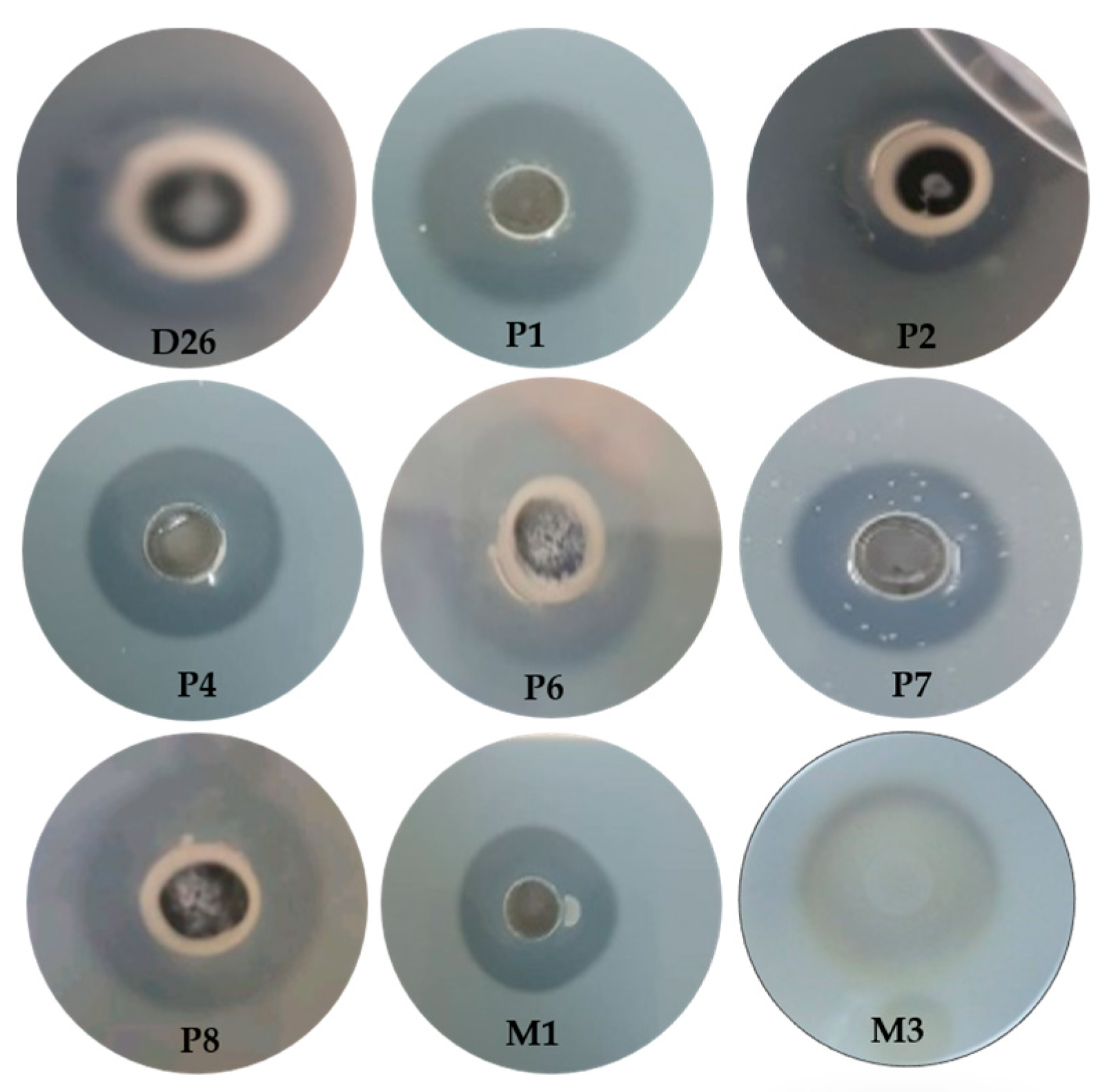
Figure 1. Showed the zones of clearance for the nine potential strains selected.
Fermentation 10 00139 g001
Figure 2. Production of Clavispora lusitaniae protease on different solid media in SmF.
Fermentation 10 00139 g002
Figure 3. Production of Clavispora lusitaniae PC 3 protease on different solid media in SSF.
Fermentation 10 00139 g003
Figure 4. Response surface graphs showing (A) Moisture vs. inoculum; (B) Inoculum vs. fermentation time; (C) Moisture vs. Fermentation time.
Fermentation 10 00139 g004
Figure 5. Multiple response optimization of protease production in Clavispora lusitaniae PC3.
Fermentation 10 00139 g005
Figure 6. SDS-PAGE analysis of purified acidic protease from Clavispora lusitaniae PC3. M: Molecular weught standard proteins. 1: Purified fraction.
Fermentation 10 00139 g006
Figure 7. Effect of pH on Protease Activity of Clavispora lusitaniae PC3 protease.
Fermentation 10 00139 g007
Figure 8. Effect of Temperature on Protease Activity and stability.
Fermentation 10 00139 g008
Figure 9. Effect of Metal Ions and Additives on Protease Activity.
Fermentation 10 00139 g009
Figure 10. Lineweaver Burk plot for partially purified protease from Clavispora lusitaniae PC3.
Fermentation 10 00139 g010
Figure 11. Action of Clavispora lusitaniae PC3 protease on gluten.
Fermentation 10 00139 g011
Table 1. Relationship between the coded levels and the real levels of the factors studied in the design of Plackett and Burman.
FactorsLevelUnit
−1+1
A: Temperature3050°C
B: Moisture5080%
C: Inoculum104108Cells/g
D: Error---
E: Glucose00.5%
F: Corn Steep Liquor00.5%
G: (NH4)2SO400.5%
H: Error---
I: NaCl00.1g/L
J: Time fermentation2472H
K: Error---
Table 2. Actual and coded values of experimental variables used CCD.
−1.682−10+1+1.682
B53.1860708086.82
C 2.08 × 1051061071080.5 × 109
J39.8148607280.18
B: Moisture (%), C: Inoculum (Cells/mL), J: Fermentation time (H).
Table 3. Identification from biochemical (ID 32C), microscopic and molecular biology characters.
Strain CodeBiochemical Characters (ID 32C)Morphological Characteristics of the Selected Isolate
Colony CharacteristicsCell ShapeVegetative ReproductionMycelium
(RAT)
Sporulation
PC3 5157
3701 17 lac-et ESC+
Smooth, glistening, butyrous, White to cream colored, entire margin.Sub-globose, ovoid to elongateBudding (unipolar and bipolar)Pseudo
mycelium
Positif
Protease productionStrain on YPGAMicroscopic Characteristics
Fermentation 10 00139 i001Fermentation 10 00139 i002Fermentation 10 00139 i003
Blast results
Id sequencesQuery (bp)SpeciesAccession number% max ident.
ITS_2156ZAB059ITS 692Clavispora lusitaniae strain CBS 6936 T PP057739 676/676(100%)
Table 4. Plackett–Burman design for the study of 11 factors with 12 experiments.
ExperimentsABC(D)EFG(H)IJ(K)Protease Activity (IU/g)
1+1+1−1+1+1+1−1−1−1+1−118,305.3
2−1+1+1−1+1+1+1−1−1−1+16567.55
3+1−1+1+1−1+1+1+1−1−1−113,621.2
4−1+1−1+1+1−1+1+1+1−1−19917.55
5−1−1+1−1+1+1−1+1+1+1−115,332.15
6−1−1−1+1−1+1+1−1+1+1+125,011.8
7+1−1−1−1+1−1+1+1−1+1+130,662.8
8+1+1−1−1−1−1+1−1+1−1+112,932.05
9+1+1+1−1−1−1+1−1+1+1−121,830.3
10−1+1+1+1−1−1−1+1−1+1+115,173.05
11+1−1+1+1+1−1−1−1+1−1+18991.2
12−1−1−1−1−1−1−1−1−1−1−114,138.3
A, B, C, E, F, G, I and J are real variables; D, H and K are dummy variables.
Table 5. CCD for optimization of protease production by Clavispora lusitaniae PC3.
RunB: MoistureC: InoculumJ: Fermentation TimeProtease Activity (IU/g)
11.6820031,856.3
200033,831.6
3001.68221,550.6
411−128,422.3
51−1126,156.6
600031,879.4
71−1−130,514.1
800032,626.5
9−1−1119,636.1
10−1−1−127,337.0
1100129,101.4
12−11022,548.0
1300032,492.0
140−1.682025,896.2
1501.682023,030.7
16−1.6820−124,251.0
17−11022,731.3
1800−131,088.8
1911128,721.6
2000−1.68229,864.7
Table 6. Purification of acidic protease from Clavispora lusitaniae PC3.
SampleTotal Activity (U)Total Protein (mg)Specific Activity (U/mg)Purification FoldYield (%)
Crude extract31,589.2168188.031100
Ammonium sulfate (60%)29,276.153552.42.93892.67
Dialysis25,821.528922.24.90481.74
Sephadex G10010,4062.83716.419.7632.94
Disclaimer/Publisher’s Note: The statements, opinions and data contained in all publications are solely those of the individual author(s) and contributor(s) and not of MDPI and/or the editor(s). MDPI and/or the editor(s) disclaim responsibility for any injury to people or property resulting from any ideas, methods, instructions or products referred to in the content.