Article

Exploring Phenotype, Genotype, and the Production of Promising GABA Postbiotics by Lactiplantibacillus plantarum: A Comprehensive Investigation

1
College of Agriculture and Forestry, Linyi University, Linyi 276005, China
2
Department of Microbiology, Soils, Water, Environment, and Microbiology Research Institute, Agriculture Research Centre, Giza 12618, Egypt
3
Department of Dairy Microbiology, Animal Production Research Institute, Agriculture Research Centre, Dokki, Giza 12618, Egypt
4
Department of Food Science and Nutrition, College of Sciences, Taif University, P.O. Box 11099, Taif 21944, Saudi Arabia
*
Authors to whom correspondence should be addressed.
These authors contributed equally to this work.
Fermentation 2024, 10(6), 309; https://doi.org/10.3390/fermentation10060309
Submission received: 11 April 2024 / Revised: 16 May 2024 / Accepted: 21 May 2024 / Published: 11 June 2024
(This article belongs to the Section Microbial Metabolism, Physiology & Genetics)

Abstract

:
This study aims to investigate the probiotic properties of various isolated strains of Lactiplantibacillus plantarum. Specifically, the focus is on examining the expression of the glutamic acid decarboxylase (GAD) gene and its role in the production of gamma-aminobutyric acid (GABA), a promising postbiotic metabolite. The investigation includes comprehensive analyses of morphology, genetics, resilience against bile, NaCl, and simulated pancreatin juice (SPJ), carbohydrate fermentation patterns, antibacterial activity, susceptibility to antibiotics, and the presence of β-D-galactosidase and GAD enzymes. Six L. plantarum strains exhibited remarkable resilience against bile, NaCl, and SPJ, as well as susceptibility to antibiotics and antagonistic behavior against pathogens. These strains also showed the presence of β-D-galactosidase. Additionally, five L. plantarum strains were found to harbor the gad gene. Further biochemical analysis of four specific L. plantarum strains revealed promising profiles consisting of antibiotics, vitamins, hormones, and a diverse array of metabolites with potential immunotherapeutic properties. This study highlights the substantial potential of Lactiplantibacillus plantarum in generating beneficial postbiotic metabolites. The identified strains offer exciting avenues for further exploration, with potential applications in functional foods and pharmaceuticals. This research opens up possibilities for harnessing the probiotic and postbiotic potential of L. plantarum to develop novel products with health-promoting properties.

1. Introduction

The gut microbiome, a complex community of microorganisms residing in the human gastrointestinal tract, establishes a symbiotic relationship with humans through the gut–brain axis, enabling communication between the central nervous system and the enteric neural system, contributing significantly to overall well-being [1]. Notably, the composition of the gut microbiome is highly individualized, akin to a unique fingerprint, despite the presence of a shared ‘core microbiome’ among individuals [2].
The brain–gut axis serves as a bidirectional communication system connecting the central nervous system (CNS) and the gastrointestinal (GI) tract. Recent research highlights the pivotal role of the gut microbiota and its interactions with the CNS in various aspects of human health, including mental health, cognition, and behavior. Complex signaling pathways, encompassing neural, hormonal, and immunological mechanisms, underpin the brain–gut axis, influencing both brain function and gut health [3].
Lactiplantibacillus plantarum, previously known as Lactobacillus arabinosus and Lactobacillus plantarum [4], is one of the beneficial LABs commonly found in the gastrointestinal tracts of humans and other mammals, as well as in saliva and various food products. It is considered safe for consumption and possesses unique characteristics that make it highly versatile. L. plantarum is Gram-positive, non-spore-forming, and catalase-negative. It can thrive in both oxygen-free and low-oxygen environments and exhibits tolerance to acidic conditions. With low G + C content, L. plantarum is extensively used in the food industry as a starter culture and preservative, offering a wide range of applications [5].
Functional foods represent a significant domain in which probiotics have gained widespread utilization. However, the realm of probiotics has expanded beyond this scope, introducing emerging concepts such as paraprobiotics, postbiotics, and psychoactives. These novel terms encompass non-living microbial cells, microbial components, or products derived from microbial cells, including lysates or metabolites. These elements offer health benefits and therapeutic effects akin to those of probiotics. By incorporating these concepts into the production of functional foods, a broader range of potential benefits can be harnessed [6].
γ-amino butyric acid (GABA), a non-protein amino acid with diverse physiological functions, has gained popularity in organic and psychological therapies as a postbiotic. LABs, which are generally recognized as safe, play a pivotal role in GABA production. This has led to the incorporation of LABs into GABA-enriched food products. However, GABA production varies among LAB strains and is influenced by fermentation conditions [7]. Optimal GABA production in the food and nutraceutical industries is achieved through biosynthesis using carefully manipulated fermentation processes [8]. The key factors for successful GABA synthesis in LABs include maintaining a slightly acidic pH (4.5–6.0) to create a favorable environment, utilizing glutamic acid as the primary precursor to enhance GAD enzyme activity, controlling fermentation temperature (30–40 °C) for balanced GABA production and LAB growth and ensuring sufficient nutrient availability to support LAB growth and promote GABA formation [9].
There is growing evidence supporting the potential benefits of postbiotics, including improved gut health, immunomodulation, anti-inflammatory effects, antimicrobial activity, metabolic regulation, and even potential anti-cancer effects. However, more research is still needed to fully understand their therapeutic applications and mechanisms of action [10].
Recently, gamma-aminobutyric acid (GABA) has gained significant attention in the field of medical and pharmaceutical research. GABA, a non-proteinogenic amino acid, serves as a well-established inhibitory neurotransmitter abundant in the central nervous system of animals [11].
Emerging evidence suggests that oral administration of GABA can potentially exert beneficial effects on the brain and psyche through the intricate gut–brain axis. GABA has garnered widespread acceptance among consumers in various markets, with reported positive effects on stress regulation and mood. Additionally, GABA has been associated with multiple physiological benefits, including neuroprotection, stress relief, sleep promotion, prevention of cell injury [12], antioxidant properties, blood pressure regulation [13], antidiabetic activity [7], and cancer protection [14]. Consequently, the combination of fundamental research and preliminary clinical findings positions GABA as an intriguing neuro-nutritional compound that warrants further investigation through clinical studies involving individuals with depression and other psychological problems [15].
GABA is a neurotransmitter that is synthesized from glutamate within the brain. It plays a vital role as an inhibitory neurotransmitter in the central nervous system. Imbalances in GABA levels have been linked to various neurological diseases, including Alzheimer’s and Parkinson’s disease, as well as psychological disorders such as anxiety, depression, and stress [16]. Until recently, it was widely believed that GABA could not cross the blood–brain barrier, leading to neglect of its potential effects on the brain [17]. Studies have shown that certain GABA-producing Lactobacilli have the ability to reduce anxiety behaviors and markers of depression in mice. These beneficial effects are accompanied by alterations in the expression of GABA receptor subunits in key brain regions involved in regulating mood and anxiety, such as the hippocampus and amygdala [18]. Based on the potential benefits of GABA and Lactobacillus plantarum, this study aimed to screen various lactic acid bacteria (LABs) from different sources to evaluate their potential as probiotics. The evaluation focused on examining their phenotype, genotype, and biochemical characteristics to investigate their ability to produce γ-amino butyric acid (GABA) as a common postbiotic substance during their metabolic processes.

2. Materials and Methods

2.1. Samples

Table 1 provides information on the diverse samples obtained from Egypt and China environments, including fermented dairy and non-dairy products, as well as fermented vegetables like kimchi and fermented soybean curd (tofu). Furthermore, it includes monogastric animals (ducks, horses), baby stool samples, and ruminant species (cows, camels, sheep). Careful measures were taken during sample transportation to maintain their integrity and prevent deterioration. The samples were stored in an ice chest at 4 °C to preserve their quality.

2.2. Isolation of LAB and Optimum Growth Conditions

One gram of each sample underwent the traditional pure culture method, involving mixing with 9 mL of peptone water buffer. This mixture was streaked onto selective media like MRS agar or M17 agar (Qingdao Hope Bio-Technology Co., Ltd., Qingdao, China) and incubated under aerobic and anaerobic conditions at 30 °C, 37 °C, and 45 °C (Shanghai Bluepard Experimental Instruments Co., Ltd., Shanghai, China) for 48 h. To ensure culture purity, three successive transfers were made onto new MRS or M17 agar plates, targeting colonies exhibiting a white and creamy appearance.

2.3. Phenotypic Characterization of LAB Isolates

The pure cultures derived from the samples underwent comprehensive characterization, involving Gram staining to determine the Gram reaction, observing cell morphologies and bacterial shapes by both light and electronic microscopy, and assessing the motility, spore-forming ability, and catalase reaction of LAB isolates. Additionally, colonies were scrutinized for attributes like color, size, and shape. Post-characterization, Gram-positive and catalase-negative isolates were chosen for further analysis. To preserve these isolates, activation and storage were performed at −80 °C in either MRS or M17 broth supplemented with 30% glycerol. Characterization experiments adhered to conventional protocols, ensuring consistency and comparability with established methods in the field [19].

2.3.1. Catalase Assay

Isolates were cultured on MRS agar and aerobically incubated overnight at 37 °C. Subsequently, a catalase test was conducted by adding two drops of 3% hydrogen peroxide to the overnight cultures on a glass slide. The presence of oxygen bubbles confirmed a positive catalase reaction, indicating the catalase enzyme’s presence. Isolates lacking gas bubble production were deemed catalase-negative and chosen for further experiments due to their absence of catalase activity, a characteristic of specific bacterial species.

2.3.2. Spore Staining and Motility Analysis

To investigate potential endospore development, isolates were initially cultured in MRS agar for 24 h at 37 °C under aerobic conditions. A single colony from each culture was selected and examined using oil immersion objectives under a light microscope to determine the isolates’ ability to form endospores, a distinctive characteristic of specific bacterial species. Furthermore, isolate motility was assessed using a stab culture method in MRS semi-solid medium. This medium, containing 1.5 g/L of agar and bromocresol purple indicator (Sangon Biotech, Shanghai, Co., Ltd., Shanghai, China), adjusted to a pH of 6.8, facilitated motility evaluation. The stab culture technique involved introducing bacterial cells into the solid medium with a sterile needle or inoculating loop, allowing observation of bacterial growth and movement within the medium to glean insights into their motility behavior [20].

2.4. In Vitro Characterization of Candidate LAB Isolates

2.4.1. Bile Tolerance Assessment

The sensitivity of isolates to bile was assessed using a modified protocol inspired by the study conducted by Abdelazez et al. [7]. The evaluation of viability included subjecting isolates to different bile concentrations (0.0, 0.3, 1.0, and 2.0% w/v). Oxgale powder (Sangon Biotech, Shanghai, Co., Ltd., Shanghai, China) was dissolved in MRS broth, centrifuged at 4 °C and 8000× g for 5 min, and filtered through a 0.45 μm sterile hydrophilic PES syringe filter (Zhejiang ALWSCI Technologies Co., Shaoxing, China) to remove remaining bile molecules. Each treatment involved the inoculation of 50 µL of 1% LAB strains (10−8 cfu) into MRS broth with corresponding bile concentrations. Viability was assessed in triplicate, and optical density (OD) was measured at 630 nm every 2 h using the ELx808™ Incubating Absorbance Microplate Reader (Creative Life Science Co., Ltd., New Taipei City, Taiwan). Optical density served as a reliable indicator of isolate viability under different bile concentrations, offering insights into microbial growth and cell density.

2.4.2. NaCl Tolerance Evaluation

The survivability of isolates in varied NaCl concentrations was explored using the method by Mulaw et al. [21] with slight adjustments. NaCl solutions were created by dissolving NaCl (Tianjin Yongda Chemical Reagent Co., Ltd., Tianjin, China) in MRS broth at concentrations of 0.0%, 4.0%, 6.0%, and 8.0% w/v. Isolates were cultivated aerobically overnight at 37 °C. Subsequently, each culture was individually inoculated with 50 µL of a 1% overnight culture and incubated at 37 °C for 48 h in MRS broth supplemented with the respective NaCl concentrations. Results were determined by measuring optical density (OD630) in triplicate every 2 h.

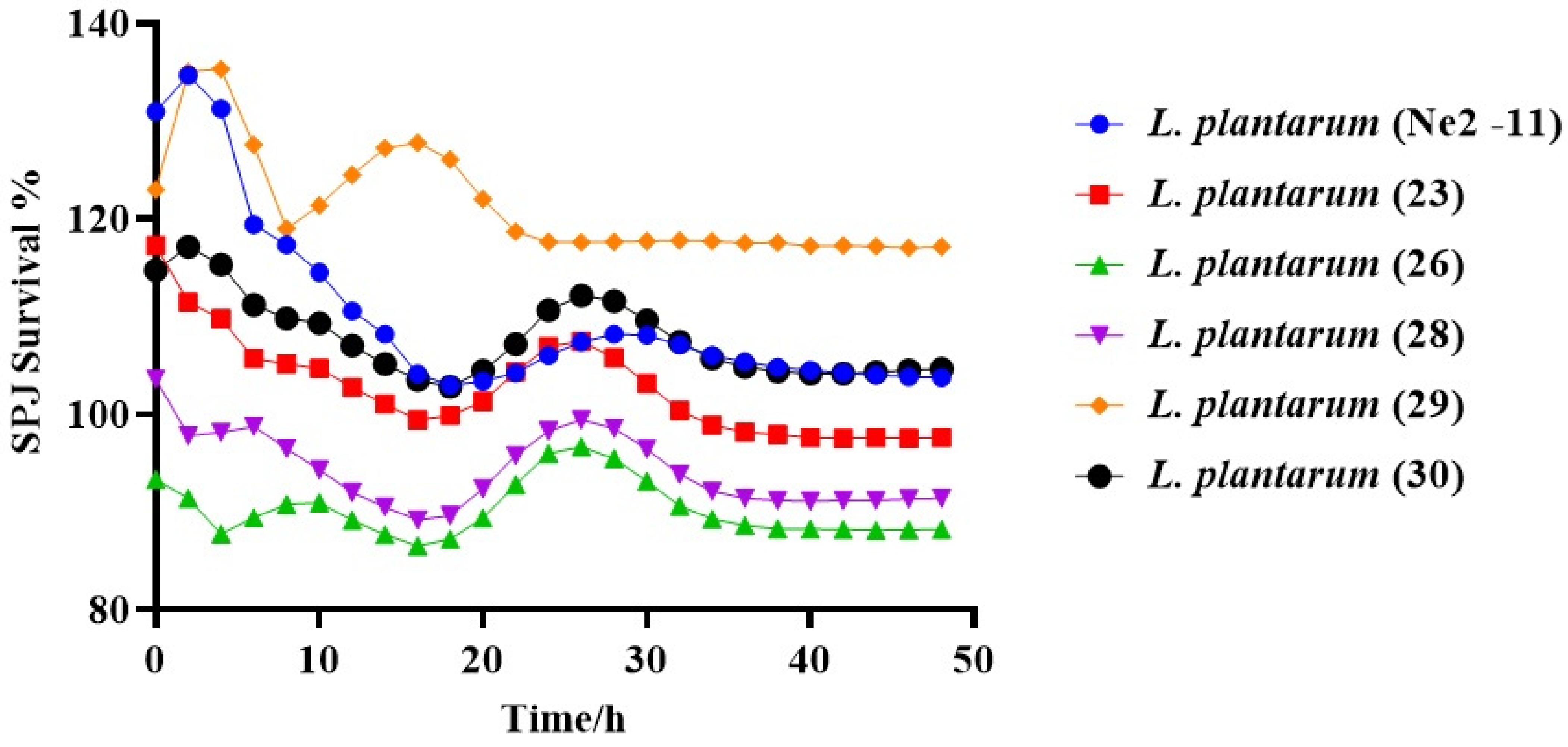
2.4.3. Tolerance of Simulated Pancreatic Juice (SPJ)

To evaluate the isolates’ tolerance to simulated pancreatic juice (SPJ), a modified approach was used, based on the methodology outlined by Rashmi and Gayathri [22]. The SPJ solution comprised bile (0.3 g/L), pancreatin (1.0 g/L), pepsin (3.0 g/L, 3000 U) (Tokyo Chemical Industry Co., Ltd., Tokyo, Japan), and NaCl (8.5 g/L). All components were dissolved in MRS broth, adjusting the pH to 8.0. Isolated LABs, individually inoculated with 50 µL of a 1% overnight culture, underwent SPJ exposure at 37 °C for 48 h. Control groups, grown in SPJ-free MRS broth, served as a baseline for comparing survival and tolerance. By contrasting results from the control group with the SPJ-exposed isolates, the impact of pancreatic juice components on growth and viability was effectively assessed. Surviving bacterial cells were quantified by measuring OD630 nm, and the experiment was performed in triplicate to ensure result reliability. The percentage of survival was calculated using the following formula:
SPJ   Survival   % = The viable cells at OD630 nm with exposure to SPJ The viable cells at OD630 nm without exposure to SPJ × 100

2.4.4. Susceptibility of Prospective Isolated Strains to Various Antibiotic Types

The antibiotic susceptibility of potential isolates was evaluated through the disc diffusion technique, employing antibiotic sensitivity test paper (Hangzhou Binhe Microbial Reagent Co., Ltd., Hangzhou, China). A variety of antibiotics, including Amikacin (7.5 µg), Polymyxin (300 µg), Cephalothin (30 µg), Sulfamethoxazole (1.25 µg), Chloramphenicol (30 µg), Spectinomycin (100 µg), Gentamicin (10 µg), Streptomycin (10 µg), Ampicillin (10 µg), Kanamycin (30 µg), Vancomycin (30 µg), Norfloxacin (10 µg), Oxacillin (1.0 µg), Tetracycline (30 µg), Clindamycin (2.0 µg), Penicillin (10 µg), and Erythromycin (15 µg), were utilized. To determine the susceptibility of LAB isolates to antibiotics, bacterial growth on MRS agar was visually inspected after 24 h. The inhibitory zones produced by the antibiotics were measured using a digital caliper (Vernier Caliper DL3944, Deliworld, Ningbo, China). The recorded results were categorized into four groups [23].
R (-) indicates cases with no inhibition zone, (R) indicates instances where the zone size indicated resistance (<7 mm), (I) indicates cases with intermediate results (zone size ranging from 7–16 mm), and (S) indicates cases displaying sensitivity (zone size > 16 mm).

2.4.5. Determination of Antimicrobial Activity

This study employed a modified semi-solid diffusion disc method to assess the antibacterial effectiveness of various isolated LAB strains [24]. Pathogenic bacterial strains targeted for evaluation included Escherichia coli ATCC 25922, Pseudomonas aeruginosa ATCC 15442, Enterococcus faecium E.980, Salmonella Typhimurium CMCC 500515, and Staphylococcus aureus ATCC 29213. Furthermore, it encompassed additional strains, including Bacillus cereus, Klebsiella spp., Citrobacter freundii, Aeromonas veronii, Citrobacter youngae, Cronobacter sakazakii, Wohlfahrtiimonas chitiniclastica, Leclercia spp., Pantoea agglomerans, and Fusobacterium nucleatum, which are all from lab stock.
A 2 µL spot of the isolated culture was placed on MRS agar and incubated at 37 °C overnight. Simultaneously, a semi-solid medium was prepared by heating Brain Heart Infusion (BHI) (Qingdao Hope Bio-Technology Co., Ltd., Qingdao, China) supplemented with 0.5% warmed agarose. A mixture of 6 mL of BHI and 100 µL of the pathogenic bacteria was then poured onto the surface of the MRS agar plate. After 24 h of incubation at 37 °C, the inhibition zone extent was measured in triplicate using a digital caliper. The results were expressed as the diameter/mm of the clear inhibition zone [25].
(-) indicates no inhibition zone (<6 mm), (+) indicates a small inhibition zone (>6–9 mm), (++) indicates a moderate inhibition zone (9–15 mm), and (+++) indicates a substantial inhibition zone (>15 mm).

2.5. Bioinformatics Analysis

2.5.1. Genetic Profiling of Isolated Strains

The potential candidates were cultured in 5 mL of their respective isolation medium overnight. Following the manufacturer’s recommendations, DNA extraction was carried out using the Ezup column bacterial genomic DNA purification kit (Sangon Biotech, Shanghai Co., Ltd., Shanghai, China).

2.5.2. PCR Amplification of the 16S rRNA Sequence and Phylogenetic Analysis of Isolates

The 16S rRNA gene amplification was carried out following the protocol provided by Mini Kit (Vazyme Biotech Co., Ltd., Nanjing, China). A specific universal primer pair (27F 5′-AGAGTTTGATCCTGGCTCAG-3′ and 1492R 5′-GGTTACCTTGTTACGACTT-3′) custom-designed by (General Biol (Anhui) Co., Ltd., Chuzhou, China) was used [26]. For the Multiplex PCR, a mixture was prepared, comprising 25 µL of 2× Taq Plus Master Mix II (Dye Plus, Vazyme Biotech., Co., Ltd., Nanjing, China), 2 µL of each primer pair (10 µM concentration each), and 2 µL of template DNA. The total volume was adjusted to 50 µL using ddH2O. DNA amplification was conducted using RT-PCR (Applied Biosystem, SimpliAmp™ Thermal Cycler, Singapore).
The amplification process involved three stages: denaturation (10 min at 95 °C), annealing (10 s at 95 °C and 15 s at 56 °C), and elongation (10 s at 72 °C), followed by 30 cycles. A final extension time of 5 min at 72 °C concluded the process. Post-amplification, an agarose gel was prepared using 1.0 g of agarose in 100 mL of 1× TAE solution. To visualize DNA bands, 4S green plus nucleic acid stain (8 µL) was added, and the gel was electrophoresed at 110 V for 30 min. Gel images were captured with a ChemiScope 6100 UV light transilluminator (Bio-Equip, Jinan, China). For strain identification, a sequence similarity analysis was conducted using BLAST (Basic Local Alignment Search Tool) software. The Multiplex PCR 16S rRNA gene sequences were compared to the NCBI (National Center for Biotechnology Information) database. Additionally, phylogenetic tree analysis was performed using MEGA 11.0.13 software. DNA sequences, along with NCBI reference sequences, were used for tree construction using methods like neighbor-joining and maximum likelihood to determine evolutionary relationships and genetic similarities among isolates.

2.5.3. Detection of Glutamate Decarboxylase (gad) Gene

Colorimetric GAD Assay

GAD enzyme activity was evaluated with a rapid colorimetric test using bromocresol green as a pH indicator [27]. Overnight bacterial cell pellets were reconstituted in a 500 µL solution of the gad reagent. The gad reagent was prepared by dissolving 1 g of L-glutamic acid (Sigma, St. Louis, MO, USA), 300 µL of Triton X-100 (Sigma), 90 g of NaCl, and 0.05 g of bromocresol green in 1 L of distilled water. The pH was adjusted to 3.4, and the color change was observed upon adding the gad reagent to bacterial cell pellets. A green color indicated mild activity, while a blue color indicated high activity of the GAD enzyme. The experiment was repeated three times for accuracy, and the averaged results were utilized for further analysis.

Molecular Identification of gad Gene

To amplify gad gene fragments, specific primers were employed in the PCR process, designed as follows:
gadF primer: 5′-CCTCGAGAAGCCGATCGCTTAGTTCG-3′
gadR primer: 5′-TCATATTGACCGGTATAAGTGATGCCC-3′
These primers were designed using Snap Gene software (6.02). DNA extracted from potentially isolated candidates served as the template for PCR amplification [28]. The PCR mixture included 25 µL of 2× Taq Plus Master Mix II (Dye Plus), 2 µL of each primer pair (gadF and gadR at 10 µM concentration each), and 2 µL of template DNA. The mixture was adjusted to a final volume of 50 µL by adding ddH2O.
The RT-PCR conditions for DNA amplification included denaturation (95 °C for 5 min), annealing (95 °C for 30 s, 55 °C for 1.30 s), and elongation (72 °C for 1.30 s). After 30 cycles, a final extension time of 10 min at 72 °C was incorporated. The same RT-PCR procedure, as mentioned earlier for 16S rRNA, was followed. To identify gad gene presence and assess sequence similarity in the GenBank database, BLAST software (http://blast.ncbi.nlm.nih.gov/Blast.cgi) was utilized.

2.6. Determination of Biochemical Analysis

2.6.1. Screening of β-D-Galactosidase

A total of 28 potential isolates were screened for β-D-galactosidase production. These isolates were cultured on MRS agar with lactose as the carbon source instead of glucose. To assess the activity of β-D-galactosidase, a chromogenic substrate called X-Gal was used. X-Gal was prepared by dissolving 20 mg/mL in dimethyl sulfoxide (DMSO) (Himedia Laboratories Pvt, Ltd., Thane, India). To induce enzyme activity, 60 µL of X-Gal and 10 µL of iso-propyl-thio β-D-galactopyranoside (IPTG, Diamond, Shanghai, China) were added to the MRS agar plates. Blue colonies indicated the presence of β-D-galactosidase, which was produced due to X-Gal hydrolysis by the enzyme. The plates were incubated for 48 h at 37 °C [29].

2.6.2. Sugar Fermentation Pattern

The Micro-Biochemical identification tube and sugar fermentation kit (Hopebio, Qingdao Hope Bio-Technology Co., Ltd., Qingdao, China) were utilized to identify and evaluate the metabolic activity of isolated strains. In the protocol, 150 μL of the indicator kit containing sugars like glucose, sucrose, galactose, mannose, maltose, lactose, arabinose, and xylose was added to each well of a 96-well plate. Subsequently, 15 μL of overnight cultures of isolated LAB strains was added to the respective wells, and thorough mixing was ensured. The plate was then incubated at 37 °C for 24 h, during which bacteria metabolized sugars, leading to detectable color changes in the kit. Observations of these changes helped determine fermentation patterns for different sugars. The experiment was replicated twice on the same plate for reproducibility. The results were expressed using the following notation: “+” for good fermentation (positive result), “+/-” for moderate fermentation (partial or intermediate result), and “-” for no fermentation (negative result).

2.6.3. Determination of Biochemical Composition and Postbiotic Metabolites of L. plantarum Strains

Four strains of Lactobacillus plantarum (Ne2-11, 23, 28, and 29) were chosen from 28 LAB candidates based on phenotype, genotype, and in vitro properties. These strains underwent anaerobic incubation in MRS broth supplemented with (1.0% w/v) monosodium glutamate for 48 h at 37 °C. Post-centrifugation (Eppendorf AG, Barkhausenweg 1, Hamburg, Germany) (8000 rpm, 15 min, 4 °C), the cell-free supernatants (‘postbiotic materials’) were collected. These materials were carefully shipped under liquid nitrogen conditions in the laboratories of Meiji Biology (Shanghai, China) for further analysis.
Metabolite extraction was performed using accurate measurements of 100 μL liquid samples. Metabolite extraction involved adding a solution of 400 μL methanol/water (4:1, v/v) and settling the mixture at −20 °C. To enhance extraction efficiency, a high-throughput tissue crusher (Wonbio-96c) was employed at 50 Hz for 6 min, followed by vortexing for 30 s and ultrasound treatment at 40 kHz for 30 min at 5 °C. Samples were then kept at −20 °C for an additional 30 min. Protein separation was achieved by centrifugation at 12,000 rpm for 15 min at 4 °C. After centrifugation, the supernatant, containing the extracted metabolites, was carefully transferred to sample vials. These vials were prepared for LC-MS/MS analysis, enabling the characterization and identification of metabolites in the samples. To ensure system conditioning and quality control, a pooled quality control sample (QC) was created by combining equal amounts from all samples.
Metabolite separation was conducted through UPLC-MS/MS analysis utilizing the ExionLCTMAD system (AB Sciex, Framingham, MA, USA) with an ACQUITY UPLC HSS T3 column (100 mm × 2.1 mm i.d., 1.7 µm) (Waters, Milford, CT, USA). Two mobile phases were employed: Solvent A with 0.1% formic acid in water, and Solvent B, a mixture of acetonitrile and isopropanol (1:1, v/v) with 0.1% formic acid. The solvent gradient program included the following stages: 0 to 3 min (95% A to 5% B), 3 to 9 min (80% A to 20% B), 9 to 13 min (5% A to 95% B), 13 to 13.1 min (maintaining 95% A and 5% B), and 13.1 to 16 min (95% A to 95% B). After 16 min, the system equilibrated at 95% A and 5% B. The analysis employed a 20 µL sample injection volume, a flow rate of 0.4 mL/min, and a constant column temperature of 40 °C. For sample stability, storage was maintained at 4 °C.
The UPLC system was coupled with a Triple TOFTM 5600 (+) mass spectrometer (AB Sciex, USA), utilizing an electrospray ionization (ESI) source operating in both positive and negative modes. Key settings included a source temperature of 500 °C, curtain gas (CUR) at 30 psi, and Ion Source GS1 and GS2 both set to 50 psi. In negative mode, the ion-spray voltage was (-) 4000 V, while in positive mode, it was 5000 V. The de-clustering potential was 80V, and collision energy (CE) ranged from 20 V to 60 V for MS/MS analysis. Data acquisition employed the Data-Dependent Acquisition (DDA) mode, with a detection range from 50 to 1000 m/z.

2.6.4. Data Preprocessing and Annotation

After UPLC-TOF/MS analyses, raw data underwent processing with Progenesis QI 2.3 software (Nonlinear Dynamics, Waters, Milford, MA, USA). This facilitated peak detection and alignment, generating a data matrix with retention time (RT), mass-to-charge ratio (m/z) values, and peak intensity. Metabolic features present in at least 80% of samples were retained. Each feature underwent normalization by sum to address intensity variations among samples. Quality control involved internal standard utilization, excluding features with a relative standard deviation (RSD) exceeding 30%. This step ensured data reliability and consistency.
The raw data were submitted to the KEGG (http://www.genome.jp/kegg) and Majorbio Cloud platforms (https://cloud.majorbio.com) for analysis of gene function, metabolic pathways, and biological networks. KEGG provided visualization and annotation capabilities for in-depth data investigation, enabling the identification and analysis of differential metabolites within specific pathways. NCBI software (https://blast.ncbi.nlm.nih.gov/Blast.cgi) was used to obtain the accession versions of isolated strains, facilitating accurate annotation and characterization of the strains. The integration of KEGG and NCBI software enhanced the understanding of gene function, metabolic pathways, and strain characterization in this study.

2.7. Statistical Analysis

The data were expressed as Mean ± SD. GraphPad Prism 8 software was used for data analysis, with statistical significance defined as p-values < 0.05. For normally distributed data, one-way ANOVA with the LSD post hoc test was conducted for multiple group comparisons. In the case of non-normally distributed data, the chi-square test was employed. These statistical approaches ensured rigorous analysis and interpretation of the results.

3. Results

3.1. Bioinformatics Analysis

The isolated strains were subjected to partial 16S rRNA gene sequencing using NCBI software (https://blast.ncbi.nlm.nih.gov/Blast.cgi). Accession versions were obtained (PP388282.1, PP388283.1, PP388284.1, PP388285.1, PP388286.1, PP388295.1), identifying the strains as L. plantarum Ne2-11, 23, 26, 28, 29, and 30, respectively. Table 2 displayed a high similarity ranging from 99.93% to 100% between the isolates and the reference strains.

3.2. Phenotypic Characteristics of Isolated Strains

The Lactiplantibacillus plantarum isolates displayed rod-shaped, non-motile, non-endospore formers and catalase-negative phenotypic characteristics. Despite being exposed to either aerobic or anaerobic conditions, all isolates exhibited robust growth at 15 °C, 37 °C, and 45 °C in MRS media, indicating their adaptability. L. plantarum strains had larger white circular colonies. Examining colony morphology is vital in determining the diversity of bacterial strains and their unique traits (Figure 1).
The isolated strains were deposited in the Institute of Microbe and Host Health, Faculty of Agriculture and Forestry, Linyi University, China, using the same names and code numbers from the phylogenetic tree for consistency and validity.
The results of the RT-PCR analysis targeting the gad gene indicated that five strains of L. plantarum, specifically Ne2-11, 23, 26, 28, and 30, exhibit a distinct 1410 bp band, indicating the presence of the gad gene. Notably, L. plantarum (29) does not possess this genetic feature. These results are consistent with the pH indicator test, which confirms that only these specific strains display a color change in the gad indicator. The identification of gad-carrying L. plantarum strains holds significant promise for future experiments focusing on the production of gamma-aminobutyric acid (GABA).
After conducting a comprehensive analysis of the whole-genome sequencing data (Table S1), it is evident that the strains exhibit a range of advantageous characteristics. These include their ability to efficiently ferment sugars and their remarkable tolerance to bile, heat shock, and the harsh conditions of gastrointestinal juice impact. Moreover, the strains harbor the gad gene, which is responsible for the production of gamma-aminobutyric acid (GABA). This further enhances their potential applications. The whole-genome sequencing reveals that the strains possess circular chromosomes with sizes ranging from 2825 to 2999 Kpb.
These findings highlight the valuable attributes of these strains, including their sugar fermentation capability, resistance to adverse conditions, antibiotic susceptibility, bacteriocin-mediated pathogen antagonism, and GABA production. As a result, they appear to be promising candidates for further investigation and application. Leveraging the whole-genome sequence of Lactobacillus plantarum strains will prove to be instrumental in harnessing their potential for diverse applications such as probiotics, the functional food industry, and antimicrobial interventions.

3.3. In Vitro Experiments

3.3.1. Bile Tolerance

The findings illustrated in Figure 2 offer compelling evidence of significant statistical differences (p < 0.001) among the Lactiplantibacillus plantarum strains when subjected to varying concentrations of bile salts. In the absence of bile, the incubation time ranged from zero to 48 h. The viability of these strains ranged from 0.788 to 1.490 nm, indicating a high growth rate. Even when exposed to a 0.3% bile concentration, these strains maintained high viability levels ranging from 0.402 to 1.445 nm, with only a slight reduction observed at a 1.0% bile concentration ranging from 0.387 to 1.123 nm. Notably, even at a higher bile concentration of 2.0%, their viability remained relatively high, ranging from 0.274 to 1.036 nm. The high tolerance of L. plantarum strains to bile salts, even at elevated concentrations, indicates their ability to withstand the potentially harmful effects of bile. These findings suggest that these strains have specific mechanisms that allow them to adapt and flourish in the presence of bile salts.

3.3.2. NaCl Resistance Assessment

Figure 3 clearly shows a significant difference in viability (p < 0.001) among the tested isolated strains after a 48 h incubation period at various NaCl concentrations (0%, 4%, 6%, and 8%). Lactiplantibacillus plantarum strains exhibited remarkable resilience, even at 0% NaCl concentration throughout the entire 48 h incubation period (0.708 to 1.543 nm). At 4% NaCl, their viability slightly decreased (0.275 to 1.440 nm), demonstrating their resistance to salt stress. Even at 6% NaCl, their viability showed minimal change (0.290 to 1.305 nm), indicating their ability to withstand higher salt concentrations. Although there was a slight decrease in viability at 8% NaCl (0.292 to 1.007 nm), the strains still maintained ongoing viability, showcasing their robustness under varying NaCl concentrations. Therefore, it can be concluded that Lactiplantibacillus plantarum strains exhibit remarkable resilience to NaCl concentrations, up to 8%, for 48 h.

3.3.3. Survivability of Lactiplantibacillus plantarum under SPJ Conditions

The results depicted in Figure 4 are statistically significant (p < 0.05) and are expressed as survivability percentages, which were calculated using the provided equation. Lactiplantibacillus plantarum strains initially displayed survivability percentages ranging from 93.29% to 130.93%. After a 48 h incubation period, these percentages decreased to a range from 88.2% to 117.12%. This suggests that the survival rate was slightly affected during incubation when exposed to the SPJ.

3.3.4. Antibiotic Susceptibility

Table 3 presents six Lactiplantibacillus plantarum strains classified into three groups based on their antibiotic reactions: sensitive, intermediate, and resistant. The provided data can be utilized for research or practical applications. However, it is noteworthy that certain antibiotics, including Tetracycline, Sulfamethoxazole, Kanamycin, Ampicillin, Erythromycin, and Clindamycin, exhibit similar reactions across the strains, either susceptible (S) or intermediate (I). All other antibiotics tested resulted in the strains being classified as resistant (R).

3.3.5. Antimicrobial Activity

Table 4 presents a statistically significant difference (p < 0.05) in the antimicrobial activity of Lactiplantibacillus plantarum strains against different pathogens. The presence of clear zones ranging from 17.38 to 40.12 mm indicates the inhibitory effect of L. plantarum isolates on the growth of pathogens. These clear zones demonstrate the ability of the strains to inhibit the growth of pathogenic bacteria, suggesting their potential application as antimicrobial agents or probiotics in various contexts.
Briefly, Lactiplantibacillus plantarum strains showed impressive resilience, surviving high concentrations of sodium chloride (up to 8%), bile salts (up to 2%), and SPJ for 48 h. These strains also demonstrated antibiotic resistance and exhibited antimicrobial activities against pathogens. These findings have important implications for food preservation, probiotic development, and the search for alternative antimicrobial agents.

3.4. Biochemical Analysis of Isolated Strains

3.4.1. Sugar Fermentation Pattern Evaluation

Table 5 presents a comprehensive overview of the diverse sugar utilization patterns. Lactiplantibacillus plantarum strains exhibit remarkable fermentation capabilities across a wide range of sugars, including glucose, sucrose, galactose, mannose, maltose, lactose, arabinose, and xylose, all within 24 h or less. The detailed analysis of sugar utilization patterns among these strains provides valuable insights into their metabolic capabilities. The ability of these strains to ferment various sugars not only highlights their adaptability but also suggests their potential applications in various fields, such as functional food production, probiotics, and biotechnology.

3.4.2. β-D-Galactosidase Detection

During a screening of 28 isolated LAB strains, it was observed that a significant number of tested strains had the presence of β-D-galactosidase to varying extents. However, the Lactiplantibacillus plantarum Ne2-11, 23, 26, 28, 29, and 30 strains exhibited the production of this enzyme in varying quantities and different times taken to express the green color among the strains. Figure 5 visually represents the differences in the intensity of the green color, indicating the variation in β-D-galactosidase production during a 24 h incubation period among the different LAB isolates.

3.4.3. Determination of Biochemical Postbiotics of Lactiplantibacillus plantarum Strains

Figure 6a illustrates the composition of postbiotics produced by L. plantarum strains Ne2-11, 23, 28, and 29 after 48 h of incubation. The graph highlights the predominant distribution of organic acids and derivatives (29.43%), lipids and lipid-like molecules (20.13%), and organic heterocyclic compounds (16.30%), among others.
Figure 6b, the relative abundance of individual amino acids is depicted. This provides insights into the amino acid profile of the strains. Figure 6c presents a heatmap tree that visually represents the key postbiotics derived from L. plantarum strains. This visualization offers a concise overview of the significant postbiotics generated.
Supplementary Table S2 provides comprehensive information on the potential biological functions and therapeutic applications of these postbiotics with references, emphasizing their value in medicine and nutrition. It summarizes the diverse bioactive metabolites produced by these strains, including the detection of hormones such as serotonin, dopamine, testosterone, etc. Additionally, the strains exhibit a range of antibiotics with potential antibacterial efficacy, along with substances and peptides displaying drug-like properties, suggesting potential therapeutic applications.
The table also highlights unique amino acid derivatives like (R)-beta-aminoisobutyric acid and gamma-aminobutyric acid-betaxanthin (GABA), as demonstrated in the principal component analysis (PCA) shown in Figure 6d. Furthermore, L. plantarum strains are identified as rich sources of various B complex vitamins, as shown in Figure 6e, carotenoids, and Vitamins D3, C, and E. Figure 6f depicts the various lipid contents, as well as polyunsaturated fatty acids, indicating their nutritional value in the strains. Additionally, Figure 6g showcases phytochemicals, alkaloids, and flavonoids, which possess biological activities such as antioxidant and anti-inflammatory properties. Overall, these figures and the table provide comprehensive insights into the postbiotic composition, their therapeutic potential, nutritional value, and bioactive metabolites. On the other hand, Figure 7 displays the profile of sufficient metabolites in each comparison of L. plantarum groups using Volcano and Venn-map software (https://grapholite.com/Diagrams/VennDiagrams).
The Volcano plot and Venn diagram show the metabolite profiles for each comparison group of Lactiplantibacillus plantarum (Figure 7). Upon analyzing the distribution of these points, a pattern emerges: metabolites with down-regulated expression tend to appear on the left side of the graph, while metabolites with up-regulated expression tend to appear on the right side. The position of a point, in terms of its distance from the center and its height, indicates the significance of the expression difference for a particular metabolite. A point that is farther to the left or right and higher in position indicates a more pronounced expression difference for the corresponding metabolite.

4. Discussion

Recently, both probiotics and their metabolites, known as postbiotics, have gained recognition for their therapeutic potential including the ability to eliminate pathogens, strengthen the immune system, and restore balance in the gut microbiome [30,31].
The morphological characteristics of bacterial cells or colonies offer valuable insights into their overall properties and shape. When combined with biochemical tests and microscopic examination, these characteristics aid in the identification and classification of LAB isolates, serving diverse applications across industries, including pharmaceuticals and food [32].
Probiotic screening involves in-depth in vitro evaluations to identify appropriate strains. Although initial morphological observations offer insights, comprehensive analysis is crucial for confirmation. This understanding aids in optimizing growth conditions and maximizing the potential benefits of bacterial strains. The study on Lactiplantibacillus plantarum aligns with previous research, describing it as a rod-shaped, Gram-positive LAB with probiotic characteristics [5].
The discovery of certain Lactiplantibacillus plantarum strains harboring the gad gene opens up promising avenues for studying GABA production. However, the significant variation in GABA production among strains of the same species indicates the need for further research to explore the various possibilities for GABA production. These findings are consistent with prior studies conducted by Abdelazez et al. [7,28], who reported that the length of the gad gene is around 1407 base pairs and also reported the promising benefits of generated GABA for developing innovative functional foods that may have health benefits.
Understanding bacterial strains is essential for researchers to unlock their potential. This process involves careful examination of morphological characteristics, microscopic analysis, and additional tests. However, evaluating potential probiotics in live organisms can be a time-consuming and costly endeavor. Thus, in vitro assessment is the primary method used for probiotic screening. While morphological characteristics provide initial identification clues, further analysis is crucial to confirm their classification. This knowledge plays a critical role in optimizing growth conditions, including temperature and ventilation, for specific applications, ultimately maximizing the potential utility of these bacterial strains [33].
The study findings highlight the superior tolerance of Lactiplantibacillus plantarum strains to NaCl and bile salts, as well as survivability under SPJ conditions, indicating their potential to withstand the harsh pancreatic environment. These findings contribute to the understanding of growth patterns under specific conditions and guide the optimization of bacterial cultures, probiotic formulation, fermentation processes, and antimicrobial research. The results are consistent with previous studies by Kouadri et al. [34], who emphasized the robustness of Lactiplantibacillus plantarum strains in in vitro tests, simulating human gastrointestinal conditions.
Nicosia et al. [35] conducted a study to test the tolerance of LAB strains to NaCl concentrations. The findings indicated that only four strains, namely L. delbruekii (P14 and P38), L. rhamnosus (P50), and L. plantarum Q3C4, were capable of growing in the presence of 10% (w/v) NaCl. These findings are consistent with the provided data, which demonstrate that L. plantarum strains displayed relatively high growth rates at 0–8% (w/v) concentrations of NaCl.
Understanding the resistance of microbes to antibiotics is crucial in selecting probiotic bacteria. Categorizing bacterial strains as resistant, sensitive, or intermediate is essential for treatment decisions, infection control, and research and development efforts. This knowledge enables researchers and healthcare professionals to make informed decisions when choosing and administering LAB strains in probiotic formulations, ensuring the efficacy and safety of these products. These considerations contribute to improved patient outcomes and public health [36].
These collective findings provide a consistent understanding of antibiotic responses and sensitivities exhibited by L. plantarum strains, contributing to the knowledge base for antibiotic selection and usage in various applications. These findings align with previous studies conducted by Abdelazez et al. [28], who confirmed the typical sensitivity of L. brevis strains to antibiotics, particularly Chloramphenicol. Similarly, de Almeida et al. [37] reported a 96% sensitivity to Chloramphenicol among the tested strains. Tulumoglu et al. [38] found that 90% of Lactobacillus strains exhibited resistance to Gentamicin while being sensitive to Ampicillin and Erythromycin. Generally, this study revealed that L. plantarum strains showed increased susceptibility to specific antibiotics such as Penicillin, cefoxitin, and Vancomycin. They are inhibited by Chloramphenicol, Erythromycin, and Tetracycline, with levels falling below or equal to the European Food Safety Authority breakpoints [39].
Lactobacillus plantarum strains exhibited significant antagonistic activity against various pathogenic bacteria [40]. Also, Silva et al. [41] demonstrated the ability of different LAB strains to suppress the growth of foodborne pathogens, with varying degrees of effectiveness.
The antimicrobial activity of the investigated L. plantarum strains has been attributed to the production of specific postbiotics, including organic acids, enzymes (e.g., proteinase K, pepsin, and trypsin), and catalase [42]. Our results align with these findings, confirming the postbiotic activity of these strains against pathogenic bacteria. Furthermore, Fossi et al. [43] emphasized the potential antimicrobial mechanisms employed by microorganisms, such as the production of antimicrobial compounds, bacteriocins, competitive nutrient action, inhibition of binding through competition, and modulation of the immune system.
These findings align with previous research by Zhang et al. [44], who demonstrated that the Lactiplantibacillus plantarum strain HOM3204 displayed plasticity in mimicking gastrointestinal and intestinal juices, indicating its capacity to withstand gastrointestinal conditions. Additionally, it exhibited strong adhesion to Caco-2 cells, which serve as a model for intestinal epithelial cells, and demonstrated notable antibacterial activity. These findings carry significant implications for maintaining a healthy gut microbiome. Overall, the study results provide valuable insights into the resilience and beneficial characteristics of Lactiplantibacillus plantarum strains, particularly their ability to survive in challenging environments and interact positively with the gastrointestinal system.
Fermentation of sugars is a significant test for characterizing and identifying different LAB strains. Lactobacillus plantarum is a type of LAB that uses fermentable carbohydrates as its primary energy source and produces lactic acid as its main byproduct. This species can adapt well to different environments, which allows it to ferment a wide variety of carbohydrates, including monosaccharides, disaccharides, and polysaccharides. Depending on the available substrates and specific conditions, L. plantarum can ferment sugars to produce other organic acids such as acetic acid and succinic acid, as well as ethanol or carbon dioxide as major metabolites. Also, it can switch between heterofermentative and homofermentative modes of metabolism depending on the carbon source [45]. The results obtained are in line with [46], who showcase the metabolic flexibility of L. plantarum strains, as they can utilize several sugar sources as a carbon source for both fermentation and growth.
Lactose, a disaccharide present in milk and milk derivatives, contributes significantly to taste, texture, and nutritional composition. However, lactose maldigestion and intolerance are common issues affecting carbohydrate digestion in the human gastrointestinal tract [47]. Several studies have shown promising results in treating lactose dysdigestion by using LABs to aid in the breakdown of lactose in the small intestine. However, infants can usually digest lactose, but this ability gradually diminishes with age, leading to lactose maldigestion, which affects about 75% of the world’s population [48]. Individuals with lactose maldigestion may experience symptoms such as abdominal bloating, pain, flatulence, diarrhea, nausea, and discomfort after consuming lactose [49].
The ability of a strain to ferment specific sugars can vary due to specific genes or the secretion of particular enzymes. By evaluating the fermentation capabilities of different strains, we can determine their unique characteristics and suitability for specific applications [19].
β-D-galactosidase is a crucial enzyme that plays a vital role in the metabolism of lactose, particularly in organisms that use lactose as their primary source of carbon. Its deficiency or absence can lead to inadequate digestion and utilization of lactose, causing lactose intolerance in humans and certain mammals. The obtained results revealed around 15 isolated LAB strains, particularly the six isolated L. planetarium strains, that demonstrated the possession of the β-D-galactosidase enzyme, which can break down lactose to glucose and galactose. The findings obtained were consistent with Plaza et al. [50], who indicated that the colonization of LABs in the intestinal tract may help alleviate clinical symptoms associated with undigested lactose and facilitate lactose hydrolysis in the small intestine. This is due to LABs’ possession of β-D-galactosidase, which plays a crucial role in managing lactose tolerance in the host intestine.
The positive results obtained from Lactiplantibacillus plantarum highlight its potential to promote intestinal health. These probiotic strains have been shown to possess antibacterial properties and the ability to survive harsh conditions such as high NaCl, bile, SPJ, and antibiotics. Based on these findings, it is important to investigate the postbiotic products produced by four specific L. plantarum strains (Ne2-11, 23, 28, and 29) to further explore their unique properties.
Postbiotics refer to a mixture of metabolic products that are secreted by probiotics in cell-free supernatants. These products include enzymes, proteins, short-chain fatty acids, vitamins, biosurfactants, amino acids, peptides, and organic acids. However, it is important to note that the term ‘postbiotics’ is often used to refer to both postbiotics and paraprobiotics without a clear distinction [31].
Postbiotics have several advantages over probiotics, including their purity, ease of production and storage, scalability for large-scale production, specific mechanisms of action, improved accessibility of Microbe-Associated Molecular Patterns (MAMPs) during recognition and interaction with Pattern Recognition Receptors (PRRs), and the ability to trigger targeted responses through specific ligand–receptor interactions [30].
Lately, probiotics and their resulting metabolites ‘postbiotics’ have showcased a wide range of therapeutic attributes. These include eradicating pathogenic microorganisms linked to various diseases, bolstering the host’s immune response, and restoring the natural equilibrium within the human gut microbiome [30]. The current focus in developing interventions for gastrointestinal microbiota disorders revolves around enhancing traditional probiotics and creating postbiotics derived from them [51]. Briefly, the obtained results presented collectively enhance comprehension of the metabolic capabilities of L. plantarum strains and their potential impact on human health. The distribution and abundance of specific postbiotic molecules offer valuable insights into the functional properties of these strains, positioning them as potential probiotics. Recent research has highlighted the potential of L. plantarum as a carrier for vaccines, offering an exciting avenue for developing anti-tumor vaccines using food-grade microorganisms [52].
The biochemical evaluation revealed that the postbiotics produced by L. plantarum encompass a diverse array of biological components. These include antibiotics, hormones, organic and amino acids, and various types of vitamins. Additionally, the postbiotics consist of different peptides and drugs that may possess therapeutic activities, potentially benefiting human health by boosting the immune system. These findings agree with those of Rozhkova et al. [51], who previously explored the postbiotics’ composition and diverse array of functional properties. Moreover, L. plantarum strains are a rich source of various vitamins, which further contribute to their nutritional value, as well as polyunsaturated fatty acids. The strains also produce unique amino acid derivatives like (R)-beta-aminoisobutyric acid and gamma-aminobutyric acid-betaxanthin, which can be explored in a wide range of immunotherapy applications [31].
The utilization of GABA-producing LAB strains presents a cost-effective solution due to their safety, ease of cultivation, and manipulation. Additionally, incorporating GABA as a postbiotic in food products offers probiotic benefits, promoting gut health, overall well-being, and the development of innovative functional food products enriched with GABA. Furthermore, GABA can play a crucial role in extending the shelf life of food products, enhancing their value within the industry [53].
Previous studies uncovered distinct biological functions between the two types of gamma-aminobutyric acid compounds owing to their unique chemical structures and properties. The first compound, known as gamma-aminobutyric acid-betaxanthin, has antioxidant activity as a betaxanthin compound. It possesses the ability to scavenge free radicals and reduce oxidative stress in cells and tissues. Besides contributing to the coloration of certain plants, such as flowers and fruits, betaxanthins provide various hues like yellow, orange, and red. Some studies suggest potential health benefits, including anti-inflammatory and anti-cancer properties. These potential benefits may be due to the presence of betaxanthin [54].
Additionally, R-beta amino iso-butyric acid (R-BAIB), the second compound, has been studied for its role in modulating cellular metabolism, including glucose and lipid metabolism. It exhibits antioxidant properties, offering protection against oxidative damage caused by free radicals. Research suggests that R-BAIB may positively impact insulin sensitivity, thereby potentially improving glucose regulation [55].
Postbiotics have the potential to be used in conjunction with probiotic microorganisms. They exhibit high digestibility and resistance to gastrointestinal conditions, making them suitable for developing products with functional properties. Approximately 95–97% of postbiotics remain unchanged and reach the colon, where their biologically active compounds immediately engage in metabolic reactions upon entering the gastrointestinal tract. This eliminates the colonization problem associated with probiotics, which can antagonize resident microbiota [30]. This study’s findings illuminate the promising probiotic, fermentative, antimicrobial, and postbiotic properties of LAB isolates, especially L. plantarum strains. The diverse bioactive components in L. plantarum supernatant signal a new era in immunotherapy research, offering exciting opportunities for novel disease treatments.

5. Conclusions

This study provides a comprehensive exploration of Lactiplantibacillus plantarum isolates, covering their morphology and genetic properties. In vitro assessments revealed the potential probiotic of six L. plantarum isolates, characterized by strong sugar fermentation capabilities and β-D-galactosidase production. These isolates also displayed notable antagonistic activity against pathogens and exhibited significant antibiotic resistance, suggesting their potential as natural defense agents in the gastrointestinal tract. Furthermore, five L. plantarum strains were found to express the gad gene, associated with the production of gamma-aminobutyric acid (GABA), while four specific strains showcased unique biological components. These findings underscore the importance of further research in fully understanding and utilizing the therapeutic potential of these strains, particularly in the field of postbiotic applications.

Supplementary Materials

The following supporting information can be downloaded at: https://www.mdpi.com/article/10.3390/fermentation10060309/s1, Table S1: Genome analysis of L. plantarum strains; Table S2: Nomenclature of the most bioactive postbiotic metabolites by L. plantarum strains. References [56,57,58,59,60,61,62,63,64,65,66,67,68,69,70,71,72,73,74,75,76,77,78,79,80,81,82,83,84,85,86,87,88,89,90,91,92,93,94,95,96,97,98,99,100,101,102,103] are cited in the supplementary materials.

Author Contributions

Conception and design were contributed to by all authors. H.A.-m., A.A., P.W., G.A. (Ghada Abady), G.A. (Garsa Alshehry), E.A., H.A. and S.A. performed conceptualization, methodology, and writing—review and editing; H.A.-m., A.A., B.Y., L.X., Y.Z. and J.N. performed investigation and formal analysis; G.A. (Ghada Abady), G.A. (Garsa Alshehry), E.A. and H.A. obtained funding for the work, analyzed data, and assisted with the initial writing and editing process; and X.Z. performed supervision. All authors have read and agreed to the published version of the manuscript.

Funding

This research was funded by Taif University, Saudi Arabia, who supported this work through project number (TU-DSPP-2024-167).

Institutional Review Board Statement

Not applicable.

Informed Consent Statement

Not applicable.

Data Availability Statement

Data are contained within the article.

Acknowledgments

The researchers would like to acknowledge the Deanship of Scientific Research, Taif University, for funding this work.

Conflicts of Interest

The authors declare no conflicts of interest.

References

  1. Patel, P.G.; Patel, A.C.; Chakraborty, P.; Gosai, H.B. Impact of Dietary Habits, Ethnicity, and Geographical Provenance in Shaping Human Gut Microbiome Diversity. In Probiotics, Prebiotics, Synbiotics, and Postbiotics: Human Microbiome and Human Health; Springer: Berlin/Heidelberg, Germany, 2023; pp. 3–27. [Google Scholar]
  2. Yuan, H.; Wang, Z.; Wang, Z.; Zhang, F.; Guan, D.; Zhao, R. Trends in Forensic Microbiology: From Classical Methods to Deep Learning. Front. Microbiol. 2023, 14, 1163741. [Google Scholar] [CrossRef] [PubMed]
  3. Loh, J.S.; Mak, W.Q.; Tan, L.K.S.; Ng, C.X.; Chan, H.H.; Yeow, S.H.; Foo, J.B.; Ong, Y.S.; How, C.W.; Khaw, K.Y. Microbiota–gut–brain axis and its therapeutic applications in neurodegenerative diseases. Signal Transduct. Target. Ther. 2024, 9, 37. [Google Scholar] [CrossRef] [PubMed]
  4. Kleerebezem, M.; Hols, P.; Bernard, E.; Rolain, T.; Zhou, M.; Siezen, R.J.; Bron, P.A. The extracellular biology of the lactobacilli. FEMS Microbiol. Rev. 2010, 34, 199–230. [Google Scholar] [CrossRef] [PubMed]
  5. Jia, F.-F.; Zhang, L.-J.; Pang, X.-H.; Gu, X.-X.; Abdelazez, A.; Liang, Y.; Sun, S.-R.; Meng, X.-C. Complete genome sequence of bacteriocin-producing Lactobacillus plantarum KLDS1.0391, a probiotic strain with gastrointestinal tract resistance and adhesion to the intestinal epithelial cells. Genomics 2017, 109, 432–437. [Google Scholar] [CrossRef] [PubMed]
  6. Abdelazez, A.; Mohamed, D.M.; Refaey, M.M.M.; Niu, J. Intervention effect of freeze-dried probiotic and unripe banana pulp combination on Set-Type bio-yogurt production during storage. J. Food Meas. Charact. 2023, 1, 18. [Google Scholar] [CrossRef]
  7. Abdelazez, A.; Abdelmotaal, H.; Evivie, S.E.; Bikheet, M.; Sami, R.; Mohamed, H.; Meng, X. Verification of Lactobacillus brevis Tolerance to Simulated Gastric Juice and the Potential Effects of Postbiotic Gamma-Aminobutyric Acid in Streptozotocin-Induced Diabetic Mice. Food Sci. Hum. Wellness 2022, 11, 165–176. [Google Scholar] [CrossRef]
  8. Pannerchelvan, S.; Rios-Solis, L.; Faizal Wong, F.W.; Zaidan, U.H.; Wasoh, H.; Mohamed, M.S.; Tan, J.S.; Mohamad, R.; Halim, M. Strategies For Improvement of Gamma-Aminobutyric Acid (GABA) Biosynthesis via Lactic Acid Bacteria (LAB) Fermentation. Food Funct. 2023, 14, 3929–3948. [Google Scholar] [CrossRef]
  9. Cha, X.; Ding, J.; Ba, W.; You, S.; Qi, W.; Su, R. High Production of γ-Aminobutyric Acid by Activating the xyl Operon of Lactobacillus brevis. ACS Omega 2023, 8, 8101–8109. [Google Scholar] [CrossRef]
  10. Żółkiewicz, J.; Marzec, A.; Ruszczyński, M.; Feleszko, W. Postbiotics—A Step Beyond Pre- and Probiotics. Nutrients 2020, 12, 2189. [Google Scholar] [CrossRef]
  11. Spiering, M.J. The discovery of GABA in the brain. J. Biol. Chem. 2018, 293, 19159–19160. [Google Scholar] [CrossRef]
  12. Hepsomali, P.; Groeger, J.A.; Nishihira, J.; Scholey, A. Effects of oral gamma-aminobutyric acid (GABA) administration on stress and sleep in humans: A systematic review. Front. Neuro. Sci. 2020, 14, 923. [Google Scholar] [CrossRef]
  13. AOKI, H.; Furuya, Y.; Endo, Y.; Fujimoto, K. Effect of γ-Aminobutyric Acid-enriched Tempeh-like Fermented Soybean (GABA-Tempeh) on the Blood Pressure of Spontaneously Hypertensive Rats. Biosci. Biotechnol. Biochem. 2003, 67, 1806–1808. [Google Scholar] [CrossRef] [PubMed]
  14. Huang, Q.; Liu, C.; Wang, C.; Hu, Y.; Qiu, L.; Xu, P. Neurotransmitter γ-aminobutyric acid-mediated inhibition of the invasive ability of cholangiocarcinoma cells. Oncol. Lett. 2011, 2, 519–523. [Google Scholar] [CrossRef] [PubMed]
  15. Liwinski, T.; Lang, U.E.; Brühl, A.B.; Schneider, E. Exploring the therapeutic potential of gamma-aminobutyric acid in stress and depressive disorders through the gut-brain axis. Biomedicines 2023, 11, 3128. [Google Scholar] [CrossRef] [PubMed]
  16. Ren, P.; Wang, J.-Y.; Chen, H.-L.; Wang, Y.; Cui, L.-Y.; Duan, J.-Y.; Guo, W.-Z.; Zhao, Y.-Q.; Li, Y.-F. Activation of σ-1 receptor mitigates estrogen withdrawal-induced anxiety/depressive-like behavior in mice via restoration of GABA/glutamate signaling and neuroplasticity in the hippocampus. J. Pharmacol. Sci. 2024, 154, 236–245. [Google Scholar] [CrossRef] [PubMed]
  17. Braga, J.D.; Thongngam, M.; Kumrungsee, T. Gamma-aminobutyric acid as a potential postbiotic mediator in the gut–brain axis. NPJ Sci. Food 2024, 8, 16. [Google Scholar] [CrossRef] [PubMed]
  18. Song, J.-H.; Kim, H.; Kong, M.-J.; Hong, Y.-T.; Oh, S.-J.; Choi, K.-C. Gamma-aminobutyric acid in rice bran extract exerts antistress effects in mouse models with depressive-like behaviors. J. Med. Food 2024, 27, 231–241. [Google Scholar] [CrossRef] [PubMed]
  19. Khushboo; Karnwal, A.; Malik, T. Characterization and selection of probiotic lactic acid bacteria from different dietary sources for development of functional foods. Front. Microbiol. 2023, 14, 1170725. [Google Scholar] [CrossRef] [PubMed]
  20. Willis, A.T. Anaerobic Bacteriology: Clinical and Laboratory Practice; Butterworth-Heinemann: Oxford, UK, 2014. [Google Scholar]
  21. Mulaw, G.; Sisay Tessema, T.; Muleta, D.; Tesfaye, A. In Vitro evaluation of probiotic properties of lactic acid bacteria isolated from some traditionally fermented Ethiopian food products. Int. J. Microbiol. 2019, 2019, 7179514. [Google Scholar] [CrossRef]
  22. Rashmi, B.S.; Gayathri, D. Molecular Characterization of Gluten Hydrolysing Bacillus sp. and Their Efficacy and Biotherapeutic Potential as Probiotics Using Caco-2 Cell Line. J. Appl. Microbiol. 2017, 123, 759–772. [Google Scholar] [CrossRef]
  23. Liu, C.; Zhang, Z.-Y.; Dong, K.; Yuan, J.-P.; Guo, X.-K. Antibiotic Resistance of Probiotic Strains of Lactic Acid Bacteria Isolated from Marketed Foods and Drugs. Biomed. Environ. Sci. 2009, 22, 401–412. [Google Scholar] [CrossRef]
  24. Mohammadi, R.; Moradi, M.; Tajik, H.; Molaei, R. Potential Application of Postbiotics Metabolites from Bioprotective Culture To Fabricate Bacterial Nanocellulose Based Antimicrobial Packaging Material. Int. J. Biol. Macromol. 2022, 220, 528–536. [Google Scholar] [CrossRef] [PubMed]
  25. Keresztény, T.; Libisch, B.; Orbe, S.C.; Nagy, T.; Kerényi, Z.; Kocsis, R.; Posta, K.; Papp, P.P.; Olasz, F. Isolation and Characterization of Lactic Acid Bacteria with Probiotic Attributes from Different Parts of the Gastrointestinal Tract of Free-Living Wild Boars in Hungary. Probiotics Antimicrob. Proteins 2023. [Google Scholar] [CrossRef] [PubMed]
  26. Angmo, K.; Kumari, A.; Savitri; Bhalla, T.C. Probiotic characterization of lactic acid bacteria isolated from fermented foods and beverage of Ladakh. LWT-Food Sci. Technol. 2016, 66, 428–435. [Google Scholar] [CrossRef]
  27. Olier, M.w.; Rousseaux, S.; Piveteau, P.; Lemaître, J.-P.; Rousset, A.; Guzzo, J. Screening of Glutamate Decarboxylase Activity and Bile Salt Resistance of Human Asymptomatic Carriage, Clinical, Food, and Environmental Isolates of Listeria monocytogenes. Int. J. Food Microbiol. 2004, 93, 87–99. [Google Scholar] [CrossRef] [PubMed]
  28. Abdelazez, A.; Abdelmotaal, H.; Evivie, S.E.; Melak, S.; Jia, F.-F.; Khoso, M.H.; Zhu, Z.-T.; Zhang, L.-J.; Sami, R.; Meng, X.-C. Screening Potential Probiotic Characteristics of Lactobacillus brevis Strains In Vitro and Intervention Effect on Type I Diabetes In Vivo. BioMed Res. Int. 2018, 2018, 7356173. [Google Scholar] [CrossRef] [PubMed]
  29. Gheytanchi, E.; Heshmati, F.; Shargh, B.K.; Nowroozi, J.; Movahedzadeh, F. Study on Β-Galactosidase Enzyme Produced by Isolated Lactobacilli from Milk and Cheese. Afr. J. Microbiol. Res. 2010, 4, 454–458. [Google Scholar]
  30. Nataraj, B.H.; Ali, S.A.; Behare, P.V.; Yadav, H. Postbiotics-Parabiotics: The New Horizons in Microbial Biotherapy and Functional Foods. Microb. Cell Fact. 2020, 19, 168. [Google Scholar] [CrossRef] [PubMed]
  31. Abdelazez, A.; Alshehry, G.; Algarni, E.; Al Jumayi, H.; Abdel-Motaal, H.; Meng, X.-C. Postbiotic Gamma-Aminobutyric Acid and Camel Milk Intervention as Innovative Trends against Hyperglycemia and Hyperlipidemia in Streptozotocin-Induced C57BL/6J Diabetic Mice. Front. Microbiol. 2022, 13, 943930. [Google Scholar] [CrossRef]
  32. Petersen, J.; McLaughlin, S. Laboratory Exercises in Microbiology: Discovering the Unseen World through Hands-On Investigation; CUNY Academic Works; City University of New York (CUNY): New York, NY, USA, 2016. [Google Scholar]
  33. Gutarowska, B.; Szulc, J.; Jastrząbek, K.; Kręgiel, D.; Śmigielski, K.; Cieciura-Włoch, W.; Mroczyńska-Florczak, M.; Liszkowska, W.; Rygała, A.; Berłowska, J. Effectiveness of Ozonation for Improving the Microbiological Safety of Fresh-Cut Parsley (Petroselinum crispum) Leaves. Appl. Sci. 2023, 13, 8946. [Google Scholar] [CrossRef]
  34. Kouadri Boudjelthia, N.; Belabbas, M.; Bekenniche, N.; Monnoye, M.; Gérard, P.; Riazi, A. Probiotic Properties of Lactic Acid Bacteria Newly Isolated from Algerian Raw Cow’s Milk. Microorganisms 2023, 11, 2091. [Google Scholar] [CrossRef] [PubMed]
  35. Nicosia, F.D.; Pino, A.; Maciel, G.L.R.; Sanfilippo, R.R.; Caggia, C.; de Carvalho, A.F.; Randazzo, C.L. Technological Characterization of Lactic Acid Bacteria Strains for Potential Use in Cheese Manufacture. Foods 2023, 12, 1154. [Google Scholar] [CrossRef] [PubMed]
  36. Zheng, M.; Zhang, R.; Tian, X.; Zhou, X.; Pan, X.; Wong, A. Assessing the Risk of Probiotic Dietary Supplements in the Context of Antibiotic Resistance. Front. Microbiol. 2017, 8, 264345. [Google Scholar] [CrossRef] [PubMed]
  37. de Almeida Júnior, W.L.G.; de Souza Ferrari ÍdS, J.V.; da Silva, C.D.A.; da Costa, M.M.; Dias, F.S. Characterization and Evaluation of Lactic Acid Bacteria Isolated from Goat Milk. Food Control 2015, 53, 96–103. [Google Scholar] [CrossRef]
  38. Tulumoglu, S.; Yuksekdag, Z.N.; Beyatli, Y.; Simsek, O.; Cinar, B.; Yaşar, E. Probiotic Properties of Lactobacilli Species Isolated from Children’s Feces. Anaerobe 2013, 24, 36–42. [Google Scholar] [CrossRef] [PubMed]
  39. Karapetkov, N.; Georgieva, R.; Rumyan, N.; Karaivanova, E. Antibiotic Susceptibility of Different Lactic Acid Bacteria Strains. Benef. Microbes 2011, 2, 335–339. [Google Scholar] [CrossRef] [PubMed]
  40. Muhammad, Z.; Ramzan, R.; Abdelazez, A.; Amjad, A.; Afzaal, M.; Zhang, S.; Pan, S. Assessment of the Antimicrobial Potentiality and Functionality of Lactobacillus plantarum Strains Isolated from the Conventional Inner Mongolian Fermented Cheese against Foodborne Pathogens. Pathogens 2019, 8, 71. [Google Scholar] [CrossRef] [PubMed]
  41. Silva, B.N.; Fernandes, N.; Carvalho, L.; Faria, A.S.; Teixeira, J.A.; Rodrigues, C.; Gonzales-Barron, U.; Cadavez, V. Lactic Acid Bacteria from Artisanal Raw Goat Milk Cheeses: Technological Properties and Antimicrobial Potential. Ital. J. Food Saf. 2023, 12, 11559. [Google Scholar] [CrossRef] [PubMed]
  42. Hu, C.-H.; Ren, L.-Q.; Zhou, Y.; Ye, B.-C. Characterization of Antimicrobial Activity of Three Lactobacillus plantarum Strains Isolated from Chinese Traditional Dairy Food. Food Sci. Nutr. 2019, 7, 1997–2005. [Google Scholar] [CrossRef]
  43. Fossi, B.T.; Ekue, N.B.; Nchanji, G.T.; Ngah, B.; Anyangwe, I.A.; Wanji, S. Probiotic Properties of Lactic Acid Bacteria Isolated from Fermented Sap of Palm Tree (Elaeis guineensis). J. Microbiol. AntiMicrob. 2015, 7, 42–52. [Google Scholar] [CrossRef]
  44. Zhang, S.; Wang, T.; Zhang, D.; Wang, X.; Zhang, Z.; Lim, C.; Lee, S. Probiotic Characterization of Lactiplantibacillus plantarum HOM3204 and Its Restoration Effect on Antibiotic-Induced Dysbiosis in Mice. Lett. Appl. Microbiol. 2022, 74, 949–958. [Google Scholar] [CrossRef] [PubMed]
  45. Wang, Y.; Wu, J.; Lv, M.; Shao, Z.; Hungwe, M.; Wang, J.; Bai, X.; Xie, J.; Wang, Y.; Geng, W. Metabolism Characteristics of Lactic Acid Bacteria and the Expanding Applications in Food Industry. Front. Bioeng. Biotechnol. 2021, 9, 612285. [Google Scholar] [CrossRef] [PubMed]
  46. Hedberg, M.; Hasslöf, P.; Sjöström, I.; Twetman, S.; Stecksén-Blicks, C. Sugar Fermentation in Probiotic Bacteria—An In Vitro Study. Oral Microbiol. Immunol. 2008, 23, 482–485. [Google Scholar] [CrossRef] [PubMed]
  47. Fassio, F.; Facioni, M.S.; Guagnini, F. Lactose Maldigestion, Malabsorption, and Intolerance: A Comprehensive Review with a Focus on Current Management and Future Perspectives. Nutrients 2018, 10, 1599. [Google Scholar] [CrossRef] [PubMed]
  48. Saburi, W.; Ota, T.; Kato, K.; Tagami, T.; Yamashita, K.; Yao, M.; Mori, H. Function and Structure of Lacticaseibacillus casei GH35 β-Galactosidase LBCZ_0230 with High Hydrolytic Activity to Lactobiose I and Galactobiose. J. Appl. Glycosci. 2023, 70, 43–52. [Google Scholar] [CrossRef] [PubMed]
  49. Romero-Velarde, E.; Delgado-Franco, D.; García-Gutiérrez, M.; Gurrola-Díaz, C.; Larrosa-Haro, A.; Montijo-Barrios, E.; Muskiet, F.A.J.; Vargas-Guerrero, B.; Geurts, J. The importance of lactose in the human diet: Outcomes of a Mexican consensus meeting. Nutrients 2019, 11, 2737. [Google Scholar] [CrossRef] [PubMed]
  50. Plaza-Vinuesa, L.; Sánchez-Arroyo, A.; Moreno, F.J.; de las Rivas, B.; Muñoz, R. Dual 6Pβ-Galactosidase/6Pβ-Glucosidase GH1 Family for Lactose Metabolism in the Probiotic Bacterium Lactiplantibacillus plantarum WCFS1. J. Agric. Food Chem. 2023, 71, 10693–10700. [Google Scholar] [CrossRef] [PubMed]
  51. Rozhkova, I.V.; Yurova, E.A.; Leonova, V.A. Evaluation of the Amino Acid Composition and Content of Organic Acids of Complex Postbiotic Substances Obtained on the Basis of Metabolites of Probiotic Bacteria Lacticaseibacillus paracasei ABK and Lactobacillus helveticus H9. Fermentation 2023, 9, 460. [Google Scholar] [CrossRef]
  52. Yue, T.; Zhang, X.; Gong, P.; Li, J.; Wang, X.; Li, X.; Ma, Y.; Chen, X.; Zhang, X.; Cheng, S.; et al. Antitumor Effect of Invasive Lactobacillus plantarum Delivering Associated Antigen Gene Shsp Between Trichinella Spiralis and Lewis Lung Cancer Cells. Int. Immunopharmacol. 2023, 115, 109708. [Google Scholar] [CrossRef]
  53. Cai, H.; Li, X.; Li, D.; Liu, W.; Han, Y.; Xu, X.; Yang, P.; Meng, K. Optimization of Gamma-Aminobutyric Acid Production by Lactiplantibacillus plantarum FRT7 from Chinese Paocai. Foods 2023, 12, 3034. [Google Scholar] [CrossRef]
  54. Sun, Y.; Mehmood, A.; Giampieri, F.; Battino, M.A.; Chen, X. Insights into the Cellular, Molecular, and Epigenetic Targets of Gamma-Aminobutyric Acid against Diabetes: A Comprehensive Review on Its Mechanisms. Crit. Rev. Food Sci. Nutr. 2023, 1–18. [Google Scholar] [CrossRef] [PubMed]
  55. Fukuyama, K.; Motomura, E.; Okada, M. A Novel Gliotransmitter, L-β-Aminoisobutyric Acid, Contributes to Pathophysiology of Clinical Efficacies and Adverse Reactions of Clozapine. Biomolecules 2023, 13, 1288. [Google Scholar] [CrossRef]
  56. Chaudhry, T.S.; Senapati, S.G.; Gadam, S.; Mannam, H.P.S.S.; Voruganti, H.V.; Abbasi, Z.; Abhinav, T.; Challa, A.B.; Pallipamu, N.; Bheemisetty, N.; et al. The Impact of Microbiota on the Gut–Brain Axis: Examining the Complex Interplay and Implications. J. Clin. Med. 2023, 12, 5231. [Google Scholar] [CrossRef]
  57. Hafza, N.; Li, N.; Luqman, A.; Götz, F. Identification of a Serotonin N-Acetyltransferase from Staphylococcus pseudintermedius ED99. Front. Microbiol. 2023, 14, 1073539. [Google Scholar] [CrossRef]
  58. Nimgampalle, M.; Chakravarthy, H.; Sharma, S.; Shree, S.; Bhat, A.R.; Pradeepkiran, J.A.; Devanathan, V. Neurotransmitter Systems in the Etiology of Major Neurological Disorders: Emerging Insights and Therapeutic Implications. Ageing Res. Rev. 2023, 89, 101994. [Google Scholar] [CrossRef]
  59. Gharahdaghi, N.; Rudrappa, S.; Brook, M.S.; Idris, I.; Crossland, H.; Hamrock, C.; Aziz, M.H.A.; Kadi, F.; Tarum, J.; Greenhaff, P.L.; et al. Testosterone Therapy Induces Molecular Programming Augmenting Physiological Adaptations to Resistance Exercise in Older Men. J. Cachexia Sarcopenia Muscle 2019, 10, 1276–1294. [Google Scholar] [CrossRef]
  60. Hani, U.; Ali Bu Sinnah, Z.; Obaidullah, A.J.; Huwaimel, B.; Alanazi, M.; Nafea Alharby, T.; Lahiq, A.A.; Alshehri, A.A. Computational Intelligence Modeling of Nanomedicine Preparation Using Advanced Processing: Solubility of Fludrocortisone Acetate in Supercritical Carbon Dioxide. Case Stud. Therm. Eng. 2023, 45, 102968. [Google Scholar] [CrossRef]
  61. Bercea, M.; Plugariu, I.-A.; Gradinaru, L.M.; Avadanei, M.; Doroftei, F.; Gradinaru, V.R. Hybrid Hydrogels for Neomycin Delivery: Synergistic Effects of Natural/Synthetic Polymers and Proteins. Polymers 2023, 15, 630. [Google Scholar] [CrossRef] [PubMed]
  62. Qiao, L.; Dou, X.; Song, X.; Chang, J.; Zeng, X.; Zhu, L.; Yi, H.; Xu, C. Replacing Dietary Sodium Selenite with Biogenic Selenium Nanoparticles Improves the Growth Performance and Gut Health of Early-Weaned Piglets. Anim. Nutr. 2023, 15, 99–113. [Google Scholar] [CrossRef]
  63. Coimbra, J.T.S.; Fernandes, P.A.; Ramos, M.J. Deciphering the Catalytic Mechanism of Virginiamycin B Lyase with Multiscale Methods and Molecular Dynamics Simulations. J. Chem. Inf. Model. 2023, 63, 6354–6365. [Google Scholar] [CrossRef]
  64. Rafique, A.; Bulbul, Y.E.; Usman, A.; Raza, Z.A.; Oksuz, A.U. Effect of Cold Plasma Treatment on Polylactic Acid and Polylactic Acid/Poly (Ethylene glycol) Films Developed as A Drug Delivery System for Streptomycin Sulfate. Int. J. Biol. Macromol. 2023, 235, 123857. [Google Scholar] [CrossRef] [PubMed]
  65. Yang, J.; Rainville, P. Liquid Chromatography–Tandem Mass Spectrometry Analysis of Aminoglycosides in Foods Using an Ethylene-Bridged Hybrid Zwitterionic Stationary Phase and Hydrophilic–Lipophilic-Balanced Solid-Phase Extraction Cartridges. J. Agric. Food Chem. 2023, 71, 7593–7603. [Google Scholar] [CrossRef] [PubMed]
  66. Qiao, L.; Tang, Q.; An, Z.; Qi, J. Minocycline Relieves Neuropathic Pain in Rats with Spinal Cord Injury via Activation of Autophagy and Suppression of PI3K/Akt/mTOR Pathway. J. Pharmacol. Sci. 2023, 153, 12–21. [Google Scholar] [CrossRef] [PubMed]
  67. Sun, C.; Yu, Y.; Hua, X. Resistance Mechanisms of Tigecycline in Acinetobacter baumannii. Front. Cell. Infect. Microbiol. 2023, 13, 1141490. [Google Scholar] [CrossRef] [PubMed]
  68. Leporati, M.; Pullara, F.; Canevaro, A.; Valesella, P.; Vitale, C.; Cosseddu, D.; Marangella, M.; Petrarulo, M. Identification of Sulfamethoxazole’s Residues in Sulfamethoxazole Induced Kidney Stones by Mass Spectrometry. Clin. Chem. Lab. Med. 2024, 62, e94–e98. [Google Scholar] [CrossRef] [PubMed]
  69. Trubitsyn, V.E.; Suzina, N.E.; Rivkina, E.M.; Shcherbakova, V.A. A New Methanogenic, Hydrogenotrophic Archaeon from Spitsbergen Permafrost. Microbiology 2023, 92, 119–128. [Google Scholar] [CrossRef]
  70. Vairo, C.; Villar Vidal, M.; Maria Hernandez, R.; Igartua, M.; Villullas, S. Colistin- and Amikacin-Loaded Lipid-Based Drug Delivery Systems For Resistant Gram-Negative Lung and Wound Bacterial Infections. Int. J. Pharm. 2023, 635, 122739. [Google Scholar] [CrossRef] [PubMed]
  71. Chang, Y.; Sun, W.; Murchie, A.I.H.; Chen, D. Genome-Wide Identification of Kanamycin B Binding RNA in Escherichia coli. BMC Genom. 2023, 24, 120. [Google Scholar] [CrossRef]
  72. Ailincai, D.; Cibotaru, S.; Anisiei, A.; Coman, C.G.; Pasca, A.S.; Rosca, I.; Sandu, A.-I.; Mititelu-Tartau, L.; Marin, L. Mesoporous Chitosan Nanofibers Loaded with Norfloxacin and Coated with Phenylboronic Acid Perform as Bioabsorbable Active Dressings to Accelerate the Healing of Burn Wounds. Carbohydr. Polym. 2023, 318, 121135. [Google Scholar] [CrossRef]
  73. Trimble, M.J.; Mlynárčik, P.; Kolář, M.; Hancock, R.E. Polymyxin: Alternative Mechanisms of Action and Resistance. Cold Spring Harb. Perspect. Med. 2016, 6, 10. [Google Scholar] [CrossRef]
  74. Ozdogan, M.S.; Gungormus, M. Salivary Opiorphin Levels in Denture-Related Traumatic Ulcers. Oral Dis. 2023, 1–6. [Google Scholar] [CrossRef] [PubMed]
  75. Matos, B.; Howl, J.; Jerónimo, C.; Fardilha, M. CAVPENET Decreases Prostate Cancer Cells Proliferation and Invasion through Modulation of Protein Phosphatase Activity. Biol. Life Sci. Forum. 2023, 21, 17. [Google Scholar] [CrossRef]
  76. Luo, M.; Zhang, Y.; Xu, Z.; Wu, C.; Ye, Y.; Liu, R.; Lv, S.; Wei, Q. Tautomycin and Enzalutamide Combination Yields Synergistic Effects on Castration-Resistant Prostate Cancer. Cell Death Discov. 2022, 8, 471. [Google Scholar] [CrossRef] [PubMed]
  77. Sun, Q.; Li, Z.; Wang, Z.; Wang, Q.; Qin, F.; Pan, H.; Lin, W.; Mu, X.; Wang, Y.; Jiang, Y.; et al. Immunosuppression by Opioids: Mechanisms of Action on Innate and Adaptive Immunity. Biochem. Pharmacol. 2023, 209, 115417. [Google Scholar] [CrossRef] [PubMed]
  78. Bhoir, S.; Uhelski, M.; Guerra-Londono, J.J.; Cata, J.P. The Role of Opioid Receptors in Cancer. Adv. Biol. 2023, 7, 2300102. [Google Scholar] [CrossRef] [PubMed]
  79. Castillo-González, J.; Ruiz, J.L.; Serrano-Martínez, I.; Forte-Lago, I.; Ubago-Rodriguez, A.; Caro, M.; Pérez-Gómez, J.M.; Benítez-Troncoso, A.; Andrés-León, E.; Sánchez-Navarro, M.; et al. Cortistatin Deficiency Reveals a Dysfunctional Brain Endothelium with Impaired Gene Pathways, Exacerbated Immune Activation, and Disrupted Barrier Integrity. J. Neuroinflamm. 2023, 20, 226. [Google Scholar] [CrossRef] [PubMed]
  80. Liu, T.; Qiao, N.; Ning, F.; Huang, X.; Luo, L. Identification and Characterization of Plant-Derived Biomarkers and Physicochemical Variations in the Maturation Process of Triadica Cochinchinensis Honey Based on UPLC-QTOF-MS Metabolomics Analysis. Food Chem. 2023, 408, 135197. [Google Scholar] [CrossRef]
  81. Leite, A.A.; da Silva, L.C.; Borgato, G.B.; Santos-Silva, A.R.; Martins, M.D.; Lopes, M.A.; Vargas, P.A. Cephaeline Sensitizes Cisplatin and Reduces Oral Squamous Cell Carcinoma Cancer Stem Cells. Oral Surg. Oral Med. Oral Radiol. 2023, 136, e53. [Google Scholar] [CrossRef]
  82. Bhardwaj, G.; Sharma, A.; Thakur, S.; Sareen, S.; Sohal, H.S.; Mutreja, V.; Sharma, A. Gentiana Kurroo Royle: Himalayan Gentian. In Immunity Boosting Medicinal Plants of the Western Himalayas; Sharma, A., Nayik, G.A., Eds.; Springer Nature: Singapore, 2023; pp. 187–204. [Google Scholar]
  83. O’Brien, M.; Mochel, J.P.; Kersh, K.; Wang, C.; Troy, J. Phenylbutazone Concentrations in Synovial Fluid Following Administration via Intravenous Regional Limb Perfusion in the Forelimbs of Six Adult Horses. Front. Vet. Sci. 2023, 10, 1236976. [Google Scholar] [CrossRef]
  84. Sharma, B.K. Synthetic and natural compounds as anti-cancer agents—A review. Asian J. Chem. 2017, 10, 699–707. [Google Scholar] [CrossRef]
  85. Meng, X.; Yu, G.; Luo, T.; Zhang, R.; Zhang, J.; Liu, Y. Transcriptomics Integrated with Metabolomics Reveals Perfluorobutane Sulfonate (PFBS) Exposure Effect During Pregnancy and Lactation on Lipid Metabolism in Rat Offspring. Chemosphere 2023, 341, 140120. [Google Scholar] [CrossRef] [PubMed]
  86. Romero-Lorca, A.; Novillo, A.; Gaibar, M.; Bandrés, F.; Fernández-Santander, A. Impacts of the Glucuronidase Genotypes UGT1A4, UGT2B7, UGT2B15 and UGT2B17 on Tamoxifen Metabolism in Breast Cancer Patients. PLoS ONE 2015, 10, e0132269. [Google Scholar] [CrossRef] [PubMed]
  87. Elks, J. The Dictionary of Drugs: Chemical Data: Chemical Data, Structures and Bibliographies; Springer: Easton, PA, USA, 2014. [Google Scholar]
  88. Blokdijk, G.J. Difluprednate: A Complete Guide; CreateSpace Independent Publishing Platform: Scotts Valley, CA, USA, 2018. [Google Scholar]
  89. Zhang, H.; Li, K.; Zhao, X.; Zou, H.; Zhao, L.; Li, X. Occurrence, Consumption Level, Fate and Ecotoxicology Risk of Beta-Agonist Pharmaceuticals in a Wastewater Treatment Plant in Eastern China. Environ. Monit. Assess. 2023, 195, 481. [Google Scholar] [CrossRef] [PubMed]
  90. Muhamad, R.; Akrivaki, A.; Papagiannopoulou, G.; Zavridis, P.; Zis, P. The Role of Vitamin B6 in Peripheral Neuropathy: A Systematic Review. Nutrients 2023, 15, 2823. [Google Scholar] [CrossRef] [PubMed]
  91. Holick, M.F. The One-Hundred-Year Anniversary of the Discovery of the Sunshine Vitamin D3: Historical, Personal Experience and Evidence-Based Perspectives. Nutrients 2023, 15, 593. [Google Scholar] [CrossRef] [PubMed]
  92. Chen, Q.; Fan, J.; Lin, L.; Zhao, M. Combination of Lycium barbarum L. and Laminaria japonica Polysaccharides as a Highly Efficient Prebiotic: Optimal Screening and Complementary Regulation of Gut Probiotics and Their Metabolites. Int. J. Biol. Macromol. 2023, 246, 125534. [Google Scholar] [CrossRef] [PubMed]
  93. Ding, Z.; Wang, S.; Shi, Y.; Fei, X.; Cheng, B.; Lu, Y.; Chen, J. Discovery of Novel d-(+)-Biotin-Conjugated Resorcinol Dibenzyl Ether-Based PD-L1 Inhibitors for Targeted Cancer Immunotherapy. J. Med. Chem. 2023, 66, 10364–10380. [Google Scholar] [CrossRef] [PubMed]
  94. Ikeda, T.; Ogawa, T.; Aono, T. Dethiobiotin Uptake and Utilization by Bacteria Possessing BioYB Operon. Res. Microbiol. 2023, 174, 104131. [Google Scholar] [CrossRef]
  95. Chen, H.; Qin, L.; Gao, R.; Jin, X.; Cheng, K.; Zhang, S.; Hu, X.; Xu, W.; Wang, H. Neurodevelopmental Effects of Maternal Folic Acid Supplementation: A Systematic Review and Meta-Analysis. Crit. Rev. Food Sci. Nutr. 2023, 63, 3771–3787. [Google Scholar] [CrossRef]
  96. Tucaliuc, A.; Cîșlaru, A.; Kloetzer, L.; Blaga, A.C. Strain Development, Substrate Utilization, and Downstream Purification of Vitamin C. Processes 2022, 10, 1595. [Google Scholar] [CrossRef]
  97. Liu, J.; Zhao, F.; Xu, Y.; Qiu, J.; Qian, Y. Gut Flora-Mediated Metabolic Health, the Risk Produced by Dietary Exposure to Acetamiprid and Tebuconazole. Foods 2021, 10, 835. [Google Scholar] [CrossRef] [PubMed]
  98. Zhu, X.; Zhao, Y.; Sun, N.; Li, C.; Jiang, Q.; Zhang, Y.; Wei, H.; Li, Y.; Hu, Q.; Li, X. Comparison of the Gut Microbiota and Untargeted Gut Tissue Metabolome of Chinese Mitten Crabs (Eriocheir sinensis) with Different Shell Colors. Front. Microbiol. 2023, 14, 1218152. [Google Scholar] [CrossRef] [PubMed]
  99. Zheng, Q.; Wang, S.; Tian, X.; Liu, W.; Gao, P. Fecal Microbiota Transplantation Confirmed That 919 Syrup Reduced the Ratio of Erucamide to 5-Avab in Hippocampus to Alleviate Postpartum Depression By Regulating Gut Microbes. Front. Immunol. 2023, 14, 1203015. [Google Scholar] [CrossRef] [PubMed]
  100. Wang, S.; Zheng, C.; Guo, D.; Chen, W.; Xie, Q.; Zhai, Q. Dose-Related Effects of Sn-2 Palmitate, A Specific Positional Distributed Human Milk Fatty Acid, Intake in Early Life on the Composition and Metabolism of the Intestinal Microbiota. J. Dairy Sci. 2023, 106, 8272–8286. [Google Scholar] [CrossRef] [PubMed]
  101. Zhou, X.; Pak, S.; Li, D.; Dong, L.; Chen, F.; Hu, X.; Ma, L. Bamboo Shoots Modulate Gut Microbiota, Eliminate Obesity in High-Fat-Diet-Fed Mice and Improve Lipid Metabolism. Foods 2023, 12, 1380. [Google Scholar] [CrossRef] [PubMed]
  102. Morizumi, H.; Okamoto, K.; Akahane, S.; Takemae, H.; Oba, M.; Okada, Y.; Kitano, Y.; Mizutani, T.; Chiba, K. Stereoselective Production of Imino-d-ribitol and C-Azanucleosides through Electrochemical C−H Functionalization. Eur. J. Org. Chem. 2023, 26, e202201046. [Google Scholar] [CrossRef]
  103. Manolios, N.; Pham, S.; Hou, G.; Du, J.; Quek, C.; Hibbs, D. Non-Antigenic modulation of antigen receptor (TCR) Cβ-FG loop modulates signalling: Implications of external factors influencing T-Cell responses. Int. J. Mol. Sci. 2023, 24, 9334. [Google Scholar] [CrossRef]
Figure 1. Phenotypic characterization of the Lactiplantibacillus plantarum L. plantarum-28 strain.
Fermentation 10 00309 g001
Figure 2. Effect of bile salts on the viability of L. plantarum strains. The blue ball represents control; the red rectangle represents 0.3%; the green triangle represents 1.0%; and the violet upside-down triangle represents 2.0% bile concentration. The dots represent the OD values measured every two hours during a 48 h incubation period.
Fermentation 10 00309 g002
Figure 3. Effect of NaCl concentration on the survival rate of L. plantarum strains. The blue ball represents control; the red rectangle represents 4%; the green triangle represents 6%; and the violet upside-down triangle represents 8.0% NaCl concentration. The dots represent the OD values measured every two hours during a 48 h incubation period.
Fermentation 10 00309 g003
Figure 4. Effect of SPJ on the survivability of L. plantarum strains.
Fermentation 10 00309 g004
Figure 5. β-D-galactosidase assessment. L.p 11, L.p 23, L.p 26, L.p 28, and L.p 30, Lactiplantibacillus plantarum strains; L.p 29, Lactiplantibacillus plantarum 29.
Fermentation 10 00309 g005
Figure 6. Determination of biochemical postbiotics.
Fermentation 10 00309 g006aFermentation 10 00309 g006b
Figure 7. Volcano and Venn-map profile of sufficient metabolites in each comparison of L. plantarum groups. The graph represents the relationship between the fold change value (log2FC) and the statistical test value (−log10 (p_value)) of metabolite expression differences between two groups. The x-axis represents the fold change value, indicating the magnitude of the expression difference, while the y-axis represents the statistical test value, indicating the significance of the difference. The values on both axes are logarithmically processed. Each point on the graph represents a specific metabolite, and the size of the point corresponds to the VIP (Variable Importance in Projection) value. Significantly up-regulated metabolites are denoted by red points, significantly down-regulated metabolites are denoted by blue points, and non-significantly different metabolites are represented by gray points. By analyzing the distribution of points, it can be observed that metabolites with down-regulated expression appear on the left side of the graph, while metabolites with up-regulated expression appear on the right side. The further to the left or right, and the higher the position of the point, the more significant the expression difference of the metabolite.
Fermentation 10 00309 g007
Table 1. Source of candidate isolates and appropriate growth conditions.
Names of LAB-Isolated StrainsSources of Isolated StrainsGrowth MediaTemperature
Lactiplantibacillus plantarum (Ne2-11)Processed cheese, EgyptMRS37, 45 °C
Lactiplantibacillus plantarum (23)Camel manure, China
Lactiplantibacillus plantarum (26)
Lactiplantibacillus plantarum (28)Sand Lake Water, China
Lactiplantibacillus plantarum (30)
Lactiplantibacillus plantarum (29)Baby stool, China
Table 2. Accession version and similarity percentage of isolated L. planetarium strains.
Isolated StrainsAccession VersionReference StrainsAccession VersionSimilarity %
(Per. Identity)
Lactiplantibacillus plantarum (Ne2-11)PP388282.1Lactiplantibacillus plantarum 3761MT538615.199.93%
Lactiplantibacillus argentoratensis M22OQ405821.1100%
Lactiplantibacillus plantarum HBUR51045OR502116.199.93%
Lactiplantibacillus plantarum (23)PP388283.1Lactiplantibacillus plantarum 3358MT613641.199.93%
Lactiplantibacillus plantarum 3549MT538442.199.93%
Lactiplantibacillus plantarum 6405MT515846.199.93%
Lactiplantibacillus plantarum (26)PP388284.1Lactiplantibacillus plantarum 1968MT597774.199.93%
Lactiplantibacillus plantarum 1863MT597690.199.93%
Lactiplantibacillus plantarum 3733MT538590.199.93%
Lactiplantibacillus plantarum (28)PP388285.1Lactiplantibacillus plantarum 8194MT538940.1100%
Lactiplantibacillus plantarum 1985MT597789.1100%
Lactiplantibacillus plantarum 8614MT464373.1100%
Lactiplantibacillus plantarum (29)PP388286.1Lactiplantibacillus plantarum 2207MT604705.1100%
Lactiplantibacillus plantarum 4294MT544837.1100%
Lactiplantibacillus plantarum VP-2.1MF191695.1100%
Lactiplantibacillus plantarum (30)PP388295.1Lactiplantibacillus plantarum Z3OR259088.199.93%
Lactiplantibacillus plantarum LP1OR354385.199.93%
Lactiplantibacillus plantarum 3482MT538386.199.93%
GenBank’s BLAST was utilized to determine the similarity percentage.
Table 3. Antibiotic susceptibility of L. plantarum isolates.
L. plantarum (Ne2-11)L. plantarum (23)L. plantarum (26)L. plantarum (28)L. plantarum (29)L. plantarum (30)
AmiIRRRRR (-)
PolyR (-)RRR (-)R (-)R (-)
CefR (-)SIISS
SulISSSSS
CmSSSSSI
SepcIR (-)RR (-)R (-)R (-)
GnIR (-)R (-)R (-)R (-)R (-)
StIR (-)R (-)R (-)R (-)R (-)
AmpSSSSR (-)S
KanIR (-)R (-)R (-)R (-)R (-)
VanR (-)R (-)R (-)R (-)R (-)R (-)
NorR (-)NDNDNDNDND
OxaR (-)ISR (-)IR (-)
TetISSISI
CliSSSIIS
PenR (-)NDNDNDNDND
EryNDSSSSS
Amikacin (Ami); Polymyxin (Poly); Cephalothin (Cef); Sulfamethoxazole (Sul); Chloramphenicol (Cm); Spectinomycin (Sepc); Gentamicin (Gn); Streptomycin (St); Ampicillin (Amp); Kanamycin (Kan); Vancomycin (Van); Norfloxacin (Nor); Oxacillin (Oxa); Tetracycline (Tet); Clindamycin (Cli); Penicillin (Pen); Erythromycin (Ery). R (-) no inhibition zone; (R) resistant < 7 mm; (I) intermediate 7–16 mm; (S) sensitive > 16 mm.
Table 4. Antagonistic activity of L. plantarum isolates.
PathogensL. plantarum Strains
(Ne2-11)(23)(26)(28)(29)(30)
E. coli++++++++++++++++++
S. Typhimurium++++++++++++++++++
E. faecium++++++++++++++++++
P. aeruginosa++++++++++++++++++
Kleb. spp.++++++++++++++++++
S. saureus++++++++++++++++++
B. cereus++++++++++++++++++
C. freundii++++++++++++++++++
A. veronii++++++++++++++++++
C. youngae++++++++++++++++++
C. sakazakii++++++++++++++++++
W. chitiniclastica++++++++++++++++++
Leclercia sp.++++++++++++++++++
P. agglomerans++++++++++++++++++
F. nucleatum++++++++++++++++++
(+++); high inhibition zone >15 mm; not detected (ND). Escherichia coli (E. coli); Salmonella Typhimurium (S. Typhimurium); Enterococcus faecium (E. faecium); Pseudomonas aeruginosa (P. aeruginosa); Klebsiella spp. (Kleb. spp.); Staphylococcus aureus (S. saureus); Bacillus ceruse (B. cereus); Citrobacter freundii (C. freundii); Aeromonas veronii (A. veronii); Citrobacter youngae (C. youngae); Cronobacter sakazakii (C. sakazakii); Wohlfahrtiimonas chitiniclastica (W. chitiniclastica); Leclercia sp. (Leclercia sp.); Pantoea agglomerans (P. agglomerans); Fusobacterium nucleatum (F. nucleatum).
Table 5. Sugar fermentation patterns of isolated L. plantarum strains.
Isolated StrainSugar Names
GlucoseSucroseGalactoseMannoseMaltoseLactoseArabinoseXylose
L. plantarum (Ne2-11)++++++++
L. plantarum (23)++++++++
L. plantarum (26)++++++++
L. plantarum (28)++++++++
L. plantarum (29)++++++++
L. plantarum (30)++++++++
“+”: This symbol indicates that the strain can utilize the corresponding sugar.
Disclaimer/Publisher’s Note: The statements, opinions and data contained in all publications are solely those of the individual author(s) and contributor(s) and not of MDPI and/or the editor(s). MDPI and/or the editor(s) disclaim responsibility for any injury to people or property resulting from any ideas, methods, instructions or products referred to in the content.

