Article

Exopolysaccharide Production in Submerged Fermentation of Pleurotus ostreatus under Red and Green Light

by
Georgios Bakratsas
,
Christoforos Tsoumanis
,
Haralambos Stamatis
and
Petros Katapodis
*
Biotechnology Laboratory, Department of Biological Applications and Technologies, University of Ioannina, 45110 Ioannina, Greece
*
Author to whom correspondence should be addressed.
Fermentation 2024, 10(6), 313; https://doi.org/10.3390/fermentation10060313
Submission received: 8 May 2024 / Revised: 2 June 2024 / Accepted: 11 June 2024 / Published: 13 June 2024
(This article belongs to the Special Issue Innovative Biotechnological Production of Microbial Polysaccharides)

Abstract

:
Light controls the developmental, physiological, morphological, and metabolic responses of many fungi. Most fungi respond primarily to blue, red, and green light through their respective photoreceptors. In this study, a screening of different light wavelengths’ effects on submerged Pleurotus ostreatus cultivation in baffled flasks was conducted. P. ostreatus growth was not inhibited in all tested conditions, while an equal or higher protein content was observed in comparison with dark conditions. Red and green light favored exopolysaccharide (EPS) production while red and blue light favored intracellular polysaccharide (IPS) production. To focus on EPS production, the effect of red and green light wavelengths on the production of the polysaccharide via submerged cultivation of P. ostreatus LGAM 1123 was tested. Submerged cultivation using red light in baffled flasks resulted in EPS production of 4.1 ± 0.4 g/L and IPS content of 23.1 ± 1.4% of dry weight (dw), while green light resulted in EPS production of 4.1 ± 0.2 g/L and 44.8 ± 5.2% dw IPS content. Similar production levels were achieved in a 3.5 L bioreactor using red light. The EPS produced using red light revealed a polysaccharide with a higher antioxidant activity compared to the polysaccharides produced by green light. In addition, the analysis of the crude polysaccharides has shown differences in biochemical composition. The structural differences and β glucan’s existence in the crude polysaccharides were confirmed by FT-IR analysis. Overall, these polysaccharides could be used in the food industry as they can enhance the functional health-promoting, physicochemical, and sensory properties of food products.

Graphical Abstract

1. Introduction

Light represents an important source of information in nature [1]. Apart from photosynthetic microorganisms that use light for photosynthesis, fungi can also sense light by using up to eleven photoreceptors. Specifically, it has been reported that fungi can sense red and far-red light using phytochromes, green light using opsin, and blue light using white-collar systems and cryptochromes. The processes that are controlled by light are nutrient uptake, pathogenicity, secondary metabolism, stress response, phototropism, circadian clock, asexual and sexual development, vegetative growth, and spore germination [2]. In addition, it has been reported that light affects polysaccharide/carbohydrate metabolism, and that carbon source availability and quality are closely interlinked with light response. The existence of certain polysaccharides, the number of glucans, and glycogen content are also regulated by light. In addition to uptake, the synthesis of carbohydrates is also altered by light via certain steps of glycolysis, the pentose phosphate pathway, or the citric acid cycle pathway [1]. Concerning mushrooms, light can be used as a signal for basidioma production as well as for the regulation of fungal metabolic pathways in the preharvest processes. In contrast, for postharvest processes, light irradiation can prolong their shelf life and improve their quality, since it improves their energy metabolism and physiological metabolism and inhibits microbial growth [3].
Intracellular polysaccharides (IPSs) and extracellular polysaccharides or exopolysaccharides (EPSs) produced by the submerged cultivation of fungi could be considered not only as prebiotics, due to the unique property of being non-digestible, but also as functional foods because of other biological properties that are present such as antioxidant, antimicrobial, antiviral, antitumor, and immunomodulating activities [4]. The most well-known polysaccharides from mushrooms are chitin, hemicellulose, α- and β-glucans, mannans, galactans, and xyloglucan [5]. A recent study about Ganoderma lucidum submerged cultivation revealed that the mechanism underlying high polysaccharide yield could be attributed to several glycoside hydrolase genes and proteins [6]. Edible mushrooms are dietary fiber sources with the most interesting functional component being β-glucans, polymers composed of glucose units linked with β-(1-3), β-(1-4), and β-(1-6) glycosidic bonds. β-glucans present antioxidative, immunomodulatory, and antidiabetic effects [7,8]. The EC50 values of antioxidant activity for β glucan range from 3–4 mg/mL for the ABTS+ protocol and 4–10 mg/mL for the DPPH protocol, depending on their solubility, molecular weight, phenolic compounds, and protein moieties [8].
β-glucans from mushrooms are part of cell walls but can also be found in the intracellular liquid as they can be secreted in the culture medium [9]. The most known glucan produced by the Pleurotus genus is pleuran, a polysaccharide with antitumor effects. A study that analyzed the glucans of the Pleurotus genus has indicated that stems are composed of more insoluble dietary fibers and more β-glucans (20–50% of dry matter) in most cases than the pilei of mushrooms [10,11]. In addition, an analytical process technology for the estimation of glucans in Pleurotus mushrooms using the ATR-FTIR technique has already been proposed [12]. Concerning the submerged cultivation of P. ostreatus, Papaspyridi et al., (2010) achieved biomass production with a high glucan content of 140 ± 4 mg/g mycelium dry weight, using a 20 L stirred tank bioreactor in optimum conditions (57 g/L xylose and 37 g/L corn steep liquor) [13]. Regarding the strain that we also used in this study, P. ostreatus LGAM 1123, Zerva et al., (2017) indicate that a semi-synthetic medium resulted in a higher total glucan content of 8.7% compared to an olive mill wastewater medium with 7.6% total glucan content [14]. Vamanu et al., (2013) studied the antioxidant effect of polysaccharides from the batch cultivation of P. ostreatus mycelium and revealed that EPS and IPS showed significant antioxidant activities in the DPPH and ABTS methods and had a chelating effect on ferrous ions [15]. In addition, a recent study suggested the use of four food industry side-streams as fermentation mediums for eight different macrofungi genera. Higher biomass and total glucan concentrations were determined in submerged cultivation using brewer’s spent grain extract as the substrate, revealing the ability to transform industry waste into valuable products [16].
The effect of light on polysaccharide and bioactive compound production has already been studied in some species of mushrooms. EPS production from the submerged cultivation of Cordyceps militaris seems to be favored by blue light, indicating growth- and non-growth-associated product formation [17]. For the same fungus, Ha et al., (2020) indicate that a mixed wavelength strategy of a combination of red and blue light leads to the highest cordycepin production [18]. Concerning the Pleurotus genus, a study on Pleurotus eryngii in submerged cultivation revealed that a decreased light wavelength led to decreased biomass and increased EPS yield [19]. For P. ostreatus, a recent study identified two genes related to blue light receptors, the white collar 1 from P. ostreatus called PoWC-1 and PoWC-2 [20]. Furthermore, another study on transcriptomic analysis in P. ostreatus has revealed that blue light upregulated most of the glycolysis and pentose phosphate pathway (PPP), whereas red light downregulated the expression of respiration and weakened glycolysis and PPP [21]. The effect of light on polysaccharide production by P. ostreatus has not been extensively studied, although there are studies about the effect of other conditions on polysaccharide production via submerged cultivation. In addition, a summary of the new extraction techniques, biological activities, and development of polysaccharides of Pleurotus spp. has already been published [22]. The structures of different Pleurotus exopolysaccharides are similar and represent a water-soluble fraction of protein–polysaccharide complexes composed of D-glucose, D-mannose, D-galactose, and protein [23]. A study has revealed that the initial volumetric oxygen mass transfer coefficient (KLa) plays a crucial role in polysaccharide production by P. ostreatus, with a lower transfer rate to enhance production [24]. A fed-batch strategy with glucose feeding based on inline data for oxygen and carbon dioxide analysis produces a two-fold higher production value than that obtained from batch culture [25]. The effect of light on polysaccharide production from the submerged cultivation of P. ostreatus LGAM 1123 has not been studied until now.
In this work, P. ostreatus LGAM 1123 underwent submerged cultivation using different light wavelengths to detect the effect of light on growth, protein content, and polysaccharide production. To focus on polysaccharide production, red and green light were chosen. Biomass, EPS production, and IPS content are estimated in all cultivation processes. The production was scaled up in a 3.5 L stirred tank bioreactor using red light. The produced polysaccharides were precipitated and lyophilized. The biochemical composition analysis of the polysaccharides was performed, and the structural differences and the existence of β-glucans were confirmed by FT-IR analysis. The biological activity of the produced polysaccharides was determined through antioxidant activity estimation via the ABTS protocol.

2. Materials and Methods

2.1. Chemicals and Reagents

Analytical-grade chemicals were used in this study. Yeast extract and potato dextrose agar (PDA) were supplied by Neogen Europe Ltd. (Scotland, UK). Zinc sulfate heptahydrate (ZnSO4·7H2O), glucose, manganese (II) sulfate heptahydrate (MnSO4·7H2O), thiamine hydrochloride (Vitamin B1), ethylenediaminetetraacetic acid disodium salt dihydrate (EDTA-Na2·2H2O) (Sodium EDTA), fructose, xylose, maltose, peptone, bovine serum albumin (BSA), phenol, and hydrochloric Acid (HCl) were supplied by Sigma-Aldrich (St. Louis, MO, USA). Sodium nitrate (NaNO3), di-potassium hydrogen phosphate anhydrous (dibasic) (K2HPO4), potassium chloride (KCl), sodium nitrate (NaNO3), potassium nitrate (KNO3), ammonium chloride (NH4Cl), and ammonium sulfate ((NH4)2SO4) were supplied by AppliChem (Darmstadt, Germany). Magnesium sulfate anhydrous (MgSO4), calcium chloride dihydrate (CaCl2·2H2O), ferrous sulfate heptahydrate (FeSO4·7H2O), ammonium molybdate tetrahydrate ((NH4)Mo7O2·4H2O), sucrose, and urea were supplied by Fluka (Buchs, Switzerland). Sodium hydroxide (NaOH) was purchased from Panreac (Barcelona, Spain). Chloroform, acetonitrile, and methanol were supplied by Thermo Fisher Scientific (Waltham, MA, USA). Sulfuric acid and ethanol were supplied by Honeywell Riedel-de Haën. Potassium bromide was supplied by Acros Organics BVBA (Geel, Belgium).

2.2. Microorganism

P. ostreatus LGAM 1123 from the Laboratory of General and Agricultural Microbiology (Agricultural Microbiology (Agricultural University of Athens, Athens, Greece)) was used in this study.

2.3. Media and Growth Conditions

The strain was kept in PDA cultures. Before an experiment, precultures were prepared as described previously [26]. The optimum condition of 54 g/L glucose and 18 g/L yeast extract with a supplement of the suitable micronutrients and Vitamin B1 was used in all experiments [26].

2.4. Biomass Determination

The biomass of the samples withdrawn from the cultures was estimated after the separation of the supernatant and cell pellets with centrifugation (4000 rpm, 10 min). The cell pellets were washed 3 times and were freeze-dried. The dry biomass was weighed using an analytical scale (Ohaus (Parsippany, NJ, USA) PX323 Pioneer analytical balance) as previously described [27].

2.5. Protein Estimation

The total protein content was determined from the freeze-dried biomass using the Dumas method and soluble proteins were quantified using the BCA method (Pierce™ BCA protein assay kit, Thermo Fisher Scientific (Waltham, MA, USA)) according to the manufacturer’s instructions as previously described [26]. Extracellular protein was estimated using the Bradford method, to avoid interfering with reducing agents that exist in the cultivation medium, using a BSA standard curve [28].
Protein production was estimated according to the below equation:
P r o t e i n   p r o d u c t i o n   g / L = P r o t e i n   c o n t e n t ( % ) × B i o m a s s   p r o d u c t i o n g / L 100

2.6. Polysaccharide Estimation and Analysis

IPSs were extracted from the biomass using a modified protocol from Fan et al., (2023) [29]. Ten milligrams of biomass were used for intracellular polysaccharide (IPS) extraction using ultrasonication (OMNI SONIC RUPTOR 400, Omni International, Kennesaw, GA, USA) at 40% intensity (8 kHz) and 80% pulse until homogenization of the sample. Cell debris was removed using centrifugation at 4000 rpm for 10 min. The intracellular content was precipitated using ethanol (1:4 v/v) and was incubated overnight at 4 °C. After the incubation period, samples were centrifugated (6000 rpm for 10 min) and the precipitated polysaccharides were kept for analysis. IPSs were determined by the phenol-sulfuric acid method using a glucose standard curve according to Dubois et al. [30].
For extracellular polysaccharides (EPSs), a modified protocol according to Giraldo et. al., (2023) was followed [31]. Specifically, the supernatant from the culture broth was mixed with ethanol (1:4 v/v) and incubated overnight at 4 °C. After the incubation period, samples were centrifugated (6000 rpm for 10 min) and the precipitated polysaccharides were kept for analysis. EPSs were determined by the phenol-sulfuric acid method using a glucose standard curve according to Dubois et al. [30].
The produced polysaccharides after precipitation with 80% ethanol and overnight incubation at 4 °C were centrifugated and the supernatant was discarded. The precipitate was freeze dried and the dry weight was estimated. Ten milligrams per milliliter stock were prepared. Sugar content was estimated using the phenol-sulfuric method, protein content was estimated using the BCA method, and total lipid was estimated using a modified Folch method as described previously [26,31,32,33].
For the structural analysis of the produced polysaccharides, FT-IR analysis was conducted with a ΚBr pellet (100 mg) with 1% (w/w) exopolysaccharide content. An FTIR-8400 infrared spectrometer (Shimadzu, Tokyo, Japan) equipped with a deuterated triglycine sulfate (DTGS) detector was used for the Fourier-transform infrared spectroscopy (FTIR) analysis. The spectra were recorded in the range of 400 to 4000 cm−1 and there was an average of 32 scans [34].

2.7. Antioxidant Activity Tests

The radical scavenging activity of samples was assessed using 2,2′-azino-bis(3-ethylbenzothiazoline-6-sulfonic acid (ABTS+), and Cupric Reducing Antioxidant Capacity (CUPRAC) assays were used to evaluate the antioxidant activity of the produced polysaccharides.
The antioxidant activity of the tested samples against ABTS+ was estimated using a modified ABTS+ protocol according to Athanasiou et al., (2024). A stock reagent containing 7 mM ABTS and 2.45 mM potassium persulphate was prepared. The reaction was performed in a 96-well plate and incubated for 30 min in the dark. The absorbance was measured at 734 nm at t0 and t30 in a microplate spectrophotometer (Multiskan Spectrum, Thermo Fisher Scientific, Waltham, MA, USA). The values were expressed as % of antioxidant activity and as trolox equivalents using a Trolox standard curve [35].
For the Cuprac assay, a modified protocol according to Fotiadou et al., (2023) was followed due to the hydrophilic nature of the samples that we tested, using water as the solvent and a Trolox standard curve. The results were expressed as Trolox equivalents (μg Trolox/mg EPS) [36].

2.8. Determination of Total Phenolic Content

Total phenolic content was measured using the Folin–Ciocalteu method, as described by Athanasiou et al., (2024), with slight modifications as described previously [37,38]. Total phenolic concentrations (mg/mL) were estimated using a gallic acid standard curve.

2.9. Cultivation in Different Light Wavelengths

The effects of five different light (white, yellow, red, green, and blue) wavelengths and a control condition of darkness were investigated in 100 mL Erlenmeyer baffled flasks. The light intensity was constant at 60 μmole·m−2·sec−1 with LEDs in the bottom of the flasks, as shown in Figure 1a, and was measured using a US-SQS/L Submersible Spherical Quantum Sensor (Heinz Walz GmbH, Effeltrich, Germany). The cultivation lasted 18 days. Measurements of biomass production, intracellular and total protein of the biomass, extracellular proteins, intracellular polysaccharides, and extracellular polysaccharides were conducted.

2.10. Cultivation Using Green LEDs

Ten Erlenmeyer baffled flasks containing 100 mL of the optimum medium were incubated with constant green light of 60 μmole·m−2·sec−1 measured using a US-SQS/L Submersible Spherical Quantum Sensor (Heinz Walz GmbH, Effeltrich, Germany) with an LED light in the front of the incubator, as shown in Figure 1c. The experiment lasted 17 days and samples were withdrawn from the cultures at specific time intervals. Measurements of biomass production, intracellular and total protein of the biomass, extracellular proteins, intracellular polysaccharides, and extracellular polysaccharides were conducted.

2.11. Cultivation Using Red LEDs

Ten Erlenmeyer baffled flasks containing 100 mL of the optimum medium were incubated with constant red light of 60 μmole·m−2·sec−1 measured using a US-SQS/L Submersible Spherical Quantum Sensor (Heinz Walz GmbH, Effeltrich, Germany) with an LED light in the front of the incubator, as shown in Figure 1b. The experiment lasted 17 days and samples were withdrawn from the cultures at specific time intervals. Measurements of biomass production, intracellular and total protein of the biomass, extracellular proteins, intracellular polysaccharides, and extracellular polysaccharides were conducted.

2.12. Scale up in a 3.5 L Stirred Tank Bioreactor Using Red LEDs

A 3.5 L stirred tank bioreactor (Ralph, Bioengineering, Zurich, Switzerland) was filled with 1.5 L of the optimum medium with a continuous red light of 60 μmole·m−2·sec−1 measured using a US-SQS/L Submersible Spherical Quantum Sensor (Heinz Walz GmbH, Effeltrich, Germany), as shown in Figure 1d. The bioreactor was inoculated with the addition of 5% (v/v) of the total volume of fermentation from a well-grown culture of 12 days. The initial growth conditions were pH 5.0, agitation of 200 rpm, and aeration of 1 vvm. The temperature was kept at 28 °C using a water jacket. The experiment lasted 8 days and samples were withdrawn from the cultures at specific time intervals. Measurements of intracellular and total protein of the biomass, extracellular proteins, intracellular polysaccharides, and extracellular polysaccharides were conducted.

3. Results

3.1. Screening of Different Light Wavelengths on Submerged Cultivation of P. ostreatus LGAM 1123

As we can see from Table 1, P. ostreatus LGAM 1123 is able to grow well in all the studied wavelengths. All the studied conditions present similar biomass production, with an average production of 20 g/L, with the red wavelength and dark conditions reaching slightly higher productions of 21.3 ± 0.4 g/L and 20.6 ± 0.4 g/L, respectively. The specific growth rates, as shown in Table 1, were higher under all light wavelengths compared to dark conditions. The specific growth rate reached up to 0.8 d−1 under red, blue, green, and white light. Concerning the effect of different light wavelengths on protein content in the submerged cultivation of P. ostreatus LGAM 1123, not many differences can be determined under the different conditions tested. Dark conditions presented slightly lower protein content compared to the other tested light wavelengths, with values up to 45% dw for cultivation under light and 37.7% dw for dark conditions (p < 0.05) (Table 1).
In addition, intracellular and extracellular polysaccharide production values were measured. The respective results are presented in Table 1. Regarding EPS, the maximum production was accomplished under green light with a value of 2.1 ± 0.3 g/L, followed by red (p > 0.05). The remaining conditions followed, with values that were lower by 0.5 g/L. IPS content reached its maximum values under red and blue light wavelengths, with values up to 30%, while submerged cultivation under dark conditions presents the lowest IPS content.

3.2. Cultivation in Baffled Flasks Using Green Light

P. ostreatus LGAM 1123 was cultivated using a green light wavelength at an intensity of 60 μmole·m−2·sec−1. LED light was supplied at the front of the incubator as shown in Figure 1b. As we can see from Figure 2a, cultivation lasted 17 days and the maximum biomass production reached a value of 24.9 ± 2.4 g/L. The protein content was decreased after the 9th day of cultivation, with a maximum value of 49.6 ± 2.4% dw, while the maximum protein production was achieved on the 13th day of cultivation with a value of 8.4 ± 0.8 g/L (Figure S1). Concerning polysaccharide production, EPS production (Figure 2a) reached its maximum value on the 9th day of cultivation with a value of 4.1 ± 0.2 g/L, decreasing afterwards. The maximum IPS content was found to be 44.8 ± 5.2% dw (Figure 2b).

3.3. Cultivation in Baffled Flasks Using Red Light

P. ostreatus LGAM 1123 was cultivated using a red light wavelength in a similar way to the green light experiment. As we can see from Figure 3a, cultivation lasted 17 days and the maximum biomass production reached a value of 19.4 ± 1.3 g/L. The protein content was almost stable for the duration of the experiment, with a maximum value on the 6th day with a value of 55.4 ± 3.9% dw, while the maximum protein production was achieved on the last day of cultivation with a value of 9.8 ± 0.8 g/L (Figure S2). Concerning polysaccharide production, EPS production (Figure 3a) reached its maximum value also on the last day of cultivation with a value of 4.1 ± 0.4 g/L. The maximum IPS content was found to be 23.1 ± 1.4% dw (Figure 3b).

3.4. Cultivation in a 3.5 L Stirred Tank Bioreactor Using Red Light

To scale up the cultivation, P. ostreatus LGAM 1123 underwent submerged cultivation in a 3.5 L stirred tank bioreactor using red light as described above. As we can see from Figure S3a, cultivation lasted 9 days and the maximum biomass production reached a value of 12.6 ± 0.6 g/L. The protein content was stable during the cultivation, with a maximum value of 50.3 ± 2.5% dw, while the maximum protein production was achieved on the 9th day of cultivation with a value of 6.3 ± 0.3 g/L (Figure S3b). Concerning polysaccharide production, EPS production (Table 2) reached its maximum value on the 8th day of cultivation with a value of 3.7 ± 0.1 g/L, decreasing afterwards (Figure S3c). The maximum IPS content was found to be 28.8 ± 5.0% dw (Table 2 and Figure S3d).

3.5. Analysis of the Produced Polysaccharides

After the precipitation of the supernatant and freeze-drying of the produced EPS by submerged cultivation of P. ostreatus under green and red light, two different EPSs were collected and analyzed. As we can see from Table 3, the production of crude EPS was similar for the two different conditions tested. Red light EPS presents the highest sugar, lipid, and protein content (50.0 ± 5.0% dw, 24.0 ± 2.3% dw, and 19.5 ± 1.1% dw, respectively) whereas the phenolic contents were almost similar, being slightly higher in red light EPS. The red light EPS presents a higher antioxidant activity compared to green light EPS, with a 0.5 mg/mL stock resulting in 78.3 ± 2.5% antioxidant activity against ABTS+ (18.7 ± 0.2 μg Trolox/mg EPS), whereas green light EPS presents only 27.7 ± 0.1% antioxidant activity against ABTS+ (7.1 ± 1.0 μg Trolox/mg EPS). The higher antioxidant activity of red light EPS was confirmed by the CUPRAC method, which resulted in antioxidant activity of 16.8 ± 0.5 μg Trolox/mg EPS in contrast to green light EPS, which only had antioxidant activity of 8.1 ± 0.4 μg Trolox/mg EPS.
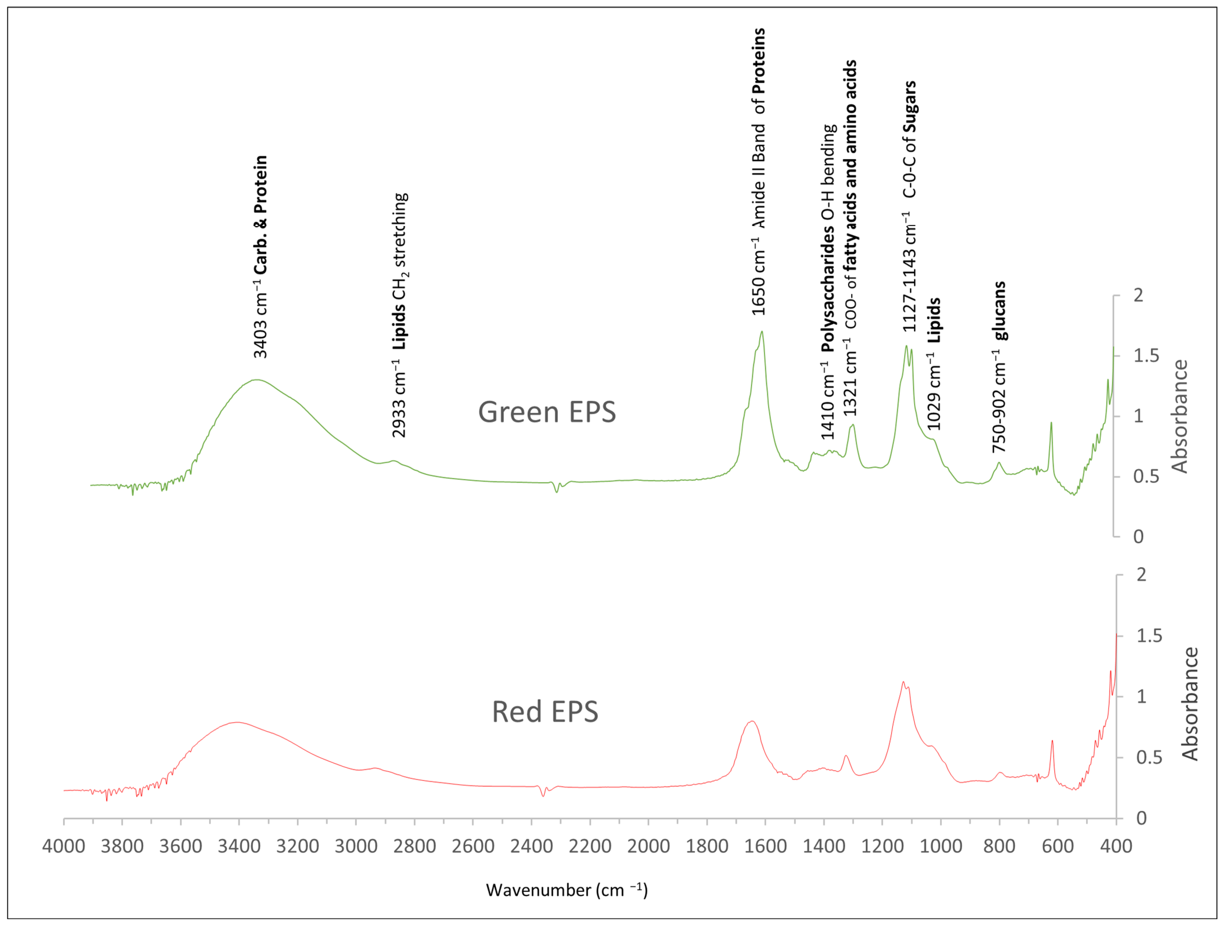
To detect structural differences between the crude exopolysaccharides, FT-IR analysis was conducted as shown in Figure 4. The FT-IR spectra contain the characteristic peaks of the Amide II band of proteins at 1650 cm−1 for all the tested conditions, and characteristic peaks of lipids at 1029 cm−1 and 2933 cm−1, indicating the existence of protein and lipid moieties in crude polysaccharides. The main differences in the crude EPSs are shown in wave numbers of 950–1200 cm−1 and the regions between 1300 and 1500 cm−1 and 1600 to 1800 cm−1. Three different peaks are detected for the green EPS at 1600 to 1800 cm−1 whereas only one peak is detected for red EPS. In the green EPS spectra, three different peaks are shown in the region from 1300 to 1500 cm−1, whereas in the red EPS, the third peak is not so distinctive. In the region of 1150 cm−1 (for sugars), we can detect two different peaks in the green EPS, whereas the second peak starts to fade out in the red EPS. In addition, the peaks attributed to the existence of glucan in the crude EPSs are detected in the region between 750 to 900 cm−1.

4. Discussion

According to our results, P. ostreatus LGAM 1123 was capable of growing well in all light conditions, with biomass production values reaching 20 g/L, similar to the control condition of darkness. All light wavelengths present higher specific growth rates in comparison with dark conditions, with maximum values reaching 0.8 d−1 for red, blue, green, and white wavelengths. The maximum growth rate when the red light was used has been confirmed in a study for Cordyceps militaris cultivation under various light-emitting diodes, in which a maximum specific growth rate of 1.47 d−1 was achieved, a value higher than that achieved under the control condition of darkness, and biomass production of 17 g/L was achieved [7]. In contrast, another study has shown that light illumination stressed Pleurotus eryngii in submerged cultivation, with only red light presenting a similar biomass value, with a value lower than ours (9.9 g/L), compared to the control conditions of darkness [9]. In addition, a study on Pleurotus spp. in liquid cultivation revealed that among seven strains of the Pleurotus genus, cultivation under green light presented lower biomass production compared to the control condition of darkness [39]. In contrast, Ha et al., (2020) have indicated that green light presents the highest biomass production, with a statistically different value compared to darkness, fluorescent light, UV-A, red, and blue light [8]. A recent study of P. ostreatus mycelia and basidioma proteomic analysis under different light wavelengths revealed that basidioma and mycelia present different responses under light, with white and blue light significantly promoting basidioma growth while red light and darkness significantly inhibited this growth. Concerning mycelia production in Petri dishes, the same study indicates that mycelia production is favored under white light while red light and darkness present morphological differentiation [40]. A study of the strain Phanerochaete chrysosporium summarizes that biomass production is favored under conditions of yellow and green light [41]. Regarding protein content, dark conditions present lower protein content compared to the other tested light wavelengths. According to the scientific literature, the P. chysosporium strain showed a decrease in protein production when red, blue, and UV light were used in aerated cultivations, whereas in oxygenated cultivations, a decrease in protein was observed for UV and white light. In addition, a study on the mycelial biomass of Lentinus crinitus under different light wavelengths revealed that protein content decreased by 33%, 29%, and 15% of dw for blue, green, and red light compared to the control condition of darkness [42]. Taking into account all these studies, we can state that with regards to biomass and protein content, different responses are observed depending on the fungal strain and the light wavelength.
Concerning polysaccharide production, EPS was favored under red and green light and suppressed under dark conditions. IPS production reached its maximum values under red and blue light wavelengths. We focus on EPS production analysis and characterization because pleuran is the most well-known β-glucan from P. ostreatus and is secreted in the cultivation medium. Consequently, in this study, we focus on red and green light wavelengths to produce exopolysaccharides in baffled flasks and in a stirred tank bioreactor.
At first, green light was used in all incubators, as shown in Figure 1c. We observed the maximum biomass production achieved in this work of 24.9 ± 2.4 g/L. The protein content was 55.4 ± 3.9% dw and EPS production reached a value of 4.1 ± 0.2 g/L, whereas IPS content reached the highest value achieved in this work of 44.8 ± 5.2% dw. The results are in agreement with other published work showing that green light can stimulate the production of biomass and polysaccharides by other fungi during submerged fermentation. Ramirez et al., (2010) confirmed that green light favored biomass production for a P. chrysosporium strain cultivated in aerated cultures [41]. When C. militaris underwent submerged cultivation using green light, biomass production of 18 g/L and EPS production of 2 g/L, values lower than those in our work, were achieved, while a higher EPS production rate compared to the other tested light wavelengths was observed [7]. Green light produces the highest EPS production for a P. eryngii strain, with values much lower than our study (0.42 g/L) [9]. In contrast to our results, a study that focuses on the effect of green light on biomass and lignocellulose decay enzymes by different Pleurotus strains shows a decrease in biomass production under green illumination but an increase in cellulolytic and xylanolytic enzyme activities. In addition, we should note that biomass production was lower compared to our results, with values up to 1.19 g/L for a P. ostreatus strain, probably due to the lower carbon concentration and lower light intensity that was used in this work (10 g/L glucose and 20 μmol m−2 sec−1) [39].
Afterward, we proceeded with a similar in-light diffusion experiment using red light instead of green. As shown in Figure 1b, red light was used in all incubators and maximum biomass production reached a value of 19.4 ± 1.3 g/L. Protein content was similar to the content achieved with green light (55.4 ± 3.9% dw), along with EPS production (4.1 ± 0.4 g/L). On the other hand, IPS content reached a value of 23.1 ± 1.4% dw. A study on the submerged cultivation of P. eryngii using red light confirmed that red light produced a similar biomass production value compared to the control condition of darkness, which had a biomass production value of only 9.1 g/L. In addition, red light shows a lower EPS production value compared to our result, with a value of 0.2 g/L of EPS concentration to be achieved. This study revealed that red light produces an EPS with high molecular weight, indicating that different polysaccharides can be produced under different wavelengths [9]. Kho et al., (2016) confirmed that red light gave the highest biomass for C. militaris under submerged cultivation, with a production value similar to our result (18.2 g/L). This also indicates that red light has the fastest EPS production speed, reaching lower values compared to ours (2 g/L) on the 10th day of cultivation [7]. Moreover, a study on the submerged cultivation of Phanerochaete chrysosporium using different wavelengths of light has shown that red light favored polysaccharide production in aerated cultures [41].
Focusing on EPS production, a scale-up study was conducted in a 3.5 L stirred tank bioreactor for P. ostreatus LGAM 1123 under conditions of submerged cultivation, using red light as shown in Figure 1d. A similar EPS production value to the experiment with red flasks was achieved; however, due to the large volume of collected supernatant at the end of fermentation, a crude polysaccharide of 2.64 g was produced. To our knowledge, this is the first time that a bioreactor has been used for EPS production with light illumination for a fungal strain, although bioreactors of semi-industrial scale have already been used for Pleuran production through the submerged cultivation of P. ostreatus in dark conditions. In conditions of non-controlled pH similar to our experiment, with an initial pH of 5.5, the maximum biomass production reached a value of 3.2 g/L whereas EPS production reached a value of 1.2 g/L, lower values than those in our results. In addition, this study revealed that a controlled pH strategy improved EPS production and pleuran production with values of 1.98 g/L and 0.4 g/g to be achieved [43]. A higher EPS production compared to our study was achieved only for a Ganoderma lucidum EPS that was produced in a 16 L stirred tank bioreactor, with a production value of 5.0 g/L after fermentation in controlled pH conditions [44]. These two studies suggest that future fermentation of P. ostreatus using red light with a controlled pH strategy process could further increase EPS production. For the same strain that we used in this study (P. ostreatus LGAM 1123), Zerva et al., (2017) suggested that in a 2.5 L bioreactor, EPS production from a 10× concentrated supernatant reached 15.9 mg/mL using a semi-synthetic medium of 57 g/L xylose, 37 g/L corn steep liquor, and 18.8 mg/mL olive mill wastewater [23]. In addition, they indicate that the composition of EPS in total glucans was 4.57 ± 0.08% for the semi-synthetic medium and 1.91 ± 0.2% for olive mill wastewater, with the β-glucans constituting the larger portion of total glucans [23].
After the fermentation process and the precipitation of the produced polysaccharides, two different crude polysaccharides were collected as mentioned above. The analysis of the crude polysaccharides has shown that protein and lipid moieties with different biochemical compositions were detected in the produced powder. These differences could explain the distinctive biological activities that polysaccharides present. In addition, it has already been stated that the antioxidant activity of polysaccharides depends on molecular weight, solubility, sugar ring structure, the existence of positively or negatively charged groups, protein moieties, and linked phenolic compounds [18]. In this work, the phenolic content of the produced polysaccharides slightly differs but confirmed that the red EPS, which presents the higher antioxidant activity, also presents the highest phenolic content. Green EPS presents a much lower antioxidant activity of 27.7 ± 0.06%. The supremacy of red light in antioxidant activity has been confirmed from a study on the effect of light on the mycelial biomass of L. crinitus using the DPPH protocol [42]. Vamanu et al., (2013) and (2012), in two studies on the antioxidant activity of EPS in four different P. ostreatus strains, have revealed that for external stocks of 10 g/L, the antioxidant activities of the PQMZ91109 strain did not exceed 40% in the ABTS reaction [24], while in the other study, the same stock presented an 80% antioxidant activity, values higher than those in our results [45]. The higher antioxidant activity of red EPS versus green EPS was also confirmed by the CUPRAC method, resulting in a value of 16.8 ± 0.5 μg Trolox/mg EPS, which is similar to the one that ABTS+ produced (18.7 ± 0.2 μg Trolox/mg EPS). From these studies, we can conclude that antioxidant activity can vary between strains of P. ostreatus.
Regarding the structural analysis of the produced polysaccharides, the FT-IR spectra revealed that the main differences are focused on the 950–1200 cm−1 region that corresponds to the sugar region [46]. In addition, the existence of peaks in the region of 750–900 cm−1 corresponds to the anomeric regions of polysaccharides [46]. We can detect some similarities in the produced polysaccharides but also some differences between the red and green EPSs corresponding to the different effects of the light wavelength in the metabolism of P. ostreatus. Three different peaks are detected for the green EPS at 1600 to 1800 cm−1 whereas only one peak is detected for Red EPS. This observation confirms the existence of protein contamination in the samples because the peaks around 1650 cm−1 correspond to the C-N bond of the amino acids. The different number of peaks could be attributed to different proteoglycans that exist in the polysaccharides produced under different lights [23]. From a study of the strain that we also used in this study (P. ostreatus LGAM 1123), the FT-IR spectra of the EPS confirmed the existence of protein moieties in the 1650 cm−1 region and lipid moieties in the region of 2920–2930 cm−1 [23]. The most well-known EPS produced from P. ostreatus is Pleuran, a β-glucan with antioxidative, antidiabetic, and immunomodulatory effects [20,43,46]. A summary of the peaks that are related to β-glucans and could correspond to Pleuran production is depicted in Table 4. The comparison between the scientific literature and our EPSs shows that all the peaks found in the scientific literature are also detected in our produced EPSs, with slight modifications in the wave numbers [20,23,46,47,48].

5. Conclusions

To sum up, this is the first report on how light affects polysaccharide production in P. ostreatus LGAM 1123. According to the results for EPS production from the screening experiment, we focus on two specific light wavelengths, red and green, which lead to the highest EPS production. The production of polysaccharides using red and green light was higher compared to other studies on the effect of light on polysaccharide production in different Pleurotus strains. The EPS that was produced by fermentation in a stirred tank bioreactor revealed similar polysaccharide production but higher productivity compared to the polysaccharide produced in the flask experiments. Furthermore, analysis of the crude polysaccharides has shown differences in biochemical composition, whereas the structural differences of the produced polysaccharides were confirmed by FT-IR analysis. Adopting the use of light illumination in the fermentation of fungi, as has already been done in plants, could enhance polysaccharide production in the food industry and lead to the exploitation of polysaccharides in food products such as meat analogs to improve their chemical composition and functional health-promoting properties, as well as increasing their physicochemical and sensory properties. Further experiments such as a pH-controlled method or the use of mixed wavelengths should be adopted to further optimize the production of polysaccharides. Finally, a purification process could be adopted to decrease the concentration of protein and lipid moieties if and only if this strategy does not reduce the biological activities of these polysaccharides.

Supplementary Materials

The following supporting information can be downloaded at: https://www.mdpi.com/article/10.3390/fermentation10060313/s1, Figure S1: Protein content and production by submerged cultivation of P. ostreatus LGAM 1123 in baffled flasks using green LED lighting; Figure S2: Protein content and production by submerged cultivation of P. ostreatus LGAM 1123 in baffled flasks using red LED lighting; Figure S3: Submerged cultivation of P. ostreatus LGAM 1123 in a 3.5 L stirred tank bioreactor using red LED lighting. a: growth curves, b: protein content and production, c: EPS production, d: IPS content.

Author Contributions

Conceptualization, P.K.; Data curation, G.B.; Formal analysis, G.B. and C.T.; Funding acquisition, H.S.; Investigation, G.B.; Methodology, G.B. and P.K.; Project administration, H.S. and P.K.; Resources, H.S. and P.K.; Supervision, H.S. and P.K.; Validation, G.B.; Visualization, G.B. and C.T.; Writing—original draft, G.B.; Writing—review & editing, H.S. and P.K. All authors have read and agreed to the published version of the manuscript.

Funding

This study was co-financed by the European Regional Development Fund of the European Union and Greek National Funds through the Operational Program Competitiveness, Entrepreneurship and Innovation, under the call RESEARCH-CREATE-INNOVATE (project code: T2EDK-02830).

Institutional Review Board Statement

Not applicable

Informed Consent Statement

Not applicable.

Data Availability Statement

The original contributions presented in the study are included in the article/Supplementary Material, further inquiries can be directed to the corresponding author.

Acknowledgments

We gratefully acknowledge Georgios I. Zervakis from the Laboratory of General and Agricultural Microbiology, Agricultural University of Athens, Iera Odos 75, 11855 Athens, Greece, for providing the fungal strain.

Conflicts of Interest

The authors declare no conflicts of interest.

References

  1. Tisch, D.; Schmoll, M. Light Regulation of Metabolic Pathways in Fungi. Appl. Microbiol. Biotechnol. 2010, 85, 1259–1277. [Google Scholar] [CrossRef]
  2. Smith, H. Light Quality, Photoperception, and Plant Strategy. Annu. Rev. Plant Physiol. 1982, 33, 481–518. [Google Scholar] [CrossRef]
  3. Feng, Y.; Xu, H.; Sun, Y.; Xia, R.; Hou, Z.; Li, Y.; Wang, Y.; Pan, S.; Li, L.; Zhao, C.; et al. Effect of Light on Quality of Preharvest and Postharvest Edible Mushrooms and Its Action Mechanism: A Review. Trends Food Sci. Technol. 2023, 139, 104119. [Google Scholar] [CrossRef]
  4. Bakratsas, G.; Polydera, A.; Katapodis, P.; Stamatis, H. Recent Trends in Submerged Cultivation of Mushrooms and Their Application as a Source of Nutraceuticals and Food Additives. Future Foods 2021, 4, 100086. [Google Scholar] [CrossRef]
  5. Singdevsachan, S.K.; Auroshree, P.; Mishra, J.; Baliyarsingh, B.; Tayung, K.; Thatoi, H. Mushroom Polysaccharides as Potential Prebiotics with Their Antitumor and Immunomodulating Properties: A Review. Bioact. Carbohydr. Diet. Fibre 2016, 7, 1–14. [Google Scholar] [CrossRef]
  6. Wang, Q.; Xu, M.; Zhao, L.; Chen, L.; Ding, Z. Novel Insights into the Mechanism Underlying High Polysaccharide Yield in Submerged Culture of Ganoderma lucidum Revealed by Transcriptome and Proteome Analyses. Microorganisms 2023, 11, 772. [Google Scholar] [CrossRef]
  7. Manzi, P.; Pizzoferrato, L. Beta-Glucans in Edible Mushrooms. Food Chem. 2000, 68, 315–318. [Google Scholar] [CrossRef]
  8. Castro-Alves, V.C.; Gomes, D.; Menolli, N.; Sforça, M.L.; do Nascimento, J.R.O. Characterization and Immunomodulatory Effects of Glucans from Pleurotus albidus, a Promising Species of Mushroom for Farming and Biomass Production. Int. J. Biol. Macromol. 2017, 95, 215–223. [Google Scholar] [CrossRef] [PubMed]
  9. Fernandes, P.A.R.; Coimbra, M.A. The Antioxidant Activity of Polysaccharides: A Structure-Function Relationship Overview. Carbohydr. Polym. 2023, 314, 120965. [Google Scholar] [CrossRef] [PubMed]
  10. Papaspyridi, L.M.; Zerva, A.; Topakas, E. Biocatalytic Synthesis of Fungal β-Glucans. Catalysts 2018, 8, 274. [Google Scholar] [CrossRef]
  11. Synytsya, A.; Míčková, K.; Jablonský, I.; Sluková, M.; Čopíková, J. Mushrooms of Genus Pleurotus as a Source of Dietary Fibres and Glucans for Food Supplements. Czech J. Food Sci. 2008, 26, 441–446. [Google Scholar] [CrossRef]
  12. Bekiaris, G.; Tagkouli, D.; Koutrotsios, G.; Kalogeropoulos, N.; Zervakis, G.I. Pleurotus Mushrooms Content in Glucans and Ergosterol Assessed by ATR-FTIR Spectroscopy and Multivariate Analysis. Foods 2020, 9, 535. [Google Scholar] [CrossRef]
  13. Papaspyridi, L.M.; Katapodis, P.; Gonou-Zagou, Z.; Kapsanaki-Gotsi, E.; Christakopoulos, P. Optimization of Biomass Production with Enhanced Glucan and Dietary Fibres Content by Pleurotus ostreatus ATHUM 4438 under Submerged Culture. Biochem. Eng. J. 2010, 50, 131–138. [Google Scholar] [CrossRef]
  14. Zerva, A.; Papaspyridi, L.M.; Christakopoulos, P.; Topakas, E. Valorization of Olive Mill Wastewater for the Production of β-Glucans from Selected Basidiomycetes. Waste Biomass Valorization 2017, 8, 1721–1731. [Google Scholar] [CrossRef]
  15. Vamanu, E. Antioxidant Properties of Polysaccharides Obtained by Batch Cultivation of Pleurotus ostreatus Mycelium. Nat. Prod. Res. 2013, 27, 1115–1118. [Google Scholar] [CrossRef]
  16. Pilafidis, S.; Tsouko, E.; Sougleri, G.; Diamantopoulou, P. Submerged Cultivation of Selected Macro-Fungi to Produce Mycelia Rich in β-Glucans and Other Bioactive Compounds, Valorizing Side Streams of the Food Industry. Carbon. Resour. Convers. 2024, 7, 100198. [Google Scholar] [CrossRef]
  17. Kho, C.H.; Kan, S.C.; Chang, C.Y.; Cheng, H.Y.; Lin, C.C.; Chiou, P.C.; Shieh, C.J.; Liu, Y.C. Analysis of Exopolysaccharide Production Patterns of Cordyceps militaris under Various Light-Emitting Diodes. Biochem. Eng. J. 2016, 112, 226–232. [Google Scholar] [CrossRef]
  18. Ha, S.Y.; Jung, J.Y.; Yang, J.K. Effect of Light-Emitting Diodes on Cordycepin Production in Submerged Culture of Paecilomyces Japonica. J. Korean Wood Sci. Technol. 2020, 48, 548–561. [Google Scholar] [CrossRef]
  19. Wu, J.; Chen, H.; Chen, M.; Kan, S.; Shieh, C. Quantitative Analysis of LED Effects on Exopolysaccharide and Mycelial Growth Productions in Pleurotus Erygii Cultures. Soc. Chem. Ind. 2012, 88, 1841–1846. [Google Scholar] [CrossRef]
  20. Qi, Y.; Sun, X.; Ma, L.; Wen, Q.; Qiu, L.; Shen, J. Identification of Two Pleurotus ostreatus Blue Light Receptor Genes (PoWC-1 and PoWC-2) and in Vivo Confirmation of Complex PoWC-12 Formation through Yeast Two Hybrid System. Fungal Biol. 2020, 124, 8–14. [Google Scholar] [CrossRef]
  21. Wang, H.; Tong, X.; Tian, F.; Jia, C.; Li, C.; Li, Y. Transcriptomic Profiling Sheds Light on the Blue-Light and Red-Light Response of Oyster Mushroom (Pleurotus ostreatus). AMB Express 2020, 10, 10. [Google Scholar] [CrossRef] [PubMed]
  22. Rodrigues Barbosa, J.; dos Santos Freitas, M.M.; da Silva Martins, L.H.; de Carvalho, R.N. Polysaccharides of Mushroom Pleurotus spp.: New Extraction Techniques, Biological Activities and Development of New Technologies. Carbohydr. Polym. 2020, 229, 115550. [Google Scholar] [CrossRef] [PubMed]
  23. Gutirrrez, A.; Prieto, A.; Martlnez, A.T. Structural Characterization of Extracellular Polysaccharides Produced by Fungi from the Genus Pleurotus. Carbohydr. Res. 1996, 281, 143–154. [Google Scholar] [CrossRef] [PubMed]
  24. Gern, R.M.M.; Wisbeck, E.; Rampinelli, J.R.; Ninow, J.L.; Furlan, S.A. Alternative Medium for Production of Pleurotus ostreatus Biomass and Potential Antitumor Polysaccharides. Bioresour. Technol. 2008, 99, 76–82. [Google Scholar] [CrossRef] [PubMed]
  25. El-Enshasy, H.; Daba, A.; El-Demellawy, M.; Ibrahim, A.; El Sayed, S.; El-Badry, I. Bioprocess Development for Large Scale Production of Anticancer Exo-Polysaccharide by Pleurotus ostreatus in Submerged Culture. J. Appl. Sci. 2010, 10, 2523–2529. [Google Scholar] [CrossRef]
  26. Bakratsas, G.; Polydera, A.; Nilson, O.; Κossatz, L.; Xiros, C.; Katapodis, P.; Stamatis, H. Single-Cell Protein Production by Pleurotus ostreatus in Submerged Fermentation. Sustain. Food Technol. 2023, 1, 377–389. [Google Scholar] [CrossRef]
  27. Bakratsas, G.; Polydera, A.; Nilson, O.; Chatzikonstantinou, A.V.; Xiros, C.; Katapodis, P.; Stamatis, H. Mycoprotein Production by Submerged Fermentation of the Edible Mushroom Pleurotus ostreatus in a Batch Stirred Tank Bioreactor Using Agro-Industrial Hydrolysate. Foods 2023, 12, 2295. [Google Scholar] [CrossRef] [PubMed]
  28. Kruger, N.J. The Bradford Method for Protein Quantitation. In The Protein Protocols Handbook; Springer Protocols Handbooks: Totowa, NJ, USA, 1996; Volume 32, pp. 15–20. [Google Scholar]
  29. Fan, X.; Yao, F.; Yin, C.; Shi, D.; Gao, H. Mycelial Biomass and Intracellular Polysaccharides Production, Characterization, and Activities in Auricularia Auricula-Judae Cultured with Different Carbon Sources. Int. J. Biol. Macromol. 2023, 244, 125426. [Google Scholar] [CrossRef] [PubMed]
  30. Dubois, M.; Gilles, K.A.; Hamilton, J.K.; Rebers, P.A.; Smith, F. Colorimetric Method for Determination of Sugars and Related Substances. Anal. Chem. 1956, 28, 350–356. [Google Scholar] [CrossRef]
  31. Giraldo, L.R.D.; Baez, P.X.V.; Forero, C.J.Z.; Arango, W.M. Production, Extraction, and Solubilization of Exopolysaccharides Using Submerged Cultures of Agaricomycetes. Bio-Protocol 2023, 13, e4841. [Google Scholar] [CrossRef]
  32. Smith, P.K.; Krohn, R.I.; Hermanson, G.T.; Mallia, A.K.; Gartner, F.H.; Provenzano, M.D.; Fujimoto, E.K.; Goeke, N.M.; Olson, B.J.; Klenk, D.C. Measurement of Protein Using Bicinchoninic Acid. Anal. Biochem. 1985, 150, 76–85. [Google Scholar] [CrossRef]
  33. Folch, J.; Lees, M.; Sloane Stanley, G.H. A Simple Method for the Isolation and Purification of Total Lipides from Animal Tissues. J. Biol. Chem. 1957, 226, 497–509. [Google Scholar] [CrossRef] [PubMed]
  34. Karageorgou, D.; Sainis, I.; Touka, A.; Vareli, K.; Stamatis, H.; Katapodis, P. Biomass and β-Glucosidase Production by the Cyanobacterium Pseudanabaena sp. under Heterotrophic Conditions. Biomass 2022, 2, 299–315. [Google Scholar] [CrossRef]
  35. Athanasiou, P.E.; Patila, M.; Fotiadou, R.; Chatzikonstantinou, A.V.; Stamatis, H. Valorization of Wine Lees: Assessment of Antioxidant, Antimicrobial and Enzyme Inhibitory Activity of Wine Lees Extract and Incorporation in Chitosan Films. Waste Biomass Valor 2024. [Google Scholar] [CrossRef]
  36. Fotiadou, R.; Lefas, D.; Vougiouklaki, D.; Tsakni, A.; Houhoula, D.; Stamatis, H. Enzymatic Modification of Pomace Olive Oil with Natural Antioxidants: Effect on Oxidative Stability. Biomolecules 2023, 13, 1034. [Google Scholar] [CrossRef] [PubMed]
  37. Athanasiou, P.E.; Gkountela, C.I.; Patila, M.; Fotiadou, R.; Chatzikonstantinou, A.V.; Vouyiouka, S.N.; Stamatis, H. Laccase-Mediated Oxidation of Phenolic Compounds from Wine Lees Extract towards the Synthesis of Polymers with Potential Applications in Food Packaging. Biomolecules 2024, 14, 323. [Google Scholar] [CrossRef] [PubMed]
  38. Bakratsas, G.; Antoniadis, K.; Athanasiou, P.E.; Katapodis, P.; Stamatis, H. Laccase and Biomass Production via Submerged Cultivation of Pleurotus ostreatus Using Wine Lees. Biomass 2024, 4, 1–22. [Google Scholar] [CrossRef]
  39. Araújo, N.L.; Avelino, K.V.; Halabura, M.I.W.; Marim, R.A.; Kassem, A.S.S.; Linde, G.A.; Colauto, N.B.; do Valle, J.S. Use of Green Light to Improve the Production of Lignocellulose-Decay Enzymes by Pleurotus spp. in Liquid Cultivation. Enzym. Microb. Technol. 2021, 149, 109860. [Google Scholar] [CrossRef]
  40. Zhu, L.; Su, Y.; Ma, S.; Guo, L.; Yang, S.; Yu, H. Comparative Proteomic Analysis Reveals Candidate Pathways Related to the Effect of Different Light Qualities on the Development of Mycelium and Fruiting Body of Pleurotus ostreatus. J. Agric. Food Chem. 2023, 72, 1361–1375. [Google Scholar] [CrossRef]
  41. Ramírez, D.A.; Muñoz, S.V.; Atehortua, L.; Michel, F.C. Effects of Different Wavelengths of Light on Lignin Peroxidase Production by the White-Rot Fungi Phanerochaete Chrysosporium Grown in Submerged Cultures. Bioresour. Technol. 2010, 101, 9213–9220. [Google Scholar] [CrossRef]
  42. Halabura, M.I.W.; Avelino, K.V.; Araújo, N.L.; Kassem, A.S.S.; Seixas, F.A.V.; Barros, L.; Fernandes, Â.; Liberal, Â.; Ivanov, M.; Soković, M.; et al. Light Conditions Affect the Growth, Chemical Composition, Antioxidant and Antimicrobial Activities of the White-Rot Fungus Lentinus crinitus Mycelial Biomass. Photochem. Photobiol. Sci. 2023, 22, 669–686. [Google Scholar] [CrossRef] [PubMed]
  43. Maftoun, P.; Malek, R.; Abdel-Sadek, M.; Aziz, R.; El Enshasy, H. Bioprocess for Semi-Industrial Production of Immunomodulator Polysaccharide Pleuran by Pleurotus ostreatus in Submerged Culture. J. Sci. Ind. Res. (India) 2013, 72, 655–662. [Google Scholar]
  44. Alsaheb, R.A.; Zjeh, K.Z.; Malek, R.A.; Abdullah, J.K.; El Baz, A.; El Deeb, N.; Dailin, D.; Hanapi, S.Z.; Sukmawati, D.; El Enshasy, H. Bioprocess Optimization for Exopolysaccharides Production by Ganoderma Lucidum in Semi-Industrial Scale. Recent. Pat. Food Nutr. Agric. 2020, 11, 211–218. [Google Scholar] [CrossRef] [PubMed]
  45. Vamanu, E. Biological Activities of the Polysaccharides Produced in Submerged Culture of Two Edible Pleurotus ostreatus Mushrooms. J. Biomed. Biotechnol. 2012, 2012, 565974. [Google Scholar] [CrossRef] [PubMed]
  46. Baeva, E.; Bleha, R.; Lavrova, E.; Sushytskyi, L.; Čopíková, J.; Jablonsky, I.; Klouček, P.; Synytsya, A. Polysaccharides from Basidiocarps of Cultivating Mushroom Pleurotus ostreatus: Isolation and Structural Characterization. Molecules 2019, 24, 2740. [Google Scholar] [CrossRef] [PubMed]
  47. Synytsya, A.; Novak, M. Structural Analysis of Glucans. Ann. Transl. Med. 2014, 2, 17. [Google Scholar] [CrossRef]
  48. Šandula, J.; Kogan, G.; Kačuráková, M.; Machová, E. Microbial (1→3)-β-d-Glucans, Their Preparation, Physico-Chemical Characterization and Immunomodulatory Activity. Carbohydr. Polym. 1999, 38, 247–253. [Google Scholar] [CrossRef]
Figure 1. Cultivation with LED lighting: (a) screening experiment in baffled flasks, (b) baffled flasks with red light, (c) baffled flasks with green light, and (d) 3.5 L STR with red light.
Fermentation 10 00313 g001
Figure 2. Submerged cultivation of P. ostreatus LGAM 1123 in baffled flasks using green LED lighting. (a) growth curves (green line) and EPS production (dotted line), (b) IPS content (green bars).
Fermentation 10 00313 g002
Figure 3. Submerged cultivation of P. ostreatus LGAM 1123 in baffled flasks using red LED lighting. (a) growth curves (red line) and EPS production (dotted line), (b) IPS content (red bars).
Fermentation 10 00313 g003
Figure 4. FT-IR spectra of the produced polysaccharides from submerged cultivation of P. ostreatus LGAM 1123 using red and green light.
Fermentation 10 00313 g004
Table 1. Effect of light wavelength on P. ostreatus LGAM 1123’s specific growth rate (μ), Biomass (g/L), Protein content (% dw), EPS production (g/L), and IPS content (% dw) in a medium composed of 54 g/L glucose and 18 g/L yeast extract. Different letters in each row indicate a significant difference (p ≤ 0.05) in Tukey’s multiple range test.
DarkRed YellowGreenBlue White
μ (d−1)0.75 ± 0.02 b0.82 ± 0.02 a0.78 ± 0.02 ab0.80 ± 0.01 a0.80 ± 0.01 a0.80 ± 0.01 a
Biomass (g/L)20.6 ± 0.4 a21.3 ± 0.4 a19.5 ± 0.4 b19.7 ± 0.4 b17.8 ± 0.4 c18.0 ± 0.4 c
Protein content (% dw)37.7 ± 4.1 b45.3 ± 3.3 a46.0 ± 1.7 a45.2 ± 2.0 a45.6 ± 4.3 a47.0 ± 2.5 a
EPS (g/L)1.6 ± 0.4 bc1.8 ± 0.2 ab1.5 ± 0.2 bc2.1 ± 0.3 a1.6 ± 0.1 bc1.7 ± 0.2 bc
IPS (% dw)8.9 ± 3.6 c27.2 ± 0.2 a15.3 ± 0.2 bc18.0 ± 3.2 b29.7 ± 6.5 a22.4 ± 1.0 ab
IPS (g/L)1.8 ± 0.0 f5.8 ± 0.1 a3.0 ± 0.1 e3.5 ± 0.1 d5.3 ± 0.1 b4.0 ± 0.1 c
Table 2. Polysaccharide production and protein content from cultivation in a 3.5 L stirred tank bioreactor using red light.
Biomass production (g/L)12.6 ± 0.6
EPS production (g/L)3.7 ± 0.1
EPS productivity (g/L/d)0.48 ± 0.02
IPS content (% dw)28.8 ± 0.1
Protein content (% dw)50.3 ± 2.5
Table 3. Exopolysaccharide analysis from submerged cultivation of P. ostreatus LGAM 1123 using light wavelengths. Different symbols in each row: *, ***, ****, and ns indicate p-values of p < 0.05, 0.001, 0.0001, and not significant, respectively, after two-way ANOVA analysis and the Sidak Test.
Polysaccharide AnalysisRed Light EPSGreen Light EPS
Production (g/L)4.16 ± 0.03 ns4.14 ± 0.06 ns
Sugar content (% dw)50.0 ± 5.0 ***42.5 ± 4.0 ***
Protein content (% dw)19.5 ± 1.1 *14.7 ± 0.9 *
Lipid content (% dw)24.0 ± 2.3 *19.2 ± 0.1 *
Antioxidant activity—ABTS (%) (0.5 mg/mL)78.3 ± 2.5 ****27.7 ± 0.1 ****
Antioxidant activity—ABTS (μg Trolox/mg EPS)18.7 ± 0.2 ****7.1 ± 1.0 ****
Antioxidant activity—CUPRAC (μg Trolox/mg EPS)16.8 ± 0.5 ****8.1 ± 0.4 ****
Phenolic content (mg GAE/mg polysaccharide)4.9 ± 0.2 ns4.32 ± 0.07 ns
Table 4. β-glucan peaks from FT-IR analyses of EPSs according to the scientific literature and our results.
β-glucan peaks (cm−1)Baeva et al. (2019) [46]Zerva et al. (2017) [23]Synytsya et al. (2014) [20]Synytsya et al. (2009) [20]Sandula et al. (1999) [48]Red EPSGreen EPS
1374--1376-13821388
1318--1317-13161313
1158–11601127–115011601162116011101126
1080-1078110010781106-
1038–1040-10441080104110201024
890–894881, 8938901040889869886
Disclaimer/Publisher’s Note: The statements, opinions and data contained in all publications are solely those of the individual author(s) and contributor(s) and not of MDPI and/or the editor(s). MDPI and/or the editor(s) disclaim responsibility for any injury to people or property resulting from any ideas, methods, instructions or products referred to in the content.