Article

Two-Step Bio-Based Production of Heme: In Vivo Cell Cultivation Followed by In Vitro Enzymatic Conversion

by
Bahareh Arab
,
Murray Moo-Young
,
Yilan Liu
and
Chih-Hsiung Perry Chou
*
Department of Chemical Engineering, University of Waterloo, Waterloo, ON N2L 3G1, Canada
*
Author to whom correspondence should be addressed.
Fermentation 2025, 11(4), 198; https://doi.org/10.3390/fermentation11040198
Submission received: 7 March 2025 / Revised: 30 March 2025 / Accepted: 2 April 2025 / Published: 8 April 2025
(This article belongs to the Special Issue Applied Microorganisms and Industrial/Food Enzymes, 2nd Edition)

Abstract

:
Heme is a chemical compound crucial for various biological processes and industrial applications. However, the microbial production of heme is often limited by its intracellular accumulation and associated toxicity. To address this, we employed a two-step approach involving in vivo cell cultivation for the production of a heme precursor (coproporphyrin III or coproheme) followed by its in vitro conversion(s) to heme. For the first step, we engineered Escherichia coli strains by implementing the coproporphyrin-dependent (CPD) pathway for bacterial cell cultivation, extracellularly producing up to 251 mg/L coproporphyrin III and 85 mg/L coproheme, respectively. For the second step, we cloned the hemH and hemQ genes for expression in E. coli, and the expressed gene products, i.e., coproheme decarboxylase (ChdC/HemH) and heme synthase (HemQ), were purified. Using the purified enzymes with modulated reaction conditions, we achieved up to a 77.2% yield to convert coproporphyrin III to coproheme and a 45.8% yield to convert coproheme to heme. This in vitro approach not only bypassed the intracellular toxicity constraint associated with in vivo cell cultivation but also enabled precise reaction control, leading to a higher efficiency and yield for heme (and coproheme) production. By applying novel strategies in strain engineering and bioprocessing to overcome inherent bioprocess challenges, this study paves the way for industrial biotechnology for the sustainable, efficient, and even large-scale bio-based production of heme.

1. Introduction

Heme, an iron-containing compound within the tetrapyrrole class, is integral to various biological systems across all domains of life [1]. Its complex architecture enables it to function in numerous biochemical processes essential for life, such as oxygen transport in hemoglobin, electron transfer in cytochromes, and catalysis in peroxidases and other heme enzymes [2,3]. Beyond its biological roles, heme is crucial in multiple industrial sectors, including pharmaceuticals and food additives [4,5]. In the medical field, synthetic heme analogs are used for treating conditions like anemia and are pivotal in the development of diagnostics and therapeutic agents [6]. Additionally, heme’s catalytic properties have garnered attention for their application in industrial chemical processes, such as environmental remediation and synthetic organic chemistry [7,8]. These application potentials have driven the initiatives for the large-scale production of heme. Currently, most heme is still produced via chemical processing [9]. However, recent advances in bio-based production have garnered significant interest in microbial heme biosynthesis, particularly through the genetic and metabolic engineering of microorganisms such as Escherichia coli and Saccharomyces cerevisiae. These biotechnological approaches aim to improve yields, reduce costs, and mitigate the environmental impact of traditional chemical synthesis methods [10,11].
The biosynthesis of heme involves three primary pathways: the porphyrin-dependent (PPD) pathway, the coproporphyrin-dependent (CPD) pathway, and the siroheme-dependent (SHD) pathway. These three pathways share similar routes to the formation of uroporphyrinogen III (UPG-III) [9] (Figure 1). The SHD pathway is primarily found in archaea and sulfate-reducing bacteria existing under anaerobic conditions [12]. In this pathway, following the formation of UPG-III, the pathway diverges and involves multiple enzymatic steps, with siroheme being produced as an intermediate, which is subsequently converted to coproheme (Fe-CP-III) and eventually to heme [13,14]. In the PPD and CPD pathways, the formed UPG-III is converted to coproporphyrinogen III (CPG-III) by the enzyme uroporphyrinogen III decarboxylase (HemE) [15]. After this enzymatic transformation, the pathways diverge into distinct routes toward heme synthesis [16]. In the PPD pathway found in Gram-negative bacteria, CPG-III is converted to protoporphyrinogen IX (PPG-IX) by coproporphyrinogen III oxidase (HemF) and subsequently to protoporphyrin IX (PP-IX) by protoporphyrinogen oxidase (HemG). Finally, ferrochelatase (HemH) catalyzes the insertion of a ferrous ion into PP-IX to produce heme [17]. In contrast, the CPD pathway found in Gram-positive bacteria offers a more streamlined route to heme production. Basically, CPG-III is converted to coproporphyrin III (CP-III) by coproporphyrinogen III dehydrogenase (HemY from Gram-positive bacteria or YfeX from Gram-negative bacteria), then to Fe-CP-III by coproheme decarboxylase (ChdC/HemH), and eventually to heme by heme synthase (HemQ) [18,19]. Unlike the SHD pathway, both the PPD and the CPD pathways involve fewer enzymatic steps, potentially making them easier for the metabolic engineering of heme biosynthesis [9]. However, the microbial biosynthesis of heme via the PPD pathway encounters significant challenges. For example, several pathway genes, such as hemB, can be subject to negative feedback regulation, limiting the overall activity of the metabolic pathway [20,21]. In addition, several reports (and our unpublished results) suggest that heme (and PP-IX) might be hard to secrete extracellularly in bacteria, particularly upon their overproduction, and the cytotoxicity arising from their intracellular accumulation can physiologically impact host cells and, therefore, heme biosynthesis [22,23]. Consequently, the CPD pathway stands out as a promising alternative for efficient and sustainable heme biosynthesis.
Studies have been conducted for the production of the key precursors of heme, including 5-aminolevulinic acid (5-ALA) [24], porphobilinogen (PBG) [25], UP-III [26,27], and CP-III [28], all of which can be extracellularly secreted by hosts upon their overproduction. These results suggest that the issue of intracellular accumulation might be specific to heme (and PP-IX) and, therefore, hard to overcome unless its secretion mechanism and channel can be properly identified for manipulation. In this study, by adopting the CPD pathway, we proposed a two-step approach for the large-scale production of heme, i.e., in vivo cell cultivation to overproduce a heme precursor (either CP-III or Fe-CP-III), followed by in vitro cell-free enzymatic conversion(s) to form heme (Figure 2). Note that cell-free systems can provide precise control over enzymatic reactions while circumventing the toxicity issues associated with intracellular heme accumulation [29]. By leveraging the modularity and scalability of cell-free systems, biochemical pathways can be implemented with a high efficiency and yield. Decoupling metabolic processes from cellular constraints enable the precise design of a reaction environment tailored to specific production requirements [30,31]. Furthermore, cell-free systems offer advantages, such as reduced conversion complexity and enhanced product stability, compared to traditional in vivo approaches [32]. The feasibility of the proposed two-step approach also lies in the prerequisites that the heme precursor can be not only overproduced in vivo but also extracellularly secreted, such that the secreted heme precursor in the cell-free medium, even without pretreatment or purification, can be ready for in vitro enzymatic conversion. Our results show that this approach can address the inherent challenges of heme production while providing a scalable framework for industrial application in bio-based production.

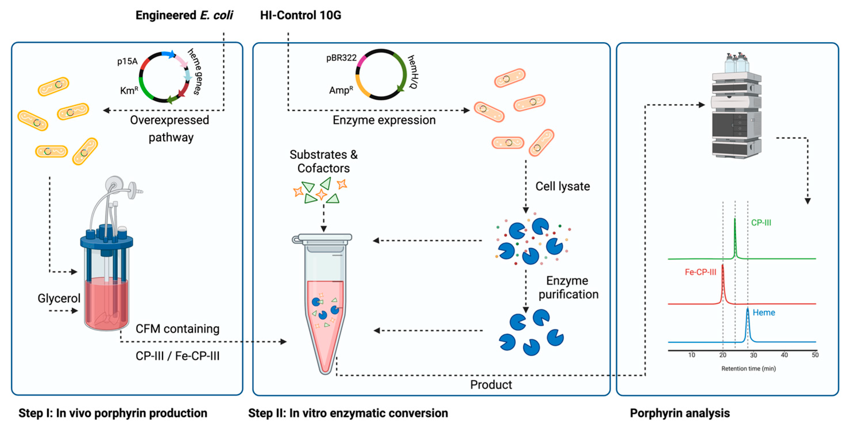
2. Materials and Methods

2.1. Bacterial Strains and Plasmids

Details of the bacterial strains and plasmids used in this study are summarized in Table 1, and the primer sequences can be found in Table S1. Taq DNA polymerase was sourced from New England Biolabs (Ipswich, MA, USA). Genomic DNA was isolated from bacterial cells using the Qiagen DNeasy Blood & Tissue Kit (Hilden, Germany), and plasmid extractions were carried out using the Qiagen Miniprep Kit. All host cells in this study were derived from CPC-Sbm through the introduction of mutation(s) in the sdhA and/or iclR genes [33]. It is noteworthy that CPC-Sbm originated from BW25113 with the ldhA gene inactivated [34]. For cloning purposes, E. coli HI-Control 10G (Lucigen, Middleton, WI, USA) was used. DNA sequencing services were provided by Plasmidsaurus (Eugene, OR, USA). Plasmid construction was performed using Gibson assembly [35], and all primers were synthesized by Integrated DNA Technologies (IDT, Coralville, IA, USA).
The plasmids pK-hemABCD, pK-hemABD-E, and pK-hemAB-E from our previous studies were used to serve as templates for the remaining plasmids [26,28].
pK-hemABD-EHQ was constructed by amplifying hemA, along with the backbone, and amplifying hemB, hemD, and hemE, using primer sets P1/P2 and P3/P4 and pK-hemABD-E as templates. The hemH and hemQ genes were amplified from the genomic DNA of Bacillus subtilis strain 168 using primer sets P5/P6 and P7/P8. These four fragments were Gibson-assembled to form pK-hemABD-EHQ. For effective coexpression, hemA, hemB, and hemD were aligned to form an operon hemABD regulated by a common strong trc promoter, and hemEHQ was on the second operon regulated by the gracmax promoter, with an individual strong RBS for each gene.
Similarly, pK-hemAB-EHQ was constructed by amplifying hemA, along with the backbone, and amplifying hemB, hemD and hemE, using primer sets P1/P2 and P3/P4 and pK-hemAB-E as templates. The hemH and hemQ genes were amplified from the genomic DNA of B. subtilis strain 168 used the primer sets P5/P6 and P7/P8. These four fragments were Gibson-assembled to form pK-hemAB-EHQ. hemAB on the first operon was regulated by the trc promoter, and hemEHQ was regulated by the gracmax promoter.
pK-hemABD-EH was constructed by amplifying hemA, along with the backbone, and amplifying hemB, hemD, hemE, and hemH, using primer sets P9/P10 and P3/P11 and pK-hemABD-EHQ as templates. These two fragments were Gibson-assembled to form pK-hemABD-EH. For effective coexpression, hemABD was expressed on the first operon regulated by the trc promoter, and hemEH was expressed on the second operon regulated by the gracmax promoter.
pK-hemAB-EH was constructed by amplifying hemA, hemB, and hemE and amplifying the backbone using primer sets P12/P13 and P9/P14 and pK-hemAB-E as a template. The hemH gene was amplified from the genomic DNA of B. subtilis strain 168 using primers P5/P15. These three fragments were Gibson-assembled to form pK-hemAB-EH. For effective coexpression, hemAB was expressed on the first operon regulated by the trc promoter, and hemEH was expressed on the second operon regulated by the gracmax promoter.
pK-hemABE-H was constructed by amplifying hemA and hemB using primers P16/P17 and amplifying hemE and the gracmax promoter separately using primer sets P18/P19 and P20/P21 and pK-hemABD-EH as a template. hemH was amplified, along with the backbone, using primers P22/P23 and pK-hemABD-EH as a template. These four fragments were Gibson-assembled to form pK-hemABE-H. hemA, hemB, and hemE were aligned to form an operon hemABE regulated by the trc promoter, and hemH was included in the second operon regulated by the gracmax promoter.
pK-hemABE-HQS.a was constructed by amplifying hemA and hemB using primers P16/P17 and amplifying hemE, the gracmax promoter, and hemH using primers P18/P6 and pK-hemABD-EH as a template. The hemQS.a was amplified from the genomic DNA of Staphylococcus aureus using primers P24/P25. The backbone was amplified from pK-hemABD-EH using primers P26/P23. These four fragments were Gibson-assembled to form pK-hemABE-HQS.a. hemABE was regulated by the trc promoter, and hemHQS.a was regulated by the gracmax promoter.
pK-hemH-His6 and pK-His6-hemH were constructed by amplifying hemH from B. subtilis strain 168 using primers P27/P28 and P29/P30, respectively. The fragment was then Gibson-assembled, with the backbone amplified from the pTrc99a plasmid using primer sets P31/P32 and P33/P34, respectively. These primers were designed to incorporate a 6x His-tag on either the C-terminus or N-terminus. A similar approach was applied for the construction of the plasmids pK-hemQ-His6, pK-His6-hemQ, pK-hemQS.a-His6, and pK-His6-hemQS.a. Briefly, hemQ was amplified using primer sets P35/P36, P37/P38, P39/P40, and P41/P42, respectively, from the genomic DNA of B. subtilis strain 168 and S. aureus. The backbone was amplified from the pTrc99a plasmid using primer sets P43/P44, P45/P46, P43/P47, and P48/P46, respectively. Each amplified gene was then Gibson-assembled with its respective backbone to generate these four plasmids.

2.2. Media and Bacterial Cell Cultivation

All medium components were obtained from Sigma-Aldrich Co. (St. Louis, MO, USA), with the exception of yeast extract and tryptone, which were obtained from BD Diagnostic Systems (Franklin Lakes, NJ, USA). E. coli strains were preserved as glycerol stocks at −80 °C and streaked on lysogeny broth (LB; 10 g/L tryptone, 5 g/L yeast extract, and 5 g/L NaCl) agar plates, then incubated at 37 °C for 14–16 h. For bioreactor cultivation, individual single colonies were picked from LB plates to inoculate 12 mL of a super broth (SB) medium (32 g/L tryptone, 20 g/L yeast extract, and 5 g/L NaCl) in a 125 mL conical flask. The culture was incubated at 37 °C and 280 rpm using a rotary shaker (New Brunswick Scientific, Edison, NJ, USA) for 4–6 h and subsequently used as a starter culture to inoculate 220 mL of the SB medium at a 2% (vol/vol) concentration in a 1 L conical flask. The seed culture was incubated at 37 °C and 280 rpm for 14–16 h. Cells were harvested by centrifugation at 4500× g and 20 °C for 8 min and resuspended in 40 mL of fresh SB medium. The resuspended culture was used to inoculate a stirred tank bioreactor (CelliGen 115, Eppendorf AG, Hamburg, Germany) with a working volume of 0.8 L at 37 °C and 430 rpm. The semi-defined production medium in the batch bioreactor contained 30 g/L glycerol, 0.23 g/L K2HPO4, 0.51 g/L NH4Cl, 49.8 mg/L MgCl2, 48.1 mg/L K2SO4, 1.52 mg/L FeSO4, 0.055 mg/L CaCl2, 2.93 g/L NaCl, 0.72 g/L tricine, 10 g/L yeast extract, 10 mM NaHCO3, and 1 mL/L trace elements (2.86 g/L H3BO3, 1.81 g/L MnCl2·4H2O, 0.222 g/L ZnSO4·7H2O, 0.39 g/L Na2MoO4·2H2O, 79 μg/L CuSO4·5H2O, and 49.4 μg/L Co(NO3)2·6H2O) [36] supplemented with 0.05 mM isopropyl β-D-1-thiogalactopyranoside (IPTG). To avoid glycine and Fe2+ limitation during cultivation, 2 g of glycine and 0.45 g of FeSO4·7H2O was supplemented into the bioreactor ~30 h post inoculation. Aerobic conditions were maintained by continuously purging the air into the bulk culture at 1 volume of air per volume of liquid per min (vvm). The pH of the bioreactor culture was maintained at 7.0 ± 0.1 using 3 M NH4OH and 3 M H3PO4.

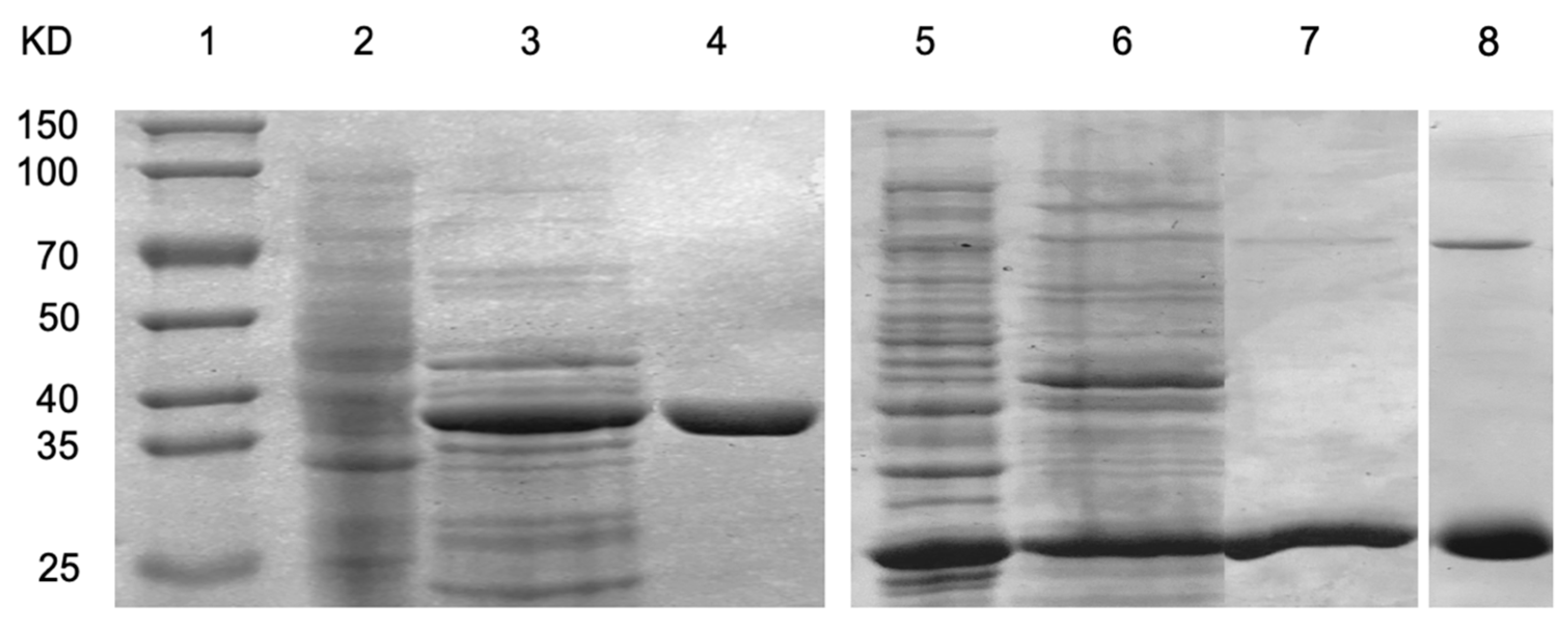
2.3. Enzyme Purification

To purify the HemH and HemQ enzymes, immobilized metal affinity chromatography (IMAC) was employed. Briefly, a bacterial strain expressing His-tagged HemH or HemQ was used to inoculate 200 mL of an LB medium, starting with an initial OD600 of approximately 0.1. The culture was grown to an OD600 of ~1, at which point protein expression was induced with isopropyl β-D-1-thiogalactopyranoside (IPTG). After 5 h post-induction, cells were harvested by centrifugation at 4 °C. The cell pellet was resuspended in a lysis buffer and subjected to sonication for 4 min to lyse the cells. Following sonication, the lysate was centrifuged at 12,000× g for 20 min at 4 °C to remove the cells. The resulting supernatant was filtered and loaded onto a 1 mL IMAC column pre-equilibrated with the lysis/wash buffer. The column was washed with a low-concentration imidazole wash buffer to remove non-specifically bound proteins. HemH and HemQ enzymes were then eluted using an imidazole-rich elution buffer. The buffer of the eluted enzyme fractions was exchanged with a non-imidazole buffer, and the enzymes were concentrated using Amicon centrifugal filters. The concentration of the purified enzymes was determined using the Bradford assay.

2.4. In Vitro Experiment Reaction Set-Up

The HemH assays were performed by quantifying the production of Fe-CP-III in reaction mixtures containing varying concentrations of CP-III and ferrous ions. Specifically, assays were performed with X μM CP-III and 2–8X μM FeSO4·7H2O, supplemented with 2–4 μM of the enzyme. In some assays, the effect of different buffers on enzyme activity was also tested.
HemQ activity was assessed by measuring the formation of heme from Fe-CP-III in the presence of hydrogen peroxide (H2O2). Each reaction mixture included X μM Fe-CP-III, 2–50X μM H2O2, and approximately 0.5–1X μM of the enzyme, using either purified enzyme or cell lysate. In some assays, the effect of different buffers on enzyme activity was also tested. In certain assays, standard reagents were substituted with cultivation broth to test the enzyme activity under varied conditions. The reaction conditions were adopted with modifications from previously established methods [37,38].

2.5. Intracellular Porphyrin Extraction

Porphyrins were extracted from the cells using a modified method from Fyrestam and Östman [39]. Briefly, cells were harvested by centrifugation and washed twice with 0.15 M saline solution. The washed cells were resuspended in 50 mM Tris-HCl buffer, chilled on ice, and subjected to ultrasonication for 4 min with 4 s pulses. The lysate was then centrifuged at 10,000× g for 10 min to separate the supernatant, which was analyzed for porphyrin content.
To extract heme, an equal volume of acetonitrile/1.7 M HCl (8:2, v/v) was added to the pellet, and the mixture was shaken for 20 min to release heme from proteins into the acetonitrile. A two-phase system was created by adding 0.25 mL of saturated MgSO4(aq) and 0.25 g of NaCl(s) for each mL of the sample. The solution was vortexed for 5 min and centrifuged again at 10,000× g for 5 min. The top organic layer was filtered and diluted with acetonitrile before analysis.

2.6. Analysis

To measure cell density (OD600), culture samples were first washed once and then diluted appropriately in 0.15 M saline before measurement using a spectrophotometer (GENESYS™ 40/50 Vis/UV-Vis, Thermo Fisher Scientific, Waltham, MA, USA). Cell-free supernatants were prepared by centrifuging the cultures at 17,000× g for 1 min, followed by filtration through 0.2 µm syringe filters. Concentrations of extracellular metabolites and glycerol were quantified using high-performance liquid chromatography (HPLC; LC-10AT; Shimadzu, Kyoto, Japan) equipped with a refractive index detector (RID; RID-10A; Shimadzu) and an Aminex HPX-87H column (Bio-Rad Laboratories, Hercules, CA, USA). The column was maintained at 35 °C, and 5 mM H2SO4 (pH 2) served as the mobile phase, flowing at 0.6 mL/min. Data acquisition and RID signal analysis were performed using Clarity Lite v.7.4.1.88 (DataApex, Prague, Czech Republic). Levels of 5-ALA and PBG in the supernatants were determined through a modified Ehrlich’s reagent method [40]. Porphyrin quantification was carried out using an HPLC system (2690 Separation Module, Waters™, Milford, MA, USA) coupled with a photodiode array detector (2996 PDA, Waters™) and a Chromolith® HighResolution RP-18 end-capped column (Supelco, Darmstadt, Germany). UV absorbance at 400 nm was monitored, and chromatographic data were processed using Empower 3 software (Waters™, Milford, MA, USA). A previously reported mobile phase protocol [41] was used with minor adjustments. The percentage yield was determined as the ratio of the produced product to the maximum theoretical yield, calculated based on the glycerol consumed in cultivations or the porphyrin substrate consumed in the in vitro experiments. For in vitro experiments, the conversion efficiency was calculated as the ratio of the porphyrin substrate consumed to its initial concentration. We used ANOVA analysis to evaluate our data. Readers are encouraged to check the Supplementary Materials for details of each analysis.

3. Results

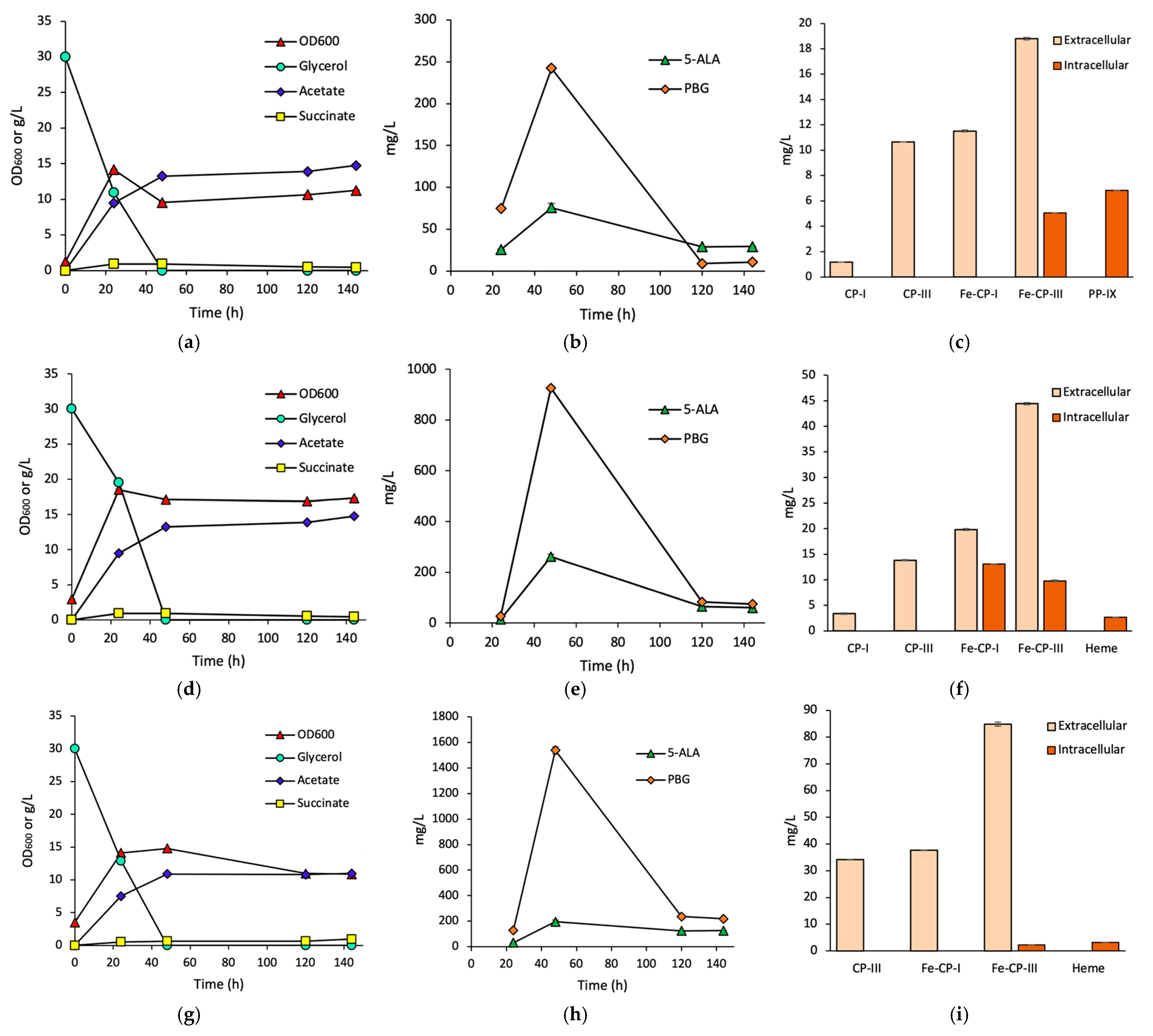
3.1. Effects of HemH on Fe-CP-III Biosynthesis

For effective Fe-CP-III biosynthesis, we derived Fe-CP-III-producing strains by the heterologous coexpression of hemA from Rhodobacter sphaeroides, hemB/D/E from E. coli, and hemH from B. subtilis. Three plasmids were constructed for this purpose, each containing two operons regulated by the strong trc and gracmax promoters, respectively, and each cloned gene had an individual strong RBS. For the plasmid pK-hemABD-EH in strain FECP1, hemA/B/D were cloned into the first operon, whereas hemE/H were placed in the second operon. Plasmid pK-hemAB-EH in FECP2 was similar to pK-hemABD-EH except that hemD was excluded from the construct. In the plasmid pK-hemABE-H in FECP3, the genes from pK-hemAB-EH were rearranged by including hemA/B/E in the first operon and hemH in the second operon. This design was made to enhance the expression of hemH by placing it as the first gene in the second operon, potentially improving Fe-CP-III biosynthesis.
Time profiles for cell growth, glycerol consumption, and by-product (acetate and succinate) formation for each strain are shown in Figure 3a,d,g. The three strains show similar glycerol consumption, with the depletion of glycerol by 48 h. Among the strains, FECP2 exhibited the highest cell growth, reaching a cell density of 18.4 OD600 at 24 h. All strains produced high levels of acetate as a by-product, while succinate accumulation remained minimal.
The time profiles of 5-ALA and PBG, two key intermediates in the Shemin/C4 pathway, are shown in Figure 3b,e,h. Among the three strains, FECP3 produced high levels of PBG, with the peak concentration reaching 1537 mg/L (104 mg/OD600/L) by 48 h. In contrast, 5-ALA production was highest in FECP2, peaking at 261 mg/L (15.2 mg/OD600/L) at 48 h. The high PBG titer in FECP3 indicates a high metabolic activity toward porphyrin biosynthesis.
Porphyrin production at 144 h for each strain is shown in Figure 3c,f,i. Among these strains, FECP3 exhibited the highest extracellular Fe-CP-III concentration of 84.9 mg/L, with intracellular accumulation of only 2.3 mg/L, indicating efficient secretion of Fe-CP-III. In contrast, both FECP1 and FECP2 showed lower extracellular Fe-CP-III titers of 18.8 mg/L and 44.4 mg/L, respectively, with intracellular Fe-CP-III titers of 5.1 mg/L and 9.8 mg/L, respectively. In addition to Fe-CP-III, various byproducts were detected in both intracellular and extracellular fractions, including CP-I, CP-III, Fe-CP-I, PP-IX, and heme. The accumulation of intracellular PP-IX and heme in these strains indicates that a small portion of the dissimilated carbon flux was diverted toward the PPD pathway, even though the flux was metabolically driven toward the CPD pathway for Fe-CP-III production.

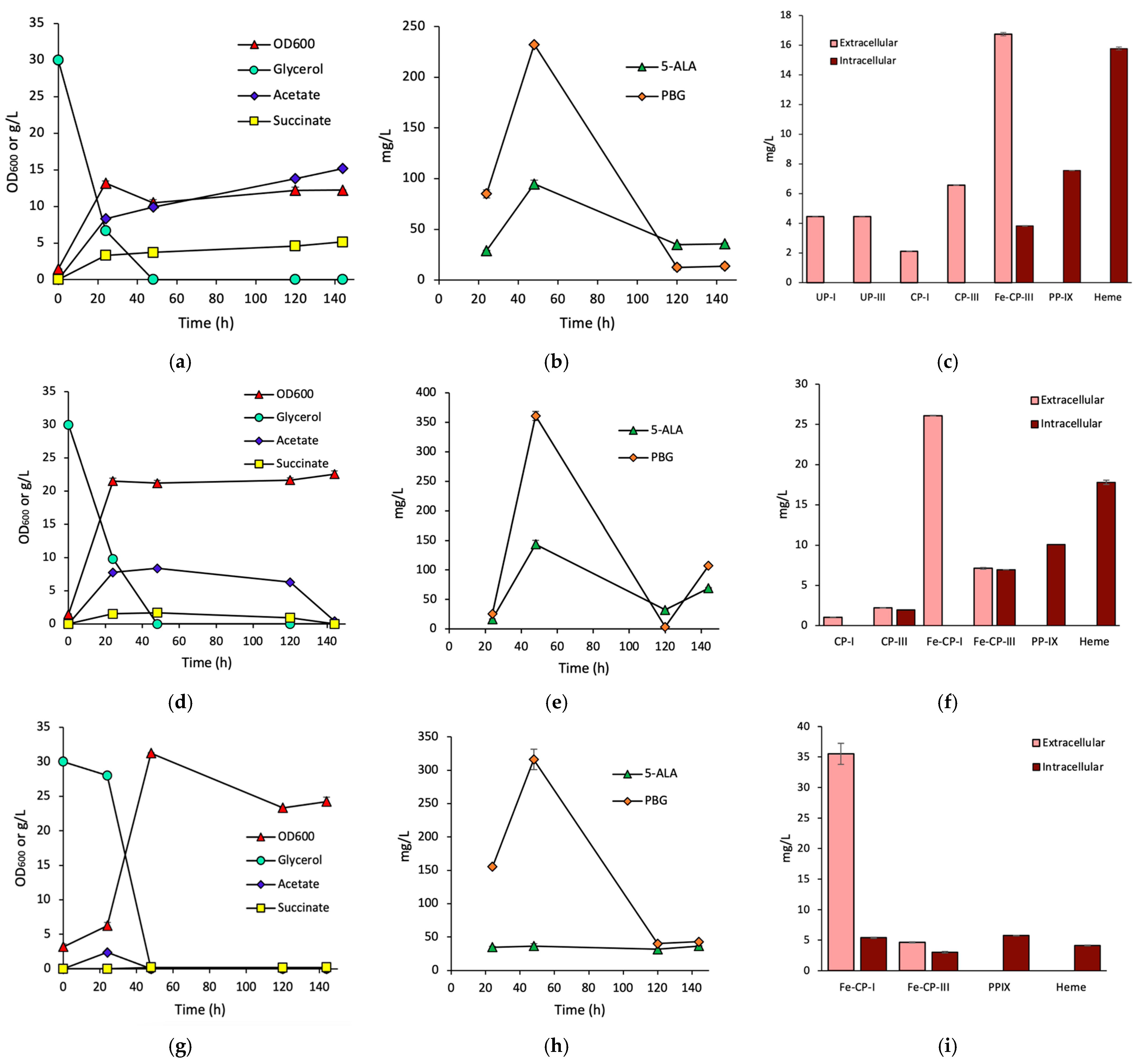
3.2. Effects of HemQ on Heme Biosynthesis

To investigate the effects of hemQ on heme biosynthesis, we derived heme-producing strains by the heterologous coexpression of hemA from R. sphaeroides, hemB/D/E from E. coli, hemH from B. subtilis, and hemQ from either B. subtilis (referred as hemQ) or S. aureus (referred to as hemQS.a). Three plasmids were constructed for this purpose, each containing two operons regulated by the strong trc and gracmax promoters, and each cloned gene had an individual strong RBS. In the first strain HEM1, hemA/B/D, and hemE/H/Q were cloned into the first and second operons, respectively, to form plasmid pK-hemABD-EHQ. In the second strain HEM2, the plasmid pK-hemAB-EHQ was similar to pK-hemABD-EHQ except that hemD was excluded. In the third strain HEM3, the harboring plasmid pK-hemABE-HQS.a was constructed by including hemA/B/E in the first operon and hemH and hemQS.a in the second operon.
The time profiles for cell growth, glycerol consumption, and by-product formation for each strain are shown in Figure 4a,d,g. All strains fully depleted glycerol by 48 h, with HEM3 showing slower consumption in the first 24 h. HEM3 exhibited the highest cell growth, reaching a peak cell density of 31.3 OD600 at 48 h. HEM1 had considerable and monotonically increasing formation of acetate and succinate, while HEM2 showed acetate depletion by the end of cultivation and minimal succinate formation. In contrast, HEM3 had negligible acetate and succinate production.
The time profiles of 5-ALA and PBG are shown in Figure 4b,e,h. All strains showed similar patterns in 5-ALA and PBG formation, with both peaking at 48 h. 5-ALA titers reached 94.7, 143, and 36.6 mg/L (9.0, 6.8, and 1.2 mg/OD600/L), while PBG titers reached 232, 366, and 316 mg/L (22.1, 17.0, and 10.1 mg/OD600/L), in HEM1, HEM2, and HEM3, respectively. Note that HEM2 produced the highest levels of both 5-ALA and PBG.
Heme production at 144 h for each strain is shown in Figure 4c,f,i. Heme was hardly detected in the extracellular fraction but was produced intracellularly at 15.8, 17.8, and 4.0 mg/L in HEM1, HEM2, and HEM3, respectively. In addition to heme, various porphyrins were also detected in both intracellular and extracellular fractions, including UP-I, UP-III, CP-I, CP-III, Fe-CP-III, and PP-IX.

3.3. In Vitro Conversion of CP-III to Fe-CP-III via HemH

To explore the in vitro enzymatic conversion of CP-III to Fe-CP-III, hemH from B. subtilis, fused with a His6 tag at either the C- or N-terminal, was PCR-amplified and cloned into plasmid pTrc99a for expression. The overexpressed HemH was purified using affinity chromatography. As illustrated in Figure 5, the insoluble fraction (lane 2), soluble fraction (lane 3), and purified protein (lane 4) were analyzed using SDS-PAGE. Clear bands corresponding to the expected molecular weight of ~35 kDa were observed, confirming functional expression and purification [42]. The expressed HemH was predominantly located in the soluble fraction, consistent with a previous report [43].
Purified HemH was applied to convert CP-III to Fe-CP-III in the presence of ferrous ions. The color change observed during the reaction from red to orange (Figure 6a) suggested successful conversion [11]. The reaction was enhanced by adjusting the buffer composition and pH. Four buffers based on citrate phosphate, Tris-HCl, sodium phosphate, and potassium phosphate at 100 mM and pH 7 were explored. The buffer based on citrate phosphate had the best performance, with a 35.6% yield (Figure 6b), whereas the other buffers had significantly lower yields. Next, the effects of pH were investigated, and high yields up to 66.3% were achieved by adjusting the pH to a range between 7.5 and 9.5 (Figure 6c). Hence, subsequent in vitro conversions of CP-III to Fe-CP-III were conducted using citrate phosphate buffer at pH 8.
In the initial trials of Experiments A-D (Figure 6d), the ratio between substrates and enzymes was examined. We began with the conversion of 50 µM CP-III, 2x ferrous ions (100 µM) as FeSO4.7H2O, and 2 µM HemH without the addition of buffer (Experiment A). This initial condition resulted in a 15.6% yield. Then, we increased the levels of ferrous ions and HemH in subsequent experiments. Raising the ferrous ion concentration to 200 µM while keeping HemH at 2 µM led to an increased yield of 35.7% (Experiment B). Further increasing the ferrous ion concentration to 400 µM decreased the yield to 11.7% (Experiment C). Similarly, doubling the enzyme concentration to 4 µM while maintaining 200 µM ferrous ions resulted in a 27.4% yield (Experiment D). These results indicate the importance of modulating the relative concentrations of substrates and HemH for in vitro conversion.
Further enhancement was made by the inclusion of citrate phosphate buffer at pH 8 to achieve a 69.2% yield (Experiment E). In Experiment F/G, the CP-III concentration was increased to 63 µM and 85 µM, respectively, while maintaining the substrate ratio (Fe2+/CP-III) constant and the enzyme concentration at 4 µM. With the increased substrate concentration, high Fe-CP-III yields of 72.9% and 77.2% were obtained.
CP-I, the stereoisomer of CP-III, could be another substrate for HemH with a 68.0% yield of Fe-CP-I (Experiment H), suggesting that HemD has a broad substrate specificity within the coproporphyrin family. Cell-free cultivation medium from CP-producing strains could be directly used for HemD conversion (Experiments I–K), with Fe-CP-III yields ranging from 37% to 61.9%. Additionally, all CP-I in the three reactions was converted to Fe-CP-I, with yields ranging from 47.6 to 64.0%. Finally, similar conversion results were obtained for both N-terminal- and C-terminal-His6-tagged HemH enzymes.

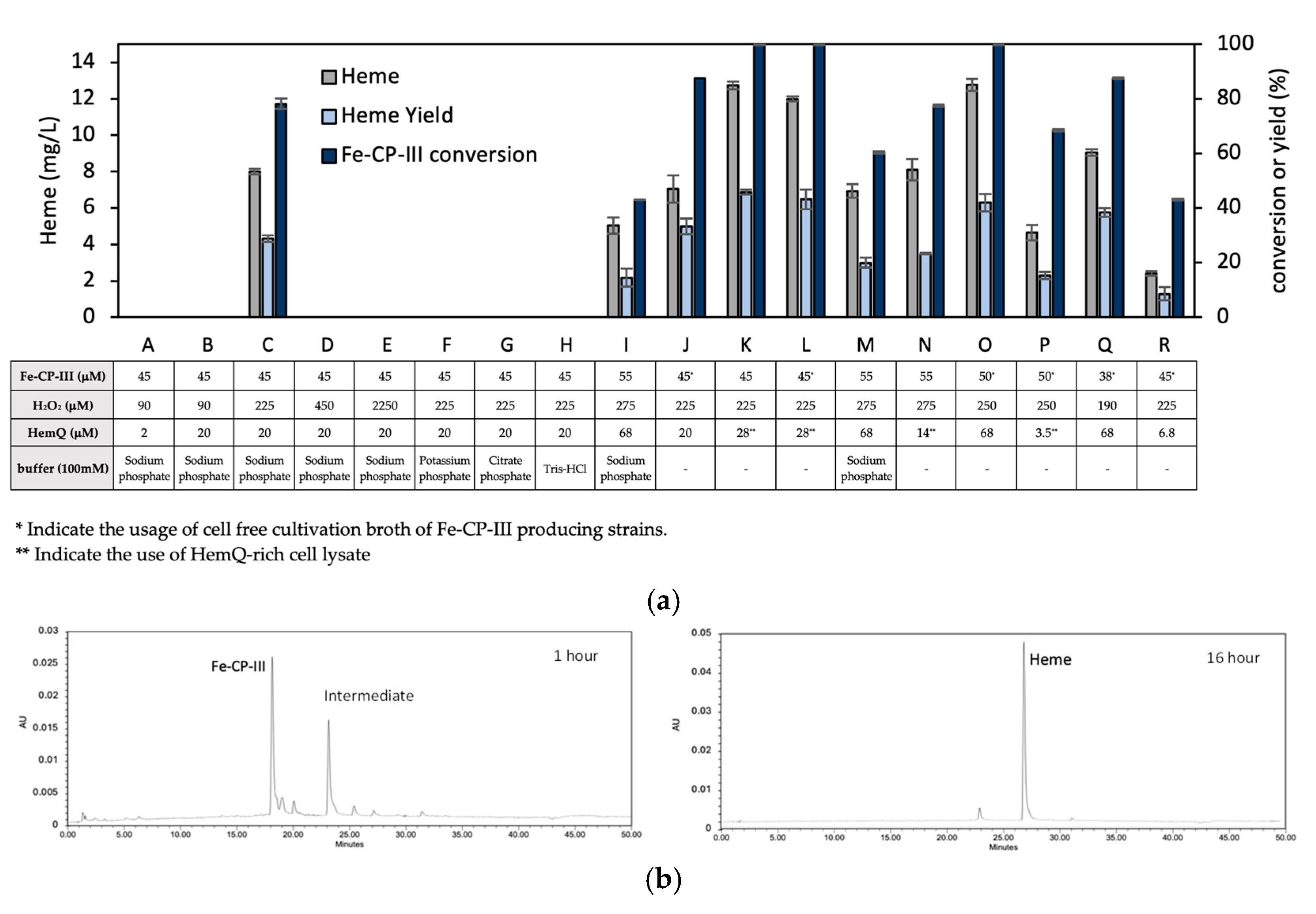
3.4. In Vitro Conversion of Fe-CP-III to Heme via HemQ

For the in vitro enzymatic conversion of Fe-CP-III to heme, hemQ from B. subtilis or S. aureus was amplified, fused with a His6 tag at either the C- or N-terminal, and cloned into pTrc99a for expression and purification. The overexpressed HemQ enzyme was purified using affinity chromatography. As shown in Figure 5, the insoluble fraction (lane 5), soluble fraction (lane 6), purified HemQ (lane 7), and purified HemQs.a (lane 8) were analyzed with SDS-PAGE. All lanes had clear bands corresponding to the expected molecular weight of ~26 kDa, confirming successful expression and purification [38]. The enzyme was predominantly located in the soluble fraction.
Purified HemQ was used in reaction mixtures containing Fe-CP-III and H2O2 under various conditions (Figure 7a). We first investigated the effects of the mole ratio of Fe-CP-III/H2O2 by adjusting it from 1/2 to 1/50 (Experiments B-E). The best performance was observed in Experiment C, with a 1/5 ratio, achieving 78.1% conversion and a heme yield of 28.7%. Increasing the ratio beyond 1/5 led to discoloration of the reaction mixture and impacted the conversion since hydrogen peroxide potentially acted as an oxidizing agent.
Among the different buffers tested, including sodium phosphate, potassium phosphate, citrate phosphate, and Tris-HCl (Experiments C and F–H), sodium phosphate was the only effective one. Further increasing the amount of HemQ did not show positive effects (Experiment I). In addition, cell-free cultivation broth of Fe-CP-III-producing strains was directly used for in vitro conversion, leading to a heme yield of 33.3% (Experiment J). Furthermore, the use of HemQ-overexpressed cell lysate could also lead to a high heme yield, either with pure Fe-CP-III or a cell-free Fe-CP-III-containing medium as the substrate, achieving 45.8% (Experiment K) and 43.1% (Experiment L), respectively. Similar in vitro conversions were conducted using HemQ from S. aureus (Experiments M–R), with different enzyme sources (i.e., purified HemQ or cell lysate) or the substrate type (i.e., pure Fe-CP-III or a cell-free cultivation broth), achieving similar heme yields, with the highest one up to 41.9% (Experiment O).
To track the in vitro conversion of Fe-CP-III to heme, HPLC analysis of a reaction mixture was conducted at 1 h and 16 h post-reaction times (Figure 7b). Partial conversion of Fe-CP-III into an intermediate with no detectable heme formation was observed at 1 h, whereas nearly all Fe-CP-III was converted to heme at 16 h. The results suggest the presence of a porphyrin intermediate for the in vitro conversion, consistent with a previous report [44].

4. Discussion

It is well documented that bio-based production using cell factories can be limited by the intracellular accumulation of toxic metabolites [45,46]. Unlike most porphyrin precursors, heme can hardly be secreted in E. coli, and its intracellular accumulation can seriously impact the physiology of host cells and, therefore, limit its biosynthesis. This study explored an integrated two-step approach for the bio-based production of Fe-CP-III (or heme), i.e., the metabolic engineering of E. coli for the in vivo biosynthesis of CP-III (or Fe-CP-III) followed by in vitro enzymatic conversion to Fe-CP-III (or heme). By decoupling the metabolic pathway into two distinct yet complementary conversion phases, this two-step approach could offer a bioprocess strategy for more effective and scalable biosynthesis of Fe-CP-III (or heme) from cheap feedstock such as glycerol.
Our results show that Fe-CP-III could be secreted in E. coli with minimal intracellular accumulation, suggesting the feasibility of using engineered E. coli for the in vivo production of Fe-CP-III. Comparative analysis of E. coli strains engineered for Fe-CP-III production reveals notable differences in culture performance, including cell growth, Fe-CP-III biosynthesis/secretion, and metabolic flux distribution. Among the strains tested, FECP3 performed best, with the highest extracellular Fe-CP-III concentration of 84.9 mg/L, which resulted from the arrangement of the pathway genes, i.e., positioning hemH at the start of the second operon. This arrangement appeared to enhance hemH expression and effective CP-III conversion to Fe-CP-III, underscoring the importance of operon design for expressing pathway genes for porphyrin biosynthesis. The efficient extracellular production of Fe-CP-III by FECP3 could not only alleviate the intracellular toxicity associated with Fe-CP-III but also facilitate downstream processing for the purification or even subsequent conversion of Fe-CP-III, underscoring the scalability of this bio-based production system for industrial application. The transient accumulation and consumption of intermediates in the Shemin/C4 pathway, including 5-ALA and PBG, potentially correlate with the metabolic activity toward porphyrin production among strains. The minimal accumulation of byproducts, such as PP-IX and heme, indicates that most of the dissimilated carbon flux was directed toward the CPD pathway, with minimal diversion into the PPD pathway. Though the extracellular production of Fe-CP-III in E. coli was successfully demonstrated in this study, the Fe-CP-III titer was relatively low, particularly compared to our previous results in the extracellular production of CP-III [28].
Our results show that heme accumulated intracellularly with minimal secretion when overproduced in light of hemQ expression in E. coli. Among engineered strains, HEM2, coexpressing hemAB-EHQ, exhibited the highest heme production of 17.8 mg/L. HemQ from S. aureus showed moderate heme yields in HEM3 compared to HemQ from B. subtilis. The intracellular accumulation of heme potentially resulted in toxicity to the cell, limiting the culture performance for heme production. The results suggest the potential advantage of in vitro enzymatic (HemQ) conversion of Fe-CP-III to heme while preventing the intracellular accumulation of heme.
The implementation of in vitro enzymatic reactions using purified HemH and HemQ enzymes enabled more precise modulation over reaction conditions while preventing the intracellular toxicity associated with in vivo heme production [32,47]. It is noteworthy that Fe-CP-III (and CP-III) in the cell-free culture medium could be used directly as the substrate for the in vitro conversions. Buffer type and pH appeared to be critical for the in vitro conversions. The use of citrate phosphate buffer at pH 8 was most effective for the HemH conversion of CP-III to Fe-CP-III since an alkaline pH could effectively neutralize the two protons released during the conversion [43]. Such a buffer design not only maintained enzyme stability and activity but also enhanced the reaction progress without disruption by the increasing acidity. On the other hand, the sodium phosphate buffer was most suitable for HemQ conversion of Fe-CP-III to heme. Our results also show that HemH exhibited a broad substrate specificity to convert both CP-I and CP-III to their respective iron-chelated forms with high yields, suggesting its potential application in synthesizing diverse porphyrin derivatives. In addition, proper tuning of the Fe2+/CP-III and Fe-CP-III/H2O2 ratios and enzyme concentrations was critical to balance substrate levels with effective conversion and minimal inhibition. For instance, we observed that excessive Fe2+ concentrations resulted in reduced yields, as well as heme degradation in the presence of excessive hydrogen peroxide, which could act as both a substrate and an oxidizing agent. Note that upon in vivo cultivation, hydrogen peroxide was not externally supplemented as it was naturally produced under aerobic conditions [48], particularly during the conversion of CPG-III to CP-III [43].
In the in vitro reactions, while we could achieve high conversion rates up to 100%, the yield of target porphyrins, particularly those involving HemQ, was notably lower. Such discrepancy might be due to the degradation of heme in the presence of hydrogen peroxide, as previously reported [37,49]. Another contributing factor could be the incomplete release of heme from HemQ due to the substrate affinity of heme toward the active site of HemQ, which not only hinders heme availability but also contributes to the degradation of protein-bound heme [37,44]. Such a phenomenon was also reported for other enzymatic reactions [50]. Future studies could focus on strategies to mitigate heme degradation, such as refining the enzyme kinetics or exploring alternative oxidants, as reported in previous studies [11,44,50]. In addition, recent studies [51] highlight advances in heme overproduction that complement and contextualize this study.

5. Conclusions

In summary, this work offers an alternative approach for the bio-based production of chemicals that are toxic to host cells. Genetically and metabolically engineered strains were first cultivated in vivo for the extracellular production of the precursor, which was enzymatically converted in vitro to form the product. The direct use of a cell-free cultivation broth as a substrate without pretreatment or purification for HemH and HemQ conversion could significantly reduce production costs, suggesting the robustness, adaptability, and scalability of the approach for synthesizing high-value compounds. Our two-step approach enhances scalability and cost-effectiveness by decoupling microbial biosynthesis from enzymatic conversion, thereby reducing intracellular toxicity and simplifying downstream processing. Despite the promising results, some limitations were observed. For instance, while the cell-free conversion of Fe-CP-III to heme was efficient, the overall yield was dependent on the substrate concentration and enzyme loading. Another limitation was the incomplete heme release and degradation during the HemQ-mediated step. Enhancing enzyme stability through protein engineering and enzyme immobilization could potentially improve the yield and reduce enzyme consumption.

Supplementary Materials

The following supporting information can be downloaded at: https://www.mdpi.com/article/10.3390/fermentation11040198/s1, Table S1: Oligomers used in this study; Table S2: Statistical analysis data for buffer selection in Fe-CP-III production; Table S3: Statistical analysis data for PH selection in Fe-CP-III production; Table S4: Statistical analysis data for Fe-CP-III production under various experimental conditions; Table S5: Statistical analysis data for Heme production under various experimental conditions.

Author Contributions

Conceptualization, B.A. and C.-H.P.C.; methodology, B.A.; validation, Y.L. and C.-H.P.C.; formal analysis, B.A.; investigation, B.A.; data curation, B.A.; writing—original draft, B.A.; writing—review and editing, B.A., Y.L. and C.-H.P.C.; supervision, C.-H.P.C.; funding acquisition, M.M.-Y. and C.-H.P.C. All authors have read and agreed to the published version of the manuscript.

Funding

This research was funded by the Natural Sciences and Engineering Research Council (NSERC) grant number RGPIN-2019-04611.

Institutional Review Board Statement

Not applicable.

Informed Consent Statement

Not applicable.

Data Availability Statement

The original contributions presented in the study are included in the article/Supplementary Materials, further inquiries can be directed to the corresponding author.

Conflicts of Interest

The authors declare that they have no known competing financial interests or personal relationships that could have appeared to influence the work reported in this paper. However, portions of the reported data were also included in a US patent application.

References

  1. Ponka, P. Cell biology of heme. Am. J. Med. Sci. 1999, 318, 241–256. [Google Scholar] [PubMed]
  2. Ajioka, R.S.; Phillips, J.D.; Kushner, J.P. Biosynthesis of heme in mammals. Biochim. Biophys. Acta (BBA)-Mol. Cell Res. 2006, 1763, 723–736. [Google Scholar]
  3. Poulos, T.L. Heme enzyme structure and function. Chem. Rev. 2014, 114, 3919–3962. [Google Scholar] [PubMed]
  4. Xing, Y.; Gao, S.; Zhang, X.; Zang, J. Dietary heme-containing proteins: Structures, applications, and challenges. Foods 2022, 11, 3594. [Google Scholar] [CrossRef]
  5. Su, H.; Chen, X.; Chen, S.; Guo, M.; Liu, H. Applications of the whole-cell system in the efficient biosynthesis of heme. Int. J. Mol. Sci. 2023, 24, 8384. [Google Scholar] [CrossRef]
  6. Abdelazim, I.A.; Abu-Faza, M.; Shikanova, S.; Zhurabekova, G.; Maghrabi, M.M. Heme-bound iron in treatment of pregnancy-associated iron deficiency anemia. J. Fam. Med. Prim. Care 2018, 7, 1434–1438. [Google Scholar]
  7. Adam, S.M.; Wijeratne, G.B.; Rogler, P.J.; Diaz, D.E.; Quist, D.A.; Liu, J.J.; Karlin, K.D. Synthetic Fe/Cu complexes: Toward understanding heme-copper oxidase structure and function. Chem. Rev. 2018, 118, 10840–11022. [Google Scholar]
  8. Hofbauer, S.; Schaffner, I.; Furtmüller, P.G.; Obinger, C. Chlorite dismutases–a heme enzyme family for use in bioremediation and generation of molecular oxygen. Biotechnol. J. 2014, 9, 461–473. [Google Scholar] [CrossRef]
  9. Yang, Q.; Zhao, J.; Zheng, Y.; Chen, T.; Wang, Z. Microbial synthesis of heme b: Biosynthetic pathways, current strategies, detection, and future prospects. Molecules 2023, 28, 3633. [Google Scholar] [CrossRef]
  10. Yang, S.; Guo, Z.; Sun, J.; Wei, J.; Ma, Q.; Gao, X. Recent advances in microbial synthesis of free heme. Appl. Microbiol. Biotechnol. 2024, 108, 68. [Google Scholar] [CrossRef]
  11. Chen, H.; Wang, Y.; Wang, W.; Cao, T.; Zhang, L.; Wang, Z.; Chi, X.; Shi, T.; Wang, H.; He, X. High-yield porphyrin production through metabolic engineering and biocatalysis. Nat. Biotechnol. 2024, 1–11. [Google Scholar] [CrossRef] [PubMed]
  12. Layer, G.; Jahn, M.; Moser, J.; Jahn, D. Radical SAM enzymes involved in tetrapyrrole biosynthesis and insertion. ACS Bio. Med. Chem. Au. 2022, 2, 196–204. [Google Scholar] [CrossRef] [PubMed]
  13. Müller, K.; Mingers, T.; Haskamp, V.; Jahn, D.; Jahn, M. Biosynthesis and insertion of heme. In Aerobic Utilization of Hydrocarbons, Oils, and Lipids; Springer: Cham, Switzerland, 2019; pp. 1–28. [Google Scholar]
  14. Bali, S.; Lawrence, A.D.; Lobo, S.A.; Saraiva, L.M.; Golding, B.T.; Palmer, D.J.; Howard, M.J.; Ferguson, S.J.; Warren, M.J. Molecular hijacking of siroheme for the synthesis of heme and d 1 heme. Proc. Natl. Acad. Sci. USA 2011, 108, 18260–18265. [Google Scholar] [CrossRef] [PubMed]
  15. Jackson, L.K.; Dailey, T.A.; Anderle, B.; Warren, M.J.; Bergonia, H.A.; Dailey, H.A.; Phillips, J.D. Exploiting differences in heme biosynthesis between bacterial species to screen for novel antimicrobials. Biomolecules 2023, 13, 1485. [Google Scholar] [CrossRef]
  16. Celis, A.I.; DuBois, J.L. Making and breaking heme. Curr. Opin. Struct. Biol. 2019, 59, 19–28. [Google Scholar] [CrossRef]
  17. Ge, J.; Wang, X.; Bai, Y.; Wang, Y.; Wang, Y.; Tu, T.; Qin, X.; Su, X.; Luo, H.; Yao, B. Engineering Escherichia coli for efficient assembly of heme proteins. Microb. Cell Factories 2023, 22, 59. [Google Scholar] [CrossRef]
  18. Aftab, H.; Donegan, R.K. Regulation of heme biosynthesis via the coproporphyrin dependent pathway in bacteria. Front. Microbiol. 2024, 15, 1345389. [Google Scholar] [CrossRef]
  19. Beas, J.Z.; Videira, M.A.; Saraiva, L.M. Regulation of bacterial haem biosynthesis. Coord. Chem. Rev. 2022, 452, 214286. [Google Scholar] [CrossRef]
  20. Zhang, J.; Kang, Z.; Chen, J.; Du, G. Optimization of the heme biosynthesis pathway for the production of 5-aminolevulinic acid in Escherichia coli. Sci. Rep. 2015, 5, 8584. [Google Scholar]
  21. Jiang, M.; Hong, K.; Mao, Y.; Ma, H.; Chen, T.; Wang, Z. Natural 5-aminolevulinic acid: Sources, biosynthesis, detection and applications. Front. Bioeng. Biotechnol. 2022, 10, 841443. [Google Scholar] [CrossRef]
  22. Choby, J.E.; Skaar, E.P. Heme synthesis and acquisition in bacterial pathogens. J. Mol. Biol. 2016, 428, 3408–3428. [Google Scholar] [PubMed]
  23. Anzaldi, L.L.; Skaar, E.P. Overcoming the heme paradox: Heme toxicity and tolerance in bacterial pathogens. Infect. Immun. 2010, 78, 4977–4989. [Google Scholar] [CrossRef] [PubMed]
  24. Miscevic, D.; Mao, J.Y.; Kefale, T.; Abedi, D.; Moo-Young, M.; Perry Chou, C. Strain engineering for high-level 5-aminolevulinic acid production in Escherichia coli. Biotechnol. Bioeng. 2021, 118, 30–42. [Google Scholar] [CrossRef] [PubMed]
  25. Lall, D.; Miscevic, D.; Bruder, M.; Westbrook, A.; Aucoin, M.; Moo-Young, M.; Perry Chou, C. Strain engineering and bioprocessing strategies for biobased production of porphobilinogen in Escherichia coli. Bioresour. Bioprocess. 2021, 8, 122. [Google Scholar]
  26. Arab, B.; Westbrook, A.W.; Moo-Young, M.; Liu, Y.; Chou, C.P. Bio-Based Production of Uroporphyrin in Escherichia coli. Synth. Biol. Eng. 2024, 2, 10002. [Google Scholar]
  27. Arab, B.; Moo-Young, M.; Liu, Y.; Chou, C.P. Manipulating Intracellular Oxidative Conditions to Enhance Porphyrin Production in Escherichia coli. Bioengineering 2025, 12, 83. [Google Scholar] [CrossRef]
  28. Arab, B.; Westbrook, A.; Moo-Young, M.; Liu, Y.; Chou, C.P. High-Level Bio-Based Production of Coproporphyrin in Escherichia coli. Fermentation 2024, 10, 250. [Google Scholar] [CrossRef]
  29. Tinafar, A.; Jaenes, K.; Pardee, K. Synthetic biology goes cell-free. BMC Biol. 2019, 17, 64. [Google Scholar]
  30. Hodgman, C.E.; Jewett, M.C. Cell-free synthetic biology: Thinking outside the cell. Metab. Eng. 2012, 14, 261–269. [Google Scholar] [CrossRef]
  31. Guterl, J.K.; Sieber, V. Biosynthesis “debugged”: Novel bioproduction strategies. Eng. Life Sci. 2013, 13, 4–18. [Google Scholar]
  32. Dudley, Q.M.; Karim, A.S.; Jewett, M.C. Cell-free metabolic engineering: Biomanufacturing beyond the cell. Biotechnol. J. 2015, 10, 69–82. [Google Scholar] [CrossRef] [PubMed]
  33. Miscevic, D.; Mao, J.Y.; Moo-Young, M.; Chou, C.H.P. High-level heterologous production of propionate in engineered Escherichia coli. Biotechnol. Bioeng. 2020, 117, 1304–1315. [Google Scholar] [CrossRef] [PubMed]
  34. Srirangan, K.; Liu, X.; Westbrook, A.; Akawi, L.; Pyne, M.E.; Moo-Young, M.; Chou, C.P. Biochemical, genetic, and metabolic engineering strategies to enhance coproduction of 1-propanol and ethanol in engineered Escherichia coli. Appl. Microbiol. Biotechnol. 2014, 98, 9499–9515. [Google Scholar] [CrossRef] [PubMed]
  35. Gibson, D.G.; Young, L.; Chuang, R.-Y.; Venter, J.C.; Hutchison, C.A.; Smith, H.O. Enzymatic assembly of DNA molecules up to several hundred kilobases. Nat. Methods 2009, 6, 343–345. [Google Scholar] [CrossRef]
  36. Neidhardt, F.C.; Bloch, P.L.; Smith, D.F. Culture medium for Enterobacteria. J. Bacteriol. 1974, 119, 736–747. [Google Scholar]
  37. Hofbauer, S.; Mlynek, G.; Milazzo, L.; Pühringer, D.; Maresch, D.; Schaffner, I.; Furtmüller, P.G.; Smulevich, G.; Djinović-Carugo, K.; Obinger, C. Hydrogen peroxide-mediated conversion of coproheme to heme b by HemQ—Lessons from the first crystal structure and kinetic studies. FEBS J. 2016, 283, 4386–4401. [Google Scholar]
  38. Dailey, T.A.; Boynton, T.O.; Albetel, A.-N.; Gerdes, S.; Johnson, M.K.; Dailey, H.A. Discovery and characterization of HemQ: An essential heme biosynthetic pathway component. J. Biol. Chem. 2010, 285, 25978–25986. [Google Scholar]
  39. Fyrestam, J.; Östman, C. Determination of heme in microorganisms using HPLC-MS/MS and cobalt (III) protoporphyrin IX inhibition of heme acquisition in Escherichia coli. Anal. Bioanal. Chem. 2017, 409, 6999–7010. [Google Scholar]
  40. Mauzerall, D.; Granick, S. The Occurence and Determination og δ-Aminolevulinic Acid and Porphobilinogen in Urine. J. Biol. Chem. 1956, 219, 435–446. [Google Scholar] [CrossRef]
  41. Kwon, S.J.; De Boer, A.L.; Petri, R.; Schmidt-Dannert, C. High-Level Production of Porphyrins in Metabolically Engineered Escherichia coli: Systematic Extension of a Pathway Assembled from Overexpressed Genes Involved in Heme Biosynthesis. Appl. Environ. Microbiol. 2003, 69, 4875–4883. [Google Scholar] [CrossRef]
  42. Hansson, M.; Hederstedt, L. Purification and characterisation of a water-soluble ferrochelatase from Bacillus subtilis. Eur. J. Biochem. 1994, 220, 201–208. [Google Scholar] [PubMed]
  43. Dailey, H.A.; Dailey, T.A.; Gerdes, S.; Jahn, D.; Jahn, M.; O’Brian, M.R.; Warren, M.J. Prokaryotic heme biosynthesis: Multiple pathways to a common essential product. Microbiol. Mol. Biol. Rev. 2017, 81, e00048-16. [Google Scholar] [CrossRef] [PubMed]
  44. Dailey, H.A.; Gerdes, S. HemQ: An iron-coproporphyrin oxidative decarboxylase for protoheme synthesis in Firmicutes and Actinobacteria. Arch. Biochem. Biophys. 2015, 574, 27–35. [Google Scholar]
  45. Santos, C.N.S.; Stephanopoulos, G. Combinatorial engineering of microbes for optimizing cellular phenotype. Curr. Opin. Chem. Biol. 2008, 12, 168–176. [Google Scholar]
  46. Ko, Y.J.; Lee, M.-E.; Cho, B.-H.; Kim, M.; Hyeon, J.E.; Han, J.H.; Han, S.O. Bioproduction of porphyrins, phycobilins, and their proteins using microbial cell factories: Engineering, metabolic regulations, challenges, and perspectives. Crit. Rev. Biotechnol. 2024, 44, 373–387. [Google Scholar] [CrossRef]
  47. Khan, A.A.; Quigley, J.G. Control of intracellular heme levels: Heme transporters and heme oxygenases. Biochim. Biophys. Acta (BBA)-Mol. Cell Res. 2011, 1813, 668–682. [Google Scholar]
  48. Sirithanakorn, C.; Imlay, J.A. Evidence for endogenous hydrogen peroxide production by E. coli fatty acyl-CoA dehydrogenase. PLoS ONE 2024, 19, e0309988. [Google Scholar]
  49. Dailey, H.A.; Gerdes, S.; Dailey, T.A.; Burch, J.S.; Phillips, J.D. Noncanonical coproporphyrin-dependent bacterial heme biosynthesis pathway that does not use protoporphyrin. Proc. Natl. Acad. Sci. USA 2015, 112, 2210–2215. [Google Scholar] [CrossRef]
  50. Hao, G.-F.; Tan, Y.; Yang, S.-G.; Wang, Z.-F.; Zhan, C.-G.; Xi, Z.; Yang, G.-F. Computational and experimental insights into the mechanism of substrate recognition and feedback inhibition of protoporphyrinogen oxidase. PLoS ONE 2013, 8, e69198. [Google Scholar] [CrossRef]
  51. Yu, F.; Zhou, Y.J. Sustainable production of porphyrins through synthetic biology. Trends Biotechnol. 2024. online. [Google Scholar] [CrossRef]
Figure 1. The Shemin/C4 pathway for porphyrin biosynthesis from succinyl-CoA and glycine. 5-ALA, 5-aminolevulinic acid; AhbA, siroheme decarboxylase α subunit; AhbB, siroheme decarboxylase β subunit; AhbC, Fe–coproporphyrin III synthase; AhbD, SAM-dependent heme synthase; CPG-I, coproporphyrinogen I; CPG-III, coproporphyrinogen III; CP-I, coproporphyrin I; CP-III, coproporphyrin III; Fe-CP-I, Fe–coproporphyrin I; Fe-CP-III, coproheme; HemA, 5-aminolevulinate synthase; HemB, porphobilinogen synthase; HemC, porphobilinogen deaminase; HemD, uroporphyrinogen III synthase; HemE, uroporphyrinogen decarboxylase; HemF, coproporphyrinogen III oxidase; HemG, protoporphyrinogen oxidase; HemH, protoporphyrin/coproporphyrin ferrochelatase; HemN, oxygen-independent coproporphyrinogen III oxidase; HemQ, coproheme decarboxylase; HemY, coproporphyrinogen III oxidase; HMB, hydroxymethylbilane; PBG, porphobilinogen; PP-IX, protoporphyrin IX; PPG-IX, protoporphyrinogen IX; UP-I, uroporphyrin I; UP-III, uroporphyrin III; UPG-I, uroporphyrinogen I; UPG-III, uroporphyrinogen III; YfeX, porphyrinogen peroxidase. BioRender.com.
Fermentation 11 00198 g001
Figure 2. Two-step heme production using in vivo porphyrin production and in vitro cell-free enzymatic conversion. Figure was created using BioRender.com.
Fermentation 11 00198 g002
Figure 3. Aerobic bioreactor cultivation of FECP1, FECP2, and FECP3 for porphyrin biosynthesis. Time profiles of (a,d,g) cell density (OD600), glycerol consumption, and acetate/succinate formation; (b,e,h) 5-ALA and PBG biosynthesis; and (c,f,i) porphyrin biosynthesis. All values are reported as means ± SD (n = 2).
Fermentation 11 00198 g003
Figure 4. Aerobic bioreactor cultivation of HEM1, HEM2, and HEM3 for porphyrin biosynthesis. Time profiles of (a,d,g) cell density (OD600), glycerol consumption, and acetate/succinate formation; (b,e,h) 5-ALA and PBG biosynthesis; and (c,f,i) porphyrin biosynthesis. All values are reported as means ± SD (n = 2).
Fermentation 11 00198 g004
Figure 5. SDS-PAGE analysis of HemH and HemQ expression and purification. Lane 1: protein ladder; Lane 2: HemH insoluble fraction; Lane 3: HemH soluble fraction; Lane 4: purified HemH from B. subtilis; Lane 5: HemQ insoluble fraction; Lane 6: HemQ soluble fraction; Lane 7: purified HemQ from B. subtilis; Lane 8: purified HemQ from S. aureus.
Fermentation 11 00198 g005
Figure 6. In vitro Fe-CP-III synthesis from CP-III. (a) Visible color change from red to orange, indicating the conversion of CP-III to Fe-CP-III. Reaction before adding enzyme (left) and after addition of enzyme (right). (b) Evaluation of buffer conditions for Fe-CP-III production. (c) Evaluation of pH conditions for Fe-CP-III production. (d) Tuning Fe2+ and HemH enzyme concentration and buffer types for Fe-CP-III production. All values are reported as means ± SD (n = 2).
Fermentation 11 00198 g006
Figure 7. In vitro heme synthesis from Fe-CP-III. (a) Tuning H2O2 and HemQ concentration and buffer types for heme production. All values are reported as means ± SD (n = 2). (b) HPLC analysis of Fe-CP-III conversion to heme at different time points: 1 h (left) and overnight (right).
Fermentation 11 00198 g007
Table 1. Strains and plasmids used in this study.
NameDescription or Relevant GenotypeSource
Host strains
HI-Control 10GmcrA, ∆(mrr-hsdRMS-mcrBC), endA1, recA1, ϕ80dlacZ∆M15, ∆lacX74, araD139, ∆(ara leu)7697, galU, galK, rpsL (StrR), nupG, λ, tonA, and Mini-F lacIq1 (GentR)Lucigen
MG1655K-12; F, λ, and rph-1Lab stock
Bacillus subtilis 168Wild typeLab stock
Staphylococcus aureusWild typeLab stock
CPC-Sbm∆iclRsdhAF, Δ(araD-araB)567, ΔlacZ4787(::rrnB-3), λ, rph-1, Δ(rhaD-rhaB)568, hsdR514, ∆ldhA, Ptrc::sbm (i.e., with the FRT-Ptrc cassette replacing the 204 bp upstream of the Sbm operon), ∆iclR, and ∆sdhA[33]
FECP1CPC-Sbm∆iclRsdhA/pK-hemABD-EHThis study
FECP2CPC-Sbm∆iclRsdhA/pK-hemAB-EHThis study
FECP3CPC-Sbm∆iclRsdhA/pK-hemABE-HThis study
HEM1CPC-Sbm∆iclRsdhA/pK-hemABD-EHQThis study
HEM2CPC-Sbm∆iclRsdhA/pK-hemAB-EHQThis study
HEM3CPC-Sbm∆iclRsdhA/pK-hemABE-HQS.aThis study
Plasmids
pK-hemABCD-Ep15A ori, KmR, Ptrc::hemABCD-Pgracmax::hemE[28]
pK-hemABD-EHp15A ori, KmR, Ptrc::hemABD-Pgracmax::hemEH -hemH from B. subtilis 168-This study
pK-hemAB-EHp15A ori, KmR, Ptrc::hemAB-Pgracmax::hemEH -hemH from B. subtilis 168-This study
pK-hemABE-Hp15A ori, KmR, Ptrc::hemABE-Pgracmax::hemH -hemH from B. subtilis 168-This study
pK-hemABD-EHQp15A ori, KmR, Ptrc::hemABD-Pgracmax::hemEHQ -hemH and hemQ from B. subtilis 168-This study
pK-hemAB-EHQp15A ori, KmR, Ptrc::hemAB-Pgracmax::hemEHQ -hemH and hemQ from B. subtilis 168-This study
pK-hemABE-HQS.ap15A ori, KmR, Ptrc::hemABE-Pgracmax::hemHQ -hemH from B. subtilis 168 and hemQ from S. aureus-This study
pK-hemH-His6pBR322 ori, AmpR, Ptrc::hemH:His6 -hemH from B. subtilis 168-This study
pK-His6-hemHpBR322 ori, AmpR, Ptrc::His6::hemH -hemH from B. subtilis 168-This study
pK-hemQ-His6pBR322 ori, AmpR, Ptrc::hemQ:His6 -hemQ from B. subtilis 168-This study
pK-His6-hemQpBR322 ori, AmpR, Ptrc::His6::hemQ -hemQ from B. subtilis 168-This study
pK-hemQS.a-His6pBR322 ori, AmpR, Ptrc::hemQ:His6 -hemQ from S. aureus-This study
pK-His6-hemQS.apBR322 ori, AmpR, Ptrc::His6::hemQ -hemQ from S. aureus-This study
Disclaimer/Publisher’s Note: The statements, opinions and data contained in all publications are solely those of the individual author(s) and contributor(s) and not of MDPI and/or the editor(s). MDPI and/or the editor(s) disclaim responsibility for any injury to people or property resulting from any ideas, methods, instructions or products referred to in the content.

