Article

High-Level Extracellular Expression of Collagenase ColH in Bacillus subtilis for Adipose-Derived Cells Extraction

1
Key Laboratory of Carbohydrate Chemistry and Biotechnology, Ministry of Education, School of Life Sciences and Health Engineering, Jiangnan University, Wuxi 214122, China
2
Wuxi Children’s Hospital, Wuxi 214023, China
3
Cytori Therapeutics LLC, Shanghai 201802, China
4
College of Biomass Science and Engineering, Sichuan University, Chengdu 610065, China
*
Authors to whom correspondence should be addressed.
Fermentation 2025, 11(5), 242; https://doi.org/10.3390/fermentation11050242
Submission received: 25 February 2025 / Revised: 7 April 2025 / Accepted: 8 April 2025 / Published: 24 April 2025
(This article belongs to the Special Issue Applied Microorganisms and Industrial/Food Enzymes, 2nd Edition)

Abstract

:
Collagenase has a wide range of applications in the medicine, cosmetic, and food industries. Inefficient expression of collagenase impedes its industrial production and commercial applications. In this study, a secretory expression system for collagenase ColH from Clostridium histolyticum was constructed in Bacillus subtilis. Signal peptide optimization effectively solved the secretion problem of large collagenase with a molecular weight of about 116 kDa, doubling the extracellular enzyme activity. Then, promoter optimization further improved the enzyme activity to 264 U/mL. By the co-optimization of the nitrogen sources and carbon sources, and employing a fed-batch fermentation strategy, the enzyme activity could reach 669 U/mL, which is, currently, the highest level reported in the industry. The recombinant collagenase ColH was purified through a purification process suitable for industrial production with a specific activity of 565.25 U/mg. Based on the purified collagenase, cells were successfully prepared from adipose tissue, indicating its potential use in cell therapy. This study provides a promising candidate for the industrial production of collagenase and highlights its potential application to extract cells from tissues.

Graphical Abstract

1. Introduction

Collagenase can specifically hydrolyze the three-dimensional helical structure of collagen without attacking other proteins and tissues, thus attracting widespread attention in the medical field [1,2,3]. It has been applied in the debridement of wounds and burns, genetic or electro-genetic therapy for cancer, the treatment of lumbar disc herniation, and even chronic complete occlusion. Moreover, collagenase shows great potential for selectively breaking down collagen in the human body, facilitating the extraction of cells from various tissues. For instance, Narwani et al. [4] successfully isolated oral mucosal epithelial cells from rabbit buccal tissue biopsies. Similarly, Jin et al. [5] discovered that treating cultured cells with different collagenases allowed for the quicker isolation of Schwann cells compared with fibroblasts, simplifying the separation process. Additionally, collagenase can be used to extract adipose-derived cells from adipocytes, which has significant implications in areas like clinical medicine [6]. The advancement of life sciences and cell immunotherapy technologies has led to a growing interest in autologous transplantation therapies, particularly those involving collagenase, within the industry.
Collagenases are classified into animal collagenases and bacterial collagenases due to their different sources. Collagenase of animal origin is often extracted and purified from fish guts and other animals. However, from the perspectives of cost and product quality, this is not ideal. The use of bacterial collagenase overcomes this problem, as microbial sources of collagenase are cheaper to produce. The M9B subfamily collagenase from C. histolyticum and the M9A subfamily collagenase from Vibrio vulnificus have received the most attention [7,8]. Particularly noteworthy are the type I collagenase ColG and type II collagenase ColH from the M9B family, which are capable of preparing cells derived from various tissues and organs [9]. The bacterial collagenase belonging to the M9B subfamily possesses the capability to cleave collagen at multiple sites, resulting in the production of short peptides and amino acids. Nevertheless, the use of collagenase obtained from pathogenic bacteria might raise some concerns because of the potential risk of pathogenicity, which makes the heterologous expression of collagenase necessary [10].
The presence of multiple types of collagenases, coupled with the diverse preferences of individual researchers, contributes to the complexity of this field. It was not until 1998 that ColH was first recombinantly expressed and purified by Jung et al. [11,12]. The researchers determined that ColH exhibits low expression levels in Escherichia coli and is readily degraded into smaller peptide fragments in Clostridium perfringens. In 2008, Eiji et al. [13] successfully expressed ColH in C perfringens, resulting in a concentration of 11 mg/L in the culture medium and an enzyme activity level of 35.7 U. In 2022, Zhao et al. [14] successfully facilitated the secretion expression of ColH in E. coli utilizing signaling peptides, resulting in an enzyme activity measurement of 0.68 U/L. It is evident that this level of production is insufficient to meet the substantial demand for ColH.
In this study, the collagenase ColH from the M9B subfamily of C. histolyticum was heterologously expressed in Bacillus subtilis. High-level enzyme expression was achieved by screening suitable signal peptides and promoters. The culture conditions were optimized at the shaking flask scale, followed by scaling up to the fermenter to investigate the industrial application potentials. The recombinant collagenase was purified through a two-step method, namely, ammonium sulfate precipitation-ion exchange chromatography. The purified collagenase was finally used in the preparation of cells from adipose tissues, indicating its potential usage in cell therapy.

2. Materials and Methods

2.1. Bacterial Strains, Plasmids, and Culture Conditions

The bacterial strains and plasmids used in this study are listed in Table 1, respectively. All E. coil strains were cultured at 37 °C in LB medium (5 g/L peptone, 10 g/L yeast extract, and 5 g/L sodium chloride). The B. subtilis strains were cultured at 37 °C in TB medium (11.8 g/L peptone, 23.6 g/L yeast extract, 9.4 g/L K2HPO4, and 2.2 g/L KH2PO4). The E. coli JM109 strain was used as a cloning host for routine DNA manipulations, such as transformation and plasmid extraction. Antibiotics were added to the media at a final concentration of 1‰ as selection markers for hosts containing the desired plasmid, specifically, kanamycin (50 μg/mL) and ampicillin (100 μg/mL).

2.2. Construction and Transformation of Plasmids for Collagenase Expression

The collagenase gene ColH was identified from the M9B family of C. histolyticum through a search of the NCBI database. The gene sequence was synthesized by GeneArt, and degenerate primers were designed. Homologous recombination was used to insert the gene into the target plasmid, which was then transformed into competent E. coli JM109 cells for amplification. The cells were cultured in LB medium at 37 °C with shaking at 220 rpm for 12 h. The amplified plasmids were extracted and transformed into the host strain for expression.

2.3. Gelatin Plate Screening

To assess collagenase production, the E. coli and B. subtilis strains expressing collagenase were spotted onto gelatin plates and incubated at 37 °C for 48 h. After incubation, a 10% trichloroacetic acid (TCA) solution was applied to the surface of the plates. The gelatinase activity of the ColH gene was qualitatively evaluated by observing the formation of clear zones on the gelatin plates, indicating enzymatic degradation of gelatin.

2.4. Quantification of Collagenase Protein and Enzyme Activity Assay

The protein concentration was quantified using the BCA Protein Assay Kit (Bioswamp, Wuhan, China), following the manufacturer’s protocol [15]. The collagenase activity was determined using a colorimetric assay with type I collagen as the substrate (1 mg/mL). First, 1 mL of collagen solution was mixed with 1 mL of Tris-HCl buffer (0.1 M (pH 7.5), containing 50 mM CaCl2). Immediately, 1 mL of the enzyme solution was added to initiate the reaction. The mixture was incubated at 37 °C for 5 h. After incubation, the reaction was terminated by adding an equal volume of 10% trichloroacetic acid (TCA). Once the reaction was complete, 500 µL of the reaction mixture was taken and mixed with 500 µL of acetic acid buffer (pH 5.4) and 500 µL of dansyl chloride reagent. The mixture was heated at 80 °C in a water bath for 20 min to allow the dansyl chloride reaction to proceed. After heating, the mixture was cooled completely in an ice bath, and then 1500 µL of 60% ethanol was added. Finally, 200 µL of the sample was transferred to a microplate, and the absorbance was measured at 570 nm to quantify collagenase activity. One unit of enzyme activity (U) was defined as the amount of enzyme required to hydrolyze collagen, producing an amount equivalent to 1 µmol of glycine per milliliter of the enzyme solution in 5 h at 37 °C and pH 7.5.

2.5. SDS-PAGE

An amount of 40 µL of the fermentation supernatant sample was transferred, 10 µL of 5× sample buffer was added, and they were mixed thoroughly by pipetting. The mixture was incubated in boiling water for 10 min. Then, 30 µL of the resulting solution was taken and separated using an 8% separating gel in SDS-PAGE. After electrophoresis, the protein gel was immersed in a staining solution (0.25% Coomassie Brilliant Blue R-250) for 30 min. After staining, the gel was rinsed with water and incubated in a decolorization solution until the protein bands were clearly visible.

2.6. Signal Peptide Engineering for Enzyme Expression

The plasmid PMA5-H-SPx (where SPx represents any one of the 11 signal peptides) was transformed into the B. subtilis WB600 host strain (Table 1). The resulting transformants were cultured in 10 mL of LB medium (containing 50 μg/mL kanamycin) at 37 °C and 220 rpm for 12 h. Subsequently, 500 μL of the culture was inoculated into 50 mL of TB medium (containing 50 μg/mL kanamycin) and incubated at 37 °C and 220 rpm for 24 h. After centrifugation, the supernatant was collected for enzyme activity assays, and strains were selected based on their quantitative activity units (U).

2.7. Promoter Engineering for Enzyme Expression

The plasmid PMA5-H-SPnpre-Px (where Px represents any 1 of the 10 promoters) was transformed into the B. subtilis WB600 host strain (Table 1). The resulting transformants were cultured in 10 mL of LB medium (containing 50 μg/mL kanamycin) at 37 °C and 220 rpm for 12 h. Subsequently, 500 μL of the culture was inoculated into 50 mL of TB medium (containing 50 μg/mL kanamycin) and incubated at 37 °C and 220 rpm for 24 h. After centrifugation, the supernatant was collected for enzyme activity assays, and strains were selected based on their quantitative activity units (U).

2.8. Purification

The supernatant from the recombinant collagenase strain underwent ammonium sulfate precipitation. Initially, the fermentation broth containing the bacterial strains was collected and subjected to centrifugation at 8000 rpm for 20 min to separate the biomass from the supernatant. The resulting supernatant was carefully retained for subsequent processing steps. Twenty percent ammonium sulfate was added to the supernatant, and the resulting precipitate was collected. Subsequently, an additional 30% ammonium sulfate was added to the remaining supernatant to precipitate further proteins, and this precipitate was also collected. The collected precipitate was dissolved in a phosphate buffer (pH 6.0), followed by desalting via ultrafiltration. Further purification was achieved using Q-Sepharose chromatography. The purified protein was concentrated and stored at −20 °C.

2.9. Optimization of Shaker Flask Culture Media and Fermenter Cultivation

The strain stored at −80 °C was revived by streaking onto LB agar plates containing kanamycin [16]. Single colonies were inoculated into 10 mL of LB medium (supplemented with kanamycin) in small flasks and incubated at 37 °C with shaking at 220 rpm for 12 h. Subsequently, 500 μL of this culture was transferred to a 250 mL Erlenmeyer flask containing 50 mL of the modified TB medium (with kanamycin) and incubated at 37 °C with shaking at 220 rpm for 18 h. The modified media formulations were screened based on their enzyme activity, measured in units (U).
For scale-up, the strain stored at −80 °C was revived similarly and used to culture 10 mL of LB medium (with kanamycin) in small flasks to prepare the primary seed culture. Following incubation, 500 μL of this culture was transferred to a 250 mL Erlenmeyer flask containing 50 mL of the modified TB medium (with kanamycin) and incubated at 37 °C with shaking at 220 rpm for 18 h to prepare the secondary seed culture. This secondary seed culture (500 mL) was then inoculated into a 7 L fermenter containing 4.5 L of the modified TB medium. Fermentation was carried out at 30 °C, pH 7.5, and 300 rpm. During fermentation, the stirring speed was adjusted between 300 and 1000 rpm, and the oxygen supply rate was regulated to maintain the dissolved oxygen levels at approximately 30%. Starting from the 8 h, the feed supplement was added at a rate of 0.5 mL/min. Samples were periodically collected for enzyme activity assays. Kanamycin was added to the fermenter every 24 h throughout the fermentation process.

2.10. Statistical Analysis

All experiments were conducted in triplicate, and the data are shown as the means ± SD. All the data were analyzed using the Origin 2018 software.

2.11. Adipose-Derived Cell Isolation

Adipocytes were isolated from the inguinal area of mice and transferred into a digestion tube. The control group received an equal volume of neutral protease solution, whereas the experimental group was treated with an equal volume of a combination of neutral protease and ColH. They were incubated at 37 °C for one hour, and then an equal amount of a low-sugar culture medium and a digestive solution was incorporated. A pipette was utilized to thoroughly mix by blowing and stirring, followed by centrifugation at 1000 rpm for 10 min to collect the cell pellet. The cells were resuspended in a basic culture medium and subsequently transferred to a 10 cm culture dish. In conditions of saturated humidity with 5% CO2 at 37 °C, the medium was replaced once the cells had adhered.

3. Results

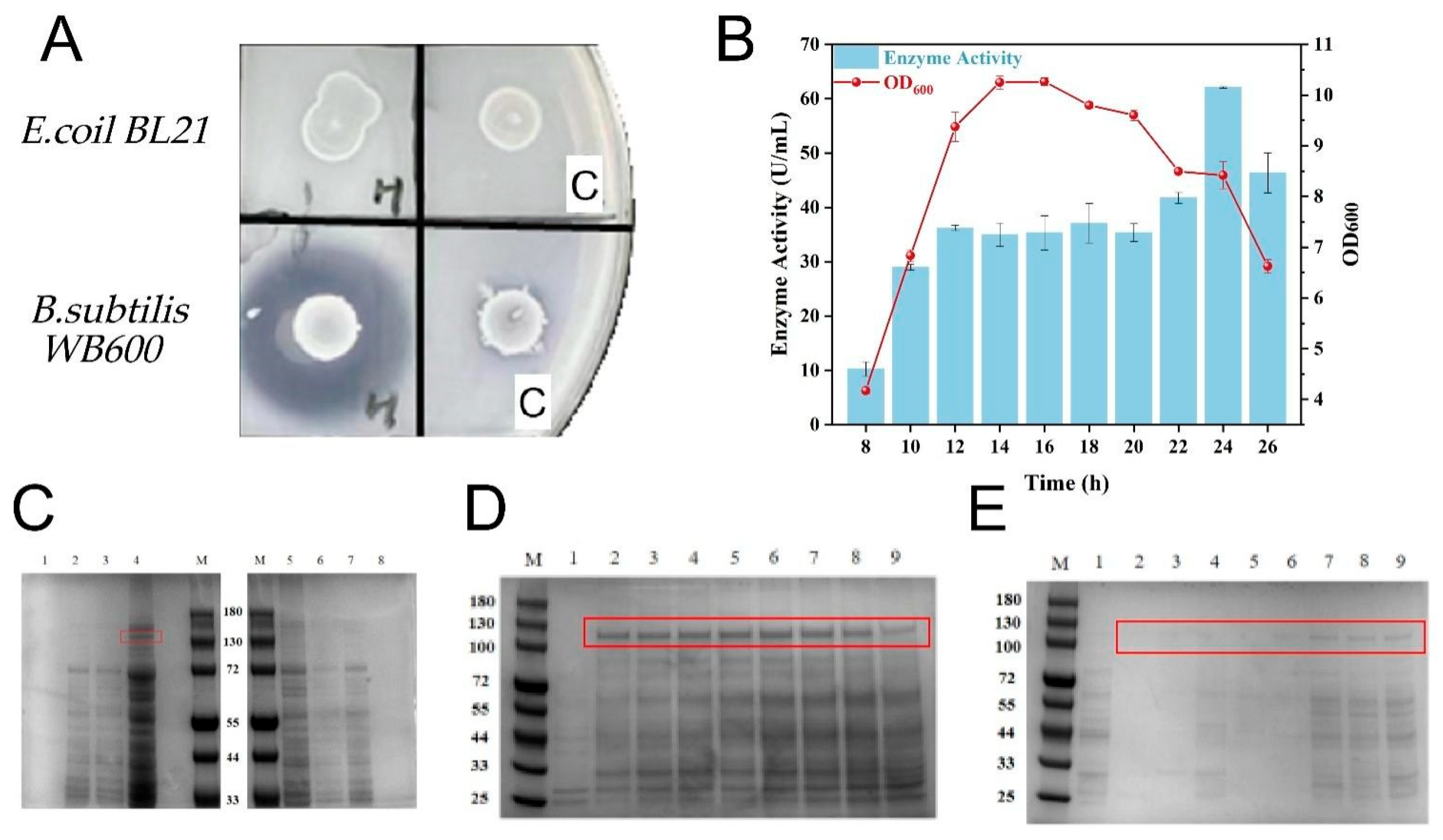
3.1. Expression of Recombinant Collagenase

The collagenase gene ColH (NCBI No. WP_171012037.1) was identified from the M9B family of C. histolyticum by searching the NCBI database, which consists of 1021 amino acids. The sequences we identified were synthesized by Talen-bio Scientific Co., Ltd. (Shanghai, China) and subjected to codon optimization for the corresponding host. To efficiently produce ColH, E. coli and B. subtilis were selected as the host strains. The results were analyzed using gelatin plate screening (Figure 1A) and SDS-PAGE (Figure 1C–E). In the E. coli expression system, no clear zones were produced on the gelatin plate, and the SDS-PAGE analysis revealed no distinct bands in the fermentation supernatant and pellet, whereas faint bands were detected in the cell lysate supernatant. Conversely, the B. subtilis expression system demonstrated the formation of clear zones on the gelatin plates, with the SDS-PAGE analysis revealing distinct bands in the fermentation supernatant. It can be hypothesized that the complex architecture of collagenase plays a significant role in its aggregation into inclusion bodies within E. coli, which subsequently impedes effective secretion. The excellent secretory ability of B. subtilis can effectively transport the correctly folded collagenase out of the cell. The WB-H strain was cultured at 37 °C with shaking at 220 rpm in TB medium. The enzyme activity assay of the WB-H strain indicated that its peak enzyme activity reached 64 U/mL after 24 h of fermentation (Figure 1B). It was also found that the protein bands became increasingly visible in the cell lysate supernatant after 24 h, indicating that with the continuous production of collagenase, some of them cannot exfoliate in time. Researchers have proposed that an inadequate adaptation of the secretion pathway could lead to the accumulation of proteins within the cells during the later phases of fermentation. Therefore, the secretion pathway was further optimized by signal peptide screening.

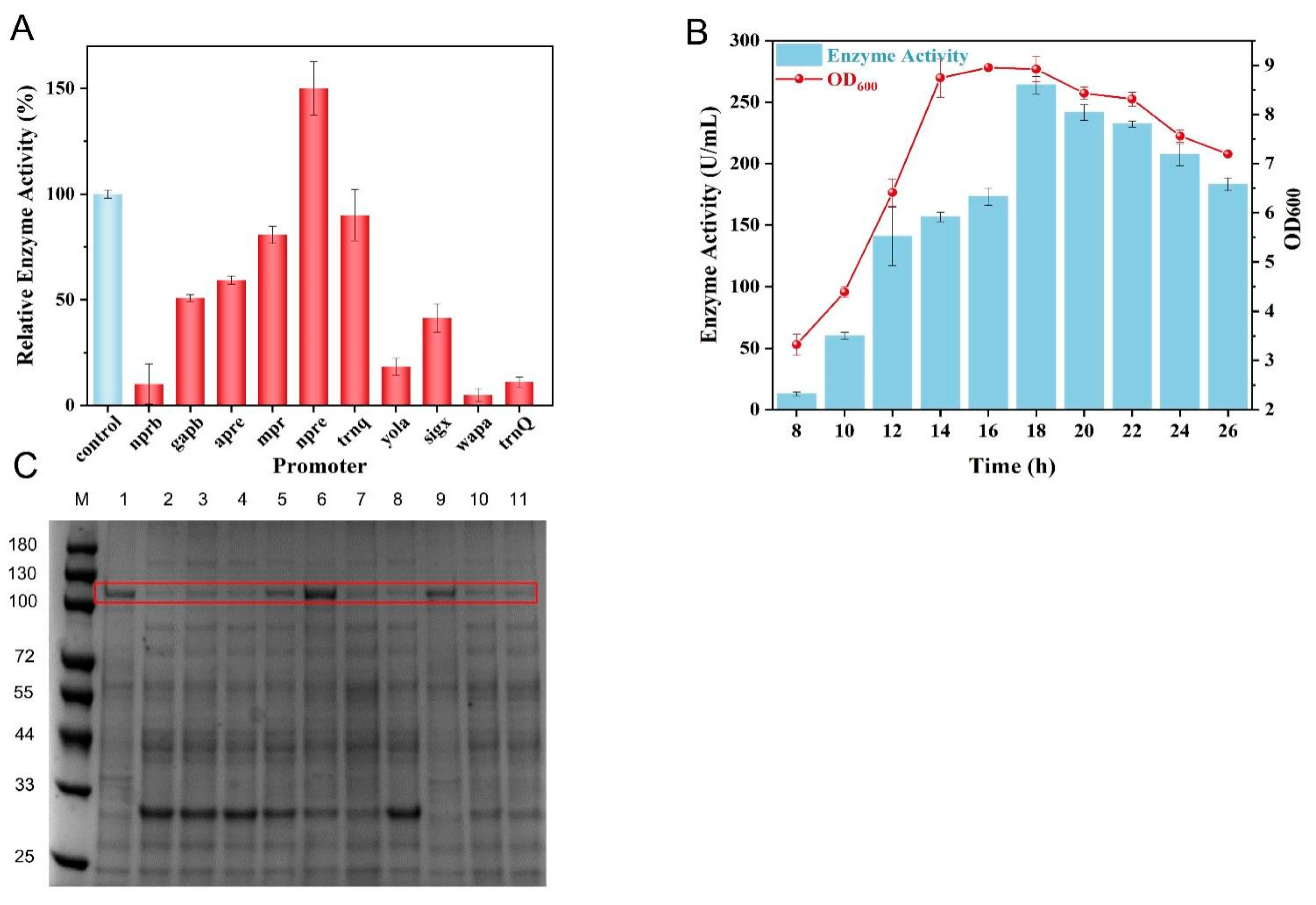
3.2. Screening of Signal Peptide for Secretory Expression of Collagenase

To promote the secretion of ColH, a total of 11 signal peptides (Appendix A: Table A1) that were previously characterized from B. subtilis were utilized in this study. Eleven recombinant strains were successfully constructed and cultured in TB medium under conditions of 220 rpm and 37 °C for fermentation. The results indicate that the WB-H-SPmdh strain demonstrated the most significant enhancement in enzyme activity (Figure 2A), achieving a maximum enzyme activity of 154 U/mL, which was double that of the initial strain. The activity curve (Figure 2B) indicated that the enzyme activity peaked at 182 U/mL after 18 h of culture, representing an advancement of 6 h compared with the initial strain. The SDS-PAGE analysis further corroborated these results (Figure 2C), revealing prominent extracellular bands for WB-H-SPmdh. Given that the WB-H-SPmdh strain exhibited the highest extracellular enzyme activity, it was selected for subsequent studies.

3.3. Optimization of Promoter to Facilitate Collagenase Expression

To improve the expression level of ColH, the promoter (Appendix A: Table A2) of the engineered strain WB-H-SPmdh was optimized. A screening of 10 promoter sequences known for their superior performance in B. subtilis was performed. In the TB medium at 220 rpm and 37 °C, the WB-H-SPmdh-Pnpre strain exhibited a significant increase in enzyme activity, achieving 1.5 times that of the WB-H-SPmdh strain (Figure 3A). The results obtained from the SDS-PAGE analysis corroborated the enzyme activity data (Figure 3C), revealing that the WB-H-SPmdh-Pnpre strain displayed the most pronounced bands of extracellular proteins. Through the optimization of the signal peptides and promoter elements, an efficient recombinant strain was successfully developed, namely, WB-H-SPmdh-Pnpre, which demonstrated nearly complete secretion of the expressed collagenase. The enzyme activity reached 264 U/mL after 18 h of culture, representing a fourfold increase compared with the original strain (Figure 3B).

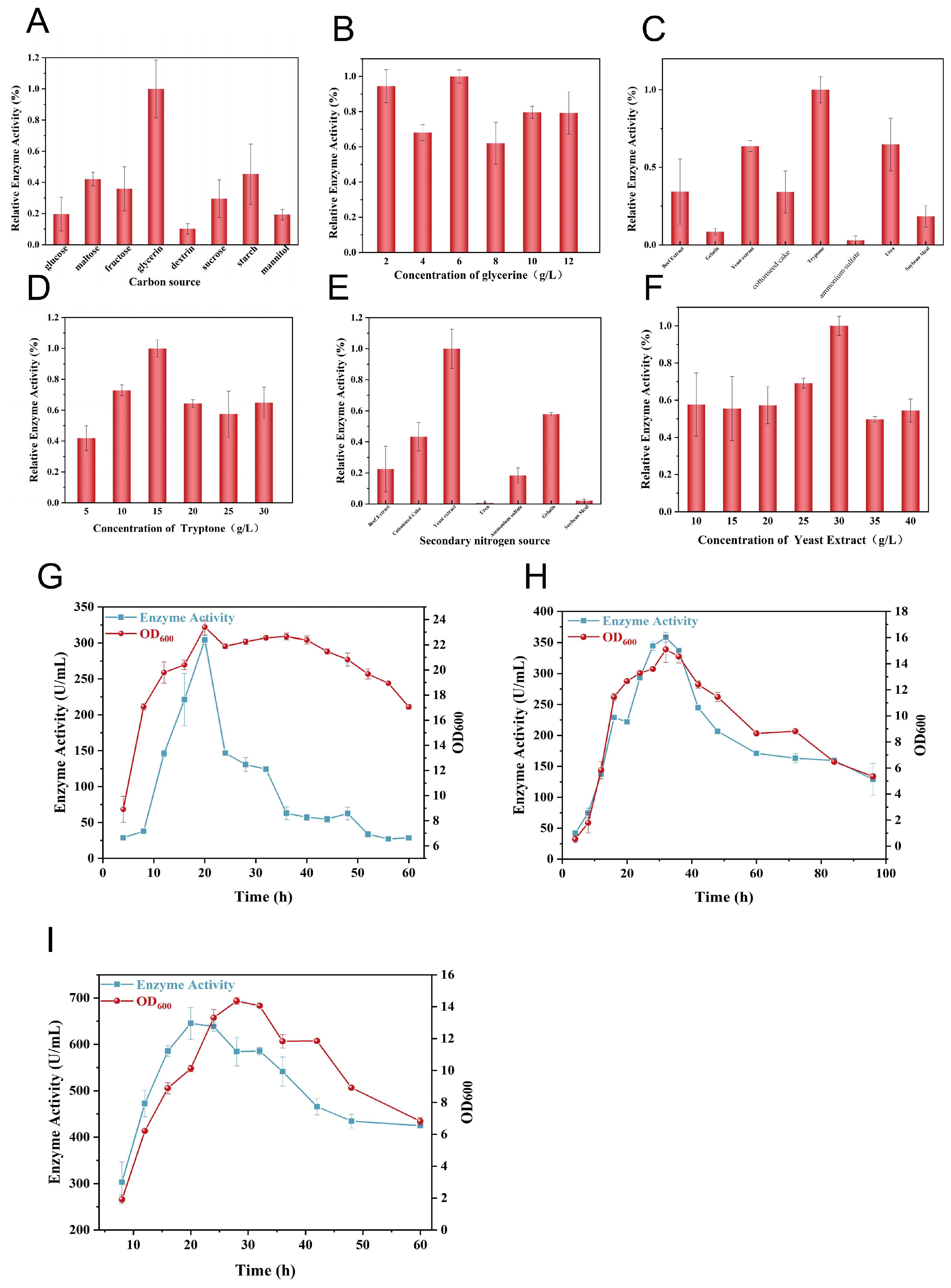
3.4. Fermentation Strategy for Collagenase Production

To improve the expression of ColH and explore its potential for large-scale production, fermentation optimization was carried out in shake flasks and fermenters. Based on the TB medium, an assessment was conducted on nitrogen sources and carbon sources. The best nitrogen source identified was 15 g/L tryptone, followed by 30 g/L yeast extract. The most effective carbon source at an optimal concentration was 6 g/L glycerin (Figure 4A–F). Furthermore, an examination of the fermentation pH revealed that the optimal pH level was 7.5. Enhanced industrial production was achieved through the utilization of batch and fed-batch fermentation processes in fermenters. In batch fermentation, a fermenter was used with an inoculation rate of 10%, employing the enhanced culture medium at a temperature of 37 °C and a pH of 7.5, with the rotation speed adjusted according to the cell density (OD). Sampling was started at 4 h and continued every 4 h. The cell density (OD) showed growth in the logarithmic phase from 4 to 20 h, transitioned to the stationary phase from 20 to 40 h, and entered the death phase from 40 to 60 h. The enzyme activity peaked at 20 h, reaching approximately 300 U/mL (Figure 4G). In fed-batch fermentation, a tank was utilized with an inoculation rate of 10%, employing the enhanced culture medium at a temperature of 37 °C and a pH of 7.5, with the rotation speed adjusted according to the cell density (OD). Supplementation was commenced at a rate of 0.5 mL/min beginning at 8 h, starting sampling at 4 h, and taking samples every 4 h, initially, and then every 12 h, later. The OD steadily increased from 4 to 48 h, followed by a significant decline after 48 h. Compared with batch fermentation, the enzyme activity peaked at 32 h, achieving a maximum of around 364 U/mL (Figure 4H). Following an examination of the conditions, we proceeded to carry out the fermentation process in a 7 L fermentation tank. The inoculation was set at 10%, with the temperature maintained at 30 °C and the pH adjusted to 7.5. The stirring speed was modulated in relation to the cell density (OD). Commencing from 8 h of fermentation, we introduced a feed supplement at a rate of 0.5 mL/min. The enzyme activity peaked at 20 h, achieving a maximum of around 669 U/mL (Figure 4I).

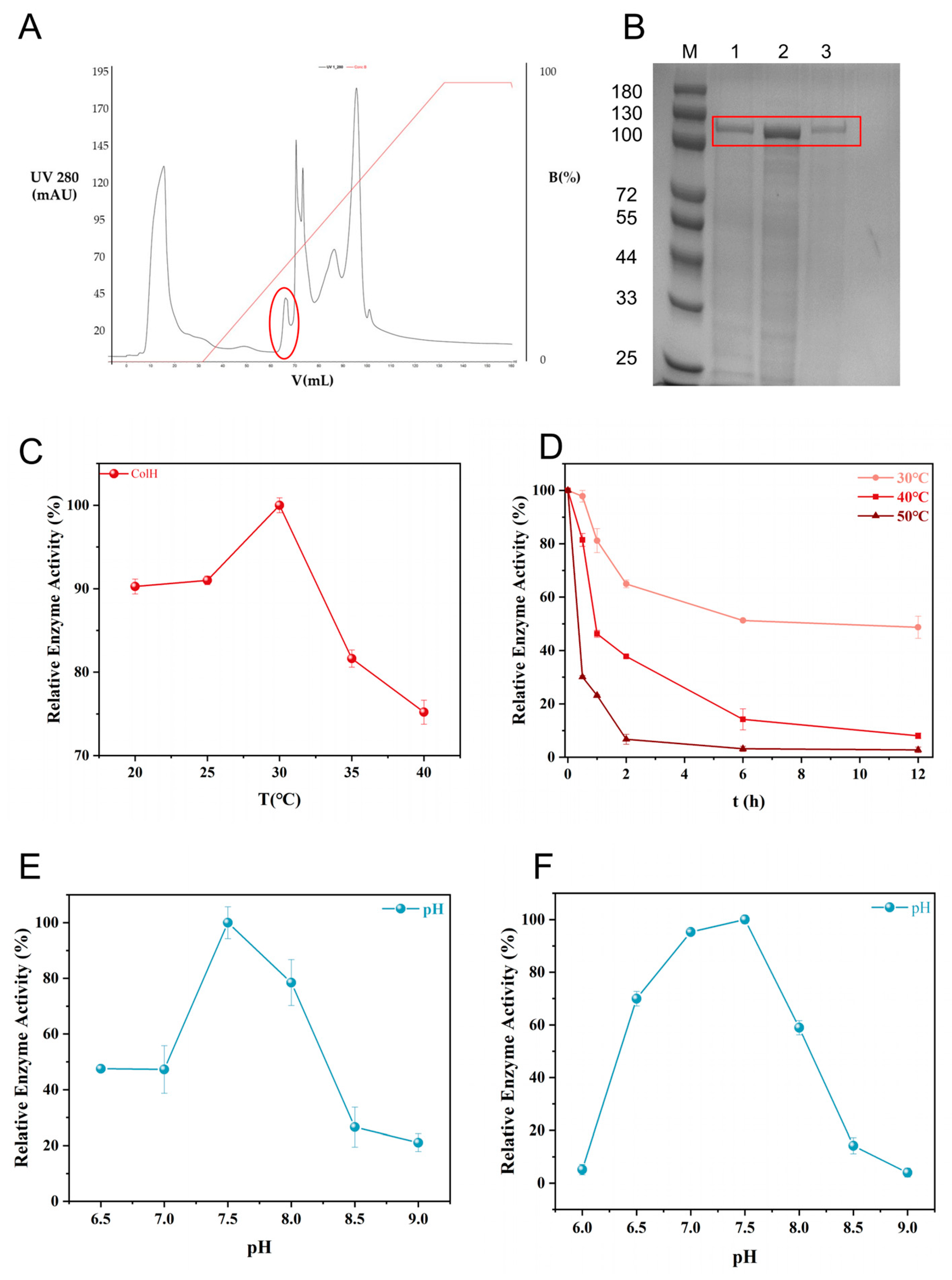
3.5. Purification and Characterization of Collagenase ColH

Purification was achieved using ammonium sulfate and Q-Sepharose chromatography. Ammonium sulfate precipitation increased the specific activity of 294.89 U/mg (Table 2). During Q-Sepharose chromatography, elution with a 0–1 M NaCl gradient generated five absorbance peaks at 280 nm, and collagenase activity was detected exclusively in the first peak (Figure 5A). The results analyzed by SDS-PAGE revealed a single prominent band with a molecular weight of about 116 kDa (Figure 5D). Simultaneously, the protein concentration was quantified using the BCA Protein Assay Kit (Bioswamp), following the manufacturer’s protocol. Using a two-step procedure, the specific activity of the enzyme increased from 21.97 U/mg (crude enzyme) to 565.25 U/mg (purified enzyme), and the enzyme was purified 81-fold with a yield of 33% from the crude extract (Table 2). The results of the temperature effect on the enzyme activity indicate that ColH exhibited optimal enzyme activity at 30 °C (Figure 5B). After incubating at 30 °C for 6 h, approximately 50% of the enzyme activity was retained. Conversely, after treatment at 50 °C for 2 h, the enzyme activity basically disappeared (Figure 5C). The effect of the pH on the enzyme activity showed that ColH exhibited optimal enzyme activity at pH 7.5 (Figure 5F), and after incubating at pH 6.5–8.0 for 5 h, more than 60% of the enzyme activity remained (Figure 5E).

3.6. Adipose Tissue Isolation

The purified ColH was applied in the extraction of cells from adipose tissue. The test group was treated with a mixture of neutral protease and ColH, while the control group was treated with an equal volume of neutral protease. After incubation at 37 °C for 1 h, both groups exhibited stratification, while the upper layer of the test group was clearer, with fewer fat particles (Figure 6A). In the test group, the cell viability was 51.6%, with a total cell count of 9.19 × 10⁶ cells/mL of adipose tissue, which showed significant improvement compared with the 16.86% cell viability and 5.26 × 10⁵ cell count of the control group. The extracted cells were fusiform after culture and could maintain their shape well after passage (Figure 6B–D). In recent years, adipose-derived cells have shown great application prospects in the field of cell therapy, which further promotes the development and production of collagenase as a key tool enzyme.

4. Discussion

In this study, we successfully expressed collagenase ColH from the M9B subfamily of C. histolyticum using Bacillus subtilis. Through the optimization of the expression elements, a recombinant strain with high collagenase production was obtained. Additionally, the potential application of this enzyme under industrial production conditions was explored and highlighted its potential application to extract cells from adipose tissue.
According to the broad definition of collagenase, that is, any enzyme that can hydrolyze collagen is considered collagenase, collagenase actually includes fungal collagenase, plant collagenase, animal collagenase, and bacterial collagenase. Nevertheless, fungal collagenase and plant collagenase do not specifically recognize collagen; they simply possess a broad substrate spectrum that includes collagen. For example, collagenases derived from filamentous fungi primarily target fungal cell wall components, exhibiting substrate specificity toward structural polysaccharides such as β-glucans. Collagenase is a complex enzyme that typically comprises four structural domains: the activation domain, the peptidase domain, the PKD (polycystic kidney disease-like) domain, and the PPC (proline-rich and calcium-binding) domain. The activation and peptidase domains together constitute the characteristic catalytic module (CM) of collagenases. Given the large molecular size and complex structure of collagenase, B. subtilis, which has a strong secretion capacity, showed better adaptation for its expression. Optimization of the signal peptide could effectively enhance the extracellular expression of the enzyme, likely due to the optimized signal peptide being more conducive to the correct folding of ColH and its efficient secretion outside the cell, thereby preventing the formation of inclusion bodies within the cell.
Interestingly, during the purification process, our initial attempts to incorporate a His-tag for nickel affinity chromatography resulted in a partial loss of enzyme activity. A literature review revealed that a similar issue was reported by Subramanian et al., who also observed a decrease in collagenase activity when a 6His tag was added to the C-terminus for purification purposes [12,17]. It is hypothesized that the collagen-binding domain (CBD) sequence at the C-terminus of collagenase may interfere with the purification tag, and it is also related to the distance between His-tags and the active center of the enzyme. Given these considerations, we selected a classical purification protocol for ColH, which is of significant importance for the industrial production and application of collagenase. Traditional column chromatography relies on multiple steps and is cost-intensive, making it essential to seek alternative methods for enzyme purification. For example, Huang et al. [18] demonstrated a column-free purification technique using self-aggregating tags and cleavable inteins for one-step, high-efficiency purification.
Due to its ability to specifically recognize and degrade collagen without affecting other proteins, collagenase has garnered significant interest among scientists. Currently, collagenase has been widely used in the isolation of fibroblasts. Our study demonstrated the efficacy of collagenase in the extraction of adipose-derived cells, which holds broad prospects in the medical field. In fact, collagenase has already found extensive applications in both the food and medical industries. For instance, in food processing, collagenase can be used to extend the shelf life of food products and develop functional foods with antihypertensive properties. In terms of health, collagenase can break down collagen into short peptides, which can help improve skin conditions and prevent osteoporosis [19,20]. In the medical field, collagenase is used in various clinical settings, such as for lumbar disc herniation, uterine fibroid prevention, and Dupuytren’s contracture [21,22,23]. For example, In February 2010, the U.S. Food and Drug Administration (FDA) approved the use of collagenase from C. histolyticum for the nonsurgical treatment of Dupuytren’s contracture [24,25]. Additionally, collagenase has been proven to be highly effective in managing burn necrosis and wounds that are difficult or even impossible to heal. In particular, for skin burns, collagenase from Clostridium can be used for debridement, which helps regulate cellular responses and promotes an anti-inflammatory microenvironment, ultimately enhancing the healing process [26]. Santyl [27], a collagenase ointment derived from C. histolyticum, is the only enzyme preparation approved by the FDA for the debridement of burns and wound trauma. Therefore, the study of collagenase holds broad market potential and significant research implications.

5. Conclusions

In this study, collagenase ColH from C. histolyticum was successfully expressed heterologously in B. subtilis. Through the optimization of the promoter and signal peptide, as well as a fed-batch fermentation strategy, the enzyme activity of ColH was increased to 669 U/mL. By using ammonium sulfate and Q purification, the recombinant collagenase was successfully purified, which showed application potential in adipocyte extraction. This work shows great promise for the industrial production of collagenase proteins and provides new prospects in the medical field.

Author Contributions

L.-F.X. and D.X.: writing—original draft, software, methodology, investigation, formal analysis, and data curation. N.C.: software, investigation, and formal analysis. C.S. and X.-D.M.: methodology. J.-S.G. (Jin-Song Gong): investigation. J.-Y.Q. and N.X.: formal analysis. Z.-Z.W.: resources. J.-S.S. (Jin-Song Shi) and Z.-H.X.: writing—review and editing, validation, resources, and conceptualization. All authors have read and agreed to the published version of the manuscript.

Funding

The authors are grateful to the financial support from the National Key Research and Development Program of China (no. 2023YFA0914500), the National Natural Science Foundation of China (no. 32301283), and the Natural Science Foundation of Jiangsu Province (no. BK20221082).

Institutional Review Board Statement

Not applicable.

Informed Consent Statement

Not applicable.

Data Availability Statement

The original contributions presented in this study are included in this article. Further inquiries can be directed to the corresponding author.

Conflicts of Interest

Authors Xu-Dong Ma and Nan Xie were employed by the company Cytori Therapeutics LLC. The remaining authors declare that the research was conducted in the absence of any commercial or financial relationships that could be construed as a potential conflict of interest.

Appendix A

Table A1. The sequences of the signal peptides.
Signal PeptideSequence
SPhruiatgagaaaatggtattttattcttttggcgggtgttttaacgtctgtcatcctcgctttcgtttatgataagacaaaagcg
SPamyeatgtttgcaaaacgattcaaaacctctttactgccgttattcgctggatttttattgctgtttcatttggttctggcagga
SPbglsatgccttatctgaaacgagtgttgctgcttcttgtcactggattgtttatgagtttgtttgcagtcactgctactgcctcagct
SPpelcttgaaaaaaatcgtgtctatcctatttatgttcggtttggttatgggtttcagccagtttcagccatcaaccgtttttgca
SPvprttgaaaaaggggatcattcgctttctgcttgtaagtttcgtcttattttttgcgttatccacaggcattacgggcgttcaggca
SPmdhatgggaaatactcgtaaaaaagtttctgttatcggagcaggttttaccggagctacaactgcatttttaatcgctcaaaaagagctggcagacgtt
SPnpregtgggtttaggtaagaaattgtctgttgctgtcgctgcttcgtttatgagtttatcaatcagcctgccaggtgttcaggct
SPdacbatgcgcattttcaaaaaagcagtattcgtgatcatgatttcttttcttattgcaaccgtaaatgtgaatacagcacatgct
SPphobttgaaaaaattcccgaagaaattactgcctatcgcggttttatcatcaattgcgttcagcagcttagccagcggcagtgtgcctgaagccagcgcc
SPyhfmatgaaaaaaatagtggcagccatcgtggtaatcggtcttgtgtttatcgcatttttttatctttacagccgatcaggcgatgtgtatcaatcggtagacgcg
SPestaatgaaatttgtaaaaagaaggatcattgcacttgtaacaattttgatgctgtctgttacatcgctgtttgcgttgcagccgtcagcaaaagcc
Table A2. The sequences of the promoters.
PromoterSequence
Pgapbgttggacaccccttatcaattatgccatcctaatttgttactatctaaaattagtataacacattaaaacaaattcgccagtacatattgataaaaatcaacgattataacgctaatttatgatgcatcttatgttttcttgcacagaatatcaggcgtaagaaccaaaaagcagccagcgctccgcactgactgccctcatgctttatgattcctgttttaacttatcattgagcacactttcttctagcacagaaaatgtatccccgtacactctgttaatatgattcgctccccaagcacacagcatatctaaaatcccttct
Pnprbcatatggagcaggggttattatttatgtcaataaaaaattagtagagtagaaaaagaagggcagagcgaaatatctctgcccttcttttttgggaaaataagaacgaagcaccacatacaagtttttgtatttttgataggatgaagaaaaatggtagattccaaaataggaaggatgtggtgtt
Pmprttagcggattacactgttgaaggattggaaaacgcactggcagtcccaaagactcaagtgcgcgtctttggaaagccgataacaaaagccggacgtcgtatggcagttgcgctttctgctgctgattcagttgaaacggcaagagagaatgcaaagaaagcgttggaccagctaattttaaaatagagtttgaacaggtcttgtcatgggacaaggcctgtttttttctttctccgtaaaagttttatcataagaatcagaaacctgattataatgtaaaagtcttccatcgatacgggtggttgacactaaaggagggagatgacaaa
Paprecgataatatccattgttctcacggaagcacacgcaggtcatttgaacgaattttttcgacaggaatttgccgggactcaggagcatttaacctaaaaaagcatgacatttcagcataatgaacatttactcatgtctattttcgttcttttctgtatgaaaatagttatttcgagtctctacggaaatagcgagagatgatatacctaaatagagataaaatcatctcaaaaaaatgggtctactaaaatattattccatctattacaataaattcacagaatagtcttttaagtaagtctactctgaatttttttaaaaggagagggtaaaga
Pyolattcctaaatcctccttggtacaagtttacatgttaaatatcgtcatttgaagggaattgtttaatatttgaaataaaaaaagaacctgcattaagcaagttcttttcatattgtcaatttattgtgaatttttaacgacaaggacatttatgtatagtataatatttcctgtacataaagtttgctcactcaagggagtcttgctcatcccctatgaaaggggtgggaaaatgactgtttacgaatcattaatgataatgatcaattttggcggattgatattaaataccgtcttgttgatcttcaatataatgatgattgtaacgtcaagccaaaagaaaaaatagaccttcccttgagtttggacacctgaagggttaggcctacgcagatttgacaacgagcaagcc
Psigxcaaagactccgggtctggcataccggaagaagatctgccatttatctttgagcggttttataaggcagataaagcgcggacaaggggcagagcaggaaccgggttagggctggctatcgttaaaaatatcgtggaagcccacaacggatcaattactgtgcacagccgaatagataaaggaacaacattttctttttatattccgacaaaacggtaaaatcgagtctgaatttgccgaagaatcttgttccataagaaacacccgctgactgagcgggtgtttttttaatagccaacattaataaaatttaaggatatgttaatataaattcccttccaaattccagttactcgtaatatagttgtaatgtaacttttcaagctattcatacgacaaaaaagtgaacggaggggtttcaa
Pnpreattgaatcagcagggtgctttgtctgcttaatataaaataacgttcgaaatgcaatacataatgactgaataactccaacacgaacaacaatcctttacttcttattaaggcctcattcggttagacagcggacttttcaaaaagtttcaagatgaaacaaaaatatctcatcttccccttgatatgtaaaaaacataactcttgaatgaaccaccacatgacacttgactcatcttgatattattcaacaaaaacaaacacaggacaatactatcaattttgtctagttatgttagtttttgttgagtattccagaatgctagtttaatataacaatataaagttttcagtattttcaaaaagggggatttatt
Pp43agcattattgagtggatgattatattccttttgatagggtggtatgttttcgcttgaacttttaaatcagccattgaacatacggttgattttaataactgacaaacatcaccctcttgctaaagcggccaaggacgctgccgccggggctgtttgcgtttttgccgtgatttcgtgtatcattggtttacttatattttttgccaaagctgtaatggctgaaaattcttacatttattttacattttttagaaatgaggcgtgaaaaaaagcgcgcgattatgtaaaataaaaagtatagcgggtacca
Pwapaatttcaatcattgtatttctcgaccccgctgtcgcgatcgtgctcgataccgtcttcacaggcttccgccctgacctctatcaaacgcttggcatcgtaatgatctttgcgggcatggccttgacgcttgtcaggaggcaggggaaggcgaatgtgacagctgagggtacggatattgaacaaatacaataaaaaatgtaaaaaggcctatgcggcctttttttgttttaggtcaattgactctcgctaatccttaaaataagataaattttctagaaaaatattgtaatgatatttcagtctagttaagattattgagtaaatattacttttattacaaaaggagagaggaa
Ptrnqgagaataaatgtcatacgctctttccccgcggttcgtttgttcaatgactgtaggtattaaattcataatgctcctccttcaccttttaggtaagtatgtagttacatgatacatttttggtcaataaaggtcaaacaaaaagctggcctgatatgcaaagtcgtctctttttcccattttccccaaaaatacaggggttcaaaccatcgtatgtcagattgccaattaagatgctttgtctatttaaaaaacggcctctcgaaatagagggttgttatttgaaaggaattatcgtataattagttgtgctgacgttctcataacgcagtctatat

References

  1. Frederick, R.E.; Bearden, R.; Jovanovic, A.; Jacobson, N.; Sood, R.; Dhall, S. Clostridium Collagenase Impact on Zone of Stasis Stabilization and Transition to Healthy Tissue in Burns. Int. J. Mol. Sci. 2021, 22, 8643. [Google Scholar] [CrossRef] [PubMed]
  2. Singh, B.; Sims, H.; Trueheart, I.; Simpson, K.; Wang, K.C.; Patzkowsky, K.; Wegman, T.; Soma, J.-M.; Dixon, R.; Jayes, F.; et al. A Phase I Clinical Trial to Assess Safety and Tolerability of Injectable Collagenase in Women with Symptomatic Uterine Fibroids. Reprod. Sci. 2021, 28, 2699–2709. [Google Scholar] [CrossRef] [PubMed]
  3. Kwak, K.; Park, J.k.; Shim, J.; Ko, N.; Kim, H.J.; Lee, Y.; Kim, J.H.; Alexander, M.; Lakey, J.R.T.; Kim, H.; et al. Comparison of islet isolation result and clinical applicability according to GMP-grade collagenase enzyme blend in adult porcine islet isolation and culture. Xenotransplantation 2021, 28, 12703. [Google Scholar] [CrossRef]
  4. Narwani, K.; Stark, J.; Cortez, D.; Yang, I.; Au, C.; Diaz, A.; Guerra, C.; Niihara, Y.; Bardag-Gorce, F. cG-CAOMECS—Clinical-grade cultured autologous oral mucosal epithelial cell sheet. Cell Tissue Res. 2021, 386, 47–57. [Google Scholar] [CrossRef]
  5. Jin, Y.-Q.; Liu, W.; Hong, T.-H.; Cao, Y. Efficient Schwann cell purification by differential cell detachment using multiplex collagenase treatment. J. Neurosci. Methods 2008, 170, 140–148. [Google Scholar] [CrossRef]
  6. Zuk, P.A.; Zhu, M.; Mizuno, H.; Huang, J.; Futrell, J.W.; Katz, A.J.; Benhaim, P.; Lorenz, H.P.; Hedrick, M.H. Multilineage cells from human adipose tissue: Implications for cell-based therapies. Tissue Eng. 2001, 7, 211–228. [Google Scholar] [CrossRef]
  7. Pal, G.K.; Pv, S. Microbial collagenases: Challenges and prospects in production and potential applications in food and nutrition. RSC Adv. 2016, 6, 33763–33780. [Google Scholar] [CrossRef]
  8. Xiao, H.; Liu, X.; Feng, Y.; Zheng, L.; Zhao, M.; Huang, M. Secretion of collagenases by Saccharomyces cerevisiae for collagen degradation. Biotechnol. Biofuels Bioprod. 2022, 15, 89. [Google Scholar] [CrossRef]
  9. Han, X.; Xiufang, L.; Lin, Z.; Mouming, Z.; Mingtao, H. Recent advances in collagenase from Clostridium histolyticum. Food Sci. 2022, 43, 316–325. [Google Scholar]
  10. Serwanja, J.; Wieland, A.C.; Haubenhofer, A.; Brandstetter, H.; Schoenauer, E. A conserved strategy to attack collagen: The activator domain in bacterial collagenases unwinds triple-helical collagen. Proc. Natl. Acad. Sci. USA 2024, 121, e2321002121. [Google Scholar] [CrossRef]
  11. Jung, C.M.; Matsushita, O.; Katayama, S.; Minami, J.; Ohhira, L.; Okabe, A. Expression of the colH Gene Encoding Clostridium histolyticum Collagenase in Bacillus subtilis and Its Application to Enzyme Purification. Microbiol. Immunol. 2013, 40, 923–929. [Google Scholar] [CrossRef] [PubMed]
  12. Matsushita, O.; Jung, C.M.; Minami, J.; Katayama, S.; Nishi, N.; Okabe, A. A study of the collagen-binding domain of a 116-kDa Clostridium histolyticum collagenase. J. Biol. Chem. 1998, 273, 3643–3648. [Google Scholar] [CrossRef] [PubMed]
  13. Tamai, E.; Miyata, S.; Tanaka, H.; Nariya, H.; Suzuki, M.; Matsushita, O.; Hatano, N.; Okabe, A. High-level expression of his-tagged clostridial collagenase in Clostridium perfringens. Appl. Microbiol. Biotechnol. 2008, 80, 627–635. [Google Scholar] [CrossRef] [PubMed]
  14. Zhao, Q.; Liu, X.; Li, S. Secretory Expression of Clostridium histolyticum Collagenase H in Escherichia coli. J. East China Univ. Sci. Technoloy Nat. Sci. Ed. 2022, 48, 797–805. [Google Scholar]
  15. Zhu, Y.; Wang, L.; Zheng, K.; Liu, P.; Li, W.; Lin, J.; Liu, W.; Shan, S.; Sun, L.; Zhang, H. Optimized Recombinant Expression and Characterization of Collagenase in Bacillus subtilis WB600. Fermentation 2022, 8, 449. [Google Scholar] [CrossRef]
  16. Hoppe, I.J.; Brandstetter, H.; Schonauer, E. Biochemical characterisation of a collagenase from Bacillus cereus strain Q1. Sci. Rep. 2021, 11, 4187. [Google Scholar] [CrossRef]
  17. Subramanian, S.R.; Singam, E.R.A.; Berinski, M.; Subramanian, V.; Wade, R.C. Identification of an Electrostatic Ruler Motif for Sequence-Specific Binding of Collagenase to Collagen. J. Phys. Chem. B 2016, 120, 8580–8589. [Google Scholar] [CrossRef]
  18. Huang, Y.; Zhang, Y.; Yan, X.; Li, Z. A high-performance protein preparation approach in a single column-free step. Trends Biotechnol. 2025, 43, 476–487. [Google Scholar] [CrossRef]
  19. Boe, C.; Blazar, P.; Iannuzzi, N. Dupuytren Contractures: An Update of Recent Literature. J. Hand Surg. 2021, 46, 896–907. [Google Scholar] [CrossRef]
  20. Holzer, L.A.; Holzer, G. Collagenase Clostridum histolyticum in the Management of Dupuytren’s Contracture. Handchir. Mikrochir. Plast. Chir. 2011, 43, 269–274. [Google Scholar]
  21. Leafblad, N.D.; Wagner, E.; Wanderman, N.R.; Anderson, G.R.; Visscher, S.L.; Kremers, H.M.; Larson, D.R.; Rizzo, M. Outcomes and Direct Costs of Needle Aponeurotomy, Collagenase Injection, and Fasciectomy in the Treatment of Dupuytren Contracture. J. Hand Surg. 2019, 44, 919–927. [Google Scholar] [CrossRef] [PubMed]
  22. Zhu, L.; Xie, Y.; Wen, B.; Ye, M.; Liu, Y.; Imam, K.M.S.U.; Cai, H.; Zhang, C.; Wang, F.; Xin, F. Porcine bone collagen peptides promote osteoblast proliferation and differentiation by activating the PI3K/Akt signaling pathway. J. Funct. Foods 2020, 64, 103697. [Google Scholar] [CrossRef]
  23. Mei, F.; Liu, J.; Wu, J.; Duan, Z.; Chen, M.; Meng, K.; Chen, S.; Shen, X.; Xia, G.; Zhao, M. Collagen Peptides Isolated from Salmo salar and Tilapia nilotica Skin Accelerate Wound Healing by Altering Cutaneous Microbiome Colonization via Upregulated NOD2 and BD14. J. Agric. Food Chem. 2020, 68, 1621–1633. [Google Scholar] [CrossRef] [PubMed]
  24. Swanson, J.W.; Watt, A.J.; Vedder, N.B. Collagenase for Recurrent Dupuytren Contracture with Skin Grafts Reply. J. Hand Surg. 2013, 38, 1264–1265. [Google Scholar] [CrossRef]
  25. Warwick, D.; Arandes-Renu, J.M.; Pajardi, G.; Witthaut, J.; Hurst, L.C. Collagenase Clostridium histolyticum: Emerging practice patterns and treatment advances. J. Plast. Surg. Hand Surg. 2016, 50, 251–261. [Google Scholar] [CrossRef]
  26. Xiong, L.; Cui, M.; Zhou, Z.; Wu, M.; Wang, Q.; Song, H.; Ding, L. Primary culture of chondrocytes after collagenase IA or II treatment of articular cartilage from elderly patients undergoing arthroplasty. Asian Biomed. 2021, 15, 91–99. [Google Scholar] [CrossRef]
  27. Sheets, A.R.; Demidova-Rice, T.N.; Shi, L.; Ronfard, V.; Grover, K.V.; Herman, I.M. Identification and Characterization of Novel Matrix-Derived Bioactive Peptides: A Role for Collagenase from Santyl® Ointment in Post-Debridement Wound Healing? PLoS ONE 2016, 11, e0159598. [Google Scholar] [CrossRef]
Figure 1. Expression of collagenase ColH. (A) Gelatin plates H: ColH, and C: control (the collagenase-negative host strain). (B) The enzyme activity curve of the WB-H strain. (C) SDS-PAGE analysis of the E. coli expression system. Lane: M, protein marker; 1, fermentation supernatant of the BL-H strain; 2, fermentation precipitate of the BL-H strain; 3, lysate supernatant of the BL-H strain; 4, lysate precipitate of the BL-H strain; 5, lysate precipitate of the BL-K; strain 6, lysate supernatant of the BL-K strain; 7, fermentation precipitate of the BL-K strain; and 8, fermentation supernatant of the BL-K strain. (D) SDS-PAGE analysis of the B. subtilis fermentation supernatant. Lane: M, protein marker; 1, fermentation supernatant of the WB-H strain; and 2–9, fermentation supernatant of the experimental group at 8 h, 12 h, 16 h, 20 h, 24 h, 28 h, 32 h, and 36 h, respectively. The red rectangles represent the target proteins. (E) SDS-PAGE analysis of the B. subtilis lysate supernatant. Lane: M, protein marker; 1, lysate supernatant of the WB-H strain; and 2–9, experimental groups of the lysate supernatant at 8 h, 12 h, 16 h, 20 h, 24 h, 28 h, 32 h, and 36 h, respectively. The red rectangles represent the target proteins.
Fermentation 11 00242 g001
Figure 2. Optimization of signal peptide. (A) Relative enzyme activity. (B) The enzyme activity curve of the WB-H-SPmdh strain. (C) SDS-PAGE analysis of signal peptide. Lane: M, protein marker; 1, control group (WB-H); and 2–12, WB-H-SPhrui, WB-H-SPamye, WB-H-SPbgls, WB-H-SPpelc, WB-H-SPvpr, WB-H-SPmdh, WB-H-SPnpre, WB-H-SPdacb, WB-H-SPphob, WB-H-SPyhfm, and WB-H-SPesta. The red rectangles represent the target proteins.
Fermentation 11 00242 g002
Figure 3. Optimization of promoter. (A) relative enzyme activity. (B) the enzyme activity curve of the WB-H-SPmdh-Pnpre strain. (C) SDS-PAGE analysis of promoter. Lane: M, protein marker; 1, control group (WB-H-SPmdh); and 2–11, WB-H-SPmdh-Pgapb, WB-H-SPmdh-Pnprb, WB-H-SPmdh-Pmpr, WB-H-SPmdh-Papre, WB-H-SPmdh-Pnpre, WB-H-SPmdh-Pyola, WB-H-SPmdh-Psigx, WB-H-SPmdh-PP43, WB-H-SPmdh-Pwapa, and WB-H-SPmdh-Ptrnq. The red rectangles represent the target proteins.
Fermentation 11 00242 g003
Figure 4. Fermentation strategy of the WB-H-SPmdh-Pnpre strain. (A) Effect of carbon sources on enzyme activity. (B) Effect of glycerin concentration on enzyme activity. (C) Effect of nitrogen sources on enzyme activity. (D) Effect of tryptone concentration on enzyme activity. (E) Effect of secondary nitrogen sources on enzyme activity. (F) Effect of yeast extract concentration on enzyme activity. (G) Batch fermentation in fermenters. (H) Fed-batch fermentation in fermenters. (I) Fed-batch fermentation in 7 L fermenters.
Fermentation 11 00242 g004
Figure 5. Purification and characterization. (A) Purification of collagenase from WB-H-SPmdh-Pnpre. The red circle indicates the peak of the target proteins. (B) SDS-PAGE analysis of purification. Lane: M, protein marker; 1, the fermentation supernatant; 2, the ammonium sulfate precipitation; and 3, Q. The red rectangles represent the target proteins. (C) Effects of temperature on enzyme activity. (D) Thermal stability. (E) pH stability. (F) Effects of pH on enzyme activity.
Fermentation 11 00242 g005
Figure 6. Application of collagenase ColH in the cell extraction from adipose tissue. (A) Adipose tissue degradation. C: control group; T: test group. The red rectangles indicate adipose tissue. (B) Primary cells in the test group on Day 2. (C) The first-generation cells in the test group on Day 2. (D) The second-generation cells in the test group on Day 2.
Fermentation 11 00242 g006
Table 1. Strains and plasmids.
Strain or PlasmidRelevant CharacteristicsSource/Reference
Strain
E. coli JM109Clone strain Our laboratory
E. coli BL21(DE3)Expression strain Our laboratory
B. subtilis 168Wild typeOur laboratory
B. subtilis WB600B. subtilis 168 derivate, deficient in nprE, aprE, epr, bpr, mpr, nprB, expression strainOur laboratory
BL-KE. coli BL21(DE3) derivative, E. coli BL21(DE3) carrying PET3bThis study
WB-KB. subtilis WB600 derivative, B. subtilis WB600 carrying PMA5This study
BL-HE. coli BL21(DE3) derivative, E. coli BL21(DE3) carrying PET3b-HThis study
WB-HB. subtilis WB600 derivative, B. subtilis WB600 carrying PMA5-HThis study
WB-H-SPhruiB. subtilis WB600 derivative, B. subtilis WB600 carrying PMA5-H-SPhruiThis study
WB-H-SPamyeB. subtilis WB600 derivative, B. subtilis WB600 carrying PMA5-H-SPamyeThis study
WB-H-SPbglsB. subtilis WB600 derivative, B. subtilis WB600 carrying PMA5-H-SPbglsThis study
WB-H-SPpelcB. subtilis WB600 derivative, B. subtilis WB600 carrying PMA5-H-SPpelcThis study
WB-H-SPvprB. subtilis WB600 derivative, B. subtilis WB600 carrying PMA5-H-SPvprThis study
WB-H-SPmdhB. subtilis WB600 derivative, B. subtilis WB600 carrying PMA5-H-SPmdhThis study
WB-H-SPnpreB. subtilis WB600 derivative, B. subtilis WB600 carrying PMA5-H-SPnpreThis study
WB-H-SPdacbB. subtilis WB600 derivative, B. subtilis WB600 carrying PMA5-H-SPdacbThis study
WB-H-SPphobB. subtilis WB600 derivative, B. subtilis WB600 carrying PMA5-H-SPphobThis study
WB-H-SPyhfmB. subtilis WB600 derivative, B. subtilis WB600 carrying PMA5-H-SPyhfmThis study
WB-H-SPestaB. subtilis WB600 derivative, B. subtilis WB600 carrying PMA5-H-SPestaThis study
WB-H-SPmdh-PgapbB. subtilis WB600 derivative, B. subtilis WB600 carrying PMA5-H-SPmdh-PgapbThis study
WB-H-SPmdh-PnprbB. subtilis WB600 derivative, B. subtilis WB600 carrying PMA5-H-SPmdh-PnprbThis study
WB-H-SPmdh-PnpreB. subtilis WB600 derivative, B. subtilis WB600 carrying PMA5-H-SPmdh-PnpreThis study
WB-H-SPmdh-PmprB. subtilis WB600 derivative, B. subtilis WB600 carrying PMA5-H-SPmdh-PmprThis study
WB-H-SPmdh-PapreB. subtilis WB600 derivative, B. subtilis WB600 carrying PMA5-H-SPmdh-PapreThis study
WB-H-SPmdh-PyolaB. subtilis WB600 derivative, B. subtilis WB600 carrying PMA5-H-SPmdh-PyolaThis study
WB-H-SPmdh-PsigxB. subtilis WB600 derivative, B. subtilis WB600 carrying PMA5-H-SPmdh-PsigxThis study
WB-H-SPmdh-Pp43B. subtilis WB600 derivative, B. subtilis WB600 carrying PMA5-H-SPmdh-Pp43This study
WB-H-SPmdh-PwapaB. subtilis WB600 derivative, B. subtilis WB600 carrying PMA5-H-SPmdh-PwapaThis study
WB-H-SPmdh-PtrnqB. subtilis WB600 derivative, B. subtilis WB600 carrying PMA5-H-SPmdh-PtrnqThis study
Plasmid
PMA5F1ori Ampr, RepB Kanr, E. coli-B. subtilis shuttle vectorOur laboratory
PET3bT7 Ampr, AmpROur laboratory
PET3b-HPET3b derivative, PET3b carrying ColH, AmpRThis study
PMA5-HPMA5 derivative, PMA5 carrying ColH, KanRThis study
PMA5-H-SPhruiPMA5 derivative, PMA5 carrying ColH-SPhrui, KanRThis study
PMA5-H-SPamyePMA5 derivative, PMA5 carrying ColH-SPamye, KanRThis study
PMA5-H-SPbglsPMA5 derivative, PMA5 carrying ColH-SPbgls, KanRThis study
PMA5-H-SPpelcPMA5 derivative, PMA5 carrying ColH-SPpelc, KanRThis study
PMA5-H-SPvprPMA5 derivative, PMA5 carrying ColH-SPvpr, KanRThis study
PMA5-H-SPmdhPMA5 derivative, PMA5 carrying ColH-SPmdh, KanRThis study
PMA5-H-SPnprePMA5 derivative, PMA5 carrying ColH-SPnpre, KanRThis study
PMA5-H-SPdacbPMA5 derivative, PMA5 carrying ColH-SPdacb, KanRThis study
PMA5-H-SPphobPMA5 derivative, PMA5 carrying ColH-SPphob, KanRThis study
PMA5-H-SPyhfmPMA5 derivative, PMA5 carrying ColH-SPyhfm, KanRThis study
PMA5-H-SPestaPMA5 derivative, PMA5 carrying ColH-SPesta, KanRThis study
PMA5-H-SPmdh-PgapbPMA5 derivative, PMA5 carrying ColH-SPmdh-Pgapb, KanRThis study
PMA5-H-SPmdh-PnprbPMA5 derivative, PMA5 carrying ColH-SPmdh-Pnprb, KanRThis study
PMA5-H-SPmdh-PmprPMA5 derivative, PMA5 carrying ColH-SPmdh-Pmpr, KanRThis study
PMA5-H-SPmdh-PnprePMA5 derivative, PMA5 carrying ColH-SPmdh-Pnpre, KanRThis study
PMA5-H-SPmdh-PaprePMA5 derivative, PMA5 carrying ColH-SPmdh-Papre, KanRThis study
PMA5-H-SPmdh-PyolaPMA5 derivative, PMA5 carrying ColH-SPmdh-Pyola, KanRThis study
PMA5-H-SPmdh-PsigxPMA5 derivative, PMA5 carrying ColH-SPmdh-Psigx, KanRThis study
PMA5-H-SPmdh-Pp43PMA5 derivative, PMA5 carrying ColH-SPmdh-Pp43, KanRThis study
PMA5-H-SPmdh-PwapaPMA5 derivative, PMA5 carrying ColH-SPmdh-Pwapa, KanRThis study
PMA5-H-SPmdh-PtrnqPMA5 derivative, PMA5 carrying ColH-SPmdh-Ptrnq, KanRThis study
Table 2. Purification of ColH from cultures of WB-H-SPmdh-Pnpre.
StepVolume (mL)Protein (mg)Activity (U)Specific Activity (U/mg)FoldYield (%)
Supernatant150183040,20021.971100
Ammonium sulfate258825,950294.895464.55
Q523.613,340565.258133.18
Disclaimer/Publisher’s Note: The statements, opinions and data contained in all publications are solely those of the individual author(s) and contributor(s) and not of MDPI and/or the editor(s). MDPI and/or the editor(s) disclaim responsibility for any injury to people or property resulting from any ideas, methods, instructions or products referred to in the content.

