Review

Lactic Acid Bacterial Production of Exopolysaccharides from Fruit and Vegetables and Associated Benefits

by
Marie Guérin
1,
Christine Robert-Da Silva
2,
Cyrielle Garcia
1,* and
Fabienne Remize
1
1
Qualisud, Université La Réunion, CIRAD, Université Montpellier, Montpellier SupAgro, Université Avignon, 2 rue Wetzell, F-97490 Sainte Clotilde, France
2
Inserm, UMR 1188 Diabète athérothrombose Thérapies Réunion Océan Indien (DéTROI), Université de La Réunion, Plateforme CYROI, F-97490 Sainte-Clotilde, France
*
Author to whom correspondence should be addressed.
Fermentation 2020, 6(4), 115; https://doi.org/10.3390/fermentation6040115
Submission received: 8 October 2020 / Revised: 15 November 2020 / Accepted: 19 November 2020 / Published: 21 November 2020
(This article belongs to the Special Issue Lactic Acid Fermentation and the Colours of Biotechnology 2.0)

Abstract

:
Microbial polysaccharides have interesting and attractive characteristics for the food industry, especially when produced by food grade bacteria. Polysaccharides produced by lactic acid bacteria (LAB) during fermentation are extracellular macromolecules of either homo or hetero polysaccharidic nature, and can be classified according to their chemical composition and structure. The most prominent exopolysaccharide (EPS) producing lactic acid bacteria are Lactobacillus, Leuconostoc, Weissella, Lactococcus, Streptococcus, Pediococcus and Bifidobacterium sp. The EPS biosynthesis and regulation pathways are under the dependence of numerous factors as producing-species or strain, nutrient availability, and environmental conditions, resulting in varied carbohydrate compositions and beneficial properties. The interest is growing for fruits and vegetables fermented products, as new functional foods, and the present review is focused on exploring the EPS that could derive from lactic fermented fruit and vegetables. The chemical composition, biosynthetic pathways of EPS and their regulation mode is reported. The consequences of EPS on food quality, especially texture, are explored in relation to producing species. Attention is given to the scientific investigations on health benefits attributed to EPS such as prebiotic, antioxidant, anti-inflammatory and cholesterol lowering activities.

1. Introduction

Lactic acid fermentation (LAF) is a liable method to improve the shelf-life of fruit and vegetables along with nutritional and sensory qualities. Bacterial strains belonging to Lactobacillus, Leuconostoc, Weissella and Bifidobacterium genera are commonly involved in food LAF. These lactic acid bacteria can produce lactic acid and other organic acids, carbon dioxide, aromatic compounds, exopolysaccharides (EPS), bacteriocins and enzymes, all involved in the modification of quality of fermented foods. It has also been shown that lactic acid bacteria (LAB) are able to modulate the composition in phenolic compounds and enhance antioxidant activity [1]. Several studies have suggested that lactic acid fermented fruit juices, which exhibit a specific phenolic composition and contain EPS, could constitute a prebiotic food and have an influence on the structure and function of the gut microbiota [2,3]. The ability of LAB to modulate nutritional status of plant-based foods depends on bacterial strains and matrix used for LAF [4]. Among the compounds produced during LAF, EPS have the ability to change rheological properties of food such as viscosity [5], which contributes to the sensory acceptability and stabilization of the products [2]. In the food industry, hydrocolloids gather polysaccharides, which improve textural stability of suspensions, rheological properties of foods, pastry and cake texture and shelf-life by swelling in aqueous medium. It has been shown that EPS produced by Lactobacillus, Leuconostoc and Weissella strains have hydrocolloid properties [6] and would deserve to be tested as an innovative alternative for food formulation.
Up to 70% of the energy of microbial cell can be used to produce EPS. However, this energy consumption is balanced by the protective effect of EPS against stress by the formation of a protecting barrier [7]. The cell is therefore protected from temperature or osmolarity shifts and from toxins and antibiotics. Both quantity and quality of EPS produced depend on sugar availability, presence of micronutrients and fermentation conditions [8].
LAB polysaccharides have interesting and attractive characteristics, including antioxidant activities, and generate an increasing interest for their possible use in the field of food and pharmaceutical industries. This review aims at exploring EPS production during fruit and vegetable LAF, the contribution of EPS-producing LAB species and the associated nutritional and sensory benefits that can result from this production.

2. EPS Classification

EPS produced by LAB are classified according to their chemical composition and their biosynthesis mechanism. Homopolysaccharides (HoPS) are produced in the extracellular medium from one type of monosaccharide, whereas heteropolysaccharides (HePS) are produced intracellularly from several monosaccharides and are then exported from the cell [2,8,9,10,11].

2.1. Homopolysaccharides

HoPS can be characterized according to the type of monosaccharide composing their structure. Glucan, namely dextran, contains only glucose, whereas fructans, namely levan and inulin, contain only fructose and polygalactan contains only galactose [8]. As HoPS are composed of a single monosaccharide, they can be further characterized according to their glycosidic bonds (Figure 1), branching, chain length, molecular weight, and polymer structure [12].
LAB producing HoPS belong to Lactobacillus, Streptococcus, Leuconostoc, Oenococcus, Pediococcus and Weissella genera. For instance, synthesis of a dextran-type glucan by a Leuconostoc pseudomesenteroides strain isolated from juçara palm fruit was confirmed using nuclear magnetic resonance (NMR) spectroscopy. More precisely, a Fourier transform-infrared (FT-IR) spectroscopy analysis identified functional groups and characterized covalent bonds of the produced EPS [13].
HoPS molecular weight ranges between 104 Da and 108 Da and is bacterial strain-dependent [8]. Indeed, dextrans from Leuconostoc mesenteroides and Weissela cibaria have a molecular weight between 6.2 and 7.1 × 10⁶ Da and between 5 × 10⁶ and 4 × 10⁷ Da, respectively. HoPS production yield can reach up to 10 g/L of bacterial culture [2].
HoPS classification takes into account the carbon atoms involved in α or β linkages (Figure 1). α-D-glucans include dextrans, mutans, reuterans and alternans and are produced by Lactobacillus, Leuconostoc and Streptococcus species. Pediococcus species produces β-glucans [14]. More precisely, dextrans are composed of a main chain of repeated glucosyl units with α-(1 → 6) linkages and of simple units or side chains with α-(1 → 2), α-(1 → 3) or α-(1 → 4) linkages [6,15]. They form compacted branched structures regardless of the number of branch chains, which explain their effect on viscosity when present at high concentrations. Lactobacillus fermentum, Lactobacillus sakei, Lactobacillus hilgardii, Lactobacillus parabuchneri and Lactobacillus curvatus produce dextrans which are mainly composed of α-(1 → 6) glycosidic bonds and ramified at the carbon atom in position 3. Dextrans confer different rheological properties depending on their solubility and structural heterogeneity. They are commonly used as plasma substitutes, thickening agents and emulsifiers. Mutans are generally linked by α-(1 → 3) bonds. They are characterized by their ability to contribute to microbial adhesion [12].
The main chain of fructans contains fructosyl fragments with β-(2 → 6) or β-(2 → 1) linkages [6]. Fructans gather inulins and levans which are synthesized by Lactobacillus, Leuconostoc, Streptococcus and Weissella species [10]. Levans are produced by some Lactobacillus reuteri and Lactobacillus sanfranciscensis strains and contained predominantly β-(2 → 6) linkages [6]. Inulins are composed of β-(2 → 1) linkages and ramifications at position β-(2 → 6) [12].

2.2. Heteropolysaccharides

HePS consist of repeated units with different degrees of polymerization and are composed of two to eight different monosaccharides such as glucose, rhamnose, galactose, fructose or mannose. HePS are produced by Lactobacillus, Lactococcus, Streptococcus and Bifidobacterium species [8]. HePS structure depends on the number and type of monosaccharides and on the kind of linkages involved [2]. They can be linear or branched with α and β linkages [8]. HePS produced by Lactobacillus species are composed of repeated units of seven monosaccharides with glucose, galactose and rhamnose as main sugars. The molecular weight of HePS ranges from 104 to 106 Da [10].
Unlike HoPS, HePS are produced at small concentrations, typically from 50 to 200 mg/L of bacterial culture [2,8,12].

3. EPS Production

3.1. Effect of the Substrate Composition

A method commonly used to demonstrate EPS production by LAB is to grow them on solid MRS medium supplemented with sugars such as sucrose, maltose, fructose, glucose or lactose and to characterize EPS produced according to the appearance of the colonies. Indeed, colonies with HoPS have a viscous appearance whereas those with HePS have a shiny aspect [2]. EPS production can also be determined by evaluating their production in a liquid medium supplemented with various nitrogen and carbon sources and vitamins. Moreover, for EPS production in foods, the influence of sugars already present in the studied matrix must be taken into account [2].
A specific substrate is required for EPS synthesis and allows the action of specific enzymes on oligosaccharide carbon sources. Sucrose is the most commonly used substrate for HoPS synthesis. EPS production can also be influenced by modifying the nature or/and the amount of nutrients available as well as pH, water activity and oxygen concentration in the culture medium [12]. Indeed, the structure of EPS and their content vary according to carbon, nitrogen, phosphorus or sulphur sources [15] and their biosynthesis depends also on the carbohydrate source [16].
For instance, a more than five-fold increase in HePS production was achieved by optimization of glucose, yeast extract and ammonium sulphate content in growth media of two Lactobacillus plantarum strains [16].
LAB are sensitive to pH and nature or amount of sugars, these factors should be considered for EPS production in fruit and vegetable products in regards to their composition. Sugar content is highly variable in these matrixes, and approximatively ranges from 0.8 in citrus to 19.6 g/100 g in black grape within fruits, and from traces in baby spinach to 6.8 g/100 g in beetroot within vegetables. The main sugars consist mostly in fructose, glucose or sucrose as it was found for litchi and red pepper, black grape and black radish, or pineapple and carrot, respectively. Apart from these major sugars, some products also contain compounds in smaller amounts, such as galactose or maltose in peach [17]. As for the pH, going from around 2.3 in citrus and 3.5 in pineapple to around 6 in cantaloupe melon and carrot [18,19]. Both pH and main sugars variation could lead to a wide diversity of EPS production from fruit and vegetables.

3.2. Effect of Bacterial Strain and Incubation Parameters on EPS Production

Apart from pH and sugar content, the type of bacteria influences HoPS production levels. Yu et al. [20] showed that only one strain of W. cibaria screened from kimchi can achieve a significant EPS production (up to 9.8 g/L) in a dose-dependent way in response to a high sucrose supplementation in the growing media. Similarly, different strains from the same species, W. cibaria MG1, MG7 and F33 isolated from cereal environments, showed approximately 2.8-fold variance in EPS production in sucrose-MRS broth [21].
Fessard and Remize [22] demonstrated the impact of temperature on EPS production using various LAB isolated from tropically grown fruits and vegetables. L. pseudomesenteroides/mesenteroides, Leuconostoc lactis and Weissella cibaria/confusa isolates were able to produce EPS at 30 °C but not at 37 °C [22]. Similarly, dextran production by L. lactis AV1n could be observed with growth media containing sucrose (but not glucose, fructose or maltose) and this production occurred at 20 and 30 °C but not at 37 °C. Dextran content was higher at 30 °C than at 20 °C, with 4.15 g/L and 2.96 g/L, respectively. Dextran production decreased to 0.41 g/L when the LAB was grown at 37 °C. These results demonstrated that dextran production by L. lactis AV1n was the highest at 30 °C [23]. Turkish-type fermented sausages, named Sucuk, were prepared using Lb. plantarum 162 R, L. mesenteroides N6 or a mixture of both strains. These LAB were chosen for their ability to produce EPS. An increase in temperature or fermentation time resulted in a rise in EPS production. Under the same fermentation conditions, EPS production was higher for L. mesenteroides compared to Lb. plantarum and the mix of both strains. Increases in temperature and incubation time led to an increase in hardness, gumminess, and chewiness and to a decrease in adhesiveness [24]. It therefore appears that among LAB species, the capacity and yields of EPS production would be a relevant parameter to consider, in order to select bacterial species and strains that match the desired properties of fermented fruits and vegetable products.
The addition of H2O2 and CaCl2 to the media led to an overproduction of HePS from Lactobacillus rhamnosus ZY [25]. Indeed, initial EPS production under anaerobic conditions at 37 °C was measured at 342.8 mg/L and increased to 567 mg/L following incubation with 3 mM H2O2 for 24 h and up to 2203 mg/L following incubation with 10 mM CaCl2 for 12 h. EPS concentration was further increased by incubation with both H2O2 and CaCl2 for 12 h reaching 2498 mg/L, which represents a 9.5-fold increase when compared to the initial condition. These results suggested a stimulation of EPS production to protect Lb. rhamnosus ZY against the cytotoxic oxidative effect of H2O2 [25]. Regarding monosaccharide composition on overall cell content, higher total carbohydrate concentrations were measured with 3 mM H2O2 but a decrease in total protein content and the molecular weight were observed when CaCl2 was present in the growth medium. Further analysis revealed that EPS produced by Lb. rhamnosus ZY were composed of rhamnose, galactose, glucose, mannose, fucose and fructose and that EPS composition varied depending on culture conditions [25].

3.3. HoPS Production Pathway

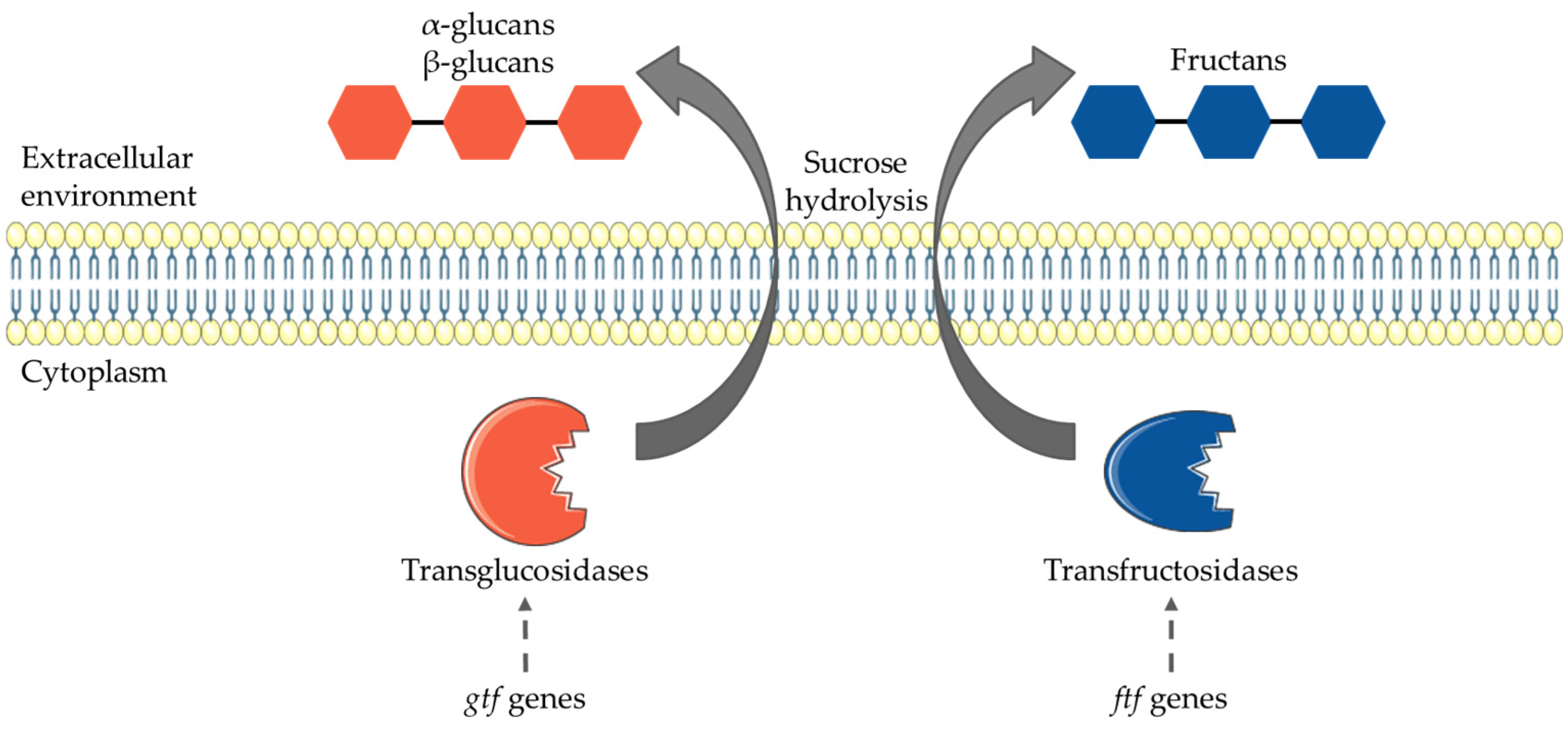
Biosynthesis of HoPS, described in Weissella, Leuconostoc, Lactobacillus and Pediococcus genera, is performed by extracellular enzymes, glucansucrases or fructansucrases (Figure 2) [6]. Fructansucrase and glucansucrase transfer a monosaccharide from a specific substrate to the growing polysaccharide chains [26]. These enzymes belong to the glycosyltransferase (GTF, E.C. 2.4x.y) group and catalyze the hydrolysis of sugars, the resulting monosaccharide residues being attached to a glycan acceptor chain. These enzymes can be categorized into transglucosidases (E.C. 2.4.1.y; glucan-synthesizing dextran-sucrases, mutansucrases, and reuteransucrases) and transfructosidases (E.C. 2.4.1.y or 2.y; fructan-catalyzing transfructosidases levansucrases and inulosucrases). These proteins are encoded by the gtf and the ftf genes, respectively [12]. Glucansucrases are responsible for glucans and fructans synthesis and belong to the 𝛼-amylase superfamily as part of the glycosides hydrolases (GH), in clan GH-H [2,11].
Glucansucrases are characterized by their ability to cleave the 𝛼-glycosidic linkage between a glucose moiety and another monosaccharide moiety using a catalytic (𝛽/𝛼)8-barrel domain which determines the reaction specificity [27]. Moreover, small differences in the amino acid sequence can influence the product specificity. Regarding the three-dimensional structures of glucansucrases, the catalytic core contains three domains to which two extra domains are attached, namely IV and V domains. Some of the domains are made up from two discontinuous segments of the polypeptide chain, resulting in a U-shaped structure [27].

3.4. HePS Production Pathway

HePS synthesis involves several steps: sugar transportation into the cytoplasm of bacterial cells, sugar nucleotide and sugar-1-phosphate (sugar nucleotide donor substrate) synthesis, repeating unit synthesis, polymerization of the sugar repeated units, and lastly, EPS exportation from the cell (Figure 3) [8,12]. During this last step, EPS can be secreted out as a slimy polymer or by adhering to the cell wall to form a capsule [12].
Enzymes, transporters and regulators are encoded by genes of plasmid replication origins in mesophilic Lactobacillus strains and of chromosomal replication origins in the thermophilic ones [12].
Transportation and phosphorylation of sugars mainly occur using the phosphoenolpyruvate-phosphotransferase system (PEP-PTS) in which two protein groups link sugar residues to the carriers and are in charge of the intermembrane transport and sugar phosphorylation [11]. Regarding sugar nucleotide formation, lactose-6-phosphate is hydrolyzed by phospho-β-galactosidase to produce glucose-6-phosphate and galactose-6-phosphate. Likewise, lactose is hydrolyzed by β-galactosidase to produce glucose and galactose. Then, galactose-6-phosphate and glucose-6-phosphate are transformed into glucose-1-phosphate, which will later be converted into UDP-glucose, UDP-galactose and dTDP-rhamnose. The glucose-6-phosphate branch-point is involved into carbon distribution between metabolic pathways. The role of NAD+/NADH balance as a regulation factor for carbon distribution into pathways was proposed and engineering of intracellular redox level was showed to be a potent way to increase HePS production level [28].
Sugar-nucleotide precursors, such as UDP-glucose and UDP-galactose, formed intracellularly, polymerize to form HePS. These precursors do not only serve for EPS production and are also used for various intracellular polysaccharides synthesis. The synthesis of precursors leading to HePS is controlled by glycosyltransferase genes which are located in an operon with genes involved in regulation, chain length determination, polymerization and cell export. Diversity of HePS produced by LAB is due to the variability of glycosyltransferase genes in the operon [2]. Indeed, overexpression of a complete eps gene cluster in Lactococcus lactis was shown to induce higher EPS production levels. Likewise, a slight increase in EPS production was observed when the NIZO B40 priming glycosyltransferase gene epsD was overexpressed [28].
Finally, the formation, transportation and polymerization of EPS-repeating units involve the Wzx/Wzy pathway. The repeating units are polymerized to long-chain HePS by several polymerases, namely Wzy, and exported from the cell by a flippase, namely Wzx (Figure 3) [11].

4. Enzyme Activities, Genes Involved and HoPS Synthesis Pathway Regulations

4.1. Dextransucrase Activity and In vitro Enzyme Properties

Several LAB selected for their ability to produce EPS showed a dextransucrase activity. These strains belong to Weissella, Leuconostoc and Streptococcus genera. Weissella species were able to ferment cellobiose, fructose, galactose, maltose, ribose, and sucrose whereas Leuconostoc species fermented melibiose and arabinose. Regarding dextransucrase activity, Leuconostoc isolates demonstrated the highest activities, followed by Weissella and Streptococcus isolates, respectively [29].
For further analyzes, the most promising strains were selected and identified as W. confusa KIBGE-IB38 and W. confusa KIBGE-IB39 [29]. To improve dextransucrase activity, the authors modified one fermentation parameter at a time. They demonstrated a maximum enzyme production of 1.508 dextransucrase units·mg−1 after 8 h of incubation. For the W. confusa strains tested, higher enzyme activity was reached at 25 °C and was not altered up to 30 °C. An increase in the pH of the medium resulted in an increase in enzyme activity. Indeed, this activity was enhanced at pH 7.5 and decreased at higher pH as well as at pH values of 6.0 and 6.5. Optimization of fermentation conditions improved dextransucrase activity and thus dextran production by W. confusa [29].
Bounaix et al. [30] characterized dextransucrase production by dextran-producing W. cibaria strains isolated from sourdough. They evaluated the production level of dextransucrase using sucrose or glucose as the carbon source and showed that dextransucrase activity was 1.5-fold higher when glucose was used instead of sucrose. Using electrophoretic analyses and zymograms, the authors determined a soluble dextransucrase at 180 kDa regardless of the carbon source used (sucrose or glucose) [30]. Using a recombinant W. cibaria DSRWC glucansucrase (rDSRWC), Kang et al. [31] assessed optimal parameters for its activity and showed the highest dextran production at pH 5.2 and 30 °C. However, 62% of the original activity remained at temperatures down to 20 °C. Likewise, 80% of the original activity remained within a pH range of 5.0–6.0 but a rapid decrease occurred outside this pH range [31].
Amari et al. [32] have studied the complete sequence of a dextransucrase from W. confusa LBAE C39-2 and W. cibaria LBAE K39 strains isolated from traditional French wheat sourdoughs. When dextransucrase gene from W. confusa LBAE C39-2 was cloned and expressed in Escherichia coli, results showed 75% of residual activity at pH 4.5–6 and 60% at pH 4.0, suggesting that this enzyme is active in an acidic environment. Using sucrose as a substrate, dextransucrase activity was enhanced at a temperature range of 35 to 40 °C. This study also illustrated that the enzyme remains during storage at 4 °C when conserved in 20 mM sodium acetate at pH 5.4 with about 10% activity loss after 6 days and 35% after 20 days of storage. The molecular weight of produced EPS was 2 × 10⁶ Da. Further analysis revealed that the dextran obtained was composed of α-(1 → 6)-linked D-glucopyranosyl units with α-(1 → 3)-linked branches [32].
Dextransucrases isolated from Lb. hordei TMW 1.1822 allowed dextran production only when sucrose was present [33]. The authors confirmed a pH-dependent release of dextransucrases in LAB and optimal activities at pH 4.0–4.5. This study suggested that controlling the pH during EPS production can be an asset to enhance production of dextrans with varying properties of dextransucrase. Maximum dextran production was obtained when Lb. hordei TMW 1.1822 was grown at pH 4.0 and 4.5 or when dextransucrases were recovered at these pH values. However, high concentrations of dextrans can be produced at pH 5.0 to pH 6.0 if the enzymes were recovered at pH 4.5, reducing the denaturation and the loss of transglycosylation activity of dextransucrases at a non-optimum pH. A pH increase resulted in an increase in molecular mass (and thus functional properties) of the dextrans produced by Lb. hordei TMW 1.1822. Likewise, the activity of a dextransucrase from W. cibaria RBA 12 isolated from pumelo pulp was influenced by the pH of the reaction mixture. In fact, a higher activity was observed at pH 5.4 and a decreased aboved this value. In addition, dextransucrase activity seems to be influenced by the temperature, as a loss of activity was observed at temperatures beyond 40 °C [34].
Taken together, these data indicate that an exhaustive comprehension of the parameters influencing biosynthesis pathways for each selected strain is needed for efficient EPS production in fermented fruit and vegetables. Fermented conditions should be optimized as, given the notable low pH, fruit and vegetables’ natural environment could be considered as unfavorable according to the strain. Their possibility of modulation could be limited by sensorial aspect and consumer acceptability of the final products.

4.2. Genes Involved in Production of HoPS

Several LAB strains, such as Lb. plantarum, L. pseudomesenteroides/mesenteroides and W. cibaria, were isolated from Turkish sourdough to determine their EPS production and the EPS genes involved were screened. All strains possessed eps genes required for EPS biosynthesis (Table 1) [35].
Most LAB producing HoPS harbor only one glucansucrase gene; however, some LAB genomes have more than one gene encoding the enzyme and are thus able to synthesize different HoPS. For instance, the L. mesenteroides NRRL B-512F strain coding both a glucansucrase and fructansucrase can produce levan in addition to dextran usually produced [2].
Leuconostoc spp. produces insoluble glucans. More precisely, L. mesenteroides NRRL B-1118 (ATCC 8293) generates insoluble dextran. Côté et al. [38] cloned the DsrI gene (referring to dextransucrase insoluble product) from L. mesenteroides NRRL B-1118 [38]. Besrour-Aouam et al. [23] characterized the dsrLL gene encoding DsrLL of L. lactis AV1n. The analysis revealed 97–99% identity of dsrLL with homologous genes from L. lactis, L. garlicum or L. mesenteroides. Dsr coding genes are also found in the chromosomes of Streptococcus, Leuconostoc and Lactobacillus species. The DsrLL enzyme is composed of 1500 amino acids. A signal peptide sequence in the N-terminal region of the protein allows translocation of the enzyme through the cell membrane leading to the protein secretion extracellularly where the Dsr acts. Further analysis showed that the DsrLL enzyme was bound to the cell wall, suggesting that this enzyme synthesizes dextran attached to the cell surface to protect the bacterial cell from extracellular stress [23]. Expression analysis revealed that dsrLL transcription was nor modulated by carbon source neither by growth temperature. An increase in EPS production level according to the presence of sucrose or low temperature probably resulted from post-transcriptional regulations, such as a higher turn-over of Dsr protein at lower temperatures [23].
The glucansucrase DSRWC was isolated from W. cibaria CMU from human saliva. The corresponding gene, namely dsrWC, encodes an extracellular enzyme that produces glucans with only α-(1 → 6) linkages [31].
W. confusa MBF8-1 strain isolated from a homemade Indonesian fermented soybean product has at least two gtf genes encoding glucansucrase enzymes. These genes, gtf8-1A and gtf8-1B, encode Gtf8-1A and Gtf8-1B glucansucrases which may then be exported by the secretory (Sec)-dependent pathway [36].
Pediococcus species EPS production is linked to the presence of plasmids containing a glycosyltransferase gene, namely dps. This gene leads to Gtf production which polymerizes UDP-glucose into β-glucans [14].
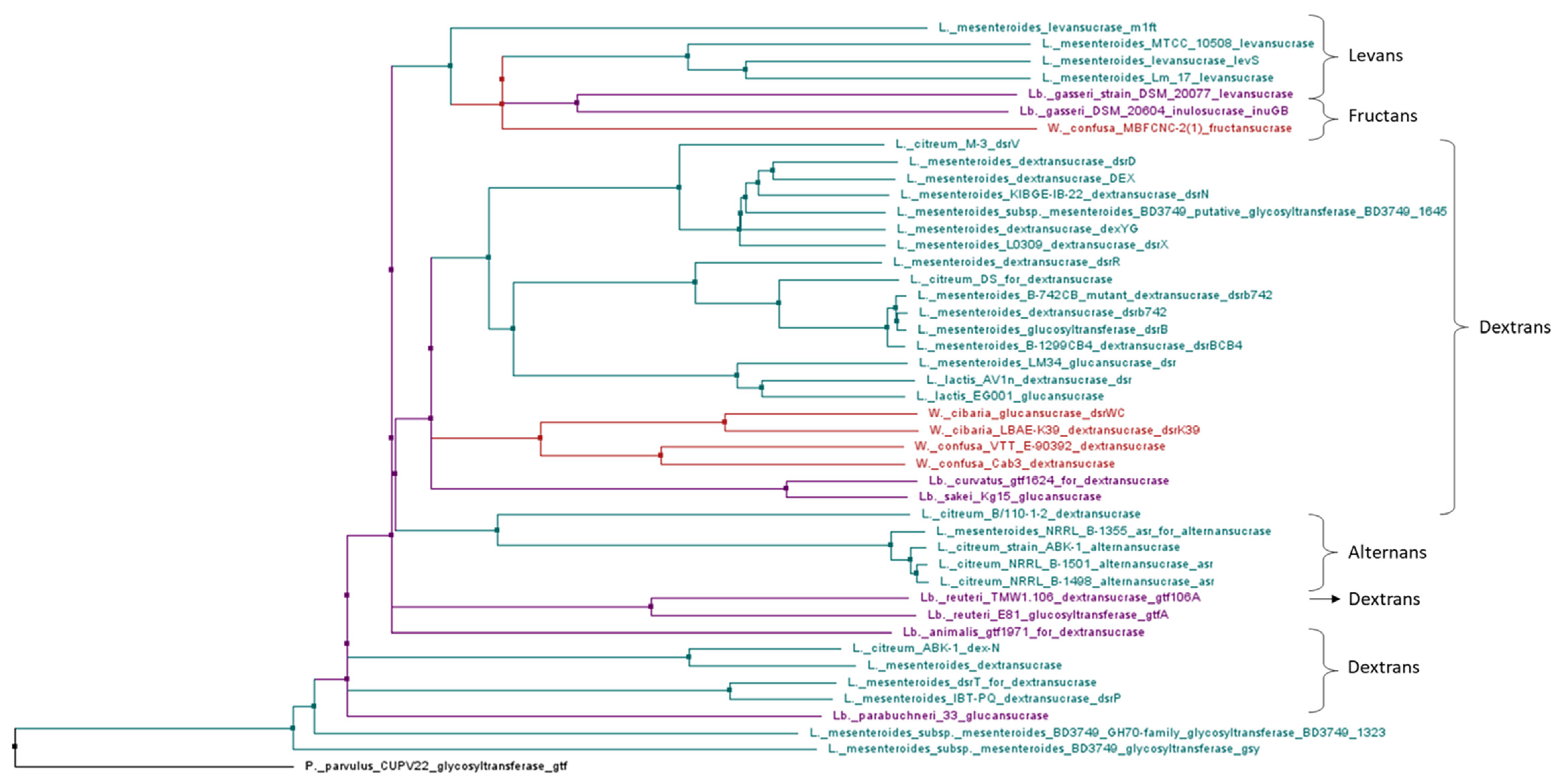
Approximately 150 glucansucrase genes have been sequenced and about a third correspond to functional genes [27]. Sequence analysis of glucansucrase encoding genes from Lactobacillus, Leuconostoc, Weissella and Pediococcus species confirmed that these LAB can be clustered according to the specificity of the EPS glycosidic bonds, rather than the 16S rRNA taxonomy (Figure 4).

5. LAB Producing EPS

Various LAB strains can be isolated from fruit and vegetables and/or perform spontaneous fermentation [39,40,41], but only a few were demonstrated to be able to produce EPS, with variable characteristics according to the strain as listed in Table 2.

5.1. L. mesenteroides and L. pseudomesenteroides

Leuconostoc were isolated from fruit and vegetables traditionally fermented in Romania, green tomato, cauliflower, and carrot. Production by L. mesenteroides/pseudomesenteroides was evaluated at 15 g/L. Results also showed that colonies belonging to Leuconostoc spp. have a mucoid appearance on sucrose-supplemented MRS [51]. Juvonen et al. [6] confirmed an important EPS production by L. pseudomesenteroides when sucrose was present in the medium. This strain produced dextran with 14% α-(1 → 4) linkages. Moreover, adjustments of saccharose concentration in fermented carrot puree suggested that L. mesenteroides also produces levan-type fructan. However, total concentration of dextran and fructan was not in line with the sucrose consumption observed for the strain. The authors hypothesized that this L. mesenteroides strain could use sucrose as an energy source or for production of a dextran with more linkages [6]. L. mesenteroides also produced lactic acid, acetic acid, and mannitol. Sensory analysis carried out on carrot puree fermented with L. mesenteroides showed a strong acidity of smell and taste and poor sweetness. Results indicated a positive correlation (R2 > 0.77) between concentrations of lactic acid plus acetic acid and the perceived acidity and a negative correlation (R2 < −0.89) with sweetness. Regarding the appearance of these purees, fermentation with L. mesenteroides enhanced the matrix thickness [6]. This bacterial species is used in the food industry because of its ability to produce dextran up to 20 g/L [2].
Recently, Abdalrahim et al. [42] showed that L. pseudomesenteroides and L. mesenteroides isolated from fermented fruits and vegetables and from dairy products (potato, gawava, molokhai, kiwi, labneh) produced EPS from sucrose. EPS concentrations of the seven tested strains of L. pseudomesenteroides/mesenteroides ranged from 18.08 to 61.9 g/L [42].

5.2. Leuconostoc lactis

EPS production of L. lactis AV1n isolated from Tunisian avocado yielded 2.25 g/L. Further characterization showed that EPS produced were 𝛼-glucans composed of glucose units. More precisely, these HoPS contain a main chain of glucopyranose units with 35% of 𝛼-(1 → 6) linkages with side chains made of a 𝛼-glucopyranose unit, allowing their classification as dextran-type HoPS [23]. L. lactis was occasionally isolated from cabbage, especially kimchi. The genome of the strain CCK940 producing EPS was sequenced, confirming the presence of genes possibly encoding enzymes of EPS synthesis [22,52].

5.3. Weissella cibaria

W. cibaria is a bacterial strain able to produce dextran up to 36 g/L which is of great interest at the agri-food level [2]. A W. cibaria strain was isolated from cabbage to assess its EPS production. Results showed dextran production of 2.4 g/dL linked to a dextransucrase activity. Dextran exhibited a linear structure with consecutive ⍺-(1 → 6) linkages and 3.4% of ⍺-(1 → 3) linked branches. Moreover, a gel permeation chromatography analysis determined the molecular weight of this dextran at approximately 2 × 10⁶ Da [43].
W. cibaria/confusa strains isolated from spontaneously fermented Malian sour milk or cassava produced glucan-type EPS in culture medium supplemented with sucrose [44]. From 123 strains assayed all producing dextran, 18 additionally produced fructan.

5.4. Lactobacillus Species

Lactobacillus sp. are less efficient producers of EPS compared to other LAB, because they produce mainly HePS known for having a small production yield, which make their production uneconomical when used for the food industry. EPS produced by these LAB are mainly synthesized during the exponential growth phase and there is a decrease in their concentration at the end of the fermentation, which suggests that EPS could be used as alternative carbon sources [12].
Lactobacillus strains isolated from human intestinal microbiota were grown in MRS-C agar (de Man, Rogosa, Sharpe supplemented with 0.25% L-cysteine) for 5 days to provide enough EPS for further characterization. Identified species include Lb. plantarum, Lactobacillus casei, Lb. rhamnosus and Lactobacillus vaginalis. All EPS isolated from these strains were composed of galactose and glucose and 52% of them contained rhamnose. This latter monosaccharide content is higher than the concentration found from bacterial strains isolated from food. The major monosaccharide was strain-dependent but glucose, and rhamnose when present, were generally the most abundant. Screening of genes involved in EPS synthesis confirmed that EPS produced by Lactobacillus were HePS [50].

5.4.1. Lactobacillus plantarum

Lb. plantarum is an EPS-producing LAB with various functions well investigated. Lb. plantarum strains are tolerant to bile and gastric juices, are able to adhere to the intestinal epithelium and have antimicrobial properties [9].
The EPS produced by Lb. plantarum NTU 102 contained fructose, arabinose, galactose, glucose, and mannose, and are, therefore, HePS [9,45]. EPS produced by Lb. plantarum YO175 and OF101 isolated from a Nigerian fermented food had a molecular weight of 1.2 × 10⁶ and 4.4 × 10⁵ Da, respectively [53]. Chemical composition revealed a total carbohydrate content of 87.1% for EPS-YO175 and 80.6% for EPS-OF101. More precisely, these EPS were composed of glucose and galactose for YO175 and glucose for OF101. Further analyses showed that these EPS have an important antioxidant capacity [53].
A Lb. plantarum strain isolated from Turkish sourdough showed an average EPS production of 1153.8 µg/107 cells at 30 °C [35]. Two strains of Lb. plantarum, NTMI05 and NTMI20, isolated from cow milk for their ability to produce EPS, showed EPS yields of 197 mg/L and 187 mg/L, respectively [16]. The greatest production was obtained when glucose (20 g/L) was used as the carbon source in the growth media. The production was also improved for the two studied strains by the addition of yeast extract. This effect was explained by yeast extract composition which includes amino acids, peptides, carbohydrates, and salts. In addition, a high EPS production was observed at an incubation time of 72 h, which corresponds to the most suitable period for enzymatic activity and polysaccharide metabolism rate. Analysis of EPS chemical composition indicated a total carbohydrate content of 95.45% for Lb. plantarum NTMI05 and 92.35% for Lb. plantarum NTMI20, the absence of protein or nucleic acid and a production of lactic acid of 14 mg/mL for NTMI05 and 11 mg/mL for NTMI20. Monosaccharide composition analysis of EPS produced by these strains revealed the presence of galactose [16].
It has been shown that EPS produced in response to environmental conditions, such as temperature, pH and light, by Lb. plantarum and W. cibaria are resistant to gastric and intestinal digestions and are also able to promote beneficial intestinal bacteria [2,3].
Characterization of an ⍺-D-glucan isolated from Lb. plantarum DM5 showed that it comprises glucose monomers with an average molecular mass of 1.11 × 10⁶ Da. They are linked through 86.5% ⍺-(1 → 6) linkage and 13.5% 𝛼-(1 → 3) branching [47].

5.4.2. Lactobacillus fermentum

Lb. fermentum stains have been isolated from the spontaneous fermentation of ‘Almagro’ eggplants [41,54] and have been assayed for their functional and technological properties [48]. EPS production by Lb. fermentum 139, Lb. fermentum 263 and Lb. fermentum 296 was 47.4 mg/L, 55.1 mg/L and 55.6 mg/L, respectively. Lb. fermentum Lf2 is able to produce EPS to levels of 1 g/L. This production can be further improved by modifying the medium composition and culture conditions. Indeed, the highest influence on EPS production was observed by changing the proportions of the nitrogen and type of carbon sources as well as the pH. EPS production at 300 and 600 mg/L from Lb. fermentum increased yogurt hardness and consistency without altering sensory and syneresis properties. EPS produced by Lb. fermentum consist of a β-glucan and two HePS composed of glucose and galactose. Lb. fermentum MC3 was studied to evaluate its ability to produce EPS using various carbohydrate sources. Supplementation of the growth medium with different sugars significantly improved EPS production, with the highest yield at 178.2 mg/L when glucose was added [48].

5.4.3. Lactobacillus rhamnosus

Lb. rhamnosus strains interest for fruit juice LAF was studied through their viability in acidic conditions [55,56,57], allowing to survive in fruit juice and gastrointestinal tract. Interestingly, the Lb. rhamnosus 9595 strain was described to produce 2.7 g/L of EPS, which is one of the highest concentrations reached by a Lactobacillus strain. The EPS obtained contained rhamnose, glucose and galactose [49].

5.5. Other LAB Species

5.5.1. Pediococcus spp.

Pediococcus pentosaseus is the species of the genus the most frequently found in fermented vegetables [58,59]. P. damnosus, P. parvulus and P. pentosaceus can cause “ropiness ˝ of wine, which is hypothesized to increase resistance to low pH, high ethanol content and sulfur dioxide [14].
Pediococcus species involved in winemaking can synthesize EPS such as β-glucans of 500–2000 kDa composed of d-glucose with β-(1 → 3) linkages and β-(1 → 2) branches made up of single units [14]. Genes encoding EPS synthesis are located on a plasmid. These EPS enhance the beverage viscosity, affect filtration, and modulate the mouthfeel from a concentration as low as 20–30 mg/L, and production above 100 mg/L results in an unpalatable wine.

5.5.2. Bifidobacterium spp.

Bifidobacterium animalis, Bifidobacterium longum and Bifidobacterium pseudocatenulatum isolated from human intestinal microbiota and grown in MRS-C agar (de Man, Rogosa, Sharpe supplemented with 0.25% L-cysteine) produced EPS composed of galactose and glucose [50]. Some of the EPS also contained rhamnose in higher proportion compared to those produced by LAB isolated from food. The identification of genes involved in synthesis of EPS produced by Bifidobacterium sp. confirmed HePS production [50].
Bifidobacterium sp. ability to survive in acidic environment is a crucial parameter for fruits LAF. One of the more acid resistant strains among Bifidobacterium species, Bifidobacterium animalis, has been investigated for stability in an orange, grape, passion fruit mixed juice [60] and showed a lower viability compared to milk and at room temperature. Therefore, these data of cultures alone do not give an accurate enough prediction of the strain versatile functionality in adverse conditions and should be more investigated. Recently, B. animalis subsp. lactis B94 was specifically selected due to it better growth and viability in Durian pulp fermentation and inoculated in combination with other LAB in order to extend this fruit preservation and to reduce the spoiling and wasting of large volume of Durian fruit [61]. The use of Bifidobacterium spp. as probiotics in non-dairy beverages is of interest for lactose-intolerant people, and research towards their incorporation in fruit or vegetable based foods is wide [62].

6. Production of EPS during Lactic Acid Fermentation of Fruit and Vegetables and Consequences on Food Quality

The production of EPS by LAB is widely considered for fermented milk-based products, where they can contribute to the texture and physical stabilization, while not being considered as food additives. The effect of EPS production in fruit and vegetable products was recently examined to achieve consumer sensory satisfaction while avoiding the use of hydrocolloid additives in industrial foods.
Depending on their rheological properties, EPS can be used as emulsifiers or stabilizers in the food industry [9,47]. EPS produced by LAB led to a modification of rheological properties linked to their composition, structure, size, and charge [6,10]. For example, β-glucans highly promoted viscosity and elasticity of food matrix regardless of their concentration, thanks to their linear structure and to their production in an acidic environment. On the contrary, bacterial strains producing dextran led to a moderate acid formation and decrease in pH [6]. Enhancing rheological properties of fermented products is an asset for consumer consumption by improving organoleptic quality, appealing appearance and pleasant mouthfeel [10], which increased the gustatory sensations of the consumers [12].
Zheng et al. [5] underlined an EPS production in fresh litchi juices fermented with Lb. casei at 5 log CFU/mL, reaching 7.07 g/L after 18 h of fermentation. Although EPS have not been characterized, this production resulted in an improvement of the viscosity of this matrix which increased from 4.7 mPas initially to 84.8 mPas after fermentation. Moreover, fructose concentration was also higher after fermentation, which showed that enzymes partially involved in EPS production were glucansucrases [5].
In another study, EPS production in vegetable matrixes was assessed in agar medium containing 30% of carrot puree and supplemented with 5% sucrose [6]. EPS production assayed for 37 LAB strains was positive for Lactobacillus spp. except Lb. plantarum strains, several L. mesenteroides strains and one W. cibaria strain. According to these results L. mesenteroides strains producing EPS were selected for carrot puree fermentation and the presence of water-soluble low branching dextrans was analyzed using an enzymatic method. Results showed a dextran concentration of 0.2% for two strains (out of six) and production of fructan-type levans for one of them. On the other hand, viscosity increased for fermented carrot purees since L. mesenteroides increased the viscosity from 50% to 270% compared to non-fermented samples. Likewise, fermentation changes the texture of purees by thickening through EPS production [6].
Similarly, Han et al. [63] showed EPS production by a L. mesenteroides strain used for fermentation of a tomato juice supplemented with 15% sucrose. Monosaccharides of the EPS produced by L. mesenteroides were identified using high-performance anion-exchange chromatography equipped with a pulsed amperometric detector (HPAEC-PAD) and the only monosaccharide present was glucose [63]. Dextran concentration obtained was 32.15 g/L, which approximately corresponds to 60% of glucosyl units of metabolized sucrose. This increased dextran concentration was consistent with the observed increases of bacterial growth and viscosity. Viscosity increased up to 451 CP (centipoises) during fermentation with L. mesenteroides, which is a modification of the matrix commonly observed during dextran production. The analysis of sugars in the tomato juice showed that after 48 h of fermentation, sucrose concentration initially at 15.10% decreased to 4.11% whereas fructose concentration increased from 1.11% to 4.93%. Observation of fructose content increase, derived from sucrose, is characteristic of dextran synthesis by this LAB [63].
EPS solubility is associated with their molecular weight and branching percentage. The porous matrix structure formed by polysaccharides chains is linked to their solubility and water holding capacity as large amounts of water can be held by hydrogen linkages. Some electrostatic interaction capacities were also described with charged proteins, resulting in the modification of the product elasticity [10].

7. Health Benefits of EPS

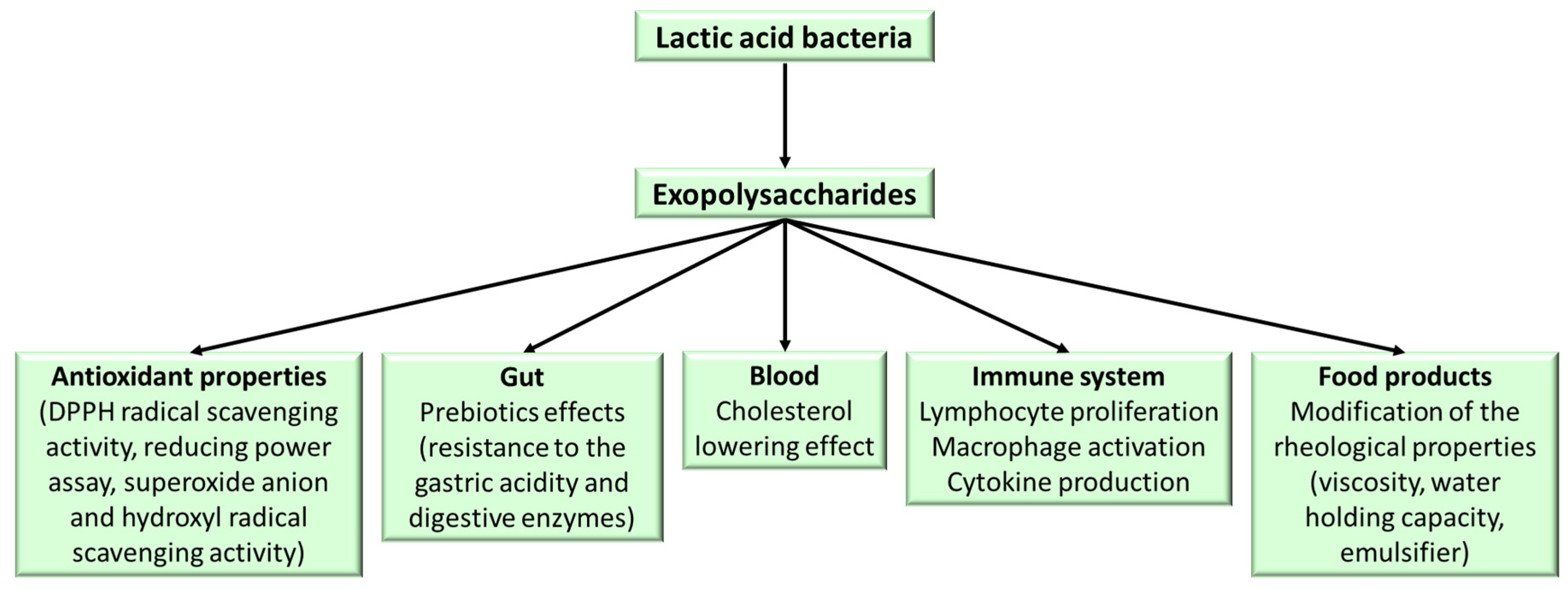
Besides technological and organoleptic properties, the EPS produced by LAB are associated to various biological properties, such as prebiotic, antioxidant, anti-inflammatory, cholesterol lowering capacities (Figure 5), and health benefits [64]. The studies conducted on EPS produced by LAB, showed a direct influence on biological potential of the chemical nature of EPS, whether it be monosaccharide composition, glycosidic linkage or chemical modifications [65]. Non-dairy sources, including fruit and vegetables, are nowadays targeted to explore their health promoting effects, as novel EPS are identified from different resources with outstanding benefits.

7.1. Prebiotic Properties

Gut microbiota play a major role in immunity against pathogens by contributing to the immune barrier role of intestinal mucosa [66,67,68]. The composition of intestinal microbiota is under the influence of multiple factors, including diet, that be linked to changes in microbial diversity [69,70]. Probiotics, but also prebiotics allowing the growth of specific microorganisms, including EPS, are widely studied in order to restore a balance in the intestinal microbiota of patients [9,69].
Das et al. [47] have shown that 𝛼-D-glucan produced by Lb. plantarum DM5 stimulates B.infantis and Lb. acidophilus growth, but also its own growth with a population increase from 6.09 to 9 log10 CFU/mL in vitro. However, the growth of non-probiotic strains, E. coli and Enterococcus aerogenes, was not stimulated. An important criterion for prebiotic selection is their resistance to gastric acidity and digestive enzymes, so that they can reach the colon and be fermented by probiotic bacteria, including hydrolysis which allows absorption. This study showed that EPS produced by Lb. plantarum DM5 were resistant to gastric juice of pH 1–4 and that an increase in pH decreased the percent hydrolysis of glucan-DM5 from 0.39% to 0.19%. Regarding the intestinal fluid, the hydrolysis percentage of glucan-DM5 was 0.21% which demonstrates this EPS high resistance to harsh intestinal environment. The hydrolysis ratio of glucan-DM5 by 𝛼-amylase increased with pH and incubation time from 0.09% to 0.22% after 1 h of incubation at pH 5–8 and from 0.13% to 0.32% after 5 h of incubation at pH 5–8. Inulin, a standard prebiotic used in this study, showed maximum hydrolysis of 19.13% at pH 8 after 5 h of incubation whereas this value was 0.32% for glucan-DM5 under the same conditions, showing significantly higher 𝛼-amylase resistance by glucan-DM5 (99.8%) compared to inulin. All together, these results suggest that 𝛼-D-glucan from Lb. plantarum DM5 will be accessible to the probiotic bacteria as a carbon source in the gastrointestinal tract [47]. As 𝛼-glucans are not degraded in the human digestive tract, they can reach the colon to be fermented by colonic bacteria. Therefore, produced HoPS can be considered as beneficial fibers with an extended liberation of glucose. Prebiotic properties of HoPS produced through glucansucrases have been assessed using in vitro fermentation of gut and colon models. Results have shown that only short oligosaccharides exert prebiotic abilities [27].
EPS are involved in mechanisms of adhesion to surfaces or to other organisms [8]. For fermented products, EPS drives an increase in the retention time in the gastro-intestinal tract resulting in an increased colonization by EPS-producing LAB [8]. In addition, EPS can alter the surface of bacterial cells by inhibiting their ability to bind to the surface of cells or to modulate expression of genes involved in biofilm formation, thus promoting their formation [9]. Resistance to antibacterial compounds is a thousand times higher for microorganisms producing biofilms [8].
Dextran produced by L. mesenteroides XG5 from homemade wine have been investigated for its ability to promote bacterial growth [71]. Supplementation of the growth media with this EPS resulted in a significant stimulation of growth of Bifidobacterium and Lactobacillus species. EPS produced by L. mesenteroides XG5 were not digested in human saliva. Likewise, simulated small intestinal juice did not affect these EPS; however, these molecules were substantially degraded during fecal fermentation. These results suggested that EPS produced by L. mesenteroides XG5 are resistant to gastro-intestinal digestion and are then utilized by the fecal microbiota. During human fecal fermentation, XG5 EPS significantly enhanced Bifidobacterium growth but not Lactobacillus growth. These EPS also enhanced the contents of short-chain fatty acids. The EPS enriched the cecum microbiota of C57BL/6J mice, leading to an increase in Firmicutes and a decrease in the Bacteroidetes to Firmicutes ratio. These microbiota modifications could be an asset for type 1 diabetes patients. These results suggested that EPS produced by L. mesenteroides XG5 could be used as prebiotics given their ability to stimulate probiotic growth [71].

7.2. Antioxidant Activity

The ability to exert antioxidant activity is widespread in LAB-derived EPSs. Imran et al. [16] evaluated the antioxidant activity of EPS produced by Lb. plantarum NTMI05 and NTMI20 by measuring free radical scavenging activity. The results showed an improvement of this activity with 500 µg/mL of EPS with a capacity of 96.62% and 91.86%, respectively, for these two strains. A hypothesis suggested that antioxidant activity could be influenced by the presence of functional groups such as hydroxyl groups [9].
An EPS composed of glucose and rhamnose sugar units produced by W. cibaria strain GA44 demonstrated moderate but significant antioxidant capacities in a dose-dependent manner in various in vitro models such as DPPH radical scavenging activity, reducing power assay, superoxide anion and hydroxyl radical scavenging activity [53]. Similarly, strong and concentration-dependent antioxidant activities were found for EPS from Lb. plantarum C88 and for HoPS from L. pseudomesenteroides JF17 [13,72].
Another study characterizing antioxidant properties of an EPS produced by Lactococcus lactis strain EPS-1 showed an important antioxidant capacity. This EPS was mainly composed of fructose and rhamnose. EPS-1 was also able to inhibit hydroxyl and superoxide radical activities. These results suggested that EPS-1 produced by Lactococcus lactis could reduce lipid peroxidation of cell membranes [73].
EPS from LAB have been studied for their health-benefit properties. It follows that their chemical structure and molecular weight (MW) are relevant characteristics to define their health-promoting functions and activities [74].

7.3. Anti-Inflammatory Activity

EPS produced by LAB are able to stimulate dendritic cells and other antigens presenting genes thanks to Toll-like receptors (TLRs) and to modulate the inflammatory response by leading to the production of pro-inflammatory cytokines [45].
Anti-inflammatory properties have been observed using in vitro and in vivo approaches for EPS. Decreased expression of pro-inflammatory markers has been shown in a rat model of inflammation [75]. Lipopolysaccharide-stimulated RAW 264.7 cells also exhibited lower contents of TNFα, IL-1b and IL-6 [76].
EPS produced by Lb. plantarum NTU 102 promote proliferation of RAW 264.7 macrophages at a concentration below 5 µg/mL but a cytotoxic effect was observed at higher concentrations. Moreover, macrophages treatment with these EPS allow phagocytosis. It was also showed that EPS produced by Lb. plantarum NTU 102 are able to induce a pro-inflammatory response by producing cytokines, such as IL-6, TNF-α and IL-1β, in a dose-dependent manner in treated macrophages [45].

7.4. Cholesterol-Lowering Activity

Recently, studies have investigated the promising cholesterol-lowering effect related to LAB-derived EPS. A high molecular weight EPS of glucomannan nature from Lb. plantarum BR2 showed a strong ability to adsorb cholesterol, exhibiting 47.5% of cholesterol lowering ability on in vitro assimilation procedure [77]. Similarly, an EPS consisting of mannose, glucose and galactose produced by Enterococcus faecium K1, an isolate from indigenously fermented milk product kalarei, also displayed a 49% cholesterol-lowering property [78].
In vivo, the effect of water-soluble EPS from kefir was determined in C57BL/6J mice by Lim et al. [79]. The EPS supplementation had significant anti-obesity effect by reducing HFD-induced body weight gain, adipose tissue weight, and plasma very-low-density lipoprotein cholesterol concentration compared to the control group in this study.
Current hypotheses on the mechanism involved in EPS cholesterol-lowering effect considers elimination of bile, cholesterol assimilation and conversion, coprecipitation effect and short fatty acids promotion [26].

8. Conclusions

EPS production by LAB involved in fruit and vegetable fermentation is gaining interest because of both effects on sensory characteristics and potential positive health effects.
The impact of EPS of food texture can be seen as a strategy to limit the use of additives, but requires a production level of several g/L. To that aim, the production of HoPS is more favorable than HePS as several LAB species, such as L. pseudomesenteroides, L. mesenteroides, W. cibaria and W. confusa produce these compounds at high levels. Moreover, production level of EPS can be enhanced through adjustment of fermentation medium composition and incubation parameters. However, this optimization should not decrease the general sensory quality of fruit or vegetable fermented foods.
EPS produced during fermentation of fruit or vegetables can exert prebiotic activity, but also antioxidant, anti-inflammatory and cholesterol-lowering activity. Most of previous results were obtained in vitro, on cell models or in mice, and will require further investigations to determine if the observed effects can be expected from human diet modifications.

Supplementary Materials

The following are available online at https://www.mdpi.com/2311-5637/6/4/115/s1, Table S1: Detailed information corresponding to the gene sequences used to achieve the phylogenetic tree of genes involved in glucansucrases synthesis.

Author Contributions

All authors contributed to writing and editing this review. All authors have read and agreed to the published version of the manuscript.

Funding

Part of this work was funded by La Reunion regional council (M.G.) and by Federation BioST from University of La Reunion.

Conflicts of Interest

The authors declare no conflict of interest.

References

  1. Fessard, A.; Kapoor, A.; Patche, J.; Assemat, S.; Hoarau, M.; Bourdon, E.; Bahorun, T.; Remize, F. Lactic fermentation as an efficient tool to enhance the antioxidant activity of tropical fruit juices and teas. Microorganisms 2017, 5, 23. [Google Scholar] [CrossRef] [PubMed]
  2. Lynch, K.M.; Zannini, E.; Coffey, A.; Arendt, E.K. Lactic acid bacteria exopolysaccharides in foods and beverages: Isolation, properties, characterization, and health benefits. Annu. Rev. Food Sci. Technol. 2018, 9, 155–176. [Google Scholar] [CrossRef] [PubMed]
  3. Caggianiello, G.; Kleerebezem, M.; Spano, G. Exopolysaccharides produced by lactic acid bacteria: From health-promoting benefits to stress tolerance mechanisms. Appl. Microbiol. Biotechnol. 2016, 100, 3877–3886. [Google Scholar] [CrossRef]
  4. Campbell-Sills, H.; El Khoury, M.; Favier, M.; Romano, A.; Biasioli, F.; Spano, G.; Sherman, D.J.; Bouchez, O.; Coton, E.; Coton, M.; et al. Phylogenomic analysis of Oenococcus oeni reveals specific domestication of strains to cider and wines. Genome Biol. Evol. 2015, 7, 1506–1518. [Google Scholar] [CrossRef] [PubMed] [Green Version]
  5. Zheng, X.; Yu, Y.; Xiao, G.; Xu, Y.; Wu, J.; Tang, D.; Zhang, Y. Comparing product stability of probiotic beverages using litchi juice treated by high hydrostatic pressure and heat as substrates. Innov. Food Sci. Emerg. Technol. 2014, 23, 61–67. [Google Scholar] [CrossRef]
  6. Juvonen, R.; Honkapää, K.; Maina, N.H.; Shi, Q.; Viljanen, K.; Maaheimo, H.; Virkki, L.; Tenkanen, M.; Lantto, R. The impact of fermentation with exopolysaccharide producing lactic acid bacteria on rheological, chemical and sensory properties of pureed carrots (Daucus carota L.). Int. J. Food Microbiol. 2015, 207, 109–118. [Google Scholar] [CrossRef]
  7. Poli, A.; Di Donato, P.; Abbamondi, G.R.; Nicolaus, B. Synthesis, production, and biotechnological applications of exopolysaccharides and polyhydroxyalkanoates by Archaea. Archaea 2011, 2011, 693253. [Google Scholar] [CrossRef] [Green Version]
  8. Ripari, V. Techno-functional role of exopolysaccharides in cereal-based, yogurt-like beverages. Beverages 2019, 5, 16. [Google Scholar] [CrossRef] [Green Version]
  9. Silva, L.A.; Lopes Neto, J.H.P.; Cardarelli, H.R. Exopolysaccharides produced by Lactobacillus plantarum: Technological properties, biological activity, and potential application in the food industry. Ann. Microbiol. 2019, 69, 321–328. [Google Scholar] [CrossRef]
  10. Singh, P.; Saini, P. Food and health potentials of exopolysaccharides derived from Lactobacilli. Microbiol. Res. J. Int. 2017, 22, 1–14. [Google Scholar] [CrossRef]
  11. Xu, Y.; Cui, Y.; Yue, F.; Liu, L.; Shan, Y.; Liu, B.; Zhou, Y.; Lü, X. Exopolysaccharides produced by lactic acid bacteria and Bifidobacteria: Structures, physiochemical functions and applications in the food industry. Food Hydrocoll. 2019, 94, 475–499. [Google Scholar] [CrossRef]
  12. Oleksy, M.; Klewicka, E. Exopolysaccharides produced by Lactobacillus sp.: Biosynthesis and applications. Crit. Rev. Food Sci. Nutr. 2018, 58, 450–462. [Google Scholar] [CrossRef]
  13. Farinazzo, F.S.; Valente, L.J.; Almeida, M.B.; Simionato, A.S.; Thereza, M.; Fernandes, C.; Saori, C.; Mauro, I.; Aparecida, A.; Tomal, B.; et al. Characterization and antioxidant activity of an exopolysaccharide produced by Leuconostoc pseudomesenteroides JF17 from juçara fruits (Euterpe edulis Martius). Process Biochem. 2020, 91, 141–148. [Google Scholar] [CrossRef]
  14. Wade, M.E.; Strickland, M.T.; Osborne, J.P.; Edwards, C.G. Role of Pediococcus in winemaking. Aust. J. Grape Wine Res. 2018, 25, 7–24. [Google Scholar] [CrossRef] [Green Version]
  15. Cerning, J. Exocellular polysaccharides produced by lactic acid bacteria. FEMS Microbiol. Lett. 1990, 87, 113–130. [Google Scholar] [CrossRef] [PubMed]
  16. Imran, M.Y.M.; Reehana, N.; Jayaraj, K.A.; Ahamed, A.A.P.; Dhanasekaran, D.; Thajuddin, N.; Alharbi, N.S.; Muralitharan, G. Statistical optimization of exopolysaccharide production by Lactobacillus plantarum NTMI05 and NTMI20. Int. J. Biol. Macromol. 2016, 93, 731–745. [Google Scholar] [CrossRef] [PubMed]
  17. Ciqual: Table de Composition Nutritionnelle des Aliments. Available online: https://ciqual.anses.fr/ (accessed on 23 October 2020).
  18. Guerrero, S.N.; Alzamora, S.M. Effect of pH, Temperature and Glucose Addition on Flow Behaviour of Fruit Purees: II. Peach, Papaya and Mango Purées. J. Food Eng. 1998, 37, 77–101. [Google Scholar] [CrossRef]
  19. Laloknam, S.; Sirisopana, S.; Phornphisutthimas, S. Learning retention in undergraduate biology using a hands-on practical “enzyme detection from vegetables and fruits”. J. Chem. 2010, 4, 29–35. [Google Scholar]
  20. Yu, Y.J.; Chen, Z.; Chen, P.T.; Ng, I.S. Production, characterization and antibacterial activity of exopolysaccharide from a newly isolated Weissella cibaria under sucrose effect. J. Biosci. Bioeng. 2018, 126, 769–777. [Google Scholar] [CrossRef] [PubMed]
  21. Zannini, E.; Mauch, A.; Galle, S.; Gänzle, M.; Coffey, A.; Arendt, E.K.; Taylor, J.P.; Waters, D.M. Barley malt wort fermentation by exopolysaccharide-forming Weissella cibaria MG1 for the production of a novel beverage. J. Appl. Microbiol. 2013, 115, 1379–1387. [Google Scholar] [CrossRef]
  22. Fessard, A.; Remize, F. Genetic and technological characterization of lactic acid bacteria isolated from tropically grown fruits and vegetables. Int. J. Food Microbiol. 2019, 301, 61–72. [Google Scholar] [CrossRef] [PubMed]
  23. Besrour-Aouam, N.; Mohedano, M.L.; Fhoula, I.; Zarour, K.; Najjari, A.; Aznar, R.; Prieto, A.; Ouzari, H.I.; López, P. Different modes of regulation of the expression of dextransucrase in Leuconostoc lactis AV1n and Lactobacillus sakei MN. Front. Microbiol. 2019, 10, 959. [Google Scholar] [CrossRef] [PubMed] [Green Version]
  24. Dertli, E.; Yilmaz, M.T.; Tatlisu, N.B.; Toker, O.S.; Cankurt, H.; Sagdic, O. Effects of in situ exopolysaccharide production and fermentation conditions on physicochemical, microbiological, textural and microstructural properties of Turkish-type fermented sausage (sucuk). Meat Sci. 2016, 121, 156–165. [Google Scholar] [CrossRef] [PubMed]
  25. Ng, I.S.; Xue, C. Enhanced exopolysaccharide production and biological activity of Lactobacillus rhamnosus ZY with calcium and hydrogen peroxide. Process Biochem. 2017, 52, 295–304. [Google Scholar] [CrossRef]
  26. Zhou, Y.; Cui, Y.; Qu, X. Exopolysaccharides of lactic acid bacteria: Structure, bioactivity and associations: A review. Carbohydr. Polym. 2019, 207, 317–332. [Google Scholar] [CrossRef] [PubMed]
  27. Leemhuis, H.; Pijning, T.; Dobruchowska, J.M.; van Leeuwen, S.S.; Kralj, S.; Dijkstra, B.W.; Dijkhuizen, L. Glucansucrases: Three-dimensional structures, reactions, mechanism, α-glucan analysis and their implications in biotechnology and food applications. J. Biotechnol. 2013, 163, 250–272. [Google Scholar] [CrossRef] [Green Version]
  28. Li, N.; Wang, Y.; Zhu, P.; Liu, Z.; Guo, B.; Ren, J. Improvement of exopolysaccharide production in Lactobacillus casei LC2W by overexpression of NADH oxidase gene. Microbiol. Res. 2015, 171, 73–77. [Google Scholar] [CrossRef]
  29. Zafar, S.B.; Siddiqui, N.N.; Shahid, F.; Qader, S.A.U.; Aman, A. Bioprospecting of indigenous resources for the exploration of exopolysaccharide producing lactic acid bacteria. J. Genet. Eng. Biotechnol. 2018, 16, 17–22. [Google Scholar] [CrossRef]
  30. Bounaix, M.S.; Robert, H.; Gabriel, V.; Morel, S.; Remaud-Siméon, M.; Gabriel, B.; Fontagné-Faucher, C. Characterization of dextran-producing Weissella strains isolated from sourdoughs and evidence of constitutive dextransucrase expression. FEMS Microbiol. Lett. 2010, 311, 18–26. [Google Scholar] [CrossRef] [Green Version]
  31. Kang, H.K.; Oh, J.S.; Kim, D. Molecular characterization and expression analysis of the glucansucrase DSRWC from Weissella cibaria synthesizing a α(1→6) glucan. FEMS Microbiol. Lett. 2009, 292, 33–41. [Google Scholar] [CrossRef] [Green Version]
  32. Amari, M.; Arango, L.F.G.; Gabriel, V.; Robert, H.; Morel, S.; Moulis, C.; Gabriel, B.; Remaud-Siméon, M.; Fontagné-Faucher, C. Characterization of a novel dextransucrase from Weissella confusa isolated from sourdough. Appl. Microbiol. Biotechnol. 2013, 97, 5413–5422. [Google Scholar] [CrossRef]
  33. Schmid, J.; Bechtner, J.; Vogel, R.F.; Jakob, F. A systematic approach to study the pH-dependent release, productivity and product specificity of dextransucrases. Microb. Cell Fact. 2019, 18, 153. [Google Scholar] [CrossRef] [PubMed]
  34. Baruah, R.; Deka, B.; Goyal, A. Purification and characterization of dextransucrase from Weissella cibaria RBA12 and its application in in vitro synthesis of prebiotic oligosaccharides in mango and pineapple juices. LWT-Food Sci. Technol. 2017, 84, 449–456. [Google Scholar] [CrossRef]
  35. Dertli, E.; Mercan, E.; Arici, M.; Yilmaz, M.T.; Sağdiç, O. Characterisation of lactic acid bacteria from Turkish sourdough and determination of their exopolysaccharide (EPS) production characteristics. LWT 2016, 71, 116–124. [Google Scholar] [CrossRef]
  36. Heng, N.C.K.; Yeh, C.W.; Malik, A. Draft genome sequence of Weissella confusa MBF8-1, a glucansucrase- and bacteriocinproducing strain isolated from a homemade soy product. Genome Announc. 2017, 5, e01497-16. [Google Scholar] [CrossRef] [Green Version]
  37. Malik, A.; Sheilla, S.; Firdausi, W.; Handayani, T.; Saepudin, E. Sucrase activity and exopolysaccharide partial characterization from three Weissella confusa strains. HAYATI J. Biosci. 2015, 22, 130–135. [Google Scholar] [CrossRef] [Green Version]
  38. Côté, G.L.; Skory, C.D. Cloning, expression, and characterization of an insoluble glucan-producing glucansucrase from Leuconostoc mesenteroides NRRL B-1118. Appl. Microbiol. Biotechnol. 2012, 93, 2387–2394. [Google Scholar] [CrossRef]
  39. Chen, Y.S.; Wu, H.C.; Pan, S.F.; Lin, B.G.; Lin, Y.H.; Tung, W.C.; Li, Y.L.; Chiang, C.M.; Yanagida, F. Isolation and characterization of lactic acid bacteria from yan-taozih (pickled peaches) in Taiwan. Ann. Microbiol. 2013, 63, 607–614. [Google Scholar] [CrossRef]
  40. Montet, D.; Ray, R.C.; Zakhia-Rozis, N. Lactic acid fermentation of vegetables and fruits. In Microorganisms and Fermentation of Traditional Foods; CRC Press: Boca Raton, FL, USA, 2014; pp. 108–140. ISBN 9781482223095. [Google Scholar]
  41. Sánchez, I.; Palop, L.; Ballesteros, C. Biochemical characterization of lactic acid bacteria isolated from spontaneous fermentation of “Almagro” eggplants. Int. J. Food Microbiol. 2000, 59, 9–17. [Google Scholar] [CrossRef]
  42. Abdalrahim, S.; Zohri, A.N.A.; Khider, M.; Kamal El-Dean, A.M.; Abulreesh, H.H.; Ahmad, I.; Elbanna, K. Phenotypic and genotypic characterization of exopolysaccharide producing bacteria isolated from fermented fruits, vegetables and dairy products. J. Pure Appl. Microbiol. 2019, 13, 1349–1362. [Google Scholar] [CrossRef] [Green Version]
  43. Ahmed, R.Z.; Siddiqui, K.; Arman, M.; Ahmed, N. Characterization of high molecular weight dextran produced by Weissella cibaria CMGDEX3. Carbohydr. Polym. 2012, 90, 441–446. [Google Scholar] [CrossRef] [PubMed]
  44. Malang, S.K.; Maina, N.H.; Schwab, C.; Tenkanen, M.; Lacroix, C. Characterization of exopolysaccharide and ropy capsular polysaccharide formation by Weissella. Food Microbiol. 2015, 46, 418–427. [Google Scholar] [CrossRef] [PubMed]
  45. Liu, C.F.; Tseng, K.C.; Chiang, S.S.; Lee, B.H.; Hsu, W.H.; Pan, T.M. Immunomodulatory and antioxidant potential of Lactobacillus exopolysaccharides. J. Sci. Food Agric. 2011, 91, 2284–2291. [Google Scholar] [CrossRef] [PubMed]
  46. Adesulu-Dahunsi, A.T.; Jeyaram, K.; Isiaka Sanni, A.; Banwo, K. Production of exopolysaccharide by strains of Lactobacillus plantarum YO175 and OF101 isolated from traditional fermented cereal beverage. PeerJ 2018, 2018, e5326. [Google Scholar] [CrossRef] [PubMed] [Green Version]
  47. Das, D.; Baruah, R.; Goyal, A. A food additive with prebiotic properties of an α-d-glucan from Lactobacillus plantarum DM5. Int. J. Biol. Macromol. 2014, 69, 20–26. [Google Scholar] [CrossRef] [PubMed]
  48. Ale, E.C.; Rojas, M.F.; Reinheimer, J.A.; Binetti, A.G. Lactobacillus fermentum: Could EPS production ability be responsible for functional properties? Food Microbiol. 2020, 90, 103465. [Google Scholar] [CrossRef]
  49. Badel, S.; Bernardi, T.; Michaud, P. New perspectives for Lactobacilli exopolysaccharides. Biotechnol. Adv. 2011, 29, 54–66. [Google Scholar] [CrossRef]
  50. Salazar, N.; Prieto, A.; Leal, J.A.; Mayo, B.; Bada-Gancedo, J.C.; de los Reyes-Gavilán, C.G.; Ruas-Madiedo, P. Production of exopolysaccharides by Lactobacillus and Bifidobacterium strains of human origin, and metabolic activity of the producing bacteria in milk. J. Dairy Sci. 2009, 92, 4158–4168. [Google Scholar] [CrossRef] [Green Version]
  51. Grosu-Tudor, S.-S.; Zamfir, M. Exopolysaccharide production by selected lactic acid bacteria isolated from fermented vegetables. Sci. Bull. Ser. Biotechnol. 2014, 18, 107–114. [Google Scholar]
  52. Lee, S.; Park, Y.S. Draft genome sequence of oligosaccharide producing Leuconostoc lactis CCK940 isolated from kimchi in Korea. Korean J. Microbiol. 2018, 54, 445–447. [Google Scholar] [CrossRef]
  53. Adesulu-Dahunsi, A.T.; Sanni, A.I.; Jeyaram, K. Production, characterization and in vitro antioxidant activities of exopolysaccharide from Weissella cibaria GA44. LWT-Food Sci. Technol. 2018, 87, 432–442. [Google Scholar] [CrossRef] [Green Version]
  54. Sánchez, I.; Seseña, S.; Palop, L. Identification of lactic acid bacteria from spontaneous fermentation of “Almagro” eggplants by SDS-PAGE whole cell protein fingerprinting. Int. J. Food Microbiol. 2003, 82, 181–189. [Google Scholar] [CrossRef]
  55. Farias, N.; Soares, M.; Gouveia, E. Enhancement of the viability of Lactobacillus rhamnosus ATCC 7469 in passion fruit juice: Application of a central composite rotatable design. LWT-Food Sci. Technol. 2016, 71, 149–154. [Google Scholar] [CrossRef]
  56. Andrade, R.; Santos, E.; Azoubel, P.; Ribeiro, E. Increased survival of Lactobacillus rhamnosus ATCC 7469 in guava juices with simulated gastrointestinal conditions during refrigerated storage. Food Biosci. 2019, 32, 100470. [Google Scholar] [CrossRef]
  57. de Andrade Pires, B.; de Almeida Bianchini Campos, R.C.; Canuto, J.W.; de Melo Carlos Dias, T.; Martins, E.M.F.; Licursi, L.; de Castro Leite Júnior, B.R.; Martins, M.L. Lactobacillus rhamnosus GG in a mixed pineapple (Ananas comosus L. Merril) and jussara (Euterpe edulis Martius) beverage and its survival in the human gastrointestinal tract. LWT 2020, 134, 110028. [Google Scholar] [CrossRef]
  58. Chen, Y.-S.; Wu, H.-C.; Yu, C.-R.; Chen, Z.-Y.; Lu, Y.-C.; Yanagida, F. Isolation and characterization of lactic acid bacteria from xi-gua-mian (fermented watermelon), a traditional fermented food in Taiwan | Italian Journal of Food Science. Ital. J. Food Sci. 2016, 28, 9–14. [Google Scholar]
  59. Xu, X.; Luo, D.; Bao, Y.; Liao, X.; Wu, J. Characterization of diversity and probiotic efficiency of the autochthonous lactic acid bacteria in the fermentation of selected raw fruit and vegetable juices. Front. Microbiol. 2018, 9, 2539. [Google Scholar] [CrossRef] [Green Version]
  60. Saarela, M.; Virkajärvi, I.; Alakomi, H.L.; Sigvart-Mattila, P.; Mättö, J. Stability and functionality of freeze-dried probiotic Bifidobacterium cells during storage in juice and milk. Int. Dairy J. 2006, 16, 1477–1482. [Google Scholar] [CrossRef]
  61. Lu, Y.; Putra, S.D.; Liu, S.Q. A novel non-dairy beverage from durian pulp fermented with selected probiotics and yeast. Int. J. Food Microbiol. 2018, 265, 1–8. [Google Scholar] [CrossRef]
  62. Aspri, M.; Papademas, P.; Tsaltas, D. Review on non-dairy probiotics and their use in non-dairy based products. Fermentation 2020, 6, 30. [Google Scholar] [CrossRef] [Green Version]
  63. Han, J.; Hang, F.; Guo, B.; Liu, Z.; You, C.; Wu, Z. Dextran synthesized by Leuconostoc mesenteroides BD1710 in tomato juice supplemented with sucrose. Carbohydr. Polym. 2014, 112, 556–562. [Google Scholar] [CrossRef] [PubMed]
  64. Zannini, E.; Waters, D.M.; Coffey, A.; Arendt, E.K. Production, properties, and industrial food application of lactic acid bacteria-derived exopolysaccharides. Appl. Microbiol. Biotechnol. 2016, 100, 1121–1135. [Google Scholar] [CrossRef] [PubMed]
  65. Angelin, J.; Kavitha, M. Exopolysaccharides from probiotic bacteria and their health potential. Int. J. Biol. Macromol. 2020, 162, 853–865. [Google Scholar] [CrossRef] [PubMed]
  66. Lynch, S.V.; Pedersen, O. The human intestinal microbiome in health and disease. N. Engl. J. Med. 2016, 375, 2369–2379. [Google Scholar] [CrossRef] [PubMed] [Green Version]
  67. Thaiss, C.A.; Zmora, N.; Levy, M.; Elinav, E. The microbiome and innate immunity. Nature 2016, 535, 65–74. [Google Scholar] [CrossRef]
  68. Honda, K.; Littman, D.R. The microbiota in adaptive immune homeostasis and disease. Nature 2016, 535, 75–84. [Google Scholar] [CrossRef]
  69. Hills, R.D.; Pontefract, B.A.; Mishcon, H.R.; Black, C.A.; Sutton, S.C.; Theberge, C.R. Gut microbiome: Profound implications for diet and disease. Nutrients 2019, 11, 1613. [Google Scholar] [CrossRef] [Green Version]
  70. Singh, R.K.; Chang, H.W.; Yan, D.; Lee, K.M.; Ucmak, D.; Wong, K.; Abrouk, M.; Farahnik, B.; Nakamura, M.; Zhu, T.H.; et al. Influence of diet on the gut microbiome and implications for human health. J. Transl. Med. 2017, 15, 1–17. [Google Scholar] [CrossRef] [Green Version]
  71. Pan, L.; Han, Y.; Zhou, Z. In vitro prebiotic activities of exopolysaccharide from Leuconostoc pseudomesenteroides XG5 and its effect on the gut microbiota of mice. J. Funct. Foods 2020, 67, 103853. [Google Scholar] [CrossRef]
  72. Zhang, L.; Liu, C.; Li, D.; Zhao, Y.; Zhang, X.; Zeng, X.; Yang, Z.; Li, S. Antioxidant activity of an exopolysaccharide isolated from Lactobacillus plantarum C88. Int. J. Biol. Macromol. 2013, 54, 270–275. [Google Scholar] [CrossRef]
  73. Pan, D.; Mei, X. Antioxidant activity of an exopolysaccharide purified from Lactococcus lactis subsp. lactis 12. Carbohydr. Polym. 2010, 80, 908–914. [Google Scholar] [CrossRef]
  74. Hidalgo-Cantabrana, C.; López, P.; Gueimonde, M.; de los Reyes-Gavilán, C.G.; Suárez, A.; Margolles, A.; Ruas-Madiedo, P. Immune modulation capability of exopolysaccharides synthesised by lactic acid bacteria and Bifidobacteria. Probiotics Antimicrob. Proteins 2012, 4, 227–237. [Google Scholar] [CrossRef] [PubMed] [Green Version]
  75. Dinic, M.; Pecikoza, U.; Djokic, J.; Stepanovic-Petrovic, R.; Milenkovic, M.; Stevanovic, M.; Filipovic, N.; Begovic, J.; Golic, N.; Lukic, J. Exopolysaccharide produced by probiotic strain Lactobacillus paraplantarum BGCG11 reduces inflammatory hyperalgesia in rats. Front. Pharmacol. 2018, 9, 1. [Google Scholar] [CrossRef] [PubMed]
  76. Cheng, R.; Wang, L.; Li, J.; Fu, R.; Wang, S.; Zhang, J. In vitro and in vivo anti-inflammatory activity of a succinoglycan Riclin from Agrobacterium sp. ZCC3656. J. Appl. Microbiol. 2019, 127, 1716–1726. [Google Scholar] [CrossRef] [PubMed]
  77. Sasikumar, K.; Kozhummal Vaikkath, D.; Devendra, L.; Nampoothiri, K.M. An exopolysaccharide (EPS) from a Lactobacillus plantarum BR2 with potential benefits for making functional foods. Bioresour. Technol. 2017, 241, 1152–1156. [Google Scholar] [CrossRef] [PubMed]
  78. Bhat, B.; Bajaj, B.K. Hypocholesterolemic and bioactive potential of exopolysaccharide from a probiotic Enterococcus faecium K1 isolated from kalarei. Bioresour. Technol. 2018, 254, 264–267. [Google Scholar] [CrossRef]
  79. Lim, J.; Kale, M.; Kim, D.H.; Kim, H.S.; Chon, J.W.; Seo, K.H.; Lee, H.G.; Yokoyama, W.; Kim, H. Antiobesity effect of exopolysaccharides isolated from kefir grains. J. Agric. Food Chem. 2017, 65, 10011–10019. [Google Scholar] [CrossRef]
Figure 1. Homopolysaccharides (HoPS) classification according to their glycosidic linkages (adapted from [10]).
Fermentation 06 00115 g001
Figure 2. HoPS biosynthesis pathway
Fermentation 06 00115 g002
Figure 3. HePS biosynthesis pathway. EPS: exopolysaccharides; HePS: heteropolysaccharides; P: phosphate PEP-PTS: phosphoenolpyruvate-phosphotransferase system; UDP: uridine diphosphate.
Fermentation 06 00115 g003
Figure 4. Phylogenetic tree of genes involved in glucansucrases synthesis. NCBI database was used to identify genes involved in glucansucrases synthesis. Only those with a complete coding sequence from “start” to “stop” were selected. Next, multiple sequence alignment was inferred with Clustal Omega program. Finally, the phylogenetic tree was calculated using Jalview software. Leuconostoc spp. sequences are shown in blue, Lactobacillus spp. sequences in purple and Weissella spp. sequences in red. Further details regarding these genes’ identification are developed in the Supplementary Materials (Table S1).
Fermentation 06 00115 g004
Figure 5. General representation of the properties attributed to EPS produced by LAB.
Fermentation 06 00115 g005
Table 1. Review of the various genes involved in HoPS production by lactic acid bacteria (LAB).
GeneFunctionEPSLAB SpeciesRef.
gtfGlucansucrase geneDextranLb. plantarum, Lb. curvatus, Lb. rossiae, Lb. sanfranciscensis, Lb. brevis, Lb. paralimentarius, W. paramesenteroides, L. mesenteroides, L. pseudomesenteroides, W. cibaria, W. confusa[35,36]
ftfFructansucraseLevanW. confusa[37]
levLevansucrase geneLevanLb. paraplantarum, Lb. sanfranciscensis, Lb. paralimentarius, W. paramesenteroides, L. mesenteroides, L. pseudomesenteroides[35]
dsrGene of extracellular Dsr enzymeDextranStreptococcus, Leuconostoc and Lactobacillus strains[23]
dpsTransmembrane glucosyltransferase (Gtf) geneβ-glucansPediococcus spp.[14]
Table 2. Characteristics of exopolysaccharide (EPS) produced by LAB.
LAB SpeciesEPS StructureProduction Improvement FactorObserved Effect on the ProductRef.
L. mesenteroidesLevan-type and fructansSucroseStrong acidity of smell and taste, poor sweetness, enhanced thickness[6,42]
L. pseudomesenteroidesDextrans with α-(1 → 4) linkagesSucrose [6,42]
L. lactisDextrans, mainly glucopyranose units with 𝛼-(1 → 6) linkages and side chains made of a 𝛼-glucopyranose unit [23]
W. cibariaDextrans with ⍺-(1 → 6) linkages and ⍺-(1 → 3) linked branches, or Fructans [43,44]
Lb. plantarumHePS containing fructose, arabinose, galactose, glucose, and mannose⍺-D-glucan with ⍺-(1 → 6) linkage and 𝛼-(1 → 3) branchingGlucose [45,46,47]
Lb. fermentumβ-glucan and two HePS composed of glucose and galactoseGlucose [48]
Lb. rhamnosusEPS containing rhamnose, glucose and galactose [49]
Pediococcus spp.β-glucans composed of D-glucose with β-(1 → 3) linkages and β-(1 → 2) branches Enhance viscosity, modulated mouthfeel[14]
Bifidobacterium spp.HePS containing galactose, glucose and rhamnose [50]
Publisher’s Note: MDPI stays neutral with regard to jurisdictional claims in published maps and institutional affiliations.