Article

The Antioxidant, Anti-Diabetic, and Anti-Adipogenesis Potential and Probiotic Properties of Lactic Acid Bacteria Isolated from Human and Fermented Foods

1
MEDIOGEN, Co., Ltd., Biovalley 1-ro, Jecheon-si 27159, Korea
2
Department of Health Functional Materials, Duksung Women’s University, Seoul 01369, Korea
3
Department of Food and Nutrition, Duksung Women’s University, Seoul 01369, Korea
*
Author to whom correspondence should be addressed.
Fermentation 2021, 7(3), 123; https://doi.org/10.3390/fermentation7030123
Submission received: 1 June 2021 / Revised: 14 July 2021 / Accepted: 18 July 2021 / Published: 21 July 2021
(This article belongs to the Section Fermentation for Food and Beverages)

Abstract

:
In this study, lactic acid bacteria (LAB) strains derived from human and fermented food sources were examined to identify their properties related to obesity, as well as establish their safety and stability as probiotics. LAB (Lacticaseibacillus rhamnosus MG4502, Lactobacillus gasseri MG4524, Limosilactobacillus reuteri MG5149, and Weissella cibaria MG5285) exhibited antioxidant activity through DPPH (>26.1%) and ABTS (>40.1%) radical scavenging assays and α-glucosidase inhibitory activities (>60.3%), respectively. The LAB strains promoted anti-adipogenesis by reducing lipid accumulation in 3T3-L1 cells by Oil Red O staining (>70.3%). In addition, we found that these LAB strains were resistant to simulated gastric and intestinal fluids (pH 3, 4, 7, and 8) and showed potential for health promotion, based on hemolysis, cell adhesion, antibiotic susceptibility, and enzyme production. Thus, LAB may be used as probiotic ingredients with beneficial effects.

1. Introduction

The World Health Organization (WHO) defines obesity and overweight as an abnormal or excessive accumulation of fat that presents a risk to the health of an individual [1]. Obesity is a significant risk factor for metabolic diseases such as type 2 diabetes (T2D), dyslipidemia, cardiovascular disease, hypertension, subclinical chronic low-grade inflammation, and cancer [2,3,4]. Many efforts have been made to reduce overweight and obesity rates and prevent related noncommunicable diseases. Many chemical drugs to manage obesity as an adjunct to lifestyle modification have been approved; however, they can have side effects such as depression, cardiovascular risks, and stroke [5]. Thus, there is an increasing need for the development or discovery of safe alternatives to prevent and treat obesity [6].
Gut dysbiosis is associated with inflammatory bowel disease, celiac disease, obesity, and metabolic disorders [7]. In addition, gut dysbiosis is linked with an increase in oxidative stress, which is known to act in a complex manner in various diseases [8]. There is evidence that the host and its microbiota have mutually beneficial and cooperative interactions. Probiotics are defined as live microorganisms that confer health benefits to the host [9]. Lactic acid bacteria (LAB), most commonly used as probiotics, have been recommended as complementary therapeutic agents. LAB, in studies on anti-adipogenesis activity, showed potential effects on the treatment and prevention of diseases without adverse consequences [10]. Additionally, LAB alleviate T2D without adverse side effects by regulating α-glucosidase activity [11,12]. In many studies, oxidative stress, such as hydroxyl radicals (•OH) and superoxide anions (•O2), increased pre-adipocyte proliferation and adipocyte differentiation in adipocytes as consequences of T2D. Therefore, antioxidant agents may have beneficial effects by controlling body weight and T2D [13,14].
To assess the beneficial effects of LAB isolated from humans and foods on obesity, we conducted a preliminary study investigating the inhibition of lipid accumulation and selected 15 candidate LAB (Table S1). In this study, the selected LAB were evaluated for their antioxidant, α-glucosidase inhibitory, and anti-adipogenic activities using 3T3-L1 cells and were examined for their potential to serve as probiotics.

2. Materials and Methods

2.1. Materials

De Man, Rogosa, and Sharpe (Lactobacilli MRS) agar, Rogosa agar, Brain Heart Infusion (BHI) agar, and tryptic soy agar (TSA) media were purchased from BD Bioscience (Franklin Lakes, NJ, USA). 3T3-L1 cells were purchased from the Korea Cell Line Bank (Seoul, Korea). Dulbecco’s modified Eagle’s medium (DMEM), bovine calf serum (BCS), and a penicillin-streptomycin mixture were purchased from GibcoBRL (Grand Island, NY, USA). 2,2-Diphenyl-1-picrylhydrazyl (DPPH) was purchased from Alfa Aesar (Haverhill, MA, USA). 2,2′-Azino-bis (3-ethylbenzothiazoline-6-sulfonic acid) diammonium salt (ABTS), α-glucosidase from Saccharomyces cerevisiae, and 4-nitrophenyl α-D-glucopyranoside (PNPG), as well 3-isobutyl-1-methylxanthine, dexamethasone, and insulin used for the preparation of the differentiation cocktail, were purchased from Sigma-Aldrich (St. Louis, MO, USA). API 50 CHL and API ZYM kits were purchased from BioMerieux (Marcy-l’Étoile, France). Minimal inhibitory concentration (MIC) test strips were purchased from Liofilchem SRL (Roseto degli Abruzzi, Italy). Sheep blood was purchased from MBCell (Seoul, Korea).

2.2. Probiotic Candidates and Culture Conditions

Probiotic candidates isolated from breast milk, human feces, and fermented foods were supplied by MEDIOGEN Co., Ltd. (Jecheon, Korea) [15,16], and are summarized in Table S1. The isolates were identified by 16S rRNA gene sequencing using universal rRNA gene primers (27F and 1492R), performed by SolGent Co. (Daejeon, Korea). The sequences were compared with available DNA sequences registered in the National Center for Biotechnology Information database (NCBI, http://www.ncbi.nlm.nih.gov/; accessed on 13 October 2020) using the Basic Local Alignment Search Tool.
Cultures were obtained from frozen stocks stored at −80 °C in MRS broth containing 25% glycerol. Under aerobic conditions, all strains (2% inoculum v/v) were cultured and maintained overnight in MRS broth at 37 °C, except for Streptococcus thermophilus, which was cultured at 42 °C.

2.3. Cell-Free Extract Preparation of the Isolates

To obtain a cell-free extract (CFE), probiotic candidates were lysed by sonicating the microbial cultures using an ultrasonic processor (Korprotech, Seoul, Korea). Briefly, the isolates were harvested by centrifugation (3470× g for 5 min at 4 °C) and washed three times with phosphate-buffered saline (PBS, pH 7.4) to remove the remaining MRS broth. The cell pellets were then resuspended in PBS at a concentration of 10 mg/mL and lysed using three cycles of sonication at 150 W for 30 s with 30 s pauses between cycles. To prepare the CFE, the lysed isolates were filtered using a 0.2 μm syringe filter (Advantech MFS, Dublin, CA, USA), and the filtrate was stored at −80 °C.

2.4. Antioxidant Activity

The DPPH radical scavenging assay was performed according to the method described by Blois [17], with slight modifications. Briefly, the selected strain was cultured and washed twice with PBS. The washed viable bacterial samples were resuspended in PBS at a known OD600. The LAB sample was added to a 0.05 mM DPPH solution (1:2) and thoroughly mixed. The mixture was incubated at room temperature in the dark for 30 min. PBS was used as the negative control. Ascorbic acid (10 μg/mL) was prepared in PBS as a positive control. The absorbance was measured at 517 nm. Each sample assay was performed in triplicate. The activity was calculated using the following formula Equation (1):
Scavenging effect (%) = (Ac − As)/Ac × 100
where As is the absorbance of the test sample and Ac is the absorbance of the control at 517 nm.
ABTS radical scavenging activity was measured using the method previously described by Re et al. [18]. Briefly, the radical cation solution was prepared by mixing 7 mM of ABTS with 2.45 mM potassium persulfate (1:1 v/v) and was left at 20–22 °C in the dark for 24 h. Then, 50 μL of the selected viable strain sample (OD600 1.0) and 100 μL of the ABTS solution were mixed and incubated for 10 min at room temperature in the dark. This procedure used the same controls as those used for the DPPH assay. The absorbance of the mixture was measured at 734 nm. Each sample assay was performed in triplicate, and the activity was calculated using the following formula Equation (2):
Scavenging rate (%) = (Ac − As)/Ac × 100
where As is the absorbance of the test sample and Ac is the absorbance of the control at 734 nm.

2.5. α-Glucosidase Inhibitory Activity

The α-glucosidase inhibitory activity of the strains was analyzed using a modified version of the method described by Chen et al. [11]. Briefly, 25 μL of CFE was added to a reaction mixture containing 150 μL of 0.01 M PBS (pH 7.0) and 75 μL of 0.02 M PNPG solution, and the resulting mixture was pre-incubated at 37 °C for 10 min. The reaction was initiated by adding 50 μL of α-glucosidase (0.17 units/mL) and the mixture was incubated at 37 °C for 10 min. The reaction was terminated by adding 1 mL of 0.1 M Na2CO3, and the amount of the released p-nitrophenol (PNP) was determined by measuring the absorbance at 405 nm. PBS was used as the control sample. Inhibition percentages were calculated using the following formula Equation (3):
Inhibition (%) = [1 − (C − D)/ (A − B)] × 100
where A is the absorbance with α-glucosidase but without the sample, B is the absorbance with neither α-glucosidase nor the sample, C is the absorbance with α-glucosidase and the sample, and D is the absorbance without α-glucosidase but with the sample.

2.6. Adipocyte Culture and Differentiation

3T3-L1 cells were cultured in DMEM supplemented with 10% BCS and 1% penicillin-streptomycin. The cells were passaged every two days to maintain the appropriate number. The 3T3-L1 cells were seeded in 6-well plates (1 × 10⁵ cells/mL) and cultured for at least two days after reaching confluence. The 3T3-L1 cells were replaced with DMEM with 0.5 mM of 3-isobutyl-1-methylxanthine, 1 μM of dexamethasone, and 1 μg/mL insulin (MDI). For the negative control, the cells were replaced with DMEM. On days two and four, the cells were replaced with a medium containing only 1 μg/mL insulin. On days zero, two, and four, the cells were treated with CFE of each strain at the concentrations of 0.1 mg/mL along with an induction medium. PBS was used as a positive control. The medium was replaced at two-day intervals for eight days to induce differentiation [19].

2.7. Anti-Adipogenesis Activity

Total lipid accumulation in differentiated 3T3-L1 cells was measured using Oil Red O staining [20]. The 3T3-L1 culture medium was removed, and the cells were washed twice with PBS (pH 7.4). Formaldehyde (1 mL, 3.7%) was added to each well, and the cells were fixed for 15 min. Formaldehyde was then removed, and the cells were washed three times with PBS. Oil Red O dye was mixed with distilled water at a ratio of 6:4 and filtered through a 0.2 μm filter. The Oil Red O solution (500 µL) was added to each well, and the cells were stained at room temperature for 30 min and washed with PBS three times. Quantification was performed by extracting the dye with 100% isopropanol and subsequently measuring the absorbance at 540 nm using a microplate reader. Lipid accumulation and anti-adipogenesis activity were calculated using the following formula Equations (4) and (5):
Lipid accumulation rate (%) = Acon or sam/AMDI × 100
Anti-adipogenic activity (%) = [1 − (Lsam − Lcon)/(LMDI − Lcon)] × 100
where Acon is the absorbance of the control without MDI, Asam is the absorbance of the sample with MDI and CFE, and AMDI is the absorbance of the control with MDI at 570 nm. Lsam is the lipid accumulation rate of the samples, Lcon is the lipid accumulation rate of the control without MDI, and LMDI is the lipid accumulation rate of the control with MDI.

2.8. Strain Survival under Simulated Human Gastrointestinal Tract Conditions

Simulated human gastric juice was prepared according to the method described by Maragkoudakis et al. [21]. Briefly, the cells were harvested by centrifugation (3750× g for 5 min at 4 °C) after culturing for 18 h and washed twice with PBS (pH 7.4). The washed cells were resuspended to 108 CFU/mL in a simulated gastrointestinal solution containing simulated gastric fluid (SGF) and simulated intestinal fluid (SIF). The SGF was prepared using sterilized PBS (adjusted to pH 3 and 4 with 1 N HCl) containing 3 g/L pepsin. SIF was prepared by mixing pancreatin (1 g/L) with sterilized PBS (adjusted to pH 7 and 8 with 1 N NaOH). The cells suspended in SGF and SIF were incubated at 37 °C for 3 h and 4 h, respectively. The resistance of the strains was evaluated by counting viable cells on MRS agar plates and reported as log10 of colony forming unit (CFU) per milliliter (log CFU/mL).

2.9. Adhesion to HT-29 Cells

The intestinal adhesion ability of LAB was evaluated using the human colorectal adenocarcinoma cell line, HT-29. The experiment was performed according to the method described by Kim et al. [22], with some modifications. HT-29 cells (1 × 10⁵ cells/mL) were aliquoted into 12-well plates and incubated in a DMEM medium without FBS and antibiotics in a 5% CO2 atmosphere at 37 °C for 24 h. LAB were cultured in MRS broth at 37 °C for 24 h. Before the strains were resuspended at 1 × 10⁸ CFU/mL in DMEM without FBS and antibiotics, the strains were washed twice with PBS (pH 7.4). The strains were individually inoculated with the HT-29 cells and incubated at 37 °C. To remove non-adherent bacteria, the cells were washed with PBS (pH 7.0) and then treated with trypsin-EDTA to detach the cells. The number of viable bacteria was measured by plate counting on MRS agar and presented as logCFU/mL. Limosilactobacillus fermentum MG4231 and MG4244 were used as reference strains. The adhesion rate of the LAB strains was calculated using the following Equation (6):
Adhesion rate (%) = (logged number of viable cells/logged initial number of cells) × 100

2.10. Enzyme Activity and Biochemical Profile Characterization

To measure the enzymatic activity and carbohydrate utilization of the isolates, the cells were incubated on MRS agar plates for 18 h at 37 °C. The colonies were selected for API ZYM and API 50 CHL kits according to the manufacturer’s instructions (BioMérieux, Marcy-l’Étoile, France). Enzyme activity and the extent of substrate hydrolysis were determined based on the intensity of the coloration.

2.11. Antibiotic Susceptibility

Antibiotic susceptibility was evaluated according to the European Food Safety Authority (EFSA) guidelines [23] using the MIC test strip method. The bacterial strains were tested for susceptibility to nine antimicrobial agents: ampicillin, chloramphenicol, clindamycin, erythromycin, gentamicin, kanamycin, streptomycin, tetracycline, and vancomycin. Bacterial cells were grown in MRS broth for 18 h at 37 °C, harvested by centrifugation at 3750× g for 5 min, washed three times with PBS (pH 7.0), and resuspended in PBS to a McFarland standard of 0.5. The cell suspensions were inoculated onto BHI agar with swabs, and the plates were allowed to dry for 10–15 min. Then, MIC test strips were placed on the agar surface according to the manufacturer’s recommendations. The plates were incubated at 37 °C for 24 h.

2.12. Hemolytic Activity

To assay hemolytic activity, the strains were streaked onto TSA agar containing 5% sheep blood and incubated at 37 °C for 48 h. Next, the plates were examined for the formation of a clear zone (β-hemolysis), greenish zone (α-hemolysis), or no zone (γ-hemolysis, non-hemolytic) around the colonies.

2.13. Statistical Analysis

All experiments were performed in triplicate, and the results are presented as mean ± standard deviation (SD, n = 3). Analysis of variance was performed to evaluate the statistical differences between A and B using R (version 3.6.2; The R Foundation for Statistical Computing, Vienna, Austria), SPSS (Statistical Package for the Social Sciences, ver. 21.0, Chicago, IL, USA), or GraphPad Prism (ver. 5.02; GraphPad Software Inc., San Diego, CA, USA). Significant differences between the results were evaluated using Tukey’s honestly significant difference test and correlation analysis based on Pearson’s correlation coefficient.

3. Results & Discussion

3.1. Antioxidant Activities of LAB

The antioxidant activity of the 15 LAB was evaluated using DPPH and ABTS radical scavenging assays. The DPPH free radical scavenging activity of LAB ranged from 20.2% to 30.8% (Figure 1A). Each datum was presented as an inhibitory ratio compared to the negative control. L. reuteri MG5149 showed the highest activity (30.8 ± 3.4%). Overall, the selected strains showed similar DPPH radical-scavenging activities. The ABTS radical scavenging activity of the LAB ranged from 12.1% to 47.1% and L. reuteri MG5149 showed the highest activity (47.1 ± 0.3%), as shown in Figure 1B.
LAB produce bioactive compounds with beneficial antioxidant activity that may be rendered by specific molecular mechanisms responsible for defense against oxidative stress based on strain specificity [24]. The mechanisms are as complex as the diversity of LAB strains. For example, LAB produce various enzymes, such as oxidase (NADH oxidases, pyruvate oxidase, and lactate oxidase), catalase (pseudocatalase, heme-dependent catalase), or superoxide dismutase (MnSODs), to eliminate H2O2 and O2. In addition, LAB reduced the damage from oxidative stress through the thioredoxin-thioredoxin reductase, glutathione-glutaredoxin, or other protective systems that contribute to the repair of damaged proteins and DNA. In addition, natural antioxidants, exopolysaccharides, ferulic acid, or carotenoids were synthesized or converted by LAB. Thus, L. reuteri MG5149, which exhibited the highest radical-scavenging activity in this study, showed antioxidant potential via these mechanisms. Additionally, it has previously been reported that antioxidant effects, glucosidase-inhibitory ability, and adipogenesis are highly correlated with each other [25,26]. Based on this result, we excluded five LAB strains (MG4548, MG5231, MG5310, MG5316, and MG5355) with low antioxidant capacity for the subsequent experiments.

3.2. α-Glucosidase Inhibition Activity of LAB

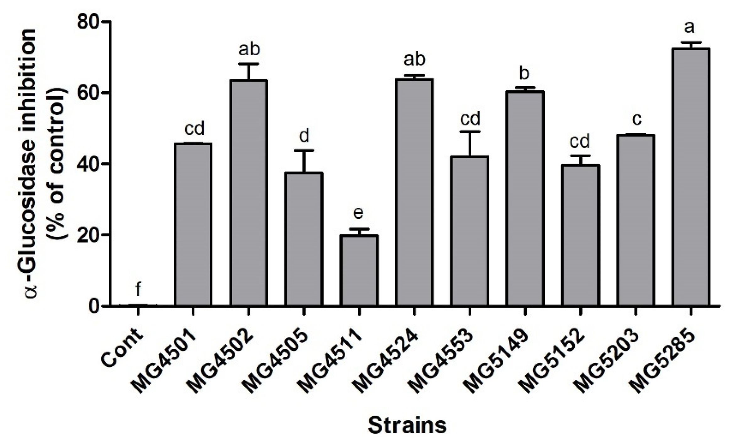
α-Glucosidase is an enzyme in the brush border membrane that catalyzes carbohydrate digestion. Inhibition of α-glucosidase activity has been demonstrated to prevent T2D by decreasing glucose absorption and reducing blood glucose levels [27]. Thus, to identify a strain with excellent hypoglycemic ability, the effects of ten LAB strains on α-glucosidase activity were examined (Figure 2). The α-glucosidase inhibition rate ranged from 19.8% to 72.3% when compared to that of the control. W. cibaria MG5285 showed the highest rate of inhibition of α-glucosidase activity (72.3 ± 1.9%). In addition, the α-glucosidase inhibition rates of L. gasseri MG4524, L. rhamnosus MG4502, and L. reuteri MG5149 were found to be 63.8 ± 1.1%, 63.4 ± 4.7%, and 60.3 ± 1.2%, respectively.
L. rhamnosus GG (LGG), one of the most well-studied probiotic LAB, significantly decreased blood glucose levels and improved hyperglycemia in neonatal streptozotocin-induced diabetic rats [28]. In addition, L. rhamnosus Z7 (29.2%), L. rhamnosus NL24 (27.6%), and Lactiplantibacillus plantarum ZF06-3 (34.9%) also exhibited α-glucosidase inhibitory activity [11,29]. Compared to that reported in other studies, four LAB strains (MG4502, MG4524, MG5149, and MG5285) showed remarkable anti-α-glucosidase activity.

3.3. LAB Inhibit Adipogenesis in 3T3-L1 Cells

The anti-adipogenic activity of the four LAB was determined during the differentiation of 3T3-L1 cells (Figure 3). The MDI group generated more adipogenesis compared to the control (p < 0.05), and four LAB displayed high anti-adipogenesis activity: L. rhamnosus MG4502 (97.3 ± 5.1%), L. gasseri MG4524 (70.3 ± 7.7%), L. reuteri MG5149 (86.7 ± 6.1%), and W. cibaria MG5285 (76.8 ± 5.5%).
Recently, LAB have been used to act as preventive and therapeutic agents against obesity by controlling enzymes involved in gut bacterial changes, increased energy absorption in the intestines, intestinal permeability changes, inflammation, and energy metabolism [30,31]. L. plantarum KY1032 decreased adipogenesis-related transcription factors in 3T3-L1 cells and induced remarkably lower body weight and total white fat mass in the treated animals compared to the high-fat diet group [32]. In addition, L. plantarum K10 significantly suppressed adipogenesis in 3T3-L1 cells and reduced fat mass in mice [33]. In this study, four LAB showed the highest inhibitory effect on adipocyte differentiation in 3T3-L1 cells.
The correlation between each activity (antioxidant, α-glucosidase inhibition, and anti-adipogenesis) of the LAB was also investigated (Table 1). The antioxidant activity of the LAB determined from the DPPH assay correlated with that indicated by the ABTS assay (0.685; p < 0.01) as well as with their α-glucosidase inhibition (0.362; p < 0.05) and anti-adipogenesis (0.380; p < 0.05) activities. These results showed that the various activities evaluated in this study are related to each other in LAB. Therefore, L. gasseri MG4524, L. reuteri MG5149, L. rhamnosus MG4502, and W. cibaria MG5285 might be used as functional food materials or supplements with various beneficial effects.

3.4. Survival under Simulated Human Gastrointestinal Tract Conditions

For functional probiotics, the four LAB strains (L. gasseri MG4524, L. reuteri MG5149, L. rhamnosus MG4502, and W. cibaria MG5285) were selected based on their antioxidant, antidiabetic, and anti-adipogenic activities. Initial cell viability ranged from 7.8 to 8.8 log CFU/mL. After the probiotic strains were exposed to simulated gastric fluid (pH 3 and 4) and intestinal conditions (pH 7 and 8), all strains showed excellent viability (Table 2); cell viability ranged from 7.3 to 8.7 log CFU/mL under gastric conditions, and from 7.8 to 8.9 log CFU/mL under intestinal conditions.
The low pH of the stomach and the antimicrobial action of pepsin provides an effective barrier for the survival of bacteria in the gastrointestinal tract. Considering that most microorganisms are eliminated by acidic gastric juices and alkaline bile juices in the duodenum, the selected probiotic strain should be able to survive under low pH and high bile conditions [34]. According to Patel et al. [35], W. cibaria has acid and bile tolerance (survival rate of 16–131% at pH 3). Lactobacillus sp. is adapted to low pH conditions caused by changes in glycolytic flux through its ability to control intracellular pH and its cell membrane ATPase [36]. In this study, the four LAB strains exhibited excellent viability in simulated gastric and intestinal conditions and demonstrated their potential as probiotics.

3.5. Adherence Ability of Probiotics

The selected strains showed high adhesion to HT-29 cells. The level of adhesive properties of the four strains ranged from 61.1 ± 0.2% to 86.2 ± 1.4% (Figure 4). There was no significant difference between the adhesion level of L. rhamnosus MG4502 and L. fermentum MG4244, the reference strain.
Adhesion of the probiotic strains to the intestinal epithelial cells is beneficial to the host because it prevents the growth of pathogenic bacteria in the intestines [37]. According to Kim et al. [22], the levels of adhesion of L. fermentum MG4231 and MG4244 were not statistically different from that of L. rhamnosus GG. L. rhamnosus MG4502 and L. fermentum MG4244 showed no statistical differences in their adhesion levels. Therefore, the adhesion ability of L. rhamnosus MG4502 to epithelial cells verifies its potential as a probiotic.

3.6. Assessment of Safety

The safety of probiotics is important for the selection of probiotic strains [9]. Therefore, evaluation of antibiotic resistance and hemolytic activity is strongly recommended. The antibiotic resistance of the four selected strains was investigated using MIC analysis (Table 3). L. gasseri MG4524 was classified as susceptible to eight antibiotics and resistant to kanamycin based on the epidemiological cut-off values suggested by EFSA [23]. L. rhamnosus MG4502, L. reuteri MG5149, and W. cibaria MG5285 were classified as susceptible to all antibiotics.
In the case of probiotics, antibiotic resistance can cause serious problems when transferred to other intestinal bacteria, especially pathogenic bacteria [38]. According to the literature, the obligate homofermentative Lactobacillus (L. gasseri), L. casei (L. rhamnosus), L. reuteri, and Leuconostoc (W. cibaria) groups commonly show intrinsic antibiotic resistance (non-transmissible), or vancomycin, kanamycin, and gentamicin [39,40]. These results support the present findings and indicate that the selected strains could be safe for host intestinal health.
All strains exhibited γ-hemolytic or non-hemolytic activity when grown on blood agar plates after 48 h of incubation (data not shown). Hemolysis, caused by pathogenic bacteria, can damage the host cell membrane, leading to cell death [41]. Since β-hemolysis is associated with pathogenicity, the test bacteria were cultured on blood agar to observe possible hemolysis. In this study, the selected probiotics did not show hemolytic activity.

3.7. Carbohydrate Utilization and Enzyme Activity

Carbohydrate utilization by the probiotic strains was assessed using the API 50 CHL system, and the results are summarized in Table 4. All strains metabolized D-galactose, glucose, and esculin. However, glycerol, erythritol, L-xylose, D-adonitol, methyl β-D-xylopyranoside, L-sorbose, L-rhamnose, methyl α-D-mannoside, methyl α-D-glucoside, inulin, D-raffinose, glycogen, xylitol, D-xylose, D-fucose, D-arabitol, L-arabitol, and 5-keto-gluconate were not metabolized by these strains. The enzymatic activity patterns of the probiotic strains were assessed using the API ZYM system (Table 5). None of the selected strains produced trypsin, β-glucuronidase, or α-mannosidase. L. rhamnosus MG4502 showed multiple enzymatic activities: esterase (C4), esterase lipase (C8), valine arylamidase, naphthol-AS-BI-phosphohydrolase, β-galactosidase, β-glucosidase, and α-fucosidase. In contrast, W. cibaria MG5285 showed only acid phosphatase activity.
The ability of probiotic strains to metabolize several carbohydrates provides them with competitive advantages to colonize numerous ecosystems [42]. In addition, probiotics can provide the enzymes necessary for digestion to people with digestive problems, such as lactose intolerance. Some LAB in fermented products can decrease the level of lactose or increase the active lactase in the small intestine. On the other hand, probiotics must be evaluated for the production of appropriate enzymes to prevent the synthesis of potentially toxic substances for safety [43]. β-Glucuronidase is a bacterial carcinogenic enzyme that exerts negative effects in the liver [38]. All LAB were safe because they did not show β-glucuronidase activity.
In conclusion, the aim of this study was to select superior probiotic strains that demonstrate their functional benefits by evaluating antioxidant, anti-obesity, and antidiabetic effects of LAB found in humans and their food. The results of our in vitro study confirmed that the four LAB strains, L. rhamnosus MG4502, L. gasseri MG4524, L. reuteri MG5149, and W. cibaria MG5285 demonstrated significant antioxidant, α-glucosidase inhibitory, and anti-adipogenesis activities. In addition, the selected strains were evaluated as probiotics, showing excellent suitability and safety. Therefore, these results suggest that the selected LAB strains could have potential as probiotics with antioxidant, anti-obesity, and anti-diabetic effects. In vivo studies are required to further evaluate the anti-obesity mechanisms and the safety of the tested probiotics as functional food supplements.

Supplementary Materials

The following are available online at https://www.mdpi.com/article/10.3390/fermentation7030123/s1, Table S1: Inhibitory effects of lipid accumulation in 3T3-L1 adipocytes cells of lactic acid bacteria (n = 3).

Author Contributions

Conceptualization, C.-H.K.; methodology, S.-I.C. and G.-H.K.; investigation, Y.J., G.W. and S.-I.C.; data curation, J.Y.L. and S.-I.C.; writing—original draft preparation, Y.J.; writing—review and editing, Y.J., J.Y.L. and H.K.; visualization, J.Y.L.; project administration, H.K., G.-H.K. and C.-H.K.; funding acquisition, C.-H.K. All authors have read and agreed to the published version of the manuscript.

Funding

This research was financially supported by the Ministry of Small and Medium-sized Enterprises (SMEs) and Startups (MSS), Korea, under the “Regional Enterprise Open-Innovative Voucher Program (R&D, Project P0010724)” supervised by the Korea Institute for Advancement of Technology (KIAT).

Institutional Review Board Statement

Not applicable.

Informed Consent Statement

Not applicable.

Data Availability Statement

Data are available in a publicly accessible repository/data are contained within the article or Supplementary Materials.

Conflicts of Interest

The authors declare no conflict of interest.

References

  1. WHO. Obesity and Overweight. Available online: https://www.who.int/en/news-room/fact-sheets/detail/obesity-and-overweight (accessed on 1 April 2020).
  2. Alford, S.; Patel, D.; Perakakis, N.; Mantzoros, C. Obesity as a risk factor for Alzheimer’s disease: Weighing the evidence. Obes. Rev. 2018, 19, 269–280. [Google Scholar] [CrossRef] [PubMed]
  3. Upadhyay, J.; Farr, O.; Perakakis, N.; Ghaly, W.; Mantzoros, C. Obesity as a disease. Med. Clin. N. Am. 2018, 102, 13–33. [Google Scholar] [CrossRef] [PubMed]
  4. Włodarczyk, M.; Nowicka, G. Obesity, DNA damage, and development of obesity-related diseases. Int. J. Mol. Sci. 2019, 20, 1146. [Google Scholar] [CrossRef] [PubMed]
  5. Derosa, G.; Maffioli, P. Anti-obesity drugs: A review about their effects and their safety. Expert Opin. Drug Saf. 2012, 11, 459–471. [Google Scholar] [CrossRef] [PubMed]
  6. Bray, G.A. Medical treatment of obesity: The past, the present and the future. Best Pract. Res. Clin. Gastroenterol. 2014, 28, 665–684. [Google Scholar] [CrossRef]
  7. Schippa, S.; Conte, M.P. Dysbiotic events in gut microbiota: Impact on human health. Nutrients 2014, 6, 5786–5805. [Google Scholar] [CrossRef]
  8. Gordo, R.; López-Andrés, N.; Fernández-Celis, A.; Gutiérrrez-Miranda, B.; Nieto, M.L.; Alarcon, T.; Alba, C.; Cachofeiro, V. The Interaction between Mitochondrial Oxidative Stress and Gut Microbiota in the Cardiometabolic Consequences in Diet-Induced Obese Rats. Antioxidants 2020, 9, 640. [Google Scholar]
  9. WHO. Report on Joint FAO/WHO Guidelines for the Evaluation of Probiotics in Food. Available online: https://www.who.int/foodsafety/fs_management/en/probiotic_guidelines.pdf (accessed on 30 April 2002).
  10. Ku, S.; Park, M.S.; Ji, G.E.; You, H.J. Review on Bifidobacterium bifidum BGN4: Functionality and nutraceutical applications as a probiotic microorganism. Int. J. Mol. Sci. 2016, 17, 1544. [Google Scholar] [CrossRef] [Green Version]
  11. Chen, P.; Zhang, Q.; Dang, H.; Liu, X.; Tian, F.; Zhao, J.; Chen, Y.; Zhang, H.; Chen, W. Screening for potential new probiotic based on probiotic properties and α-glucosidase inhibitory activity. Food Control 2014, 35, 65–72. [Google Scholar] [CrossRef]
  12. Koh, W.Y.; Utra, U.; Ahmad, R.; Rather, I.A.; Park, Y.-H. Evaluation of probiotic potential and anti-hyperglycemic properties of a novel Lactobacillus strain isolated from water kefir grains. Food Sci. Biotechnol. 2018, 27, 1369–1376. [Google Scholar] [CrossRef]
  13. Burgos-Morón, E.; Abad-Jiménez, Z.; Martinez de Maranon, A.; Iannantuoni, F.; Escribano-López, I.; López-Domènech, S.; Salom, C.; Jover, A.; Mora, V.; Roldan, I. Relationship between oxidative stress, ER stress, and inflammation in type 2 diabetes: The battle continues. Clin. Med. 2019, 8, 1385. [Google Scholar] [CrossRef] [Green Version]
  14. Furukawa, S.; Fujita, T.; Shimabukuro, M.; Iwaki, M.; Yamada, Y.; Nakajima, Y.; Nakayama, O.; Makishima, M.; Matsuda, M.; Shimomura, I. Increased oxidative stress in obesity and its impact on metabolic syndrome. J. Clin. Investig. 2017, 114, 1752–1761. [Google Scholar] [CrossRef]
  15. Chang, C.E.; Sylvia, I.P.; Lin, T.; Eun-Ki, K.; Seung, C.K.; Hyun, S.Y.; Jae-Seong, S. Molecular identification of vaginal Lactobacillus spp. isolated from Korean women. J. Microbiol. Biotechnol. 2002, 12, 312–317. [Google Scholar]
  16. Kim, H.; Kim, J.-S.; Kim, Y.; Jeong, Y.; Kim, J.-E.; Paek, N.-S.; Kang, C.-H. Antioxidant and Probiotic Properties of Lactobacilli and Bifidobacteria of Human Origins. Biotechnol. Bioprocess. Eng. 2020, 25, 421–430. [Google Scholar] [CrossRef]
  17. Blois, M.S. Antioxidant determinations by the use of a stable free radical. Nature 1958, 181, 1199–1200. [Google Scholar] [CrossRef]
  18. Re, R.; Pellegrini, N.; Proteggente, A.; Pannala, A.; Yang, M.; Rice-Evans, C. Antioxidant activity applying an improved ABTS radical cation decolorization assay. Free Radic. Biol. Med. 1999, 26, 1231–1237. [Google Scholar] [CrossRef]
  19. Lee, J.; Kim, A.-R.; Lee, J.-J. Ramie leaf extracts suppresses adipogenic differentiation in 3T3-L1 cells and pig preadipocytes. Asian Australas. J. Anim. Sci. 2016, 29, 1338. [Google Scholar] [CrossRef] [Green Version]
  20. Rizzatti, V.; Boschi, F.; Pedrotti, M.; Zoico, E.; Sbarbati, A.; Zamboni, M. Lipid droplets characterization in adipocyte differentiated 3T3-L1 cells: Size and optical density distribution. Eur. J. Histochem. 2013, 57, e24. [Google Scholar] [CrossRef] [Green Version]
  21. Maragkoudakis, P.A.; Zoumpopoulou, G.; Miaris, C.; Kalantzopoulos, G.; Pot, B.; Tsakalidou, E. Probiotic potential of Lactobacillus strains isolated from dairy products. Int. Dairy J. 2006, 16, 189–199. [Google Scholar] [CrossRef]
  22. Kim, S.; Choi, S.-I.; Jang, M.; Jeong, Y.; Kang, C.-H. Anti-adipogenic effect of Lactobacillus fermentum MG4231 and MG4244 through AMPK pathway in 3T3-L1 preadipocytes. Food Sci. Biotechnol. 2020, 29, 1541–1551. [Google Scholar] [CrossRef]
  23. EFSA Panel on Additives and Products; Substances Used in Animal Feed (FEEDAP); Rychen, G.; Aquilina, G.; Azimonti, G.; Bampidis, V.; Bastos, M.d.L.; Bories, G.; Chesson, A.; Cocconcelli, P.S.; et al. Guidance on the characterisation of microorganisms used as feed additives or as production organisms. EFSA J. 2018, 16, e05206. [Google Scholar]
  24. Feng, T.; Wang, J. Oxidative stress tolerance and antioxidant capacity of lactic acid bacteria as probiotic: A systematic review. Gut Microbes 2020, 12, e1801944. [Google Scholar] [CrossRef]
  25. Ji, Y.-J.; Lee, E.Y.; Lee, J.Y.; Lee, Y.J.; Lee, S.E.; Seo, K.H.; Kim, H.D. Antioxidant and Anti-Diabetic Effects of Agastache rugosa Extract. J. East. Asian Soc. Diet. Life 2020, 30, 297–305. [Google Scholar] [CrossRef]
  26. Lee, J.Y.; Park, J.-Y.; Seo, H.T.; Seong, H.-A.; Ji, Y.-J.; Lee, S.E.; Seo, K.H.; Kim, H.D. Samnamul (Shoots of Aruncus dioicus) Inhibit Adipogenesis by Downregulating Adipocyte-Specific Transcription Factors in 3T3-L1 Adipocytes. Processes 2020, 8, 1576. [Google Scholar] [CrossRef]
  27. Marella, S.; Hema, K.; Shameer, S.; Prasad, T. Nano-ellagic acid: Inhibitory actions on aldose reductase and α-glucosidase in secondary complications of diabetes, strengthened by in silico docking studies. 3 Biotech 2020, 10, 439. [Google Scholar] [CrossRef]
  28. Tabuchi, M.; Ozaki, M.; Tamura, A.; Yamada, N.; Ishida, T.; Hosoda, M.; Hosono, A. Antidiabetic effect of Lactobacillus GG in streptozotocin-induced diabetic rats. Biosci. Biotechnol. Biochem. 2003, 67, 1421–1424. [Google Scholar] [CrossRef]
  29. Zeng, Z.; Luo, J.; Zuo, F.; Zhang, Y.; Ma, H.; Chen, S. Screening for potential novel probiotic Lactobacillus strains based on high dipeptidyl peptidase IV and α-glucosidase inhibitory activity. J. Funct. Foods 2016, 20, 486–495. [Google Scholar] [CrossRef]
  30. Ejtahed, H.-S.; Angoorani, P.; Soroush, A.-R.; Atlasi, R.; Hasani-Ranjbar, S.; Mortazavian, A.M.; Larijani, B. Probiotics supplementation for the obesity management; A systematic review of animal studies and clinical trials. J. Funct. Foods 2019, 52, 228–242. [Google Scholar] [CrossRef]
  31. Jung, S.-P.; Lee, K.-M.; Kang, J.-H.; Yun, S.-I.; Park, H.-O.; Moon, Y.; Kim, J.-Y. Effect of Lactobacillus gasseri BNR17 on overweight and obese adults: A randomized, double-blind clinical trial. Korean J. Fam. Med. 2013, 34, 80. [Google Scholar] [CrossRef] [Green Version]
  32. Park, D.-Y.; Ahn, Y.-T.; Huh, C.-S.; Jeon, S.-M.; Choi, M.-S. The inhibitory effect of Lactobacillus plantarum KY1032 cell extract on the adipogenesis of 3T3-L1 Cells. J. Med. Food. 2011, 14, 670–675. [Google Scholar] [CrossRef]
  33. Kim, S.; Huang, E.; Park, S.; Holzapfel, W.; Lim, S.-D. Physiological characteristics and anti-obesity effect of Lactobacillus plantarum K10. Korean J. Food Sci. Anim. Resour. 2018, 38, 554. [Google Scholar]
  34. Morelli, L. In vitro assessment of probiotic bacteria: From survival to functionality. Int. Dairy J. 2007, 17, 1278–1283. [Google Scholar] [CrossRef]
  35. Patel, A.; Lindström, C.; Patel, A.; Prajapati, J.; Holst, O. Probiotic properties of exopolysaccharide producing lactic acid bacteria isolated from vegetables and traditional Indian fermented foods. Int. J. Fermented Foods 2012, 1, 87–101. [Google Scholar]
  36. Kang, C.-H.; Kim, Y.G.; Han, S.H.; Jeong, Y.; Paek, N.-S. Antibacterial activity and probiotic properties of lactic acid bacteria from Korean Intestine Origin. KSBB J. 2017, 32, 153–159. [Google Scholar] [CrossRef]
  37. Srisesharam, S.; Park, H.S.; Soundharrajan, I.; Kuppusamy, P.; Kim, D.H.; Jayraaj, I.A.; Lee, K.D.; Choi, K.C. Evaluation of probiotic Lactobacillus plantarum against foodborne pathogens and its fermentation potential in improving Lolium multiflorum silage quality. 3 Biotech 2018, 8, 443. [Google Scholar] [CrossRef] [PubMed]
  38. Kang, M.-S.; Yeu, J.-E.; Hong, S.-P. Safety evaluation of oral care probiotics Weissella cibaria CMU and CMS1 by phenotypic and genotypic analysis. Int. J. Mol. Sci. 2019, 20, 2693. [Google Scholar] [CrossRef] [PubMed] [Green Version]
  39. Gunyakti, A.; Asan-Ozusaglam, M. Lactobacillus gasseri from human milk with probiotic potential and some technological properties. LWT 2019, 109, 261–269. [Google Scholar] [CrossRef]
  40. Shi, Y.; Cui, X.; Gu, S.; Yan, X.; Li, R.; Xia, S.; Chen, H.; Ge, J. Antioxidative and probiotic activities of lactic acid bacteria isolated from traditional artisanal milk cheese from Northeast China. Probiot. Antimicrob. Proteins 2019, 11, 1086–1099. [Google Scholar] [CrossRef]
  41. Yasmin, I.; Saeed, M.; Khan, W.A.; Khaliq, A.; Chughtai, M.F.J.; Iqbal, R.; Tehseen, S.; Naz, S.; Liaqat, A.; Mehmood, T. In Vitro Probiotic Potential and Safety Evaluation (Hemolytic, Cytotoxic Activity) of Bifidobacterium Strains Isolated from Raw Camel Milk. Microorganisms 2020, 8, 354. [Google Scholar] [CrossRef] [Green Version]
  42. Oak, S.J.; Jha, R. The effects of probiotics in lactose intolerance: A systematic review. Crit. Rev. Food Sci. Nutr. 2018, 59, 1675–1683. [Google Scholar] [CrossRef]
  43. Yang, S.-J.; Lee, J.-E.; Lim, S.-M.; Kim, Y.-J.; Lee, N.-K.; Paik, H.-D. Antioxidant and immune-enhancing effects of probiotic Lactobacillus plantarum 200655 isolated from kimchi. Food Sci. Biotechnol. 2019, 28, 491–499. [Google Scholar] [CrossRef]
Figure 1. Antioxidant activities of the selected strains. (A) DPPH scavenging activity and (B) ABTS radical scavenging activity compared to control. The control was measured by adding PBS instead of LAB. All values are presented as the means ± SD from three independent experiments. Different letters indicate significant differences between means at p < 0.05 by Tukey’s multiple comparison tests. Cont; control. A.A.; ascorbic acid (10 μg/mL).
Fermentation 07 00123 g001
Figure 2. α-Glucosidase inhibitory activity of probiotic candidates. All values are presented in comparison with the control and as the means ± SD from three independent experiments. The control was measured by adding a buffer instead of LAB. Different letters indicate significant differences between means at p < 0.05 by Tukey’s multiple comparison tests. Cont; control.
Fermentation 07 00123 g002
Figure 3. Inhibitory effects of probiotic candidates on adipogenesis in 3T3-L1 cells. Con is the control without MDI treatment and MDI is the control with MDI treatment. Samples were treated with MDI and CFE. All values are presented as the means ± SD from three independent experiments. Different letters indicate significant differences between means at p < 0.05 by Tukey’s multiple comparison tests.
Fermentation 07 00123 g003
Figure 4. Adhesion ability of the selected strains. Adhesion ability to HT-29 cell was measured. All values are presented as the mean ± SD from three independent experiments. Different letters indicate significant differences between means at p < 0.05 by Tukey’s multiple comparison tests.
Fermentation 07 00123 g004
Table 1. Correlation analysis of the biological activities of the lactic acid bacteria.
Factors aDPPHABTSα-GlucosidaseAdipogenesis
DPPH1.0000.685 **0.362 *0.380 *
ABTS 1.0000.2480.533 **
α-Glucosidase 1.0000.337 *
Adipogenesis 1.000
Significance was determined using SPSS by Pearson’s correlation coefficient; * p < 0.05 and ** p < 0.01. a Values of the activities indicate inhibition rate (%).
Table 2. Survivability of selected strains under simulated gastrointestinal conditions.
StrainsInitialViable Count
Simulated Gastric Fluid aSimulated Intestinal Fluid b
pH 3pH 4pH 7pH 8
MG45028.6 ± 0.18.6 ± 0.08.6 ± 0.28.7 ± 0.18.6 ± 0.0
MG45248.3 ± 0.07.6 ± 0.17.5 ± 0.18.9 ± 0.18.8 ± 0.0
MG51498.8 ± 0.08.7 ± 0.08.7 ± 0.18.8 ± 0.08.8 ± 0.0
MG52857.8 ± 0.17.3 ± 0.17.7 ± 0.17.8 ± 0.07.9 ± 0.1
The results are expressed as average ± standard deviation; each data point is the average of three repeat measurements from three independently replicated experiments. All values are shown as viable counts (log CFU/mL) for each strain. Initial values evaluated at 0 h. a Simulated gastric tolerance results were obtained in PBS (pH 3 and pH 4) after 3 h. b Simulated intestinal tolerance results were obtained in PBS (pH 7 and pH 8) after 4 h.
Table 3. Minimum inhibitory concentrations (MICs) of various antibiotics for the selected strains.
AntibioticsIn This Study
MG4502MG4524MG5149MG5285
Ampicillin0.750.1250.50.25
Gentamicin4222
Kanamycin64324816
Streptomycin841224
Tetracycline0.5181
Chloramphenicol4344
Erythromycin0.0640.0160.0230.064
Vancomycinn.r.0.75n.r.n.r.
Clindamycin0.190.190.0160.047
The susceptibility of all strains was evaluated according to EFSA guidelines [23]. The inhibitory zones around the antibiotic strips were measured and determined according to the standard index. n.r., not required.
Table 4. Summary of the carbohydrate fermentation profiles of the selected probiotic candidate strains as measured using the API 50 CHL system.
SubstrateMG4524MG5149MG4502MG5285
d-arabinose+
l-arabinose++
d-ribose++
d-xylose+
d-fructose+++
d-mannose+++
Dulcitol+
Inositol+
d-mannitol++
d-sorbitol++
N-acetyl-glucosamine+++
Amygdalin+++
Arbutin+++
Salicin+++
d-cellobiose+++
d-maltose+++
d-lactose++
d-melibiose+
d-sucrose+++
d-trehalose+++
d-melezitose+
Starch+
Gentiobiose+++
d-turanose++
d-tagatose++
l-fucose+
Gluconate+++
2-keto-gluconate+
+: reaction; −: no reaction. All strains fermented d-galactose, d-glucose, and esculin. None of the strains fermented glycerol, erythritol, l-xylose, d-adonitol, methyl-β-d-xylopyranoside, l-sorbose, l-rhamnose, methyl-α-d-mannoside, methyl-α-d-glucoside, inulin, d-raffinose, glycogen, xylitol, d-xylose, d-fucose, d-arabitol, l-arabitol, and 5-keto-gluconate.
Table 5. Enzyme activities of the selected strains as measured using the API ZYM system.
Enzyme Assayed forMG4524MG5149MG4502MG5285
Alkaline phosphatase0020
Esterase (C4)1130
Esterase Lipase (C8)0130
Lipase (C14)0020
Leucine arylamidase4150
Valine arylamidase1350
Crystine arylamidase1030
α-Chymotrypsin0010
Acid phosphatase1234
Naphtol-AS-BI-Phosphohydrolase1251
α-Galactosidase3020
β-Galactosidase1430
α-Glucosidase1120
β-Glucosidase5050
N-Acetyl-β-glucosaminidase3000
α-Fucosidase0030
Enzyme activities were recorded from 0 (no activity) to 5 (≥40 nM of product released) at 10 nM intervals in the API ZYM color reaction chart; 3, 4, or 5 were considered positive reactions. All strains showed negative responses to trypsin, β-glucuronidase, and α-mannosidase.
Publisher’s Note: MDPI stays neutral with regard to jurisdictional claims in published maps and institutional affiliations.