Article

The Interactions among Isolates of Lactiplantibacillus plantarum and Dairy Yeast Contaminants: Towards Biocontrol Applications

1
Department of Cheese Technologies, Dairy Research Institute, Ltd., Ke Dvoru 12 a, 16000 Praha, Czech Republic
2
Milcom, Ltd., Culture Collection of Dairy Microorganisms, Ke Dvoru 12 a, 16000 Praha, Czech Republic
*
Author to whom correspondence should be addressed.
Fermentation 2022, 8(1), 14; https://doi.org/10.3390/fermentation8010014
Submission received: 22 October 2021 / Revised: 15 December 2021 / Accepted: 18 December 2021 / Published: 30 December 2021
(This article belongs to the Special Issue Lactic Acid Fermentation and the Colours of Biotechnology 3.0)

Abstract

:
Yeast diversity in the cheese manufacturing process and in the cheeses themselves includes indispensable species for the production of specific cheeses and undesired species that cause cheese defects and spoilage. The control of yeast contaminants is problematic due to limitations in sanitation methods and chemicals used in the food industry. The utilisation of lactic acid bacteria and their antifungal products is intensively studied. Lactiplantibacillus plantarum is one of the most frequently studied species producing a wide spectrum of bioactive by-products. In the present study, twenty strains of L. plantarum from four sources were tested against 25 species of yeast isolated from cheeses, brines, and dairy environments. The functional traits of L. plantarum strains, such as the presence of class 2a bacteriocin and chitinase genes and in vitro production of organic acids, were evaluated. The extracellular production of bioactive peptides and proteins was tested using proteomic methods. Antifungal activity against yeast was screened using in vitro tests. Testing of antifungal activity on artificial media and reconstituted milk showed significant variability within the strains of L. plantarum and its group of origin. Strains from sourdoughs (CCDM 3018, K19-3) and raw cheese (L12, L24, L32) strongly inhibited the highest number of yeast strains on medium with reconstituted milk. These strains showed a consistent spectrum of genes belonging to class 2a bacteriocins, the gene of chitinase and its extracellular product 9 LACO Chitin-binding protein. Strain CCDM 3018 with the spectrum of class 2a bacteriocin gene, chitinase and significant production of lactic acid in all media performed significant antifungal effects in artificial and reconstituted milk-based media.

Graphical Abstract

1. Introduction

Lactic acid bacteria (LAB) have been utilised in food and feed processing for centuries [1]. LAB are used as starter cultures for the fermentation of milk, meat, vegetables, and fruits and as adjunct cultures to improve the sensory and health properties of foods. At present, LAB with antimicrobial and antifungal effects are frequently applied as protective agents to limit the use of chemical preservatives and antibiotics, attain green labels, and prolong the shelf life of milk and bakery products [2,3]. Lactiplantibacillus plantarum, as representative LAB, is one of the most intensively studied species for its functional properties, which can enhance the quality, safety, and health benefits of foods [1,4,5]. The production of specific organic compounds (organic acids, fatty acids, hydrogen peroxide, bioactive peptides, and proteins) and mutual synergistic effects are strain-specific traits related to the antifungal effect [2,4]. The antifungal effect of L. plantarum is supposed to be associated also with the production of bacteriocins [1,4,6] that are primarily related to antimicrobial activity against bacterial contaminants [7,8]. The characterisation of chitinolytic enzymes and its genes [3,9,10] serves other possible products explaining the antifungal activity of lactobacilli. The genetic background and the resulting metabolic and proteomic expression of these traits are objects of current research [1]. The antifungal effect of L. plantarum is frequently verified against filamentous fungi that contaminate dairy and bakery products [3,5,6,7]. However, fewer studies have examined the antifungal activity of L. plantarum against yeast contaminants.
Yeasts are widespread eucaryotic organisms used in the dairy industry as starter and adjunct cultures to manufacture fermented drinks and specific cheeses. Yeasts are responsible for the fermentation process, ripening, and sensory properties of cheeses [11,12]. However, contamination with undesired yeast species remains a problem that affects the quality and safety of dairy products [13]. Detrimental yeast can be introduced from the air, raw material, and dairy equipment. Although hazards analysis and critical control points (HACCP) system is an obligatory hygienic control standard in dairies, the total elimination of yeast in dairy plants is impossible because of their ability to form biofilms in inaccessible technological components, such as tanks and pipes. Yeasts are also present in salt baths, salt brines, and whey [12,13]. The resistance of yeast to sanitation chemicals is also an emerging problem [14]. In well-established, controlled, and stable dairy operations, selected yeast populations are established, which are adapted to specific technology and sanitary conditions. These yeast populations are usually part of the dairy microbial environment, and they do not cause any defects in cheeses. However, the yeast population can increase and transform into undesirable contaminants when the environment or technology is disturbed due to insufficient sanitation or/and technological changes. Besides problems associated with chemical elimination and yeast resistance in dairy, the market demand for clean label products requires the testing and utilisation of bioprotective agent generally recognized as safe (GRAS) in Europe’s qualified presumption as safety list (QPS) [15], including Lactiplantibacillus plantarum. The application of verified L. plantarum strains with functional properties, including antifungal activity against yeast spoilers, is the desired step approaching the sustainable production of fermented dairy products [16,17].
In this work, 20 strains of L. plantarum of various origins were screened for genes coding class 2a bacteriocins and chitinase. These strains were tested against 25 yeast strains representing contaminants in cheeses and cheese manufacturing facilities. The extracellular expression of peptides and proteins was verified by using Tricine–SDS electrophoresis. Extracellular peptides and proteins were identified in 30 kDa cell-free filtrates by mass chromatography. The ability to change the pH of the substrate and the production of lactic and acetic acids were measured. The antifungal effects of twenty strains of L. plantarum were screened against 25 strains of yeasts isolated from contaminated cheeses and cheese manufacturing facilities. Interactions among yeast and L. plantarum strains were first tested by in vitro assays on artificial media [18]. Modified assays with reconstituted milk were used to simulate the matrix and conditions of dairy products [12,19]. The genetic background of bioactive peptides associated with L. plantarum and the ability to produce organic acids were used as covariate factors to assess the antifungal effect against yeast. Principal component analysis (PCA) was used to create scatter plots of the strains of L. plantarum based on their characteristics.
The aims of this study were to demonstrate that the variability in the antifungal effect of L. plantarum strains is related to the origin of the strain and functional traits driven by genes and their phenotypic expression. The study was aimed at specific yeast contaminants belonging to Candida spp., Trichosporon spp., Debaryomyces spp., Kluyveromyces spp., and Geotrichum sp., obtained from dairy products.

2. Materials and Methods

2.1. The Functional Traits of L. plantarum Strains

2.1.1. Physicochemical Parameters

The physicochemical parameters were measured for cultures of L. plantarum strains cultured in three media: MRS broth (Merck KGaA, Darmstadt, Germany), MRS supplemented with fructose (Lachner, Neratovice, Czech Republic) glucose (Lachner, Neratovice, Czech Republic), sodium gluconate (Sigma Aldrich, Steinheim, Germany) and maltose (Glentham, Corsham, UK) (MRS-FGGM), and 10% reconstituted milk (RSM) at 30 °C within 24 h. The pH values of the three cultivation media were measured in triplicate using the inoLab pH730 (WTW, Germany) pH meter. Lower fatty acids were detected directly from the three media (MRS, MRS enriched with sugars, and reconstituted milk) by an isotachophoretic analyser (EA 02), VILLA Labeco, Nitra, Slovakia). The results are presented as mg·100 mL−1.

2.1.2. PCR Amplification of Class 2a Bacteriocin Genes

Class 2a bacteriocin genes were amplified using different primer pairs adopted from the method of a prior study of Wieckowicz et al., 2011 [20]. PCR was performed in a 25 µL reaction volume (0.5 µL of each primer (10 µM), 1 µL of DNA template, 12.5 µL of 2× PPP Taq MasterMix (TopBio, Prague, Czech Republic), and 10.5 µL of ddH2O). The amplification conditions were as follows: pre-heating at 95 °C for 2 min, 35 cycles each of denaturation at 95 °C for 30 s, annealing for 30 s at 44 °C, and extension at 72 °C for 60 s, followed by a final extension at 72 °C for 8 min. PCR products were separated and visualised according to the method described above. PCR products were separated on a GelRed®-stained (Biotium, Fremont, QC, Canada) 2% SeaKem® LE agarose gel (SeaKem® LE; Lonza, Rockland, ME, USA) at 60 V for 120 min and viewed using the Gene Genius Bio Imaging System (Syngene, Frederick, MD, USA).

2.1.3. PCR Amplification of the Chitinase Gene

Two different primer pairs were used to detect the presence of the chitinase (chiA) gene, namely, chiAF (5′-ACCCTTCCCACTTTCAAGCC-3′), chiAR (5′-ATATGAGCGTCAGCTCCTCC-3′), chiFEMSF (5′-GATATCGACTGGGAGTTCCC-3′), and chiFEMSR (5′-CATAGAAGTCGTAGGTCATC-3′) according to method used in a previous study [9]. PCR was performed in a 25 µL reaction volume (0.5 µL of each primer (10 µM), 1 µL of DNA template, 12.5 µL of 2× PPP Taq MasterMix (TopBio, Prague, Czech Republic), and 10.5 µL of ddH2O). The amplification conditions were as follows: pre-heating at 95 °C for 2 min, 35 cycles each of denaturation at 95 °C for 30 s, annealing for 30 s at 52/46 °C, and extension at 72 °C for 60 s, followed by a final extension at 72 °C for 8 min. PCR products were separated on a GelRed®-stained (Biotium, Fremont, QC, Canada) 2% SeaKem® LE agarose gel (SeaKem® LE; Lonza, Rockland, ME, USA) at 60 V for 120 min and viewed using the GENE GENIUS Bio Imaging System (Syngene, Frederick, MD, USA).

2.1.4. Phenotypic Expression of Bioactive Compounds—Proteomics and Extracellular Production of Bioactive Compounds

Cell-free supernatants were obtained from strains of L. plantarum (Table 1).
Cultures grown for 24 h were centrifuged for 15 min (11.000 rpm). The supernatants were filtered through a microbial filter (0.20 µL) (Chromafil CA/S, Macherey-Nagel, Germany). According to the instruction manual, the Amicon ProAffinity Concentrator 30.000 NMWL (Merck Millipore Ltd., Tullagreen, Ireland) was used to obtain the optimal size (kDa) and concentration of extracellular proteins and peptides. Proteins and peptides were isolated from concentrate using ProteoExtract Protein Precipitation Kit (Merck KGaA, Darmstadt, Germany). The obtained pellets were dried and resuspended in 20 µL of Tricine sample buffer (BIO-RAD, Hercules, CA, USA) supplemented with 2-mercaptoethanol (BIO-RAD, USA) (2% v/v) and heated at 70 °C for 10 min. Resolving (15%) and stacking gels (4%) and buffers were prepared according to a previous study [21]. Precision Plus Protein™DualXtra Standards (BIO-RAD, Hercules, CA, USA) and Polypeptide SDS-PAGE Molecular Weight StandardsTM (BIO-RAD, Hercules, CA, USA) were loaded in wells together with samples (8 µL) under a constant voltage of 125 V until the dye front touched the bottom. The Mini-Protean Tetra Cell Apparatus for 1-D vertical electrophoresis (BIO-RAD) was used. The gels were washed and fixed in a fixing solution (50:10:40/methanol–acetic acid–H2O) for 25–30 min and repeatedly washed in deionised water. The products were stained by GelCode™Blue Safe Protein (Thermo Scientific, Rockford, IL, USA) in a rocking bath for 1 h. The gels were destained in 10% acetic acid solution. The peptides and proteins were determined directly from the concentrates with 30.000 NMWL on TimsTOF Pro (Bruker Daltonik, Bremen, Germany) with Ultra-High-Performance Liquid Chromatography UltiMate 3000 nano UHPLC (Thermo Fisher Scientific, Waltham, MA, USA) in the Faculty of Science, Dpt. of Chemistry, University of South Bohemia, Czech Republic. MaxQuant software (Max Plankt Institute of Biochemistry, Martinsried, Germany) and the UniProt database were used for data analysis.
The chitinolytic activity of L. plantarum strains was tested on basic chitin medium (BCM) (modified method [22]) after 10 days of cultivation at 30 °C. Instead of the original spot-on lawn method, the agar well diffusion method was used. The hydrolysation of chitin by strains of L. plantarum was identified as zones around wells in the presence of L. plantarum suspension.

2.2. Antifungal Activity Assays

2.2.1. Lactiplantibacillus plantarum Strains and Cultures

Twenty strains of Lactiplantibacillus plantarum isolated from silages, artisanal cheeses, milk, and sourdoughs were obtained from the CCDM collection (CCDM®, Milcom Ltd., Prague, Czech Republic) (Table 1). The reference strain ATCC14917 was used to compare bacteriocins profiles with other strains L. plantarum based on the original primer design and other comparative studies [20,23].
All strains were previously identified by sequencing the 16 S rRNA gene. The 16 S rRNA gene was amplified using fD1 (5′-ccg aat tcg aca acA GAG TTT GAT CCT GGC TCA G-3′) and rP2 (5′-ccc ggg atc caa gct tAC GGC TAC CTT GTT ACG ACT T-3′) primers [24]. The PCR products were treated with 2 µL of ExoSAP-ITTM according to the manufacturer’s reference manual (ThermoFisher Scientific, Baltic UAB, Vilnius) and sequenced by Eurofins Genomics Germany GmbH (Ebersberg, Germany). Cultures of L. plantarum were deposited in the CCDM® collection as deep-frozen (−70 °C) and lyophilised samples.
Lyophilised cultures were revitalised in 16% reconstituted milk at 30 °C for 24 h firstly. Then, L. plantarum strains were inoculated (1% v/v) in MRS broth (MERCK, Darmstadt, Germany) and cultivated at 30 °C. MRS + FGGM enriched with sugars (Fructose, Glucose, sodium Gluconate, Maltose) according to the M638 recipe (DSMZ, German Collection of Microorganisms and Cell Cultures GmbH, Germany) was used to increase the production of bioactive proteins [25].

2.2.2. Yeast Strains and Cultures

Yeast contaminants comprised 25 strains previously isolated from dairy matrices and environments (Table 2). The contaminants were provided by the Culture Collection of Dairy and Bakery Contaminants (CCDBC) (Milcom Ltd., Prague, Czech Republic). The taxonomic affiliation was confirmed by sequencing the ITS region rDNA (ITS1, 5.8 S rDNA, and ITS2) [26,27] by identifying ITS sequences using BLAST alignment tools. The phenotypic characteristics were defined according to a previous study [28]. The strains were deposited on agar slants with supplements. The yeasts were cultivated in yeast malt broth [29] for experimental purposes at 25 °C for 24 h.

2.2.3. The Overlay Method

L. plantarum strains cultivated at 30 °C for 24 h in MRS media were used in the modified overlay method [3,13,33]. The L. plantarum strains were streaked in two lines on MRS agar and cultivated anaerobically at 30 °C for 48 h. Cooled 0.7% malt extract agar was inoculated with yeast strains at a concentration of 104 CFU in 1 mL and gently poured on MRS medium with L. plantarum strains. Plates were cultivated at 25 °C in aerobic conditions. The growth of yeasts was assessed after 12, 24, 48, and 72 h. The final effect of L. plantarum strains on the yeast was evaluated according to an index scale of 1–7 (Table 3). Each combination of lactobacilli and yeast strain included three replicates.

2.2.4. The Agar Layer-Diffusion Method

A reconstituted milk was used in the modified agar layer diffusion method [34]. The strains of L. plantarum were cultivated overnight in 10% reconstituted milk at 30 °C. Then, the cultures were mixed with malt extract agar cooled to 45 °C; thus, the final concentration of the medium was 30% reconstituted milk with the tested strain of L. plantarum. To improve the visualisation of live yeast cells, 10 µL of neutral red solution (3% v/v) (Sigma-Aldrich, Darmstadt, Germany) was added to the mixture [35]. Yeast cultures grown for 24 h in yeast malt broth were diluted in Ringer’s solution (Sigma-Aldrich, Darmstadt, Germany) to 1 × 10⁵ CFU in 1 mL. Four drops (5 µL) were placed on solidified media containing 30% reconstituted milk with L. plantarum strains. The Petri dishes were incubated at 25 °C. The growth of the yeast colonies was assessed every 12 h and stopped after 72 h. Yeast grown on media with milk without lactobacilli and on MEA (0.7%) (Sigma-Aldrich, Darmstadt, Germany) were used as control variants. Each combination of L. plantarum strain and yeast strain was analysed in triplicate.

2.3. Statistical Analyses

Data for physicochemical parameters were subjected to statistical analysis using ANOVA with a factorial design in Statistica Software v 12.0 (StatSoft, Tulsa, OK, USA). Significant differences among the tested groups were determined using Tukey’s HSD test at significance p ≤ 0.05. Variability in the antifungal effect based on index scale values was analysed by ANOVA with a factorial design, Statistica Software v 12.0 (StatSoft Europe, Hamburg, Germany). Index values were first log-transformed and presented as the mean with standard error of the means (s.e.m.) because of discrepancy of sample’s means [17,36]. Significant differences among the tested groups were determined using Tukey’s HSD test at significance p ≤ 0.05. Analysis of covariance (ANCOVA) was used to compare the combinatory effect of functional traits of lactobacilli (organic acids, pH, bacteriocin genes, and chitinase genes) on the variability in the antifungal effect. Parameter values were first log-transformed. The significance was tested at p ≤ 0.05 (post hoc Tukey’s HSD Test). The antifungal effects of L. plantarum strains on artificial media were divided into three categories: i.e., total inhibition, inhibition, and no inhibition. The effects were first summarised by using principal component analysis (PCA) using their correlation matrix. The functional traits were converted to a log-transformed dataset to summarise the multiplicative changes.
The Hedonic rating scale was used for data scoring and analysed in Statistica Software 12.0 (StatSoft Europe, Hamburg, Germany) using multivariate techniques [23]. The variability in the average colony size (mm) of yeast was the main parameter used to assess the antifungal activity of lactobacilli in the RSM medium. The dataset obtained in this experiment was managed as described above. The average colony size and the zone around the yeast colony were summarised by the PCA model using their correlation matrix and visualised as PCA biplots and PCA scatter plots. The sensitivity of 25 yeast strains to strains of L. plantarum with defined traits was analysed by ANCOVA for groups represented by yeast genera to avoid statistical error during non-parametric testing because of unequal N.

3. Results and Discussion

First, the functional traits of Lactiplantibacillus plantarum were tested. The production of lactic acid, acetic acid, and changes in the substrate pH by strains of L. plantarum were evaluated in three tested media. Genomic methods were used to detect the genes responsible for producing bioactive peptides and chitinase.
The presence of extracellular bioactive peptides in cell-free filtrates was verified by using Tricine -SDS-PAGE on 15% tricine gels. The spectra of extracellular peptides in cell-free filtrates were determined by using mass spectrometry. The results obtained from these analyses were used as characteristics of L. plantarum strains in statistical models to help us clarify the background of their antifungal activity.

3.1. The Functional Traits of L. plantarum Strains

3.1.1. Physicochemical Parameters

The physicochemical parameters of lactobacilli were tested and compared among strains of different origins cultured in the three media types. The production of lactic and acetic acid and the pH of media varied significantly for all tested factors (Table 4), i.e., strains of L. plantarum, the group of origin, and substrate.
Production of lactic acid varied significantly among all strains of L. plantarum and the groups of origin (Figure A2). The acetic acid production varied significantly within the strains of L. plantarum, but there were no evident differences among the origin groups. The variability in lactic acid production is apparent among all the strains depending on the cultivation substrate. The strains originating from milk (CCDM 384, 391) produced the highest amount of lactic acid in MRS and MRS + FGGM. Despite that, these strains made the lowest amount of lactic acid in RSM. The addition of sugars (FGGM) significantly increased lactic acid production by all the strains of L. plantarum in MRS. In this medium, the strains from milk and silages were significant producers of lactic acid. In RSM, the highest amount of lactic acid was measured when strains from silage (CCDM 182), cheese wild types (L16, L17) and CCDM 3018 from sourdough were cultivated. The pH value of MRS media (5.57) was reduced by L. plantarum strains up to 30% without significant difference. The significant variability among the strains within the groups of origin was noted in RSM (Table A1, Figure A1). The strains obtained from sourdoughs decreased the original pH value (6.62) of RSM by about 44% at maximum. The strains of L. plantarum can utilize substrates rich in starch and sugars to produce lactic acid [37] and other short-chain organic acids. Lactic acid production is supposed to be the base of the antifungal effect [21]. Besides the lactic acid, the production of benzoic acid and diacetyl were confirmed in dairy matrices with L. casei [38]. The content of lactic acid in dairy matrix can be increased when supplements such as whey or milk powder are added [39]. The antifungal effect of lactobacilli is described mainly at the species-level under in vitro conditions [19,40]. Thus, the production of organic acids with possible antifungal effects remains optimal in these tests but becomes changed under technological conditions in dairies (salt, temperature, and microbial cultures).

3.1.2. Bacteriocins

Bioactive peptides from L. plantarum with antimicrobial and antifungal effects have been described in recent studies [1,20,23]. Most of the defined peptides were also detected when lactobacilli were tested against filamentous fungi [41]. The production of antifungal peptides by L. rhamnosus and L. paracasei in milk matrices targeted against Debaryomyces hansenii was described by McNair et al. [42]. Although many functional properties of L. plantarum and other lactobacilli based on the production of bioactive peptides have been described [1,7], the specificity of strains against targeted yeast contaminants is species-specific and variable. The gene coding for the related antifungal peptides is unknown or strain-specific; thus, screening L. plantarum strains for such traits is ineffective. As bacteriocins are the best-described bioactive peptides with a broad spectrum of antimicrobial and antifungal effects, the tested strains of L. plantarum were screened for their genetic backgrounds. Table 5 summarizes the characteristics of L. plantarum strains based on the presence of bacteriocin genes belonging to class 2a.
All L. plantarum strains encoded class 2a bacteriocin genes belonging to clade 1 (Figure A3). The profiles of PCR products in individual clades showed that not a single strain of L. plantarum has the same pattern as reference strain ATCC 14917. In addition, more than half of our strains encoded bacteriocin from clade 2, excluding strains CCDM 182, 185, 381, 383, 384, 387, 391, and 3018, which did not show amplified products (Figure A4). Clade 4 bacteriocin was successfully amplified using the first primer pair in all strains except CCDM 381 (Figure A5). Amplification with the second primer pair failed in most strains, excluding L12 and L16. L12 yielded a product size comparable to the PCR products in Więckowicz et al, 2011 [20]. Strain L16 produced a band of smaller size than expected (Figure A6). Non-specific primer annealing occurred during the amplification of bacteriocins from clade 5, regardless of the forward primer used (Figure A7 and Figure A8). Variability in PCR products among lactobacilli was detected after using the first primer pair (Figure A7). Half of the strains yielded PCR products of approximately 50 bp, corresponding to the values presented in previous study [20]. Strains K19-3 and CCDM 3018 produced a band with a size close to that of the expected product. Strains L12-16 and CCDM 381, 383, and 384 only yielded a spectrum of larger non-specific bands. A similar result was observed when using the second primer pair (Figure A8). The DNA of a single strain, CCDM 381, was not amplified using a second primer pair. Single bands of approximately 500 bp and 1000 bp were produced by strains K19-3 and K20-4, respectively. The sizes of both products exceeded the size declared in [20]. Strains K19-1, K19-2, CCDM 191 and 196, and L12-17 produced a band of the appropriate size. The functional diversity of strains L. plantarum was confirmed recently by comparative genomic analyses of 54 genome sequences pointed on the biosynthesis of bacteriocins. The three evolutionary lineages of L. plantarum varied in the biosynthesis of plantaricin [43]. Thus, strains from individual lineages performed diverse functional traits. The tricine-SDS-PAGE method was used to screen the extracellular expression of bacteriocin genes into the substrate. The extracellular proteins and peptides isolated from cell-free supernatants using AMICON ultrafilters with 30 MWO corresponded to sizes of 25–20 kDa, 20 kDa, 10 kDa, and less than 5 kDa (Figure 1).
Mass spectrometry analysis on filtrates and products from tricine gels showed a broad spectrum of proteins that are metabolic products of L. plantarum described in previous studies [7,40,44]. Although the bands less than 5 kDa were evident in extracellular filtrates of L. plantarum no known bacteriocin-like or antifungal peptides were identified among these peptides.

3.1.3. Chitinase

Currently, there are only a few studies about the detection of chitin-binding proteins (CBPs) and the chitinolytic activity of L. plantarum [9,45]. In most of the studied L. plantarum strains, the chiA gene was successfully amplified using chiFEMSF and chiFEMSR primers [9] (Table 6, Figure A9), from which it was assumed that these strains encode the chiA gene. However, the amplification with the ChiAF and ChiAR primers based on Lactococcus lactis subsp. lactis chiA sequences [30] failed, possibly because they are not compatible with L. plantarum DNA. The chiA gene was present in all strains from silages, whereas the lack of chiA was noted in most of the strains isolated from milk. The extracellular proteins and peptides isolated from cell-free supernatants using AMICON ultrafilters with 30 MWO corresponded to sizes of 25–20 kDa, 20 kDa, 10 kDa, and less than 5 kDa (Figure 1).
In most of the tested L. plantarum strains, chitinase was the only expressed extracellular protein with antifungal activity detected in the products. Although the 9 LACO Chitin-binding protein (22.199 kDa) was identified in most concentrates of cell-free supernatants from L. plantarum strains with the chiA gene, there were some discrepancies in strains CCDM 387 and L32. In these two strains, the chiA gene was not detected, but the 9 LACO Chitin-binding protein was detected in the concentrate and directly from 20 kDa products on 15% tricine gels. The visibility of chitinase on 15% Tricine gels depends on the absolute quantity of the peptide in the sample, i.e., the previous concentration of cells in the medium and conditions for the phenotypic expression of genes [46]. The conditions for the expression of CBP genes in artificial media and milk matrices are still unknown. The number of chitinase gene copies in genomic DNA might differ from strain to strain, so the result depends on the sensitivity of the PCR method. TimsTOF Pro with Ultra-High-Performance Liquid Chromatography UltiMate 3000 nano UHPLC remains a highly sensitive method to screen peptides or proteins present in small amounts in the concentrate [47,48]. The strains of L. plantarum were screened for chitinase activity in vitro using colloidal chitin in BCM medium combined with the agar well diffusion method. Clearing zones have never occurred; instead, the irregular hyaline zones occurred around the wells with suspensions. Seven L. plantarum strains (L17, L16, K19-2, 191, 3018, 185, and 187) hydrolysed colloidal chitin. The same strains were tested positively for the presence of chitinase genes. There are no known conditions for chitinolytic activity of CBP nonhydrolytic proteins against the yeast and fungi in milk matrices. Primarily CBP proteins bind to N-acetylglucosamine residues in chitin structures in the fungal cell walls [45]. The research on Candida albicans and G. glabrata confirmed that the inhibition of candida growth by chitinase represented by hydrolase Msp1 from L. rhamnosus GG is supported with lactic acid, particularly L-lactic acid isomer that potentially increases the sensitivity of hyphal surface to chitinase [10].

3.2. Antifungal Activity Assays

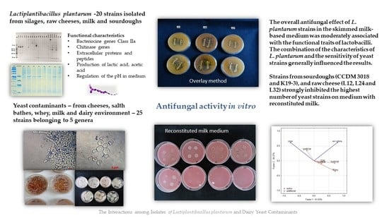
3.2.1. Antifungal Activity of Lactiplantibacillus plantarum Strains

The antifungal activity of twenty strains belonging to L. plantarum was tested against 25 strains of yeast contaminants. The overlay method on artificial media represented the optimal in vitro conditions for interacting organisms, lactobacilli, and yeast. In overlay assay, the inhibitory effect against yeast varied significantly among L. plantarum strains (Figure 2, Table 7) and yeast strains. The numbers of non-inhibited, partially inhibited, and fully inhibited yeast strains are presented in Figure 2. The results show that the number of fully inhibited yeast strains was highest for L. plantarum strains isolated from sourdough (3018 and K19-3), silage (196), and cheese-wild types (L16, L24). The strain L16 inhibited eight yeast strains which is a maximal number of total inhibitions. The strains originating from raw milk proved rather partial inhibitions against the yeast inhibited and less rate of total inhibition. Strain CCDM 384 weakly suppressed yeast growth on artificial media, reflected by the high number of low-indexed inhibitions (Figure 2). This strain isolated from milk can partially inhibit a broad spectrum of yeast strains at a low level.
The overall inhibitory effect of L. plantarum, including partial and total inhibition, reached 92% (K19-3) and 88% (384). Although the strains from milk inhibited yeast more potently than those from raw cheese and silages, the inhibitory effect was partial and temporary. The results from the overlay method confirmed that all strains of lactobacilli inhibited all yeast contaminants in general compared with the control variants (Figure 3). The data presented in Figure 3 are supported with detailed statistical parameters included in Table A2. The complete dataset related to testing 20 strains of L. plantarum against 25 strains of the yeast obtained from analyses of variance is supplemented as Table S3.
The intensity of the inhibitory effect was shown to be driven by different functional traits of lactobacilli [47,49]. The functional properties of lactobacilli, such as pH, production of LA and AA, bacteriocin and chitinase genes, were tested as covariates. The dimensional variability and interactions among the traits were evaluated by principal component analyses (PCA) (Figure 4). Partial inhibition (index value 2–5) is distinct from total inhibition (index value 5–6) and no inhibition (index value 1). The first two components explained 99.9% of the variance, with a cophenetic coefficient of 0.97. The first component explained 61.56% of the variability and showed that the inhibitory effect (total inhibition, partial inhibition, or no inhibition) of lactobacilli on yeast is influenced by different functional properties of L. plantarum strains. The total inhibition of yeast was positively correlated with the spectrum of genes for bacteriocins class 2a and chitinase. Although the spectrum of genes was shown as the significant trait allied with the antifungal activity, the base of this phenomenon is unclear. The six strains of L. plantarum (3018, K19-3, 196, L12, L16, L24) with significant antifungal activity varied in clades 4-1 and 5-1 of bacteriocins class 2a. The role and importance of individual genes for the final antimicrobial and antifungal effects are unknown. The strain L24 lacked the chitinase gene and protein. Partial inhibition was positively correlated with the production of lactic acid and acetic acid. The second component explained 38.44% of the original variability and positively correlated with the pH of the medium and the content of lactic and acetic acids. The distribution of L. plantarum strains (Figure 5) according to index values in the factorial space explained the indexed inhibitory effect correlated with functional traits. The six strains of L. plantarum that inhibited the most yeast strains are cumulated at the bottom right corner of the scatter plot. The studies aimed at antifungal effect are using evaluating scales representing mostly just the occurrence of inhibition zones. The yeast species representing dairy contaminants remain a group of diverse strains that performed unlike properties such as adaptability, tolerance, and sensitivity. To evaluate mutual interactions with lactobacilli strains, a detailed evaluating scale respecting both organisms is necessary.
The data shown in Figure 6 are supported by Table A3 including detailed statistical parameters. The complete dataset of analysis of variance based on the average of yeast colonies on media with RSM is supplemented as Table S4. The significance of factors influencing the variability within strains of L. plantarum and groups of origin is summarised in Table 8. The strains from sourdoughs (3018, K19-3) and cheese-wild types (L12, L24, L32) suppressed the yeast growth on RSM enriched medium significantly compared with control and others. The spectrum of effective strains from sourdoughs on RSM medium is the same compared to overlay assay on artificial media. The effective strains belonging to cheese-wild types (L12, L24, L32) differed from those effective on synthetic media. Notably, the strain L32 inhibited yeast growth significantly under RSM conditions and showed a weak antifungal effect on artificial media. The strains originating from silages suppressed the yeast growth weakly in RSM.
The PCA analyses were used to relate the yeast growth with characteristics of L. plantarum strains in RSM matrix (Figure 7). Inhibitory zones were observed in only a few combinations of lactobacilli and yeast strains. The proteolytic activity of yeast strain can also induce the clearing zone; thus, the comparison with the control variant is necessary. The first two components represented 99.9% variability. The first component explained 61.63% of the variability. The growth of yeast colonies was negatively correlated with the production of organic acids and bioactive proteins, and peptides. The second component demonstrated 38.37% of the variability and was positively correlated with the pH of RSM. The strains of L. plantarum in the scatter plot of PCA (Figure 8) are clustered according to their inhibitory effect against the yeast. The strains in the bottom left corner (L12, L32, L24, K19-3, 3018) represent the most effective strains. They are positively correlated with bacteriocins and organic acids, whereas the strains in the upper left corner are correlated with pH value and the occurrence of the zone. The antifungal effect of strains from sourdoughs, 3018 and K19-3, was the most effective on artificial and RSM-based medium. Besides the bacteriocins and chitinase profile, strain 3018 produced the lactic acid in MRS and RSM at the highest stable amount to compare the rest of the strains. Thus, concerning medical research on C. albicans [10], the intensive production of lactic acid can also support chitinase’s role in antifungal action.
The sources of isolates/strains and strain-associated traits such as bile resistance, adaptability, and the production of extracellular proteins play essential roles in phenotypic characteristics [49]. The strains isolated from sourdoughs had the most substantial suppressive effect against yeast. In our study, the strains of L. plantarum originating from sourdoughs inhibited up to 90% of the 25 yeast strains. Most published studies on L. plantarum strains producing bioactive compounds and exhibiting antifungal activity originated from sourdoughs and related bakery material [3,4,31]. In addition, wild strains of L. plantarum isolated from raw ewe cheeses inhibited yeast growth on both media tested. The functional properties of lactobacilli isolated from raw milk and artisanal cheeses have been characterised in recent studies [7,50]. Strains 191 and 196 from silages exerted effective antifungal activity against yeast in an overlay test on artificial media. The L. plantarum strains from silages reduced the yeast growth in RSM medium to a significantly lesser extent than those from raw cheeses and sourdoughs. The properties of lactobacilli isolated from silage were successfully tested to improve animal feed quality [51]. The strains obtained from milk showed different profiles of bioactive proteins and the weakest antifungal effect among the groups of origin. The combination of the characteristics of L. plantarum and the sensitivity of yeast strains generally influenced the results. Potential antifungal compounds (peptides and organic acids) produced by strains of L. paracasei and L. rhamnosus [42], L. rhamnosus, and L. jensenii [13,52] were identified in dairy matrices in recent studies [4]. The spectrum of bioactive peptides produced under the RSM condition might differ from that in optimised growing media. The inhibition of yeast growth can be limited by time; i.e., lactobacilli and their products may only postpone development, and/or they can influence the morphological features of yeast, such as the density and size of the yeast colonies. In a recent study, the genomic comparison based on clusters of orthologous groups did not show intraspecific diversity of plant-associated strains belonging to L. plantarum, but the variability was significantly supported by phenotypic studies [53]. The inhibitory activity against Saccharomyces cerevisiae confirmed significant variability within strains [53] from fermented fruits and grains.

3.2.2. The Sensitivity of Yeast Strains

As the L. plantarum strains showed variability in their antifungal effects, yeast contaminants showed variable sensitivity to L. plantarum strains and their products. Five genera of yeast (Candida spp., Trichosporon spp., Debaryomyces spp., Kluyveromyces spp., and Geotrichum sp.) including the unequal number of strains were tested. The sensitivity of the yeast contaminants during the test on artificial media (Figure 9) was significantly different among yeast strains (Table A4). Significant variability in yeast growth based on index scale value was noted within five yeast genera (Table S1). Trichosporon species shown to be the most sensitive to L. plantarum species. High index values were also obtained for Candida apicola, C. atlantica, Debaryomyces subglobosus, and D. hansenii strain 47. On the contrary, C. krusei, C. inconspicua, Kluyveromyces marxianus strains 270 and 258, and Galactomyces candidum strain 1061 demonstrated the maximum tolerance to L. plantarum strains and their products in the overlay method on artificial media.
The influences of the functional properties of lactobacilli on yeast genera were tested as covariate factors. Most of the tested yeast strains belonging to Candida spp., Galactomyces spp, and Debaryomyces spp. proved tolerant to the lactic and acetic acid content produced by lactobacilli. The bacteriocin profiles of lactobacilli significantly influenced sensitivity of almost all yeast strains except for Kluyveromyces. The presence of chitinase genes was found to be a significant trait only for the suppression of Trichosporon spp. and Debaryomyces spp. on RSM media. The significant variability among yeast indicates that the sensitivity of yeast to L. plantarum is an interspecific and intraspecific property, depending on origin of isolates and environmental factors. The overlay method based on artificial media was optimised for the growth of lactobacilli and yeast in vitro. Thus, the results are far different from those obtained in the dairy matrices. The development of yeast contaminants on RSM with lactobacilli was suppressed by up to 60% compared with controls (Figure 10). Significant variability in the average sizes of yeast colonies was noted within five yeast genera (Table S2). The functional properties of lactobacilli were tested as covariate factors. For yeast growth on media with RSM, the amount of lactic and acetic acids produced by lactobacilli were determined to be non-significant factors. The variability in the size of colonies was significant in the tested genera of yeast except for Kluyveromyces. The detailed information related to interactions among 20 L. plantarum strains and 25 yeast strains in overlay method are summarized in Supplementary Table S3. Most studies on antifungal effects have targeted filamentous fungi. The antifungal activity of lactic acid bacteria against a broad spectrum of potential yeast contaminants was tested in dairy-mimicking models in a previous study [19]. Even though the yeast and filamentous fungi belong to ascomycetous and basidiomycetous fungi there are important differences in their lifestyle. The spore germination and hyphal growth, including synthesis of the fungal cell wall, is the most sensitive stage to environmental changes. In the case of yeast, the cell wall is completed during the budding process. Thus, this step can cause difference in sensitivity of the yeast and filamentous fungi. The high sensitivity of Candida albicans to products of lactobacilli during the filamentous stage is declared in a previous study [44]. The interactions between yeast and L. plantarum are conditioned by intraspecific and interspecific characteristics of both inter-acting organisms, i.e., lactobacilli and yeast [13,31]. Yeast species such as C. apicola, C. atlantica, T. asahii, D. subglobosus, and D. hansenii 615 showed high sensitivity to almost all strains of L. plantarum tested. The sensitivity of these strains significantly influenced the results. C. apicola and C. atlantica were identified in brines [54] and as part of cheese micro-flora in cheese with a long ripening period (up to 60 days) [55,56]. However, their effect on cheeses is not confirmed to be detrimental. The interaction of these two species with LAB has not yet been tested. T. asahii was identified in indigenous cheeses, brines, and whey [1]. In our study, T. asahii showed significant sensitivity to strains of L. plantarum. A variety of Lactobacillus spp. in a dairy matrix was tested by [52]. The colony size of T. domesticum and T. coremiiforme strains growing on reconstituted milk with L. plantarum differed from that of the control by 65%, whereas T. asahii differed by 93%.
The in vitro studies confirmed that C. krusei, C. inconspicua, C. zeylanoides, and strains of K. marxianus and G. candidum were the most tolerant to L. plantarum strains on artificial media (Table A4, Supplementary Table S3). In contrast, the inhibitory effect was apparent on RSM media, although the colony size was only about 30% less than the control. These findings reveal that a milk matrix supports the inhibitory effect of L. plantarum, and the inhibitory effect observed for each combination can differ from tests on artificial media [1]. The strains representing obligate pathogens, C. krusei, C. parapsilosis, and C. inconspicua, were included in experiments because they are frequently identified in dairy matrices. In general, these three strains were observed to be the most tolerant species to L. plantarum. The different by-products of L. rhamnosus, L. gasseri, L. acidophillus, L. paracasei, L. crispatus, and L. jensenii were tested to explore the biofilm adhesion and inhibition of non-albicans species in models simulating the environment of the human body [57]. Similarly, T. asahii is an obligate pathogen with importance in human and veterinary medicine [58]. However, T. asahii and other Trichosporon spp. are frequent in raw milk products, cheeses, and brines [1]. T. asahii was the most sensitive strain that did not grow on reconstituted milk with L. plantarum strains. In addition, the in vitro dual tests showed that T. asahii was highly sensitive to L. plantarum. The strain-specific sensitivity also occurred in Debaryomyces and Kluyveromyces spp., which represent common yeast species in the dairy environment and products. Geotrichum/Galactomyces spp. were strongly suppressed by L. plantarum strains on RSM media when compared with artificial media

4. Conclusions

The screening of L. plantarum for functional traits followed by testing on artificial and RSM matrix enables the selection of effective strains with antifungal activity against yeast contaminants. As shown, the strains that performed the antifungal activity in synthetic media cannot display the same effect on RSM media as shown in the case of L. plantarum strains originating from silages and raw milk cheeses. The strain 3018 from sourdough positively provided the best antifungal effect on both the media tested. All the tested strains encode the spectrum of Class 2a bacteriocin genes with some variability in clades. In MRS medium, only chitinase, non-target peptides, and proteins were produced extracellularly. There are no existing comparative data for extracellular production of bacteriocins, chitinase, and the antifungal effect of L. plantarum in RSM media. The results indicate that the bacteriocin spectrum might predict the antimicrobial and antifungal ability of L. plantarum. The competitive resources such as silages, raw milk cheeses and sourdoughs with naturally occurring fungal organisms can provide more effective strains than the uniform environment of the milk. Although lactic acid production is the renowned trait of the antifungal effect, this study showed that its production in artificial media and RSM media is highly variable within the strains and their groups of origin. Only strain 3018 from sourdough produced significantly high amount of lactic and acetic acid in MRS and RSM medium. Thus, the final antifungal effect results from the synergistic interactions based on the enzymatic and metabolic activity of strains under specified conditions. The sensitivity and tolerance of the yeast strains also play essential roles in evaluating antifungal effects. The testing of high species richness of the yeast contaminants, including the isolates from specific dairy matrices or environments, can confirm the broad spectrum of antifungal activity of L. plantarum strain compared with studies employing only a few strains. Then, the strain exhibiting the antifungal activity should be tested for technological properties (salt tolerance, temperature, interactions with starter cultures, Aw) stepwise when supposed to be practically used. Although the dairy products are naturally populated with L. plantarum, the antifungal activity of strains originating from milk was weak and insufficient compared with strains from a fermentative environment. The application of protective strains of L. plantarum with adequate technological properties reflects the current demands on dairy products, including probiotic potential and microbial biocontrol against yeast spoilers. Thus, the supplementation of fermented dairy products with the suitable culture of L. plantarum significantly contribute to sustainable development in the field of fermented foods [59]. All in all, there is no existing uniform strain of L. plantarum that suppressed or eliminated all the spectrum of yeast contaminants but only the strain L. plantarum that perform antifungal effect against the broad spectrum of yeast contaminants under defined conditions. The identification of functional traits of L. plantarum strains has been shown to be an innovative and effective tool for pointed selection and understanding the interaction among lactobacilli and yeast contaminants.

Supplementary Materials

The following are available online at https://www.mdpi.com/article/10.3390/fermentation8010014/s1. Table S1: The factorial analysis within the yeast genera. The categorical predictors—functional properties of L. plantarums strains are employed as covariates. The variability in inhibition is based on logarithm of index scale value for overlay method. ANCOVA, p ≤ 0.05, Statistica Software v. 12.1. The index letters ns means—non-significant differences. Table S2 The factorial analysis within the yeast genera. The categorical predictors—functional properties of lactobacili in milk matrice are employed as covariates. The variability in the inhibition is based on the averages of yeast colony (mm) on modified overlay method with skimmed milk. ANCOVA, p ≤ 0.05, Statistica Software v. 12.1. The index letters ns means—non-significant differences. Table S3 The interactions among L. plantarum strains and yeast strains based on the logarithm of index scale value by mean, standard deviation (s.d.) and standard error of means (s.e.m.). The data resulted from Factorial ANOVA, Statistica 12v., Significant at α ≤ 0.05. Table S4 The variability of yeast colony average (mm) characterised by mean, standard deviation (s.d.) and standard error of means (s.e.m.). The data resulted from Factorial ANOVA, Statistica 12v., Significant at α ≤ 0.05.

Author Contributions

Conceptualization, M.K., J.C., O.B. and V.D.; methodology, M.K., J.C., O.B. and V.D.; software, J.C., M.K.; validation, J.C., M.K. and O.B.; formal analysis, M.K., O.B. and J.C.; investigation, V.D.; resources, Z.D.; data curation, J.C., M.K.; writing—original draft preparation, M.K. and J.C.; writing, M.K. and J.C.; visualization, M.K. and J.C.; supervision, M.K. and V.D.; project administration, M.K. and V.D.; funding acquisition, M.K. and V.D. All authors have read and agreed to the published version of the manuscript.

Funding

This research was funded by THE MINISTRY OF AGRICULTURE OF THE CZECH REPUBLIC, MZE-RO1421, THE NATIONAL AGENCY FOR AGRICULTURAL RESEARCH OF CZECH REPUBLIC, grant numbers QK1910036, QK19110024 and The National Programme on Conservation and Utilization of Plant, Animal and Microbial Genetic Resources Important for Food and Agriculture (NPGR) belonging to Ministry of Agriculture of the Czech Republic.

Institutional Review Board Statement

Not applicable.

Informed Consent Statement

Not applicable.

Data Availability Statement

The data that supports the findings of this study are present in Appendix A and in the Supplementary Materials.

Conflicts of Interest

All the authors declare that they have no relevant conflict of interest. The funders had no role in the design of the study; in the collection, analyses, or interpretation of data; in the writing of the manuscript, or in the decision to publish the results.

Appendix A

Table A1. Production of organic acids by strains of L. plantarum in different media. ANOVA, Statistica v. 12, Post Hoc Tukey’s HSD Test. The data represents logarithmically transformed index scale values, standard deviation (SD) and standard error of means (SEM). The index letters indicate the significant differences. Significant at α ≤ 0.05.
OriginStrainLactic Acid (mg.100 mL−1)Acetic Acid (mg.100 mL−1)
MRSMRS + FGGMRSMMRSMRS + FGGMRSM
Silage1911172.94 ± 0.021387.54 ± 0.02 b959.38 ± 0.01 b484.64 ± 0.02582.63 ± 0.02 ba181.49 ± 0.02 a
1961356.38 ± 0.02 b1410.04 ± 0.05 b977.42 ± 0.10 ab484.86 ± 0.03574.32 ± 0.02 ab199.88 ± 0.02 a
1821169.07 ± 0.031431.95 ± 0.05 a1009.59 ± 0.02 a578.92 ± 0.06 a621.50 ± 0.06 a171.85 ± 0.08 b
1851232.11 ± 0.011459.72 ± 0.02 a985.53 ± 0.12 ab568.36 ± 0.02 a533.62 ± 0.08175.62 ± 0.06 b
1871133.74 ± 0.021554.79 ± 0.03 a911.02 ± 0.06 bc523.60 ± 0.02 ab603.99 ± 0.07 a176.34 ± 0.02 b
Raw cheeseL161267.44 ± 0.05 b1381.41 ± 0.02 b1016.68 ± 0.12 a566.20 ± 0.06 ab643.32 ± 0.06 a160.88 ± 0.13
L171311.20 ± 0.05 b1292.19 ± 0.071008.47 ± 0.04 a601.05 ± 0.09632.12 ± 0.09 a166.53 ± 0.10
L241185.13 ± 0.021309.29 ± 0.09956.61 ± 0.03 b537.91 ± 0.12593.13 ± 0.02 a136.43 ± 0.02
L32928.19 ± 0.061197.13 ± 0.02983.76 ± 0.07 ab513.72 ± 0.03592.66 ± 0.05 a190.99 ± 0.05 a
L121333.80 ± 0.02 b1401.14 ± 0.06 b422.56 ± 0.02188.10 ± 0.05373.62 ± 0.03114.07 ± 0.02
Milk3811345.53 ± 0.06 b1497.48 ± 0.05 a821.42 ± 0.02498.13 ± 0.06516.03 ± 0.0294.32 ± 0.09
3831353.22 ± 0.02 b1450.37 ± 0.02 a406.71 ± 0.02503.27 ± 0.02 b409.29 ± 0.0229.90 ± 0.07
3841526.51 ± 0.07 a1491.59 ± 0.04 a354.68 ± 0.02563.49 ± 0.03 a482.58 ± 0.0242.51 ± 0.05
3911545.06 ± 0.04 a1487.34 ± 0.03 a406.71 ± 0.05537.35 ± 0.02 ab605.83 ± 0.05 ab94.32 ± 0.05
SourdoughsK19-11047.83 ± 0.061286.92 ± 0.02975.77 ± 0.06 ab590.32 ± 0.05 a613.09 ± 0.02 a135.65 ± 0.02
K19-21009.88 ± 0.021236.48 ± 0.05685.02 ± 0.02539.09 ± 0.09 ab611.01 ± 0.02 a162.56 ± 0.03
K19-31008.35 ± 0.071296.72 ± 0.04724.97 ± 0.07540.13 ± 0.07 ab550.78 ± 0.03123.86 ± 0.12
K20-41116.30 ± 0.021020.33 ± 0.04513.50 ± 0.06540.13 ± 0.03 ab511.89 ± 0.11150.60 ± 0.02
30181367.88 ± 0.01 b1416.70 ± 0.02 a1025.10 ± 0.08 a537.56 ± 0.06 ab592.99 ± 0.12 b171.60 ± 0.02 b
Table A2. The variability in antifungal effect of 20 strains of L. plantarum in in vitro tests on artificial media against 25 yeast strains. The data represent logarithmically transformed index scale values, standard deviation (s.d.) and standard error of means (s.e.m.). F(24,1050) = 96.87; p ≤ 0.00 E ≤ 0.05. (Factorial ANOVA, Statistica v. 12. Post Hoc Tukey’s HSD Test). The index letter indicates significant differences. Significant at α ≤ 0.05.
L. plantarum StrainsLog Values.d.s.e.m.
CCDM 1911.07 bc0.770.09
CCDM 1961.16 b0.740.09
CCDM 1821.03 bc0.710.08
CCDM 1850.99 bc0.690.08
CCDM 1871.01 bc0.720.08
L121.07 bc0.710.08
L161.16 b0.720.08
L171.03 bc0.790.09
L241.04 bc0.760.09
L320.96 bc0.790.09
CCDM 3810.94 c0.680.08
CCDM 3830.94 c0.710.08
CCDM 3840.86 d0.660.08
CCDM 3911.05 bc0.670.08
CCDM 3870.99 bc0.690.08
K19-11.24 b0.620.07
K19-21.18 bc0.610.07
K19-31.23 b0.590.07
K20-41.11 bc0.630.07
CCDM 30181.32 b0.550.06
controls0.00 a00
Table A3. The variability in the average size of yeast colonies (mm) including standard deviation (s.d.) and standard error of mean (s.e.m.) on RSM medium with L. plantarum strains, including the control variant, for all yeast strains. Factorial ANOVA, Statistica Software v. 12, F(20,1530) = 45.604; p ≤ 0.5 ≤ α. The index letter indicates significant differences. Significant at α ≤ 0.05.
Strain of L. plantarum Average (mm)±s.d.±s.e.m.
CCDM 1917.56 b6.400.74
CCDM 1968.52 b6.490.75
CCDM 1825.71 d6.480.51
CCDM 1856.91 bc5.760.98
CCDM 1875.72 d4.560.53
L123.61 ef2.670.42
L166.89 bd4.460.52
L178.18 b4.230.49
L244.02 e3.450.51
L324.34 e3.510.50
CCDM 3815.34 de4.510.52
CCDM 3835.19 de4.220.53
CCDM 3844.96 de3.520.60
CCDM 3916.50 d4.530.53
CCDM 3875.64 de4.240.56
K19-14.99 de3.210.37
K19-24.45 e3.940.46
K19-33.11 e2.440.40
K20-48.34 b4.780.55
CCDM 30182.17 f3.610.42
controls17.95 a2.390.27
Table A4. The variability in sensitivity 25 yeast strains to all strains of L. plantarum. The data represents logarithmically transformed index scale values, s.d. and s.e.m. F(24, 1050) = 400.009; p ≤ 0.00 E ≤ 0.05. (Factorial ANOVA, Statistica v. 12. Post Hoc Tukey’s HSD Test). The index letter indicates significant differences. Significant at α ≤ 0.05.
Yeast StrainLog Value±s.d.±s.e.m.
Candida krusei 6000.24 f0.460.06
C. inconspicua 6010.39 f0.470.06
C. parapsilosis 6121.32 bc0.450.06
C. intermedia 6221.42 b0.50.06
C. apicola 6201.66 a0.540.07
C. zeylanoides 6230.59 ef0.460.06
C. atlantica 6111.58 ab0.520.07
Trichosporon asahii 6241.77 a0.410.05
T. domesticum 10621.69 a0.390.05
T. coremiiforme 6071.53 ab0.380.05
T. coremiiforme 6081.61 a0.40.05
Debaryomyces subglobosus 20271.75 a0.460.06
D. hansenii 7420.51 e0.530.07
D. hansenii 6151.12 c0.440.06
D. hansenii 471.79 a0.410.05
Kluyveromyces lactis 10540.92 cd0.470.06
K. lactis 6170.82 d0.350.04
K. marxianus 6200.85 d0.450.06
K. marxianus 2700.15 f0.290.04
K. marxianus 2580.08 g0.220.03
Geotrichum candidum 8780.66 d0.530.07
G. candidum 8701.19 c0.480.06
G. candidum 8320.89 cd0.540.07
G. candidum 10530.83 d0.420.05
G. candidum 10610.09 g0.230.03
Figure A1. Modification of the pH value by L. plantarum strains with respect to strain origin and three cultivation media-MRS, MRS + FGGM and reconstituted milk.
Fermentation 08 00014 g0a1
Figure A2. Variability in production of lactic acid by L. plantarum strains with respect to strain origin and three cultivation media- MRS, MRS + FGGM and reconstituted milk.
Fermentation 08 00014 g0a2
Figure A3. Class 2a bacteriocin gene belonging to the Clade 1 amplification. O’RangeRulerTM 50 bp DNA Ladder was used as a molecular marker. PC-positive control by strain ATTCC 14917.
Fermentation 08 00014 g0a3
Figure A4. Class 2a bacteriocin gene belonging to the Clade 2 amplification. O’RangeRulerTM 50 bp DNA Ladder was used as a molecular marker. PC-positive control by strain ATTCC 14917.
Fermentation 08 00014 g0a4
Figure A5. Class 2a bacteriocin gene belonging to the Clade 4 amplified using first primer pair (Table 3). O’RangeRulerTM 50 bp DNA Ladder was used as a molecular marker.
Fermentation 08 00014 g0a5
Figure A6. Class 2a bacteriocin gene belonging to the Clade 4 amplified using second primer pair (Table 3). O’RangeRulerTM 50 bp DNA Ladder was used as a molecular marker. PC-positive control by strain ATTCC 14917.
Fermentation 08 00014 g0a6
Figure A7. Class 2a bacteriocin gene belonging to the Clade 5 amplified using first primer pair (Table 3). O’RangeRulerTM 50 bp DNA Ladder was used as a molecular marker. PC-positive control by strain ATTCC 14917.
Fermentation 08 00014 g0a7
Figure A8. Class 2a bacteriocin gene belonging to the Clade 5 amplified using second primer pair (Table 3). O’RangeRulerTM 50 bp DNA Ladder was used as a molecular marker. PC-positive control by strain ATTCC 14917.
Fermentation 08 00014 g0a8
Figure A9. Chitinase (chiA) amplification. Gene encoding chitinase was detected in L. plantarum strains using primers chiFEMSF and chiFEMSR. GeneRulerTM DNA Ladder Mix was used as a molecular marker.
Fermentation 08 00014 g0a9

References

  1. Behera, S.S.; Ray, R.C.; Zdolec, N. Lactobacillus plantarum with Functional Properties: An Approach to Increase Safety and Shelf-Life of Fermented Foods. BioMed Res. Int. 2018, 2018, 9361614. [Google Scholar] [CrossRef] [Green Version]
  2. Gao, M.; Hou, H.; Teng, X.; Zhu, Y.; Hao, H.; Zhang, G. Microbial diversity in raw milk and traditional fermented dairy products (Hurood cheese and Jueke) from Inner Mongolia, China. Genet. Mol. Res. 2017, 16, gmr16019451. [Google Scholar] [CrossRef] [PubMed]
  3. Russo, P.; Arena, M.P.; Fiocco, D.; Capozzi, V.; Drider, D.; Spano, G. Lactobacillus plantarum with broad antifungal activity: A promising approach to increase safety and shelf-life of cereal-based products. Int. J. Food Microbiol. 2017, 247, 48–54. [Google Scholar] [CrossRef] [PubMed]
  4. Scano, P.; Pisano, M.; Murgia, A.; Cosentino, S.; Caboni, P. GC-MS Metabolomics and Antifungal Characteristics of Autochthonous Lactobacillus Strains. Dairy 2021, 2, 326–335. [Google Scholar] [CrossRef]
  5. Cosentino, S.; Viale, S.; Deplano, M.; Fadda, M.E.; Pisano, M.B. Application of Autochthonous Lactobacillus Strains as Biopreservatives to Control Fungal Spoilage in Caciotta Cheese. BioMed Res. Int. 2018, 2018, 3915615. [Google Scholar] [CrossRef] [Green Version]
  6. Guimarães, A.; Venancio, A.; Abrunhosa, L. Antifungal effect of organic acids from lactic acid bacteria on Penicillium nordicum. Food Addit. Contam.-Part A Chem. Anal. Control Expo. Risk Assess. 2019, 35, 1803–1818. [Google Scholar] [CrossRef] [PubMed] [Green Version]
  7. Raveschot, C.; Cudennec, B.; Coutte, F.; Flahaut, C.; Fremont, M.; Drider, D.; Dhulster, P. Production of Bioactive Peptides by Lactobacillus Species: From Gene to Application. Front. Microbiol. 2018, 9, 2354. [Google Scholar] [CrossRef] [Green Version]
  8. Rocchetti, M.T.; Russo, P.; Capozzi, V.; Drider, D.; Spano, G.; Fiocco, D. Bioprospecting Antimicrobials from Lactiplantibacillus plantarum: Key Factors Underlying Its Probiotic Action. Int. J. Mol. Sci. 2021, 22, 12076. [Google Scholar] [CrossRef]
  9. Horvath-Szanics, E.; Perjéssy, J.; Klupács, A.; Takács, K.; Nagy, A.; Koppány-Szabó, E.; Hegyi, F.; Németh-Szerdahelyi, E.; Du, M.; Wang, Z.; et al. Study of chitinase and chitinolytic activity of Lactobacillus strains. Acta Aliment. 2020, 49, 214–224. [Google Scholar] [CrossRef]
  10. Allonsius, C.N.; Vandenheuvel, D.; Oerlemans, E.F.M.; Petrova, M.I.; Donders, G.G.G.; Cos, P.; Delputte, P.; Lebeer, S. Inhibition of Candida albicans morphogenesis by chitinase from Lactobacillus rhamnosus GG. Sci. Rep. 2019, 9, 2900. [Google Scholar] [CrossRef]
  11. Ryssel, M.; Johansen, P.; Abu Al-Soud, W.; Sørensen, S.; Arneborg, N.; Jespersen, L. Microbial diversity and dynamics throughout manufacturing and ripening of surface ripened semi-hard Danish Danbo cheeses investigated by culture-independent techniques. Int. J. Food Microbiol. 2015, 215, 124–130. [Google Scholar] [CrossRef]
  12. Geronikou, A.; Srimahaeak, T.; Rantsiou, K.; Triantafillidis, G.; Larsen, N.; Jespersen, L. Occurrence of Yeasts in White-Brined Cheeses: Methodologies for Identification, Spoilage Potential and Good Manufacturing Practices. Front. Microbiol. 2020, 11, 582778. [Google Scholar] [CrossRef]
  13. Garnier, L.; Valence, F.; Mounier, J. Diversity and Control of Spoilage Fungi in Dairy Products: An Update. Microorganisms 2017, 5, 42. [Google Scholar] [CrossRef] [PubMed] [Green Version]
  14. Zara, G.; Budroni, M.; Mannazzu, I.; Fancello, F.; Zara, S. Yeast biofilm in food realms: Occurrence and control. World J. Microbiol. Biotechnol. 2020, 36, 134. [Google Scholar] [CrossRef] [PubMed]
  15. Ben Said, L.; Gaudreau, H.; Dallaire, L.; Tessier, M.; Fliss, I. Bioprotective Culture: A New Generation of Food Additives for the Preservation of Food Quality and Safety. Ind. Biotechnol. 2019, 15, 138–147. [Google Scholar] [CrossRef]
  16. Timmis, K.; De Vos, W.M.; Ramos, J.L.; Vlaeminck, S.; Prieto, A.; Danchin, A.; Verstraete, W.; De Lorenzo, V.; Lee, S.Y.; Brüssow, H.; et al. The contribution of microbial biotechnology to sustainable development goals. Microb. Biotechnol. 2017, 10, 984–987. [Google Scholar] [CrossRef] [PubMed]
  17. Shoemaker, W.R.; Jones, S.E.; Muscarella, M.E.; Behringer, M.G.; Lehmkuhl, B.K.; Lennon, J.T. Microbial population dynamics and evolutionary outcomes under extreme energy limitation. Proc. Natl. Acad. Sci. USA 2021, 118, e2101691118. [Google Scholar] [CrossRef] [PubMed]
  18. Balouiri, M.; Sadiki, M.; Ibnsouda, S.K. Methods for in vitro evaluating antimicrobial activity: A review. J. Pharm. Anal. 2015, 6, 71–79. [Google Scholar] [CrossRef] [Green Version]
  19. Salas, M.L.; Thierry, A.; Lemaître, M.; Garric, G.; Harel-Oger, M.; Chatel, M.; Lê, S.; Mounier, J.; Valence, F.; Coton, E. Antifungal Activity of Lactic Acid Bacteria Combinations in Dairy Mimicking Models and Their Potential as Bioprotective Cultures in Pilot Scale Applications. Front. Microbiol. 2018, 9, 1787. [Google Scholar] [CrossRef] [Green Version]
  20. Więckowicz, M.; Schmidt, M.; Sip, A.; Grajek, W. Development of a PCR-based assay for rapid detection of class IIa bacteriocin genes. Lett. Appl. Microbiol. 2011, 52, 281–289. [Google Scholar] [CrossRef]
  21. Kurien, B.T.; Scofield, R.H. Protein Electrophoresis; Springer: Berlin/Heidelberg, Germany, 2012. [Google Scholar] [CrossRef]
  22. Liu, P.; Cheng, D.; Miao, L. Characterization of Thermotolerant Chitinases Encoded by a Brevibacillus laterosporus Strain Isolated from a Suburban Wetland. Genes 2015, 6, 1268–1282. [Google Scholar] [CrossRef] [PubMed] [Green Version]
  23. Polak-Berecka, M.; Kubik-Komar, A.; Gustaw, K.; Michalak, M.; Kazimierczak, W.; Waśko, A. Functional traits of Lactobacillus plantarum from fermented Brassica oleracea var. capitata L. in view of multivariate statistical analysis. Eur. Food Res. Technol. 2018, 244, 1719–1727. [Google Scholar] [CrossRef] [Green Version]
  24. Weisburg, W.G.; Barns, S.M.; Pelletier, D.A.; Lane, D.J. 16S ribosomal DNA amplification for phylogenetic study. J. Bacteriol. 1991, 173, 697–703. [Google Scholar] [CrossRef] [PubMed] [Green Version]
  25. Abbasiliasi, S.; Tan, J.S.; Ibrahim, T.A.T.; Bashokouh, F.; Ramakrishnan, N.R.; Mustafa, S.; Ariff, A.B. Fermentation factors influencing the production of bacteriocins by lactic acid bacteria: A review. RSC Adv. 2017, 7, 29395–29420. [Google Scholar] [CrossRef]
  26. Gardes, M.; Bruns, T.D. ITS primers with enhanced specificity for basidiomycetes—Application to the identification of mycorrhizae and rusts. Mol. Ecol. 1993, 2, 113–118. [Google Scholar] [CrossRef]
  27. White, T.J.; Bruns, T.; Lee, S.; Taylor, J.W. Amplification and Direct Sequencing of Fungal Ribosomal RNA Genes for Phylogenetics. In PCR Protocols: A Guide to Methods and Applications; Academic Press, Inc.: New York, NY, USA, 1990; pp. 315–322. [Google Scholar]
  28. Kurtzman, C.P.; Robnett, C.J. Phylogenetic relationships among yeasts of the ‘Saccharomyces complex’ determined from multigene sequence analyses. FEMS Yeast Res. 2003, 3, 417–432. [Google Scholar] [CrossRef] [Green Version]
  29. Kurtzman, C.P.; Fell, J.W.; Boekhout, T.; Robert, V. Methods for Isolation, Phenotypic Characterization and Maintenance of Yeasts; Elsevier: Amsterdam, The Netherlands, 2011; Volume 1, ISBN 9780444521491. [Google Scholar]
  30. Mayo, B.; Rodríguez, J.; Vázquez, L.; Flórez, A. Microbial Interactions within the Cheese Ecosystem and Their Application to Improve Quality and Safety. Foods 2021, 10, 602. [Google Scholar] [CrossRef] [PubMed]
  31. Fröhlich-Wyder, M.-T.; Arias-Roth, E.; Jakob, E. Cheese yeasts. Yeast 2018, 36, 129–141. [Google Scholar] [CrossRef]
  32. Deák, T. Handbook of Food Spoilage Yeasts; CRC Press: Boca Raton, FL, USA, 2007; ISBN 9781420044942. [Google Scholar]
  33. Magnusson, J.; Schnürer, J. Lactobacillus coryniformis subsp. coryniformis Strain Si3 Produces a Broad-Spectrum Proteinaceous Antifungal Compound. Appl. Environ. Microbiol. 2001, 67, 1–5. [Google Scholar] [CrossRef] [Green Version]
  34. Tropcheva, R.; Nikolova, D.; Evstatieva, Y.; Danova, S. Antifungal activity and identification of Lactobacilli, isolated from traditional dairy product “katak”. Anaerobe 2014, 28, 78–84. [Google Scholar] [CrossRef] [Green Version]
  35. Kwolek-Mirek, M.; Zadrag-Tecza, R. Comparison of methods used for assessing the viability and vitality of yeast cells. FEMS Yeast Res. 2014, 14, 1068–1079. [Google Scholar] [CrossRef] [PubMed]
  36. Tonner, P.D.; Darnell, C.L.; Engelhardt, B.E.; Schmid, A.K. Detecting differential growth of microbial populations with Gaussian process regression. Genome Res. 2017, 27, 320–333. [Google Scholar] [CrossRef] [Green Version]
  37. Wang, Y.; Wu, J.; Lv, M.; Shao, Z.; Hungwe, M.; Wang, J.; Bai, X.; Xie, J.; Wang, Y.; Geng, W. Metabolism Characteristics of Lactic Acid Bacteria and the Expanding Applications in Food Industry. Front. Bioeng. Biotechnol. 2021, 9, 612285. [Google Scholar] [CrossRef]
  38. Rouxel, M.; Barthe, M.; Marchand, P.; Juin, C.; Mondamert, L.; Berges, T.; Blanc, P.; Verdon, J.; Berjeaud, J.-M.; Aucher, W. Characterization of antifungal compounds produced by lactobacilli in cheese-mimicking matrix: Comparison between active and inactive strains. Int. J. Food Microbiol. 2020, 333, 108798. [Google Scholar] [CrossRef]
  39. Vénica, C.I.; Perotti, M.C.; Bergamini, C.V. Organic acids profiles in lactose-hydrolyzed yogurt with different matrix composition. Dairy Sci. Technol. 2014, 94, 561–580. [Google Scholar] [CrossRef] [Green Version]
  40. Siedler, S.; Balti, R.; Neves, A.R. ScienceDirect Bioprotective mechanisms of lactic acid bacteria against fungal spoilage of food. Curr. Opin. Biotechnol. 2019, 56, 138–146. [Google Scholar] [CrossRef]
  41. Gräslund, S.; Nordlund, P.; Weigelt, J.; Hallberg, B.M.; Bray, J.; Gileadi, O.; Knapp, S.; Oppermann, U.; Arrowsmith, C.; Hui, R.; et al. Protein production and purification. Nat. Methods 2008, 5, 135–146. [Google Scholar] [CrossRef] [PubMed]
  42. McNair, L.K.F.; Siedler, S.; Vinther, J.M.O.; Hansen, A.M.; Neves, A.R.; Garrigues, C.; Jäger, A.K.; Franzyk, H.; Staerk, D. Identification and characterization of a new antifungal peptide in fermented milk product containing bioprotective Lactobacillus cultures. FEMS Yeast Res. 2018, 18, foy094. [Google Scholar] [CrossRef] [PubMed] [Green Version]
  43. Choi, S.; Baek, M.-G.; Chung, M.-J.; Lim, S.; Yi, H. Distribution of bacteriocin genes in the lineages of Lactiplantibacillus plantarum. Sci. Rep. 2021, 11, 20063. [Google Scholar] [CrossRef] [PubMed]
  44. Crowley, S.; Mahony, J.; Van Sinderen, D. Comparative analysis of two antifungal Lactobacillus plantarum isolates and their application as bioprotectants in refrigerated foods. J. Appl. Microbiol. 2012, 113, 1417–1427. [Google Scholar] [CrossRef] [PubMed]
  45. Sánchez, B.; González-Tejedo, C.; Ruas-Madiedo, P.; Urdaci, M.C.; Margolles, A. Lactobacillus plantarum Extracellular Chitin-Binding Protein and Its Role in the Interaction between Chitin, Caco-2 Cells, and Mucin. Appl. Environ. Microbiol. 2011, 77, 1123–1126. [Google Scholar] [CrossRef] [Green Version]
  46. Hamid, R.; Khan, M.A.; Ahmad, M.; Ahmad, M.M.; Abdin, M.Z.; Musarrat, J.; Javed, S. Chitinases: An update. J. Pharm. Bioallied Sci. 2013, 5, 21–29. [Google Scholar] [CrossRef] [PubMed]
  47. Guidone, A.; Zotta, T.; Ross, R.P.; Stanton, C.; Rea, M.C.; Parente, E.; Ricciardi, A. Functional properties of Lactobacillus plantarum strains: A multivariate screening study. LWT-Food Sci. Technol. 2013, 56, 69–76. [Google Scholar] [CrossRef]
  48. Sandow, J.J.; Infusini, G.; Dagley, L.F.; Larsen, R.; Webb, A.I. Simplified high-throughput methods for deep proteome analysis on the timsTOF Pro. bioRxiv 2019. [Google Scholar] [CrossRef]
  49. Palla, M.; Cristani, C.; Giovannetti, M.; Agnolucci, M. Large Genetic Intraspecific Diversity of Autochthonous Lactic Acid Bacteria and Yeasts Isolated from PDO Tuscan Bread Sourdough. Appl. Sci. 2020, 10, 1043. [Google Scholar] [CrossRef] [Green Version]
  50. Reis, N.A.; Saraiva, M.A.F.; Duarte, E.A.A.; De Carvalho, E.A.; Vieira, B.B.; Evangelista-Barreto, N.S. Probiotic properties of lactic acid bacteria isolated from human milk. J. Appl. Microbiol. 2016, 121, 811–820. [Google Scholar] [CrossRef]
  51. Chaves-López, C.; Serio, A.; Rossi, C.; Pepe, A.; Compagnone, E.; Paparella, A. Interaction between Galactomyces geotrichum KL20B, Lactobacillus plantarum LAT3 and Enterococcus faecalis KE06 during Milk Fermentation. Fermentation 2017, 3, 52. [Google Scholar] [CrossRef] [Green Version]
  52. Arasu, M.V.; Jung, M.-W.; Ilavenil, S.; Jane, M.; Kim, D.-H.; Lee, K.-D.; Park, H.-S.; Hur, T.-Y.; Choi, G.-J.; Lim, Y.-C.; et al. Isolation and characterization of antifungal compound from Lactobacillus plantarum KCC-10 from forage silage with potential beneficial properties. J. Appl. Microbiol. 2013, 115, 1172–1185. [Google Scholar] [CrossRef] [PubMed]
  53. Garnier, L.; Salas, M.L.; Pinon, N.; Wiernasz, N.; Pawtowski, A.; Coton, E.; Mounier, J.; Valence, F. Technical note: High-throughput method for antifungal activity screening in a cheese-mimicking model. J. Dairy Sci. 2018, 101, 4971–4976. [Google Scholar] [CrossRef]
  54. Yu, A.O.; Goldman, E.A.; Brooks, J.T.; Golomb, B.L.; Yim, I.S.; Gotcheva, V.; Angelov, A.; Kim, E.B.; Marco, M.L. Strain diversity of plant-associated Lactiplantibacillus plantarum. Microb. Biotechnol. 2021, 14, 1990–2008. [Google Scholar] [CrossRef]
  55. Cardoso, V.M.; Borelli, B.M.; Lara, C.A.; Soares, M.A.; Pataro, C.; Bodevan, E.C.; Rosa, C.A. The influence of seasons and ripening time on yeast communities of a traditional Brazilian cheese. Food Res. Int. 2015, 69, 331–340. [Google Scholar] [CrossRef] [Green Version]
  56. Larsen, T.O.; Smedsgaard, J.; Lund, F.; Frisvad, J.C.; Gareis, M. Chemical characterisation of cheese associated fungi. Mycotoxin Res. 2000, 16, 109–112. [Google Scholar] [CrossRef] [PubMed]
  57. Zangl, I.; Pap, I.-J.; Aspöck, C.; Schüller, C. The role of Lactobacillus species in the control of Candida via biotrophic interactions. Microb. Cell 2020, 7, 1–14. [Google Scholar] [CrossRef] [PubMed]
  58. Colombo, A.L.; Padovan, A.C.B.; Chaves, G.M. Current Knowledge of Trichosporon spp. and Trichosporonosis. Clin. Microbiol. Rev. 2011, 24, 682–700. [Google Scholar] [CrossRef] [PubMed] [Green Version]
  59. Capozzi, V.; Fragasso, M.; Bimbo, F. Microbial Resources, Fermentation and Reduction of Negative Externalities in Food Systems: Patterns toward Sustainability and Resilience. Fermentation 2021, 7, 54. [Google Scholar] [CrossRef]
Figure 1. The proteomic profile of extracellular products of L. plantarum strains isolated from silages on 15% Tricine-SDS-PAGE gel. Chitinase bands in the 20 kDa range are visible in samples 191 and 196. Sample 157 is not included in this study. Precision Plus ProteinTM Dual Xtra Standards was used as the peptide standard. Polypeptide Molecular Weight StandardsTM represents insulin b chain (3.96 kDa), α-lactalbumin (14.437 kDa), myoglobin (16.95 kDa) and triosephosphate isomerase (26.62 kDa).
Fermentation 08 00014 g001
Figure 2. The numbers of yeast strains (25) showing no inhibition, total inhibition (6–7), and partial inhibition (2–5) by L. plantarum strains (20) based on index scale values from the overlay method (Table 3) on artificial media.
Fermentation 08 00014 g002
Figure 3. The variability in the antifungal effect against 25 yeast strains among L. plantarum strains from different origins based on index scale values from the overlay method. The data represent the means and SEM (ANOVA, Statistica v. 12, Post Hoc Tukey’s HSD Test). The index letter indicates significant differences. Significant at α ≤ 0.05.
Fermentation 08 00014 g003
Figure 4. Biplot representing the first two components of PCA performed on the standardised values of index logarithms representing the interaction among lactobacilli and yeast contaminants in the overlay method. The functional properties of lactobacilli are additional factors—pH, LA (lactic acid), AA (acetic acid), Bciny (bacteriocins), Chit (chitinase).
Fermentation 08 00014 g004
Figure 5. Principal component analysis (PCA) on the standardised value of index logarithms representing the interaction among lactobacilli and yeast contaminants in the overlay method. Scatter plot of the position of strains of L. plantarum is based on correlations among its functional properties. The interaction between the L. plantarum strains and yeast contaminants under the RSM conditions significantly influenced the average size of yeast colonies (mm). All L. plantarum strains significantly suppressed the growth of yeast colonies compared with the control (Figure 6, Table 8).
Fermentation 08 00014 g005
Figure 6. The differences in average sizes (mm) of yeast colonies growing on media with reconstituted milk and lactobacilli strains included in groups of origin (Factorial ANOVA, Statistica Software v. 12, Post Hoc Tukey’s HSD Test). The bars represent means and s.e.m. The index letter indicates significant differences. Significant at α ≤ 0.05.
Fermentation 08 00014 g006
Figure 7. Biplot representing the first two components of PCA performed on the standardised values of index logarithms representing the interaction among lactobacilli and yeast contaminants in the overlay method. The functional properties of L. plantarum strains are included as additional factors. Asterisk notes the functional traits—pH, LA (lactic acid), AA (acetic acid), Bciny (bacteriocins), Chit (chitinase).
Fermentation 08 00014 g007
Figure 8. Principal component analysis (PCA) on the standardised values of the average size of yeast colonies (mm) representing the interaction among lactobacilli and yeast contaminants in RSM matrix. Scatter plot of the position of strains of L. plantarum is based on correlations among its functional properties.
Fermentation 08 00014 g008
Figure 9. The sensitivity of yeast strains to L. plantarum strains in in vitro overlay tests. The bars represent the means of index value and SEM. F(24, 1050) = 400.009; p ≤ 0.00 E ≤ 0.05 (Factorial ANOVA, Statistica Software v. 12, Post Hoc Tukey’s HSD Test). The index letter indicates significant differences. Significant at α ≤ 0.05.
Fermentation 08 00014 g009
Figure 10. The comparison of the average sizes (mm) of the yeast colonies growing on malt extract agar supplemented with reconstituted milk with L. plantarum strains in and control variants without L. plantarum. The dotted bars represent the average of the yeast colony, blue part represents the control variant without lactobacilli.
Fermentation 08 00014 g010
Table 1. The list of strains of Lactiplantibacillus plantarum from the CCDM collection, including their origins and accession numbers.
L. plantarumCCDMOriginGB Accession Number
K19-13030SourdoughOK189688
K19-23031SourdoughOK189689
K19-33048SourdoughOK189690
K20-43049SourdoughOK189691
CCDM30183018SourdoughOK189687
CCDM 182182SilageOK189677
CCDM 185185SilageOK189678
CCDM 187187SilageOK189679
CCDM 191191SilageOK189680
CCDM 196196SilageOK189681
L121091Livanjski cheeseMG825683
L161092Livanjski cheeseMG825698
L17434Livanjski cheeseMG825687
21 L241094Livanjski cheeseMG825701
L321095Livanjski cheeseMG825720
CCDM 381381Non-pasteurised milkOK189682
CCDM 383383Non-pasteurised milkOK189683
CCDM 384384Non-pasteurised milkOK189684
CCDM 387387Non-pasteurised milkOK189685
CCDM 391391Non-pasteurised milkOK189686
ATCC14917ATCC14917Reference strain[20,23]
Table 2. The list of spoilage yeasts in dairy products used in experiments including GenBank Accession numbers and the references.
GeneraSpeciesAcronymOriginGenBank
Accession Numbers
References
CandidakruseiCCDBC 600Smeared cheeseOL687493[10,11,14,30,31,32]
inconspicuaCCDBC 601Salt bathOL687494
parapsilosisCCDBC 612Cheese contaminantOL687495
intermediaCCDBC 622Cheese contaminantOL687496
atlanticaCCDBC 611Salt bathOL687497
apicolaCCDBC 610Salt bathOL687498
zeylanoidesCCDBC 623Vegetarian cheeseOL687499
TrichosporondomesticumCCDM 1062CheeseOL687500
coremiiformeCCDBC 607Salt bathOL687501
CCDBC 608Salt bathOL687502
asahiiCCDBC 624Salt bathOL687503
DebaryomycessubglobosusCCDM 2027Traditional Stayer cheeseOL687504
hanseniiCCDM742CheeseOL687505
CCDBC 615Dairy wastewaterOL687506
CCDBC 47KefirOL687507
KluyveromyceslactisCCDM 1054MilkOL687508
CCDBC 617WheyOL687509
marxianusCCDBC 620WheyOL687510
CCDM 270MilkOL687511
CCDM 258MilkOL687512
GeotrichumcandidumCCDM 878CamembertOL687513
CCDM 870Smeared cheeseOL687514
GalactomycescandidumCCDM 832 DSmeared cheeseOL687515
candidumCCDM 1053CheeseOL687516
CCDM 1061KefirOL687517
Table 3. The index scale with a description of scale values from the overlay method on artificial media. Three levels of inhibition represent the level of inhibition for PCA analysis.
DescriptionIndex ScaleInhibition
Yeast colonies cover the whole Petri dish1No inhibition
Yeast colonies are sparse between L. plantarum colonies compared with the rest of the Petri dish2Partial inhibition
Yeast colonies occur outside L. plantarum colonies3
Yeast colonies occur until 5 mm from the outer edges of L. plantarum colonies4
Yeast colonies occur up to 5 mm from the outer edges of L. plantarum colonies5
Yeast colonies are only on the edges of the Petri dish6Total inhibition
No yeast colonies on the Petri dish7
Table 4. Physicochemical parameters of media inoculated with L. plantarum strains (ANOVA, Factorial design, Post Hoc Tukey’s HSD Test, Statistica v.12.0). Factors (F1, F2, F3) are represented by four groups of lactobacilli origin (F1), twenty strains of lactobacilli (F2), both summarised in Table 1, and three types of the cultivation media (F3) (MRS, MRS + FGGM, milk). The asterisk indicates significant differences. * Significant at α ≥ 0.05.
Factors Tested Lactic AcidAcetic AcidpH
dfFpFpFp
Groups (F1)32.110.44 *2.190.1013.140.033 *
Strains (F2)1941.040.001 *6.30.001 *12.800.001 *
Medium (F3)158.900.001 * 230.10.001 *246.360.001 *
F1 × F2610.750.001 *3.880.001 *14.500.001 *
F1 × F363.160.01 *1.030.425.210.001 *
F2 × F363.430.006 *1.700.136128.400.003 *
F1 × F2 × F348.220.001 *1.590.07916.300.001 *
Table 5. The presence of bacteriocin Class 2a genes in cell-free filtrate with 30.000 NMWL.
Bacteriocins Class 2a
L. plantarumClade 1Clade 2Clade
4-1
Clade
4-2
Clade 5-1Clade 5-2
K19-1+++++
K19-2+++++
K19-3++++50 bp, +150 bp+(cca 500 bp)
K20-4++++50 bp+(cca 1000 bp)
CCDM 3018+++50 bp, +150 bpmultiple bands
CCDM 182+++multiple bands
CCDM 185+++multiple bands
CCDM 187++++multiple bands
CCDM 191+++++
CCDM 196+++++
L12++++150 bp,
−50 bp
multiple bands+
L16+++−150 bp,
+50 bp
multiple bands+
L17+++multiple bands+
L24+++multiple bandsmultiple bands
L32+++multiple bandsmultiple bands
CCDM 381+multiple bands
CCDM 383++multiple bandsmultiple bands
CCDM 384++multiple bandsmultiple bands
CCDM 387++multiple bandsmultiple bands
CCDM 391+++multiple bands
ATCC14917++++150++(cca 500 bp)
Table 6. The presence of chitinase chiA in DNA of tested L. plantarum strains. Presence of 9 LACO Chitin-binding protein (22.199 kDa) in cell-free filtrate with 30.000 NMWL.
Chitinase
L. plantarumchiA9 LACO Chitin-Binding Protein OS
K19-1+
K19-2++
K19-3++
K20-4++
CCDM 3018++
CCDM 182++
CCDM 185++
CCDM 187++
CCDM 191++
CCDM 196++
L12++
L16++
L17++
L24
L32+
CCDM 381++
CCDM 383
CCDM 384+
CCDM 387+
CCDM 391
Table 7. The interactions between lactobacilli and yeast strains in the in vitro overlay method, including two control variants for each yeast strain. ANOVA, Statistica Software v. 12, Post Hoc Tukey’s HSD Test, * Significant at α ≤ 0.05.
FactorDFFp
L. plantarum strains (F1)2094.50.001 *
L. plantarum-groupes of origin (F2)4358.60.001 *
Yeast strains (F3)24424.50.001 *
Table 8. The factors influencing in the average size of yeast colonies (mm) on RSM medium with L. plantarum strains, including the control variant, for each yeast strain. Factorial ANOVA, Statistica Software v. 12, Post Hoc Tukey’s HSD Test, * Significant at α ≤ 0.05.
FactorDFFp
L. plantarum strains (F1)2094.50.001 *
Groups of origin (F2)4424.50.001 *
F1 × F25045.830.001 *
Publisher’s Note: MDPI stays neutral with regard to jurisdictional claims in published maps and institutional affiliations.

