Article

Comparison of Aqueous and Lactobacterial-Fermented Mercurialis perennis L. (Dog’s Mercury) Extracts with Respect to Their Immunostimulating Activity

by
Peter Lorenz
1,*,
Ilona Zilkowski
2,
Lilo K. Mailänder
1,
Iris Klaiber
3,
Sven Nicolay
4,
Manuel Garcia-Käufer
5,
Amy M. Zimmermann-Klemd
4,
Claudia Turek
2,
Florian C. Stintzing
1,†,
Dietmar R. Kammerer
1,† and
Carsten Gründemann
4,†
1
Department of Analytical Development & Research, Section Phytochemical Research, WALA Heilmittel GmbH, DE-73087 Bad Boll/Eckwälden, Germany
2
Pharmacological and Clinical Research, WALA Heilmittel GmbH, DE-73087 Bad Boll/Eckwälden, Germany
3
Mass Spectrometry Unit, Core Facility Hohenheim, University of Hohenheim, DE-70593 Stuttgart, Germany
4
Translational Complementary Medicine, Department of Pharmaceutical Sciences, University of Basel, CH-4058 Basel, Switzerland
5
Institute for Infection Prevention and Hospital Epidemiology, Medical Center-University of Freiburg, Faculty of Medicine, University of Freiburg, DE-79106 Freiburg, Germany
*
Author to whom correspondence should be addressed.
The authors are members of the MOCS (‘more than one constituent substances’) initiative (www.vielstoffgemische.de).
Fermentation 2023, 9(2), 190; https://doi.org/10.3390/fermentation9020190
Submission received: 31 January 2023 / Revised: 14 February 2023 / Accepted: 14 February 2023 / Published: 18 February 2023
(This article belongs to the Special Issue Recent Trends in Lactobacillus and Fermented Food)

Abstract

:
Lactic acid (LA) fermentation of dog’s mercury (M. perennis L.) herbal parts was investigated in samples inoculated with either Lactobacteria (Lactobacillus plantarum and Pediococcus pentosaceus, LBF) or whey (WF). Depending on fermentation time, LA concentrations were monitored in a range of 3.4–15.6 g/L with a concomitant pH decline from 6.5 to 3.9. A broad spectrum of cinnamic acids depsides containing glucaric, malic and 2-hydroxyglutaric acids along with quercetin and kaempferol glycosides were detected by LC-DAD-ESI-MSn. Moreover, in this study novel constituents were also found both in unfermented and fermented extracts. Furthermore, amino acids and particular Lactobacteria metabolites such as biogenic amines (e.g., putrescine, 4-aminobutyric acid, cadaverine) and 5-oxoproline were assigned in WF extracts by GC-MS analysis after silylation. Enhanced NFκB and cytokine expression (IL-6, TNFα, IL-8 and IL-1β) was induced by all extracts, both non-fermented and fermented, in NFκB-THP-1 reporter cells, showing a concentration-dependent immunostimulatory effect. The WF extracts were tested for micronuclei formation in THP-1 cells and toxicity in luminescent bacteria (V. fischeri), whereby no mutagenic or toxic effects could be detected, which corroborates their safe use in pharmaceutical remedies.

1. Introduction

Wound healing is a complex process comprising three main phases: inflammation, formation of new tissue and remodeling. The inflammatory phase starts right after an injury with the activation, coagulation and attraction of immune cells. Neutrophils are the first to arrive, shortly followed by monocytes, which then differentiate to macrophages. The latter play a crucial role in further modulating the inflammatory response. Differentiation as well as modulation are triggered by an array of cytokines, which are regulated by the activation of the transcription factor NFκB [1,2,3,4]. In contrast to acute inflammatory events, which are necessary for wound healing and usually self-limiting, persistent inflammation processes may require specific therapeutic strategies. Depending on the condition of the wound, appropriate medicinal therapies should be chosen. Topical treatments with plant extracts have traditionally been used to support wound healing by interaction with different phases of the healing process, with a particular emphasis on the inflammatory phase [4].
While anti-inflammatory plant constituents, such as phenylpropanoids [5,6,7] and terpenes [8,9] or other have been in the focus of research for years, immuno-stimulating natural products are a research topic of increasing relevance [10,11]. For instance, beta-glucans, arabino-galactans and other bioactive polysaccharides from plants may exhibit strong immuno-modulating/stimulating effects [12,13] to fulfil wound healing capacities.
In search of plant extracts with wound healing properties, our focus was drawn on the less known medicinal plant dog‘s mercury (Mercurialis perennis L.), which has been particularly used in remedies of the European folk medicine. M. perennis belongs to the Euphorbiaceae (spurge) family, growing in shady beech forests on calcareous soils. In the Middle Ages the plant was primarily applied as a strong laxative and for the treatment of menstrual cramps, but was also used against amenorrhea, anorexia, oedema, rheumatism, gout and bronchial catarrh, etc. [14]. For medical purposes, flowering herbal parts of M. perennis are harvested in early spring (March–April). However, the herb is considered weakly poisonous because of strong laxative effects when used internally [15,16]. Nowadays, fermented extracts of the herb are applied in complementary medicine for the topical treatment of hard-to-heal wounds, burns, inflammations, suppurations, hemorrhoids and also against conjunctivitis of the eye [14]. Therefore, lacto-fermented M. perennis extracts were prepared according to an official pharmaceutical procedure (German Homoeopathic Pharmacopoeia, GHP no. 34 c [17]). For that purpose, crushed plant material was blended with water and whey, resulting in spontaneous lactic acid (LA) fermentation by the action of the genuine microflora, while subsequently microbial enzymes disintegrated the plant cellular matrix and leached secondary metabolites into the extracts. We recently investigated the impact of LA fermentation on the conversion of certain M. perennis constituents such as alkaloids and cinnamic acid depsides [18,19].
The current study focused on investigations of both non-fermented and fermented herbal M. perennis extracts in monocytic immune cells as a model to study immune-triggered inflammatory processes. In addition, the spectrum of natural constituents of the extracts was thoroughly analyzed by LC-MSn and GC-MS and safety issues were assessed.

2. Materials and Methods

2.1. General

BSTFA (N,O-bis(trimethylsilyl)trifluoroacetamide) for silylation, containing 1% trimethylchlorosilane, was obtained from Sigma-Aldrich (Merck KGa, St. Louis, MO, USA). MRS bouillon was from VWR International (Leuven, Belgium). Strains of L. plantarum and P. pentosaceus, gene bank accession numbers: MK841313.1 and MK841045.1, respectively, were formerly isolated from a fermentation batch of M. perennis and characterized by DNA genotypization (16S rRNA gene sequencing) [18].

2.2. Plant Material

Herbal parts of M. perennis were collected in the mountain forest above Bad Boll/Eckwaelden (Germany) in March 2021. The herb was cleaned from impurities, rinsed with cold tap water, wiped with paper towels and stored at −80 °C until investigation. A voucher specimen (voucher number: HOH-020290) was identified by Dr. R. Duque-Thüs and deposited at the herbarium of the Institute of Botany, Hohenheim University (Stuttgart, Germany).

2.3. Preparation of Aqueous Extracts (AE) and Model Fermentation after Inoculation with Lactobacteria Starter Cultures (LBF)

For extraction and fermentation, the frozen herb material of M. perennis (−80 °C) was pre-crushed with a hammer. Then, deionized water (500 mL, 0.055 µS/cm) was added to the ground plant material (100.0 g), the slurry was bubbled 15 min with nitrogen (N2) to prevent air oxidation, then minced by Ultra-turrax® treatment (3 min at 15.000 rpm, IKA-Werke GmbH and Co. KG, Staufen, Germany) and again treated with N2. For preparation of the AE, the slurry was left over night in the refrigerator and then filtered over Celite by vacuum suction to yield the aqueous extract. For model fermentation (LBF), the slurry was directly inoculated with 1 d cultures of L. plantarum and P. pentosaceus in MRS bouillon, with a bacteria count of 1.7 × 10⁸ cells/mL and 2.1 × 10⁹ cells/mL, respectively. Incubation was performed for 7 d at 33 °C with shaking of the culture broth once a day (appox. 30 s). To monitor pH values and lactic acid concentrations, samples (approx. 12 mL) were taken daily, filtered over paper filter and frozen at −80 °C until investigation. After 7d the culture broth was filtered as explained above.
For cell culture experiments all samples were repeatedly (twice) frozen, thawed and filtered through a sterile 0.2 µm cellulose acetate membrane filter (VWR International GmbH, Darmstadt, Germany) to reduce endotoxin contents.

2.4. Whey-Inoculated Fermentation (WF) According to GHP Procedure No. 34c

Whey was prepared according to an in-house procedure. In brief, non-pasteurized cow milk (1 L) was inoculated with a mixture of pure cultures of Lactococcus lactis and Lactobacillus helveticus (IP Ingredients GmbH, Süderlügum, Germany) and incubated for 2 d at 28 °C. After filtration, the whey (pH 3.9) was directly used in fermentation batches. Mercurialis herbal parts were processed in 2021 according to official procedure GHP no. 34 c (GHP = German Homoeopathic Pharmacopoeia, [17]), comprising comminution of the herb (1.0 kg) in water (750 g), addition of whey (500 g), fermentation (7 days), filtration and storage for 180 days. For a detailed process description see Sauer et al. 2022 [20].

2.5. D-/L-Lactic Acid (LA) Quantitation

LA was determined enzymatically via a reaction with lactate dehydrogenase (LDH, without differentiation of enantionmers) by using a commercial assay kit (EnzytecTM Liquid D-/L-Lactic acid, R-Biopharm AG, Darmstadt, Germany). The produced NADH was quantitated spectrophotometrically at 340 nm (reaction principle:
D-/L-lactate + NAD+ - LDHFermentation 09 00190 i001Pyruvate + NADH + H+. The recovery rate for D-/L-LA amounted to 101.13% (SD 0.03) utilizing the EnzytecTM Liquid multi-acid standard high (R-Biopharm AG). Prior to analysis the aqueous samples or reference standard were diluted with water (1:50 or 1:100 (v/v)).

2.6. HPLC(DAD)-ESI-MSn Analysis of Secondary Metabolites

LC-MS analyses were performed on an Agilent 1200 HPLC system (Agilent Technologies Inc., Palo Alto, CA, USA), equipped with a binary pump, a micro vacuum degasser, an autosampler, a thermostatic column compartment and a UV-VIS diode array detector. The LC system was coupled to an HCTultra ion trap mass spectrometer (Bruker Daltonik GmbH, Bremen, Germany) with an ESI source operating in the negative ionization mode, applying the following parameters: capillary voltage: +4000 V, dry gas (N2) flow: 9.00 L/min with a capillary temperature of 365 °C; nebulizer pressure: 50 psi. Full scan mass spectra (mass range m/z 50–1300 Da) of HPLC eluates were recorded during chromatographic separation yielding [M-H] ions. To obtain structural information, collision-induced dissociation (CID) experiments were performed. MSn data were acquired in the auto MS/MS mode. The instruments were controlled by Agilent Chemstation and EsquireControl software (V7.1). A Kinetex® core-shell C18 100 Å reversed-phase column (2.6 µm particle size, 150 × 2.1 mm i.d., Phenomenex Ltd., Aschaffenburg, Germany) was used for chromatographic separation at 25 °C and a flow rate of 0.21 mL/min. UV detection of the constituents was performed at wavelengths of 200–600 nm. The mobile phase consisted of HCOOH/H2O, 0.1/99.9 (v/v; eluent A) and MeCN (eluent B). The injection volume of each sample was 10 µL, and the gradient applied was as follows: 0–8 min, 0–10% B; 8–20 min, 10% B; 20–51 min, 10–23% B; 51–70 min, 23–60% B; 70–80 min, 60–100% B; 80–85 min, 100% B; 85–90 min, 100–0% B; 90–100 min, 0% B. Samples were centrifuged for 10 min at 18.213 rcf (12.700 rpm) prior to injection into the HPLC system.

2.7. GC-MS Analysis of Low Molecular Metabolites (<350 Dalton)

Samples of aqueous extracts (AE) and of whey-inoculated fermentation (WF, 25 mL each) were evaporated to dryness with a vacuum rotoevaporator. Water residues were removed by distillation with toluene (3 × 50 mL) and the residues were further dried 1 h at low pressure (p < 5 mbar, T = 45 °C). For derivatization (silylation) samples of the dry residues (approx. 20 mg) were suspended in DMF and treated with BSTFA (300 µL). After incubation at 105 °C (15 min) samples were injected into the GC-MS system. GC-MS analyses were performed with a PerkinElmer Clarus 500 gas chromatograph with split injection (split ratio 30:1, injection volume 1.0 µL), coupled to a single quadrupole mass detector. The column used was a Zebron ZB-5 MS capillary column (60 m × 0.25 mm i. d. × 0.25 µm film thickness, 5% phenylpolysiloxane and 95% dimethylpolysiloxane coating; Phenomenex, Torrance, USA). Carrier gas: helium at a flow rate of 1 mL/min. The injector used was a PSS (temperature-programmed split/splitless injector, temperature: 250 °C). The temperature program for the column oven was 100 to 320 °C with a linear gradient of 4 °C/min and a final hold time of 30 min. The mass spectrometer was run in electron ionization (EI) mode (70 eV). The software Turbomass (v. 5.4.2, PerkinElmer Inc., MA, USA) was used for data acquisition and processing.

2.8. Cell Culture of THP-1 NFκB-eGFP Reporter Cells

The THP-1 NFκB-eGFP human acute monocytic leukemia cell line (cat. # SCC223) was purchased from Merck (Darmstadt, Germany). It is characterized by the fusion of eGFP to the NFκB response element. This allows flow cytometric quantification of the NFκB activity. Cells were cultured in an RPMI medium (Fisher Scientific, Reinach, Switzerland) supplemented with 10% heat-inactivated fetal calf serum (Bioconcept, Allschwil, Switzerland), 2 mM L-glutamine (Sigma Aldrich, Buchs SG, Switzerland), 100 U/mL penicillin (Sigma Aldrich) and 100 U/mL streptomycin (Sigma Aldrich) in T75 flasks at a cell density of 0.5–1.0 × 10⁶/mL.

2.9. NFκB Activity Assay

THP-1 NFκB-eGFP reporter cells (5 × 10⁵ cells/mL) were stimulated with either lipopolysaccharides (LPS, 100 ng/mL; Sigma Aldrich, Buchs SG, Switzerland), or medium (unstimulated control), dexamethasone (10−5 M; Sigma Aldrich) or the test samples at various concentrations (1, 3, 10, 30, 100, 300, 1000 µg/mL) for 24 h. A FACS buffer of 50 µL was added, 130 µL of the cell suspension was transferred to a 96-well plate with a U-bottom and the green fluorescent protein (GFP) expression (NFκB activity) was measured with a CytoFLEX flow cytometer (Beckmann Coulter International Sa, Nyon, Switzerland).

2.10. Analysis of Cytokines

THP-1 NFκB-eGFP reporter cells (5 × 10⁵ cells/mL) were stimulated with LPS (1 µg/mL; except for the unstimulated control) and incubated with serum-free medium (unstimulated, stimulated), dexamethasone (10−5 M; Sigma Aldrich) or the test substances at various concentrations (1, 3, 10, 30, 100, 300, 1000 µg/mL) for 24 h. Then, supernatants were harvested and frozen at −80 °C. Cytokine content was determined using LEGENDplex™ according to the manufacturer’s instructions (BioLegend, San Diego, CA, USA).

2.11. Viability Assay

THP-1 NFκB-eGFP reporter cells (5 × 10⁵ cells/mL) were incubated for 24 h with medium (unstimulated), dexamethasone (10−5 M; Sigma Aldrich, Buchs SG, Switzerland) or test samples at various concentrations (1, 3, 10, 30, 100, 300, 1000 µg/mL). Then, cells were washed with PBS and resuspended in 100 µL WST medium (RPMI 1640 without phenol red, supplemented with 10% (v/v) heat-inactivated fetal calf serum (Bioconcept, Allschwil, Switzerland), 2 mM L-glutamine (Sigma Aldrich), 100 U/mL penicillin (Sigma Aldrich), 100 U/mL streptomycin (Sigma Aldrich) and 10% (v/v) WST-1 reagent (Sigma Aldrich). Cells were incubated for 120 additional minutes and the colorimetric measurement was performed on a microplate reader (Tecan Infinite M200, Tecan Group Ltd., Männedorf, Switzerland).

2.12. Micronucleus Test

The in vitro mammalian cell micronucleus test was performed in accordance with the OECD 487 guideline [21], without use of an exogenous source of metabolic activation. Flow cytometry was employed as a micronucleus scoring procedure. To begin with, THP-1 cells were seeded at an initial density of 1.5 × 10⁴ cells per 100 µL and well (F-bottom 96-well plate; Sarstedt AG, Nümbrecht, Germany). After 24 h of precultivation, the cells were submitted to sample treatment, i.e., exposure to the extracts for 48 h, ensuring 1.5–2 normal cell cycle lengths. Addition of 50 µL of extract solutions led to the desired concentrations in the final volume (150 µL). Previously, the extracts (WF1, WF2 and WF3) were pre-adjusted according to their initial dry weight to volume ratio by dilution with culture medium, to reach 1000 µg/mL at the highest concentration level, followed by semi-logarithmic dilutions down to 1 µg/mL. Following 48 h extract exposure, cells were harvested by centrifugation and then submitted to a stepwise lysis and staining protocol, according to [22].
Per sample and replicate 5000 diploid nuclei were counted as a reference value in order to allow a normalization of the occurrence of micro- and hyponuclei. Extranuclear DNA-bodies (non-apoptotic) were classified into two types according to a potential clastogenic or aneugenic mode of action, that were detected by size discrimination of the induced micronuclei (micro- and hyponuclei, respectively). This allowed the concurrent assessment of micronucleus induction and a genotoxic mode of action [23]. In order to demonstrate assay performance and proficiency, a positive control substance (vinblastine sulphate, 1.25 nM, Cayman Chemicals, Ann Arbor, MI, USA) was always included. As the micronucleus mutagenicity assay assumes that sufficient treated cells have undergone mitosis during exposure and in order to ensure that the treatments were conducted at appropriate levels of toxicity, accompanying cell viability was assessed (see above).

2.13. Luminescent Bacteria Toxicity Test

The luminescent bacteria test was chosen for determining the acute toxicity potential of extract samples (WF1, WF2 and WF3) against the marine bacterium Vibrio fischeri (Aliivibrio fischeri, strain NRRL B-11177, dsm 7151, Leibniz Institute DSMZ-German Collection of Microorganisms and Cell Cultures GmbH, Braunschweig, Germany) and was performed in accordance with ISO 11348 guidelines [24]. After 30 min of contact time between bacteria and the respective extracts, bioluminescence was measured in a microplate reader (Tecan Infinite M200). Validation of the assay was ensured by complying to the specified toxicity threshold concentrations for positive control chemicals (ZnSO4 and 2,4-dichlorophenol).

2.14. Statistical Analysis

Statistical data analysis was performed using the PRISM software (version 9.3.1; GraphPad Software, Inc., San Diego, CA, USA). The Shapiro–Wilk test was applied to check for normal distribution of the data. A multiple group comparison, using Brown–Forsythe and Welch ANOVA for non-normally distributed data, and standard ANOVA for normally distributed data in combination with the Dunnett’s T3 post-hoc test, was performed. Statistical significance was considered for * p < 0.05, ** p < 0.01, *** p < 0.001, **** p < 0.0001.
For the bacteria luminescence inhibition test, the half-maximum inhibitory responses (IC50) induced by the extracts were determined by means of nonlinear regression (Probit analysis).

3. Results and Discussion

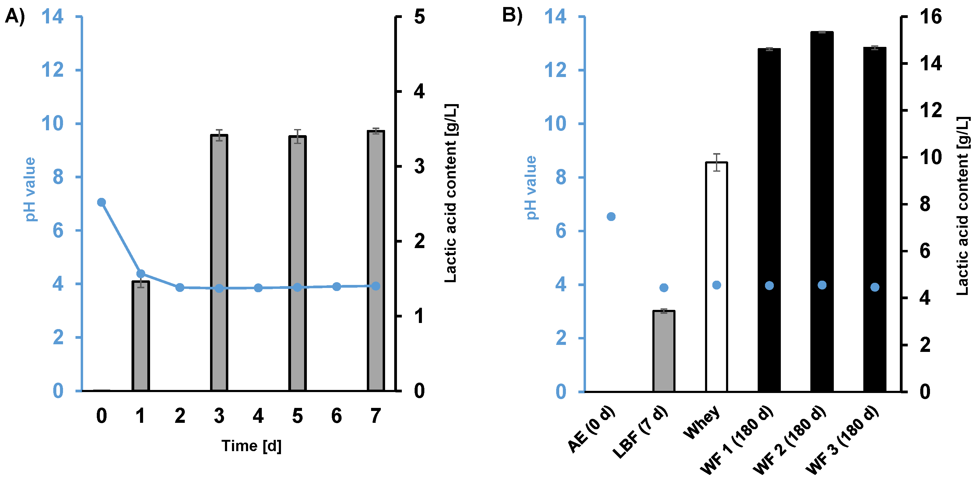
3.1. pH Change and Lactic Acid Formation in Fermentation Samples Inoculated with Lactobacteria (LBF) and Whey (WF)

The fermentation of M. perennis herbal material was investigated in two separate experiments: the first experiment was performed by inoculation with isolated Lactobacteria strains (Lactobacillus plantarum and Pediococcus pentosaceus) and sampled over a fermentation period of 7 d and the second by the addition of whey, sampled after 180 d. The latter experiment was performed according to the official protocol of the German Homoeopathic Pharmacopoeia, GHP no. 34 c [17,20].
Freshly prepared aqueous extracts (AE) of M. perennis herbal parts (plant material:water ratio = 1:5, w/w) displayed pH values between 6.5 and 7.0. In the first experiment, the sample was inoculated with L. plantarum and P. pentosaceus in order to facilitate fermentation [25,26]. Both bacterial strains have recently been isolated from fermented batches of M. perennis [18]. In these LBF the pH dropped rapidly from 7.0 to 3.8 within 3 days (Figure 1A) and then remained virtually unchanged until day 7 (pH 3.9). Simultaneously, the LA concentration increased from 0.0 g/L (day 0) to 3.4 g/L (day 3) and remained constant until day 7 (Figure 1A).
In the second experiment, whey-inoculated fermentation (WF) of M. perennis (according to GHP procedure no. 34 c [17]) was investigated. Deviating from the LBF samples, the WF differed in the composition of the fermented materials (plant material : whey : water ratio = 100 : 50 : 75, w/w/w) and the fermentation time (180 d). Supplementation of whey is an interesting option to facilitate LA production upon medicinal plant fermentation [20], setting up optimum starting conditions for Lactobacteria microflora by providing high lactose contents. Thus, the WF approach reached a 4.3–4.5 fold higher LA concentration (14.6–15.3 g/L, respectively, Figure 1B) compared to LBF without whey.

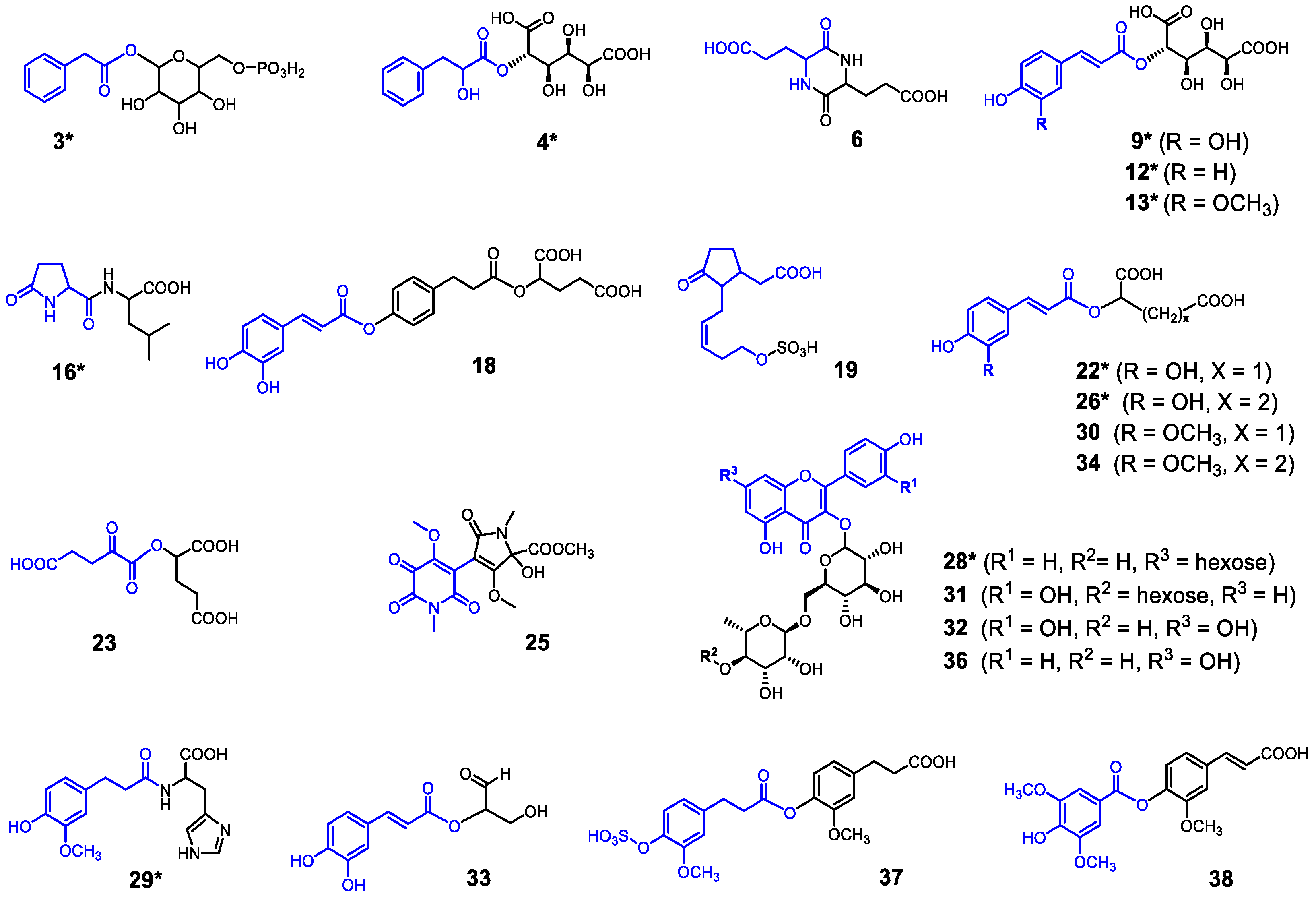
3.2. Characterization of Depsides, Flavonoid Glycosides and Fermentation Metabolites in AE, LBF and WF Samples by LC-MSn

Several classes of natural compounds have been identified in M. perennis, i.e., alkaloid constituents [27,28,29], sterols, terpenes, carotenoids, n-alkylresorcinols [28,30], depsides and flavonoid glycosides [31]. To assign a broad spectrum of high- and low-molecular metabolites, both aqueous unfermented (AE) and fermented extracts (LBF and WF) were comprehensively analyzed in the current study by LC-MSn and GC-MS.
First, the AE of M. perennis was investigated by LC-MSn (negative ionization mode) to monitor all water extractable constituents. In the base peak chromatogram (BPC) of these extracts (Figure 2A) a multitude of peaks were detected, being mainly characterized as depsides, i.e., condensation products of substituted cinnamic acids together with glucaric, malic or 2-hydroxyglutaric acids. Several of them have already been described in previous studies [31,32,33]. Glucaric acid depsides with caffeic, p-coumaric and ferulic acids (isomers of 9*, 12* and 13*, respectively; for structures see Figure 3) were assigned based on their MS fragmentation and distinct UV spectra (Table S1, Figure S2A, Supplementary Materials) in a retention time range between 8.5 and 21.0 min (Figure 2A).
Since many signals were overlapped in a retention time range between 7 and 25 min, extracted ion chromatograms (EIC) were generated (Figure 4). Accordingly, EIC of [M-H] ions at m/z 371, 355 and 385 revealed numerous isomers of 9*, 12* and 13*, respectively (Figure 4B–D). These signals refer to constitutional isomers and diastereomers of the latter [33].
Furthermore, two intense peaks of the main depsides, phaseolic acid (trans-caffeoyl malate, 22*) and mercurialis acid (trans-caffeoyl 2-hydroxyglutarate, 26*), were recorded at tR 26.5 and 34.3 min, respectively (Figure 2A). Moreover, novel depsides were also detected in unfermented Mercurialis extracts. For instance, at tR 39.6, 45.7 and 49.6 min three ferulic acid depsides, i.e., feruloyl malate (30), feruloyl 2-hydroxyglutarate (34) and feruloyl syringate (38), were assigned based on their ESI-MS fragmentation patterns (Table S1, Figure S2C, Supplementary Materials). These depsides have not been described for M. perennis so far. In addition, another novel depside of dihydro-ferulic acid was detected at tR 48.4 min (37, Figure 2A and Figure 3). A neutral loss of 80 Da from the [M-H] ion at m/z 453 in the collision-induced dissociation (CID) experiments yielded a fragment ion at m/z 373, which points to a sulfuric acid residue (HSO3), (Supplementary Materials, Figure S2C). A subsequent loss of 178 Da upon further fragmentation resulted in an ion at m/z 195, indicating the molecule consisted of two dihydro-ferulic acid moieties. Therefore, 37 was tentatively assigned to a bis-dihydro-feruloyl sulfate. The structure of 37 was supported by a HR-MS experiment (Figure S3G).
Besides the depsides, six known kaempferol and quercetin glycosides (Figure 3) were detected in the BPC of the AE between tR 35.0 and 47.2 min (Figure 2A), which have formerly been described for M. perennis ethanolic extracts [31]. Moreover, by comparison of MS data with the literature [34] 12-hydroxyjasmonate sulfate 19 (Figure 3 and Figure S3D, Table S1) was identified (at tR 21.3 min) for the first time in the AE of M. perennis.
When evaluating the fermented samples (LBF and WF) it became apparent that several of the depsides and flavonoids were partly degraded to different extents (Figure 2B,C), while at the same time novel metabolites were formed. For instance, in the BPC of LBF samples, three novel signals 16* (two isomers at tR 18.8 and 20.8 min) and 25 (tR 32.4 min) appeared. The mass spectrum of 16* exhibited the pseudomolecular ions [2M-H] and [M-H] at m/z 483 and 241 (Figure S2B). A subsequent release of CO2 (44 Da) upon CID, yielding a fragment ion at m/z 197, indicated the presence of a carboxylic acid. By comparison with the literature data [35,36] 16* was assigned to 5-oxoprolyl-leucine. For the assignment of compound 25, high resolution MS data were recorded in HR-ESIpos/neg-MS experiments (for details see Supplementary Materials, Figure S3E,F). The corresponding [M-H] and [M+H]+ ions exhibited m/z ratios at 367.07855 (calcd for C15H15O9N2, m/z 367.07830) and 369.09274 (calcd for C15H17O9N2+, m/z 369.09286) accordingly. The molecular formula C15H16O9N2 deduced from these results implies nine units of unsaturation (double bonds or ring systems). Based on these data 25 was tentatively allocated to a novel hermidin alkaloid artefact (see Figure 3, Table S1). An ethylester derivative of 25 has recently been identified from hydroalcoholic extracts of M. perennis [29].
A more complex composition was found for the 180 d WF samples. Several novel constituents were detected such as compound 3* (phenylacetic acid hexoside phosphate), caffeic acid (17) and again isomers of 16* (Figure 2C). Moreover, the depsides 4* (glucaroyl phenyllactate isomers), 18 (caffeoyl p-dihydro-coumaroyl 2-hydroxyglutarate), 23 (2-ketoglutaroyl 2-hydroxyglutarate) and 33 (caffeoyl glycerinaldehyde) were tentatively assigned (Figure 3, Table S1). The latter were probably formed by pH-dependent acyl migration [37] and condensation of carboxylic functions from other depsides. Apart from these constituents, a few N-containing metabolites (6, 10, 15*, 20*, 29*, 39) were also detected, which showed characteristic even-numbered m/z ratios of their [M-H] ions. These compounds are probably formed upon reaction of whey peptides with depsides or from alkaloid artefacts. Some of them could not be further characterized, except compounds 6 and 29*, one of which was tentatively identified as glutamic acid diketopiperazine (6, tR 4.5 min) due to the loss of a [glutamic acid-H2O] moiety (129 Da) from the [M-H] ion at m/z 257 and the other as condensation product of dihydro-ferulic acid and histidine (29*, two isomers at tR 38.6 an 39.6 min, Figure 2C and Figure 3, Table S1). Accordingly, the mass spectrum of 29* exhibited the elimination of a histidine moiety (155 Da) from the [M-H] ion at m/z 332, yielding a fragment at m/z 177 (Figure S2B). The latter corresponds to a dihydro-feruloyl unit. In addition, the UV absorption maximum at 274 nm, which is similar to the chromophore of free histidine [38], corroborates the structural assignment of the isomers of compound 29*.

3.3. GC-MS Assignment of Low Molecular Weight Compounds in AE and WF Samples

For analyzing low molecular weight constituents, aqueous and a whey-inoculated fermented extract (AE and WF, respectively) were dried in vacuo, silylated with BSTFA and then investigated by GC-MS (Figure 5). Compound assignment was based on comparison of the respective mass spectra with the NIST database. At first glance, sugar constituents (fructose, galactose, glucose, sucrose) were assigned via MS and comparison with reference compounds (Figure 5C). In the total ion chromatograms (TICs) of the unfermented M. perennis water extract (AE) the monosaccharides fructose, glucose and myo-inositol were detected, together with the disacharides sucrose and melibiose, at tR 28.8, 30.8, 35.5, 46.6 and 54.8 min, respectively (Figure 5A).
In the WF samples, fructose, glucose, sucrose and melibiose were almost completely metabolized while galactose was formed (Figure 5B). The latter constitutes a microbial degradation product, which is released from melibiose or lactose (the latter originate from whey). Galactose is obviously accumulated in the fermentate, since only certain Lactobacteria species (LAB) are able to further metabolize this monosaccharide [39,40]. Interestingly, myo-inositol, which may originate from phytic acid (inositol hexaphosphate) [41,42], was also essentially not converted upon fermentation.
Moreover, considerable amounts of lactic acid (LA) were detected in the TIC together with free amino acids, which were mainly analyzed in the WF extract (see Supplementary Materials, Figure S4). Remarkably, lower amounts of biogenic amines, such as putrescine (1,4-diaminobutane) and cadaverin (1,5-diaminopentane) as well as 4-aminobutyric acid could also be detected as decarboxylation products of their specific precursor amino acids ornithine, lysine and glutamic acid, respectively (Figure S4B). It is known from the literature that these amines may be produced by the action of Lactobacillus decarboxylases [43,44,45]. This is also corroborated by the fact that they were only detected in fermented, but not in unfermented extracts. Moreover, 5-oxoproline (pyroglutamic acid) was assigned as a further distinctive Lactobacteria marker metabolite [46], apparently formed from glutamic acid due to microbial enzymatic dehydration [47]. It has been reported in the literature that 5-oxoproline exhibits similar antimicrobial activity like LA and inhibits the growth of many spoilage bacteria, e.g., of the Enterobacter and Pseudomonas families [47]. Furthermore, 5-oxoproline-containing dipeptides such as compound 16*, assigned in the previous chapter, produced by L. plantarum have recently been shown to exhibit immunomodulatory activity in vivo [48].

3.4. Stimulation of NFκB Activity of THP-1 Cells by Treatment with AE, LBF and WF Extracts

To study the immunomodulating effects of M. perennis extracts, THP-1 cells were utilized as a model for human monocytes [49]. THP-1 cells were formerly applied, e.g., to screen diverse plant extracts regarding anti-inflammatory activity [50,51,52].
Proinflammatory mediator expression is regulated via molecular pathways including MAP kinases and NF-κB signaling. The binding of the transcription factor NFκB to the response element is essential for the transcription of different proinflammatory genes [2,3]. In this study, the influence of the extracts on NFκB activity was investigated using an immune reporter cell line (THP-1), characterized by the fusion of GFP to the NFκB response element.
First, the effect of extracts on the cell viability of THP-1 cells was determined by a WST assay measuring the mitochondrial activity. Cell viability was significantly diminished in cells treated with AE, LBF and WF1 extracts at the highest concentration (1000 µg/mL), indicating a cytotoxic effect (see Supplementary Materials, Figure S5). For whey, as well as the WF2 and WF3 extracts, no effect on the viability of THP-1 cells could be detected in the entire concentration range investigated (Figure S5).
For all extracts, a concentration-dependent increase in NFκB activity was observed at the lower concentrations (1–100 µg/mL for AE and LBF, 1–300 µg/mL for whey and WF1, WF2 and WF3, respectively) (Figure 6). The decrease in NFκB activity at the higher concentrations (1000 µg/mL) is due to the cytotoxic effects of the M. perennis extracts. However, it can be assumed that a toxicity-related impact on the function of the cells already occurs at somewhat lower concentrations. No differences in activity were found between the AE, LBF and WF samples, indicating that fermentation has no influence on the ingredients causing NFκB activation.

3.5. Effect of Extracts on Cytokine Expression

The secretion of cytokines is one of the important functions of activated immune cells to fulfil its function. For further characterization of the extracts, their impact on the proinflammatory cytokine expression of LPS-stimulated THP-1 cells was investigated (Figure 7). Cytokine expression was moderately affected in the concentration range of 1–100 µg/mL and clearly exceeded the LPS-stimulated control only in the case of LBF (IL-6) and whey (IL-6, IL-1β, TNFα). At higher concentrations (1000 µg/mL and 300 µg/mL, respectively), a strong increase in cytokine secretion was detected in some cases, which, however, was also based on cytotoxic effects. In contrast to the extracts obtained from M. perennis, whey showed a clear concentration-dependent effect on all cytokines in the whole concentration range. An immunostimulatory effect of whey protein extracts on primary peripheral blood mononuclear cells has already been described in the literature [53,54,55], while the accumulation of the cytokines IL-10, IL-1β, IL-8, IL-6 and TNFα was observed.
In our study a tendency towards a concentration-dependent stimulating effect on cytokine expression by the M. perennis extracts was registered in the case of IL-8, IL-6 (AE and LBF only) and IL-1β (AE only). The TNFα expression remained unaffected by treatment with the extracts. IL-1β, IL-6 and TNFα are known as endogen pyrogens that can induce fever or a local inflammation, but also have an impact on the coagulation cascade and activation of other immune cells. IL-8 acts on migration and proliferation of keratinocytes and fibroblasts and has a chemotactic effect on granulocytes, all relevant in wound healing.
Endotoxins, which may occur as autolysis products of Gram(-) bacteria [56] can mimic immunostimulating effects of fermented plant extracts. Hence, they must be taken into account when interpreting the results. Since in the present study no or only small amounts of endotoxins were detected in the samples tested, as evidenced by the Limulus amoebocyte lysate (LAL) assay (Table S2, Supplementary Materials), the observed immunostimulatory effect in vitro was not associated with endotoxins (bacterial LPS).

3.6. Bacterial Toxicity: Inhibition of Bioluminescence Induced by WF Extracts in V. fischeri

The marine Gram-negative bacterium Vibrio fischeri (strain NRRL B-11177) is widely used as a test organism for screening the aquatic toxicity of compounds and environmental samples. However, this nontarget organism test has been also discussed as an in vitro tool to support toxicology studies of xenobiotics, due to its sensitivity and predictive power. The rationale behind its implementation is based on the fact, that V. fisheri toxicity data have a good correlation with in vivo data of human toxicity [57]. Furthermore, this assay might give an indication for an antibacterial potential of an extract. In this study, the extracts WF 1–3 were evaluated against V. fischeri based on the bioluminescence read-out as a (anti-bacterial) toxicity endpoint. As expected, varying extract-specific bacterial toxicity was found, based on IC50 values (WF1 > WF2 > WF3), but far from physiologically and systemically relevant concentrations (Figure 8).

3.7. Genotoxic Potential: In Vitro Micronucleus Test

WF extracts from M. perennis, processed according to GHP procedure no. 34 c, are used in pharmaceutical products for dermal applications or in ophthalmic drugs. To evaluate the mutagenic potential of the WF extracts these were tested in an AMES test and found to be non-mutagenic (data not shown) [58]. For further evaluation of genotoxic potential in the human system, the in vitro micronucleus test conducted with THP-1 monocytic cells was performed.
Exposure to a genotoxic test substance may result in damages to chromosomes or spindle apparatus of cells. During regular cell division, this type of damage can create a smaller micro- or hyponucleus, apart from the main nucleus. Therefore, the micronucleus test is an in vitro method that provides a comprehensive basis for investigating chromosome damage, because both aneugens and clastogens can be detected in cells that have undergone cell division during or after exposure to the test sample. Micronuclei represent damages that have been transmitted to daughter cells and in either case, the changes may not be compatible with cell survival.
As confirmed by the viability assessment (see above), none of the tested WF extract concentrations had the capability of producing artifactual positive responses by concurrent excessive cytotoxicity (Figure 9). Therefore, as the acceptability criteria for the assay were fulfilled (according to OECD 487; [21]), it can be emphasised that none of the test concentrations of WF1–3 exhibited a statistically significant increase in micro- and hyponuclei frequency, compared with the control background.
However, a visible, though non-significant decrease compared to the background control, as observed in WF1, was probably associated with reduced proliferation rather than specific chemopreventive or anti-genotoxic properties. Thus, the WF extracts were clearly negative, being unable to induce chromosome breaks (clastogenicity) and/or gain or loss micronuclei and hyponuclei (aneugenicity). Significant induction of both micro- and hyponuclei, i.e., chromosome breakage and whole chromosome loss, respectively, was however achieved by the subtoxic treatment of THP-1 cells with vinblastine sulphate (positive control, PC), demonstrating the sensitivity of the test system (Figure 9).

4. Conclusions

In the study presented here, the lacto-fermentation of the medicinal plant M. perennis was investigated with respect to its specific metabolite profile and the evaluation of immunomodulatory activity. Lacto-fermentation provides enzymes facilitating aqueous extraction by digestion of the plant cellular matrix and goes along with LA formation, which preserves the extracts by pH decrease and antimicrobial effects.
Fermentation of M. perennis by inoculation with either two Lactobacteria strains (LBF) or whey (WF) yielded extracts whose microbial stability was caused by low pH (3.9) and high LA concentration (maximum 15.6 g/L). Analysis of the compound profiles of LBF and WF extracts by LC-MSn and GC-MS revealed a partial degradation of plant depsides and flavonoid glycosides. This went along with the formation of several novel compounds via fermentative conversion. Among others, novel substituted cinnamic acid depsides, but also nitrogen-containing metabolites, i.e., amino acid derivatives, biogenic amines, alkaloid artefacts, etc., could be tentatively assigned. However, some of the latter remain unknown and their exact structural assignment requires further studies. All extracts (AE, LBF and WF) enhanced the NFκB activity of LPS-stimulated THP-1 NFκB reporter cells in a concentration-dependent manner, while differences between unfermented (AE) and fermented (LBF or WF) extracts were not observed. Accordingly, bioactive constituents were not substantially affected by LA fermentation. Simultaneously, cytokine secretion was detected (most evidently for IL-8) for AE, LBF and WF in some cases, although this effect was partly superimposed by cytotoxicity (at 1000 µg/mL). However, whey exhibited a strong immunostimulating effect for all cytokines (IL-8, IL-6, IL-1β and TNFα). Further investigations are needed to understand potential correlations between the constituents of the fermented extracts and the observed immunomodulating activity. Conclusions about the contribution of the observed effects to specific phases of the healing process cannot be drawn yet. More complex in vitro models have to be applied to gain further understanding about the impact on other involved cell types and their interaction.
Finally, the toxicity and genotoxicity of the WF extracts was assessed in a V. fisheri assay and an in vitro micronucleus test with THP-1 cells, respectively. No toxic or mutagenic effects could be determined which supports the safety of WF extracts for their use, e.g., in dermal pharmaceutical remedies. The results of the study presented here significantly expand our knowledge on the technology of producing LA-fermented medicinal plant extracts and their biological activity.

Supplementary Materials

The following supporting information can be downloaded at: https://www.mdpi.com/article/10.3390/fermentation9020190/s1, Figure S1: HPLC-MSn fingerprints (BPC = base peak chromatograms) of M. perennis extracts. Figure S2A-C: Selections of MSn(ESIneg) mass spectra and UV spectra of individual peaks, detected in the HPLC-DAD-MSn(ESI) chromatograms of the none-fermented (AE) and fermented extracts (LBF, WF) of M. perennis. Table S1: Tentative assignment of polyphenolic constituents (depsides and flavonoids) in the water extract and lactic acid fermented extract, obtained from herbal parts of M. perennis, according HPLC-DAD-ESI(negative)-MSn data. Method: HR-ESI-HPLC-MS analyses. Figure S3A: Base peak chromatogram of a Lactobacteria inoculated fermentation extract (LBF) of M. perennis with extracted ion chromatogram (mass scan on m/z 243; HR-ESI-MS of the peak (tR 8.2 min) in the positive mode, with calculated masses and proposed molecular formula for metabolite 16* (isomer 1). Figure S3B: Base peak chromatogram of a Lactobacteria inoculated fermentation extract (LBF) of M. perennis with extracted ion chromatogram (mass scan on m/z 243; HR-ESI-MS of the peak (tR 9.2 min) in the positive mode, with calculated masses and proposed molecular formula for metabolite 16* (isomer 2). Figure S3C: Structure annotation from FISh scoring node on MS2 spectrum of 16*. The fragmentation was simulated with the Compound Discoverer software, Version 3.3 of Thermo Scientific. Figure S3D: Base peak chromatogram of a Lactobacteria inoculated fermentation extract (LBF) of M. perennis with extracted ion chromatogram (mass scan on m/z 305; HR-ESI-MS of the peak (tR 8.0 min) in the negative mode, with calculated masses and proposed molecular formula for metabolite 19. Figure S3E: Base peak chromatogram of a Lactobacteria inoculated fermentation extract (LBF) of M. perennis with extracted ion chromatogram (mass scan on m/z 369; HR-ESI-MS of the peak (tR 12.3 min) in the positive mode, with calculated masses and proposed molecular formula for metabolite 25. Figure S3F: Structure annotation from FISh scoring node on MS2 spectrum of 25. The fragmentation was simulated with the Compound Discoverer software, Version 3.3 of Thermo Scientific. Figure S3G: Base peak chromatogram of a Lactobacteria inoculated fermentation extract (LBF) of M. perennis with extracted ion chromatogram (mass scan on m/z 453; HR-ESI-MS of the peak (tR 15.7 min) in the negative mode, with calculated masses and proposed molecular formula for metabolite 37. Figure S3H: Structure annotation from FISh scoring node on MS2 spectrum of 37. The fragmentation was simulated with the Compound Discoverer software, Version 3.3 of Thermo Scientific. Figure S4: GC-MS total ion chromatogram sections of a WF extract, obtained after a HAB procedure, showing low molecular constituents (TMS derivatization). Method: Viability assay. Figure S5: Effects of test substances on cell viability of THP-1 NFκB reporter cells determined by WST assay. Method: Determination of evaporation residues (dry weight) of liquid extracts. Method: Endotoxin (LAL Limulus) assay. Table S2: Chemical and physical parameters of tested samples.

Author Contributions

Design of the study: P.L., I.Z., C.T., D.R.K., F.C.S. and C.G.; preparation of extracts, fermentation experiments: P.L. and I.Z.; data acquisition: P.L. and L.K.M.; HR-MS experiments and data interpretation: I.K. and P.L.; cell culture experiments, data equisition and interpretation: A.M.Z.-K., M.G.-K., S.N. and C.G.; toxicity (V. fisheri) and genotoxicity tests: M.G.-K.; evaluation of the data and preparation of the manuscript: P.L., I.Z., L.K.M., D.R.K., F.C.S., A.M.Z.-K., M.G.-K. and C.G. All authors have read and agreed to the published version of the manuscript.

Funding

S.N., A.M.Z.K. and C.G. were financially supported by a PRIAM-based consortium of multiple funders.

Institutional Review Board Statement

Not applicable.

Informed Consent Statement

Not applicable.

Data Availability Statement

Not applicable.

Acknowledgments

The authors wish to acknowledge Sonja Dahlin and Anja Bohrer for performance and data interpretation of the LAL Assay and Sabrina Eberle (all WALA Heilmittel GmbH) for support with fermentation experiments. We are grateful to Rhinaixa Duque-Thüs (Institute of Botany, University of Hohenheim, Germany) for identifying the herbarium specimen.

Conflicts of Interest

P.L., I.Z., L.K.M., C.T., F.C.S. and D.R.K. are employed at WALA Heilmittel GmbH. The authors declare that the research was conducted in the absence of any commercial or financial relationships that could be construed as a potential conflict of interest.

References

  1. Gurtner, G.C.; Werner, S.; Barrandon, Y.; Longaker, M.T. Wound repair and regeneration. Nature 2008, 453, 314–321. [Google Scholar] [CrossRef] [PubMed]
  2. Lawrence, T. The nuclear factor NF-kappaB pathway in inflammation. Cold Spring Harb. Perspect. Biol. 2009, 1, a001651. [Google Scholar] [CrossRef] [Green Version]
  3. Mitchell, S.; Vargas, J.; Hoffmann, A. Signaling via the NFκB system. Wiley Interdiscip. Rev. Syst. Biol. Med. 2016, 8, 227–241. [Google Scholar] [CrossRef] [Green Version]
  4. Embacher, K.; Zilkowski, I.; Turek, C.; Stintzing, F.C. Topische Anwendung von Heilpflanzen zur Unterstützung der Wundheilung. Z. Für Phytother. 2021, 42, 249–262. [Google Scholar] [CrossRef]
  5. Talhouk, R.S.; Karam, C.; Fostok, S.; El-Jouni, W.; Barbour, E.K. Anti-inflammatory bioactivities in plant extracts. J. Med. Food 2007, 10, 1–10. [Google Scholar] [CrossRef] [PubMed]
  6. Korkina, L.; Kostyuk, V.; de Luca, C.; Pastore, S. Plant phenylpropanoids as emerging anti-inflammatory agents. Mini Rev. Med. Chem. 2011, 11, 823–835. [Google Scholar] [CrossRef] [PubMed]
  7. Recio, M.C.; Andujar, I.; Rios, J.L. Anti-inflammatory agents from plants: Progress and potential. Curr. Med. Chem. 2012, 19, 2088–2103. [Google Scholar] [CrossRef]
  8. Miranda, R.d.S.; de Jesus, B.d.S.M.; da Silva Luiz, S.R.; Viana, C.B.; Adão Malafaia, C.R.; Figueiredo, F.d.S.; Carvalho, T.D.S.C.; Silva, M.L.; Londero, V.S.; da Costa-Silva, T.A.; et al. Antiinflammatory activity of natural triterpenes-An overview from 2006 to 2021. Phytother. Res. 2022, 36, 1459–1506. [Google Scholar] [CrossRef]
  9. Li, X.; Li, X.; Huang, N.; Liu, R.; Sun, R. A comprehensive review and perspectives on pharmacology and toxicology of saikosaponins. Phytomedicine 2018, 50, 73–87. [Google Scholar] [CrossRef]
  10. Zhang, A.; Yang, X.; Li, Q.; Yang, Y.; Zhao, G.; Wang, B.; Wu, D. Immunostimulatory activity of water-extractable polysaccharides from Cistanche deserticola as a plant adjuvant in vitro and in vivo. PLoS ONE 2018, 13, e0191356. [Google Scholar] [CrossRef] [Green Version]
  11. Gründemann, C.; Garcia-Käufer, M.; Sauer, B.; Scheer, R.; Merdivan, S.; Bettin, P.; Huber, R.; Lindequist, U. Comparative chemical and biological investigations of β-glucan-containing products from shiitake mushrooms. J. Funct. Foods 2015, 18, 692–702. [Google Scholar] [CrossRef]
  12. Huang, K.; Li, Y.; Tao, S.; Wei, G.; Huang, Y.; Chen, D.; Wu, C. Purification, characterization and biological activity of polysaccharides from Dendrobium officinale. Molecules 2016, 21, 701. [Google Scholar] [CrossRef] [Green Version]
  13. Kardošová, A.; Ebringerová, A.; Alföldi, J.; Nosál’ová, G.; Matáková, T.; Hříbalová, V. Structural features and biological activity of an acidic polysaccharide complex from Mahonia aquifolium (Pursh) Nutt. Carbohydr. Polym. 2004, 57, 165–176. [Google Scholar] [CrossRef]
  14. Lorenz, P.; Beckmann, C.; Felenda, J.; Meyer, U.; Stintzing, F. Das Waldbingelkraut (Mercurialis perennis L.)—Pharmakognosie einer alten Arzneipflanze. Z Phytother 2013, 34, 40–46. [Google Scholar] [CrossRef]
  15. Rugman, F.; Meecham, J.; Edmondson, J. Mercurialis perennis (dog’s mercury) poisoning: A case of mistaken identity. Br. Med. J. 1983, 287, 1924. [Google Scholar] [CrossRef] [Green Version]
  16. Watson, P.J. Suspected dog’s mercury (Mercurialis perennis) poisoning in cattle. Vet. Rec. 1998, 142, 116–117. [Google Scholar] [CrossRef] [PubMed]
  17. German Homoeopathic Pharmacopoeia: GHP; WVG medPharm Scientific Publishers: Stuttgart, Germany, 2005; ISBN 978-3-8047-5088-3.
  18. Lorenz, P.; Bunse, M.; Sauer, S.; Conrad, J.; Stintzing, F.C.; Kammerer, D.R. Conversion of plant secondary metabolites upon fermentation of Mercurialis perennis L. extracts with two Lactobacteria strains. Fermentation 2019, 5, 42. [Google Scholar] [CrossRef] [Green Version]
  19. Lorenz, P.; Conrad, J.; Stintzing, F.C. Metabolic fate of depsides and alkaloid constituents in aqueous extracts from Mercurialis perennis L. during fermentation. Chem. Biodivers. 2013, 10, 1706–1723. [Google Scholar] [CrossRef]
  20. Sauer, S.; Dlugosch, L.; Milke, F.; Brinkhoff, T.; Kammerer, D.R.; Stintzing, F.C.; Simon, M. Succession of bacterial and fungal communities during fermentation of medicinal plants. Fermentation 2022, 8, 383. [Google Scholar] [CrossRef]
  21. Test No. 487: In Vitro Mammalian Cell Micronucleus Test; OECD Publishing: Paris, France, 2016; ISBN 9789264264861.
  22. Bryce, S.M.; Bemis, J.C.; Avlasevich, S.L.; Dertinger, S.D. In vitro micronucleus assay scored by flow cytometry provides a comprehensive evaluation of cytogenetic damage and cytotoxicity. Mutat. Res. 2007, 630, 78–91. [Google Scholar] [CrossRef] [Green Version]
  23. Hashimoto, K.; Nakajima, Y.; Matsumura, S.; Chatani, F. An in vitro micronucleus assay with size-classified micronucleus counting to discriminate aneugens from clastogens. Toxicol. Vitr. 2010, 24, 208–216. [Google Scholar] [CrossRef]
  24. ISO 11348-1:2007; Water Quality—Determination of the Inhibitory Effect of Water Samples on the Light Emission of Vibrio fischeri (Luminescent bacteria test)—Part 1: Method Using Freshly Prepared Bacteria. Beuth Verlag GmbH: Berlin, Germany, 2009.
  25. Laitila, A.; Sweins, H.; Vilpola, A.; Kotaviita, E.; Olkku, J.; Home, S.; Haikara, A. Lactobacillus plantarum and Pediococcus pentosaceus starter cultures as a tool for microflora management in malting and for enhancement of malt processability. J. Agric. Food Chem. 2006, 54, 3840–3851. [Google Scholar] [CrossRef] [PubMed]
  26. Song, G.; He, Z.; Wang, X.; Zhao, M.; Cao, X.; Lin, X.; Ji, C.; Zhang, S.; Liang, H. Improving the quality of Suancai by inoculating with Lactobacillus plantarum and Pediococcus pentosaceus. Food Res. Int. 2021, 148, 110581. [Google Scholar] [CrossRef] [PubMed]
  27. Swan, G.A. Isolation, structure, and synthesis of hermidin, a chromogen from Mercurialis perennis L. J. Chem. Soc. Perkin Trans. 1 1985, 16, 1757–1766. [Google Scholar] [CrossRef]
  28. Lorenz, P.; Hradecky, M.; Berger, M.; Bertrams, J.; Meyer, U.; Stintzing, F.C. Lipophilic constituents from aerial and root parts of Mercurialis perennis L. Phytochem. Anal. 2010, 21, 234–245. [Google Scholar] [CrossRef]
  29. Lorenz, P.; Conrad, J.; Duckstein, S.; Kammerer, D.R.; Stintzing, F.C. Chemistry of hermidin: Insights from extraction experiments with the main alkaloid of Mercurialis perennis L. tracked by GC/MS and LC/MSn. HCA 2014, 97, 1606–1623. [Google Scholar] [CrossRef]
  30. Lorenz, P.; Knödler, M.; Bertrams, J.; Berger, M.; Meyer, U.; Stintzing, F.C. n-Alkylresorcinol occurrence in Mercurialis perennis L. (Euphorbiaceae). Z. Naturforsch. C J. Biosci. 2010, 65, 174–179. [Google Scholar] [CrossRef]
  31. Lorenz, P.; Conrad, J.; Bertrams, J.; Berger, M.; Duckstein, S.; Meyer, U.; Stintzing, F.C. Investigations into the phenolic constituents of dog’s mercury (Mercurialis perennis L.) by LC-MS/MS and GC-MS analyses. Phytochem. Anal. 2012, 23, 60–71. [Google Scholar] [CrossRef]
  32. Lorenz, P.; Duckstein, S.; Conrad, J.; Knödler, M.; Meyer, U.; Stintzing, F.C. An approach to the chemotaxonomic differentiation of two European dog’s mercury species: Mercurialis annua L. and M. perennis L. Chem. Biodivers. 2012, 9, 282–297. [Google Scholar] [CrossRef]
  33. Chamandy, A.; Zhao, M.; Rammal, H.; Ennahar, S. Hyphenated LC-ABTS·+ and LC-DAD-HRMS for simultaneous analysis and identification of antioxidant compounds in Astragalus emarginatus Labill. extracts. J. Pharm. Anal. 2022, 12, 253–262. [Google Scholar] [CrossRef]
  34. Kapp, K.; Hakala, E.; Orav, A.; Pohjala, L.; Vuorela, P.; Püssa, T.; Vuorela, H.; Raal, A. Commercial peppermint (Mentha × piperita L.) teas: Antichlamydial effect and polyphenolic composition. Food Res. Int. 2013, 53, 758–766. [Google Scholar] [CrossRef]
  35. Frerot, E.; Chen, T. Identification and quantitation of new glutamic acid derivatives in soy sauce by UPLC/MS/MS. Chem. Biodivers. 2013, 10, 1842–1850. [Google Scholar] [CrossRef] [PubMed]
  36. Kiyono, T.; Hirooka, K.; Yamamoto, Y.; Kuniishi, S.; Ohtsuka, M.; Kimura, S.; Park, E.Y.; Nakamura, Y.; Sato, K. Identification of pyroglutamyl peptides in Japanese rice wine (sake): Presence of hepatoprotective pyroGlu-Leu. J. Agric. Food Chem. 2013, 61, 11660–11667. [Google Scholar] [CrossRef] [PubMed]
  37. Deshpande, S.; Jaiswal, R.; Matei, M.F.; Kuhnert, N. Investigation of acyl migration in mono- and dicaffeoylquinic acids under aqueous basic, aqueous acidic, and dry roasting conditions. J. Agric. Food Chem. 2014, 62, 9160–9170. [Google Scholar] [CrossRef]
  38. Nakano, H.; Tamai, N.; Tsukamoto, M.; Abe, N. Change in ultraviolet absorption spectra of histidine in aqueous solution as a result of femtosecond laser irradiation. JJSLSM 2010, 31, 390–393. [Google Scholar] [CrossRef] [Green Version]
  39. Turner, K.W.; Martley, F.G. Galactose fermentation and classification of thermophilic lactobacilli. Appl. Environ. Microbiol. 1983, 45, 1932–1934. [Google Scholar] [CrossRef] [Green Version]
  40. Iskandar, C.F.; Cailliez-Grimal, C.; Borges, F.; Revol-Junelles, A.-M. Review of lactose and galactose metabolism in lactic acid bacteria dedicated to expert genomic annotation. Trends Food Sci. Technol. 2019, 88, 121–132. [Google Scholar] [CrossRef]
  41. Loewus, F.A. Inositol and plant cell wall polysaccharide biogenesis. Subcell. Biochem. 2006, 39, 21–45. [Google Scholar] [CrossRef]
  42. Hadi Alkarawi, H.; Zotz, G. Phytic acid in green leaves. Plant Biol. 2014, 16, 697–701. [Google Scholar] [CrossRef]
  43. Arena, M.E.; Manca de Nadra, M.C. Biogenic amine production by Lactobacillus. J. Appl. Microbiol. 2001, 90, 158–162. [Google Scholar] [CrossRef] [PubMed]
  44. Ruiz-Capillas, C.; Jiménez-Colmenero, F. Biogenic amines in meat and meat products. Crit. Rev. Food Sci. Nutr. 2004, 44, 489–499. [Google Scholar] [CrossRef] [Green Version]
  45. Capozzi, V.; Russo, P.; Ladero, V.; Fernández, M.; Fiocco, D.; Alvarez, M.A.; Grieco, F.; Spano, G. Biogenic amines degradation by Lactobacillus plantarum: Toward a potential application in wine. Front. Microbiol. 2012, 3, 122. [Google Scholar] [CrossRef] [PubMed] [Green Version]
  46. Mucchetti, G.; Locci, F.; Massara, P.; Vitale, R.; Neviani, E. Production of pyroglutamic acid by thermophilic lactic acid bacteria in hard-cooked mini-cheeses. J. Dairy Sci. 2002, 85, 2489–2496. [Google Scholar] [CrossRef] [PubMed]
  47. Yang, Z.; Suomalainen, T.; Mäyrä-Mäkinen, A.; Huttunen, E. Antimicrobial activity of 2-pyrrolidone-5-carboxylic acid produced by lactic acid bacteria. J. Food Prot. 1997, 60, 786–790. [Google Scholar] [CrossRef] [PubMed]
  48. Zvanych, R.; Lukenda, N.; Kim, J.J.; Li, X.; Petrof, E.O.; Khan, W.I.; Magarvey, N.A. Small molecule immunomodulins from cultures of the human microbiome member Lactobacillus plantarum. J. Antibiot. 2014, 67, 85–88. [Google Scholar] [CrossRef] [PubMed]
  49. Bosshart, H.; Heinzelmann, M. THP-1 cells as a model for human monocytes. Ann. Transl. Med. 2016, 4, 438. [Google Scholar] [CrossRef] [PubMed] [Green Version]
  50. Kartini; Piyaviriyakul, S.; Thongpraditchote, S.; Siripong, P.; Vallisuta, O. Effects of Plantago major extracts and its chemical compounds on proliferation of cancer cells and cytokines production of lipopolysaccharide-activated THP-1 macrophages. Pharmacogn. Mag. 2017, 13, 393–399. [Google Scholar] [CrossRef] [PubMed] [Green Version]
  51. D’Eliseo, D.; Pannucci, E.; Bernini, R.; Campo, M.; Romani, A.; Santi, L.; Velotti, F. In vitro studies on anti-inflammatory activities of kiwifruit peel extract in human THP-1 monocytes. J. Ethnopharmacol. 2019, 233, 41–46. [Google Scholar] [CrossRef] [Green Version]
  52. Albrahim, T.; Alnasser, M.M.; Al-Anazi, M.R.; ALKahtani, M.D.; Alkahtani, S.; Al-Qahtani, A.A. Potential anti-inflammatory and anti-apoptotic effect of Coccinia grandis plant extract in LPS stimulated-THP-1 cells. Environ. Sci. Pollut. Res. Int. 2020, 27, 21892–21904. [Google Scholar] [CrossRef]
  53. Chun, S.-H.; Lee, H.A.; Lee, K.B.; Kim, S.H.; Park, K.-Y.; Lee, K.-W. Effects of glycated whey protein concentrate on pro-inflammatory cytokine expression and phagocytic activity in RAW264.7 macrophages. Biol. Pharm. Bull. 2016, 39, 199–206. [Google Scholar] [CrossRef] [Green Version]
  54. Rusu, D.; Drouin, R.; Pouliot, Y.; Gauthier, S.; Poubelle, P.E. A bovine whey protein extract stimulates human neutrophils to generate bioactive IL-1Ra through a NF-kappaB- and MAPK-dependent mechanism. J. Nutr. 2010, 140, 382–391. [Google Scholar] [CrossRef] [PubMed] [Green Version]
  55. Kiewiet, M.B.G.; Dekkers, R.; Gros, M.; van Neerven, R.J.J.; Groeneveld, A.; de Vos, P.; Faas, M.M. Toll-like receptor mediated activation is possibly involved in immunoregulating properties of cow’s milk hydrolysates. PLoS ONE 2017, 12, e0178191. [Google Scholar] [CrossRef] [PubMed] [Green Version]
  56. Kimbrell, M.R.; Warshakoon, H.; Cromer, J.R.; Malladi, S.; Hood, J.D.; Balakrishna, R.; Scholdberg, T.A.; David, S.A. Comparison of the immunostimulatory and proinflammatory activities of candidate Gram-positive endotoxins, lipoteichoic acid, peptidoglycan, and lipopeptides, in murine and human cells. Immunol. Lett. 2008, 118, 132–141. [Google Scholar] [CrossRef] [PubMed] [Green Version]
  57. Kahru, A. Ecotoxicological tests in non-ecotoxicological research: Contribution to the three Rs. Use of luminescent photobacteria for evaluating the toxicity of 47 MEIC reference chemicals. ALTEX 2006, 23, 302–308. [Google Scholar]
  58. In Vitro Evaluation of Genotoxicity and Irritation Potential of Eye Drops Containing Aqueous Plant Extracts, 9th ed.; Zilkowski, I.; Müller, A.; Mörbt, N.; Turek, C.; Stintzing, F.C. (Eds.) 11th World Congress (WC11), Maastricht; ALTEX Proceedings: Kreuzlingen, Switzerland, 2021. [Google Scholar]
Figure 1. (A) pH decline (blue curve) and increase in LA concentration (grey bars) in a Lactobacteria-inoculated fermentation (LBF) of M. perennis, with two LAB strains (L. plantarum and P. pentosaceus; n = 3, measured in triplicate). (B) pH values (blue dots) and lactic acid concentrations in AE, LBF and WF samples (bars, measured in triplicate). AE = aqueous extract, LBF = Lactobacteria-inoculated fermentation; WF = whey-inoculated fermentation.
Fermentation 09 00190 g001
Figure 2. HPLC-(DAD)/ESI-MSn base peak chromatograms (BPC) of aqueous (AE), Lactobacteria (LBF) and whey-inoculated (WF) fermentation extracts obtained from herbal parts of M. perennis. (A) BPC of AE; (B) BPC of LBF after 7 d; (C) BPC of WF after 180 d (according to GHP procedure no. 34 c). The peak numbering corresponds to the structural formulas illustrated in Figure 3. For compound assignment see Table S1 (Supplementary Materials). Compounds with green numbers were merely detected in LBF or WF extracts. Isomers are marked with an asterisk (*).
Fermentation 09 00190 g002
Figure 3. Chemical structures of depsides, carboxylic esters and flavonoid glycosides (and others) identified in non-fermented and fermented extracts of M. perennis. For better comprehensibility of structures one acyl moiety of each structure is drawn in blue color. Isomers are marked with an asterisk (*).
Fermentation 09 00190 g003
Figure 4. Detection of cinnamoyl glucarates by HPLC-(DAD)/ESI-MSn in the AE of M. perennis herbal parts. (A) Base peak chromatogram (BPC). Extracted ion chromatograms (EIC) of [M-H] ions at m/z 371, 355 and 385 showing caffeoyl glucarate isomers 9* (B), p-coumaroyl glucarate isomers 12* (C) and feruloyl glucarate isomers 13* (D), respectively.
Fermentation 09 00190 g004
Figure 5. GC-MS sections of AE (A) and WF extracts (B) and reference compounds (C) after silylation. Asterisks mark the furanose form of the respective monosaccharides (beside pyranose form).
Fermentation 09 00190 g005
Figure 6. Effects of extracts on NFκB activity of THP-1 NFκB immune reporter cells. THP-1 cells were stimulated with LPS (100 mg/mL) and incubated for 24 h with medium (unstimulated, stimulated), dexamethasone (Dex) or extracts. GFP expression was quantified by flow cytometry. The percentage of cells was compared, normalized to the stimulated control and presented as mean ± standard deviation. n = 3; * p < 0.05, ** p < 0.01, *** p < 0.001, **** p < 0.0001.
Fermentation 09 00190 g006
Figure 7. Effects of extracts on cytokines of THP-1 NFκB-eGFP reporter cells. THP-1 cells were stimulated with LPS (1 µg/mL) and incubated for 24 h with serum-free medium (unstimulated, stimulated), dexamethasone (Dex; 10−5 M) or extracts. The levels of IL-8 (A), IL-6 (B), IL-1β (C) and TNFα (D) in the supernatants were determined via the LEGENDplex™ (flow cytometry). Results are presented as mean ± standard deviation compared to stimulated control (stim.). n = 4; * p < 0.05; ** p < 0.01, *** p < 0.001, **** p < 0.0001.
Fermentation 09 00190 g007
Figure 8. Toxicity of WF 1–3 on V. fischeri after 30 min of acute exposure as determined by bioluminescence. Data represent biological triplicates, each comprising technical quadruplicates.
Fermentation 09 00190 g008
Figure 9. In vitro micronucleus test (genotoxicity) of WF extracts. Induction of micro- and hyponucleated cells was assessed by flow cytometric analysis and displayed as induction factor (IF) in comparison to the unstimulated control (IF = 1). PC: vinblastine sulphate (1.25 nM). Values represent mean ± SEM of three independent experiments (n = 3); **** p < 0.0001.
Fermentation 09 00190 g009
Disclaimer/Publisher’s Note: The statements, opinions and data contained in all publications are solely those of the individual author(s) and contributor(s) and not of MDPI and/or the editor(s). MDPI and/or the editor(s) disclaim responsibility for any injury to people or property resulting from any ideas, methods, instructions or products referred to in the content.

