Article

Exploring the Inhibitory Activity of Selected Lactic Acid Bacteria against Bread Rope Spoilage Agents

by
Giovanna Iosca
1,2,
Joanna Ivy Irorita Fugaban
2,
Süleyman Özmerih
2,
Anders Peter Wätjen
2,
Rolf Sommer Kaas
2,
Quốc Hà
2,
Radhakrishna Shetty
2,
Andrea Pulvirenti
1,
Luciana De Vero
1,* and
Claus Heiner Bang-Berthelsen
2,*
1
Department of Life Sciences, University of Modena and Reggio Emilia, 42122 Reggio Emilia, Italy
2
National Food Institute, Technical University of Denmark, Kemitovet, DK-2800 Kongens Lyngby, Denmark
*
Authors to whom correspondence should be addressed.
Fermentation 2023, 9(3), 290; https://doi.org/10.3390/fermentation9030290
Submission received: 28 February 2023 / Revised: 13 March 2023 / Accepted: 14 March 2023 / Published: 16 March 2023
(This article belongs to the Special Issue Development and Application of Starter Cultures)

Abstract

:
In this study, a wide pool of lactic acid bacteria strains deposited in two recognized culture collections was tested against ropy bread spoilage bacteria, specifically belonging to Bacillus spp., Paenibacillus spp., and Lysinibacillus spp. High-throughput and ex vivo screening assays were performed to select the best candidates. They were further investigated to detect the production of active antimicrobial metabolites and bacteriocins. Moreover, technological and safety features were assessed to value their suitability as biocontrol agents for the production of clean-label bakery products. The most prominent inhibitory activities were shown by four strains of Lactiplantibacillus plantarum (NFICC19, NFICC 72, NFICC163, and NFICC 293), two strains of Pediococcus pentosaceus (NFICC10 and NFICC341), and Leuconostoc citreum NFICC28. Moreover, the whole genome sequencing of the selected LAB strains and the in silico analysis showed that some of the strains contain operons for bacteriocins; however, no significant evidence was observed phenotypically.

1. Introduction

Bread is considered an essential staple food in many cultures, being a valuable source of proteins, lipids, vitamins, and minerals [1]. Unluckily, bread and most of the bakery products available on the market are characterized by short shelf life. Generally, bread quality can be affected by physicochemical decomposition, known as staling, and microbiological contamination, with daily losses ranging from 9.7% to 14.4% [2]. Among bread-spoilage bacteria involved in bakery product loss, those belonging to Bacillus spp., including B. subtilis, B. licheniformis, B. cereus, B. amyloliquefaciens, B. mycoides, B. pumilus, and a few species of the genera Paenibacillus and Lysinibacillus, contribute to two-thirds of total food waste, along with fruit and vegetables [3,4,5,6,7]. Contamination by these microorganisms leads to “ropy” bread, which is characterized by an unpleasant ripe fruity odor similar to an overripe pineapple, melon, valerian, or honey, with a sticky, soft, and discolored crumb due to enzymatic degradation of starch, proteins, and exopolysaccharides (EPS) [8]. These bacteria are normally present in the bakery environment, surfaces, and atmosphere [9]. Furthermore, raw materials such as wheat, seeds, semolina, and brewer’s yeast during harvesting and processing conditions are an optimal colonizing source for bacteria, which can prosper during storage periods. Humidity and heat enable bacteria growth to reach high levels of contamination in the flour after the milling process, leading to a higher speed of the rotting process in bread (24–48 h) [7]. The successful colonizing ability of these bacteria is associated with their endospores, a highly thermoresistant structure, which allows retention of their viability during the baking process [10]. The ubiquitous presence and the spore-forming ability of these microorganisms, in combination with the increasing demand for eco-friendly ways of handling food and food products themselves, make the elimination of these spoilage agents a challenge, both at the artisanal and industrial levels. Furthermore, the increasing trend of “green consumerism” demands to be met particularly by the food industries. With this, chemical preservatives are now being rejected, which results in “new” standards considered for food safety and extended shelf-life [11,12]. Ideally, the need for alternative preservatives should be obtained from naturally occurring sources that can be achievable using microorganisms or/and their metabolites [13]. In this context, selected lactic acid bacteria (LAB) can be exploited to prevent spoilage of bread and contribute both to the production of sensory properties and microbial safety of bakery products [14,15]. The use of LAB as a starter culture has a long history in a variety of fermented foods. Particularly in sourdough, lactic acid fermentation is considered one of the most prominent antispoilage “technologies” due to the production of lactic acid, acetic acid, fatty acids, short peptides, and the pH reduction, which lead to the suppression of several spoilage agents [16,17,18]. Several authors have already investigated different LAB cultures for sourdough as an additive-free method to avoid rope development in bakery products [19,20,21,22]. In this study, a wide pool of LAB strains, provided by two recognized culture collections in Italy and Denmark, were tested against some common bread spoilage bacteria, specifically belonging to Bacillus spp., Paenibacillus spp., and Lysinibacillus spp. High-throughput and ex vivo screening assays were performed to select the best candidates. They were further investigated to detect the production of active antimicrobial metabolites and bacteriocins. Moreover, the technological and safety features of the selected strains were assessed. The final goal was the detection of candidate LAB strains with an inhibitory activity to be potentially used as biocontrol agents for the production of clean-label bakery products.

2. Materials and Methods

2.1. Microorganisms Used in This Study

2.1.1. Lactic Acid Bacteria Strains

A total of 18 LAB strains from the Lactobacillus, Pediococcus, and Leuconostoc groups were selected to be tested against bread spoilage agents (Table 1). Five strains were provided by the Unimore Microbial Culture Collection (UMCC), University of Modena and Reggio Emilia (Italy), and the remaining strains were provided by the National Food Institute Culture Collection (NFICC), Technical University of Denmark (Table 1). UMCC strains were identified and characterized by 16 s RNA gene sequencing in previous works and selected for their antispoilage activity [14,15]. Regarding NFICC strains, they were identified by using the MALDI Biotyper® sirius IVD System (BRUKER, Roskilde, Denmark) or through whole-genome sequencing, and selected for their suitability to ferment plant-based substrates [23]. The original culture of the strains is maintained in their respective collections by cryopreservation at −80 °C in cryovials containing De Man, Rogosa, Sharpe (MRS) broth (Oxoid, Milan, Italy) mixed with 25% (v/v) glycerol. An active culture of each strain was used for all the reported screenings.

2.1.2. Spoilage Bacteria Strains

According to previous works by Saranraj and Gheeta [24] and Valerio et al. [6], a total of 29 different bread spoilage bacterial strains were chosen for the screenings (Table 2). They were provided by the NFICC collection and the German Collection of Microorganisms and Cell Cultures (DSMZ).

2.2. Screenings for Antibacterial Activity of the LAB Strains

2.2.1. High-Throughput Screening Assay with LAB Cell-Free Supernatants (CFS)

The preliminary screening of the selected LAB activity against 29 bread spoilage strains was performed by using the LAB cell-free supernatants (CFS), following the protocol of Inglin et al. [25] with some modifications. The different spoilage strains were inoculated in a definite medium, specifically, brain heart infusion (BHI) broth (Oxoid, Milan, Italy) (Bacillus spp.), M17 broth (Oxoid, Milan, Italy) (Lysinobacillus spp.), and potato dextrose (PD) broth (Oxoid, Milan, Italy) (Paenibacillus spp.), and incubated at 30 °C for 24 h. The LAB strains were incubated in MRS broth at 30 °C for 48 h. LAB supernatants were obtained by centrifuge at 6000× g 15 min following sterile filtration (0.20 µm) in new Eppendorfs (ThermoFisher, Waltham, MA USA). After 50 μL of BHI/M17/PD containing 0.5% of an overnight culture of the chosen spoilage agent was transferred to a 200 μL clear-glass, flat-bottom 96-well microtiter plate (ThermoFisher, Waltham, MA, USA) using a multichannel pipette. Then, 30 μL of LAB supernatant was transferred to each well. Optical density at 600 nm (OD600) was measured at time zero (t0) using a plate reader infinite M200PRO (Tecan, Männedorf, Switzerland), and results were analyzed using the formula 1.5 × OD600t0. Then, the plates were incubated at 30 °C (optimal condition for the spoilage strains), and the OD600 was subsequently controlled after 24 h and 48 h. For each strain, OD600 value below the formula threshold indicated an inhibition activity.

2.2.2. Double-Agar-Layer Screening Assay

The double-agar-layer screening assay was performed by adapting the protocol described by Iosca et al. [15]. Briefly, spoilage strains were incubated in their respective optimal media mentioned previously (BHI broth, M17 broth, and PD broth) at 30 °C for 24 h. After incubation, initial OD600 was measured and adjusted to a final concentration of 105 CFU/mL. LAB strains were grown in MRS broth at 30 °C for 24 h, then OD600 was measured and adjusted as needed to obtain 108 CFU/mL. After, 1 mL of MRS agar was poured into 24-well plates (ThermoFisher, Waltham, MA, USA) and overlaid with 500 μL of the specific growth medium of the spoilage strains (BHI agar for Bacillus spp.; M17 agar for Lysinobacillus spp.; and PD agar for Paenibacillus spp.). After solidification, 10 μL of the spoilage agents was spread on each well and left to dry. Subsequently, a hole of 4 mm was made and 5 μL of each LAB strain was inoculated in the wells. Results were taken after 48 h of incubation at 30 °C, wherein growth inhibitions were noted. Scores were designated as follows: complete inhibition of the spoilage agents was scored 3, strong inhibition was scored 2, and moderate to weak inhibition was scored 1, while no inhibition was scored 0.

2.3. Confirmatory Assay of Antibacterial Activity in Bread Medium (BM)

To evaluate the ex vivo antibacterial activity of the LAB strains selected from the previous screenings, a culture medium from plain wheat bread was simulated following the protocol designed by Verni et al. [26] and Iosca et al. [17] with some modifications. Briefly, 600 g of wheat bread was homogenized using a blender to facilitate the enzyme treatment, and water was added in a 1:4 ratio (w/w). After the blended mixture, Neutrase, an endoprotease (1.5 AU/g) from Bacillus amyloliquefaciens (Novozymes, Bagsværd, Denmark) was used at 1.5 mL/L, while Amylase (Novozymes, Bagsværd, Denmark) was added at 0.5 g/L. Proteolytic and amylolytic enzymes were then added to facilitate nutrient compound availability. Bread suspensions were then incubated at 55 °C for 18 h, considered the enzymes optimal conditions. After the incubation, mixtures were centrifuged (6000 rpm for 20 min) and supernatants were collected. The medium pH was adjusted to around 5.7, filtered, and sterilized at 121 °C for 15 min. Preselected lactic acid bacteria CFS, obtained as previously described, were then tested in ex vivo conditions using BM to detect the best performers. As above, Inglin et al.’s [25] protocol was performed. LAB and selected Bacillus spp. spoilage agents were grown in BM. All screening assays were carried out in triplicate. Data were analyzed and compared using a one-tailed t-test. Strains with significant inhibitory activities were selected and further analyzed.

2.4. Assessment of the LAB Bioactive Compounds

2.4.1. High-Performance Liquid Chromatography (HPLC) Detection of the Main Compounds Produced in MRS Fermentation

To detect and identify the main compounds produced by the most prominent inhibitory LAB strains, the CFS of the evaluated strains were examined by HPLC after 14 h, 18 h, and 24 h of fermentation in MRS at 30 °C. HPLC was equipped with an Aminex HPX-87H column (300 × 7.8 mm column) (Bio-Rad, Hercules, CA, USA), and a Shodex RI-101 refractive index detector was used. The flow rate of the mobile phase (5 mM H2SO4) was 0.5 mL/min, and the column oven temperature was maintained at 60 °C. All reagents were analytically pure, standard curves were first identified individually, and retention times were calculated. Oxalic acid, tartaric acid, formic acid, lactic acid, citric acid, acetic acid, and succinic acid were the key acids chosen for the analysis, as suggested by Hui-Hu et al. [27]. All the samples were loaded in triplicate. MRS broth was also analyzed as a control. Statistical analysis was performed using Kruskal–Wallis nonparametric analysis. A p-value of <0.05 was considered statistically significant.

2.4.2. Bacteriocins Production

The potential bacteriocin production from the various LAB was evaluated by treating LAB’s CFS with proteolytic enzymes, including proteinase K, trypsin, and α-chymotrypsin at a final concentration of 0.1 mg/mL. The preparation of CFS was performed as previously described. Subsequently, after 1 h incubation at 37 °C, heat treatment at 95 °C × 10 min was performed to terminate the enzymatic processes before spotting it on a plate [28].
Moreover, another assay of CFS spotted on a BHI was performed according to the method described by Fugaban et al. [28]. Briefly, the selected LAB were cultured in MRS broth at 30 °C for 18 h, and the CFS was obtained by centrifugation (8000× g, 10 min). Using sterile 1 M NaOH, supernatant’s pH was adjusted to 6.5 and heat treated at 80 °C for 10 min to inactivate putative proteolytic enzymes and eliminated hydrogen peroxide. The supernatant was then filtered with 0.2 µm syringe filters (Sartorius Ministart Syringe hydrophilic Filter, Göttingen, Germany). Ten microliters of the CFS were then spotted on a BHI plate with 1% agar seeded with appropriative test organisms at a final viable cell count of ~105 CFU/mL and left to dry. Plates were then incubated for 24 h at 30 °C to observe the formation of inhibition zones. The experiment was conducted in triplicate.

2.4.3. Kinetic Screening Assay

To further confirm that the antibacterial activity of the selected LAB inhibits spoilage growth, the method proposed by Fugaban et al. [28] was followed. Test organisms, B. cereus DSM 2301, B. thuringensis NFICC816, B. weihenstephanensis NFICC855, B. amyloliquefaciens NFICC1130 and NFICC1127, and B. licheniformis NFICC1549, were grown individually in sterile 96-well flat-bottom plates. After 3 h, LAB-CFS, obtained as previously described, was added to the appropriate wells. Sterile BHI inoculated with 10% test organisms was dispensed in the first 10 columns of the plate, leaving the last two for sterility control and growth control. Experiments were performed in two independent set-ups for each LAB-CFS, with the test organism being treated. Plates were incubated at 30 °C for 18 h and, simultaneously, the OD600 nm were measured for 24 h.

2.5. Phenotypical Characterization of the LAB Strains

2.5.1. API Test

Carbohydrate utilization was characterized by using API 50 CHL panel test (bioMérieux, Marcy l’Etoile, France) according to the protocol suggested by the manufacturers. Briefly, all the LAB strains were initially grown in MRS broth for 18 h at 30 °C. Subsequently, cells were collected by centrifugation and washed twice in 0.85% NaCl saline solution. Cell concentrations were adjusted to 0.5 McFarland. Suggested volumes of inoculum were added to each corresponding well. Set-ups were incubated at 30 °C, and color changes were monitored after 24 h and 48 h. Results were interpreted based on the manufacturers’ recommendations.

2.5.2. Determination of Acidification Ability in BM USING ICINAC

Acidification profiles were generated for the antibacterial strain in BM. Exponentially growing LAB, previously incubated in MRS at 30 °C for 18 h, were washed as mentioned before, and 1 mL was inoculated in 40 mL of sterile BM, obtained as already described in the previous paragraph. The fermentation was performed in 40 mL of liquid Bread Media and monitored with the iCinac system (AMS alliance, Frepillon, France), which allows monitoring of acidification kinetics during fermentation [29,30]. The cultures were incubated in a water bath at 30 °C during all procedures, and sampling was recorded every 30 min for 48 h.

2.6. Safety Assessment

2.6.1. Hemolytic Activity

The hemolytic activity of the LAB present in this study was evaluated using Columbia Blood Agar (Oxoid LTD, Basingstoke, UK) with 5% defibrinized horse blood according to Fugaban et al. [31]. Strains grown for 18 h in MRS at 30 °C were spot-plated (10 µL) on the agar surface. Positive hemolytic activity was indicated by clear yellow zones around the bacterial growth (β-hemolysis). The reference strains used were Staphylococcus aureus NFICC1477, B. cereus DMS 2301, and Lc. citreum NFICC88 as controls for α-, β-, and γ- hemolysis, respectively. All experiments were performed in triplicate.

2.6.2. LAB Antimicrobial Susceptibility

The antimicrobial susceptibility testing (AST) was performed according to the suggestions set by the European Food Safety Authority (EFSA) in the set guidelines for the assessment of AST of human and veterinary significant microorganisms [32]. The assay was performed in a 96-well microplate using microbroth dilution with specific antibiotics from Sigma-Aldrich (ampicillin, chloramphenicol, ciprofloxacin, gentamycin, kanamycin, streptomycin, and vancomycin) on cation-adjusted Mueller–Hinton broth supplemented with MRS (5.0 g/L). The assay included 10 antibiotic dilutions in two-fold and controls (growth and sterility controls). Inocula were adjusted to 0.5 McFarland units and disseminated appropriately to obtain a final concentration of 105 CFU/mL. The plates were incubated following EFSA guidance (35 ± 1 °C for 18 h). The lowest concentration with complete bacterial inhibition was recorded as the MIC and analyzed according to the standards set for LAB.

2.7. Whole-Genome Sequencing Analysis: In Silico Screening for Functional Genes and Virulence Genes

Whole-genome sequencing of the selected LAB strains was performed using Illumina technology. Libraries for paired-end sequencing were constructed using the Nextera XT kit (Illumina, CA, USA) guide 15031942v01. The pooled Nextera XT libraries were loaded onto an Illumina NextSeq reagent cartridge using the NextSeq 500/550 Mid Output Kit v2.5 (300 Cycles) with a standard flow cell. Raw FASTQ files were trimmed using standard Trimmomatic settings, and genome assembly was performed using Unicycler [33,34]. Subsequently, annotation and conversion of the genome in protein sequence were obtained using the Bacterial and Viral Bioinformatics Resource Center (BV-BRCbeta, https://www.bv-brc.org/, accessed on 13 January 2023) [35,36]. To detect putative bacteriocins’ sequence and ribosomally synthesized and post-translationally modified peptides (RiPPs), BAGEL4 (http://bagel4.molgenrug.nl, accessed on 15 December 2022) was used in combination with UniProt Consortium and the National Center for Biotechnology Information (NCBI, https://www.ncbi.nlm.nih.gov/, accessed on 1 December 2022) using BLAST protein [37,38].

3. Results

3.1. Screening Assays

3.1.1. High-Throughput Preliminary Screening

The high-throughput screening assay suggested by Inglin et al. [25], which aims for an efficient screening for putative antispoilage LAB, was conducted using 96-well plates. The obtained results were analyzed based on the formula: 1.5 × OD600 t0. Results are reported in Table S1. All the strains in which the OD600 value after 24 h and 48 h resulted to be under the cut-off derived from the formula were considered to be putative inhibitory candidates. All results obtained in this preliminary screening were further evaluated. In this assay, the best antagonistic strains were L. plantarum UMCC 2996 and NFICC 19, 27, 72, 163, 207, and 293. Additionally, P. pentosaceus UMCC 3010 and NFICC 10, 58, 103, and 341, along with Lc. citreum NFICC28 and NFICC94, demonstrated having the highest inhibitory activity in the typical growth media used.

3.1.2. Antibacterial Activity of LAB Assessed by Double-Agar-Layer Screening Assay

Additional assessment of the antibacterial activity of the LAB strains was assessed against Bacillus spp. using well diffusion assay in 24-well microtiter plates. Inhibitions were analyzed and graded based on the scores previously mentioned, and results are indicated in Table S2. Assessments of activities showed that the strain P. pentosaceus NFICC341 had the broadest inhibition (66% of the spoilage agents), followed by 66% for P. pentosaceus UMCC 3010 and 57% for Lc. citreum UMCC 3011 and F. sanfranciscensis UMCC 2990. A strong inhibition was also detected for F. rossiae UMCC 3002 and L. plantarum NFICC207, with a percentage inhibition of 52% for both strains. For the other evaluated strains, even though inhibitory abilities were detected, due to a percentage under 50%, no significant antagonistic activity was observed.

3.2. Antibacterial Activity of LAB by Confirmatory Assay in BM

To confirm the activities of the CFS of the selected LAB candidates, an ex vivo experiment was performed using liquid BM. This is to mimic the nutritional bread conditions to allow the evaluation of the actual antimicrobial potential of the candidates. Results obtained after 48 h were investigated with the use of one-tailed t-test as reported in Figure 1, Figure 2, Figure 3, Figure 4, Figure 5 and Figure 6. Based on the results obtained by the first screenings, Bacillus cereus DSM 2301 was tested against the CFS of five LAB (Figure 1). The set-up treated with L. plantarum NFICC19 showed the strongest inhibitory activity, with a significant difference compared with the control DSM 2301 in BM.
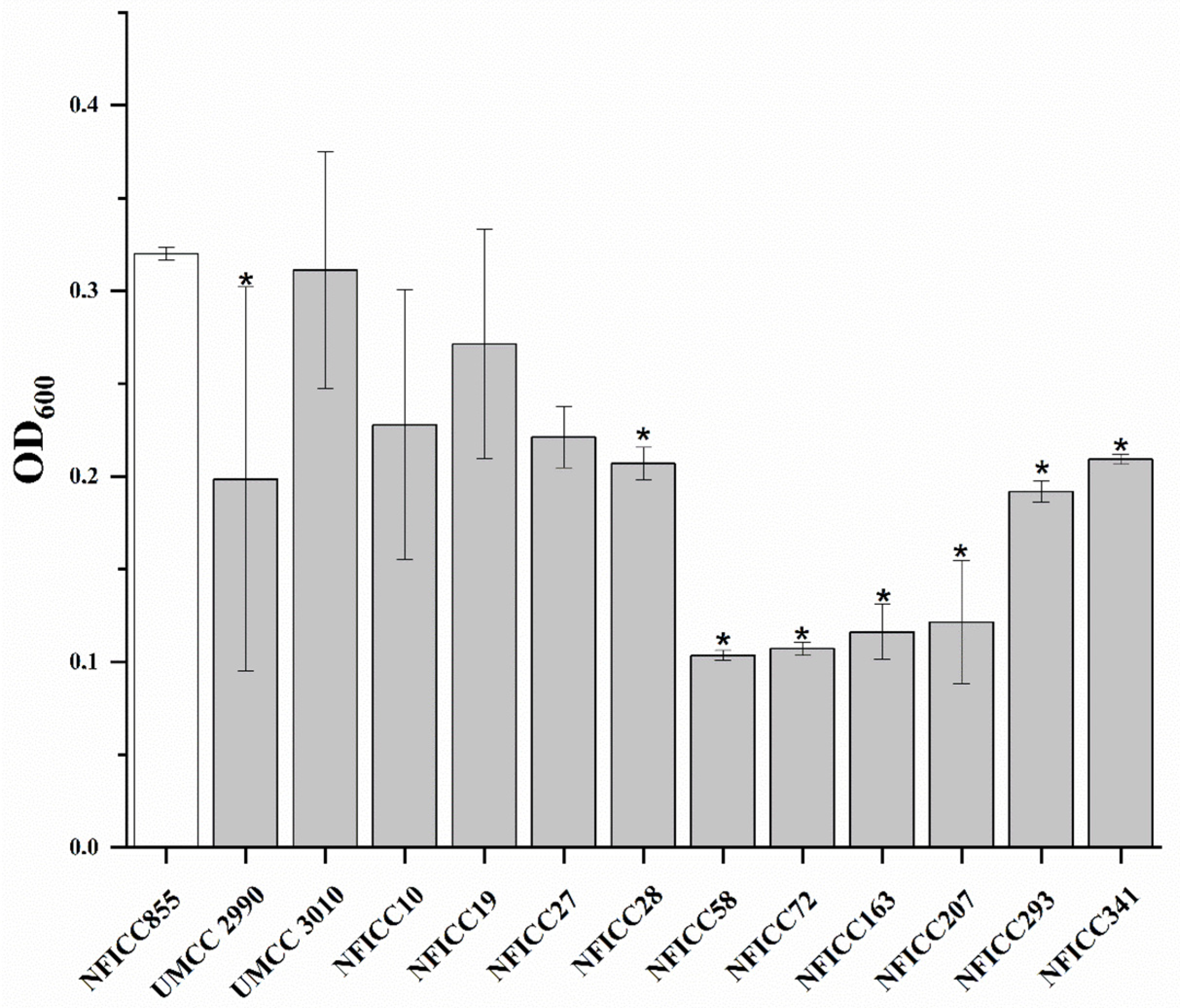
Similar action was detected for L. plantarum UMCC 2996, Lc. citreum NFICC28, and F. sanfranciscensis UMCC 2990. The lowest activity was shown by L. plantarum NFICC293. B. thuringiensis NFICC816 was challenged with the CFS of six LAB strains, specifically, L. plantarum NFICC19, 27, 293, Lc. citreum NFICC28, and P. pentosaceus NFICC341 (Figure 2). Here, only Lc. citreum NFICC28 was able to significantly prevent the growth of the spoilage organism. Additionally, B. weihenstephanensis NFICC855 was challenged with various LAB-CFS, including L. plantarum strains (NFICC19, 27, 72, 207, and 293), F. sanfranciscensis UMCC 2990, Lc. citreum NFICC28, and P. pentosaceus strains (UMCC 3010, NFICC58, and 341). As reported in Figure 3, significant results were obtained. Candidates from all four species were able to inhibit pathogen growth, particularly L. plantarum NFICC72, NFICC163, and P. pentosaceus NFICC58.
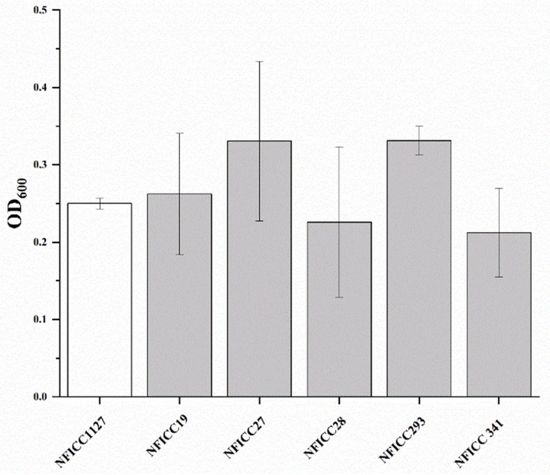
Two strains of B. amyloliquefaciens, NFICC1130 and NFICC1127, were used in these experiments (Figure 4 and Figure 5), recording completely different results even if challenged with the same LAB-CFS strains. The majority of the LAB were able to inhibit B. amyloliquefaciens NFICC1130; the highest inhibitory activities were recorded for P. pentosaceus NFICC10 and L. plantarum NFICC19. On the contrary, B. amyloliquefaciens NFICC1127 showed no inhibition with the same LAB-CFS. The CFS of different LAB strains, including L. plantarum, Lc. Citreum, and P. pentosaceus, were tested against B. licheniformis NFICC1549, as reported in Figure 6. L. plantarum NFICC72 and NFICC293 appear to be the best inhibitory agents, along with Lc. citreum NFICC94.

3.3. Detection of Bioactive Compounds

3.3.1. Metabolites Detected by HPLC during Fermentation in MRS

LAB’s ability to produce organic acids, other potentially antimicrobial compounds, and sugar consumption was assessed (Table 3). The CFS of the evaluated strains were examined by HPLC after 14 h, 18 h, and 24 h of fermentation in MRS. Observation of the results shows a corresponding ratio between amounts of lactic acid and acetic acid after 24 h. In congruence with the assessment for acidification, L. plantarum strains NFICC72, 163, 293, and 19 showed a strong and fast acidification activity. The present results also show that the ethanol detected is too low or not produced at all. Other compounds such as citric acid, oxalate, tartaric acid, and succinic were detected in trace amounts only.

3.3.2. Evaluation of the Bacteriocins Production and Kinetic Screening Assay

The screening of potential bacteriocin production revealed no antibacterial protein-associated inhibitions. Moreover, the assay of CFS spotted on a BHI confirmed the inhibitory activity of acids compounds and the absence of produced bacteriocins due to the nonformed inhibition zones. The results obtained from the kinetics assay highlight the bacteriostatic activity of the selected LAB. In fact, observations of the results demonstrates the presence of an extended lag phase, supporting the presence of organic acids in the cell-free supernatants and the absence of bacteriocins.

3.4. Assessment of Technological Features of the Candidate LAB Strains

3.4.1. Sugar Fermentation Profiling (API Test)

Sugar fermentation profiles obtained by API 50 CHL galleries were assessed for all the selected LAB, as indicated in Table 4, wherein the most common sugars in bakery products are highlighted (complete API profiles after 48 h are shown in Table S3). Results show that the most utilized sugars by the strains include glucose, fructose, maltose, and cellobiose.

3.4.2. Acidification Ability in BM

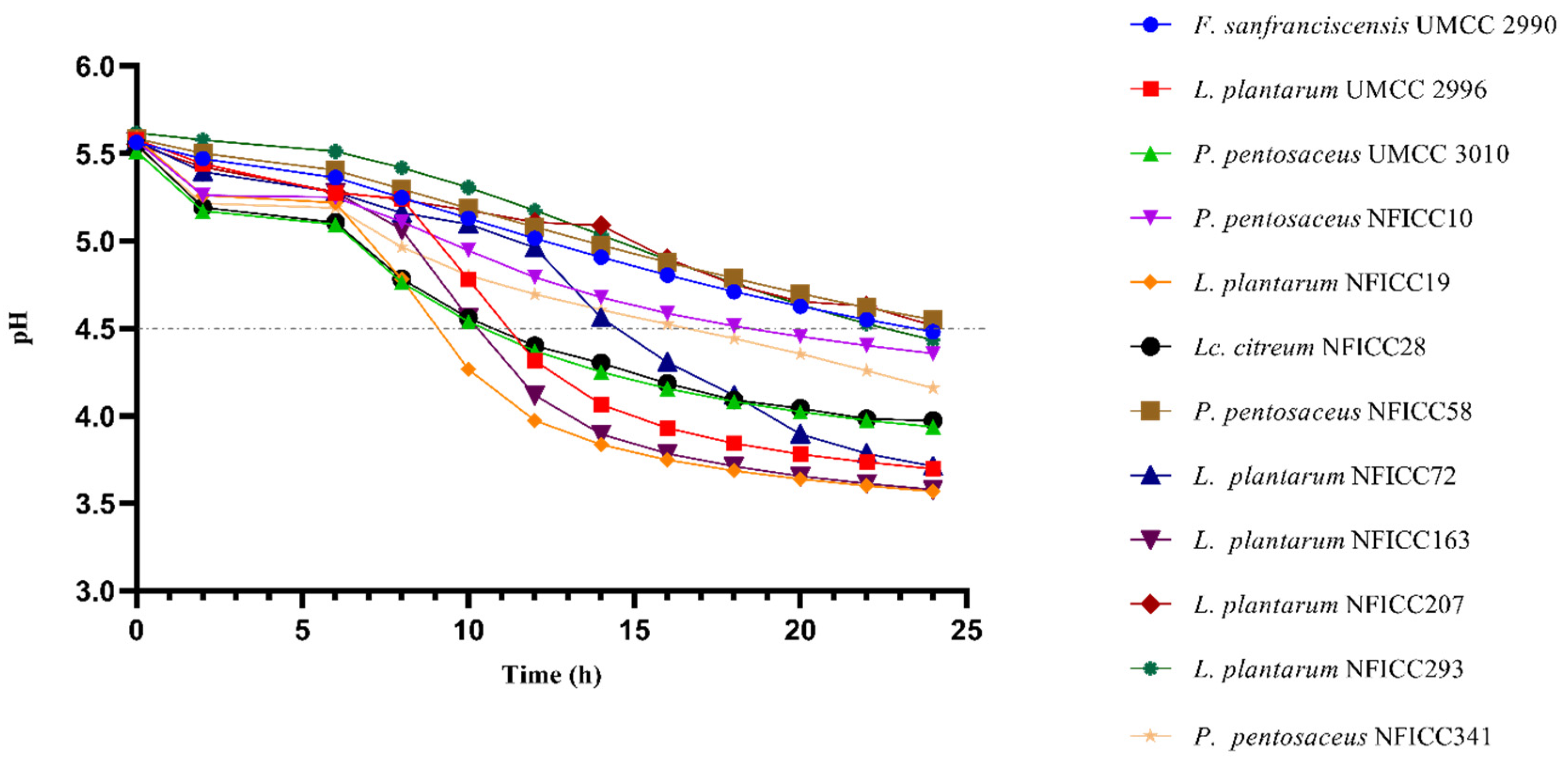
To further assess the strains’ performance for dough fermentation, their acidification ability was tested in BM during a period of 24 h. In Figure 7, only the best performers are reported. A decrease in pH ≈ 4.5, which is considered to be safe, was noted. The strains considered to be the fastest acidifiers were L. plantarum (UMCC 2996, NFICC19, and 163), Lc. citreum NFICC28, and P. pentosaceus UMCC 3010, which reached a pH ≈ 4.5 with at least 10 h of fermentation.

3.5. Safety Profile of the Selected LAB Strains

3.5.1. Hemolytic Activity Profile

Nonhemolytic activity is considered a safe prerequisite for the selection of new strains for food starter culture [39,40]. Results indicate that all the examined strains did not show signs of β-hemolytic activity when grown in Columbia Blood Agar (Table 5). Eleven LAB strains exhibited green-hued zones around colonies showing α-hemolytic activity (partial hemolysis), while seven strains including F. rossiae UMCC 3002, Lc. citreum UMCC 3011, NFICC28, NFICC94, and P. pentosaceus NFICC58 and NFICC341 were γ- hemolytic (nonhemolysis).

3.5.2. Antimicrobial Susceptibility Profiles (AST)

The sensitivity of LAB strains was determined against various antibiotics (streptomycin, ampicillin, vancomycin, gentamicin, kanamycin, and chloramphenicol), and the obtained results were compared based on the cut-offs, as specified in the guidance on the assessment of bacterial susceptibility to antimicrobials of human and veterinary importance from EFSA [32]. Sensitivity profiles are shown in Table 6. Briefly, almost all of the tested LAB show resistance against streptomycin (99%), except P. pentosaceus NFICC341 and vancomycin (100%), as already reported by different studies. However, against ampicillin (81%), kanamycin (76%), chloramphenicol (99%), and gentamycin (57%), the majority of them were in accordance with the EFSA cut-off and could be considered susceptible.

3.5.3. In Silico Screening for Resistance Genes

All 18 isolates were analyzed for the presence of acquired resistance genes using ResFinder version 4.2.3 and ResFinder database version 2.0.1 [41] using genome assemblies. Parameters used for a matching identity on the database were required to be at least 80%, whereas the coverage of a matching gene in the database was required to be at least 60%.

3.6. Whole-Genome Sequencing Analysis: Functional Gene and Potential Virulence In Silico Screening

The presence of bacteriocins was initially tested following the protocol by Fugaban et al. [28] with no positive results. In addition, to further confirm the possible absence of bacteriocins, sequences of the whole genome obtained from all the evaluated LAB were analyzed using the web server BAGEL4. Although phenotypically no bacteriocin-associated inhibitions were observed, in silico analysis showed the presence of bacteriocins belonging to class IIb and Iic (particularly penocin and plantaricins A, E, F, K, J, and N) (Table 7). The structures of the putative operons are shown in Figure S1.

4. Discussion

Bacillus spp. are well associated with ropy bread and bakery product spoilage [6,7,42]. Clean-label strategies to control microbial spoilage in bakery industries are important, accordingly, some starter cultures, including LAB, are promising alternative biocontrol agents thanks to their ability to produce bacteriocins and organic acids [22,43]. To screen candidates efficiently, we adapted the high-throughput screening assay developed by Inglin et al. [25], which was identified as a fast, low-cost, and accurate primary screening. Based on the results, most of the LAB strains found to be active against Bacillus spp. strains originated from fruits and vegetables (NFICC strains) or sourdoughs (UMCC strains). Their ability to adapt to these highly variable and stressful environments has aided in their capacity to grow into different niches, which can be exploited for their beneficial features, such as acidification ability and competitiveness [11,44,45].
To further assess the ability of the strains, a well diffusion assay in 24-well microtiter plates was conducted using live cells, confirming 18 active strains comprised of representatives from Lactobacillus, Pediococcus, and Leuconostoc genera, from both screening assays. Observations in this study were similar to those of Adesulu-Dahunsi et al. [46] against Bacillus spp., Listeria monocytogenes, and Escherichia coli.
The capacity of the P. pentosaceus strain NFICC341 to inhibit the growth of the majority of the spoilage microorganisms can be traced back to its origin, which was Brewers’ spent grain. The potential of LAB strains isolated from the vegetable matrix was already demonstrated by Puntillo et al. [47]. In accordance with previous studies, similar behavior can be associated with strains of Lc. citreum, L. plantarum, and F. sanfranciscensis, isolated from plants and sourdoughs, which exhibited promising inhibitory activity [48,49]. Furthermore, it was noted that L. plantarum and P. pentosaceus are well-known species that are able to produce antimicrobial compounds and organic acids, which can influence the safety of food [44,45,50,51]. Differences between the activities of the strains from the same species might be related to the isolation matrix of the strains and their adaptability to different stress conditions [15,52]. While strains UMCC 2996, UMCC 2990, and NFICC28 were isolated from sourdough and shared a similar antimicrobial activity, sample NFICC19 resulted to be the best acidifier.
A confirmation assay was conducted to further assess the activity of the bioactive metabolites produced by the LAB; however, contrary to the initial observations obtained on the high-throughput screening employed, only bacteriostatic activity was observed, in accordance with the previous literature findings [53]. Thus, identification of the nature of the bioactive compound was performed.
The evaluation of the metabolites in the CFS samples analyzed by using HPLC revealed the presence of organic acids in the supernatant, and therefore suggests they may play a significant role in the inhibition activity of the LAB strains tested [19,54]. Organic acids, in particular lactic acid, reduce the matrix’s pH without causing a strong sour taste. A fast acidification of the product is responsible for the inhibition of B. cereus, as showed by Yang et al. [45], where a reduction in the initial CFU/mL of the pathogen was detected after LAB fermentation. Treatment of proteolytic enzymes on the CFS showed no significant changes in the inhibitory activity of the bioactive strains—ruling out the possible involvement of protein-based bioactive compounds, including bacteriocins and bacteriocin-like inhibitory substances. Additionally, the elimination of heat-labile antimicrobials was excluded by heat treatment (80 °C) of the CFS.
Even though no bacteriocinogenic inhibitions were detected based on the assays performed, the necessity for further investigation of which other possible metabolites can be further exploited, for future potential application of these studied LAB against different types of Gram-positive pathogens and spoilage bacteria, is needed [55]. Therefore, in silico search for possible antibacterial metabolites was conducted, identifying the presence of Class IIb and Class IIc bacteriocin operons on some of the studied LAB strains.
Class II bacteriocins are nonlantibiotics, heat-stable, and small (≤10 kDa) hydrophobic peptides divided into four subclasses. Albeit these strains are detected on the genome, one of the possibilities for not observing bacteriocins from the CFS might be due to the presence of nonsatisfactory environmental conditions or an incomplete expression of all operons required for bacteriocin synthesis, transport, and regulation [56].
The effective biocontrol activity of the LAB strains evaluated would be associated with their rapid rate of acidification, as this is fundamental in food processing to prevent and avoid spoilage contaminants. Sourdough’s pH varies depending on the state of fermentation; however, it typically ranges between 3.5 and 5. Harmful microorganisms such as botulism bacteria, E. coli, and spoilage fungi cannot develop in an environment with a pH below 4.6, as this acidity keeps them away [5,57]. We observed that the majority of the strains were able to lower the pH around 4–4.5 (considered a safe pH to avoid spoilage) after 10 h, exhibiting a high acidification ability and underlining the massive production of acids (lactic and acetic acid). In fact, low pH and high acidity are the major factors for avoiding/delaying the growth of ropy bacterial agents, thus making the rate of pH reduction during the early stage of fermentation crucial for optimum inhibition [45,53,58]. Organic acids were the key metabolites that were identified in this study. The significance of pH reduction in food stability and preservation has been widely researched and recognized by the scientific community [59,60]. Furthermore, pH value also impacts the pKa of the various acids, leading them to a lower or higher dissociation, affecting the safety and taste of the final product [61].
Before the application of any microorganisms in food systems, an assessment of their safety and technological features is important. In this study, we profiled bioactive strains, identifying their sugar fermentations and their ability to be functional for their intended application in simulated ex vivo models. As observed, these strains can use a wide array of carbohydrates primarily found in bakery products, allowing them to be applied efficiently in this system. Consequently, acidification profiles highlight their ability to thrive in bread and produce necessary metabolites for possible bio-protection [13,62]. On the other hand, safety features were assessed as follows: antimicrobial susceptibility and hemolytic activity. Findings indicate that the evaluated strains have a broad range of resistance against streptomycin and vancomycin. Although this might be concerning, it should be noted that resistance to antibiotics in the majority of LAB is intrinsic. Additional assessment, particularly the identification of the location of associated resistance genes, should be performed to identify the occurrence of acquired resistance [63,64]. However, an in silico search for the presence of antibiotic resistance genes showed no matches on all the genomes of the 18 strains evaluated. Evaluation of hemolytic activity is one of the key factors that should be considered when assessing the safety of functional strains [39]. In this study, we identified partial hemolysis (α-hemolysis) on 11 out of 18 strains, whereas the rest showed no hemolysis. Thus, further confirmation, particularly the presence of genes involved in these putative virulence factors, should be investigated to assess potential risk for the application of the strains, especially for food consumption.

5. Conclusions

Cereals and bakery goods are a fundamental part of the human diet, and their higher susceptibility to microbial spoilage could lead to economic losses and health issues. Generally, LAB starter cultures and their metabolites are found to be promising for controlling spoilage agents. In this study, several LAB strains able to contrast the growth of some common rope spoilage agents were selected by using high-throughput and ex vivo screening assays. Specifically, L. plantarum NFICC19, NFICC72, NFICC293, Lc. citreum NFICC28, and P. pentosaceus NFICC58 and NFICC341 showed the best inhibitory activity. The assessment of their technological and safety features supported their suitability for fermentation processes and the production of bakery products. Moreover, the whole-genome sequencing of the selected LAB strains and the in silico analysis showed that some of the strains contain operons for bacteriocins, yet no significant evidence was observed phenotypically, suggesting that additional analysis needs to be performed to better understand the inhibitory mechanisms involved and validate the application of the strains as potential biocontrol agents.

Supplementary Materials

The following supporting information can be downloaded at: https://www.mdpi.com/article/10.3390/fermentation9030290/s1, Figure S1: Gene encoding for bacteriocins detected in the genome of the best candidate strains. Table S1: Results of the high-throughput preliminary screening of LAB-CFS against spoilage bacteria in common media. Table S2: Results of the dual-plate agar high-throughput screening. Table S3: Results of the complete API test profile after 48 h of incubation at 30 °C.

Author Contributions

Conceptualization, methodology, data curation, investigation, formal analysis, writing—original draft, G.I.; Conceptualization, methodology, data curation, investigation, writing—review and editing, J.I.I.F.; formal analysis, data curation, S.Ö.; data curation, visualization, A.P.W.; investigation, data curation, R.S.K.; data curation, Q.H.; validation, formal analysis, data curation, R.S.; resources, supervision, A.P.; conceptualization, writing—review and editing, supervision, L.D.V.; conceptualization, resources, writing—review and editing, supervision, funding acquisition, C.H.B.-B. All authors have read and agreed to the published version of the manuscript.

Funding

G.I. acknowledges the award of a PhD scholarship from the Department of Life Sciences, University of Modena and Reggio Emilia. R.S. and S.O. were funded by FBCD FFBI grant Apple Pom (project ID 20080). R.S.K. was funded by the European Union’s Horizon 2020 research and innovation program (Grant: 874735). C.H.B.-B. has received funding from MycoProtein (Grant Number 34009-20-1682). Lastly, J.I.I.F. was funded by an industrial PhD scholarship (23 IFD LAB DuPont PhD project number 110063) from the Danish innovation foundation. S.Ö. and Q.H. were working as research assistants. Both L.D.V. and A.P. are employees of the Department of Life Sciences, University of Modena and Reggio Emilia.

Institutional Review Board Statement

Not applicable.

Informed Consent Statement

Not applicable.

Data Availability Statement

Not applicable.

Acknowledgments

The authors would like to thank Christina Aaby Svendsen from the Research Group for Genomic Epidemiology, DTU National Food Institute for the technical assistance with performing full-genome Illumina sequencing.

Conflicts of Interest

The authors declare no conflict of interest.

References

  1. Zain, M.Z.M.; Shori, A.B.; Baba, A.S. Potential functional food ingredients in bread and their health benefits. Biointerface Res. Appl. Chem. 2022, 12, 6533–6542. [Google Scholar] [CrossRef]
  2. Goryńska-Goldmann, E.; Gazdecki, M.; Rejman, K.; Łaba, S.; Kobus-Cisowska, J.; Szczepański, K. Magnitude, causes and scope for reducing food losses in the baking and confectionery industry a multi-method approach. Agriculture 2021, 11, 936. [Google Scholar] [CrossRef]
  3. Cicatiello, C.; Franco, S.; Pancino, B.; Blasi, E.; Falasconi, L. The dark side of retail food waste: Evidences from in-store data. Resour. Conserv. Recycl. 2017, 125, 273–281. [Google Scholar] [CrossRef]
  4. Muthappa, D.M.; Lamba, S.; Sivasankaran, S.K.; Naithani, A.; Rogers, N.; Srikumar, S.; Macori, G.; Scannell, A.G.M.; Fanning, S. 16S rRNA Based Profiling of Bacterial Communities Colonizing Bakery-Production Environments. Foodborne Pathog. Dis. 2022, 19, 485–494. [Google Scholar] [CrossRef] [PubMed]
  5. Pepe, O.; Blaiotta, G.; Moschetti, G.; Greco, T.; Villani, F. Rope-producing strains of Bacillus spp. from wheat bread and strategy for their control by lactic acid bacteria. Appl. Environ. Microbiol. 2003, 69, 2321–2329. [Google Scholar] [CrossRef] [PubMed] [Green Version]
  6. Valerio, F.; De Bellis, P.; Di Biase, M.; Lonigro, S.L.; Giussani, B.; Visconti, A.; Lavermicocca, P.; Sisto, A. Diversity of spore-forming bacteria and identification of Bacillus amyloliquefaciens as a species frequently associated with the ropy spoilage of bread. Int. J. Food Microbiol. 2012, 156, 278–285. [Google Scholar] [CrossRef] [PubMed]
  7. Valerio, F.; Di Biase, M.; Huchet, V.; Desriac, N.; Lonigro, S.L.; Lavermicocca, P.; Sohier, D.; Postollec, F. Comparison of three Bacillus amyloliquefaciens strains growth behaviour and evaluation of the spoilage risk during bread shelf-life. Food Microbiol. 2015, 45, 2–9. [Google Scholar] [CrossRef] [PubMed]
  8. Iurie, R.; Cahul, B.P.H.; Uniersity, S.; Turtoi, M.; Dunarea, U.; Galati, D.J. The occurrence of the rope spoilage of bread at Cahulpan bread-making company. Sci. J. Cahul State Univ. “Bogdan Petriceicu Hasdeu” Econ. Eng. Stud. 2021, 2, 107–116. [Google Scholar]
  9. Thompson, J.M.; Waites, W.M.; Dodd, C.E.R. Detection of rope spoilage in bread caused by Bacillus species. J. Appl. Microbiol. 1998, 85, 481–486. [Google Scholar] [CrossRef]
  10. Pereira, A.P.M.; Stradiotto, G.C.; Freire, L.; Alvarenga, V.O.; Crucello, A.; Morassi, L.L.P.; Silva, F.P.; Sant’Ana, A.S. Occurrence and enumeration of rope-producing spore forming bacteria in flour and their spoilage potential in different bread formulations. LWT 2020, 133, 110108. [Google Scholar] [CrossRef]
  11. De Vero, L.; Iosca, G.; Gullo, M.; Pulvirenti, A. Functional and healthy features of conventional and non-conventional sourdoughs. Appl. Sci. 2021, 11, 3694. [Google Scholar] [CrossRef]
  12. Ibrahim, S.A.; Ayivi, R.D.; Zimmerman, T.; Siddiqui, S.A.; Altemimi, A.B.; Fidan, H.; Esatbeyoglu, T.; Bakhshayesh, R.V. Lactic acid bacteria as antimicrobial agents: Food safety and microbial food spoilage prevention. Foods 2021, 10, 3131. [Google Scholar] [CrossRef] [PubMed]
  13. Wang, Y.; Wu, J.; Lv, M.; Shao, Z.; Hungwe, M.; Wang, J.; Bai, X.; Xie, J.; Wang, Y.; Geng, W. Metabolism Characteristics of Lactic Acid Bacteria and the Expanding Applications in Food Industry. Front. Bioeng. Biotechnol. 2021, 9, 612285. [Google Scholar] [CrossRef] [PubMed]
  14. De Vero, L.; Iosca, G.; La China, S.; Licciardello, F.; Gullo, M.; Pulvirenti, A. Yeasts and lactic acid bacteria for panettone production: An assessment of candidate strains. Microorganisms 2021, 9, 1093. [Google Scholar] [CrossRef] [PubMed]
  15. Iosca, G.; De Vero, L.; Di Rocco, G.; Perrone, G.; Gullo, M.; Pulvirenti, A. Anti-spoilage activity and exopolysaccharides production by selected lactic acid bacteria. Foods 2022, 11, 1914. [Google Scholar] [CrossRef]
  16. De Vuyst, L.; Leroy, F. Bacteriocins from lactic acid bacteria: Production, purification, and food applications. J. Mol. Microbiol. Biotechnol. 2007, 13, 194–199. [Google Scholar] [CrossRef]
  17. Iosca, G.; Turetta, M.; De Vero, L.; Bang-berthelsen, C.H.; Gullo, M.; Pulvirenti, A. Valorization of wheat bread waste and cheese whey through cultivation of lactic acid bacteria for bio-preservation of bakery products. LWT 2023, 176, 114524. [Google Scholar] [CrossRef]
  18. Zotta, T.; Parente, E.; Ricciardi, A. Viability staining and detection of metabolic activity of sourdough lactic acid bacteria under stress conditions. World J. Microbiol. Biotechnol. 2009, 25, 1119–1124. [Google Scholar] [CrossRef]
  19. Cizeikiene, D.; Juodeikiene, G.; Paskevicius, A.; Bartkiene, E. Antimicrobial activity of lactic acid bacteria against pathogenic and spoilage microorganism isolated from food and their control in wheat bread. Food Control 2013, 31, 539–545. [Google Scholar] [CrossRef]
  20. Katina, K.; Sauri, M.; Alakomi, H.L.; Mattila-Sandholm, T. Potential of lactic acid bacteria to inhibit rope spoilage in wheat sourdough bread. LWT 2002, 35, 38–45. [Google Scholar] [CrossRef]
  21. Menteş, Ö.; Ercan, R.; Akçelik, M. Inhibitor activities of two Lactobacillus strains, isolated from sourdough, against rope-forming Bacillus strains. Food Control 2007, 18, 359–363. [Google Scholar] [CrossRef]
  22. Valerio, F.; De Bellis, P.; Lonigro, S.L.; Visconti, A.; Lavermicocca, P. Use of Lactobacillus plantarum fermentation products in bread-making to prevent Bacillus subtilis ropy spoilage. Int. J. Food Microbiol. 2008, 122, 328–332. [Google Scholar] [CrossRef] [PubMed]
  23. Madsen, S.K.; Priess, C.; Wätjen, A.P.; Øzmerih, S.; Mohammadifar, M.A.; Heiner Bang-Berthelsen, C. Development of a yoghurt alternative, based on plant-adapted lactic acid bacteria, soy drink and the liquid fraction of brewers’ spent grain. FEMS Microbiol. Lett. 2021, 368, fnab093. [Google Scholar] [CrossRef] [PubMed]
  24. Saranraj, P.; Gheeta, M. Microbial Spoilage of Bakery Products and Its Control by Preservatives. Int. J. Pharm. Biol. 2012, 3, 38–48. [Google Scholar]
  25. Inglin, R.C.; Stevens, M.J.A.; Meile, L.; Lacroix, C.; Meile, L. High-throughput screening assays for antibacterial and antifungal activities of Lactobacillus species. J. Microbiol. Methods 2015, 114, 26–29. [Google Scholar] [CrossRef]
  26. Verni, M.; Minisci, A.; Convertino, S.; Nionelli, L.; Rizzello, C.G. Wasted Bread as Substrate for the Cultivation of Starters for the Food Industry. Front. Microbiol. 2020, 11, 293. [Google Scholar] [CrossRef] [PubMed] [Green Version]
  27. Hui-Hu, C.; Ren, L.Q.; Zhou, Y.; Ye, B.C. Characterization of antimicrobial activity of three Lactobacillus plantarum strains isolated from Chinese traditional dairy food. Food Sci. Nutr. 2019, 7, 1997–2005. [Google Scholar] [CrossRef] [Green Version]
  28. Fugaban, J.I.I.; Vazquez Bucheli, J.E.; Holzapfel, W.H.; Todorov, S.D. Characterization of Partially Purified Bacteriocins Produced by Enterococcus faecium Strains Isolated from Soybean Paste Active Against Listeria spp. and Vancomycin-Resistant Enterococci. Microorganisms 2021, 9, 1085. [Google Scholar] [CrossRef]
  29. Corrieu, G.; Spinnler, H.E.; Picque, D.; Jomier, Y. Automated system to follow up 403 and control the acidification activity of lactic acid starters. Fr. Pat. FR 1988, 2, 612. [Google Scholar]
  30. Madsen, S.K.; Thulesen, E.T.; Mohammadifar, M.A.; Bang-Berthelsen, C.H. Chufa drink: Potential in developing a new plant-based fermented dessert. Foods 2021, 10, 3010. [Google Scholar] [CrossRef]
  31. Fugaban, J.I.I.; Vazquez Bucheli, J.E.; Kim, B.; Holzapfel, W.H.; Todorov, S.D. Safety and beneficial properties of bacteriocinogenic Pediococcus acidilactici and Pediococcus pentosaceus isolated from silage. Lett. Appl. Microbiol. 2021, 73, 725–734. [Google Scholar] [CrossRef] [PubMed]
  32. European Food Safety Authority (EFSA). Guidance on the assessment of bacterial susceptibility to antimicrobials of human and veterinary importance. EFSA J. 2012, 10, 2740. [Google Scholar] [CrossRef]
  33. Bolger, A.M.; Lohse, M.; Usadel, B. Trimmomatic: A flexible trimmer for Illumina sequence data. Bioinformatics 2014, 30, 2114–2120. [Google Scholar] [CrossRef] [PubMed] [Green Version]
  34. Wick, R.R.; Judd, L.M.; Gorrie, C.L.; Holt, K.E. Unicycler: Resolving bacterial genome assemblies from short and long sequencing reads. PLoS Comput. Biol. 2017, 13, e1005595. [Google Scholar] [CrossRef] [Green Version]
  35. VanOeffelen, M.; Nguyen, M.; Aytan-Aktug, D.; Brettin, T.; Dietrich, E.M.; Kenyon, R.W.; Machi, D.; Mao, C.; Olson, R.; Pusch, G.D.; et al. A genomic data resource for predicting antimicrobial resistance from laboratory-derived antimicrobial susceptibility phenotypes. Brief. Bioinform. 2021, 22, bbab313. [Google Scholar] [CrossRef] [PubMed]
  36. Wattam, A.R.; Davis, J.J.; Assaf, R.; Boisvert, S.; Brettin, T.; Bun, C.; Conrad, N.; Dietrich, E.M.; Disz, T.; Gabbard, J.L.; et al. Improvements to PATRIC, the all-bacterial bioinformatics database and analysis resource center. Nucleic Acids Res. 2017, 45, D535–D542. [Google Scholar] [CrossRef] [PubMed]
  37. Bateman, A.; Martin, M.J.; Orchard, S.; Magrane, M.; Agivetova, R.; Ahmad, S.; Alpi, E.; Bowler-Barnett, E.H.; Britto, R.; Bursteinas, B.; et al. UniProt: The universal protein knowledgebase in 2021. Nucleic Acids Res. 2020, 49, D480–D489. [Google Scholar] [CrossRef]
  38. Van Heel, A.J.; De Jong, A.; Song, C.; Viel, J.H.; Kok, J.; Kuipers, O.P. BAGEL4: A user-friendly web server to thoroughly mine RiPPs and bacteriocins. Nucleic Acids Res. 2018, 46, W278–W281. [Google Scholar] [CrossRef]
  39. Bindu, A.; Lakshmidevi, N. Identification and in vitro evaluation of probiotic attributes of lactic acid bacteria isolated from fermented food sources. Arch. Microbiol. 2021, 203, 579–595. [Google Scholar] [CrossRef]
  40. Estifanos, H. Isolation and identification of probiotic lactic acid bacteria from curd and in vitro evaluation of its growth inhibition activities against pathogenic bacteria. Afr. J. Microbiol. Res. 2014, 8, 1419–1425. [Google Scholar] [CrossRef] [Green Version]
  41. Bortolaia, V.; Kaas, R.S.; Ruppe, E.; Roberts, M.C.; Schwarz, S.; Cattoir, V.; Philippon, A.; Allesoe, R.L.; Rebelo, A.R.; Florensa, A.F.; et al. ResFinder 4.0 for predictions of phenotypes from genotypes. J. Antimicrob. Chemother. 2020, 75, 3491–3500. [Google Scholar] [CrossRef]
  42. Dubey, S.; Sharma, N.; Thakur, S.; Patel, R.; Reddy, B.M. Bacillus cereus food poisoning in Indian perspective: A review. Pharma Innov. J. 2021, 10, 970–975. [Google Scholar]
  43. Qian, M.; Liu, D.; Zhang, X.; Yin, Z.; Ismail, B.B.; Ye, X.; Guo, M. A review of active packaging in bakery products: Applications and future trends. Trends Food Sci. Technol. 2021, 114, 459–471. [Google Scholar] [CrossRef]
  44. Corsetti, A.; Gobbetti, M.; Smacchi, E. Antibacterial activity of sourdough lactic acid bacteria: Isolation of a bacteriocin-like inhibitory substance from Lactobacillus sanfrancisco C57. Food Microbiol. 1996, 13, 447–456. [Google Scholar] [CrossRef]
  45. Yang, Y.; Tao, W.Y.; Liu, Y.J.; Zhu, F. Inhibition of Bacillus cereus by lactic acid bacteria starter cultures in rice fermentation. Food Control 2008, 19, 159–161. [Google Scholar] [CrossRef]
  46. Adesulu-Dahunsi, A.T.; Sanni, A.I.; Jeyaram, K. Diversity and technological characterization of Pediococcus pentosaceus strains isolated from Nigerian traditional fermented foods. LWT 2021, 140, 110697. [Google Scholar] [CrossRef]
  47. Puntillo, M.; Gaggiotti, M.; Oteiza, J.M.; Binetti, A.; Massera, A.; Vinderola, G. Potential of Lactic Acid Bacteria Isolated From Different Forages as Silage Inoculants for Improving Fermentation Quality and Aerobic Stability. Front. Microbiol. 2020, 11, 586716. [Google Scholar] [CrossRef]
  48. Arena, M.P.; Silvain, A.; Normanno, G.; Grieco, F.; Drider, D.; Spano, G.; Fiocco, D. Use of Lactobacillus plantarum strains as a bio-control strategy against food-borne pathogenic microorganisms. Front. Microbiol. 2016, 7, 464. [Google Scholar] [CrossRef] [Green Version]
  49. Messens, W.; De Vuyst, L. Inhibitory substances produced by Lactobacilli isolated from sourdoughs—A review. Int. J. Food Microbiol. 2002, 72, 31–43. [Google Scholar] [CrossRef]
  50. Lindgren, S.E.; Dobrogosz, W.J. Antagonistic activities of lactic acid bacteria in food and feed fermentations. FEMS Microbiol. Lett. 1990, 87, 149–163. [Google Scholar] [CrossRef]
  51. Thao, T.T.P.; Thoa, L.T.K.; Ngoc, L.M.T.; Lan, T.T.P.; Phuong, T.V.; Truong, H.T.H.; Khoo, K.S.; Manickam, S.; Hoa, T.T.; Tram, N.D.Q.; et al. Characterization halotolerant lactic acid bacteria Pediococcus pentosaceus HN10 and in vivo evaluation for bacterial pathogens inhibition. Chem. Eng. Process. Process Intensif. 2021, 168, 108576. [Google Scholar] [CrossRef]
  52. Ruiz Rodríguez, L.G.; Mohamed, F.; Bleckwedel, J.; Medina, R.; De Vuyst, L.; Hebert, E.M.; Mozzi, F. Diversity and functional properties of lactic acid bacteria isolated from wild fruits and flowers present in northern Argentina. Front. Microbiol. 2019, 10, 1091. [Google Scholar] [CrossRef] [PubMed]
  53. Gao, Z. Evaluation of different kinds of organic acids and their antibacterial activity in Japanese Apricot fruits. Afr. J. Agric. Res. 2012, 7, 4911–4918. [Google Scholar] [CrossRef] [Green Version]
  54. Rishi, L.; Mittal, G.; Agarwal, R.K.; Sharma, T. Melioration in Anti-staphylococcal Activity of Conventional Antibiotic(s) by Organic Acids Present in the Cell Free Supernatant of Lactobacillus paraplantarum. Indian J. Microbiol. 2017, 57, 359–364. [Google Scholar] [CrossRef]
  55. Parada, J.L.; Caron, C.R.; Medeiros, A.B.P.; Soccol, C.R. Bacteriocins from lactic acid bacteria: Purification, properties and use as biopreservatives. Braz. Arch. Biol. Technol. 2007, 50, 521–542. [Google Scholar] [CrossRef] [Green Version]
  56. Zhang, T.; Zhang, Y.; Li, L.; Jiang, X.; Chen, Z.; Zhao, F.; Yi, Y. Biosynthesis and Production of Class II Bacteriocins of Food-Associated Lactic Acid Bacteria. Fermentation 2022, 8, 217. [Google Scholar] [CrossRef]
  57. Pacher, N.; Burtscher, J.; Johler, S.; Etter, D.; Bender, D.; Fieseler, L.; Domig, K.J. Ropiness in Bread—A Re-Emerging Spoilage Phenomenon. Foods 2022, 11, 3021. [Google Scholar] [CrossRef]
  58. Cho, W.-I.; Chung, M.-S. Antimicrobial effect of a combination of herb extract and organic acid against Bacillus subtilis spores. Food Sci. Biotechnol. 2017, 26, 1423–1428. [Google Scholar] [CrossRef]
  59. Amit, S.K.; Uddin, M.M.; Rahman, R.; Islam, S.M.R.; Khan, M.S. A review on mechanisms and commercial aspects of food preservation and processing. Agric. Food Secur. 2017, 6, 1–22. [Google Scholar] [CrossRef]
  60. Gould, G.W. Methods for preservation and extension of shelf life. Int. J. Food Microbiol. 1996, 33, 5–64. [Google Scholar] [CrossRef]
  61. Su, X.; Wu, F.; Zhang, Y.; Yang, N.; Chen, F.; Jin, Z.; Xu, X. Effect of organic acids on bread quality improvement. Food Chem. 2019, 278, 267–275. [Google Scholar] [CrossRef] [PubMed]
  62. Ayivi, R.; Gyawali, R.; Krastanov, A.; Aljaloud, S.; Worku, M.; Tahergorabi, R.; Da Silva, R.; Ibrahim, S. Lactic Acid Bacteria: Food Safety and Human Health Applications. Dairy 2020, 1, 202–232. [Google Scholar] [CrossRef]
  63. Abriouel, H.; Casado Muñoz, M.D.C.; Lavilla Lerma, M.L.; Pérez Montoro, B.; Bockelmann, W.; Pichner, R.; Kabisch, J.; Cho, G.-S.; Franz, C.M.A.P.; Galvez, A.; et al. New insights in antibiotic resistance of Lactobacillus species from fermented foods. Food Res. Int. 2015, 78, 465–481. [Google Scholar] [CrossRef]
  64. Mathur, S.; Singh, R. Antibiotic resistance in food lactic acid bacteria—A review. Int. J. Food Microbiol. 2005, 105, 281–295. [Google Scholar] [CrossRef] [PubMed]
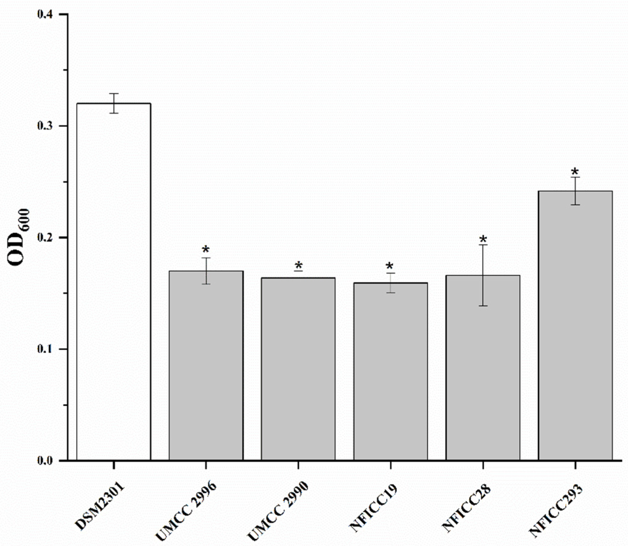
Figure 1. Antibacterial activity of the selected LAB’ cell-free supernatant in Bread Media (BM) against B. cereus DSM 2301. One-tail t-test was performed comparing OD600 value after 48 h growth of B. cereus DSM 2301 in BM and the same pathogens challenged with CFS of L. plantarum (UMCC 2996, NFICC19, and 293), F. sanfranciscensis (UMCC 2990), and Lc. citreum (NFICC28). The mean value of all the treated samples was significantly lower than the control sample (untreated sample); t(2) = −2.92 and p = 0.05. Bars with * are significantly different.
Fermentation 09 00290 g001
Figure 2. Antibacterial activity of the selected LAB’s cell-free supernatant in Bread Media (BM) against spoilage agents B. thuringensis NFICC816. One-tail t-test was performed on the obtained triplicates comparing OD600 value after 48 h growth of NFICC816 in BM and the same pathogens challenged with CFS of L. plantarum (NFICC19, 27, and 293) and P. pentosaceus (NFICC341). The mean value of one treated sample of Lc. citreum (NFICC28) was significantly lower than the control sample (untreated sample), while the other samples respected the null hypothesis; t(2) = −2.92 and p = 0.05. Bars with * are significantly different.
Fermentation 09 00290 g002
Figure 3. Antibacterial activities of the selected LAB’s cell-free supernatant in Bread Media (BM) against spoilage agents B. weihenstephanensis NFICC855. One-tail t test was performed on the obtained triplicates comparing OD600 value after 48 h growth of NFICC855 in BM and the same pathogens challenged with CFS of L. plantarum (NFICC19, 27, 72, 207, and 293), F. sanfranciscensis (UMCC 2990), Lc. citreum (NFICC28), and P. pentosaceus (UMCC 3010, NFICC 58, and 341). The mean value of the samples represented by * was significantly lower than the control sample (untreated sample); t(2) = −2.92 and p = 0.05.
Fermentation 09 00290 g003
Figure 4. Antibacterial activity of the selected LAB’s cell-free supernatant in Bread Media (BM) on spoilage agent B. amyloliquefaciens NFICC1130. One-tail t-test was performed on the obtained triplicates comparing OD600 value after 48 h growth of NFICC1130 in BM and the same pathogens challenged with CFS of L. plantarum (NFICC9, 27, 72, 163, 207, and 293), Lc. citreum (NFICC28), and P. pentosaceus (UMCC 3010, NFICC10, 58, and 341). Bars with * are significantly different, with a mean value significantly lower than the control sample (untreated sample), while samples NFICC 27, 163, 207, 293, and 341 respect the null hypothesis; t(2) = −2.92 and p = 0.05.
Fermentation 09 00290 g004
Figure 5. Antibacterial activity of the selected LAB’s cell-free supernatant in Bread Media (BM) on spoilage agent B. amyloquiefaciens NFICC 1127. One-tail t test was performed on the obtained triplicates comparing OD600 value after 48 h growth of NFICC1127 in BM and the same pathogens challenged with CFS of L. plantarum (NFICC19, 27, and 293), Lc. citreum (NFICC28), and P. pentosaceus (NFICC341). No significant differences between the control and treated samples were observed at t(2) = −2.92 and p = 0.05.
Fermentation 09 00290 g005
Figure 6. Antibacterial activity of the selected LAB’s cell-free supernatant in Bread Medium (BM) on spoilage agent B. licheniformis NFICC1549. One-tail t-test was performed on the obtained triplicates comparing OD600 value after 48 h growth of NFICC1549 in BM and the same spoilage agent challenged with CFS of L. plantarum (NFICC27, 72, 163, and 293), Lc. citreum (NFICC28 and 94), and P. pentosaceus (UMCC 3010 and NFICC341). The * indicate measurements that are significantly different, with a mean lower than the control samples; t(2) = −2.92, p = 0.05.
Fermentation 09 00290 g006
Figure 7. Acidification performance in Bread Medium showed by the best LAB candidates against bread rope-producing strains of Bacillus spp.
Fermentation 09 00290 g007
Table 1. Lactic acid bacteria strains tested in the present study. They were provided by the National Food Institute Culture Collection (NFICC), Technical University of Denmark and by Unimore Microbial Culture Collection (UMCC), University of Modena and Reggio Emilia (Italy).
Strain CodeSpeciesIsolation Source
NFICC10Pediococcus pentosaceusSourdough
NFICC19Lactiplantibacillus plantarumDill
NFICC27Lactiplantibacillus plantarumSourdough
NFICC28Leuconostoc citreumSourdough
NFICC58Pediococcus pentosaceusSourdough
NFICC72Lactiplantibacillus plantarumGooseberry
NFICC87Leuconostoc citreumBeetroot
NFICC94Leuconostoc citreumSpinach
NFICC103Pediococcus pentosaceusPumpkin
NFICC163Lactiplantibacillus platarumField pea
NFICC207Lactiplantibacillus plantarumGlasswort
NFICC293Lactiplantibacillus plantarumDragsholm plant
NFICC341Pediococcus pentosaceusBrewer’s spent grain
UMCC 2990Fructilactobacillus sanfranciscensisSourdough type I
UMCC 2996Lactiplantibacillus plantarumDough for Panettone
UMCC 3002Furfurilactobacillus rossiaeDough for Panettone
UMCC 3010Pediococcus pentosaceusGluten-free sourdough
UMCC 3011Leuconostoc citreumDough for Panettone
Table 2. Selected bread spoilage bacteria tested in the present study. They were provided by the German Collection of Microorganisms and Cell Cultures (DSMZ) and the National Food Institute Culture Collection (NFICC).
Strain CodeSpeciesIsolation Source
DSM 2301Bacillus cereusFood poisoning incident
DSM 4222Bacillus cereus-
DSM 4312Bacillus cereusVomit
DSM 22905Bacillus cytotoxicusVegetable puree
NFICC119Lysinibacillus fusiformisBeetroot
NFICC432Paenibacillus polymyxaWalnut
NFICC503Bacillus mycoidesBeech leaves
NFICC510Bacillus altitudinisPlant
NFICC526Bacillus mycoidesRed fir
NFICC528Bacillus subtilisSourdough
NFICC529Lysinibacillus sphaericusCommon Juniper
NFICC530Bacillus pumilusCommon Juniper
NFICC531Bacillus simplexCommon Juniper
NFICC532Lysinibacillus fusiformisCommon Juniper
NFICC740Bacillus cereusPlant
NFICC781Bacillus cereusKombucha
NFICC816Bacillus thuringiensisAnimal feces
NFICC855Bacillus weihenstephanensisPotato
NFICC869Bacillus amyloliquefaciensPasteurized BSG
NFICC871Lysinibacillus sphaericusPasteurized BSG
NFICC879Lysinibacillus boronitoleransPotato
NFICC882Lysinibacillus fusiformisPotato
NFICC889Lysinibacillus boronitoleransPotato
NFICC906Bacillus simplexPotato
NFICC1127Bacillus amyloliquefaciensPasteurized BSG *
NFICC1130Bacillus amyloliquefaciensPasteurized BSG
NFICC1525Bacillus subtilisHerring Garum
NFICC1534Bacillus subtilisMiso
NFICC1549Bacillus licheniformisApple pulp
* BSG: brewers’ spent grains.
Table 3. Different compounds (g/L) detected by HPLC after 14, 18, and 24 h of fermentation in MRS broth. The creation of the heat map is based upon the detected MRS broth concentration: green color highlights the production, red color the consumption, and yellow color was used when the detected amount was equal to MRS broth. The measurement were with three independent experiments, and Kruskal–Wallis statistical analysis was performed to detect the significant strains with respect to plain MRS. The significance is marked with an *.
Sample/timeMRSF. sanfranciscensis UMCC 2990L. plantarum UMCC2 996P. pentosaceus UMCC 3010 P. pentosaceus NFICC10L. plantarum NFICC19Lc. citreum NFICC28P. pentosaceus NFICC58L. plantarum NFICC72L. plantarum NFICC163L. plantarum NFICC207L. plantarum NFICC293P. pentosaceus NFICC341
OXALATE14H0.310.370.360.380.390.95 *0.340.320.390.57 *0.390.300.34
18H0.370.330.340.40 *0.93 *0.39 *0.380.380.290.360.63 *0.39 *
24H0.370.440.440.48 *1.38 *0.57 *0.410.70 *0.69 *0.320.55 *0.37
CITRIC ACID14H1.860.00 *0.00 *0.00 *0.00 *1.771.041.180.00 *0.00 *0.00 *0.00 *0.38 *
18H1.130.00 *1.061.121.051.051.860.92 *0.09 *1.040.00 *0.00 *
24H0.990.90 *1.040.961.600.40 *2.150.00 *0.00 *0.81 *0.00 *1.10
TARTARIC ACID14H0.001.12 *1.081.13 *1.12 *1.171.47 *1.001.10 *1.071.19 *0.971.08
18H1.12 *1.001.031.08 *0.811.32 *0.001.020.611.17 *0.000.00
24H0.000.000.000.001.22 *0.000.000.000.000.000.000.00
GLUCOSE14H14.6214.339.34 *12.2511.566.32 *6.97 *8.00 *13.397.02 *13.626.04 *10.13
18H14.997.06 *11.19 *11.13 *0.95 *5.37 *5.67 *11.284.87 *11.745.23 *8.59 *
24H14.0211.869.04 *8.98 *0.98 *2.78 *4.03 *0.00 *0.00 *9.524.07 *7.61 *
SUCROSE14H1.491.790.761.331.570.00 *0.00 *0.00 *1.570.931.030.511.14
18H1.680.751.261.250.710.00 *0.00 *0.49 *0.51 *0.920.49 *0.95
24H1.721.331.221.340.70 *0.00 *0.000.87 *0.83 *1.061.111.08
GLUTAMIC ACID14H0.230.08 *0.250.06 *0.07 *0.00 *0.230.150.08 *0.100.00 *0.170.18
18H0.130.130.11 *0.12 *0.210.10 *0.12 *0.130.11 *0.09 *0.130.18
24H0.090.00 *0.00 *0.00 *0.230.00 *0.00 *0.200.200.00 *0.00 *0.00 *
SUCCINIC14H0.000.000.000.000.000.000.000.000.000.182.07 *0.000.00
18H0.75 *0.590.70 *0.74 *0.000.000.070.440.481.10 *0.440.00
24H0.000.000.000.001.22 *0.000.080.000.001.15 *0.000.00
FORMIC ACID14H0.000.030.000.000.000.000.000.07 *0.000.000.09 *0.070.15 *
18H3.76 *0.010.13 *0.15 *0.000.000.11 *0.030.000.030.000.08
24H1.44 *0.010.23 *0.18 *0.000.000.122.02 *0.000.010.000.14
ACETIC ACID14H0.004.364.77 *4.73 *4.484.90 *5.44 *0.164.344.73 *5.14 *4.414.56
18H4.56 *4.45 *4.38 *4.50 *4.86 *5.52 *2.234.354.49 *4.89 *4.49 *4.47 *
24H4.394.414.404.55 *4.79 *4.77 *4.294.60 *4.65 *4.84 *4.384.51 *
1,3 PROPANDIOL14H0.000.710.85 *0.83 *0.82 *0.86 *0.92 *0.710.750.86 *0.89 *0.84 *0.81
18H0.790.780.810.820.89 *0.86 *0.810.530.85 *0.83 *0.88 *0.75
24H0.670.740.79 *0.80 *0.84 *0.670.84 *0.700.84 *0.83 *0.86 *0.82 *
2,3 BUTANDIOL14H0.000.050.060.21 *0.20 *0.000.000.100.18 *0.000.29 *0.000.18 *
18H0.18 *0.000.16 *0.18 *0.000.000.130.140.000.26 *0.000.06
24H0.000.060.19 *0.17 *0.000.000.150.000.000.26 *0.000.08
LACTIC ACID14H0.000.5912.05 *6.145.5014.73 *5.474.972.8814.25 *2.9513.33 *7.98 *
18H0.8613.08 *5.596.3419.78 *6.167.91 *6.5616.14 *3.1314.77 *9.18 *
24H0.785.219.3810.0819.90 *16.91 *12.27 *20.32 *20.78 *4.6916.11 *11.43
ETOH14H0.000.000.000.000.000.001.32 *0.000.000.000.000.000.00
18H0.000.000.000.000.001.58 *0.000.000.000.000.000.00
24H0.000.000.000.000.000.37 *0.000.000.001.74 *0.000.00
Table 4. Sugar fermentation profiles of the tested LAB strains obtained by API 50 CHL after 48 h of incubation at 30 °C.
LABD(+)-GlucoseD(−)-FructoseD(+)-CellobioseMaltoseLactoseD(+)-MelibioseSaccharoseD(+)-RaffinoseStarchD(+)-Xylose
F. sanfranciscensis UMCC 2990++++
L. plantarum UMCC 2996++++++++
F. rossiae UMCC 3002++/−++/−+
P. pentosaceus UMCC 3010++++
Lc. citreum UMCC 3011++/−++
P. pentosaceus NFICC10++++++++
L. plantarum NFICC19++++++++
L. plantarum NFICC27++++++++
Lc. citreum NFICC28+++++++++
L. plantarum NFICC58+++++++++
L. plantarum NFICC72++++++++
P. pentosaceus NFICC87++++++
Lc. citreum NFICC94++++++
P. pentosaceus NFICC103++++++++/−
P. pentosaceus NFICC163+++++++++/−
L. plantarum NFICC207+++++++++
L. plantarum NFICC293++++++++
P. pentosaceus NFICC341+++++++++/−
Table 5. Hemolytic capacity * of the tested strains.
Tested Strainsγ-Hemolysisα-Hemolysisβ-Hemolysis
F. sanfranciscensis UMCC 2990 x
L. plantarum UMCC2996 x
F. rossiae UMCC 3002x
P. pentosaceus UMCC 3010 x
Lc. citreum UMCC 3011x
P. pentosaceus NFICC10 x
L. plantarum NFICC19 x
L. plantarum NFICC27 x
Lc. citreum NFICC28x
L. plantarum NFICC58x
L. plantarum NFICC72 x
P. pentosaceus NFICC87x
Lc. citreum NFICC94x
P. pentosaceus NFICC103 x
P. pentosaceus NFICC163 x
L. plantarum NFICC207 x
L. plantarum NFICC293 x
P. pentosaceus NFICC341x
S. aureus NFICC1477x
Lc. citreum NFICC88 x
B. cereus DMS 2301 x
* The occurrence of the specific hemolytic activity is marked by “x”.
Table 6. Minimum inhibitory concentration (MIC) (µg/mL) of the antibiotic susceptibility of the selected LAB. The values reported in bold and blue color are under or equal to recommended EFSA cut-offs and underline the susceptibility of the strains.
Tested LABTested Antibiotics
StreptomycinAmpicillinKanamycinVancomycinChloramphenicolGentamycin
F. sanfranciscensis UMCC 2990≤128≤16≤32512≤4≤4
L. plantarum UMCC 2996≤128≤32≤64512Resistant≤256
F. rossiae UMCC 3002≤128≤4≤32512≤4≤4
P. pentosaceus UMCC 3010≤128≤2≤16512≤4≤4
Lc. citreum UMCC 3011≤128≤4≤32512≤8≤8
P. pentosaceus NFICC10≤128≤1≤16512≤4≤4
L. plantarum NFICC19≤64≤1≤16512≤4≤4
L. plantarum NFICC27≤64≤1≤16512≤4≤4
Lc. citreum NFICC28≤128≤1≤32512≤4≤4
L. plantarum NFICC58≤128≤1≤32512≤4≤4
L. plantarum NFICC72≤64≤1≤16512≤4≤4
Lc. citreum NFICC87≤128≤1≤32512≤4≤4
Lc. citreum NFICC94≤128≤1≤32512≤4≤8
P. pentosaceus NFICC103≤128≤2≤32512≤4≤4
P. pentosaceus NFICC163≤64≤1≤16512≤4≤4
L. plantarum NFICC207≤64≤1≤16512≤4≤4
L. plantarum NFICC293≤128≤1≤8≤8≤4≤4
P. pentosaceus NFICC341≤64≤2≤32512≤4≤4
Table 7. Bacteriocins produced by the LAB strains displaying the highest inhibitory activity against ropy agents.
StrainSpeciesBacteriocins Predicted by BAGEL4
NFICC28Lc. citreumNone
NFICC10P. pentosaceusPenocin A
NFICC58P. pentosaceusNone
NFICC19L. plantarumPlantaricin E, Plantaricin F Plantaricin K,
putative class IIc bacteriocin, putative class IIb bacteriocin
NFICC72L. plantarumPlantaricin E, Plantaricin F Plantaricin K, putative class IIc bacteriocin, putative class IIb bacteriocin
NFICC163L. plantarumPlantaricin E, Plantaricin F Plantaricin K, putative class IIc bacteriocin, putative class IIb bacteriocin
NFICC293L. plantarumPlantaricin A, Plantaricin E, Plantaricin F, Plantaricin J, Plantaricin K, Plantaricin N
UMCC 2996L. plantarumPlantaricin A, Plantaricin E, Plantaricin F, Plantaricin J, Plantaricin K, Plantaricin N
Disclaimer/Publisher’s Note: The statements, opinions and data contained in all publications are solely those of the individual author(s) and contributor(s) and not of MDPI and/or the editor(s). MDPI and/or the editor(s) disclaim responsibility for any injury to people or property resulting from any ideas, methods, instructions or products referred to in the content.