Article

Biofilm Inhibition, Antibacterial and Antiadhesive Properties of a Novel Biosurfactant from Lactobacillus paracasei N2 against Multi-Antibiotics-Resistant Pathogens Isolated from Braised Fish

by
Hippolyte Tene Mouafo
1,*,
Alphonse Tegang Sokamte
2,
Linda Manet
1,
Arsene Joseph Manga Mbarga
3,4,
Sachivkina Nadezdha
3,*,
Somashekhar Devappa
5 and
Augustin Mbawala
6
1
Centre for Food, Food Security and Nutrition Research, Institute of Medical Research and Medicinal Plants Studies, Yaoundé P.O. Box 13033, Cameroon
2
Department of Food Engineering and Quality Control, University Institute of Technology, University of Ngaoundéré, Ngaoundéré P.O. Box 455, Cameroon
3
Department of Microbiology V.S. Kiktenko, Medical Institute, RUDN University Named after Patrice Lumumba, 119435 Moscow, Russia
4
Research Institute of Molecular and Cellular Medicine, Peoples’ Friendship University of Russia, 117198 Moscow, Russia
5
Department of Microbiology and Fermentation Technology, CSIR-Central Food Technological Research Institute, Mysore 570020, Karnataka, India
6
Department of Food Sciences and Nutrition, National School of Agro-Industrial Sciences, University of Ngaoundéré, Ngaoundéré P.O. Box 455, Cameroon
*
Authors to whom correspondence should be addressed.
Fermentation 2023, 9(7), 646; https://doi.org/10.3390/fermentation9070646
Submission received: 23 June 2023 / Revised: 6 July 2023 / Accepted: 7 July 2023 / Published: 9 July 2023
(This article belongs to the Section Microbial Metabolism, Physiology & Genetics)

Abstract

:
This study aimed to assess the antibiotic susceptibility and biofilm formation ability of pathogens isolated from braised fish as well as characterize and evaluate the antibacterial, antiadhesive, and antibiofilm activities of the biosurfactant from Lactobacillus paracasei subsp. tolerans N2 against these pathogens. The susceptibility of six pathogens isolated from braised fish (Escherichia coli EM2, Staphylococcus aureus SA1, Salmonella enteritidis PE1, Pseudomonas aeruginosa CT3, Yersinia enterolitica MH5, Proteus mirabilis MR2, and Klebsiella pneumoniae AG5) to 16 antibiotics revealed multiple resistances with an MAR index greater than 0.3. These pathogens were able to form biofilms with S. aureus SA1, which showed the highest ability. Using biochemical and elemental analyses, FTIR, GC-MS, 1H NMR and high-resolution mass spectrometry, the biosurfactant was characterized as a novel glycolipoprotein made of two congeners of mass 482.28 and 507.27 m/z, respectively. They showed bactericidal and antiadhesive activities against all pathogens. The biosurfactant inhibited biofilm formation by these pathogens and eradicated mature biofilms.

1. Introduction

Braised fish is among the highly appreciated and consumed street foods in Cameroon [1]. However, to satisfy the increasing demand for braised fish by consumers, antibiotics are abusively used in farming activities [2]. This has resulted in the presence of antibiotic residues in fish flesh [3] and thus the presence of multiple antibiotics-resistant microorganisms [4]. Hence, their consumption might lead to the development of antibiotics resistance. In addition, braised fish are air-exposed on an aluminum or woody surface during their sale [5]. An improper cleaning process applied to these surfaces could favor the adhesion and formation of biofilms by microorganisms. Biofilms are complex structures composed of microorganisms embedded in a polymeric matrix, where they can survive [6]. Microorganisms present in biofilms are 1000 times more resistant to antimicrobial agents than planktonic microorganisms. The presence of biofilms on the surface, if not removed, could lead to the continuous contamination of food products. In the food industry, there are many issues associated with pathogens and spoilage microorganisms in biofilms [7]. Hence, to avoid food contamination due to the biofilms forming microorganisms, there is an urgent need to search for efficient methods against pathogens and biofilms [7]. The strategies reported in the literature to control biofilms are expensive, especially for traditional food producers, and they sometimes require skills and the use of chemicals that are not always safe for humans, animals, and the environment [8]. In the search for efficient and safe compounds endowed with antimicrobial and antibiofilm properties, biosurfactants from lactobacilli due to their GRAS status appear to be a great and green alternative to avoid the phenomenon of antibiotic resistance and biofilms’ formation. Biosurfactants are amphiphilic compounds produced by several bacteria, yeasts, and molds, and they are endowed with surface properties. According to their chemical composition, they can be glycolipids, glycoproteins, lipopeptides, glycolipoproteins, or a complex mixture of compounds including proteins, sugars, lipids, and phosphates [9]. They possess antimicrobial, antiadhesive and antibiofilm activities [10,11,12]. In the search for new biosurfactants with cost-effective production, low-cost substrates and agro-industrial by-products are used. In this study, glycerol, a by-product from biodiesel and oleochemicals production which is currently raised, was used as a substrate to reduce the production cost while increasing the yield. In addition, the close relationship between the substrate used and the structure of the produced biosurfactant [13] might lead not only to an improvement in the production yield but also to the development of novel compounds with interesting functionality. In this light, the present research was designed and aimed at (i) assessing the antibiotic susceptibility profile of pathogens from braised fish, (ii) evaluating their ability to form biofilms, (iii) producing and characterizing biosurfactant from a lactobacilli strain using glycerol as substrate, and (iv) determining the antibacterial, antiadhesive, and antibiofilm abilities of the biosurfactant against these pathogens.

2. Materials and Methods

2.1. Microorganisms

The strain Lactobacillus paracasei subsp. tolerans N2 (accession number MH142620), isolated from a traditional fermented milk “pendidam” and identified by Mouafo et al. [14], was used to produce a biosurfactant. The strain stored at −20 °C in Man Rogosa and Sharpe (MRS) broth containing glycerol was sub-cultured twice in MRS broth (LiofilChem, Roseto degli Abruzzi, Italy) at 37 °C.
Pathogenic strains of Escherichia coli EM2, Staphylococcus aureus SA1, Salmonella enteritidis PE1, Pseudomonas aeruginosa CT3, Yersinia enterolitica MH5, Proteus mirabilis MR2, and Klebsiella pneumoniae AG5 were isolated and identified from braised fish sold in Yaoundé [5]. The strains were sub-cultured twice in Brain–Heart Infusion broth (BHI, LiofilChem, Roseto degli Abruzzi, Italy) before being used.

2.2. Antibiotics’ Resistance Profile of the Pathogens

The susceptibility of these pathogens to 16 commonly used antibiotics in human diseases and fish farming (Amoxycillin-clavulanic acid (AMC, 30 µg), Amoxycillin (AX, 30 µg), Ampicillin (AMP 10 µg), Cefroxadine (FX, 10 µg), Ciprofloxacin (CIP, 5 µg), Cefuroxime (CXM, 30 µg), Ceftazidime (CAZ, 10 µg), Chloramphenicol (C, 30 µg), Cotrimoxazole (SXT, 25 µg), Erythromycin (ERY, 10 µg), Gentamicin (GEN, 10 µg), Imipenem (IMP, 10 µg), Nalidixic acid (NA, 30 µg), Nitrofurantoin (F, 300 µg), Norfloxacin (NOR, 10 µg) and Tetracycline (TE, 10 µg)) was tested using the disk diffusion method of the American Society for Microbiology [15]. Based on the inhibition diameters, the pathogens were classified as susceptible, intermediate or resistant [16]. Their Multiple Antibiotic Resistance (MAR) indexes were calculated as the ratio of the number of antibiotics for which a strain showed resistance to the total number of antibiotics tested. The method of Krumperman [17] was used to characterize the risk associated with the consumption of braised fish where these pathogens were isolated.

2.3. Biofilm Formation Ability of the Pathogens

The crystal violet method described by Sambanthamoorthy et al. [18] with some modifications was used to assess the ability of pathogens isolated from braised fish to form biofilms. Pathogens were cultured in BHI broth for 24 h at 37 °C. The cells were then collected by centrifugation (7000× g, 4 °C, 20 min), washed twice with sterile PBS, suspended in PBS and adjusted to 8 Log CFU/mL. Two hundred microliters of sterile BHI broth containing the pathogen suspension (7 Log CFU/mL) were added to each well of a 96-well flat-bottomed plastic tissue culture plate (Sigma, Munich, Germany). Negative control wells were filled with sterile BHI broth free of pathogens. The plates were then incubated at 37 °C for 24 h. After incubation, the broth was discarded and the plates were washed three times with sterile PBS and air-dried at room temperature (25 ± 1 °C) for 2 h under sterile conditions of the laminar flow. Two hundred microliters of analytical grade ethanol 95% (Sigma, Germany) was added to each well to fix the biofilm cells followed by incubation at room temperature (25 ± 1 °C) for 10 min. After removing ethanol, the plates were air-dried and stained with 200 µL of 1% (w/v) crystal violet for 10 min. Thereafter, the dye was removed and the wells were washed three times with sterile PBS and air-dried. Finally, 200 µL of glacial acetic acid (33%) was added to each well, and the optical density of the plates was read at 595 nm (Spark® 10M Multimode Microplate Reader, Tecan, Switzerland). The strains were classified as no biofilm producers when the OD of the sample wells (ODs) was lower than the OD of the negative control wells (ODnc); weak biofilm producers when ODnc ≤ ODs ≤ 2 × ODnc; moderate biofilm producers when 2 × ODnc ≤ ODs ≤ 4 × ODnc; and strong biofilm producers when 4 × ODnc < ODs [19].

2.4. Production of Biosurfactant

The biosurfactant was produced according to the protocol described by Mouafo et al. [14]. Fifty (15) milliliters of a young culture (37 °C for 16 h) of L. paracasei subsp. tolerans N2 at a load of 6 Log CFU/mL was introduced into 600 mL of sterile broth prepared with glycerol 9% (w/v) as a carbon source. Incubation was performed at 37 °C for 72 h and 150 oscillations/min (Kottermann D-3162, Munich, Germany). The culture was centrifuged (7000× g, 20 min, 4 °C), and the supernatant was collected and filtered (0.22 µm, Millipore, Munich, Germany), acidified at pH 2 with HCl 6N and stored at 4 °C. The biosurfactant was extracted from the supernatant using ethyl acetate/methanol (4:1) following the method described by Mouafo et al. [14]. The crude extract was dissolved in methanol, centrifuged (13,000× g, 20 min, 4 °C), filtered (0.22 µm, Millipore, Germany), and dried under nitrogen. The dried powder obtained was purified by reverse-phase high-performance liquid chromatography (RP-HPLC) using the method described by Mouafo et al. [10] and stored for analyses.

2.5. Characterization of the Biosurfactant

The surface tension, critical micellar concentration and emulsification index of the biosurfactant were measured according to the protocol reported by Mouafo et al. [14]. The elemental composition (C, H, N, S, and O) of the biosurfactant was assessed following the method previously reported by Mouafo et al. [10]. The total sugar, lipid and protein contents of the biosurfactant were determined following the methods described by Dubois et al. [20], Lobna et al. [21] and AACC [22], respectively.
A Fourier transform infrared spectroscopy (FTIR) spectrometer (Tensor II, IFS 25, Bruker, Ettlingen, Germany) equipped with a deuterated triglycine sulfate (DTGS) detector and operating in attenuated total reflectance mode was used to identify the functional groups of the biosurfactants following the method described by Mouafo et al. [10].
The fatty acid and monosaccharide profiles of the biosurfactants were determined by gas chromatography–mass spectrometry (GC–MS) after derivatization into fatty acid methyl esters (FAME) and alditol acetates, respectively, using the method described by Mouafo et al. [10]. The 1H NMR profile of the biosurfactant was determined using a Bruker Avance II 400 MHz NMR Spectrometer (Billerica, MA, USA) operating at a frequency of 400 MHz.
Mass spectra were obtained using a Triple TOF 5600 Mass Spectrometer (AB Sciex, Framingham, MA, USA) in both positive and negative modes. The sample was dissolved in HRMS grade methanol (3 ng/mL), and 500 µL was directly infused with a syringe pump at a flow rate of 10 µL/min. N2 was used as nebulization and collision gas. The source voltage and capillary voltage were 3.5 kV and 90 V in positive ion mode, while they were 3.5 kV and 100 V in negative ion mode. The MS scan range was 100 to 2000 m/z. MS/MS fragmentation was performed in positive and negative modes, and different collision energies (ranging from 20 to 70 eV) were tested to study the fragmentation.
The thermal behavior of the biosurfactant was assessed using differential scanning calorimetry. In the protocol, 5 mg of the biosurfactant was introduced into an aluminum DSC pan and sealed. The DSC pan was scanned under a nitrogen atmosphere at temperatures ranging from 30 to 300 °C at a gradient speed of 5 °C/min. A Perkin Elmer DSC 8000 (Waltham, MA, USA) was used to routinely record scans in duplicate. A free DSC pan was used as the control.

2.6. Antimicrobial Activity of Biosurfactant

The minimum inhibitory concentration (MIC) and the minimum bactericidal concentration (MBC) of the biosurfactant against different pathogens were determined following the modified method of the American Society for Microbiology [15] as reported by Mouafo et al. [10]. Briefly, 0.2 mL of an overnight suspension of the pathogen in PBS (8 Log CFU/mL) was introduced in tubes containing 0.5 mL of sterile MH broth. Then, 1 mL of biosurfactant in PBS at different concentrations (0.5, 1.0, 2.5, 5.0, 7.5, 10.0, 12.5, 15.0, 17.5, 20.0, 25.0 and 50.0 mg/mL) was added. The final volume of the tubes was adjusted to 2 mL using sterile MH broth, and the tubes were homogenized and incubated at 37 °C for 24 h. The negative growth control was a tube free of pathogens, while the positive one was a tube free of biosurfactants. The MIC, which corresponds to the tubes where no visible growth was observed, was determined by measuring the absorbance at 600 nm (UV-1800 UV Vis Spectrophotometer; Shimadzu, Vernon Hills, IL, USA) as the tube containing the lowest concentration of biosurfactant that completely inhibited measurable growth (A600 = 0).
For MBC determination, 100 µL of culture was taken from the tubes for which no visible growth was noticed (A600 = 0) and introduced into novel tubes containing 1.9 mL sterile MH broth. After homogenization, the inoculated tubes were incubated at 37 °C for 24 h, and the MBC was determined as the tube containing the lowest biosurfactant concentration for which no growth was observed (A600 = 0).
To assess the antimicrobial mechanism of the biosurfactant, pathogen cells treated with the biosurfactant at the MBC were submitted to scanning electron microscope analysis (Leo 435 VP Electron Microscopy Limited, Cambridge, UK). Under high vacuum and a temperature of 24 ± 2 °C, the images were recorded at 10 kV with a magnification of 25 KX.

2.7. Antiadhesive Activity of Biosurfactant

The ability of the biosurfactant to inhibit the adhesion of pathogens to surfaces was tested using the modified method of Gudiña et al. [23] reported by Mouafo et al. [11]. In the protocol, 200 µL of biosurfactant solutions in PBS at different concentrations (0, 0.5, 1.0, 2.5, 5.0, 7.50, 10.0, 12.5, 15.0, 17.5 and 20.0 mg/mL) was added to the wells of a sterile 96-wells flat-bottomed plastic tissue culture plate (Sigma, Germany). After incubation at 4 °C for 1 h, the wells of the plate were washed two times with sterile PBS. The washed wells were filled with 200 µL of an overnight culture of each pathogen suspended in PBS (8 Log CFU/mL), and the plate was incubated at 4 °C for 24 h. After double washing with sterile PBS, 200 µL of analytical grade methanol (99%, Sigma, Germany) was added to each well, and the plate was left on the bench (25 ± 1 °C) for 15 min. Methanol was then removed, and the plate was dried and stained with 200 µL of a 33% crystal violet solution. The crystal violet was discarded. Then, the plate was washed three times with PBS and air-dried, and 200 µL of glacial acetic acid (33%) was added to each well. The optical density of the suspension in each plate well was measured at 595 nm (Spark® 10M Multimode Microplate Reader, Tecan, Switzerland). PBS free of biosurfactant was used as the control. The formula was used to calculate the inhibition of microbial adhesion:
M i c r o b i a l i n h i b i t i o n % = A 0 A i A 0 × 100
where A0 refers to the absorbance of the control wells, and Ai refers to the absorbance of the wells containing biosurfactant at concentration i.

2.8. Antibiofilm Activity of Biosurfactant

2.8.1. Inhibition of Biofilm Formation

The ability of biosurfactants to inhibit the formation of biofilms by pathogens was assessed following the method described by Kim et al. [24] with some modifications. Briefly, wells of the microtiter plates were filled with 100 µL of sterile BHI broth containing 7 Log CFU/mL of the pathogen. Then, 100 µL of biosurfactants in PBS at different concentrations (0, 2.5, 5.0, 10.0, 15.0 and 20.0 mg/mL) was added to the wells of the plates, which was followed by incubation at 37 °C for 24 h. Wells containing pathogens and free of biosurfactants (0 g/L) and wells free of pathogens and biosurfactants were used as the positive and negative controls, respectively. After incubation, the plates were proceeded as described above for the biofilm formation. The optical density of the plates was measured at 595 nm, and the following formula was used to calculate the biofilm inhibition percentage:
B i o f i l m i n h i b i t i o n % = O D 0 O D i O D 0 × 100
where OD0 refers to the absorbance of the positive control wells, and ODi refers to the absorbance of the wells containing biosurfactant at concentration i.

2.8.2. Eradication of Mature Biofilms

The ability of the biosurfactant to eradicate mature biofilms formed by pathogens was assessed following the method described by Sambanthamoorthy et al. [18] with some modifications. Biofilms were formed in the wells of the microtiter plate as previously described (Section 2.3). Briefly, the wells of the microtiter plates were filled with 200 µL of sterile BHI broth containing 7 Log CFU/mL of the pathogen followed by incubation at 37 °C for 72 h to allow the formation of mature biofilms. The culture was then aspired, and the wells were washed thrice with sterile PBS. Afterwards, 200 µL of biosurfactants in PBS at different concentrations (0, 5.0, 10.0, 20.0, 30.0, 40.0 and 50.0 mg/mL) was introduced into the wells of the plate followed by incubation at 37 °C for 24 h. Wells containing pathogens and free of biosurfactants (0 g/L) and wells free of pathogens and biosurfactants were, respectively, used as positive and negative controls. The plates proceeded as described above for the biofilm formation. The optical density of the plates was read at 595 nm, and the following formula was used to calculate the biofilm inhibition percentage.
B i o f i l m e r a d i c a t i o n % = O D 0 O D i O D 0 × 100
where OD0 refers to the absorbance of the positive control wells, and ODi refers to the absorbance of the wells containing biosurfactant at concentration i.

2.9. Statistical Analysis

Experiments were performed in triplicate, and the results were expressed as mean ± standard deviation. Duncan’s multiple-range test was performed to compare means at a statistical significance level of 95% using the software Statgraphic centurion XVI version 16.1.18 (StatPoint Technologies, Inc., Chicago, IL, USA).

3. Results and Discussion

3.1. Antibiotics’ Susceptibility Profile of the Pathogens

The results depicted in Table 1 show that all pathogens were resistant to at least six different antibiotics. This observation could arise from the abusive use of antibiotics by non-qualified and unexperimented personnel involved in fish farming activities. Indeed, the subjective use of antibiotics in fish farming activities in Cameroon has been highlighted [2]. The consequences include the presence of multiple antibiotics-resistant bacteria in raw fish flesh [4] and fish products [25].
Gentamicin, erythromycin, and imipenem with a susceptibility score of 86% followed by ciprofloxacin with a score of 71% were the most active antibiotics against the tested pathogens. The high susceptibility to these antibiotics could be associated with their specific antimicrobial mechanism against both Gram-negative and Gram-positive bacteria. Indeed, imipenem modifies bacterial cell wall formation and membrane permeability [26], and it interferes with bacterial DNA replication and transcription by inhibiting DNA gyrase and topoisomerase IV [27]. Gentamicin blocks the synthesis of proteins by binding to the 30S RNA subunit [28]. Erythromycin acts on the translation by inhibiting the assembly of the 50S ribosomal subunit [29].
The highest resistance scores were recorded with ampicillin (100%), tetracycline (86%), nalidixic acid (86%), cotrimoxazole (71%) and norfloxacin (71%). This resistance could be explained by the ability of bacteria to produce enzymes such as extended-spectrum β-lactamases that destroy antibiotics or to harbor AmpC mutations [30]. The expression of efflux pumps and modification of porin genes leading to a reduction in the permeability of bacterial cells could also explain the resistance observed with tetracycline [30]. Fluoroquinolones (nalidixic acid, and norfloxacin) are extensively used in animals and humans because of their large spectrum of action against Gram-positive and Gram-negative bacteria. The resistance observed in this study could result from the ability of some bacteria to induce mutations in the genes encoding type II topoisomerases (gyrA, gyrB, parC, and parE), which are the most likely targets of these fluoroquinolones [31].
The MAR indexes of the pathogens tested in this study vary from 0.37 (P. mirabilis MR2 and K. pneumoniae AG5) to 0.75 (E. coli EM2). MAR indexes greater than 0.2 were also reported by Pahane et al. [25] and Tsafack et al. [4] with pathogens isolated from braised fish and fish flesh, respectively. These observations probably ascribe to the abuse the use of antibiotics in fish farming activity, suggesting that these products are high risk sources of multiple antibiotic resistance and their consumption might lead to a possible transfer of resistance genes to human pathogens. The direct consequence is the continuous increase in multidrug resistance phenomena as observed worldwide.

3.2. Formation of Biofilms by the Pathogens

The biofilm formation ability of the multiple antibiotic-resistant pathogens isolated from braised fish is shown in Figure 1. The highest biofilm contents were formed by S. aureus SA1 (8.40 ± 0.30) followed with P. aeruginosa CT3 (7.10 ± 0.50) and E. coli EM2 (6.25 ± 0.75). The lowest biofilm contents were recorded with the strains P. mirabilis MR2 (2.23 ± 0.17) and Y. enterolitica MH5 (1.50 ± 0.10). According to the classification of Kamali et al. [19], the strains P. mirabilis MR2 and Y. enterolitica MH5 can be classified as moderate biofilm producers, while S. aureus SA1, P. aeruginosa CT3, E. coli EM2, K. pneumoniae AG5 and S. enteritidis PE1 can be classified as strong biofilm producers. A similar observation was reported by Salem et al. [32]. The highest biofilm formation ability recorded in this study with S. aureus SA1 could be ascribed to surface proteins (biofilm-associated protein (bap), surface protein G and staphylococcal protein A) which are involved in the biofilm formation process [33]. Moreover, S. aureus has been reported to possess several regulators (Agr, CodY, CcpA, Rot, Sae, SarA, and SigB) that are involved in the strengthening of its biofilm formation ability [34]. The motility of some bacteria such as E. coli EM2, S. enteritidis PE1 and P. aeruginosa CT3 can also explain their high biofilm formation ability observed in this study. Indeed, the initial adhesion of flagella owning pathogens to surfaces (abiotic or biotic), which is the first step in the biofilm formation process, is eased by their motility [35].

3.3. Characterization of Biosurfactant

The biosurfactant produced by L. paracasei N2 with glycerol as substrate (2.77 ± 0.03 g/L) reduced the surface tension of water from 72 to 38.05 ± 0.56 mN/m with a CMC of 15 mg/mL and an emulsification index of 74.25 ± 1.64%. The chromatogram obtained from the RP-HPLC of the biosurfactant from L. paracasei N2 showed one peak at a retention time of 2.42 min corresponding to the solvent and another one at 2.69 min corresponding to the biosurfactant (Figure 2). This result indicates the purity of the biosurfactant.
Biochemical analysis revealed that the biosurfactant contained proteins (46.07 ± 1.95%), sugars (38.78 ± 0.07%), and lipids (15.13 ± 0.01%). The absence of ash after incineration at 550 °C indicates the salt removal efficiency of the extraction and purification process of the biosurfactant. Data from the elemental analysis showed the presence of carbon (58.46 ± 0.01%), nitrogen (4.97 ± 0.01%), hydrogen (18.71 ± 0.02%) and sulfur (0.01 ± 0.00%). These results demonstrate that the biosurfactant from L. paracasei N2 cultured on media with glycerol as the carbon source might be a complex mixture of proteins, lipids, and carbohydrates. This suggests its glycolipoprotein nature.
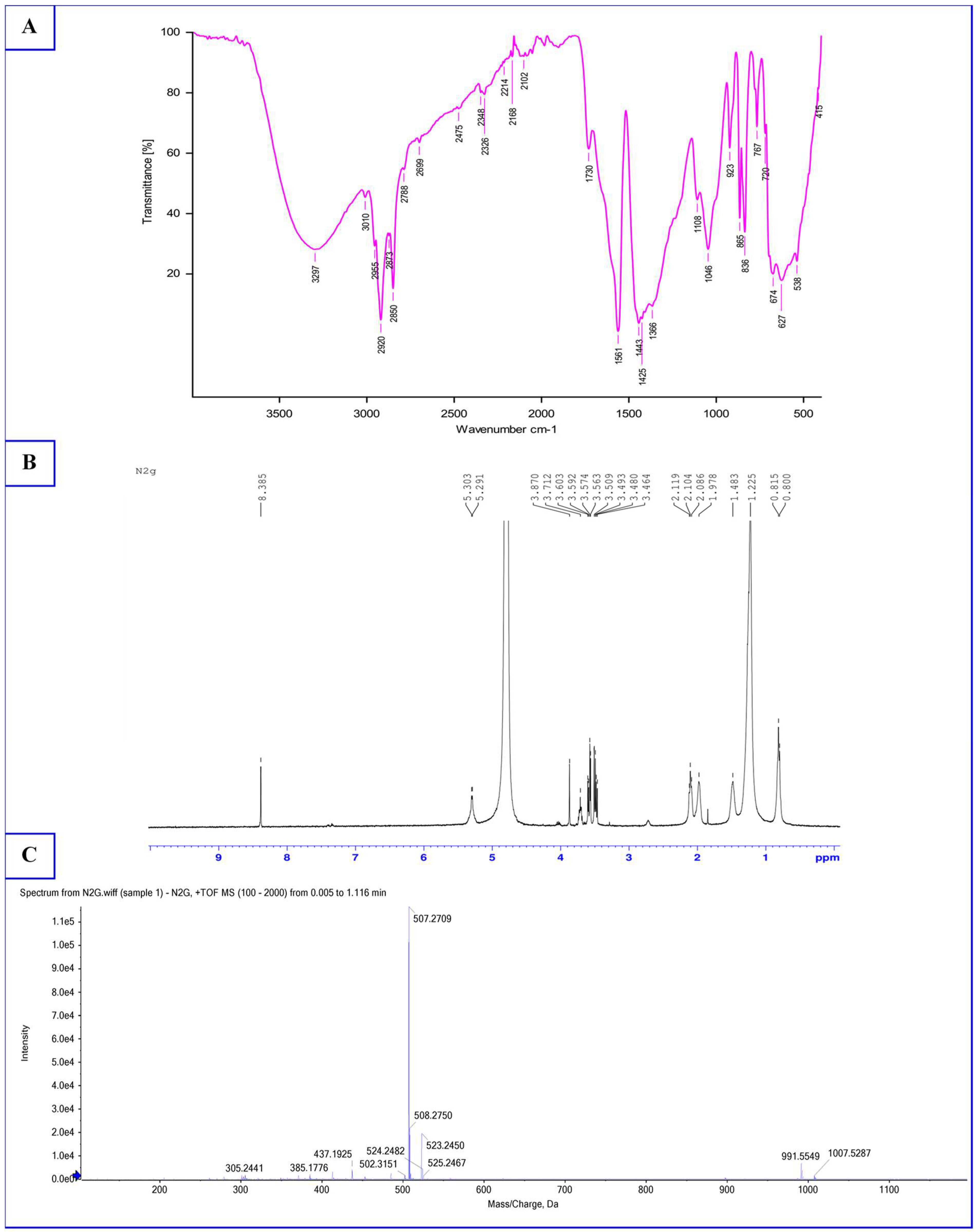
Bands characteristics of proteins (O–H stretching at 3293 cm−1 and N–H stretching at 3010 cm−1, C–N stretching mode of amide II band at 1561 cm−1), aliphatic chains of lipids (symmetric stretch C–H at 2920 and 2850 cm−1, –CH2–CH3 stretching vibrations at 1443 cm−1, and –C–CH3 1425 cm−1), and the glycosidic linkage of sugars (C–O stretching vibrations at 1108 cm−1 and vibrations in the C–O–C group at 1046 cm−1) were observed (Figure 3A). The peak observed at 1730 cm−1 corresponding to C=O stretching absorption in ester groups might indicate that the COOH group of fatty acids is involved in ester liaison with an –OH function of sugars or proteins that constitute the biosurfactant. Peaks that might correspond to C–S and S–H stretching were observed at 2475 and 720 cm−1, respectively, suggesting the presence of sulfur amino acids such as cysteine and methionine in the biosurfactant. Similar spectra for glycolipoprotein biosurfactants were reported in the literature by Mouafo et al. [14] with the biosurfactant from L. paracasei subsp. tolerans N2 using sugar cane molasses as substrate and Ferreira et al. [36] with the biosurfactant from L. paracasei using corn steep liquor as substrate. The results of the FTIR analysis obtained in this study are in a direct line with data gathered from biochemical and elemental analyses, thus confirming the glycolipoprotein nature of the biosurfactants from L. paracasei N2 as reported in the literature by several authors with these biomolecules derived from lactic acid bacteria [9,14,36].
The GC-MS analysis of lipids present in the biosurfactant from L. paracasei N2 revealed that it contains seven fatty acids (lauric acid, myristic acid, palmitic acid, 15-methyl palmitic acid, oleic acid, linoleic acid and homo-γ-linolenic acid) with oleic and palmitic acids being the most representative with percentages of 29.26 and 27.90%, respectively (Table 2). Palmitic acid was also reported in the literature as the main fatty acid of biosurfactants derived from lactic acid bacteria grown on different substrates [10,37,38].
Regarding the monosaccharide profile of the biosurfactant, it can be observed in Table 2 that six monosaccharides were detected with 1,6-anhydro-α-D-glucopyranose, which was the most important monosaccharide (47.70%). The monosaccharides identified in this study differed from those reported in the literature [10,37,39]. This could be associated to the variation from one strain to another of metabolic activities leading to the biosynthesis of biosurfactants [38].
The chemical shift corresponding to the proton of the amine –NH2 from amino acid was observed at 8.38 ppm, thus suggesting a peptide backbone in the constitution of the biosurfactant (Figure 3B). The signal at 1.48 ppm which could be attributed to the chemical shift of the proton –C–SH [40,41] confirms the presence of cysteine in the constitution of the biosurfactant. Indeed, elemental analysis and FTIR revealed the presence of sulfur in the biosurfactant. The signal at 4.1 ppm might correspond to the proton of amino acid –OOC–CH–N [42,43,44] and the signal at 2.08 ppm might correspond to the proton of an aliphatic chain located at position α of the carbonyl of a peptidic link (–CH2–CO–NH–) [43]. The signals at 5.30 and 5.29 ppm could be assigned to the proton of anomeric carbon (C1) of glucose, while protons resonating at 3.71, 3.59, 3.56, 3.57 and 1.97 ppm might refer to their respective C2, C3, C4, C5 and C6. A similar observation was reported by Garcia-Vello et al. [45] with cell surface glycans from L. plantarum IMB19. The signals at 0.8–0.85 ppm that might be inferred to methyl (–CH3) associated to those at 1.22 ppm corresponding to methylene protons (–(CH2)n–) clearly indicate the presence of aliphatic chain from fatty acids, as reported in the literature [42]. The chemical shifts at 2.10–2.11 ppm might be assigned to the proton of methylene close to carbonyl (–CH2–C=O) [10,37,46]. This 1H NMR spectrum of the biosurfactant confirms its glycolipoprotein nature. A chemical structure cannot be proposed at this level, as there are currently no standards or chemical structures proposed for the glycolipoprotein type of biosurfactants in the literature.
The HRMS spectrum of the biosurfactant showed six main ions with the greatest abundances at m/z 1007.52, 991.55, 525.24, 523.24, 508.27 and 507.27 (Figure 3C). The ion observed at m/z 991.55 could correspond to the mass of the glycolipoprotein biosurfactant. As depicted in Figure 3C, the biosurfactant might contain two main congeners: M1 and M2. The abundant fragment at m/z 507.27 might correspond to the congener M1 of the biosurfactant, the ones at m/z 508.27 might correspond to [M1 + H], and the ones at m/z 525.24 might correspond to [M1 + H2O]. The second congener M2 with a mass of 482.28 m/z could be identified through peaks at m/z 523.24 corresponding to the potassium adduct [M2 + K] and at m/z 1007.52 corresponding to [2M2 + K]. The predicted mass of the biosurfactant proposed in this study (991.55 m/z) is not in direct line with the low molecular weight character ascribed to biosurfactants from lactic acid bacteria that were structurally characterized in the literature. In fact, to date, the biosurfactants from lactic acid bacteria for which a structure has been proposed in the literature are either glycolipids or lipopeptides [9]. This result confirms the complexity of identifying biosurfactants composed of a mixture of several compounds including proteins, sugars and lipids. Although the mass proposed for the biosurfactant is higher than that obtained by the other authors, the mass of congeners M1 and M2 is not highly different to the 476 m/z obtained by Saravanakumari and Mani [39] with a biosurfactant from Lactococcus lactis, the 391.32 m/z noticed by Sharma et al. [46] with a biosurfactant produced by L. helveticus MRTL91, and the 446 m/z recorded by Sharma et al. [37] derived from a biosurfactant from E. feacium MRTL9.
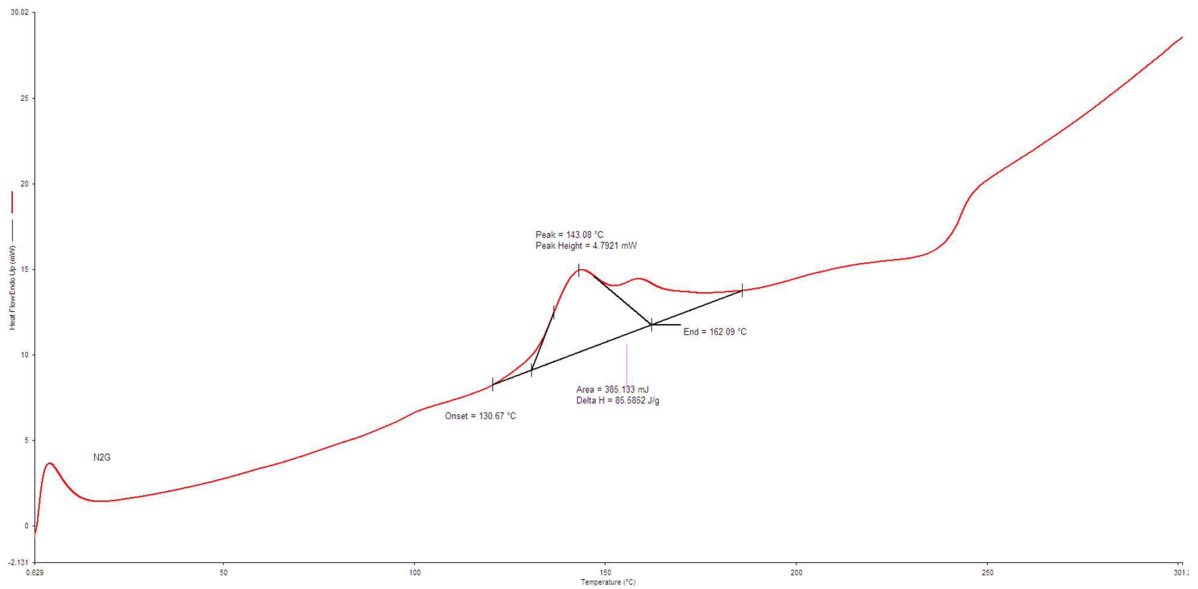
Differential scanning calorimetry was used to assess the behavior of the biosurfactant while submitted at high temperatures (30–300 °C). The thermogram obtained (Figure 4) showed the most visible transition temperature of the biosurfactant at 143.08 °C and another at approximately 250 °C. These two transitions might be attributed to the evaporation of water (143.08 °C) and the degradation of the side chains of the constituent elements of the biosurfactant which are proteins, sugars and lipids (250 °C). A similar behavior was observed for glycolipopeptide biosurfactants by Gil et al. [47].

3.4. Antimicrobial Activity of Biosurfactant

The ability of the biosurfactant from L. paracasei N2 to inhibit the proliferation of multidrug-resistant and biofilm-producing pathogens was assessed. Table 3 shows that the biosurfactant was active against all pathogens with MIC values ranging from 0.5 to 10 mg/mL and MBC values ranging from 1.0 to 15 mg/mL. With MBC/MIC ratios lower than 4, the biosurfactant was bactericidal against all pathogens. The bactericidal activity of the biosurfactant might result from its ability to alter bacterial cell walls and membranes as observed on images from SEM analysis performed using the most multiple antibiotic-resistant strain E. coli EM2 (Figure 5). Cell lysis leading to the loss of intracellular materials, disruption of transport proteins, formation of micellar aggregates, pores’ formation and disintegration of microbial cell walls and membranes have been highlighted as possible antimicrobial mechanisms of biosurfactants [48].
The sensitivity to biosurfactant observed in the present study was as follows: Y. enterolitica MH5 ˃ P. mirabilis MR2 ˃ S. enteritidis PE1 ˃ K. pneumoniae AG5 ˃ P. aeruginosa CT3 ˃ E. coli EM2 ˃ S. aureus SA1. This result differs from those generally reported in the literature. Indeed, Gram-negative bacteria are known to be more resistant to antimicrobials because of the lipopolysaccharide layer present on their outer surface membrane, which acts as an effective barrier [49,50]. However, in this study, they were more sensitive to the biosurfactant than the Gram-positive bacteria. The highest resistance observed in this study for S. aureus SA1 could be explained by its high biofilm-forming ability. Indeed, biofilm-producing S. aureus strains have been reported to display increased resistance to antibiotics and other antimicrobials [51].

3.5. Antiadhesive Activity of Biosurfactant

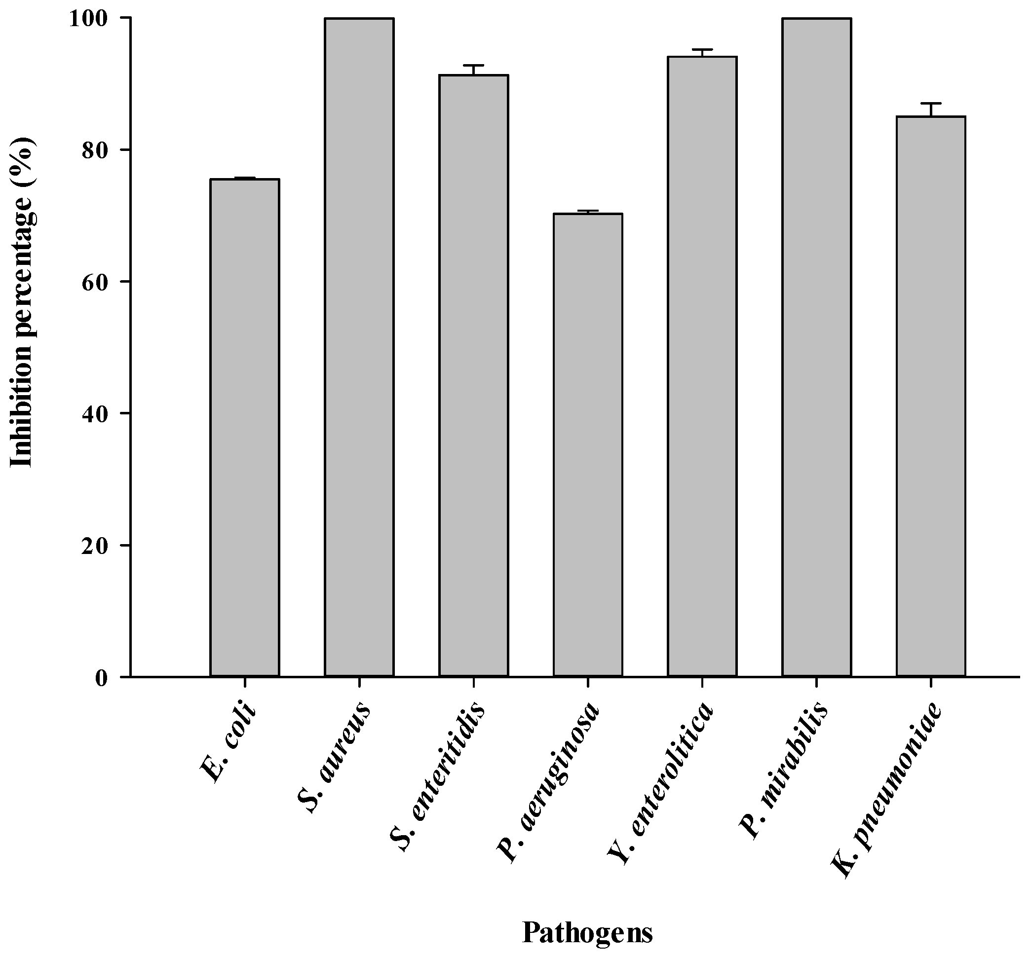
Microbial adhesion to surfaces is the first step in biofilm formation and mucosal colonization [24]. In this study, the ability of the biosurfactant to inhibit the adhesion of pathogens to the surface of a polystyrene 96-wells microtiter plate was assessed. Globally, the biosurfactant produced by L. paracasei N2 with glycerol as the substrate showd an inhibition of the adhesion of all the pathogens even at the concentration of 0.5 mg/mL (unpublished data). That inhibition of microbial adhesion evolved in a concentration-dependent manner. As shown in Figure 6, inhibition percentages greater than 70% were recorded at 10 mg/mL against the tested pathogens. This antiadhesive activity could arise from the ability of the biosurfactant to adsorb at the interface between the pathogens and the surface of the wells of the microplate and then decrease the interfacial tension that enables microbial adhesion. As the concentration of biosurfactant increases, the interfacial tension decreases, leading to a proportional release of microbes that adhere to the surface [52]. Sharma and Saharan [53] also highlighted an antiadhesive mechanism involving the modification of the wettability of the surface due to the formation of a thin film by the biosurfactant.
The highest antiadhesive activity (100%) was recorded against S. aureus SA1 and K. pneumoniae AG5, while the lowest antiadhesive activity was observed against P. aeruginosa CT3 and E. coli EM2 (Figure 6). Similar observations were reported by Sambanthamoorthy et al. [18]. This could be ascribed to the fact that Gram-negative bacteria, once adhered to surfaces, produce high quantities of compounds such as acylated homoserine lactone, which confer resistance to antiadhesive molecules [54].

3.6. Inhibition of Biofilm Formation by Biosurfactant

Biofilm formation by microorganisms is a survival strategy that generally results from harsh environmental conditions. Once biofilms are formed, microorganisms are protected from external stress, particularly the penetration of antimicrobials [55]. As shown in Table 4, a significant (p ˂ 0.05) increase in the inhibition of biofilm formation occurred with increasing biosurfactant concentration. This inhibition of biofilm formation could be related to the ability of the biosurfactant to interfere with cell-to-cell communication, generally called quorum sensing, which is a key factor that contributes to biofilm formation by several microorganisms. The inhibition of microbial cells’ communication by biosurfactants was reported by Valle et al. [56]. The highest inhibition of biofilm formation observed against S. aureus SA1 could be explained by the ability of the biosurfactant to inhibit their initial adhesion to the surface. Indeed, the initial adhesion to the surface, which is a critical step in biofilm formation [24], was completely inhibited (100%) in this study with the biosurfactant from L. paracasei N2. The highest resistance at the biosurfactant concentration of 20 mg/mL was recorded with the biofilm from P. aeruginosa CT3 (40.20 ± 1.60%). This could be attributed to the ability of these bacteria to produce compounds that stabilize the structure of their biofilm. Indeed, the polymeric components (pyoverdine, Pel polysaccharides, alginates, rhamnolipid, eDNA, lectins and Psl polysaccharides) produced by P. aeruginosa, as well as their interactions, might contribute to strengthening their biofilm structure [57,58] and thus explain the highest resistance to antibiofilm treatments. Despite the highest resistance of P. aeruginosa CT3 biofilm, the antibiofilm activity observed in this study against this strain could be explained by the iron-chelating ability of the biosurfactant from L. paracasei N2. Such iron-chelating activity embedded in biosurfactants has been reported by Mouafo et al. [10]. Indeed, iron plays a vital role in biofilm formation by P. aeruginosa. Hence, iron chelation disrupts the viability of bacteria, thus disturbing its biofilm formation [58].

3.7. Eradication of Mature Biofilms by Biosurfactant

Table 5 shows that the biosurfactant was able to eradicate the biofilms formed by the pathogens, although inhibition percentages of less than 10% were recorded at a biosurfactant concentration of 10 mg/mL, which corresponded to the MIC recorded against the most resistant pathogen. The activity against mature biofilms formed by pathogens could result from the ability of biosurfactants to disrupt the membrane potential of cells embedded in the biofilm. This leads to the inhibition of the transport of binding surface proteins and degradation of the polysaccharide involved in the formation of biofilms. The inhibition of bacterial signaling systems and the downregulation of the expression of genes involved in biofilm formation as a consequence of the degradation of the membrane potential of bacterial cells embedded in the biofilm were used by Yasir et al. [59] to explain the eradication of mature biofilms by antimicrobial peptides. The ability of biosurfactants to inhibit the expression of genes involved in biofilm formation could also explain the eradication of biofilms observed in this study. Indeed, biosurfactants from lactic acid bacteria such as L. plantarum and P. acidilactici were reported to be effective at 12.5 mg/mL in reducing of the expression of agrA and icaA genes involved in biofilm formation by S. aureus CMCC26003 [60]. Inhibition of the expression genes dltB and cidA involved in the adhesion and release of eDNA, which are involved in the biofilm formation by S. aureus CMCC26003, has also been observed [60]. Moreover, the authors highlighted that at 50 mg/mL, the biosurfactant P. acidilactici can inhibit quorum sensing in biofilms through their interference with the autoinducer-2 (AI-2) molecule involved in cells communication and biofilm formation in some microorganisms such as S. aureus [60]. Another possible mechanism by which the biosurfactants have gradually eradicated the biofilm could be the formation of micelles. Indeed, biosurfactants owning to their amphiphilic nature can anchor to surfaces where biofilms are formed. As the concentration of biosurfactant increases, the hydrophobic part is inserted into the extracellular polymeric substances (EPSs) (lipopolysaccharide which is mainly responsible for the resistance of biofilms through the limitation of diffusion of compounds) and detaches biofilms onto the surfaces through the formation of micelles [61].
Compared to the inhibition of biofilm formation in which biosurfactant and pathogens were added at the same time in the wells, biofilms already formed in the wells were very resistant to the biosurfactant. At 5 mg/mL, the eradication of biofilms formed by P. aeruginosa CT3, P. mirabilis MR2 and K. pneumoniae AG5 was null and void (Table 5). The strains for which the inhibition of biofilm formation was more sensitive to biosurfactant at 5 mg/mL such as S. aureus SA1 (30.70 ± 1.00%) and Y. enterolitica MH5 (28.90 ± 2.10%), showed a pronounced resistance in mature biofilms with inhibition percentages of 2.25 ± 0.15% and 2.30 ± 0.10%, respectively, at the same concentration of biosurfactant (5 mg/mL). These results demonstrated that once biofilms are formed, they become more resistant to antimicrobials. Generally, in our hands as in others, the concentrations required to inhibit and eradicate biofilms formed by pathogens were significantly higher than those obtained in planktonic conditions. This highlights the high resistance of biofilms to antimicrobials.
Among the pathogens, the eradication of biofilm formed by P. aeruginosa CT3 was least affected by the biosurfactant. A similar observation highlighting the highest resistance of biofilms formed by P. aeruginosa CT3 compared to other pathogens such as S. aureus SA1 and K. pneumoniae AG5 was also noticed by Salem et al. [32]. Singh and Sharma [62] reported in their studies that the efficiency of biofilm eradication with biosurfactants varies significantly from one pathogen to another.
The treatment of infections related to these biofilm-forming bacteria with high antibiotic resistance, as well as the eradication of biofilms formed by these bacteria on surfaces, is very difficult [50]. The results obtained in this study suggest the potential use of the biosurfactant from L. paracasei N2 in the medical field, where bacteria colonize the surface of medical devices, as well as in the food industry, where biofilms are currently of great concern.

4. Conclusions

This study demonstrated the presence of multidrug-resistant bacteria in food intended for human consumption as well as their biofilm-forming ability. It also indicates the antibacterial, antiadhesive and antibiofilm properties of biosurfactants from lactobacilli and suggests their potential use in the food industry, as well as in the medical field, as an alternative therapeutic approach against multidrug-resistant pathogens and their biofilms.

Author Contributions

Conceptualization, H.T.M. and A.M.; Data curation, H.T.M.; Formal analysis, H.T.M.; Funding acquisition, H.T.M., S.D. and A.M.; Investigation, H.T.M.; Methodology, H.T.M.; Software, A.T.S.; Supervision, S.D. and A.M.; Writing—original draft, H.T.M.; Writing—review and editing, A.T.S., L.M., A.J.M.M. and S.N. All authors have read and agreed to the published version of the manuscript.

Funding

This project was funded by the TWAS-CSIR, India, Postgraduate fellowship under grant number 3240293592.

Institutional Review Board Statement

Not applicable.

Informed Consent Statement

Not applicable.

Data Availability Statement

Upon request, the data will be made available from the corresponding author.

Acknowledgments

The authors are grateful to the Institute of Medical Research and Medicinal Plant Studies (Cameroon), Central Food Technological Research Institute (India) and the National School of Agro-industrial Sciences of the University of Ngaoundéré (Cameroon) for facilities provide for the completion this project.

Conflicts of Interest

The authors declare no conflict of interest.

References

  1. Mbassi, J.E.G.; Tsafack, A.L.S.; Maboune, A.S.; Eyenga, E.F.; Sophie, N.E.; Bongse, K.P. Quality evaluation of local Cameroonian mackerel (Scomber scombrus) processed by different methods. Int. J. Food Sci. Nutr. 2019, 4, 162–167. [Google Scholar]
  2. Ntsama, B.I.S.; Tambe, B.A.; Tsafack, T.J.J.; Medoua, G.N.; Kansci, G. Characteristics of fish farming practices and agrochemicals usage therein in four regions of Cameroon. Egypt. J. Aquat. Res. 2018, 44, 145–153. [Google Scholar] [CrossRef]
  3. Tsafack, T.J.J.; Gondam, K.M.; Yangoua, M.H.; Medjo, K.E.; Ntsama, B.S.I.; Tatfo, K.F.P.; Sasanya, J.; Medoua, N.G. Abridged validation of charm II screening tests for the detection of veterinary drug residues in fish farmed in Cameroon. Food Addit. Contam. Part A 2022, 39, 1691–1704. [Google Scholar] [CrossRef] [PubMed]
  4. Tsafack, T.J.J.; Tchuenchieu, A.D.K.; Mouafo, T.H.; Baomog, A.M.B.; Adjele, J.J.B.; Medjo, E.K.; Djuikoo, I.L.N.; Ndakoh, B.T.; Matchawe, C.; Sasanya, J.; et al. Microbial assessment and antibiotic susceptibility profile of bacterial fish isolates in an aquaculture production site in Mefou Afamba division of Cameroon. J. Environ. Sci. Eng. 2021, B10, 20–30. [Google Scholar] [CrossRef]
  5. Maffouo, S.T.M.; Mouafo, T.H.; Mouokeu, R.S.; Manet, L.; Tchuenchieu, K.A.D.; Simo, B.N.; Djeuachi, H.T.; Medoua, G.N.; Tchoumbougnang, F. Evaluation of sanitary risks associated with the consumption of street food in the city of Yaoundé (Cameroon): Case of braised fish from Mvog-Ada, Ngoa Ekélé, Simbock, Ahala and Olézoa. Heliyon 2021, 7, e07780. [Google Scholar] [CrossRef]
  6. Mouafo, T.H.; Adjele, B.J.J.; Hell, R.H.; Baomog, B.A.M.; Tchuenchieu, K.A.D.; Kamgnia, J.A.; Manet, L.; Bonny, P.; Baleba, M.R.M.; Medoua, G.N. Popular cleaning systems of bottles reused for traditional food packaging in the city of Yaoundé (Cameroon) and study of their prospective effectiveness on biofilms. Front. Food Sci. Technol. 2022, 2, 1060880. [Google Scholar] [CrossRef]
  7. Guan, C.; Zhang, W.; Su, J.; Li, F.; Chen, D.; Chen, X.; Huang, Y.; Gu, R.; Zhang, C. Antibacterial and antibiofilm potential of Lacticaseibacillus rhamnosus YT and its cell-surface extract. BMC Microbiol. 2023, 12, 12. [Google Scholar] [CrossRef]
  8. Carrascosa, C.; Raheem, D.; Ramos, F.; Saraiva, A.; Raposo, A. Microbial biofilms in the food industry—A comprehensive review. Int. J. Environ. Res. Public Health 2021, 18, 2014. [Google Scholar] [CrossRef]
  9. Mouafo, T.H.; Sokamte, T.A.; Mbawala, A.; Ndjouenkeu, R.; Somashekar, D. Biosurfactants from lactic acid bacteria: A critical review on production, extraction, structural characterization and food application. Food Biosci. 2022, 46, 101598. [Google Scholar] [CrossRef]
  10. Mouafo, T.H.; Mbawala, A.; Somashekar, D.; Tchougang, H.M.; Harohally, N.V.; Ndjouenkeu, R. Biological properties and structural characterization of a novel rhamnolipid like-biosurfactants produced by Lactobacillus casei subsp. casei TM1B. Biotechnol. Appl. Biochem. 2020, 68, 585–596. [Google Scholar] [CrossRef]
  11. Mouafo, T.H.; Baomog, A.M.B.; Adjele, J.J.B.; Sokamte, T.A.; Mbawala, A.; Ndjouenkeu, R. Microbial profile of fresh beef sold in the markets of Ngaoundéré, Cameroon, and antiadhesive activity of biosurfactants against selected bacterial pathogens. J. Food Qual. 2020, 2020, 5989428. [Google Scholar] [CrossRef]
  12. Patel, M.; Siddiqui, A.J.; Hamadou, W.S.; Surti, M.; Awadelkareem, A.M.; Ashraf, S.A.; Alreshidi, M.; Snoussi, M.; Rizvi, S.M.D.; Bardakci, F.; et al. Inhibition of Bacterial Adhesion and Antibiofilm Activities of a Glycolipid Biosurfactant from Lactobacillus rhamnosus with Its Physicochemical and Functional Properties. Antibiotics 2021, 10, 1546. [Google Scholar] [CrossRef]
  13. Santos, D.K.; Rufino, R.D.; Luna, J.M.; Santos, V.A.; Sarubbo, L.A. Biosurfactants: Multifunctional Biomolecules of the 21st Century. Int. J. Mol. Sci. 2016, 17, 401. [Google Scholar] [CrossRef] [Green Version]
  14. Mouafo, T.H.; Mbawala, A.; Tchougang, M.H.; Ndjouenkeu, R.; Somashekar, D. Application of response surface methodology to improve the production of antimicrobial biosurfactants by Lactobacillus paracasei subsp. tolerans N2 using sugar cane molasses as substrate. Bioresour. Bioprocess. 2018, 5, 48. [Google Scholar] [CrossRef] [Green Version]
  15. Cavalieri, S.J.; Rankin, I.D.; Harbeck, R.J.; Sautter, R.L.; McCarter, Y.S.; Sharp, S.E.; Ortez, J.H.; Spiegel, C.A. Manual of Antimicrobial Susceptibility Testing; American Society for Microbiology: Washington, DC, USA, 2005. [Google Scholar]
  16. CLSI (Clinical and Laboratory Standards Institute). Performance Standards for Antimicrobial Susceptibility Testing, 30th ed.; CLSI Supplement M100; Clinical and Laboratory Standards Institute: Wayne, PA, USA, 2020. [Google Scholar]
  17. Krumperman, P.H. Multiple antibiotic resistance indexing of Escherichia coli to identify high-risk sources of fecal contamination of foods. Appl. Environ. Microbiol. 1983, 46, 165–170. [Google Scholar] [CrossRef] [PubMed] [Green Version]
  18. Sambanthamoorthy, K.; Feng, X.; Patel, R.; Patel, S.; Paranavitana, C. Antimicrobial and antibiofilm potential of biosurfactants isolated from lactobacilli against multi-drug-resistant pathogens. BMC Microbiol. 2014, 14, 197. [Google Scholar] [CrossRef] [PubMed] [Green Version]
  19. Kamali, E.; Jamali, A.; Ardebili, A.; Ezadi, F.; Mohebbi, A. Evaluation of antimicrobial resistance, biofilm forming potential, and the presence of biofilm-related genes among clinical isolates of Pseudomonas aeruginosa. BMC Res. Notes 2020, 13, 27. [Google Scholar] [CrossRef]
  20. Dubois, M.; Gilles, K.; Hamilton, J.; Rebers, P.; Smith, F. Colorimetric method for determination of sugars and related substances. Anal. Chem. 1956, 28, 350–356. [Google Scholar] [CrossRef]
  21. Lobna, A.M.; Ahmed, A.A.A. Identification and characterization of biosurfactants produced by Rodococcus equi and Bacillus Methylotrophicus. J. Biol. Chem. Environ. Sci. 2013, 8, 341–358. [Google Scholar]
  22. AACC (American Association of Cereal Chemists). Crude Protein-Kjeldahl Method; AACC International Method 46-12.01; American Association of Cereal Chemists: Eagan, MN, USA, 1999; pp. 1–3. [Google Scholar]
  23. Gudiña, E.J.; Fernandes, E.C.; Teixeira, J.A.; Rodrigues, L.R. Antimicrobial and anti-adhesive activities of cell-bound biosurfactant from Lactobacillus agilis CCUG31450. RSC Adv. 2015, 5, 90960–90968. [Google Scholar] [CrossRef] [Green Version]
  24. Kim, G.; Xu, Y.J.; Farha, A.K.; Sui, Z.Q.; Corke, H. Bactericidal and antibiofilm properties of Rumex japonicus Houtt. on multidrug-resistant Staphylococcus aureus isolated from milk. J. Dairy Sci. 2022, 105, 2011–2024. [Google Scholar] [CrossRef] [PubMed]
  25. Pahane, M.M.; Mouafo, T.H.; Djuikoue, I.C.; Nyondo, C.; Tchoumbougnang, F.; Tatsadjieu, L.N.; Njintang, N.Y. Assessment of the microbiological quality of braised products from Bangangté city and antibiotic susceptibility of selected pathogens isolated therein. Asian J. Biotechnol. Bioresour. Technol. 2021, 7, 30–50. [Google Scholar] [CrossRef]
  26. Jain, N.; Jansone, I.; Obidenova, T.; Simanis, R.; Meisters, J.; Straupmane, D.; Reinis, A. Antimicrobial resistance in nosocomial isolates of Gram-negative bacteria: Public health implications in the Latvian context. Antibiotics 2021, 10, 791. [Google Scholar] [CrossRef]
  27. Rehman, A.; Patrick, W.M.; Lamont, I.L. Mechanisms of ciprofloxacin resistance in Pseudomonas aeruginosa: New approaches to an old problem. J. Med. Microbiol. 2019, 68, 1–10. [Google Scholar] [CrossRef] [PubMed]
  28. Beganovic, M.; Luther, M.K.; Rice, L.B.; Arias, C.A.; Rybak, M.J.; LaPlante, K.L. A review of combination antimicrobial therapy for Enterococcus faecalis bloodstream infections and infective endocarditis. Clin. Infect. Dis. 2018, 67, 303–309. [Google Scholar] [CrossRef] [Green Version]
  29. Champney, W.S. Antibiotics targeting bacterial ribosomal subunit biogenesis. J. Antimicrob. Chemother. 2020, 75, 787–806. [Google Scholar] [CrossRef] [PubMed]
  30. Gaibani, P.; Giani, T.; Bovo, F.; Lombardo, D.; Amadesi, S.; Lazzarotto, T.; Coppi, M.; Rossolini, G.M.; Ambretti, S. Resistance to ceftazidime/avibactam, meropenem/vaborbactam and imipenem/relebactam in Gram-negative MDR bacilli: Molecular mechanisms and susceptibility testing. Antibiotics 2022, 11, 628. [Google Scholar] [CrossRef] [PubMed]
  31. Urban-Chmiel, R.; Marek, A.; Stepien-Pysniak, D.; Wieczorek, K.; Dec, M.; Nowaczek, A.; Osek, J. Antibiotic Resistance in Bacteria—A Review. Antibiotics 2022, 11, 1079. [Google Scholar] [CrossRef]
  32. Salem, S.S.; Badawy, M.S.E.M.; Al-Askar, A.A.; Arishi, A.A.; Elkady, F.M.; Hashem, A.H. Green biosynthesis of selenium nanoparticles using orange peel waste: Characterization, antibacterial and antibiofilm activities against multidrug-resistant bacteria. Life 2022, 12, 893. [Google Scholar] [CrossRef]
  33. Avila-Novoa, M.G.; Iñíguez-Moreno, M.; Solís-Velázquez, O.A.; González-Gomez, J.P.; Guerrero-Medina, P.J.; Gutiérrez-Lomelí, M.J. Biofilm formation by Staphylococcus aureus isolated from food contact surfaces in the dairy industry of Jalisco, Mexico. J. Food Qual. 2018, 2018, 1746139. [Google Scholar] [CrossRef] [Green Version]
  34. Idrees, M.; Sawant, S.; Karodia, N.; Rahman, A. Staphylococcus aureus Biofilm: Morphology, genetics, pathogenesis and treatment strategies. Int. J. Environ. Res. Public Health 2021, 18, 7602. [Google Scholar] [CrossRef]
  35. Bai, J.R.; Zhong, K.; Wu, Y.P.; Elena, G.; Gao, H. Antibiofilm activity of shikimic acid against Staphylococcus Aureus. Food Control 2019, 95, 327–333. [Google Scholar] [CrossRef]
  36. Ferreira, A.; Vecino, X.; Ferreira, D.; Cruz, J.M.; Moldes, A.B.; Rodrigues, L.R. Novel cosmetic formulations containing a biosurfactant from Lactobacillus paracasei. Colloids Surf. B Biointerfaces 2017, 155, 522–529. [Google Scholar] [CrossRef] [Green Version]
  37. Sharma, D.; Saharan, B.S.; Chauhan, N.; Procha, S.; Lal, S. Isolation and functional characterization of novel biosurfactant produced by Enterococcus faecium. SpringerPlus 2015, 4, 4. [Google Scholar] [CrossRef] [Green Version]
  38. Vecino, X.; Rodríguez-López, L.; Gudiña, E.J.; Cruz, J.M.; Moldes, A.B.; Rodrigues, L.R. Vineyard pruning waste as an alternative carbon source to produce novel biosurfactants by Lact. Paracasei. J. Ind. Eng. Chem. 2017, 55, 40–49. [Google Scholar] [CrossRef] [Green Version]
  39. Saravanakumari, P.; Mani, K. Structural characterization of a novel xylolipid biosurfactant from Lactococcus lactis and analysis of antibacterial activity against multi-drug resistant pathogens. Bioresour. Technol. 2010, 101, 8851–8854. [Google Scholar] [CrossRef]
  40. Hopkinson, R.J.; Leung, I.K.; Smart, T.J.; Rose, N.R.; Henry, L.W.; Claridge, T.D.; Schofield, C.J. Studies on the glutathione-dependent formaldehyde-activating enzyme from Paracoccus denitrificans. PLoS ONE 2015, 10, e0145085. [Google Scholar] [CrossRef] [Green Version]
  41. White, M.D.; Klecker, M.; Hopkinson, R.J.; Weits, D.A.; Mueller, C.; Naumann, C.; Neill, R.O.; Wickens, J.; Yang, J.; Brooks-bartlett, J.C.; et al. Plant cysteine oxidases are dioxygenases that directly enable arginyl transferase-catalysed arginylation of N-end rule targets. Nat. Commun. 2017, 8, 14690. [Google Scholar] [CrossRef] [PubMed] [Green Version]
  42. Joshi, S.J.; Al-wahaibi, Y.M.; Al-bahry, S.N.; Elshafie, A.E.; Ennis, C.J. Production, characterization, and application of Bacillus licheniformis W16 biosurfactant in enhancing oil recovery. Front. Microbiol. 2016, 7, 1853. [Google Scholar] [CrossRef] [PubMed]
  43. Thies, S.; Rausch, S.C.; Kovacic, F.; Wilhelm, S.; Rosenau, F.; Daniel, R.; Streit, W.; Pietruszka, J.; Jaeger, K. Metagenomic discovery of novel enzymes and biosurfactants in a slaughterhouse biofilm microbial community. Sci. Rep. 2016, 6, 27035. [Google Scholar] [CrossRef] [Green Version]
  44. Wang, K.; Li, R.; Ma, J.H.; Jiana, Y.K.; Che, J.N. Extracting keratin from wool by using l-cysteine. Green Chem. 2016, 18, 476–481. [Google Scholar] [CrossRef]
  45. Garcia-Vello, P.; Sharma, G.; Speciale, I.; Molinaro, A.; Im, S.H.; De Castro, C. Structural features and immunological perception of the cell surface glycans of Lactobacillus plantarum: A novel rhamnose-rich polysaccharide and teichoic acids. Carbohydr. Polym. 2020, 233, 115857. [Google Scholar] [CrossRef] [PubMed]
  46. Sharma, D.; Saharan, B.S.; Chauhan, N.; Bansal, A.; Procha, S. Production and structural characterization of Lactobacillus helveticus derived biosurfactant. Sci. World J. 2014, 2014, 493548. [Google Scholar] [CrossRef] [PubMed] [Green Version]
  47. Gil, C.V.; Rebocho, A.T.; Esmail, A.; Sevrin, C.; Grandfils, C.; Torres, C.A.V.; Reis, M.A.M.; Freitas, F. Characterization of the thermostable biosurfactant produced by Burkholderia thailandensis DSM 13276. Polymers 2022, 14, 2088. [Google Scholar] [CrossRef]
  48. Bhadra, S.; Chettri, D.; Verma, A.K. Biosurfactants: Secondary metabolites involved in the process of bioremediation and biofilm removal. Appl. Biochem. Biotechnol. 2022, 1–27. [Google Scholar] [CrossRef]
  49. Mouafo, T.H.; Mbawala, A.; Ndjouenkeu, R. Effect of different carbon sources on biosurfactants’ production by three strains of Lactobacillus spp. Biomed Res. Int. 2018, 2018, 5034783. [Google Scholar] [CrossRef] [Green Version]
  50. Shen, Y.; Li, P.; Chen, X.; Zou, Y.; Li, H.; Yuan, G.; Hu, H. Activity of sodium lauryl sulfate, rhamnolipids, and n-acetylcysteine against biofilms of five common pathogens. Microb. Drug Resist. 2019, 26, 290–299. [Google Scholar] [CrossRef] [PubMed]
  51. Neopane, P.; Nepal, H.P.; Shrestha, R.; Uehara, O.; Abiko, Y. In vitro biofilm formation by Staphylococcus aureus isolated from wounds of hospital-admitted patients and their association with antimicrobial resistance. Int. J. Gen. Med. 2018, 11, 25–32. [Google Scholar] [CrossRef]
  52. Cochis, A.; Fracchia, L.; Martinotti, M.G.; Rimondini, L. Biosurfactants prevent in vitro Candida albicans biofilm formation on resins and silicon materials for prosthetic devices. Oral Med. 2012, 113, 755–761. [Google Scholar] [CrossRef]
  53. Sharma, D.; Saharan, B.S. Functional characterization of biomedical potential of biosurfactant produced by Lactobacillus helveticus. Biotechnol. Rep. 2016, 11, 27–35. [Google Scholar] [CrossRef] [Green Version]
  54. Biswa, P.; Doble, M. Production of acylated homoserine lactone by gram-positive bacteria isolated from marine water. FEMS Microbiol. Lett. 2013, 343, 34–41. [Google Scholar] [CrossRef] [Green Version]
  55. Rabin, N.; Zheng, Y.; Opoku-Temeng, C.; Du, Y.; Bonsu, E.; Sintim, H.O. Biofilm formation mechanisms and targets for developing antibiofilm agents. Future Med. Chem. 2015, 7, 493–512. [Google Scholar] [CrossRef] [PubMed]
  56. Valle, J.; Re, S.D.; Henry, N.; Fontaine, T.; Balestrino, D.; Latour-Lambert, P.; Ghigo, J.M. Broad-spectrum biofilm inhibition by a secreted bacterial polysaccharide. Proc. Natl. Acad. Sci. USA 2006, 103, 12558–12563. [Google Scholar] [CrossRef] [PubMed]
  57. Bissong, M.E.A.; Ateba, C.N. Genotypic and phenotypic evaluation of biofilm production and antimicrobial resistance in Staphylococcus aureus isolated from milk, north west province, South Africa. Antibiotics 2020, 9, 156. [Google Scholar] [CrossRef] [PubMed] [Green Version]
  58. Thi, M.T.T.; Wibowo, D.; Rehm, B.H.A. Pseudomonas aeruginosa Biofilms. Int. J. Mol. Sci. 2020, 21, 8671. [Google Scholar] [CrossRef]
  59. Yasir, M.; Willcox, M.D.P.; Dutta, D. Action of antimicrobial peptides against bacterial biofilms. Materials 2018, 11, 2468. [Google Scholar] [CrossRef] [Green Version]
  60. Yan, X.; Gu, S.; Cui, X.; Shi, Y.; Wen, S.; Chen, H.; Ge, J. Antimicrobial, anti-adhesive and anti-biofilm potential of biosurfactants isolated from Pediococcus acidilactici and Lactobacillus plantarum against Staphylococcus aureus CMCC26003. Microb. Pathog. 2019, 127, 12–20. [Google Scholar] [CrossRef]
  61. Oppenheimer-Shaanan, Y.; Steinberg, N.; Kolodkin-Gal, I. Small molecules are natural triggers for the disassembly of biofilms. Trends Microbiol. 2013, 21, 594–601. [Google Scholar] [CrossRef]
  62. Singh, A.K.; Sharma, P. Disinfectant-like activity of lipopeptide biosurfactant produced by Bacillus tequilensis strain SDS21. Colloids Surf. B Biointerfaces 2020, 185, 110514. [Google Scholar] [CrossRef]
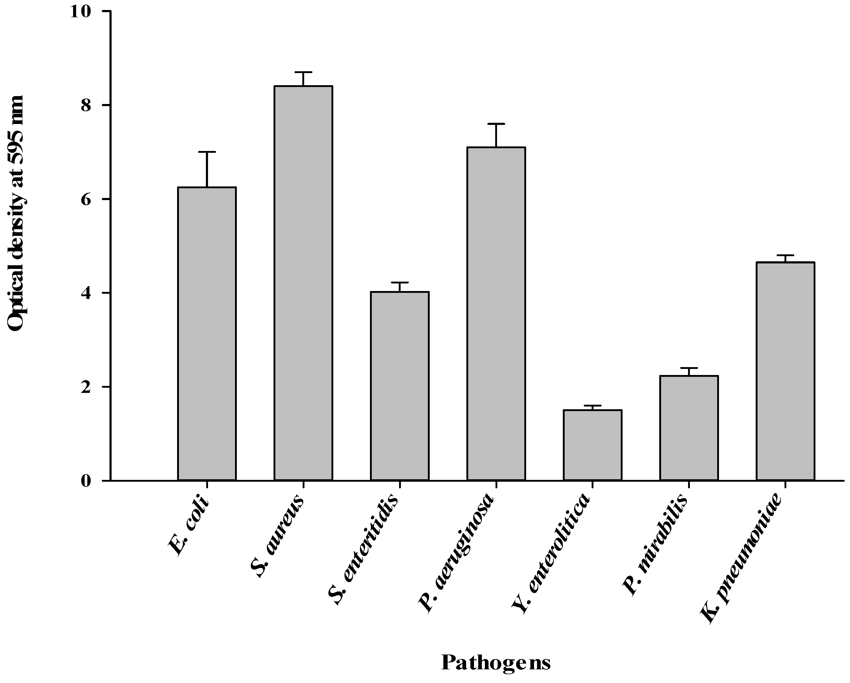
Figure 1. Quantity of biofilms (in terms of absorbance at 595 nm) formed by the different pathogens.
Fermentation 09 00646 g001
Figure 2. RP-HPLC chromatogram showing the purity of the biosurfactant produced by L. paracasei subsp. tolerans N2 with glycerol as substrate.
Fermentation 09 00646 g002
Figure 3. FTIR spectrum (A), 1H NRM spectrum (B) and HRMS spectrum (C) of the biosurfactant produced by L. paracasei subsp. tolerans N2 with glycerol as substrate.
Fermentation 09 00646 g003
Figure 4. DSC thermogram of biosurfactant produced by L. paracasei subsp. tolerans N2 with glycerol as substrate.
Fermentation 09 00646 g004
Figure 5. SEM images showing untreated cells of E. coli EM2 (A) and cells of E. coli EM2 treated with biosurfactant produced by L. paracasei subsp. tolerans N2 with glycerol as substrate (B). Arrows referred to the cell wall and membrane disintegrations, while circles indicated cell lysis.
Fermentation 09 00646 g005
Figure 6. Inhibition percentages obtained from the antiadhesive assays with biosurfactant produced by L. paracasei subsp. tolerans N2 at 10 mg/mL.
Fermentation 09 00646 g006
Table 1. Antibiotics’ resistance profile of the pathogens isolated from braised fish.
PathogensAntimicrobial Resistance Profile *MAR Index
E. coli EM2AMP, AX, C, CXM, F, FX, GEN, IMP, NA, NOR, SXT, TE0.75
S. enteritidis PE1AMC, AMP, C, CAZ, F, NOR, SXT, TE0.50
S. aureus SA1AMC, AMP, AX, CAZ, CXM, F, FX, NA, NOR, TE0.62
P. aeruginosa CT3AX, AMP, CIP, CXM, F, SXT, FX, IMP, NA, NOR, TE0.68
Y. enterolitica MH5AMP, CAZ, ERY, NA, NOR, SXT, TE0.43
P. mirabilis MR2AMP, AMC, C, CIP, NA, SXT0.37
K. pneumoniae AG5AMC, AMP, AX, CAZ, NA, TE0.37
AMC = Amoxycillin–clavulanic acid, AMP = Ampicillin, AX = Amoxycillin, C = Chloramphenicol, CAZ = Ceftazidime, CIP = Ciprofloxacin, CXM = Cefuroxime, ERY = Erythromycin, F = Nitrofurantoin, FX = Cefroxadine, GEN = Gentamicin, IMP = Imipenem, NA = Nalidixic acid, NOR = Norfloxacin, SXT = Cotrimoxazole, TE = Tetracycline. * Bacteria with intermediate resistance were considered as susceptible. MAR index was the ratio of antimicrobial resistance profile to the total number of antibiotics used.
Table 2. Fatty acids and monosaccharides profiles of biosurfactant from L. paracasei subsp. tolerans N2 with glycerol as substrate.
ParametersCompositionRetention Time (min)Proportions (%)
Fatty acids
Lauric acid4.5119.71
Myristic acid6.755.38
Palmitic acid9.3827.90
15-Methyl palmitic acid12.282.77
Oleic acid12.8129.26
Linoleic acid14.014.26
γ-Homo linolenic acid18.392.89
Monosaccharides
Maltol4.342.50
2-Deoxy-D-ribose,6.4414.18
DL-Arabinose, 8.852.05
D-Glucitol, 1,4-anhydro-9.2247.70
1,4-Anhydro-d-galactitol9.661.26
Methyl 2,4-di-O-acetyl-3,6-dideoxy-D-glucopyranoside9.9512.51
Table 3. MIC and MBC of biosurfactant from L. paracasei subsp. tolerans N2 using glycerol as substrate against the multiple antibiotics-resistant pathogens isolated from braised fish.
PathogensMIC (mg/mL)MBC (mg/mL)MBC/MIC
Escherichia coli EM27.5010.001.33
Staphylococcus aureus SA110.0015.001.5
Salmonella enteritidis PE12.505.002
Pseudomonas aeruginosa CT37.5010.001.33
Yersinia enterolitica MH50.501.002
Proteus mirabilis MR21.002.502.5
Klebsiella pneumoniae AG55.007.501.5
Table 4. Percentage of inhibiting biofilm formation by the pathogens with biosurfactant from L. paracasei subsp. tolerans N2 using glycerol as substrate.
PathogensBiosurfactant Concentrations (mg/mL)
02.55.010.015.020.0
E. coli EM20.00 ± 0.00 aA10.25 ± 0.10 cB22.75 ± 1.50 eC41.20 ± 1.30 dD48.50 ± 0.77 dE62.10 ± 1.80 eF
S. aureus SA10.00 ± 0.00 aA15.25 ± 0.25 dB30.70 ± 1.00 fC45.63 ± 1.50 eD61.30 ± 2.50 eE81.90 ± 3.60 fF
S. enteritidis PE10.00 ± 0.00 aA1.50 ± 0.00 bB14.30 ± 0.00 dC25.80 ± 0.62 cD35.80 ± 1.09 cE55.90 ± 2.00 dF
P. aeruginosa CT30.00 ± 0.00 aA0.00 ± 0.00 aA2.90 ± 0.10 aB12.60 ± 0.26 aC26.70 ± 0.20 aD40.20 ± 1.60 aE
Y. enterolitica MH50.00 ± 0.00 aA18.30 ± 0.50 eB28.90 ± 2.10 fC47.30 ± 0.95 eD62.75 ± 0.74 eE80.25 ± 1.01 fF
P. mirabilis MR20.00 ± 0.00 aA0.00 ± 0.00 aA4.90 ± 0.40 bB15.50 ± 1.10 bC29.30 ± 0.00 bD42.80 ± 1.70 bE
K. pneumoniae AG50.00 ± 0.00 aA0.00 ± 0.00 aA7.50 ± 0.90 cB17.20 ± 0.85 bC30.90 ± 1.70 bD47.90 ± 0.10 cE
Values followed by the same lowercase letter in superscript on the same column or by the same capital letter in superscript on the same line are not significantly different (p < 0.05).
Table 5. Percentage of eradication of biofilms formed by the pathogens with biosurfactant from L. paracasei subsp. tolerans N2 using glycerol as substrate.
PathogensBiosurfactant Concentrations (mg/mL)
05.010.020.030.040.050.0
E. coli EM20.00 ± 0.00 aA0.25 ± 0.01 cB3.15 ± 0.10 cC13.90 ± 0.10 bD28.50 ± 0.77 dE42.10 ± 0.10 cF60.50 ± 0.80 dG
S. aureus SA10.00 ± 0.00 aA2.25 ± 0.15 dB6.13 ± 0.40 dC25.93 ± 1.90 dD42.10 ± 2.50 eE58.90 ± 1.60 eF75.40 ± 2.10 eG
S. enteritidis PE10.00 ± 0.00 aA0.10 ± 0.01 bB3.10 ± 0.00 cC15.80 ± 0.12 cD25.70 ± 0.39 cE45.80 ± 1.00 dF59.90 ± 2.00 dG
P. aeruginosa CT30.00 ± 0.00 aA0.00 ± 0.00 aA0.90 ± 0.02 aB9.70 ± 0.36 aC20.40 ± 0.50 aD35.30 ± 0.60 aE50.60 ± 2.60 aF
Y. enterolitica MH50.00 ± 0.00 aA2.30 ± 0.10 dB8.20 ± 0.10 eC27.30 ± 2.25 dD42.25 ± 0.14 eE60.25 ± 2.01 eF78.15 ± 0.81 eG
P. mirabilis MR20.00 ± 0.00 aA0.00 ± 0.00 aA2.90 ± 0.20 bB13.80 ± 2.10 bcC22.30 ± 0.03 bD40.70 ± 0.30 bE54.50 ± 0.70 bF
K. pneumoniae AG50.00 ± 0.00 aA0.00 ± 0.00 aA3.10 ± 0.10 cB14.40 ± 1.84 bcC23.90 ± 0.10 bD39.10 ± 0.90 bE57.20 ± 0.50 cF
Values followed by the same lowercase letter in superscript on the same column or by the same capital letter in superscript on the same line are not significantly different (p < 0.05).
Disclaimer/Publisher’s Note: The statements, opinions and data contained in all publications are solely those of the individual author(s) and contributor(s) and not of MDPI and/or the editor(s). MDPI and/or the editor(s) disclaim responsibility for any injury to people or property resulting from any ideas, methods, instructions or products referred to in the content.