Article

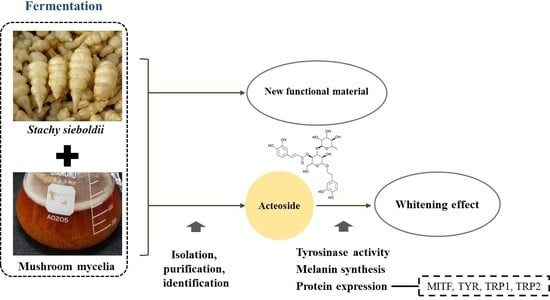
Whitening Activity of Acteoside from Stachys sieboldii Fermented with Hericium erinaceus Mycelia on Melanocytes

1
Department of Pharmacy, Sunchon National University, 255 Jungang-Ro, Suncheon 57922, Republic of Korea
2
Jangheung Research Institute for Mushroom Industry, Jangheung 59338, Republic of Korea
3
Department of Biomedical Science, Sunchon National University, 255 Jungang-Ro, Suncheon 57922, Republic of Korea
4
Suncheon Research Center for Bio Health Care, Jungang-Ro, Suncheon 57922, Republic of Korea
*
Author to whom correspondence should be addressed.
Fermentation 2023, 9(8), 697; https://doi.org/10.3390/fermentation9080697
Submission received: 30 June 2023 / Revised: 21 July 2023 / Accepted: 22 July 2023 / Published: 25 July 2023
(This article belongs to the Special Issue Fermentation in Cosmetics)

Abstract

:
Skin whitening has recently renewed attention on Chinese herbal medicines with whitening activity for esthetic applications. Stachys sieboldii has been used as herbal medicine since ancient times and has the potential for development as a cosmetic material because of its astringent effect. In this study, with an aim to develop new functional materials with whitening effects, S. sieboldii water extracts were fermented with different mushroom mycelia. Fermented with Hericium erinaceus mycelia showed the strongest tyrosinase inhibition effect and the lowest melanin content. Thus, H. erinaceus mycelia, the most potent inhibitor of melanogenesis, was used for large-scale fermentation and fractionated. The ethyl acetate fraction, which had the strongest whitening activity, was separated and purified using HPLC. Finally, the single compound was isolated and identified as acteoside, which has promising whitening activity. Acteoside inhibited melanin synthesis and intracellular tyrosinase activity in a dose-dependent manner. The effects of acteoside on the expression of TYR, TRP-1, TRP-2, and MITF were analyzed using Western blot analysis, which showed that acteoside reduced the protein in a dose-dependent manner. Our findings reveal the potential applicability of S. sieboldii extract fermented with H. erinaceus mycelia and its useful component, which is an acteosid, for skin lightening and the treatment of pigmentation.

Graphical Abstract

1. Introduction

The skin is the largest organ in the human body; it is involved in touch sensation and temperature maintenance and provides protection from chemicals, microorganisms, and UV radiation [1]. Skin aging is influenced by various factors, including changes in physiological function and metabolic processes. It can be divided into natural or intrinsic aging and extrinsic aging, which is induced by environmental factors [2], including ultraviolet (UV) radiation, environmental pollution, smoking, and alcohol abuse. Among these factors, UV light is the most important. Thus, extrinsic aging is often referred to as photoaging [3]. Skin that is unexposed to sunlight only undergoes intrinsic aging; photoaging is a superposition of aging induced by UV radiation over natural aging. Both intrinsic and extrinsic skin aging can lead to decreased structural integrity and a loss of physiological function [4].
Melanin, the primary determinant of skin color, is synthesized by specialized organs called melanosomes within melanocytes [5]. Melanin plays an important role in protecting human skin from UV damage by absorbing UV light, removing reactive oxygen species (ROS), and providing protection from toxic drugs and chemicals [6]. However, dysregulation in melanin synthesis can result in several skin manifestations, including freckles, melasma, age spots, and other hyperpigmentation syndromes [7]. Recent reports demonstrated that melanin also affects melanoma progression by promoting metastasis [8]. Melanocytes are specialized cells located in the hair follicles and epidermis that synthesize and transfer melanin pigment to surrounding keratinocytes [9]. Melanocytes are melanin-producing dendritic cells that are involved in pigmentation and determine the color of the skin, hair, and eyes [10]. One melanocyte is linked and can transfer melanin to approximately 36 neighboring keratinocytes, which is called an epidermal-melanin unit. Differences in skin color among different races result from variations in the number of melanocytes [11,12].
Fermentation, an ancient technique used for processing and preserving food, has been used to make wine in the Old East for thousands of years [13]. This technique not only preserves food but also leads to significant changes in the nutritional value and sensory characteristics of foods [14,15]. Fermentations have found extensive application in the food industry for the production of biologically active compounds. However, recently fermented products are more frequently being used in cosmetics, as they may improve product quality and facilitate the absorption of active substances [16]. Interestingly, fermentation can significantly decrease the allergenicity of foods and improve the physicochemical properties and nutritional value [17,18]. Therefore, we fermented a natural material using mushroom mycelia and evaluated its whitening activity.
Mushroom mycelia, which were used as a microbial source in this study, are composed of clusters of multinucleate hyphae, which typically manifest as slender strands or fine filaments. As mycelia grow, it secretes various compounds into the substrate, altering its chemical nature [19]. The appeal of edible mushroom fruiting bodies and their mycelia as functional foods are attributed to their low toxicity and rich content of bioactive compounds, which has garnered significant interest among many consumers. Investigators have reported antitumor, antimutagenicity, antiviral, and antioxidant activities in various mushroom fruiting bodies and mycelia. Mushroom mycelia have been investigated for their potential applications in foods or cosmetics, and they are used to create fermented products, such as cosmetics, health products, foods, and alcoholic drinks [20].
S. sieboldii MiQ, Korean name Cho Seok Jam, which was used as a fermentation substrate in this study, is a food and herbal medicine widely used in China, South Korea, and Japan [21]. The plant is used in Korea as a food additive to improve the taste and nutritional value of traditional porridge, noodles, rice cakes, and bread [22]. S. sieboldii contains several bioactive compounds, including flavonoids [23], terpenes [24], and phenolic compounds [25], which are directly associated with its antimicrobial [26] and antioxidant [27] properties. However, the therapeutic potential of S. sieboldii and the mechanisms underlying its whitening effect are largely unknown. Therefore, we generated new functional materials by fermenting S. sieboldii using mushroom mycelia and then assessed the whitening effect of the ferments compared to the raw material to search for potential whitening compounds.

2. Materials and Methods

2.1. Chemicals and Reagents

Ascorbic acid (vitamin C), arbutin, dimethyl sulfoxide (DMSO), α-melanocyte stimulating hormone (α-MSH), and L-DOPA were purchased from Sigma-Aldrich (St. Louis, MO, USA). Dulbecco’s modified Eagle’s medium (DMEM), phosphate-buffered saline (PBS), fetal bovine serum (FBS), and antibiotic/antimycotic (anti/anti) were obtained from Gibco BRL (Gaithersburg, MD, USA). Potato dextrose agar (PDA) and potato dextrose broth (PDB) were purchased from BD DIFCO (NJ, USA). Radioimmunoprecipitation assay buffer; protease and phosphatase inhibitor cocktail; BCA protein assay kit; 4–12% bis-tris plus polyacrylamide gels; nitrocellulose membranes; 20× TBS Tween™ 20 Buffer; Starting Block T20 (TBS) Blocking Buffer; enhanced chemiluminescence kit; and the following antibodies: MITF (#PA5-38294), TYR (#PA5-99493), TRP1 (#PA5-69298), and TYRP2 (#PA5-69368), β-actin, and horseradish peroxidase (HRP)-conjugated secondary antibody (#G-21234) were acquired from Thermo (Rockford, IL, USA). All other chemical buffers and reagents used for extraction and analysis were of analytical grade or purer and were purchased from J. T. Baker Chemicals (Center Valley, PA, USA).

2.2. Preparation of S. sieboldii

S. sieboldii Miq. Tubers were purchased from Dongbu Herbal Market (Suncheon City, Republic of Korea). In order to remove dust and other accumulated substances on the roots from the natural environment, the collected tubers underwent a thorough washing procedure using tap water. The decontaminated tubers were freeze-dried and ground with a mortal to a coarse powder.

2.3. Extraction and Fermentation

A sufficient quantity of tuber extract was obtained by using distilled water. To prepare an aqueous extract of S. sieboldii tuber, the coarse powder (100 g) was mixed with 1 L of hot water and incubated at 70 °C for 6 h. The four mushroom mycelia (H. erinaceus, Wolfiporia extensa, Lentinula edodes, and Ganoderma lucidum) used in this study were obtained from the Korean Agricultural Culture Collection (KACC; Suwon, Republic of Korea). The freeze-dried mycelia were suspended in 0.5 mL of sterile water and then plated on PDA and incubated for 2 weeks at 25 °C. The mycelia that developed on PDA were extracted using a cork borer with a diameter of 5 mm. Five punched discs were then transferred to PDB and incubated at 25 °C for a duration of two weeks. Mycelia grown in the liquid media were transferred to S. sieboldii hot water extract and fermented for 2 weeks at 25 °C on a fermentation shaker at 250 rpm. After incubation, the ferments were filtered under reduced pressure and lyophilized, and extract yield was measured. We determined the solid content of both S. sieboldii hot water extract and S. sieboldii hot water extract fermentation using mushroom mycelial as the extraction yield.
The mycelia with the most potent melanogenesis inhibition activity were selected for large-scale fermentation. To prepare a large-scale aqueous extract of S. sieboldii tubers, a large amount of the coarse powder (2 kg) was mixed with 20 L of hot water and incubated at 70 °C for 6 h. H. erinaceus mycelia grown on PDA plates was punched with the cork borer, and five punched discs were transferred to 500 mL of PDB and incubated for 2 weeks at 25 °C in a shaking incubator at 250 rpm. The liquid medium was transferred to S. sieboldii tuber extract and incubated for 2 weeks at 25 °C in a shaking incubator at 250 rpm.

2.4. Liquid–Liquid Partition

The ferments were filtered by vacuum filtration through Whatman No. 2 filter paper in a Büchner funnel. The obtained supernatant was utilized to prepare a crude extract. The crude extract was transferred to a separating funnel for fractionation using different organic solvents with increasing polarity (n-hexane, chloroform, ethyl acetate, n-butanol, and distilled water), and their anti-melanogenesis activities were evaluated. The crude extract was put in a 2 L separating funnel with an equal volume of each organic solvent. The separating funnel was shaken for 15 min and then allowed to settle at room temperature until the organic solvent layer was clear and colorless (about 1 h). The resulting organic solvent fractions were concentrated to dryness in vacuo, and the obtained residues were stored in an air-tight container in a freezer. The fractions were concentrated, followed by freeze-drying to remove the solvent. The dry fractions were then dissolved in DMSO to create a stock solution. Subsequently, the stock solution was diluted with PBS to achieve the desired concentration.

2.5. Isolation and Purification

In order to identify useful whitening compounds, the solvent fractions were simultaneously isolated and analyzed. MPLC (Biotage-Isolera One system SE-751 03; Uppsala, Sweden) and LC-MS (Agilent Technologies 6410 Triple Quad, CA, USA) were used for separation and isolation, respectively. A YMC C18 triart reverse phase column was eluted with a gradient system consisting of distilled water:acetonitrile (95:5, v/v, with increasing acetonitrile %) to yield seven sub-fractions. The sub-fraction with the highest whitening activity was further separated and purified using LC-MS under the following parameters: LC-MS separation of sub-fractions with distilled water:acetonitrile (95:5 v/v, Table 1) for 32 min at 0.4 mL/min, to obtain compound 1, which was obtained to >97% purity.

2.6. NMR Analysis

The chemical structures of compounds were determined by nuclear magnetic resonance (NMR) analysis. NMR spectra were recorded in CD3OD solvent on a Bruker 600 MHz NMR spectrometer at 1H frequencies of 600 MHz or 13C frequencies of 150 MHz using a conventional series of two-dimensional (2D) H 1–N 15 HSQC (Heteronuclear Single Quantum Coherence) for compound 1. Detailed analysis of zero-filled, resolution-enhanced spectra (peak picking, integration, and multiplet analysis) were performed using Topspin 3.1 (Bruker, Germany). Compound 1 was dissolved in CD3OD, and its chemical shift was determined using tetramethyl silane (TMS) as an internal standard.

2.7. Cell Culture

B16F10 mouse melanocytes were obtained from American Type Culture Collection (ATCC; Manassas, VA, USA) and cultured in Dulbecco’s modified Eagle’s medium (DMEM) supplemented with 10% FBS and 1% anti/anti at 37 °C in a humidified atmosphere containing 5% CO2. The medium was changed once every other day for culture maintenance. Cells were not used beyond passage 10.

2.8. Cell Viability Assay (MTS Assay)

Prior to evaluating the whitening activity of the sample, we conducted cytotoxicity screening using the MTS (Promega; Annandale, NSW, Australia) assay to identify any potential cytotoxic effects. In summary, B16F10 cells were initially seeded at a density of 5 × 10⁴ cells/mL in a 96-well plate and allowed to adhere for 24 h. The test samples were diluted in the culture medium to achieve a final concentration of 0.1% DMSO and subsequently added to the cell monolayers. Following a 48 h incubation period, the culture medium was carefully removed and replaced with 100 μL of fresh medium. Subsequently, 20 μL of MTS reagent was added to each well, and the plate was incubated at 37 °C for 30 min. The absorbance of aliquots was read at 490 nm using a microplate reader, and cell viability was determined by calculating the absorbance value relative to the control and expressing it as a percentage.

2.9. Mushroom Tyrosinase Activity Assay

The direct effect of the sample on diphenolase activity was evaluated using mushroom tyrosinase as a source of an enzyme with L-DOPA as the substrate according to a previously described method [28]. In summary, for each sample, 20 µL aliquots at various concentrations, prepared in 0.1 M sodium phosphate buffer (pH 6.5), were combined with 40 µL of prepared substrate solution (1.5 mM L-DOPA in phosphate buffer) in a 96-well microplate. The reaction was initiated by adding 20 µL of mushroom tyrosinase (at a final concentration of 22 U/mL), followed by incubation at 37 °C for 30 min. Ascorbic acid served as the positive control in the experiment. Tyrosinase inhibitory activity was calculated using the following equation:
Mushroom tyrosinase inhibition (%) = ([(A − B) − (C − D)])/((A − B)) × 100, where A is the optical density (OD490) without extract; B is the OD490 with tyrosinase and without extract; C is the OD490 with test extract; and D is the OD490 with both tyrosinase and test extract.

2.10. Intracellular Tyrosinase Activity Assay

Intracellular tyrosinase activity was measured as described previously [29]. Briefly, B16F10 cells were plated at a density of 3 × 10⁴ cells/mL in 12-well plates and treated with various concentrations of sample in the presence of α-MSH. Cells were washed with PBS and lysed with 1% Triton X-100 in PBS (pH 7.4). The cell lysates were collected and centrifuged at 12,000× g for 30 min. The collected supernatant (50 μL) was mixed with 100 μL of 10 mM L-DOPA in a 96-well plate and incubated at 37 °C for 30 min. Then, the absorbance at 475 nm was measured. Ascorbic acid served as the positive control in the experiment.

2.11. Measurement of Melanin Content

B16F10 cells were plated in 12-well culture plates at a density of 3 × 10⁴ cells/mL in DMEM supplemented with 10% FBS and 1% anti-anti solution. The cells were then incubated at 37 °C with 5% CO2. For a duration of 48 h, the cells were incubated with a medium containing α-MSH along with different concentrations of the samples. α-MSH is a substance that induces melanogenesis and is used as a negative control. Subsequently, 1 N NaOH was added to each well to induce cell lysis, and the plates were incubated at 70 °C for 30 min. The amount of melanin in each lysate was measured as the absorbance at 405 nm. Arbutin served as the positive control in the experiment.

2.12. Western Blotting

Western blotting was performed to measure the expression levels of melanogenesis-related proteins in B16F10 cells. In summary, B16F10 cells were seeded in 6-well plates at a density of 1 × 10⁵ cells/well per well and incubated for a period of 24 h. The cells were then treated with acteoside for 48 h. After the treatment, the cells were washed twice with cold PBS and then lysed using RIPA buffer supplemented with phosphatase and protease inhibitors. The supernatant was obtained by centrifugation (14,000× g and 4 °C for 20 min), and the protein was quantified using the BCA protein assay kit. An equal amount of protein (20 µg) was combined with 5x SDS loading buffer and subjected to heat at 95 °C for 5 min. Subsequently, the protein samples were separated on 4–12% bis-tris plus polyacrylamide gels and transferred onto nitrocellulose membranes. The membranes were then incubated with Block T20 (TBS) blocking buffer for 3 h at room temperature, followed by overnight incubation at 4 °C with primary antibodies against MITF (1:1000), tyrosinase (1:2000), TRP1 (1:500), and TRP2 (1:2000). Following three washes with TBST buffer, each for a duration of 15 min, and the membranes were incubated with HRP-conjugated secondary antibodies (1:10,000) for 1 h at room temperature. Then, the membranes were washed twice, again for 15 min each, and treated with an enhanced chemiluminescence reagent to facilitate protein visualization. Signal intensities were determined using a chemiDoc imaging system (BioRad, Hercules, CA, USA).

2.13. Statistical Analysis

Unless otherwise specified, data are expressed as the mean and standard deviation (SD) from at least triplicate experiments. Statistical differences between groups were analyzed using Student’s t-test. p values less than 0.05 were considered statistically significant (*** and ### p < 0.001, ** and ## p < 0.01, and * and # p < 0.05).

3. Results

3.1. Effects of Fermentation Using Different Mushroom Mycelia on Extract Yields

S. sieboldii tubers were homogenized and extracted with hot water for use as a substrate for mushroom mycelia fermentation. After 2 weeks of fermentation, the ferment was filtered under reduced pressure and lyophilized (Figure 1). Extract yield was evaluated by measuring the weight of the remaining residue after lyophilization of each ferment (Table 2). The four ferments with S. sieboldii extracts had higher extraction yields than S. sieboldii extract alone.

3.2. Effect of S. sieboldii Extract Fermented with Mushroom Mycelia

The safety of functional materials plays a critical role in determining their suitability for applications in food and cosmetics. As a result, it is crucial to establish safe concentrations of anti-melanogenesis agents for future in vitro and in vivo studies. As shown in Figure 2A, the mushroom mycelia ferments of S. sieboldii extracts had no significant effect on cell viability. The positive control, arbutin, also had no effect on cell viability.
To evaluate the potential inhibitory effect of mushroom mycelia ferments on melanin synthesis, we measured the melanin content of B16F10 cells with different concentrations of mushroom mycelia ferments of S. sieboldii extracts. The results are shown in Figure 2B, and melanin content was increased in cells treated with the melanogenesis inducer α-MSH. The melanin content of α-MSH-stimulated B16F10 cells significantly decreased following treatment with the ferment samples in a concentration-dependent manner. Additionally, the mushroom mycelia S. sieboldii ferments inhibited melanin synthesis more than unfermented S. sieboldii extract. At the highest tested concentration, the percent melanin content of cells incubated with S. sieboldii extract alone, S. sieboldii extract fermented by H. erinaceus mycelia, W. extensa mycelia, L. edodes mycelia, and G. lucidum mycelia was 75.2, 57.4, 61.9, 64.9, and 63.1%, respectively. Incubation with S. sieboldii extract fermented by H. erinaceus mycelia was the lowest melanin content.
The mechanism of melanogenesis involves enzymatic oxidation of tyrosine and DOPA to DOPA quinone. Melanin formation could be reduced through the inhibition of tyrosinase or DOPA oxidase. Evaluation of the effects of mushroom mycelia ferments on these enzymes showed that tyrosinase activity was reduced in a dose-dependent manner (Figure 2C), and the mushroom mycelia ferments of S. sieboldii extracts inhibited tyrosinase activity more than S. sieboldii extract alone. These results were consistent with the greater inhibition of melanin synthesis by these mushroom mycelia ferments. S. sieboldii extracts fermented with H. erinaceus mycelia had the strongest tyrosinase activity-inhibiting effect.

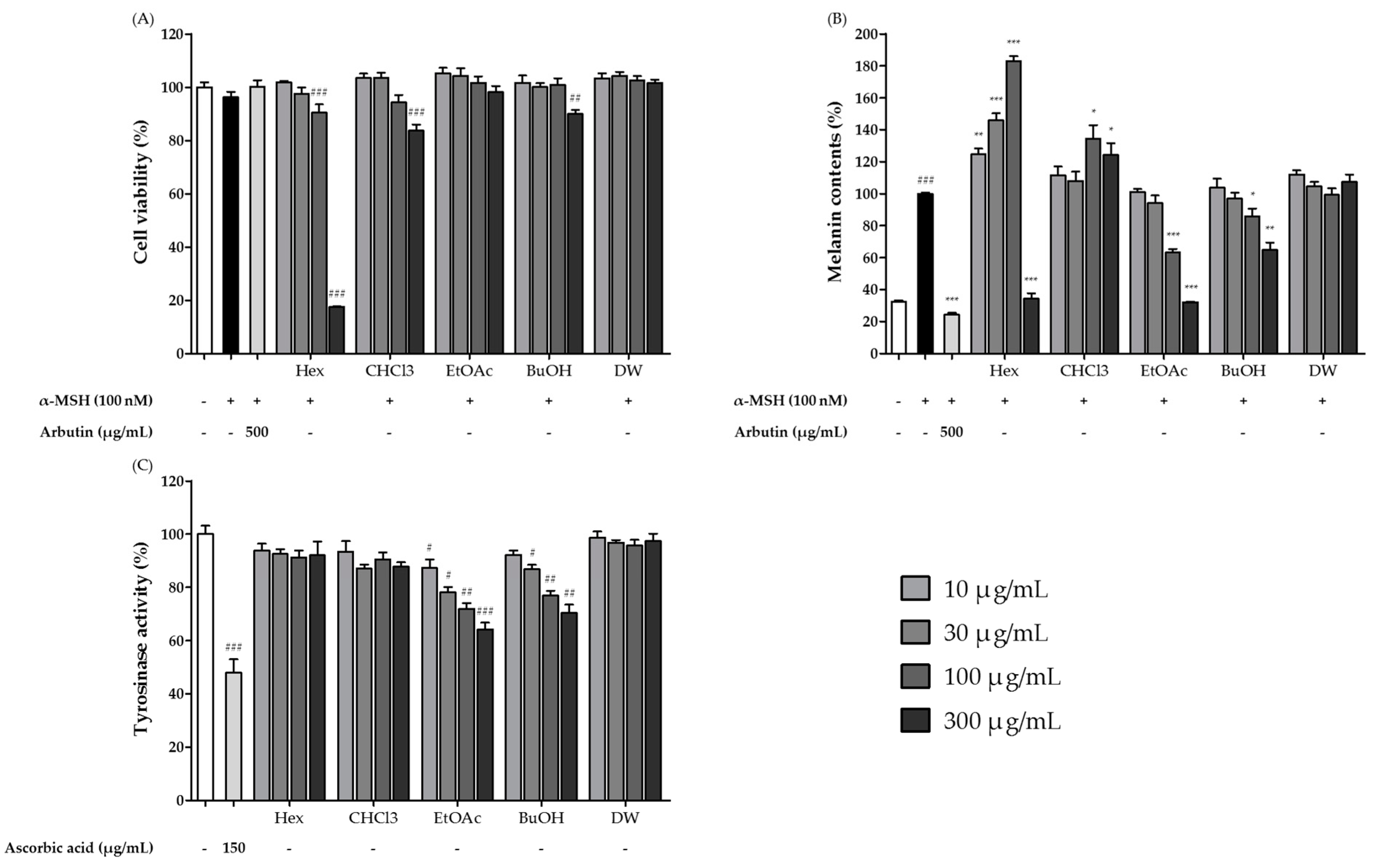
3.3. Cytotoxic and Whitening Effects of Solvent Fractions of S. sieboldii Extract Fermented by H. erinaceus Mycelia

The S. sieboldii extract fermented with H. erinaceus mycelia was extracted with five different solvents, and the effects of the extracts on cell viability, melanin synthesis, and tyrosinase activity were evaluated (Figure 3). The solvents used for extraction were hexane, chloroform, ethyl acetate, butanol, and water. The cytotoxicity of the solvent-extracted fractions in B16F10 melanocytes was evaluated using the MTS assay. The hexane, chloroform, and butanol fractions of the ferment significantly reduced the viability of B16F10 cells, whereas the ethyl acetate and water fractions did not decrease viability (Figure 3A). Thus, high concentrations of the hexane, chloroform, and butanol fractions affected the viability of B16F10 melanocytes.
We then assessed the effects of these five fractions on melanogenesis. As shown in Figure 3B, only the ethyl acetate and butanol fractions decreased melanin content in a dose-dependent manner. The ethyl acetate fraction was the strongest inhibitor of melanin synthesis. The hexane fraction, when added at 10–100 µg/mL, actually increased melanin content. We then assessed the tyrosinase-inhibitory effects of the sequential fractions (Figure 3C). As with the melanin content, only the ethyl acetate and butanol fractions decreased tyrosinase activity in a dose-dependent manner, and the ethyl acetate fraction had the strongest tyrosinase inhibitory activity. Based on these results, we speculated that certain compounds in the ethyl acetate fraction were responsible for the melanogenesis-inhibiting activity of the ferment.

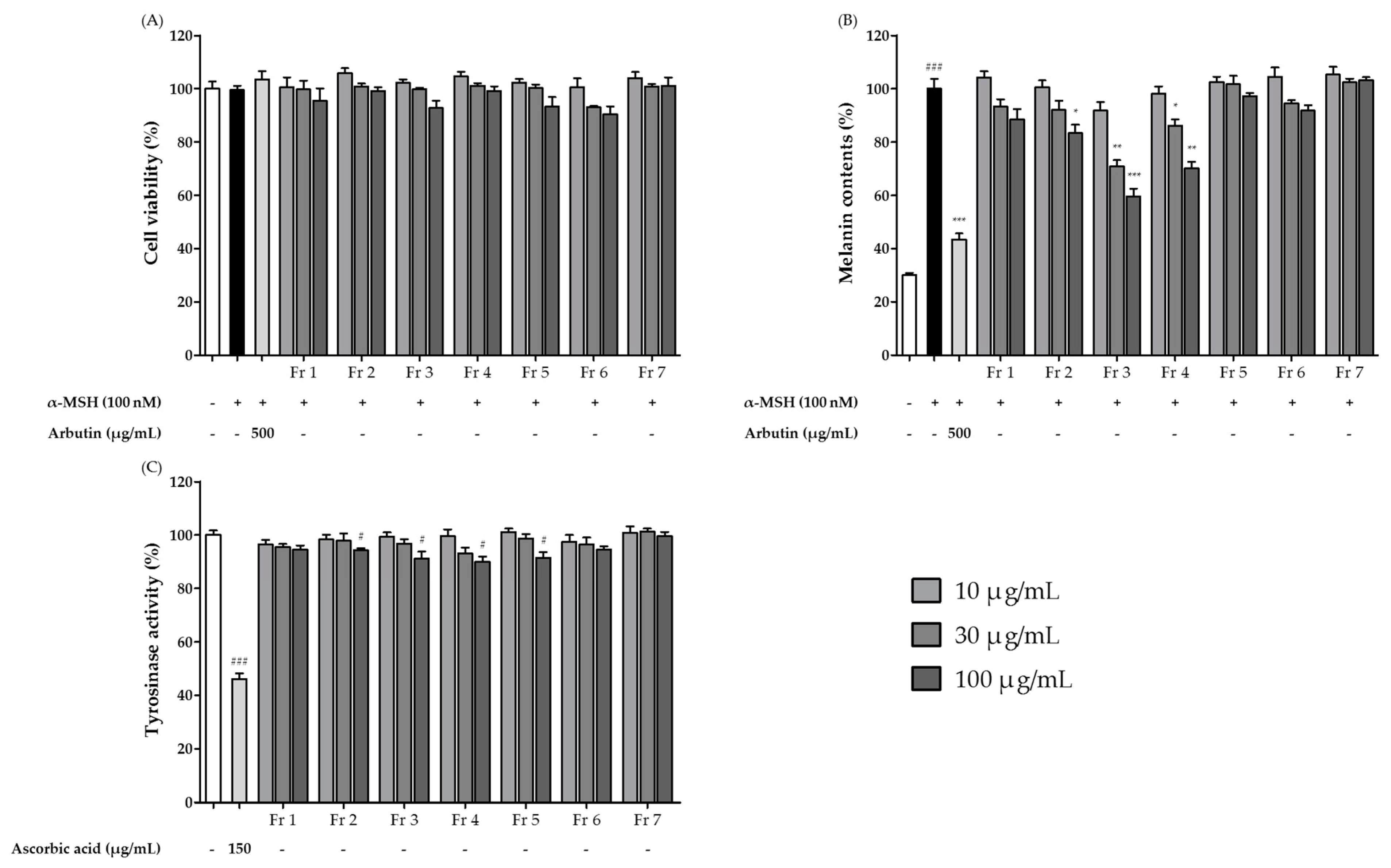
3.4. Isolation and Purification of the Major Compounds in the Ethyl Acetate Extract Sub-Fractions

The ethyl acetate extract was separated into seven fractions using MPLC. These fractions did not decrease the viability of B16F10 cells (Figure 4A). Sub-fractions Fr 2, Fr 3, and Fr 4 inhibited melanin production in B16F10 cells in a dose-dependent manner (Figure 4B), whereas sub-fractions Fr 1, Fr 5, Fr 6, and Fr 7 did not have dose-dependent melanogenesis-inhibiting effects. Sub-fractions of the ethyl acetate extract were evaluated for their tyrosinase inhibitory effects (Figure 4C). Subfractions Fr 2, Fr 3, Fr 4, and Fr 5 showed significant tyrosinase inhibition at the maximum concentration (300 µg/mL). Subfraction Fr 3 showed the strongest whitening activity; thus, fraction Fr 3 was further separated to identify the major compounds and further investigate their whitening effects. Fr 3 was fractionated using LC-MS with a kromasil C18 column (3.0 mm × 150 mm, 3.0 μm), which yielded one main compound. The compound was dissolved in DMSO and stored at −70 °C until use. The final DMSO concentration did not exceed 0.1%.

3.5. Structural Characterization of the Isolated Compound

A single compound was isolated as a white powder. The molecular weight of the compound was deduced from the [M-H]- ion at m/z 622.9 in negative ESI ionization mode. The 1H NMR, 13C NMR, and DEPT spectra of the compound were consistent with the molecular formula C29H36O15. The 1H and 13C NMR spectra of the compound are listed in Table 3. The chemical structure of the compound was determined to be β-(3′,4′-dihydroxyphenyl) ethyl-O-α-L-rhamnopyranosyl-(1→3)-β-D-(4-O-caffeoyl)-gluco pyranoside. The chemical shifts of acteoside were compared to 1H and 13C NMR data from a previous study [30]. Acteoside is a member of the dihexose family, which has been isolated and purified from plant materials. A recent report showed that acteoside inhibits a-MSH-induced melanin production in murine melanoma cells by inhibiting adenylyl cyclase activity [31]. However, the precise mechanisms through which acteoside regulates melanogenesis and melanocyte pigmentation remain unclear.

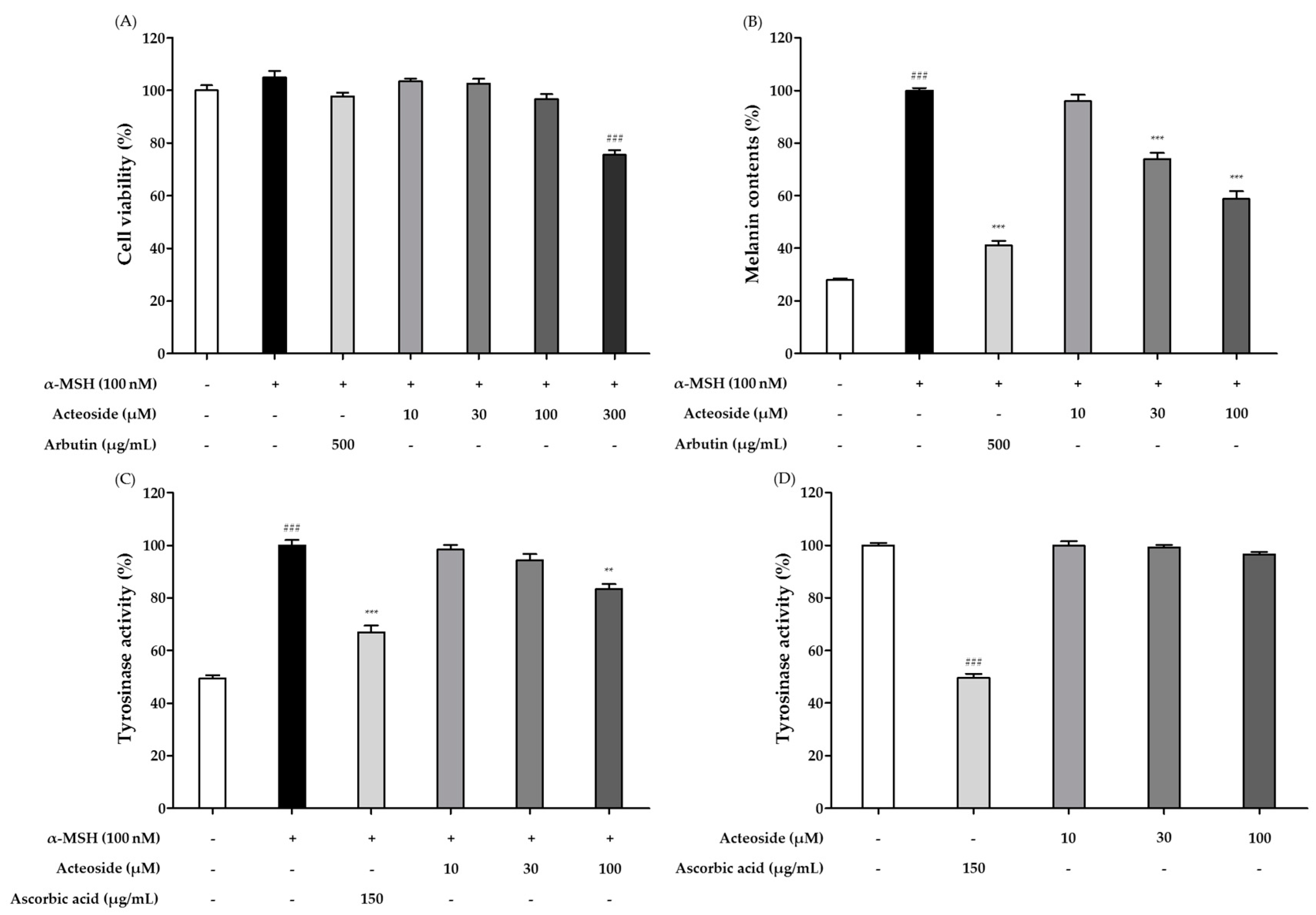
3.6. Effects of Acteoside on Cell Viability and Whitening Effect

To exclude melanogenesis inhibition caused by cytotoxicity, we first evaluated the cytotoxicity of acteoside in B16F10 cells (Figure 5A). The cells were treated with different concentrations of acteoside, and the results showed that 300 μM acteoside caused cytotoxicity in B16F10 cells. Thus, acteoside at 10–100 μM was used in subsequent experiments to determine its effects on melanogenesis and tyrosinase activity. As shown in Figure 5B, acteoside at 30 and 100 μM also significantly inhibited α-MSH-induced melanin production in a dose-dependent manner (by 26.2% and 41.3%, respectively, compared with the levels in α-MSH-treated cells). The tyrosinase activity in B16F10 cells stimulated with α-MSH was notably increased two-fold compared to the activity observed in the control cells (Figure 5C). Acteoside induced a dose-dependent reduction in tyrosinase activity in B16F10 cells, with 16.5% inhibition at 100 μM. These results suggest that acteoside can suppress melanin synthesis and intracellular tyrosinase activity in B16F10 cells. We then assessed its effect on mushroom tyrosinase activity to confirm a direct effect on tyrosinase enzyme (Figure 5D). The results showed that mushroom tyrosinase activity was not suppressed by acteoside in our cell-free assay system. These results suggest that acteoside does not directly inhibit tyrosinase activity but inhibits melanogenesis through other pathways, such as MITF regulation.

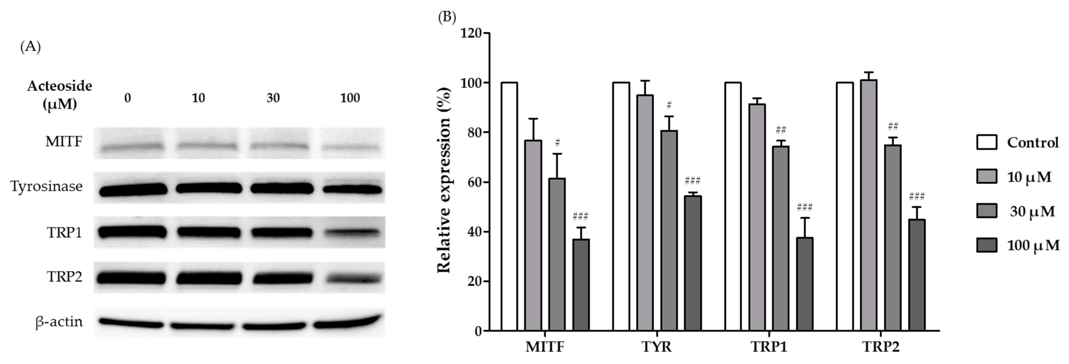
3.7. Effects of Acteoside on the Expression of Melanogenesis-Related Proteins

Melanogenic enzymes, such as tyrosinase, TRP-1, and TRP-2, play important roles in melanogenesis. MITF is a transcriptional factor that impacts melanogenesis by regulating the expression of these melanogenic enzymes. To assess the impact of acteoside on the expression of these proteins, we performed a Western blot assay. Protein levels of MITF were clearly decreased after treatment with acteoside at 10, 30, and 100 μM in a dose-dependent manner (Figure 6A). Expression levels of the tested melanogenic enzymes in B16F10 cells were also significantly decreased in a concentration-dependent manner. The highest concentration of acteoside (100 μM) significantly reduced protein expression levels relative to those in untreated control cells (Figure 6B). Based on these findings, it can be inferred that acteoside exerts a suppressive effect on the expression of MITF, tyrosinase, TRP-1, and TRP-2 in B16F10 cells at the translational levels. This ultimately leads to a decrease in cellular tyrosinase activity and melanin content.

4. Discussion

When formulating functional foods or cosmetics with plant extracts or phytochemical compounds, safety must be considered. Hydroquinone, which is used in the cosmetics industry as a skin whitening agent for hyperpigmentation, has numerous side effects, including skin irritation, contact dermatitis, and exogenous ochronosis in dark-skinned people. Because of these side effects, the US Food and Drug Administration prohibited the sale of hydroquinone in 2006 [32]. Kojic acid, a depigmenting agent that inhibits tyrosinase activity, can also induce dermatitis [33]. Thus, many studies have aimed to identify new materials for the treatment of hyperpigmentation disorders with fewer side effects.
The original purpose of fermentation was for preservation. With the development of alternative preservation technologies, foods were fermented for their unique flavors, aromas, and textures, which are appreciated by consumers. Fermentation is widely employed in the food industry as a means to produce biologically active compounds with significant functional properties, but more recently in cosmetics, as fermentation can improve product safety and quality and facilitate the absorption of active substances by the human body [16]. With these advantages in mind, we used mushroom mycelia to ferment an extract of S. sieboldii, which has biological activity when used as a cosmetic material. We aimed to increase its safety and whitening activity through fermentation. The results showed that fermentation of an S. sieboldii hot water extract with four kinds of mushrooms led to a stronger whitening effect than the unfermented extract. It seems that conducting fermentation with mushroom mycelial in S. sieboldii resulted in an increase in useful components, and it has been demonstrated that the extraction yields also increased. There is a study showing that the fermentation product exhibited higher solid content compared to the control group, which is attributed to the leaching of water-soluble components, such as vitamins, during fermentation [34]. The fermentation with H. erinaceus showed the strongest inhibitory effects on melanin synthesis and mushroom tyrosinase activity. This new fermented functional material was fractionated, and the potential whitening compound was separated, purified, and identified. A single compound was identified, acteoside, which has promising whitening activity.
Acteoside is abundant in many dicotyledonous plants, including plants in the families Oleaceae, Bignoniaceae, Verbenaceae, and Labiatae [35]. The biological activity of acteoside has attracted interest from diverse fields. Acteoside is an important secondary metabolite in medicinal plants and a natural bioactive ingredient [36]. It possesses numerous beneficial activities for human health, including wound-healing, neuro protective, antioxidant, anti-inflammatory, and antineoplastic properties [37]. Although there is little information on the whitening effect of acteoside, it has been reported to prevent hyperpigmentation and have an astringent effect. We investigated the whitening activity of isolated acteoside on B16F10 and found that acteoside inhibited both melanin synthesis and intracellular tyrosinase activity in a dose-dependent manner. However, in contrast with the inhibition of melanin production and cellular tyrosinase activity in cultured melanocytes [38], acteoside did not affect mushroom tyrosinase activity using L-DOPA as a substrate, suggesting that acteoside does not directly inhibit tyrosinase activity. Several studies have shown that the expression of melanogenic enzymes, such as tyrosinase, TRP-1, and TRP-2, is transcriptionally regulated by MITF, resulting in decreased melanogenic enzyme expression and inhibition of melanogenesis in B16F10 melanoma cells [39,40,41,42]. We hypothesized that acteoside inhibits tyrosinase activity by regulating MITF expression. As expected, acteoside significantly reduced the expression levels of MITF and the downstream enzymes tyrosinase, TRP-1, and TRP-2 at the translational level. These results suggested that acteoside reduces melanogenesis in B16F10 cells by inhibiting the expression of tyrosinase, TRP-1, and TRP-2 through inactivation of MITF or some other upstream signal. To verify our hypothesis, further studies should be directed to elucidate the pathway(s) regulated by acteoside in melanocytes.
In summary, we found that an S. sieboldii extract fermented with mushroom mycelia improved its whitening activity, and when fermented by H. erinaceus mycelia, its active components, acteoside, possessed a strong inhibitory effect on melanogenesis without cytotoxicity. The ability of acteoside to effectively reduce skin pigmentation even at low concentrations indicates that targeting melanogenesis could be a promising approach for regulating skin pigmentation.

5. Conclusions

The present study confirms the improvement of the safety and whitening effect of S. sieboldii extract through mushroom mycelia fermentation. Although little has been reported on the whitening effect of S. sieboldii, it is known to have an astringent effect. Among the four tested mushroom mycelia, S. sieboldii extract fermented with H. erinaceus had the strongest inhibitory effect on melanin synthesis and tyrosinase activity. Based on these results, we performed a large-scale fermentation, fractionation, and separation and found a single compound that was expected to inhibit melanogenesis and pigmentation. The compound isolated in this study, acteoside, inhibited melanin synthesis in B16F10 cells. Although acteoside did not directly inhibit mushroom tyrosinase activity, its dose-dependently suppressed intracellular tyrosinase activity in B16F10 cells. This suggested that acteoside does not directly affect tyrosinase but affects upstream signaling. Specifically, acteoside reduced the expression levels of the melanogenic enzymes tyrosinase, TRP-1, and TRP-2 by downregulating the expression of MITF in B16F10 cells.
In conclusion, fermentation by mushroom mycelia can be used exploitation and valorization of S. sieboldii for its use in cosmetics formulations or the production of enriched extracts. Moreover, acteoside exhibited no cytotoxic activity; thus, we conclude that acteoside is a potentially useful natural depigmentation agent.

Author Contributions

Conceptualization, S.-T.Y.; data curation, S.-B.I.; funding acquisition, K.-S.S. and K.-J.K.; investigation, S.-B.I., S.-K.M., N.-I.H. and H.-Y.J.; project administration, S.-T.Y.; resources, K.-Y.K. and K.-W.P.; supervision, S.-T.Y.; writing—original draft preparation, S.-B.I.; writing—review and editing, S.-T.Y. and K.-S.S. All authors have read and agreed to the published version of the manuscript.

Funding

This research received external funding from Jangheung Mushroom Industry Research (Grant Number: JMI 2023-01).

Institutional Review Board Statement

Not applicable.

Informed Consent Statement

Not applicable.

Data Availability Statement

Not applicable.

Acknowledgments

This study was carried out with the support of ‘Industry-Academia-Research Collabo R&D (Project No. RS-2023-00223217)’ provided by the Korea Technology and Information Promotion Agency for SMEs.

Conflicts of Interest

The authors declare no conflict of interest.

References

  1. Krutmann, J.; Humbert, P. Nutrition for Healthy Skin; Springer: Berlin/Heidelberg, Germany, 2011. [Google Scholar]
  2. Wu, P.-Y.; Lin, T.-Y.; Hou, C.-W.; Chang, Q.-X.; Wen, K.-C.; Lin, C.-Y.; Chiang, H.-M. 1, 2-bis [(3-methoxyphenyl) methyl] ethane-1, 2-dicarboxylic acid reduces uvb-induced photodamage in vitro and in vivo. Antioxidants 2019, 8, 452. [Google Scholar] [CrossRef] [PubMed] [Green Version]
  3. Penna, I.; Albanesi, E.; Bertorelli, R.; Bandiera, T.; Russo, D. Cytoprotective, anti-inflammatory, and antioxidant properties of high-molecular-weight hyaluronan enriched with red orange extract in human fibroblasts exposed to ultra violet light B irradiation. Biotechnol. Appl. Biochem. 2019, 66, 273–280. [Google Scholar] [CrossRef] [PubMed]
  4. Durai, P.C.; Thappa, D.M.; Kumari, R.; Malathi, M. Aging in elderly: Chronological versus photoaging. Indian J. Dermatol. 2012, 57, 343. [Google Scholar] [PubMed]
  5. Slominski, A.; Tobin, D.J.; Shibahara, S.; Wortsman, J. Melanin pigmentation in mammalian skin and its hormonal regulation. Physiol. Rev. 2004, 84, 1155–1228. [Google Scholar] [CrossRef]
  6. Brown, D.A. Skin pigmentation enhancers. Compr. Ser. Photosciences 2001, 3, 637–675. [Google Scholar]
  7. Chiang, H.-M.; Chien, Y.-C.; Wu, C.-H.; Kuo, Y.-H.; Wu, W.-C.; Pan, Y.-Y.; Su, Y.-H.; Wen, K.-C. Hydroalcoholic extract of Rhodiola rosea L.(Crassulaceae) and its hydrolysate inhibit melanogenesis in B16F0 cells by regulating the CREB/MITF/tyrosinase pathway. Food Chem. Toxicol. 2014, 65, 129–139. [Google Scholar] [CrossRef] [PubMed]
  8. Sarna, M.; Zadlo, A.; Hermanowicz, P.; Madeja, Z.; Burda, K.; Sarna, T. Cell elasticity is an important indicator of the metastatic phenotype of melanoma cells. Exp. Dermatol. 2014, 23, 813–818. [Google Scholar] [CrossRef]
  9. Bandyopadhyay, D.; Medrano, E.E. Melanin accumulation accelerates melanocyte senescence by a mechanism involving p16INK4a/CDK4/pRB and E2F1. Ann. N. Y. Acad. Sci. 2000, 908, 71–84. [Google Scholar] [CrossRef]
  10. Bonaventure, J.; Domingues, M.J.; Larue, L. Cellular and molecular mechanisms controlling the migration of melanocytes and melanoma cells. Pigment Cell Melanoma Res. 2013, 26, 316–325. [Google Scholar] [CrossRef]
  11. Gillbro, J.; Olsson, M. The melanogenesis and mechanisms of skin-lightening agents–existing and new approaches. Int. J. Cosmet. Sci. 2011, 33, 210–221. [Google Scholar] [CrossRef]
  12. Thody, A.J.; Higgins, E.M.; Wakamatsu, K.; Ito, S.; Burchill, S.A.; Marks, J.M. Pheomelanin as well as eumelanin is present in human epidermis. J. Investig. Dermatol. 1991, 97, 340–344. [Google Scholar] [CrossRef] [PubMed] [Green Version]
  13. Sõukand, R.; Pieroni, A.; Biró, M.; Dénes, A.; Dogan, Y.; Hajdari, A.; Kalle, R.; Reade, B.; Mustafa, B.; Nedelcheva, A. An ethnobotanical perspective on traditional fermented plant foods and beverages in Eastern Europe. J. Ethnopharmacol. 2015, 170, 284–296. [Google Scholar] [CrossRef] [PubMed]
  14. Bleotu, A.; Mandravel, C.; Ciuculescu, C. Characterization of some glycoside iridoids by mass spectrometry. Rom. Biotechnol. Lett. 2006, 11, 2643. [Google Scholar]
  15. Chung, H.-J.; Kim, W.K.; Park, H.J.; Cho, L.; Kim, M.-r.; Kim, M.J.; Shin, J.-S.; Lee, J.H.; Ha, I.-H.; Lee, S.K. Anti-osteoporotic activity of harpagide by regulation of bone formation in osteoblast cell culture and ovariectomy-induced bone loss mouse models. J. Ethnopharmacol. 2016, 179, 66–75. [Google Scholar] [CrossRef] [PubMed]
  16. Ziemlewska, A.; Nizioł-Łukaszewska, Z.; Bujak, T.; Zagórska-Dziok, M.; Wójciak, M.; Sowa, I. Effect of fermentation time on the content of bioactive compounds with cosmetic and dermatological properties in Kombucha Yerba Mate extracts. Sci. Rep. 2021, 11, 18792. [Google Scholar] [CrossRef]
  17. Chen, L.; Song, P.; Jia, F.; Wang, J.S. Reducing the allergenicity from food by microbial fermentation. Adv. Mater. Res. 2012, 524, 2302–2305. [Google Scholar] [CrossRef]
  18. El-Ghaish, S.; Ahmadova, A.; Hadji-Sfaxi, I.; El Mecherfi, K.E.; Bazukyan, I.; Choiset, Y.; Rabesona, H.; Sitohy, M.; Popov, Y.G.; Kuliev, A.A. Potential use of lactic acid bacteria for reduction of allergenicity and for longer conservation of fermented foods. Trends Food Sci. Technol. 2011, 22, 509–516. [Google Scholar] [CrossRef]
  19. Benson, K.F.; Stamets, P.; Davis, R.; Nally, R.; Taylor, A.; Slater, S.; Jensen, G.S. The mycelium of the Trametes versicolor (Turkey tail) mushroom and its fermented substrate each show potent and complementary immune activating properties in vitro. BMC Complement. Altern. Med. 2019, 19, 342. [Google Scholar] [CrossRef] [Green Version]
  20. Matsui, T. Development of functional foods by mushroom fermentation. Mushroom Sci. Biotechnol. 2017, 24, 169–175. [Google Scholar]
  21. Yin, J.; Yang, G.; Wang, S.; Chen, Y. Purification and determination of stachyose in Chinese artichoke (Stachys Sieboldii Miq.) by high-performance liquid chromatography with evaporative light scattering detection. Talanta 2006, 70, 208–212. [Google Scholar] [CrossRef]
  22. Jeon, K.-S.; Lee, N.-H.; Park, S.-I. Quality characteristics of white pan bread with Chinese artichoke (Stachys sieboldii MIQ) powder. Culin. Sci. Hosp. Res. 2015, 21, 1–15. [Google Scholar]
  23. Lee, J.E.; Jin, S.Y.; Han, Y.S. Antioxidant activities and quality characteristics of tofu supplemented with Chinese artichoke powder. Korean J. Food Nutr. 2014, 27, 10–21. [Google Scholar] [CrossRef] [Green Version]
  24. Goren, A.C.; Piozzi, F.; Akcicek, E.; Kılıç, T.; Çarıkçı, S.; Mozioğlu, E.; Setzer, W.N. Essential oil composition of twenty-two Stachys species (mountain tea) and their biological activities. Phytochem. Lett. 2011, 4, 448–453. [Google Scholar] [CrossRef]
  25. Na, B.-R.; Lee, J.-H. Antioxidative capacities of Stachys sieboldii MIQ and ginseng powders and their effects on quality characteristics of cookies. J. Korean Soc. Food Sci. Nutr. 2017, 46, 68–76. [Google Scholar] [CrossRef]
  26. Slobodianiuk, L.; Budniak, L.; Marchyshyn, S.; Demydiak, O. Investigation of the anti-inflammatory effect of the dry extract from the herb of Stachys sieboldii Miq. Pharmacologyonline 2021, 2, 590–597. [Google Scholar]
  27. Baek, H.; Song, S.; Na, Y.; Ryu, B. Antioxidant activities of Stachys sieboldii MIQ. stalks. Korean J. Biotechnol. Bioeng. 2003, 18, 266–271. [Google Scholar]
  28. Kim, H.; Choi, J.; Cho, J.K.; Kim, S.Y.; Lee, Y.-S. Solid-phase synthesis of kojic acid-tripeptides and their tyrosinase inhibitory activity, storage stability, and toxicity. Bioorg. Med. Chem. Lett. 2004, 14, 2843–2846. [Google Scholar] [CrossRef]
  29. Lin, C.-H.V.; Ding, H.-Y.; Kuo, S.-Y.; Chin, L.-W.; Wu, J.-Y.; Chang, T.-S. Evaluation of in vitro and in vivo depigmenting activity of raspberry ketone from Rheum officinal e. Int. J. Mol. Sci. 2011, 12, 4819–4835. [Google Scholar] [CrossRef] [Green Version]
  30. Hart, S.N.; Therneau, T.M.; Zhang, Y.; Poland, G.A.; Kocher, J.-P. Calculating sample size estimates for RNA sequencing data. J. Comput. Biol. 2013, 20, 970–978. [Google Scholar] [CrossRef] [Green Version]
  31. Langmead, B.; Salzberg, S.L. Fast gapped-read alignment with Bowtie 2. Nat. Methods 2012, 9, 357–359. [Google Scholar] [CrossRef] [Green Version]
  32. Barwick, B.; Abramovitz, M.; Kodani, M.; Moreno, C.; Nam, R.; Tang, W.; Bouzyk, M.; Seth, A.; Leyland-Jones, B. Prostate cancer genes associated with TMPRSS2–ERG gene fusion and prognostic of biochemical recurrence in multiple cohorts. Br. J. Cancer 2010, 102, 570–576. [Google Scholar] [CrossRef]
  33. Young, M.D.; Wakefield, M.J.; Smyth, G.K.; Oshlack, A. Gene ontology analysis for RNA-seq: Accounting for selection bias. Genome Biol. 2010, 11, 1–12. [Google Scholar] [CrossRef] [PubMed] [Green Version]
  34. Zhou, X.; Tan, J.; Gou, Y.; Liao, Y.; Xu, F.; Li, G.; Cao, J.; Yao, J.; Ye, J.; Tang, N. The biocontrol of postharvest decay of table grape by the application of kombucha during cold storage. Sci. Hortic. 2019, 253, 134–139. [Google Scholar] [CrossRef]
  35. Wu, A.Y.T.; Ueda, K.; Lai, C.P.K. Proteomic analysis of extracellular vesicles for cancer diagnostics. Proteomics 2019, 19, 1800162. [Google Scholar] [CrossRef] [PubMed] [Green Version]
  36. Klindworth, A.; Pruesse, E.; Schweer, T.; Peplies, J.; Quast, C.; Horn, M.; Glöckner, F.O. Evaluation of general 16S ribosomal RNA gene PCR primers for classical and next-generation sequencing-based diversity studies. Nucleic Acids Res. 2013, 41, e1. [Google Scholar] [CrossRef]
  37. Paul, S.; Andrew, M.; Owen, O. Baliga Nitin S, Wang Jonathan T, Ramage Daniel, Amin Nada, Schwikowski Benno, Ideker Trey. Cytoscape: A software environment for integrated models of biomolecular interaction networks. Genome Res. 2003, 13, 2498–2504. [Google Scholar]
  38. Guo, H.; Liu, A.-H.; Li, L.; Guo, D.-A. Simultaneous determination of 12 major constituents in Forsythia suspensa by high performance liquid chromatography—DAD method. J. Pharm. Biomed. Anal. 2007, 43, 1000–1006. [Google Scholar] [CrossRef]
  39. Ashraf, Z.; Rafiq, M.; Nadeem, H.; Hassan, M.; Afzal, S.; Waseem, M.; Afzal, K.; Latip, J. Carvacrol derivatives as mushroom tyrosinase inhibitors; synthesis, kinetics mechanism and molecular docking studies. PLoS ONE 2017, 12, e0178069. [Google Scholar] [CrossRef] [Green Version]
  40. MTH, Z.S.B.A.K. Munoz-munoz j. Garcia-molina f. Garcia-canoras f. Sabour aa j. Enzym. Inhib. Med. Chem. 2019, 34, 279–309. [Google Scholar]
  41. Song, H.S.; Sim, S.S. Acetoside inhibits α-MSH-induced melanin production in B16 melanoma cells by inactivation of adenyl cyclase. J. Pharm. Pharmacol. 2009, 61, 1347–1351. [Google Scholar] [CrossRef]
  42. Wu, S.-C.; Chen, R.-J.; Lee, K.-W.; Tung, C.-C.; Lin, W.-P.; Yi, P. Angioembolization as an effective alternative for hemostasis in intractable life-threatening maxillofacial trauma hemorrhage: Case study. Am. J. Emerg. Med. 2007, 25, 988.e1–988.e5. [Google Scholar] [CrossRef] [PubMed]
Figure 1. S. sieboldii fermentation using four different mushroom mycelia. Mushroom mycelia of four different species were mixed with a hot water extract of S. sieboldii for fermentation. Then, the ferment was prepared for use as an experimental material.
Fermentation 09 00697 g001
Figure 2. Effect of ferments on cell viability and whitening activity: (A) The cell viability of B16F10 cells exposed to various concentrations of S. sieboldii extracts fermented by four different mushrooms was assessed via MTS assay. (B) Melanin inhibitory effects of fermented product extracts on B16F10 cells induced with α-MSH (100 nM) for 48 h. The sample treated with arbutin was used as a positive control. (C) The direct effects of the fermented product extracts on tyrosinase activity were measured in a cell-free system. The sample treated with ascorbic acid was used as a positive control. All experimental data were presented as average values ± SD; n = 3. #, ##, and ### indicate differences for p < 0.05, p < 0.01, and p < 0.001 in comparison as control. *, **, and *** indicate differences for p < 0.05, p < 0.01, and p < 0.001 in comparison to the cell treated with α-MSH as negative control. A—S. sieboldii extracts, B—S. sideboldii extracts + H. erinaceus mycelia, C—S. sieboldii extracts + W. extensa mycelia, D—S. sideboldii extracts + L. edodes mycelia, E—S. sideboldii extracts + G. lucidum mycelia.
Fermentation 09 00697 g002
Figure 3. Effect of solvent fractions on cell viability and whitening activity: (A) The cell viability of B16F10 cells exposed to various concentration solvent fractions was assessed via MTS assay. (B) Melanogenesis-inhibiting effects of the solvent fractions (10, 30, 100, and 300 μg/mL) on B16F10 cells treated with α-MSH (100 nM) for 48 h. The sample treated with arbutin was used as a positive control. (C) The effects of the solvent fractions on tyrosinase activity were measured in a cell-free system. The sample treated with ascorbic acid was used as a positive control. All experimental data were presented as average values ± SD; n = 3. #, ##, and ### indicate differences for p < 0.05, p < 0.01, and p < 0.001 in comparison as control. *, **, and *** indicate differences for p < 0.05, p < 0.01, and p < 0.001 in comparison to the cell treated with α-MSH as negative control.
Fermentation 09 00697 g003
Figure 4. Activity analysis of the ethyl acetate extract subfractions of S. sieboldii extract fermented with H. erinaceus mycelia: (A) The viability of B16F10 cells exposed to various concentrations of ethyl acetate layer subfractions for 48 h was assessed via MTS assay. (B) Effects of various concentrations of ethyl acetate extract subfractions on melanin synthesis in B16F10 cells treated with α-MSH (100 nM) for 48 h. Cells treated with arbutin were used as a positive control. (C) Effects of ethyl acetate extract subfractions on tyrosinase activity in a cell−-free system. Samples treated with ascorbic acid were used as a positive control. # and ### indicate significant differences (p < 0.05 and p < 0.001, respectively) vs. the positive control. *, **, and *** indicate significant differences (p < 0.05, p < 0.01, and p < 0.001, respectively) vs. the negative control (cells treated with α−-MSH). Data are presented as mean ± SD; n = 3.
Fermentation 09 00697 g004
Figure 5. Effects of purified acteoside from S. sieboldii extract fermented with H. erinaceus mycelia: (A) The viability of B16F10 cells exposed to various concentrations (10, 30, 100, and 300 μM) of acteoside as assessed via the MTS assay. (B) The effect of acteoside (10, 30, and 100 μM) on melanin content in B16F10 cells treated with α-MSH (100 nM) for 48 h. (C) The effect of acteoside on intracellular tyrosinase activity in B16F10 cells. (D) The direct effects of acteoside at 10, 30, and 100 μM on tyrosinase activity were measured in a cell-free system. The results for the control cells cultured with medium are shown on the left. Data are presented as mean ± SD; n = 3. ### indicates a significant difference (p < 0.001) vs. the control. ** and *** indicate significant differences (p < 0.01 and p < 0.001, respectively) vs. the negative control (cells treated with α-MSH).
Fermentation 09 00697 g005
Figure 6. Effects of acteoside on the expression levels of melanogenesis-related proteins in B16F10 cells: (A) Protein levels of MITF, tyrosinase, TRP1, and TRP2 were measured by Western blotting in B16F10 cells treated with 10, 30, and 100 μM acteoside for 48 h. Protein expression levels relative to β-actin levels are shown. (B) Quantification of MITF, tyrosinase, TRP1, and TRP2 protein expression levels. Data are presented as mean ± SD; n = 3. #, ##, and ### indicate significant differences (p < 0.05, p < 0.01, and p < 0.001, respectively) vs. the control (cells treated with culture medium).
Fermentation 09 00697 g006
Table 1. Operating conditions for LC-MS/MS analysis.
ParameterCondition
InstrumentAgilent Technologies 6410 Triple Quad
ColumnKromasil C18 (3.0 mm × 150 mm, 3.0 μm)
Solvent *A: Distilled water B: Acetonitrile
Gradient Time (min)B%
105
230100
332100
Flow rate0.4 mL/min
Injection volume5 µL
* Solvents included 1 mL of formic acid per L.
Table 2. Extract yields from the S. sieboldii hot water extract fermentations using four strains of mushroom mycelia.
Sample (1)Extract Yield (%)
S. sieboldii hot water extract5.44
S. sieboldii hot water extractH. erinaceus mycelia7.65
W. extensa mycelia6.3
L. edodes mycelia8.06
G. lucidum mycelia5.91
(1) S. sieboldii extract was fermented using four mushroom mycelia; each ferment was filtered and freeze-dried.
Table 3. 1H and 13C-NMR spectra data of compound.
Compound
PositionδHδC
1-129.9
26.68 (d, J = 1.8 Hz)114.7
3-143.1
4-145.3
56.66 (d, J = 7.8 Hz)115.6
66.55 (dd, J = 7.8, 1.8 Hz)119.7
72.79 (m)35.0
84.04 (m)70.8
1′-126.1
2′7.04 (d, J = 1.8 Hz)113.7
3′-144.6
4′-148.2
5′6.70 (d, J = 7.8 Hz)114.9
6′6.94 (dd, J = 7.8, 1.8 Hz)121.7
7′7.58 (d, J = 15.6 Hz)146.5
8′6.26 (d, J = 15.6 Hz)113.1
c = 0-166.7
1″4.36 (d, J = 7.8 Hz)102.7
2″3.54 (m)74.5
3″3.80 (t-like, J = 8.9 Hz)80.1
4″4.91 (t-like, J = 8.9 Hz)69.0
5″3.38 (t-like, J = 8.9 Hz)74.7
6″3.70 (m), 3.60 (m)60.8
1‴5.17 (d, J = 1.9 Hz)101.5
2‴3.71 (m)70.5
3‴3.90 (m)70.7
4‴3.28 (t-like, J = 9.5 Hz)72.2
5‴3.55 (m)68.9
6‴1.08 (d, J = 5.9 Hz)16.9
Disclaimer/Publisher’s Note: The statements, opinions and data contained in all publications are solely those of the individual author(s) and contributor(s) and not of MDPI and/or the editor(s). MDPI and/or the editor(s) disclaim responsibility for any injury to people or property resulting from any ideas, methods, instructions or products referred to in the content.

