Article

The Microbial Community in a Substrate of Solid-State Fermentation by Lentinula edodes: A Preliminary Study

1
Department of Forest Biomaterials and Technology, Swedish University of Agricultural Sciences, SE-901 83 Umeå, Sweden
2
Department of Biosystems and Technology, Swedish University of Agricultural Sciences, SE-230 53 Alnarp, Sweden
3
Department of Aquatic Sciences and Assessment, Division of Microbial Ecology, Swedish University of Agricultural Sciences, SE-750 07 Uppsala, Sweden
4
Department of Forest Ecology and Management, Swedish University of Agricultural Sciences, SE-901 83 Umeå, Sweden
*
Author to whom correspondence should be addressed.
Current address: Department of Biochemistry and Biotechnology, University of Thessaly, Biopolis, 41500 Larissa, Greece.
Current address: School of Horticulture, Anhui Agricultural University, Hefei 230036, China.
Fermentation 2023, 9(8), 736; https://doi.org/10.3390/fermentation9080736
Submission received: 20 June 2023 / Revised: 2 August 2023 / Accepted: 4 August 2023 / Published: 6 August 2023
(This article belongs to the Special Issue Solid State Fermentation for Microbial Synthesis)

Abstract

:
Edible-fungal-based solid-state fermentation holds promise for sustainable food and biofuel production. Understanding the role of microbial communities in fungal substrates is crucial. Birch-based substrates were treated with autoclaving (121 °C, at 2 bar) or hot air pasteurization (75–100 °C), followed by incubation with and without shiitake (Lentinula edodes) inoculum. Mycelial growth was monitored by CO2 release and microbial biomass by phosphate-lipid fatty acid (PLFA). DNA sequencing was used to analyze the microbial communities. Results showed successful colonization of shiitake on all substrates, regardless of pasteurization temperatures and coexisting microbes. Total microbial respiration (CO2) and PLFA biomass showed no significant differences between pasteurization regimes. However, significant microbial differences were found between shiitake-inoculated and non-inoculated treatments. DNA sequencing revealed the dominance of Phyllobacterium, Sphingomonas, and Pelomonas genera in all inoculated substrates, while non-inoculated substrates were abundant in Bacillus spp. and Paenibacillus spp. of the Firmicutes phylum. This study provides preliminary insights into the microbial community in birch-based shiitake substrates, facilitating further investigation of bacteria involved in shiitake mycelium growth promotion and biochemical conversion for biofuel production.

1. Introduction

The global situations regarding increasing greenhouse gases and growing populations require actions to develop climate-smart food and energy production systems. Recent studies [1,2] have demonstrated a sustainable approach in fungal biotechnology, where local non-food biomass residues are first used as substrates for growing shiitake (Lentinula edodes (Berk.) Pegler) and, after harvesting of shiitake fruit bodies, spent mushroom substrates (SMS) are then recycled as feedstock for production of biofuels such as biogas or cellulosic ethanol. The key underlying process in this system is that the lignin is selectively degraded by the fungi, thereby enabling an efficient bioconversion of the cellulose feedstock to biofuels [3].
Production of shiitake mushroom requires a deactivation of competitive microbes in the growing substrate prior to inoculation. Globally, pressurized steam at 121 °C is often used for substrate sterilization. However, steam production requires intensive energy input and operation of sterilization requires high costs for pressurized equipment as well as maintenance. Our previous study [2] demonstrated that hot-air pasteurization at 75–100 °C and atmospheric pressure could be an alternative solution. The hot air pasteurization of substrate led to faster growth of mycelia, earlier fructification and higher or comparable yield of shiitake compared with steam-sterilized treatment. It was also observed that hot-air pasteurization at as low as 75–85 °C had a comparable effect to autoclaving with over-pressured steam at 121 °C on substrate lignocellulose degradation by shiitake [4]. Hot air production and operation are much easier and cheaper than steam, for example, it can be obtained from recovered/waste heat, or directly from a hot water boiler. In contrast, steam autoclaving requires 60% more energy compared to 85 °C hot-air pasteurization (784 vs. 341 kJ per kg wet substrate) and additional costs for pressurized equipment [4]. This suggests the use of hot air pasteurization may considerably increase the energy efficiency, cost effectiveness and environmentally friendliness for integrated production of food and biofuel. However, the reasons for why positive effects on mushroom production were observed after hot air pasteurization are unclear. It is known that the mild heat (<100 °C) of pasteurization can deactivate a part microorganisms including vegetative bacteria, but other part and most bacteria spore may survive the process [5]. This contrasts to heat sterilization by saturated steam that can more efficiently destroy microbes including heat-resistant bacteria [5]. Therefore, one hypotheses of this study is that some symbiotic microbes may exist in the substrate and promote the shiitake mycelium growth, possibly due to the low-temperature heat treatment allowing a higher survival rate of certain microbial species. Thus, the effect of different pasteurization regimes on the microbial community in mushroom substrate is the first focus of this study.
Numerous studies have demonstrated symbiotic interactions between edible fungi and bacteria in the growth substrates of, for example, Agaricus bisporus, e.g., [6,7], Pleurotus ostreatus [8,9] and P. eryngii [10,11]. The symbiotic bacteria can promote the fungi by facilitating nutrient uptake, mycelial growth, primordia development and fruiting body formation, and preventing pathogen contamination ([12] and literature reviewed therein). It was suggested that a low temperature (<70 °C) pasteurization would benefit a symbiotic microbial composition in the substrate for A. bisporus, in which Pseudomonas spp. was found to be responsible for promoting mycelial growth [6,12]. In the substrate used for Pleurotus spp. that is often pasteurized at <70 °C, several bacterial species, such as Bacillus spp. and Pseudomonas spp. were attributed to stimulating mycelial growth and inducing fruiting body formation [11,13]. Similar to plant-growth promoting rhizobacteria, it has been suggested that the capability of bacteria to produce hormone-like compounds, and/or to dissolve bound phosphate and to secrete siderophores present examples of the mechanisms behind the promoting effects [13,14]. Some bacterial genera, such as Bacillus, have the ability, through endospore formation, to tolerate high temperature environment [14]. Thus, it is not unlikely that some bacteria can survive from the hot air pasteurization and coexist in the mushroom growing substrates. To the best of our knowledge, studies on the microbial communities in shiitake substrates are so far rarely published.
The main goal of this study was to explore the compositional structure of the microbial community in shiitake growth substrates. For this, an incubation experiment of shiitake mycelia was performed in pretreated substrates and an environmentally-controlled respirator. Microbial respiration, biomass, and species composition in active substrates were investigated. A literature review on three major co-exiting species was also performed to discuss their potential roles in mushroom substrates and implications for future studies.

2. Materials and Methods

2.1. Materials and Experiment Settings

The initial substrate was composed of downy birch (Betula pubescens Ehrh.) sawdust (≤4 mm) at 80% dry mass (DM), wheat (Triticum aestivum) grain (3–8 mm) at 10% DM, wheat bran (≤2 mm) at 10% DM and CaCO3 (<0.2 mm) at <0.35% DM. The mixture of all ingredients was then moistened to 65% water content (wet mass basis). An ultimate analysis on the substrate composition can be found in Xiong et al. (2019) [2]. The mixed substrate had a pH value of 6.34, C/N ratio of 95 and N/P ratio of 3.3. Grain spawn of commercial shiitake strain M3790 (Mycelia BVBA, Brussel, Belgium http://www.mycelia.be/ (accessed on 1 August 2023)) was used as the inoculum.
The main experiment was composed of 8 treatments of the initial substrate, resulting from combinations of 4 pasteurization regimes and 2 options of inoculation (i.e., with vs. without shiitake inoculum) (Table 1). The well-mixed substrate was divided into four parts, each containing 100–150 g in a separate polypropylene plastic bag. One bag was heat-treated in an autoclave (i.e., treatment T121) by saturated steam. Three other bags were hot-air pasteurized in an oven at 100 °C (T100), 85 °C (T85) and 75 °C (T75), respectively. All heat treatments were employed for one hour. After the heat treatments, the substrates were cooled to room temperature. Then, 5 g of each heat-treated substrate was taken to fill in one pre-sterilized polypropylene cups (4.5 cm in diameter and 5 cm in height), with a bulk density of around 600 g/L. Eight replicated cups were prepared for each heat treatment regime, including 4 cups with inoculation of 0.5 g of grain spawn of shiitake per cup and 4 cups without any inoculation. The inoculation was performed under a sterile bench. In total 32 cups of the substrates plus 4 empty cups (control) were installed in the Respicond respirometer (Figure 1; A. Nordgren Innovations AB, Sweden, http://www.respicond.com/ (accessed on 1 August 2023)).

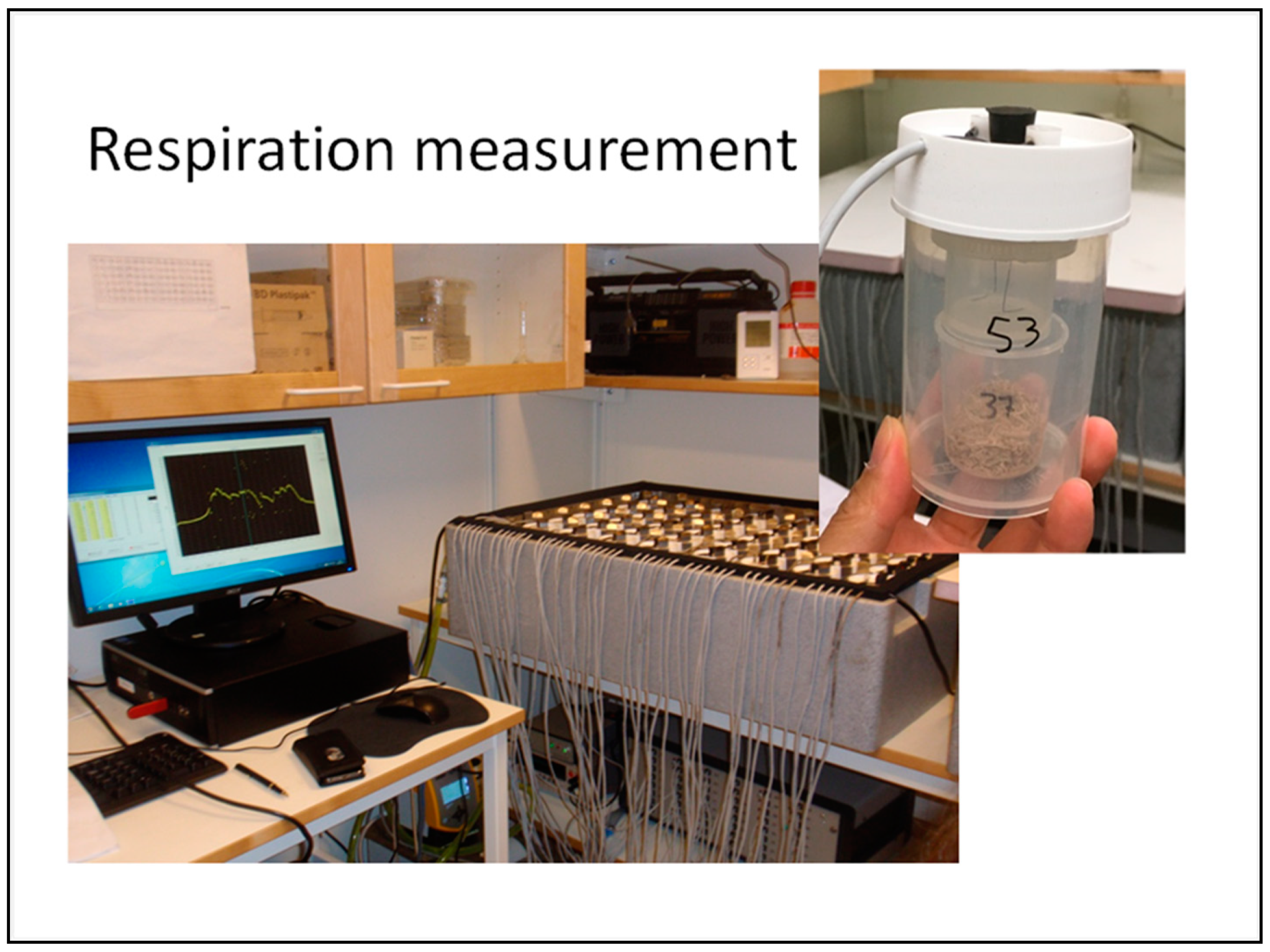
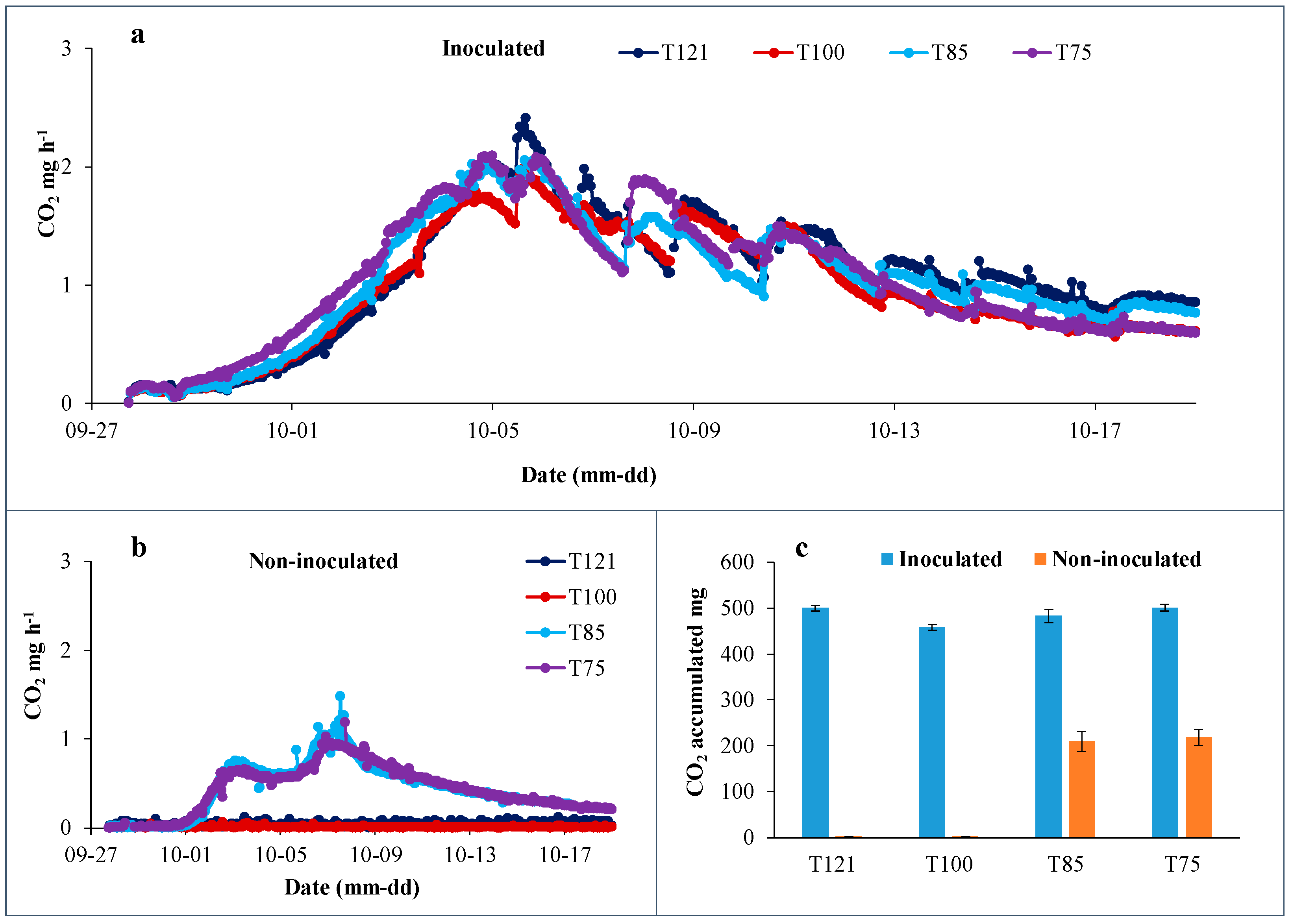
2.2. Microbial Incubation and Respiration Measurement

The Respicond respirometer [15] was used to incubate and measure the microbial respiration rate during a period of 24 days. The respiration rate was quantified by CO2 released from microbial activities that was trapped in KOH solution, which could be calculated by measuring the change in conductance of the KOH traps at regular intervals. The equipment has been previously adapted to soil microbiological studies, which allowed multiple samples (up to 96, one in each vessel), to be measured simultaneously and automatically. Thus, an incubation of microbes in the respirometer allowed a digital measurement of microbial growth. The polypropylene substrate cups (without lid), filled with substrates after inoculation, were randomly distributed inside pre-sterilized vessels (8 cm in diameter and 10 cm in height) of the Respicond respirator (Figure 1). All vessels were placed in a common water bath at 20 °C to maintain homogeneous and constant incubation temperature. Care was taken to avoid external microbial contaminations when handling substrates and inoculation as well as setting up vessels.
The respiration ratio was registered hourly. On 24 days of incubation, when CO2 emission curves became leveled out and stable, every individual substrate together with the cup was entirely removed from the respirator and lyophilized immediately using Christ Freeze Dryer (Martin Christ, Germany). After drying, each substrate was individually sealed and kept in the same cup and stored at room temperature before further analysis. Accumulated CO2 was quantified by summing up hour-ratios of CO2 emission (i.e., respiration ratio), using the formula:
A c c u m l a t e d   C O 2 = d = 1 24 h = 1 24 C O 2 ( m g h 1 )
where d = day and h = hour.

2.3. Profiling of Microbial Community Structure and Composition

Two different methods were used to characterize the microbial communities in a selection of the different experiments as depicted in Table 1: phospholipid fatty acid (PLFA) analysis and DNA sequencing of the overall microbial taxonomy.
PLFA analysis. Phospholipid fatty acid (PLFA) analysis was performed to characterize the structure of the microbial community in the different substrates after 24 days of incubation in the respirator. PLFA has been used for analyzing structure and activities of microbial communities in soils [16] and in composts [7,17], since PLFAs can serve as molecular markers of a selection of groups of living microbes. The samples of lyophilized substrates from Respicond trials, 0.5 g of each replicate, were analyzed on a gas chromatograph from Perkin Elmer with a FID detector and an Elite-5MS column (L 30 m ID 0.25 DF 0.25). For each substrate, the abundance of each fatty acid extracted was expressed as relative nmoles per g of dry substrate mass.
The identification of microbial groups depending on their PLFA profile pattern followed the classification system developed by Frostegård and Bååth [16] and Kroppenstedt [18]. The PLFAs i15:0, a15:0, 15:00, i16:0, 16:1w9, 16:1w7t, 16:1w7c, i17:0, a17:0, cy17:0, 18:1w7, and cy19:0 were used to indicate and calculate the sum of relative bacterial biomass, 18:2w6 was for fungal biomass, and 10Me17:0 and 10Me18:0 were for actinomycete biomass. PLFAs of 16:0, 17:0, and 18:0, previously used as bacterial biomass markers and 16:1w5 and 18:1w9 as fungal biomarkers [19], were not included in the current analysis as they were found in shiitake [20] or other organisms [21]. The PLFAs i-15:0, a-15:0, i-16:0, i-17:0 and a17:00 were included in the calculations for the sum of Gram-positive bacteria, while the PLFAs 16:1w9, 16:1w7c, 16:w7t and 18:1w7 were used for sum of Gram-negative bacteria.
DNA sequencing of the microbial communities. To identify fungal and bacterial species that co-existed in the different substrates after 24 days incubation in the Respicond, DNA sequencing on the lyophilized substrates was performed by the Next-Generation sequencing company Novagene; Hong Kong (https://en.novogene.com/, accessed on 15 May 2023). Four individual samples of each treatment were pooled proportionally (20% each), under a sterile hood, into one mixed sample that was then used for DNA sequencing. For elucidation of the phylogenetic affiliation, 16S rRNA analysis was performed for bacterial species and ITS18S rRNA analysis for fungal species. 16S rRNA/18SrRNA/ITS genes of distinct regions (16SV4/16SV3/16SV3-V4/16SV4-V5, 18S V4/18S V9, ITS1/ITS2, Arc V4) were amplified using specific primer pairs (e.g., 16S V4: 515F-806R, 18S V4: 528F-706R, and 18S V9: 1380F-1510R) with sequencing barcodes. All PCR reactions were carried out with the Phusion® High-Fidelity PCR Master Mix (New England Biolabs, Ipswich, MA, USA). The detailed procedures for DNA extraction, amplification, amplicon library and sequencing can be found in Novogene’s website (https://en.novogene.com/services/research-services/metagenome-sequencing/16s-18s-its-amplicon-metagenomic-sequencing/, accessed on 15 May 2022). The amplicon sequencing data were deposited at the European Nucleotide Archive (ENA, https://www.ebi.ac.uk/ena/browser/home, accessed on 31 May 2022) under the accession PRJEB53156.

2.4. Data Analysis

ANOVA followed by post hoc multiple comparisons (Tukey’s test) was conducted to analyze the differences between the heat treatments and between inoculated and non-inoculated substrates in microbial respiration (accumulation CO2) and biomass (PLFA) using SPSS statistical analysis software (IBM SPSS version 24.0).

3. Results

3.1. Microbial Activities in Shiitake Substrates

After 24 days of incubation in the respirator “Respicond”, a full colonization of shiitake mycelia was observed in all inoculated substrates. There was a significant difference between inoculated and non-inoculated substrates in both respiration ratios, measured as CO2 mg/h (Figure 2a,b), and accumulated CO2 release (Figure 2c). It was observed that T85 and T75 had a couple of days earlier start of colonization than T100 and T121, which is consistent to the pattern of CO2 emission during the first week (Figure 2a). However, CO2 emissions from high-temperature treatment especially T121 exhibited at a higher ratio than T85 and T75. No significant differences were observed for accumulated CO2 during incubation of 24 days between the heat treatments for the inoculated substrates. In non-inoculated substrates, considerable microbial respiration was measured with treatments of 75 °C and 85 °C in contrast to the treatments of 100 °C and 121 °C that resulted in negligible CO2 release. This suggests that the high-temperature treatments remained relatively sterile during the entire incubation period. Therefore, non-inoculated T100 and T121 are not included in the following discussion on compositions of the microbial communities.

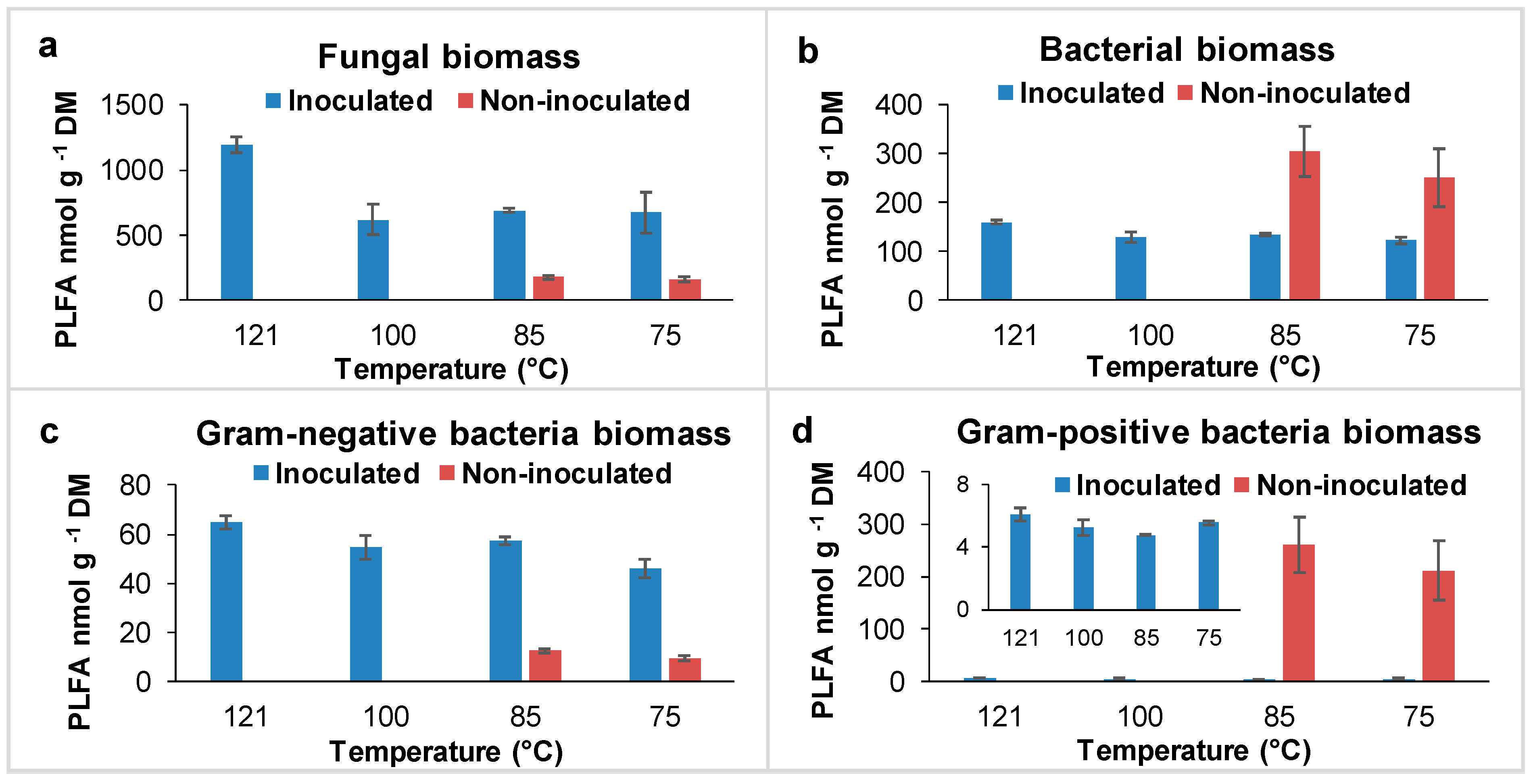
3.2. Microbial Community Structure Profile Based on PFLA Studies

PLFA analysis indicated that all four inoculated substrates and two non-inoculated substrates contained both fungi and bacteria (Figure 3a–d). The treatments of inoculated versus non-inoculated affected the dominance/existence of the studied organism groups significantly (p < 0.001). Fungal species were expectedly dominant in inoculated substrates where shiitake mycelium was growing. Compared with the effects of inoculation, the effects of temperature for pasteurization on fungal biomass was minor and not significant (p > 0.05) within inoculated treatments (Figure 3a). In the non-inoculated substrates (thus not supposed to contain shiitake), the PLFA of other fungal species was found in similar quantity (161.84 ± 17.41 vs. 178.48 ± 12.72 nmol g −1 DM) in the substrates subjected to 75–85 °C pasteurization.
The bacterial biomass was also more affected by inoculation than pasteurization temperature. There were no significant differences (p > 0.05) in bacterial biomass between the heat treatments regardless the inoculated and non-inoculated substrates (Figure 3b). However, the bacterial biomass was significantly higher (p < 0.05) than the fungal biomass in the two non-inoculated substrates, where the ratios of fungi to bacteria were 0.61 and 0.69 for 75 °C and 85 °C pasteurizations, respectively. The growth of inoculated shiitake had an inhibitory effect on the bacteria as a whole, which may explain a blooming of bacteria in the non-inoculated and 75–85 °C pasteurized substrates.
Figure 3c,d demonstrate the observed changes in the subgroups of Gram-negative and Gram-positive bacteria in different treatments. In the inoculated treatments, the weight of Gram-negative bacteria was >10 times higher (p < 0.001) than that of Gram-positive species. In contrast, the Gram-positive bacteria dominated in the non-inoculated and 75–85 °C pasteurized substrates.

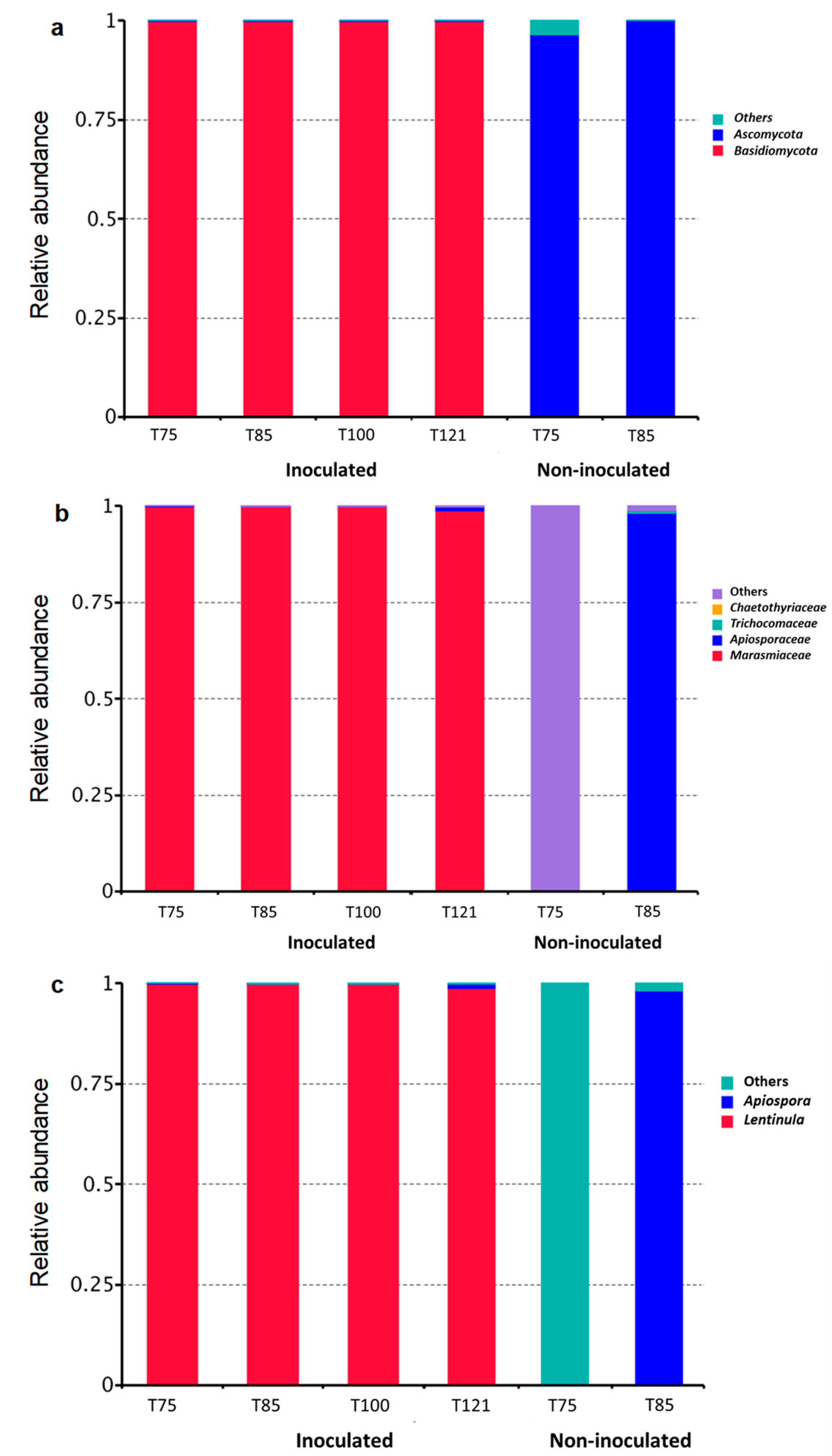
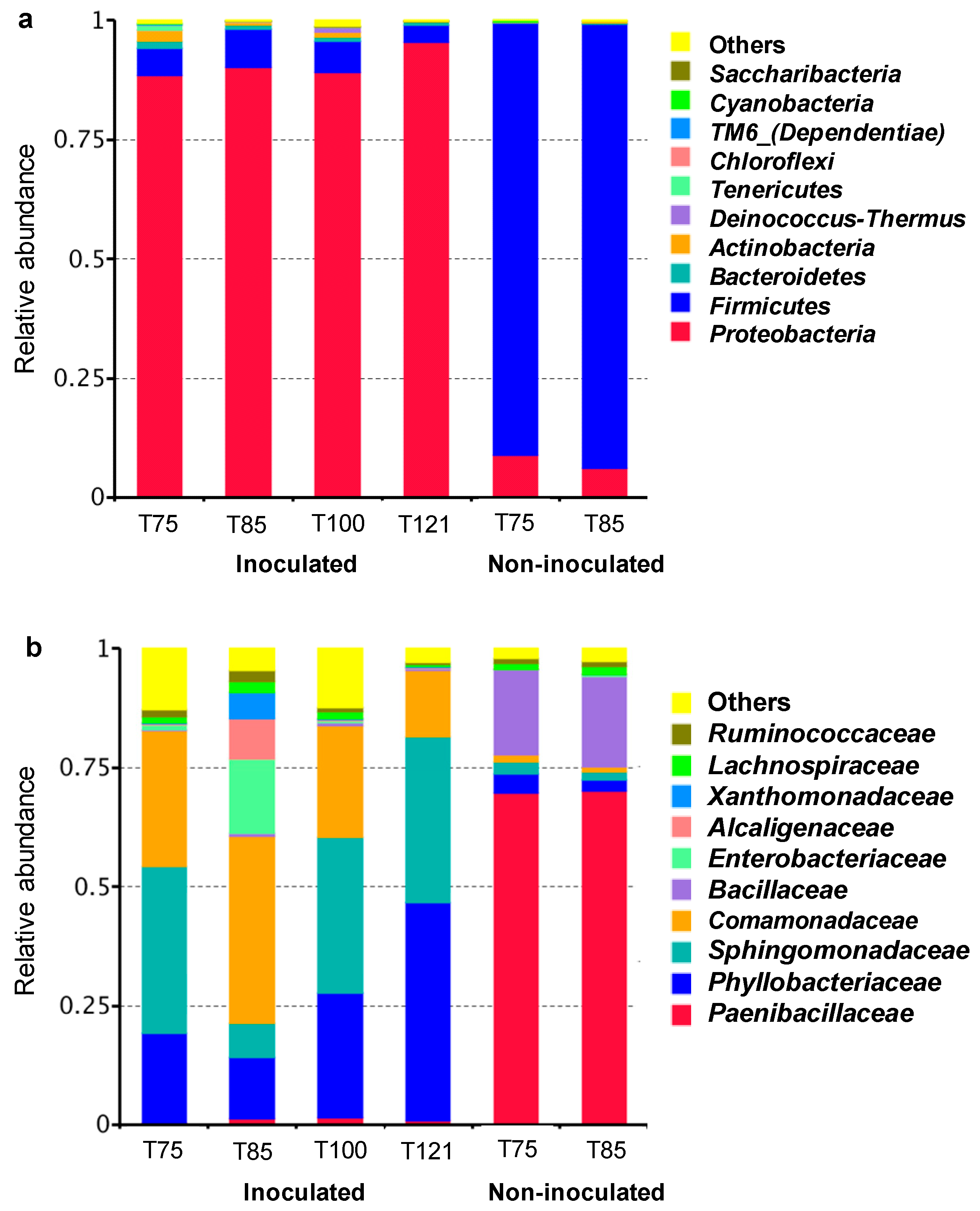
3.3. Microbial Community Composition Based on DNA Sequencing

Fungal taxa in different substrates. The DNA sequencing (targeting the ITS within the 18S ribosomal RNA (rRNA) gene) generated 17 different fungal taxa in total from 6 treatments. Seven of the 17 taxa were not identifiable by blasting trials. The remaining 10 strains showed significant affiliations to two fungal phyla: Basidiomycota (Figure 4a) in the inoculated treatments and Ascomycota in non-inoculated ones of 75 °C and 85 °C. The two phyla covered 87.21% of all retrieved gene sequences across all six treatments. Four families and two genera could be ascertained (Figure 4b,c) by blast hits, representing 83.09% and 82.99% of all sequenced strains, respectively. The taxa that could not be identified with regard to the genus or family taxonomical level are shown as “others” in Figure 4. L. edodes was identified as the sole species of genus Lentinula in the inoculated substrates, while Apiospora montagnel represented the only species of Apiospora in both inoculated and non-inoculated substrates.
Bacterial taxa in different substrates. Across all 6 treatments, more than 238 bacterial taxa affiliated to 23 phyla were detected in the substrates by DNA sequencing of the 16S rRNA genes. The top 10 phyla representing 99.69% of all sequences are listed in Figure 5a. The most dominant bacterial taxa in the inoculated substrates belonged to the phylum Proteobacteria. In the non-inoculated 75 °C and 85 °C treatments, however, the most dominant bacterial taxa belonged to the phylum Firmicutes.
Figure 5b shows 10 of the 126 detected bacterial families with the highest abundance of sequences across all 6 treatments. The top 10 families represent 93.77% of all bacterial sequences. It is evident that the abundant distribution of the dominant bacterial families in the substrates varied considerably with treatments. Six of the top ten families belonged to Proteobacteria phylum and were dominant in the inoculated substrates, while the other four families belonged to Firmicutes and dominated in non-inoculated substrates, where Paenibacillaceae had a >70% of relative abundance.
Further analysis on the species abundance of the top 10 dominant genera is demonstrated in Table 2, which covers at least 17 species and represents 89.71% of all bacterial taxa found across all treatments. A few phenomena are worth paying attention to. (1) For all 4 inoculated substrates, Phyllobacterium spp., Sphingomonas leidyi, Pelomonas spp. were the most dominant bacterial species among the top 10 genera found. (2) For the non-inoculated substrates, the dominant species were Cohnella xylanilytica, Paenibacillus favisporus and Bacillus spp. in 85 °C pasteurized substrate; while Paenibacillus spp., Bacillus circulans and Paenibacillus odorifer dominated in 75 °C pasteurized substrate. In contrast, these species showed a considerable low abundance in the inoculated substrates subjected to the same heat treatments of 75–85 °C, suggesting they might be inhibited by shiitake mycelia. (3) For 75–85 °C pasteurized substrates, the relative abundance of most of the seven genera/species affiliated to Proteobacteria, except for Achromobacter xylosoxidans at 75 °C, was higher in the shiitake inoculated substrate than in non-inoculated treatments; thus, it is likely that these bacteria were enhanced by shiitake growth. (4) Except for Bacillus subterraneus, all other 16 identified bacterial species (Table 2) were common in both inoculated and non-inoculated treatments, despite differences in abundance. This suggests that these 16 species could have an origin in the initial substrate ingredients, e.g., birch sawdust, wheat grain and bran. However, B. subterraneus showed an uncertainty of its origin. It could be still originated from substrate but become eliminated by stronger competitors in non-inoculated, although an alternative hypothesis of grain-spawn origin cannot be exclude by this study.

4. Discussion

4.1. The Microbial Community after Different Heat Treatments

After 24 days of incubation in a respirator at 20 °C, shiitake mycelia colonized well in all inoculated substrates (Figure 2a); however, a significant difference in growth speed could be observed. A faster growth (up to 3–4 days earlier) was observed in the 75–85 °C pasteurized substrates. The respiration patterns in the inoculated substrates (Figure 2a) and the non-inoculated substrates (Figure 2b) differed clearly. In the inoculated substrates there was a steady increase in carbon dioxide emission already during the first days. This pattern agrees well with the colonization of prepared inoculum of white-rot fungi (i.e., shiitake spawn) already adapted for growth on this type of lignocellulosic substrate. In the non-inoculated substrates, a lack of respiration was observed in the high-temperature treatments, evidently demonstrating that the sterilization treatments were applied successfully. For the low-temperature treatments, survival of some heat resistant bacteria (e.g., endospore-forming bacteria) can be expected and this agrees well with the fact that respiration is confirmed in these treatments even in the absence of shiitake inoculum. However, for most of the microorganisms, besides the white-rot fungi, lignocellulosic substrates are too complex to degrade [22]. Thus, as demonstrated in Figure 2b compared to Figure 2a, a longer lag-phase can be expected. Furthermore, the second lag-phase in Figure 2b suggests that a phenomenon similar to diauxic shift occurs. This shift may be explained by metabolization of more easily available carbohydrates in the first step followed by either a change in microbial community structure or induction of lignin-degrading enzymes in the existing microflora. Based on the PLFA analysis, high amounts of Gram-positive bacteria were observed in the non-inoculated low-temperature treatments after 24 days of growth (Figure 3d). This may suggest that the endospore-forming bacteria that survived the low temperature regimes also were successful in colonizing and metabolizing the lignocellulosic substrate. Our knowledge about bacterial degradation of lignocellulosic substrates is scarce, compared to the knowledge about the fungal degradation of these substrates, but recent research indicate that lignin degradation can be performed by specific bacterial consortia in different environments [22]. The general 16S rRNA gene sequence data presented in Table 2 suggest that the abundance of Bacillus spp. and Paenibacillus spp. are high in the non-inoculated treatments. Additionally, C. xylanilytica, a Gram-positive and endospore-producing species, belonging to the family Paenibacillaceae, was observed. None of the bacteria, which were tentatively identified to species level in these treatments (B. circulans, P. favisporus, P. odorifer and C. xylanilytica), have so far been recognized for metabolization of lignin. However, some Paenibacillus spp. and Bacillus spp. are known for the production of lignin-degrading and lignin-modifying enzymes [23,24] and several of the identified species are known to be able to degrade hemicellulose.
The accumulated CO2 from microbial respiration (Figure 2c) and PLFA of total fungal and bacterial biomass (Figure 3a,b) in the inoculated media were not significantly different between the pasteurization regimes by the end of 24 days. As the PLFA can generally only be found in living microbial cells, it is evident that many bacteria and few actinomycetes coexisted with shiitake in the growth media, but they did not seem to impair shiitake colonization. However, the heating temperatures had a significant effect on some co-existing microbes; for example, significantly more Gram-negative bacteria (p < 0.05) were found in the substrates subjected to high-temperature treatments (Figure 3c). As there was an absence of respiration in the non-inoculated controls of the high-temperature treatments, it can be expected that living cells or spores were rarely present in these treatments at the time of the spawning. Thus, the microbial community that developed might be derived from the spawn. This is supported by the fact that the identified major bacterial species and their relative abundance show high similarity between the two inoculated high-temperature treatments (Table 2). Spawn production is usually a very carefully controlled process, striving for complete sterility, as infection at this stage in practice may risk the whole production [25]. The substrate used for propagation of the spawn, often cereal grain, is generally subjected to prolonged autoclavation. However, since the fungal growth is not compromised, it cannot be ruled out if the fungus, which is added to the carefully sterilized substrate, may carry unrecognized symbiotic bacteria. A new experiment should be helpful to examine this hypothesis in a future study.

4.2. Coexisting Strains of Bacteria in Shiitake Substrates

DNA sequencing of the whole microbial communities demonstrated that Phyllobacterium spp., S. leidyi and Pelomonas spp., all belonging to the Gram-negative Proteobacteria phylum, were the most dominant bacterial species in the inoculated substrates where shiitake was the sole dominant fungal species, across all heating treatments. These dominant bacterial species had a much higher relative abundance in the substrates with growing shiitake than without, although there were differences to some extent between the pasteurization temperatures. These traits hint at plausible habitat-specific symbionts and the potential conferred by the microbial genetic makeup. These bacterial species are distributed in diverse ecosystems ranging from the harsh environment to eco-friendly for growth. Our survey of current literature (Table 3) suggests broader beneficial associations of plants with different species of the Phyllobacterium spp., Sphingomonas spp. and Pelomonas spp. These bacteria, especially Phyllobacterium spp., have been reported for their significant roles in plant growth promotion (reference in Table 3).
Studies have shown that Phyllobacterium spp. are endophytic species that reside within plant tissues [26]. It is widely considered as rhizobacteria, which possess numerous stress tolerant genes, and are adapted to various environmental conditions. Rhizobacteria are well known for their role in promoting plant growth and metabolism. The several beneficial rhizospheric and endophytic bacterial communities improve adaptation and resilience under changing conditions and promote sustainable systems. The study also revealed that Phyllobacterium sp. appears to be linked with a genus of N2-fixing plant-growth-promoting bacteria [27]. They could establish their role as an N2-fixing organisms in the roots of land plants and mangrove rhizosphere [28,29,30]. The recent study performed by Makino et al. [31] on Pelomonas strains MRB1 and MRB3 as the first report on plant growth promoting bacteria (PGPB) displayed a strong symbiotic effect concerning the growth, biomass and chlorophyll content of Lemna minor. Asaf et al. [32] demonstrated a symbiotic relationship between Sphingomonas and plant roots, combined with a fungal association, which promotes the ability to avoid stress and degrades pollutants. Interestingly, S. leidyi was described as an endosymbiont harboured in fungus Stachylidium bicolor mycelium [33]. All these characteristics are similar to those bacterial promoters of mycelia growth of, e.g., P. ostreatus and A. bisporus [12,13,14]. However, no definitive conclusion can be drawn yet from the literature survey on potential symbiotic relationship between most of these species strains and a fungus (Table 3), whilst, S. leidyi seems to be the only one that was identified as an endosymbiont in fungal host.
The DNA sequencing showed that Gram-negative bacteria Proteobacteria dominated in the inoculated substrate samples. This is in agreement with the general PLFA analyses (Figure 3c). Plausibly, some of these dominant Gram-negative species (e.g., Phyllobacterium spp., S. leidyi and Pelomonas spp.) in the inoculated substrates might therefore be involved in some kind of symbiotic relationship with shiitake at least during the stage of mycelial colonization, keeping in mind that PLFA can be measured generally only in living organisms. However, for the time being our current data do not allow a deeper interpretation on the role of the identified taxa based on these two methods. There could be inherent biases and discrepancies between DNA-based and PFLA-based databases, due to the fact that DNA based studies can also include dead microbes, since some DNA fractions of dead bacteria may not be completely degraded under high temperature [57,58]. Furthermore, 16S rRNA gene targets are too conservative to allow further speculation about the function of sequenced organisms. This study represents a preliminary attempt to determine the existence of bacterial species within birch-based substrates of shiitake, revealing new information on fungal-bacterial community in shiitake substrates. To complement this information, further studies should be conducted to distinguish the bacterial composition, functions and behavior of these dominant bacterial growths in the presence of shiitake. Additionally, these co-culture models will provide clear insights into microbial dynamics changes in fungi and bacteria relationships.

4.3. Implication for Biorefinery Development

Increased mushroom production holds promise for circular and sustainable production of protein and is of importance for developing a sustainable biobased society. The findings reported in the present study are of interest considering that the common practice in shiitake production is often high-temperature treatment of the substrate before inoculating the spawn. However, the pretreatment of the substrate in high temperature is energy demanding; decreasing this demand will inevitably increase sustainability in production. Our previous study [2,4] demonstrated that low-temperature treatment of the substrate resulted in earlier fructification and higher or comparable yield of shiitake, as well as a similar degree of substrate degradation, compared to the more energy demanding steam sterilization of the substrate. The low temperature hot air pasteurization is apparently beneficial even when considering the potential for developing a circular production system and reusing the substrate for biofuel production. Thus, to understand the mechanisms behind this viable method and increase knowledge on microbial interactions in the mushroom substrate may assist in optimizing mushroom production and consequent biorefinery using spent mushroom substrate.

Author Contributions

S.X. concepted and managed the project and together with L.E. designed Experiment. L.E., M.S., M.H. and F.C. performed and involved variable analysis work. S.X., M.H. and N.L. involved drafting the manuscript. P.S. performed literature survey and reviews. N.L. and M.S. provided comments for manuscript writing. All authors have read and agreed to the published version of the manuscript.

Funding

This research was co-financed by the Swedish State Department of Innovation (VINNOVA), Swedish State Energy Agency and Swedish Research Council (FORMAS) (2013-02015, 2016-05104, 2017-02705, P37816, P42481, P41845), through national research program BioInnovation and Re:Source. The fundings from Kempestiftelserna (JCK-2125, JCK22-0028) and Jordbruksverket (2019-3031) are appreciated.

Data Availability Statement

The amplicon sequencing data on metagenome were deposited at the European Nucleotide Archive (ENA, https://www.ebi.ac.uk/ena/browser/home) under the accession PRJEB53156.

Acknowledgments

The authors would like to thank Anders Nordgren for guidance for using the Respicond system, Jonas Lundholm at SLU Department of Forest Ecology and Management for performing PLFA analysis, and Carina Jonsson and Maogui Wei at SLU Department of Forest Biomaterials and Technology for helping with variable lab work. The authors are grateful to Nicolas Delhomme and Judith Lundberg-Felten, Umeå Plant Science Center, for their helps for data depositions of the DNA sequencing.

Conflicts of Interest

The authors declare no conflict of interest.

References

  1. Lin, Y.; Ge, X.; Liu, Z.; Li, Y. Integration of shiitake cultivation and solid-state anaerobic digestion for utilization of wood biomass. Bioresour. Technol. 2015, 182, 128–135. [Google Scholar] [CrossRef]
  2. Xiong, S.J.; Martín, C.; Eilertsen, L.; Wei, M.; Myronycheva, O.; Larsson, S.; Lestander, T.; Atterhem, L.; Jönsson, L. Energy-efficient substrate pasteurisation for combined production of shiitake mushroom (Lentinula edodes) and bioethanol. Bioresour. Technol. 2019, 274, 65–72. [Google Scholar] [CrossRef]
  3. Wan, C.X.; Li, Y.B. Fungal pretreatment of lignocellulosic biomass. Biotechnol. Adv. 2012, 30, 1447–1457. [Google Scholar] [CrossRef] [PubMed]
  4. Wei, M.; Xiong, S.J.; Chen, F.; Geladi, P.; Eilertsen, L.; Myronycheva, O.; Lestander, T.A.; Thyrel, M. Energy smart hot-air pasteurisation as effective as energy intense autoclaving for fungal preprocessing of lignocellulose feedstock for bioethanol fuel production. Renew. Energy 2020, 155, 237–247. [Google Scholar] [CrossRef]
  5. Fellows, P.J. Food Processing Technology Principles and Practice. In Food Science, Technology and Nutrition, 5th ed.; Woodhead Publishing: Cambridge, UK, 2017; pp. 343–383. [Google Scholar]
  6. Vieira, F.R.; Pecchia, J.A. An exploration into the bacterial community under different pasteurization conditions during substrate preparation (compostingphase II) for Agaricus bisporus cultivation. Microb. Ecol. 2018, 75, 318–330. [Google Scholar] [CrossRef] [Green Version]
  7. Carrasco, J.; García-Delgado, C.; Lavega, R.; Tello, M.L.; De Toro, M.; Barba-Vicente, V.; Rodríguez-Cruz, M.S.; Sánchez-Martín, M.J.; Pérez, M.; Preston, G.M. Holistic assessment of the microbiome dynamics in the substrates used for commercial champignon (Agaricus bisporus) cultivation. Microb. Biotechnol. 2020, 13, 1933–1947. [Google Scholar] [CrossRef] [PubMed]
  8. Kong, W.L.; Sun, B.; Zhang, J.Y.; Zhang, Y.T.; Gu, L.K.; Bao, L.J.; Liu, S.X. Metagenomic analysis revealed the succession of microbiota and metabolic function in corncob composting for preparation of cultivation medium for Pleurotus ostreatus. Bioresour. Technol. 2020, 306, 123156. [Google Scholar] [CrossRef] [PubMed]
  9. Wu, S.; Zhou, R.; Ma, Y.; Fang, Y.; Xie, G.P.; Gao, X.Z.; Xiao, Y.Z.; Liu, J.J.; Fang, Z.M. Development of a consortium-based microbial agent beneficial to composting of distilled grain waste for Pleurotus ostreatus cultivation. Biotechnol. Biofuels 2021, 14, 242. [Google Scholar] [CrossRef]
  10. Lee, C.K.; Haque, M.A.; Choi, B.R.; Lee, H.Y.; Eun, C. Molecular diversity of endobacterial communities in edible part of king oyster mushroom (Pleurotus eryngii) based on 16S rRNA. Korean J. Microbiol. 2015, 51, 148–155. [Google Scholar] [CrossRef] [Green Version]
  11. Chen, L.; Yan, M.; Qian, X.; Yang, Z.; Xu, Y.; Wang, T.; Cao, J.; Sun, S. Bacterial community composition in the growth process of Pleurotus eryngii and growth-promoting abilities of isolated bacteria. Front. Microbiol. 2022, 13, 787628. [Google Scholar] [CrossRef]
  12. Carrasco, J.; Preston, G.M. Growing edible mushrooms: A conversation between bacteria and fungi. Environ. Microbiol. 2020, 22, 858–872. [Google Scholar] [CrossRef] [PubMed] [Green Version]
  13. Torres-Ruiz, E.; Sánchez, J.E.; Guillén-Navarro, G.K.; Ramos-Pérez, D.G.; Royse, D.J. Microbial promoters of mycelial growth: Fruiting and production of Pleurotus ostreatus. Sydowia 2016, 68, 151–161. [Google Scholar]
  14. Kertesz, M.A.; Thai, M. Compost bacteria and fungi that influence growth and development of Agaricus bisporus and other commercial mushrooms. Appl. Microbiol. Biotechnol. 2018, 102, 1639–1650. [Google Scholar] [CrossRef]
  15. Nordgren, A. Apparatus for the continuous, long-term monitoring of soil respiration rate in large number of samples. Soil Biol. Biochem. 1988, 20, 955–957. [Google Scholar] [CrossRef]
  16. Frostegård, Å.; Bååth, E. The use of phospholipid analysis to estimate bacterial and fungal biomass in soil. Biol. Fertil. Soils 1996, 22, 59–65. [Google Scholar] [CrossRef]
  17. Steger, K.; Jarvis, A.; Smårs, S.; Sundh, I. Comparison of signature lipid methods to determine microbial community structure in compost. J. Microbiol. Methods 2003, 55, 371–382. [Google Scholar] [CrossRef] [PubMed]
  18. Kroppenstedt, R.M. Fatty acid menaquinone analysis of actinomycetes and related organisms. In Chemical Methods in Bacterial Systematics; Goodfellow, M., Minnikin, D.E., Eds.; Academic Press: New York, NY, USA, 1985; pp. 173–199. [Google Scholar]
  19. Willers, C.; Jansen van Rensburg, P.; Claassens, S. Phospholipid fatty acid profiling of microbial communities—A review of interpretations and recent applications. J. Appl. Microbiol. 2015, 119, 1207–1218. [Google Scholar] [CrossRef]
  20. Chung, I.M.; Kim, S.Y.; Han, J.G.; Kong, W.S.; Jung, M.Y.; Kim, S.H. Fatty acids and stable isotope ratios in shiitake mushrooms (Lentinula edodes) indicate the origin of the cultivation substrate used: A preliminary case study in Korea. Foods 2020, 9, 1210. [Google Scholar] [CrossRef]
  21. Frostegård, Å.; Tunlid, A.; Bååth, E. Use and misuse of PLFA measurements in soils. Soil Biol. Biochem. 2011, 43, 1621–1625. [Google Scholar] [CrossRef]
  22. Silva, J.P.; Ticona, A.R.P.; Hamann, P.R.V.; Quirino, B.F.; Noronha, E.F. Deconstruction of lignin: From enzymes to microorganisms. Molecules 2021, 26, 2299. [Google Scholar] [CrossRef]
  23. Grady, E.N.; MacDonald, J.; Liu, L.; Richman, A.; Yuan, Z.C. Current knowledge and perspectives of Paenibacillus: A review. Microb. Cell Factories 2016, 15, 203. [Google Scholar] [CrossRef] [PubMed] [Green Version]
  24. Srinivasan, P.; Selvankumar, T.; Kamala-Kannan, S.; Mythili, R.; Sengottaiyan, A.; Govarthanan, M.; Senthilkumar, B.; Selvam, K. Production and purification of laccase by Bacillus spp. using millet husks and its pesticide degradation application. 3 Biotech 2019, 9, 396. [Google Scholar] [CrossRef] [PubMed]
  25. Sánchez, C. Cultivation of Pleurotus ostreatus and other edible mushrooms. Appl. Microbiol. Biotechnol. 2010, 85, 1321–1337. [Google Scholar] [CrossRef] [PubMed]
  26. Shi, Y.; Zhang, X.; Lou, K.; Adams, M. Isolation, characterization and insecticidal activity of an endophyte of drunken horse grass: Achnatherum inebrians. J. Insect Sci. 2013, 13, 1. [Google Scholar] [CrossRef] [Green Version]
  27. Ettinger, C.L.; Eisen, J.A. Fungi, bacteria and oomycota opportunistically isolated from the seagrass, Zostera marina. PLoS ONE 2020, 15, e0236135. [Google Scholar] [CrossRef]
  28. Mantelin, S.; Saux, M.F.; Zakhia, F.; Bena, G.; Bonneau, S.; Jeder, H.; de Lajudie, P.; Cleyet-Marel, J.C. Emended description of the genus Phyllobacterium and description of four novel species associated with plant roots: Phyllobacterium bourgognense sp. nov., Phyllobacterium ifriqiyense sp. nov., Phyllobacterium leguminum sp. nov. and Phyllobacterium brassicacearum sp. nov. Int. J. Syst. Evol. Microbiol. 2006, 56, 827–839. [Google Scholar]
  29. Holguin, G.; Guzman, M.A.; Bashan, Y. Two new nitrogen-fixing bacteria from the rhizosphere of mangrove trees: Their isolation, identification and in vitro interaction with rhizosphere Staphylococcus sp. FEMS Microbiol. Lett. 1992, 101, 207–216. [Google Scholar] [CrossRef]
  30. Rojas, A.; Holguin, G.; Glick, B.R.; Bashan, Y. Synergism between Phyllobacterium sp. (N2-fixer) and Bacillus licheniformis (P-solubilizer), both from a semiarid mangrove rhizosphere. FEMS Microbiol. Ecol. 2001, 35, 181–187. [Google Scholar] [CrossRef]
  31. Makino, A.; Nakai, R.; Yoneda, Y.; Toyama, T.; Tanaka, Y.; Meng, X.Y.; Mori, K.; Ike, M.; Morikawa, M.; Kamagata, Y.; et al. Isolation of aquatic plant growth-promoting bacteria for the floating plant duckweed (Lemna minor). Microorganisms 2022, 10, 1564. [Google Scholar] [CrossRef]
  32. Asaf, S.; Numan, M.; Khan, A.L.; Al-Harrasi, A. Sphingomonas: From diversity and genomics to functional role in environmental remediation and plant growth. Crit. Rev. Biotechnol. 2020, 40, 138–152. [Google Scholar] [CrossRef]
  33. Almeida, C.; Silva Pereira, C.; Gonzalez-Menendez, V.; Bills, G.; Pascual, J.; Sánchez-Hidalgo, M.; Kehraus, S.; Genilloud, O. Unveiling concealed functions of endosymbiotic bacteria harbored in the ascomycete Stachylidium bicolor. Appl. Environ. Microbiol. 2018, 84, e00660-18. [Google Scholar] [CrossRef] [Green Version]
  34. Jiao, Y.S.; Yan, H.; Ji, Z.J.; Liu, Y.H.; Sui, X.H.; Zhang, X.X.; Wang, E.T.; Chen, W.X.; Chen, W.F. Phyllobacterium sophorae sp. nov.; a symbiotic bacterium isolated from root nodules of Sophora flavescens. Int. J. Syst. Evol. Microbiol. 2015, 65, 399–406. [Google Scholar] [CrossRef]
  35. Larcher, M.; Muller, B.; Mantelin, S.; Rapior, S.; Cleyet-Marel, J.C. Early modifications of Brassica napus root system architecture induced by a plant growth-promoting Phyllobacterium strain. New Phytol. 2003, 160, 119–125. [Google Scholar] [CrossRef] [PubMed]
  36. Flores-Félix, J.D.; Velázquez, E.; García-Fraile, P.; González-Andrés, F.; Silva, L.R.; Rivas, R. Rhizobium and Phyllobacterium bacterial inoculants increase bioactive compounds and quality of strawberries cultivated in field conditions. Food Res. Int. 2018, 111, 416–422. [Google Scholar] [CrossRef] [PubMed]
  37. Bresson, J.; Varoquaux, F.; Bontpart, T.; Touraine, B.; Vile, D. The PGPR strain Phyllobacterium brassicacearum STM 196 induces a reproductive delay and physiological changes that result in improved drought tolerance in Arabidopsis. New Phytol. 2013, 200, 558–569. [Google Scholar] [CrossRef]
  38. Kechid, M.; Desbrosses, G.; Gamet, L.; Castaings, L.; Varoquaux, F.; Djekoun, A.; Touraine, B. Arabidopsis growth-promotion and root architecture responses to the beneficial rhizobacterium Phyllobacterium brassicacearum strain STM196 are independent of the nitrate assimilatory pathway. Plants 2022, 11, 128. [Google Scholar] [CrossRef] [PubMed]
  39. Valverde, A.; Velázquez, E.; Fernández-Santos, F.; Vizcaino, N.; Rivas, R.; Mateos, P.F.; Martínez-Molina, E.; Igual, J.M.; Willems, A. Phyllobacterium trifolii sp. nov.; nodulating Trifolium and Lupinus in Spanish soils. Int. J. Syst. Evol. Microbiol. 2005, 55, 1985–1989. [Google Scholar] [CrossRef] [PubMed] [Green Version]
  40. Khan, M.A.; Yadav, S.; Sharma, R.; Dalela, M.; Celin, S.M.; Sharma, A.; Sharma, S. Augmentation of stimulated Pelomonas aquatica dispersible granules enhances remediation of hexahydro-1, 3, 5-trinitro-1, 3, 5-triazine (RDX) contaminated soil. Environ. Technol. Innov. 2020, 17, 100594. [Google Scholar] [CrossRef]
  41. Somal, M.K.; Karnwal, A. Effect of stress tolerance endophytic bacteria on the growth of Rheum emodi under abiotic stress. Plant Biol. J. 2022. [Google Scholar] [CrossRef]
  42. Doudoroff, M. The oxidative assimilation of sugars and related substances by Pseudomonas saccharophila with a contribution to the problem of the direct respiration of di-and polysaccharides. Enzymologia 1940, 9, 59–72. [Google Scholar]
  43. Nagar, S.; Shaw, A.K.; Anand, S.; Celin, S.M.; Rai, P.K. Biodegradation of octogen and hexogen by Pelomonas aquatica strain WS2-R2A-65 under aerobic condition. Environ. Technol. 2020, 43, 1003–1012. [Google Scholar] [CrossRef]
  44. Gomila, M.; Bowien, B.; Falsen, E.; Moore, E.R.; Lalucat, J. Description of Pelomonas aquatica sp. nov. and Pelomonas puraquae sp. nov., isolated from industrial and haemodialysis water. Int. J. Syst. Evol. Microbiol. 2007, 57, 2629–2635. [Google Scholar] [CrossRef] [PubMed]
  45. Abraham, W.R.; Estrela, A.B.; Rohde, M.; Smit, J.; Vancanneyt, M. Prosthecate sphingomonads: Proposal of Sphingomonas canadensis sp. nov. Int. J. Syst. Evol. Microbiol. 2013, 63, 3214–3219. [Google Scholar] [CrossRef] [Green Version]
  46. Willison, J.C. Isolation and characterization of a novel sphingomonad capable of growth with chrysene as sole carbon and energy source. FEMS Microbiol. Lett. 2004, 241, 143–150. [Google Scholar] [CrossRef] [Green Version]
  47. Wittich, R.M.; Wilkes, H.; Sinnwell, V.; Francke, W.; Fortnagel, P. Metabolism of dibenzo-p-dioxin by Sphingomonas spp. strain RW1. Appl. Environ. Microbiol. 1992, 58, 1005–1010. [Google Scholar] [CrossRef] [PubMed]
  48. Ashtaputre, A.A.; Shah, A.K. Studies on a viscous, gel-forming exopolysaccharide from Sphingomonas paucimobilis GS1. Appl. Environ. Microbiol. 1995, 61, 1159–1162. [Google Scholar] [CrossRef] [PubMed]
  49. Tangaromsuk, J.; Pokethitiyook, P.; Kruatrachue, M.; Upatham, E.S. Cadmium biosorption by Sphingomonas paucimobilis biomass. Bioresour. Technol. 2002, 85, 103–105. [Google Scholar] [CrossRef] [PubMed]
  50. Li, D.; Feng, L.; Liu, K.; Cheng, Y.; Hou, N.; Li, C. Optimization of cold-active CMCase production by psychrotrophic Sphingomonas sp. FLX-7 from the cold region of China. Cellulose 2016, 23, 1335–1347. [Google Scholar] [CrossRef]
  51. Halo, B.A.; Khan, A.L.; Waqas, M.; Al-Harrasi, A.; Hussain, J.; Ali, L.; Adnan, M.; Lee, I.J. Endophytic bacteria (Sphingomonas sp. LK11) and gibberellin can improve Solanum lycopersicum growth and oxidative stress under salinity. J. Plant. Interact. 2015, 10, 117–125. [Google Scholar] [CrossRef] [Green Version]
  52. Yang, S.; Zhang, X.; Cao, Z.; Zhao, K.; Wang, S.; Chen, M.; Hu, X. Growth-promoting Sphingomonas paucimobilis ZJSH 1 associated with Dendrobium officinale through phytohormone production and nitrogen fixation. Microb. Biotechnol. 2014, 7, 611–620. [Google Scholar] [CrossRef]
  53. Luo, Y.; Wang, F.; Huang, Y.; Zhou, M.; Gao, J.; Yan, T.; Sheng, H.; An, L. Sphingomonas sp. Cra20 increases plant growth rate and alters rhizosphere microbial community structure of Arabidopsis thaliana under drought stress. Front. Microbiol. 2019, 10, 1221. [Google Scholar] [CrossRef] [PubMed] [Green Version]
  54. Videira, S.S.; De Araujo, J.L.S.; da Silva Rodrigues, L.; Baldani, V.L.D.; Baldani, J.I. Occurrence and diversity of nitrogen-fixing Sphingomonas bacteria associated with rice plants grown in Brazil. FEMS Microbiol. Lett. 2009, 293, 11–19. [Google Scholar] [CrossRef] [Green Version]
  55. Tsavkelova, E.A.; Egorova, M.A.; Leontieva, M.R.; Malakho, S.G.; Kolomeitseva, G.L.; Netrusov, A.I. Dendrobium nobile Lindl. seed germination in co-cultures with diverse associated bacteria. Plant Growth Regul. 2016, 80, 79–91. [Google Scholar] [CrossRef]
  56. Lowman, S.; Kim-Dura, S.; Mei, C.; Nowak, J. Strategies for enhancement of switchgrass (Panicum virgatum L.) performance under limited nitrogen supply based on utilization of N-fixing bacterial endophytes. Plant Soil 2016, 405, 47–63. [Google Scholar] [CrossRef]
  57. Karni, M.; Zidon, D.; Polak, P.; Zalevsky, Z.; Shefi, O. Thermal degradation of DNA. DNA Cell Biol. 2013, 32, 298–301. [Google Scholar] [CrossRef] [PubMed] [Green Version]
  58. Bauer, T.; Weller, P.; Hammes, W.P.; Hertel, C. The effect of processing parameters on DNA degradation in food. Eur. Food Res. Technol. 2003, 217, 338–343. [Google Scholar] [CrossRef]
Figure 1. Respicond experiment setting for the estimation of microbial respiration.
Fermentation 09 00736 g001
Figure 2. Ratio of microbial respiration in inoculated (a) and non-inoculated substrates (b); as well as accumulation (c) of CO2 released by microorganism respiration in substrates subjected to different heat treatments during the incubation of 24 days. Data are averages of four replicates for each treatment.
Fermentation 09 00736 g002
Figure 3. Microbial biomass (measured by phospholipid fatty acid (PLFA) analysis) in the substrates subjected to different heat treatments. Microbial groups were classified based on their PLFA-signatures (ad) and grouped based on whether the growth substrate was inoculated with Shiitake fungus or not.
Fermentation 09 00736 g003
Figure 4. Relative abundance of fungal species belonging to the top 10 fungal phyla (a), families (b) and genera (c) based on sequencing of the ITS 18S rRNA gene. The top 10 taxa in the different taxonomic ranks represent 87.21% of all detected fungal taxa.
Fermentation 09 00736 g004
Figure 5. Relative abundance of bacteria grouped in top 10 phyla (a) and top 10 families (b) in the substrates subjected to different treatments. Data are based on a 16S rRNA gene sequence analysis.
Fermentation 09 00736 g005
Table 1. Experimental set-ups and performed analyses.
Heat treatment T121: Steam 121 °C with 2 bar pressure
(sterilization/pasteurization)T100: Hot air 100 °C for 1 h
T85: Hot air 85 °C for 1 h
T75: Hot air 75 °C for 1 h
Shiitake inoculationInoculated vs. non-inoculated
IncubationAt 20 °C; in Respicond respirator
Measurement/analysisCO2 release ratio (mg/h); CO2 release accumulated (mg); Profile of microbial community structure and taxonomy by PLFA analysis; and ITS 18S and 16S rRNA gene sequencing.
Table 2. Survey of major bacterial species and their relative abundance (%) in the top 10 genera found in different treatments. Data are based on a 16S rRNA analysis.
InoculatedNon-Inoculated
PhylumClasssOrderFamilyGenusSpeciesT121T100T85T75T85T75
FirmicutesBacilliBacillalesBacillaceaeBacillusB. circulans0.200.350.310.110.4918.60
B. subterraneus<0.01<0.010.01<0.01
B. spp.0.180.320.250.0919.910.88
CohnellaC. xylanilytica0.300.440.480.1549.950.42
PaenibacillaceaePaenibacillusP. alginolyticus0.010.01<0.01<0.010.010.20
P. favisporus0.120.210.170.0920.963.28
P. lautus0.010.020.01<0.010.030.63
P. odorifer0.110.130.360.060.2312.02
P. spp. i760.010.010.01<0.010.011.48
P. spp.0.370.780.310.312.3653.49
Proteobacteriaα-proteobacteriaRhizobialesPhyllobacteriaceaePhyllobacteriumPh. Spp.47.5730.9414.3222.632.504.32
SphingomonadalesSphingomonadaceaeSphingomonasS. leidyi36.4038.87.7341.361.742.76
β-proteobacteriaBurkholdderialesAlcaligenaceaeAchromobacterA. xylosoxidans<0.010.099.180.030.040.04
ComamonadaceaeDelftiaD. spp.0.160.3926.110.170.060.04
PelomonasP. spp.14.2427.2117.4634.331.191.54
γ-proteobacteriaEnterobacterialesEnterobacteriaceaeSerratiaS. spp.0.100.1517.240.330.130.05
PseudomonasP. spp.0.090.076.020.150.030.03
Ratio of the identified ones to all bacterial sequences of the top 10 genera % 96.0184.2690.3682.9492.8291.84
Table 3. Literature survey of the studies on Phyllobacterium spp., Sphingomonas spp. and Pelomonas spp.
BacteriumHabitatSymbiosisMajor Finding/CharacteristicReference
Phyllobacterium spp.
Ph. spp. CLE16Root of Zostera marinaPGPBNitrogen fixing [27]
Ph. sophorae spp. CCBAU 3422Root nodule of Sophora flavescensPGPRNodule formation and nitrogen fixation[34]
Ph. spp. (29-15)Root of Brassica napusPGPBEnhanced root length [35]
Ph. (PEPV15)Legume nodules of Phaseolus vulgarisPGPBIncreased in citric acid. vitamin C. epicatechin and other bioactive compounds [36]
Ph. rubiacearumBlack, white, and red mangrovesPGPBCo-cultivation increased nitrogen fixation and phosphate solubilization.[30]
Ph. Brassicacearum STM196Root of Brassica napusPGPRIncreased in biomass. drought resistance and water-use efficiency [37]
Ph. brassicacearum STM196Roots of canola plantsPGPRStimulating growth[38]
Ph. strain PETP02Nodules of Trifolium pratensePGPRInduced nodules in roots[39]
Pelomonas spp.
P. aquatica strain 12868SoilRemediation of hexahydro-1.3.5-trinitro-1.3.5-triazine (RDX) [40]
P. spp. MRB1Roots of aquatic plants PGPBIncreased in growth. biomass and chlorophyll content [31]
P. spp. MRB3Roots of aquatic plants PGPBEnhanced in biomass. chlorophyll and high growth-promoting effect [31]
P. aquatica AIS1SAbutilon indicumPGPRSeed germination under salt and heavy metal stress conditions[41]
P. saccharophilaMudHydrogen-oxidizing bacterium[42]
P. aquatica strain WS2-R2A-65Nitramine explosive-contaminated effluentBiodegradation of octogen and hexogen[43]
P. strains CCUG 52769THaemodialysis waterPresence of nifH (nitrogenase) and hoxG (hydrogenase) genes[44]
Sphingomonas spp.
S. canadensis strain FWC47TPulp mill sludge pondPositive towards α-chymotrypsin. β-galactosidase activities and hydrolysed aesculin[45]
S. spp. strain CHY-1Soil (heavily contaminated with PAHs from a coal gasification site)Capable of degrading PAHs such as naphthalene. phenanthrene and chrysene[46]
S. wittichii RW1Water from the river Elbe, GermanyBiodegradation of biaryl ethers dibenzo-p-dioxin and dibenzofuran[47]
S. paucimobilis GS1Soil Produce EPS with high mechanical and heat resistance[48]
S. paucimobilis strain BKK1Municipal wastewater treatment plantHighly efficient for cadmium removal from solution[49]
S. spp. FLX-7Soil Degrades cellulose at low temperatures[50]
S. spp. LK11Leaves of Tephrosia apollineaPGPRImprove salinity tolerance to tomato crops, increase biomass and growth.[51]
S. paucimobilis ZJSH1Root of Dendrobium officinalePGPBPhytohormone production and nitrogen fixation [52]
S. spp. Cra20Root of Leontopodium leontopodioidesPGPRPromotes growth rate under water-deficit[53]
S. spp. strains BR12245. BR12249. BR12253. BR12195. and BR12200Washed root of rice plant PGPRNitrogen fixing [54]
S. spp. (JQ660212.1)Aerial roots of Dendrobium moschatumPGPRPromotion of seed germination[55]
S. spp. strain NSLSeeds of Panicum virgatum cv. AlamoPGPBNitrogen fixation and promote growth under low nitrogen[56]
S. leidyiStachylidium bicolor myceliumEndo-bacterial symbiosisSecondary metabolites in Ascomycota host[33]
Not applicable (−); plant growth-promoting bacteria (PGPB); plant growth-promoting rhizobacteria (PGPR).
Disclaimer/Publisher’s Note: The statements, opinions and data contained in all publications are solely those of the individual author(s) and contributor(s) and not of MDPI and/or the editor(s). MDPI and/or the editor(s) disclaim responsibility for any injury to people or property resulting from any ideas, methods, instructions or products referred to in the content.

