Article

Isolation and Characterization of Enterococcus faecium from Fermented Korean Soybean Paste with Antibacterial Effects

Department of Bio-Health Convergence, Kangwon National University, Chuncheon 24341, Republic of Korea
*
Author to whom correspondence should be addressed.
These authors contributed equally to this work.
Fermentation 2023, 9(8), 760; https://doi.org/10.3390/fermentation9080760
Submission received: 13 July 2023 / Revised: 10 August 2023 / Accepted: 11 August 2023 / Published: 14 August 2023
(This article belongs to the Section Fermentation for Food and Beverages)

Abstract

:
In the present study, a total of eight Enterococcus faecium (OQ940301, OQ940302, OQ940303, OQ940304, OQ940305, OQ940308, OQ940309, and OQ940310) were isolated from soybean paste, a traditional Korean fermented food, and evaluated for their probiotic properties. The results showed that all the E. faecium strains survived in simulated human gastrointestinal conditions (4.1–5.59 log10 CFU/mL). In addition, the range of auto-aggregation was 5–25%, the hydrophobicity was around 94%, and it exhibited significant co-aggregation ability with Salmonella enterica and Staphylococcus aureus. However, all the isolates were shown to be resistant to Gentamycin. The bacterial cell-free supernatant showed antibacterial activity against S. enterica, Escherichia coli, Bacillus cereus, Listeria monocytogenes, and S. aureus. Furthermore, E. faecium exhibited potent anti-oxidant activity by scavenging 2,2′-Azino-bis(3-ethylbenzothiazoline-6-sulfonic acid) diammonium salt (ABTS) free radicals. In addition, safety was confirmed by evaluating the hemolytic activity in blood agar; none of the bacterial isolates showed hemolysis. These results demonstrated that E. faecium (OQ940301 and OQ940309) isolated from soybean paste showed a higher probiotic potential.

1. Introduction

The gut microbiota has a variety of effects on the health of the host by protecting them from infection, reducing intestinal permeability, and promoting digestion [1]. On the other hand, harmful bacteria interfere with the digestive function of the host and induce various diseases [2]. A well-known fact is that diet is the biggest factor in changing the intestinal microbial community in both a positive and negative direction. Therefore, the circumstances of harmful and beneficial bacteria in the intestine became widely known, leading to an interest in functional foods that can improve intestinal function and lead to a healthy life through intake [3,4]. In addition, fermented foods have a large number of lactic acid bacteria (LAB) that are intended to change or improve the beneficial bacterial dominance [5,6]. However, an unbalanced diet with processed foods and excessive use of antibiotics affect the balance of the gut microbiota and the interaction between the host and microorganisms, making it difficult to maintain homeostasis (immune control, metabolic activity, and overall health of the body) [7,8,9]. Furthermore, probiotic strains as future supplements can be tailored to each individual’s microbiota content for specific diseases and used to promote human health [10]. For example, Lactobacillus strains can increase the biological availability of calcium, increase calcium absorption in the body, and improve bone density, which can have a beneficial effect on osteoporosis [11]. Hence, research is underway to isolate promising probiotics with biological properties using traditional fermented foods such as kimchi and fruit syrup [12]. The consumption of probiotics increases the diversity of the gut microbiota and influences the beneficial development of the gut, helping to promote health. In addition, the major probiotics found in the body break down Glycosaminoglycan (GAG), a component of the intestinal cells and matrix, attach to the intestine, and establish themselves as dominant species of the intestinal microflora, providing beneficial functions to the host [5,13]. For example, probiotics isolated from fermented foods have antioxidant, anti-diabetic, anti-inflammatory, anti-cancer, and anti-allergic activities [14,15,16,17].
Since probiotics resistant to antibiotics can survive in the presence of antibiotics, they can be used to eliminate pathogens by co-administering probiotics to prevent diseases [18]. Antibiotic resistance in probiotics can also transfer the resistance genes to intestinal pathogens and cause severe diseases, but if the resistant factors/mechanisms are inherent in the probiotic, it does not cause a serious safety problem in the body [19]. Therefore, probiotics are being evaluated for antibiotic resistance before being recommended for commercial use [20]. In addition, the organic acid (low pH) produced through probiotics lowers the viability of intestinal pathogens that do not have acid resistance ability and serves to inhibit the colonization of pathogenic bacteria in the intestine [21].
Among the LAB, Enterococcus spp., or enterococci, are common in fermented foods derived from plants or animals and have high heat resistance and viability. Enterococcus spp. plays a role in enhancing organic properties in the cheese ripening process and has useful biotechnological properties (enzymatic and proteolytic activities) unique to bacteria, such as being used as a component of cheese starter culture, and is important in the dairy industry [22,23]. Enterococci are important in food decay, and certain enterococci strains are used as probiotics to improve human or animal health because they have beneficial effects such as irritable bowel syndrome, diarrhea, immune balance, and overall health improvement [24]. Among the various enterococcal species, E. faecium and E. faecalis are representative strains used as probiotics and are very important in terms of medical and food microbiological perspectives [24,25]. However, limited studies have been reported to isolate probiotic candidates from fermented soybeans. Therefore, in this study, probiotic bacteria were isolated from Korean fermented soybean paste and evaluated for their physiological characteristics through pH tolerance, hydrophobicity, auto-aggregation, and co-aggregation capabilities. In addition, biotechnological applications of isolates were tested for antibacterial and antioxidant abilities. Furthermore, the biosafety properties of isolated bacteria were investigated through antibiotic resistance and hemolysis experiments.

2. Materials and Methods

2.1. Chemicals, Reagents, and Materials

de Man Rogosa Sharpe Agar (MRSA, Oxoid, UK), de Man Rogosa Sharpe Broth (MRSB, Oxoid, UK), bromocresol purple (SIGMA, St. Louis, MO, USA), Mueller Hinton Broth (MHB, MB cell, Republic of Korea), Nutrient Broth (NB, MB cell, Republic of Korea), Brain Heart Infusion Broth (BHIB, MB cell, Republic of Korea), hydrochloric acid (HCl, Daejung, Republic of Korea), Bile salt (Sigma, USA), xylene (Duksan, Republic of Korea), Ampicillin sodium salt (Sigma, USA), Gentamicin sulfate (Sigma, USA), Erythromycin (Sigma, USA), Vancomycin (Sigma, USA), Tetracyclin (Boehringer, Germany), phosphate buffer saline (PBS, Corning, USA), and sheep blood (Carlina, Republic of Korea). All these chemicals were purchased from the local chemical vendor (Koram Biotech Scientific Utility Network) in Chuncheon, Republic of Korea.

2.2. Isolation of Probiotics from Fermented Food

The isolation of probiotics from fermented food was determined following the previous report with some modifications [26,27]. In brief, soybean paste samples with different fermentation periods (1, 2, 3, and 4 years) were collected, and among them, two-year-old homemade soybean paste (10 g) was selected and mixed with 150 mL MRSB for 24 h at 37 °C under an anaerobic condition for the enrichment of bacterial growth. The remaining samples did not show a bacterial colony on initial screening and were omitted for further analysis. After incubation, the suspension medium of two-year-old soybean paste was spread on MRSA plates containing bromocresol-purple (0.04%). Further, the plates were incubated for 24–36 h at 37 °C. Bromocresol-purple stain was used to distinguish the acid-producing bacteria. When they grow on the plate, the pH changes caused by organic acids turn the surrounding area of the colony yellow. Colonies were selected randomly and purified by streaking on MRSA plates.

2.3. 16s rRNA Sequencing for Identification of Probiotics

The probiotics isolated from soybean paste were identified through 16S rRNA gene sequence analysis following the previous report [12]. The DNA of selected probiotic isolates was extracted using a bacterial DNA extraction kit (GenelixTM, Korea) and amplified using forward primer 27F (5′-AGA GTT TGA TCM TGG CTC AG-3′) and reverse primer 1492R (5′-TAC GGY TAC CTT GTT ACG ACT T-3′) by polymerase chain reaction (PCR). The obtained PCR product was purified using electrophoresis and 16S rRNA sequences performed at COSMOGENTECH (Korea). The 16S rRNA sequences of probiotics isolated were identified by nucleotide BLAST searches. Furthermore, these sequences were deposited at the NCBI GeneBank. Further, the evolutionary distances among the sequences were determined using a neighbor-joining method by MEGA X software (version 11).

2.4. pH and Bile Tolerance of Probiotics

The isolates’ tolerance to low pH and bile salt was determined following the earlier report with some modifications [28]. In the case of low pH, the LAB isolates grown in MRSB (1.5%) were cultured in 10 mL of fresh MRSB adjusted to pH 3.0 using hydrochloric acid (1.0 M) for 3 h at 37 °C. The cell suspension was withdrawn at 0 and 3 h and cultured on MRSA plates at 37 °C for 48 h, and the number of colonies was counted. For the bile salt environment, LAB isolates were inoculated (2% v/v) into MRSB (10 mL) containing 1% bile salt and pH adjusted to 8 using NaOH (1.0 M). After incubating for 24 h at 37 °C, the cell suspension was withdrawn to measure the cell population. Samples were incubated on MRSA plates at 37 °C for 48 h, and the number of colonies was enumerated.

2.5. Co-Aggregation and Auto-Aggregation of Probiotics

Each isolate was cultured in 10 mL of MRSB for 24 h. The bacterial cells were collected by centrifugation at 6000 rpm for 20 min and washed twice with 10 mL of PBS (pH 7.4). After that, the cells were adjusted to an absorbance of 0.5 at 600 nm. The cells were incubated at 37 °C for 24 h in a stagnant position. Moreover, 100 μL of cell suspension was collected carefully at each predetermined time (1 h, 2 h, 3 h, and 24 h) to measure the absorbance (600 nm). For the co-aggregation analysis, probiotics were incubated with each pathogen (S. enterica and S. aureus) under the above-mentioned conditions. Further, the co-aggregation ability of isolates was checked according to the previous report [29].

2.6. Hydrophobicity of Probiotics

To measure the adhesion properties of microorganisms in the human intestine, the hydrophobicity of the cell surface was evaluated by culturing the isolates with xylene (98.5%), a non-polar solvent. Each bacterial isolate was cultured in MRS broth medium overnight and collected by centrifugation at 6000 rpm for 10 min. Collected bacterial cells were washed with PBS (pH 7.4) and suspended in 10 mL. Cell suspension (1 mL) and organic solvent (3 mL) were mixed and incubated at room temperature for 1 h. The upper organic phase was carefully removed, and the lower aqueous phase was measured at 610 nm to calculate hydrophobicity according to previous reports [30].

2.7. Antibiotic Susceptibility Assay

To evaluate the antibiotic susceptibility of each bacterial isolate, the disk diffusion method was used according to CLSI regulations [31]. The types of antibiotics used in this experiment were tetracycline hydrochloride (TCH), erythromycin (ERY), ampicillin sodium salt (AMP), gentamicin sulfate (GEN), and vancomycin hydrochloride (VAN), and Sigma-Aldrich (St. Louis, MO, USA) was purchased. Each bacterial strain was cultured overnight in MRS broth medium; the cell number was adjusted to 1 × 10⁸ CFU/mL and seeded on MHA plates. Discs (TCH (30 μg), VAN (30 μg), ERY (15 μg), GEN (10 μg), and AMP (10 μg) containing antibiotics were placed on the inoculated medium and incubated at 37 °C for 24 h. Each zone of inhibition was measured, and the experiment was repeated three times. The antibiotic sensitivities were determined according to previous reports [28,32,33].

2.8. Antibacterial Activity

The antibacterial potential of each probiotic was evaluated by the agar-well diffusion method [12,34,35]. Briefly, probiotics were inoculated into an MRS broth medium and incubated at 37 °C for 24 h. After culturing, the supernatant (cell-free supernatant, CFS) was collected by centrifugation (6000× g, 4 °C, 20 min) and filtered through a 0.22 μm syringe filter to remove residual cells. In addition, 1 mL of CFS was adjusted to pH 6.5 with 0.1 M NaOH and designated as (neutralized cell-free supernatant, nCFS). Gram-positive bacteria S. aureus (ATCC 19095), B. cereus (ATCC 14576), L. monocytogenes (ATCC 15313), and Gram-negative bacteria E. coli (ATCC 43888) and S. enterica (ATCC 14028) were used as pathogens, and for analysis, they were inoculated into NB broth medium and cultured at 37 °C for 24 h. The cultured pathogens were uniformly inoculated onto the MHA plate using a sterile cotton swab, and then wells were created using a cork borer. After adding 50 μL of CFS and nCFS to each well, the plate was incubated overnight at 37 °C. Antibacterial abilities were evaluated by measuring the zone of inhibition. CFS adjusted to pH 6.5 was used as a control.

2.9. Antioxidant Activity

The antioxidant activity of CFS was determined by the ABTS radical scavenging assay [36,37]. The solution (ABTS, 7 mM) was mixed with potassium persulfate (K2S2O8) and then kept overnight in a dark environment to produce ABTS free radicals. After diluting the mixture in methanol, the absorbance was adjusted to 0.70 ± 0.02 at 734 nm by a UV-visible spectrophotometer. For the assay, 100 µL of ABTS+ solution and 100 µL of CFS were mixed and reacted in dark conditions at room temperature for 10 min. Thereafter, the absorbance was confirmed at 734 nm to determine the ABTS+ scavenging ability of the sample.

2.10. Hemolysis Assay

To evaluate the hemolytic activity, a blood agar medium was prepared by including 5% (v/v) of fresh sheep blood in the brain heart infusion agar (BHIA) medium. The eight bacterial isolates, Lactobacillus rhamnosus (the reference probiotic strain), and the pathogenic strain (S. aureus), were each streaked on the blood agar medium, and all plates were incubated at 37 °C for 24 h. The hemolytic properties of the bacterial isolates/strains were determined as follows: α-hemolysis (green in medium); γ-hemolysis is considered non-hemolytic (no change); β-hemolysis is considered hemolytic (blood lysis, clear zone) [38].

2.11. Statistical Analysis

All the experiments were performed in triplicate, and the results were presented as the mean with a standard deviation. The descriptive statistics, analysis of variance (ANOVA), and post hoc tests were performed using SPSS.

3. Result and Discussion

3.1. Isolation and Identification of Probiotics

For the isolation of probiotics, homemade soybean pastes with different manufacturing years (1, 2, 3, and 4 years) were chosen. Among them, a bacterial colony was observed in one soybean paste made 2 years ago (Figure S1). Further, the 8 bacterial isolates were isolated on an MRSA medium, and the molecular identification of each strain was confirmed by 16S rRNA gene sequencing (primers 27F and 1492R). As a result, it was shown that eight isolated strains belonged to the Enterococcus genus (Table 1). Enterococcus faecium is a Gram-positive coccus belonging to the Lactobacillus family and is the third-largest genus of Lactobacillus and Streptococcus [39]. Enterococcus has been found in dairy products, meat, and unleavened vegetables [40,41,42]. Enterococcus can be used to improve intestinal microbial balance, treat gastroenteritis in humans or animals, and may occur in fermented foods or be added intentionally [23,43,44,45]. Further, the evolutionary relationship revealed that it is highly similar to that of Enterococcus faecium (OQ940301–OQ940305 and OQ940308–OQ940310) compared to NR 114742.1 Enterococcus faecium DSM 20477 (Figure 1).

3.2. Resistance to Low pH and Bile Salt

Gastric juice exhibits a higher acidic pH, which plays an important role in maintaining the gut microbial community by filtering out new microbial taxa before entering the intestine [46]. In addition, the cell membranes of microorganisms are composed of lipids and fatty acids, which can be dissolved by bile salts that play a major role in fat digestion [47]. Therefore, it is considered that for a strain to function as a probiotic in the human intestine, it must exhibit a high survival rate in gastric conditions, including low pH and bile salts. First, bacterial isolates were incubated at lower pH conditions (pH 3.0), similar to gastric juice, to evaluate resistance in the stomach (Figures S2 and S3). Results showed that all isolates survived for 3 h in a low pH medium (Table 2). The viability of the isolates ranged from 5.50–5.68 (log10 CFU/mL) and 5.49–5.59 (log10 CFU/mL) at 0 and 3 h, respectively. Most of the strains did not show a significant change in survival rate. In addition, bacterial isolates were incubated at pH 8.0 with 1% bile salts to evaluate resistance (Table 2). The range of viability of the isolates was 4.7–5.44 (log10 CFU/mL) and 4.15–5.32 (log10 CFU/mL) in control and bile salt, respectively. Most of the isolates showed no significant change in survival rate, but OQ940310 showed the lowest survival rate after 3 h, and OQ940303 showed the least change. Overall, through studies on the viability of probiotics, it was found that the viability in the stomach and intestines was highly dependent on each strain. Previous studies have also confirmed that Enterococcus faecium can remain viable at pH 3.0 for 3 h, supporting the results of this experiment [48].

3.3. Autoaggregation, Coaggregation, and Hydrophobicity Properties

Autoaggregation refers to a multicellular mass produced by bacteria colonizing a culture medium. Aggregation is mediated by proteins or exopolysaccharides, and the aggregated bacteria are protected from environmental stress and host responses [49]. All bacterial isolates were tested for autoaggregation at different time intervals (1, 3, and 5 h) (Table 3). All isolates increased their autoaggregation rate over time. The autoaggregation rate increased from a minimum of 5% to a maximum of 25%. OQ940302 and OQ940309 showed the highest initial aggregation rate and the highest aggregation rate even after 5 h. On the other hand, OQ940301 showed the lowest initial aggregation rate and the lowest aggregation rate even after 5 h. In addition, OQ940304 showed the smallest increase in autoaggregation rate for 5 h. These results suggest that bacterial isolates with high autoaggregation rates are likely to survive and adhere to the gastrointestinal tract. Co-aggregation between pathogens and probiotic strains, as well as autoaggregation, is regarded as one of the important indicators for the use of probiotics [50]. The isolated bacterial strain and the pathogenic strain were co-cultured to confirm co-aggregation ability at different time intervals (1, 3, and 5 h) (Table 3). As pathogenic strains, Gram-negative bacteria, Salmonella entrica, and Gram-positive bacteria, Staphylococcus aureus, were used. Similar to the auto-aggregation rate, the co-aggregation rate also increased over time. In Gram-negative bacteria, the change in aggregation rate increased from a minimum of ~4.15% to a maximum of ~28.10%, and in Gram-positive bacteria, it increased from a minimum of ~4.29% to a maximum of ~25.62%. OQ940301 and OQ940309 showed the highest co-aggregation rates with Gram-negative bacteria, and OQ940309 showed the highest co-aggregation rates with Gram-positive bacteria. On the other hand, the co-aggregation rate with Gram-negative bacteria showed the lowest initial rate and the smallest increase rate in OQ940308. As for the co-aggregation rate with Gram-positive bacteria, OQ940304 showed the lowest initial aggregation rate. Both autoaggregation and co-aggregation abilities were strain-specific and varied over time. For cell aggregation and adhesion, there is an important correlation with the hydrophobicity of the cell surface [51]. Therefore, the bacterial isolates were incubated with an organic solvent (xylene), and their hydrophobicity was measured to confirm their ability to attach to the cell surface (Table 3). In general, all strains showed similar hydrophobicity around 94%, and hydrophobicity appeared strain-specific.

3.4. Hemolytic Property

In the “Guidelines for the Evaluation of Probiotics in Food” (2002) published by the FAO and WHO, it is also required to confirm the vessel activity of the evaluation strain to ensure safety. Hemolysis is a common toxic factor among pathogens and can cause anemia and edema in the host. Previous studies have also shown α-hemolysis in the blood among Lactobacillus strains derived from dairy products [52]. For blood safety evaluation, all isolates were tested for hemolytic activity in a base medium supplemented with 5% sheep blood (Figure 2). α-hemolysis is characterized by the occurrence of a green, opaque background around bacterial colonies due to damage to RBC, and β-hemolysis dissolves RBC, making the area around bacterial colonies transparent. As a result, none of the bacterial isolates exhibited α-hemolysis and were therefore considered non-hemolytic (γ-hemolytic), while Staphylococcus aureus, a representative pathogenic strain, exhibited β-hemolysis. These results suggest that all of the isolated bacteria can be used as safe probiotics.

3.5. Antibiotic Susceptibility

Antibiotic sensitivity is also one of the important factors in securing the safety of probiotics. If probiotics are resistant to commonly used antibiotics, they can easily die when taking antibiotics from the host. On the other hand, in the case of probiotics with high antibiotic resistance, antibiotic multi-resistance probiotics cannot be considered safe because resistance genes can be transmitted to pathogens to produce resistant pathogens [53]. It was confirmed whether antibiotic susceptibility was observed during the cultivation of the isolated strain by treatment with five representative antibiotics (Figure S4 and Table 4). All strains were found to be resistant to Gentamycin. Additionally, OQ940302 and OQ940305 showed resistance to Erythromycin, and OQ940310 showed resistance to Vancomycin. Strains such as Lactobacillus, Lactococcus, Weissella, and Bifidobacterium, which are classified into the LAB group, show strain-specific resistance to tetracycline, gentamicin, kanamycin, and chloramphenicol [54]. Antibiotic-resistant probiotics may be beneficial in restoring the gut microbiome after antibiotic treatment, but they could potentially constitute a reservoir of resistance to pathogens, resulting in safety concerns. Therefore, antibiotic resistance screening is required for the commercial use of probiotics [20]. Research on antibiotic sensitivity is already underway in probiotic products sold as health food supplements [55]. In addition, prior studies have shown that probiotic yeast derived from fermented foods has multi-drug resistance and superbug management abilities [56].

3.6. Antibacterial Activity

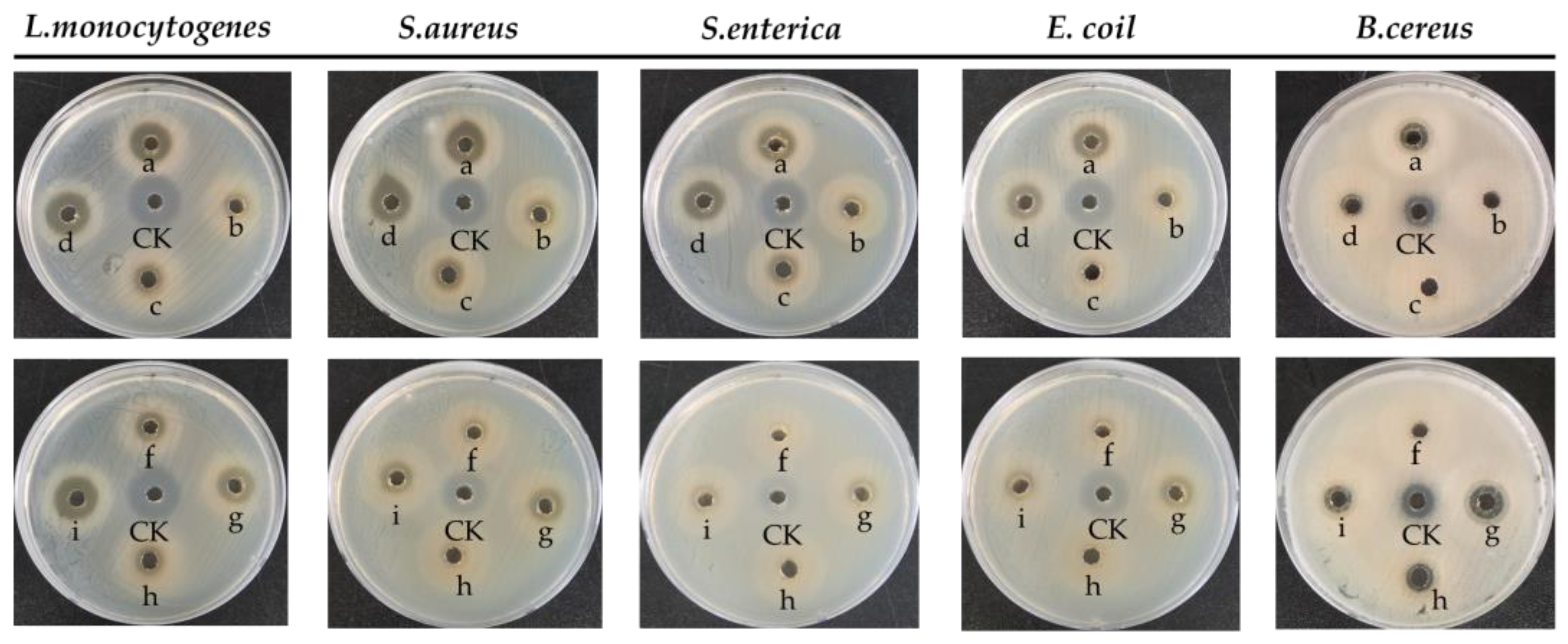
The antibacterial activity was confirmed using the cell-free supernatant (CFS) of the isolated strain and five pathogenic Gram-positive and Gram-negative bacteria (Figure 3 and Figure S5). To confirm the effect of the pH of the cell-free supernatant on the antibacterial activity, the antibacterial activity was evaluated by dividing CFS and nCFS adjusted to pH 6.5 (Table S1). The CFS of the isolated bacteria that were not neutralized with NaOH showed high levels of antibacterial activity against all pathogens. In particular, it showed high antibacterial activity against L. monocytogenes. This corresponds to previous studies that reported that enterococci have antibacterial activity against pathogenic organisms such as L. monocytogenes, S. aureus, and Bacillus cereus [57,58,59,60]. On the other hand, nCFS neutralized to pH 6.5 showed no antibacterial activity or minimal activity (6–10 mm inhibition zone). LAB produces various antimicrobial metabolites during fermentation. These metabolites counteract pathogenic microorganisms by creating an unfavorable environment for their growth [61]. LAB and CFS of LAB inhibit the growth and activity of pathogens and reduce adhesion to epithelial cells, thus having an important effect on pathogen colonization [62]. Antibacterial properties of probiotics are under clinical trials using multiple strains in COVID-19, which is also a promising property of probiotics that enables prevention and treatment at the same time [63]. These results suggest that the isolated strain produces physiologically active metabolites, and these metabolites have antibacterial activity.

3.7. Antioxidant Activity

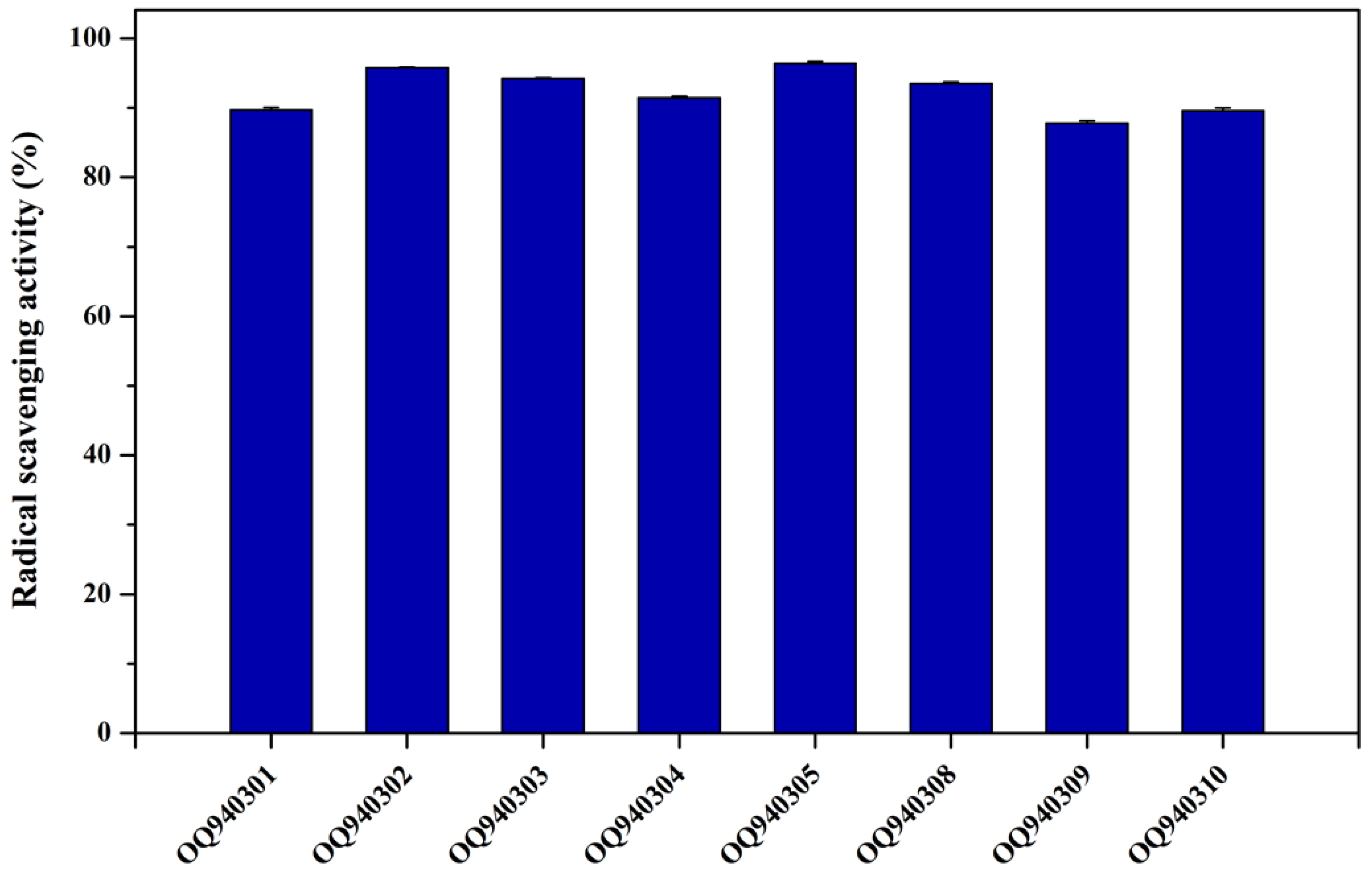
Free radicals (reactive oxygen species (ROS)) are continuously formed in the human body and are related to the pathology of various human diseases such as cancer, rheumatoid arthritis, and neurodegenerative diseases [64]. Antioxidant molecules help maintain the balance of the intestinal microbiome by removing ROS to prevent oxidative damage in the body [65]. An ABTS free radical scavenging assay was conducted using suspension to evaluate the antioxidant properties of bacterial strains isolated from soybean (Figure 4). As a result, it was possible to confirm a high ABTS free radical scavenging ability of more than 80% in all strains. Among them, OQ940302 (95.7%) and OQ940305 (96.3%) showed high antioxidant properties, and OQ940309 (87.7%) showed the lowest antioxidant properties, confirming the overall high ABTS free radical scavenging ability. The fermented Enterococcus faecium lowered oxidation stress compared to conventional food and showed high antioxidant activity in experiments using EPS of Enterococcus faecium separated from traditional fish [66,67]. In addition, research was conducted on probiotics obtained from vegetable-fermented oils such as soybeans and almonds and bio-fermented milk with high antioxidant properties [68]. In this study, the high antioxidant activity of Enterococcus faecium isolated from soybeans was also confirmed.

4. Conclusions

In this study, a total of eight Enterococcus faecium isolates were isolated from fermented soybean paste and identified using 16S rRNA gene sequencing. However, this study finds species-level variants in fermented soybean paste instead of different genera. Among them, most of the bacterial isolates showed tolerance to both acidic and basic environments (~5 log10 CFU/mL) compared to the control. In addition, E. faecium OQ940309 showed higher auto-aggregation (24.52 ± 6.21% at 5 h), co-aggregation (27.62 ± 3.46% at 5 h (S. enterica); 25.62 ± 1.62% at 5 h (S. aureus), and hydrophobicity (94.84 ± 0.04% with xylene). Similarly, E. faecium OQ940301 also showed probiotic potential, and the nCFS of E. faecium OQ940301 exhibited significant antibacterial activity and antioxidant activity against bacterial pathogens and ABTS radicals, respectively. In addition, none of the isolates showed hemolytic activity. Hence, this study concludes that Enterococcus faecium isolates (OQ940301 and OQ940309) have potential probiotic properties and suggests evaluating probiotic criteria under in vivo conditions before recommending them for commercial application.

Supplementary Materials

The following supporting information can be downloaded at: https://www.mdpi.com/article/10.3390/fermentation9080760/s1. Figure S1: Plate image of isolated probiotics from Korean fermented soybean paste; Figure S2: The plate picture of isolates before and after exposure to a low pH environment; Figure S3: The plate picture of isolates before and after exposure to a bile salt environment; Figure S4: Antibiotic susceptibility assays: a-TCH (Tetracycline, 30 μg/mL), b-VAN (Vancomycin, 30 μg/mL), c-Ery (Erythromycin, 15 μg/mL), d-GEN (Gentamycin, 10 μg/mL), e-Amp (Ampicillin, 10 μg/mL); Figure S5: Antibacterial activity of cell-free supernatant adjusted to pH 6.5. a-So1, b-So2, c-So3, d-So4, f-So5, g-So8, h-So9, i-So10, CK-TCH (50 μg/mL); Table S1: Antibacterial activity of cell-free supernatant of E. faecium isolates. +++ = zone dia > 15 mm, ++ = zone dia >10–15 mm, + = zone dia > 5–10 mm, and - = no zone of inhibition.

Author Contributions

Conceptualization, A.S. and M.-H.W.; methodology, A.S. and S.P.; software, K.H.; validation, A.S. and M.-H.W.; formal analysis, S.P. and K.H.; investigation, S.P. and K.H.; resources, M.-H.W.; data curation, S.P. and K.H.; writing—original draft preparation, S.P. and K.H.; writing—review and editing, A.S.; visualization, K.H.; supervision, A.S. and M.-H.W.; project administration, M.-H.W.; funding acquisition, M.-H.W. All authors have read and agreed to the published version of the manuscript.

Funding

This work was supported by the National Research Foundation of Korea (2021R1I1A1A01057742; 2022R1A2C2091029).

Institutional Review Board Statement

Not applicable.

Informed Consent Statement

Not applicable.

Data Availability Statement

Data is contained within the article or Supplementary Materials.

Conflicts of Interest

The authors declare no conflict of interest.

References

  1. Gomes, A.C.; Bueno, A.A.; de Souza, R.G.M.; Mota, J.F. Gut microbiota, probiotics and diabetes. Nutr. J. 2014, 13, 60. [Google Scholar] [CrossRef] [Green Version]
  2. Dubois, A. Spiral bacteria in the human stomach: The gastric helicobacters. Emerg. Infect. Dis. 1995, 1, 79–85. [Google Scholar] [CrossRef] [PubMed] [Green Version]
  3. Salminen, S.; Bouley, C.; Boutron, M.-C.; Cummings, J.H.; Franck, A.; Gibson, G.R.; Isolauri, E.; Moreau, M.-C.; Roberfroid, M.; Rowland, I. Functional food science and gastrointestinal physiology and function. Br. J. Nutr. 1998, 80 (Suppl. S1), S147–S171. [Google Scholar] [CrossRef] [Green Version]
  4. Tripathi, M.K.; Giri, S.K. Probiotic functional foods: Survival of probiotics during processing and storage. J. Funct. Foods 2014, 9, 225–241. [Google Scholar] [CrossRef]
  5. Kawai, K.; Kamochi, R.; Oiki, S.; Murata, K.; Hashimoto, W. Probiotics in human gut microbiota can degrade host glycosaminoglycans. Sci. Rep. 2018, 8, 10674. [Google Scholar] [CrossRef] [Green Version]
  6. Beena Divya, J.; Varsha, K.K.; Nampoothiri, K.M.; Ismail, B.; Pandey, A. Probiotic fermented foods for health benefits. Eng. Life Sci. 2012, 12, 377–390. [Google Scholar] [CrossRef]
  7. Patangia, D.V.; Ryan, C.A.; Dempsey, E.; Ross, R.P.; Stanton, C. Impact of antibiotics on the human microbiome and consequences for host health. MicrobiologyOpen 2022, 11, e1260. [Google Scholar] [CrossRef] [PubMed]
  8. Wu, Q.L.; Chen, T.; El-Nezami, H.; Savidge, T.C. Food ingredients in human health: Ecological and metabolic perspectives implicating gut microbiota function. Trends Food Sci. Technol. 2020, 100, 103–117. [Google Scholar] [CrossRef]
  9. Wolfe, W.; Xiang, Z.; Yu, X.; Li, P.; Chen, H.; Yao, M.; Fei, Y.; Huang, Y.; Yin, Y.; Xiao, H. The Challenge of Applications of Probiotics in Gastrointestinal Diseases. Adv. Gut Microbiome Res. 2023, 2023, 1–10. [Google Scholar] [CrossRef]
  10. Liang, D.; Wu, F.; Zhou, D.; Tan, B.; Chen, T. Commercial probiotic products in public health: Current status and potential limitations. Crit. Rev. Food Sci. Nutr. 2023, 1–22. [Google Scholar] [CrossRef]
  11. Harahap, I.A.; Suliburska, J. Can probiotics decrease the risk of postmenopausal osteoporosis in women? PharmaNutrition 2023, 24, 100336. [Google Scholar] [CrossRef]
  12. Sathiyaseelan, A.; Saravanakumar, K.; Han, K.; Naveen, K.V.; Wang, M.-H. Antioxidant and Antibacterial Effects of Potential Probiotics Isolated from Korean Fermented Foods. Int. J. Mol. Sci. 2022, 23, 10062. [Google Scholar] [CrossRef]
  13. Shin, D.; Chang, S.Y.; Bogere, P.; Won, K.; Choi, J.-Y.; Choi, Y.-J.; Lee, H.K.; Hur, J.; Park, B.-Y.; Kim, Y.; et al. Beneficial roles of probiotics on the modulation of gut microbiota and immune response in pigs. PLoS ONE 2019, 14, e0220843. [Google Scholar] [CrossRef] [Green Version]
  14. Won, G.; Choi, S.-I.; Park, N.; Kim, J.-E.; Kang, C.-H.; Kim, G.-H. In Vitro Antidiabetic, Antioxidant Activity, and Probiotic Activities of Lactiplantibacillus plantarum and Lacticaseibacillus paracasei Strains. Curr. Microbiol. 2021, 78, 3181–3191. [Google Scholar] [CrossRef]
  15. Grimoud, J.; Durand, H.; de Souza, S.; Monsan, P.; Ouarné, F.; Theodorou, V.; Roques, C. In vitro screening of probiotics and synbiotics according to anti-inflammatory and anti-proliferative effects. Int. J. Food Microbiol. 2010, 144, 42–50. [Google Scholar] [CrossRef] [PubMed]
  16. Jafari-Nasab, T.; Khaleghi, M.; Farsinejad, A.; Khorrami, S. Probiotic potential and anticancer properties of Pediococcus sp. isolated from traditional dairy products. Biotechnol. Rep. 2021, 29, e00593. [Google Scholar] [CrossRef]
  17. Lee, N.K.; Kim, S.-Y.; Han, K.J.; Eom, S.J.; Paik, H.-D. Probiotic potential of Lactobacillus strains with anti-allergic effects from kimchi for yogurt starters. LWT-Food Sci. Technol. 2014, 58, 130–134. [Google Scholar] [CrossRef]
  18. Courvalin, P. Antibiotic resistance: The pros and cons of probiotics. Dig. Liver Dis. 2006, 38 (Suppl. S2), S261–S265. [Google Scholar] [CrossRef]
  19. Gueimonde, M.; Sánchez, B.; de Los Reyes-Gavilán, C.G.; Margolles, A. Antibiotic resistance in probiotic bacteria. Front. Microbiol. 2013, 4, 202. [Google Scholar] [CrossRef] [Green Version]
  20. Sharma, P.; Tomar, S.K.; Goswami, P.; Sangwan, V.; Singh, R. Antibiotic resistance among commercially available probiotics. Food Res. Int. 2014, 57, 176–195. [Google Scholar] [CrossRef]
  21. Kareem, K.Y.; Ling, F.H.; Chwen, L.T.; Foong, O.M.; Asmara, S.A. Inhibitory activity of postbiotic produced by strains of Lactobacillus plantarum using reconstituted media supplemented with inulin. Gut. Pathog. 2014, 6, 23. [Google Scholar] [CrossRef] [PubMed] [Green Version]
  22. Giraffa, G.; Carminati, D.; Neviani, E. Enterococci Isolated from Dairy Products: A Review of Risks and Potential Technological Use. J. Food Prot. 1997, 60, 732–738. [Google Scholar] [CrossRef] [PubMed]
  23. Giraffa, G. Enterococci from foods. FEMS Microbiol. Rev. 2002, 26, 163–171. [Google Scholar] [CrossRef] [PubMed]
  24. Franz, C.M.; Huch, M.; Abriouel, H.; Holzapfel, W.; Gálvez, A. Enterococci as probiotics and their implications in food safety. Int. J. Food Microbiol. 2011, 151, 125–140. [Google Scholar] [CrossRef] [PubMed] [Green Version]
  25. Moreno, M.F.; Sarantinopoulos, P.; Tsakalidou, E.; De Vuyst, L. The role and application of enterococci in food and health. Int. J. Food Microbiol. 2006, 106, 1–24. [Google Scholar] [CrossRef]
  26. Park, S.; Saravanakumar, K.; Sathiyaseelan, A.; Park, S.; Hu, X.; Wang, M.-H. Cellular antioxidant properties of nontoxic exopolysaccharide extracted from Lactobacillales (Weissella cibaria) isolated from Korean kimchi. LWT 2022, 154, 112727. [Google Scholar] [CrossRef]
  27. Ashraf, R.; Shah, N.P. Selective and differential enumerations of Lactobacillus delbrueckii subsp. bulgaricus, Streptococcus thermophilus, Lactobacillus acidophilus, Lactobacillus casei and Bifidobacterium spp. in yoghurt—A review. Int. J. Food Microbiol. 2011, 149, 194–208. [Google Scholar] [CrossRef]
  28. Mohammed, S.; Con, A.H. Isolation and characterization of potential probiotic lactic acid bacteria from traditional cheese. LWT-Food Sci. Technol. 2021, 152, 112319. [Google Scholar] [CrossRef]
  29. Vlkova, E.; Rada, V.; Šmehilová, M.; Killer, J. Auto-aggregation and co-aggregation ability in bifidobacteria and clostridia. Folia Microbiol. 2008, 53, 263–269. [Google Scholar] [CrossRef]
  30. Krausova, G.; Hyrslova, I.; Hynstova, I. In Vitro Evaluation of Adhesion Capacity, Hydrophobicity, and Auto-Aggregation of Newly Isolated Potential Probiotic Strains. Fermentation 2019, 5, 100. [Google Scholar] [CrossRef] [Green Version]
  31. Humphries, R.M.; Ambler, J.; Mitchell, S.L.; Castanheira, M.; Dingle, T.; Hindler, J.A.; Koeth, L.; Sei, K.; on behalf of the CLSI Methods Development and Standardization Working Group of the Subcommittee on Antimicrobial Susceptibility Testing. CLSI Methods Development and Standardization Working Group Best Practices for Evaluation of Antimicrobial Susceptibility Tests. J. Clin. Microbiol. 2018, 56, e01934-17. [Google Scholar] [CrossRef] [Green Version]
  32. Plessas, S.; Nouska, C.; Karapetsas, A.; Kazakos, S.; Alexopoulos, A.; Mantzourani, I.; Chondrou, P.; Fournomiti, M.; Galanis, A.; Bezirtzoglou, E. Isolation, characterization and evaluation of the probiotic potential of a novel Lactobacillus strain isolated from Feta-type cheese. Food Chem. 2017, 226, 102–108. [Google Scholar] [CrossRef]
  33. Rojo-Bezares, B.; Sáenz, Y.; Poeta, P.; Zarazaga, M.; Ruiz-Larrea, F.; Torres, C. Assessment of antibiotic susceptibility within lactic acid bacteria strains isolated from wine. Int. J. Food Microbiol. 2006, 111, 234–240. [Google Scholar] [CrossRef] [PubMed]
  34. Soundharrajan, I.; Yoon, Y.H.; Muthusamy, K.; Jung, J.-S.; Lee, H.J.; Han, O.-K.; Choi, K.C. Isolation of Lactococcus lactis from Whole Crop Rice and Determining Its Probiotic and Antimicrobial Properties towards Gastrointestinal Associated Bacteria. Microorganisms 2021, 9, 2513. [Google Scholar] [CrossRef]
  35. Divyashree, S.; Anjali, P.; Somashekaraiah, R.; Sreenivasa, M. Probiotic properties of Lactobacillus casei—MYSRD 108 and Lactobacillus plantarum-MYSRD 71 with potential antimicrobial activity against Salmonella paratyphi. Biotechnol. Rep. 2021, 32, e00672. [Google Scholar] [CrossRef] [PubMed]
  36. Li, J.Y.; Jin, M.-M.; Meng, J.; Gao, S.-M.; Lu, R.-R. Exopolysaccharide from Lactobacillus planterum LP6: Antioxidation and the effect on oxidative stress. Carbohydr. Polym. 2013, 98, 1147–1152. [Google Scholar] [CrossRef] [PubMed]
  37. Sathiyaseelan, A.; Park, S.; Saravanakumar, K.; Mariadoss, A.V.A.; Wang, M.-H. Evaluation of phytochemicals, antioxidants, and antidiabetic efficacy of various solvent fractions of Gynura procumbens (Lour.) Merr. Process Biochem. 2021, 111, 51–62. [Google Scholar] [CrossRef]
  38. Mangia, N.P.; Saliba, L.; Deiana, P. Functional and safety characterization of autochthonous Lactobacillus paracasei FS103 isolated from sheep cheese and its survival in sheep and cow fermented milks during cold storage. Ann. Microbiol. 2019, 69, 161–170. [Google Scholar] [CrossRef]
  39. Al Atya, A.K.; Drider-Hadiouche, K.; Ravallec, R.; Silvain, A.; Vachee, A.; Drider, D. Probiotic potential of Enterococcus faecalis strains isolated from meconium. Front. Microbiol. 2015, 6, 227. [Google Scholar] [CrossRef]
  40. Gaaloul, N.; Ben Braiek, O.; Berjeaud, J.M.; Arthur, T.; Cavera, V.L.; Chikindas, M.L.; Hani, K.; Ghrairi, T. Evaluation of Antimicrobial Activity and Safety Aspect of E nterococcus Italicus GGN 10 Strain Isolated from T unisian Bovine Raw Milk. J. Food Saf. 2014, 34, 300–311. [Google Scholar] [CrossRef]
  41. Pieniz, S.; Andreazza, R.; Okeke, B.C.; Camargo, F.A.O.; Brandelli, A. Antimicrobial and antioxidant activities of Enterococcus species isolated from meat and dairy products. Braz. J. Biol. 2015, 75, 923–931. [Google Scholar] [CrossRef] [PubMed] [Green Version]
  42. Tamang, J.P.; Watanabe, K.; Holzapfel, W. Review: Diversity of microorganisms in global fermented foods and beverages. Front. Microbiol. 2016, 7, 377. [Google Scholar] [CrossRef] [PubMed] [Green Version]
  43. Giraffa, G. Functionality of enterococci in dairy products. Int. J. Food Microbiol. 2003, 88, 215–222. [Google Scholar] [CrossRef]
  44. Bhardwaj, A.; Malik, R.K.; Chauhan, P. Functional and safety aspects of enterococci in dairy foods. Indian J. Microbiol. 2008, 48, 317–325. [Google Scholar] [CrossRef] [PubMed] [Green Version]
  45. Martinez-Villaluenga, C.; Torino, M.I.; Martín, V.; Arroyo, R.; Garcia-Mora, P.; Estrella Pedrola, I.; Vidal-Valverde, C.; Rodriguez, J.M.; Frias, J. Multifunctional Properties of Soy Milk Fermented by Enterococcus faecium Strains Isolated from Raw Soy Milk. J. Agric. Food Chem. 2012, 60, 10235–10244. [Google Scholar] [CrossRef]
  46. Beasley, D.E.; Koltz, A.M.; Lambert, J.E.; Fierer, N.; Dunn, R.R. The Evolution of Stomach Acidity and Its Relevance to the Human Microbiome. PLoS ONE 2015, 10, e0134116. [Google Scholar] [CrossRef]
  47. Hofmann, A.F.; Mysels, K.J. Bile acid solubility and precipitation in vitro and in vivo: The role of conjugation, pH, and Ca2+ ions. J. Lipid Res. 1992, 33, 617–626. [Google Scholar] [CrossRef]
  48. Strompfova, V.; Laukova, A. In vitro study on bacteriocin production of Enterococci associated with chickens. Anaerobe 2007, 13, 228–237. [Google Scholar] [CrossRef]
  49. Trunk, T.; Khalil, H.S.; Leo, J.C. Bacterial autoaggregation. AIMS Microbiol. 2018, 4, 140–164. [Google Scholar] [CrossRef]
  50. Collado, M.C.; Meriluoto, J.; Salminen, S. Measurement of aggregation properties between probiotics and pathogens: In vitro evaluation of different methods. J. Microbiol. Methods 2007, 71, 71–74. [Google Scholar] [CrossRef]
  51. Van Loosdrecht, M.C.; Lyklema, J.; Norde, W.; Schraa, G.; Zehnder, A.J. The role of bacterial cell wall hydrophobicity in adhesion. Appl. Environ. Microbiol. 1987, 53, 1893–1897. [Google Scholar] [CrossRef] [PubMed]
  52. Maragkoudakis, P.A.; Zoumpopoulou, G.; Miaris, C.; Kalantzopoulos, G.; Pot, B.; Tsakalidou, E. Probiotic potential of Lactobacillus strains isolated from dairy products. Int. Dairy J. 2006, 16, 189–199. [Google Scholar] [CrossRef]
  53. Curragh, H.J.; Collins, M. High levels of spontaneous drug resistance in Lactobacillus. J. Appl. Bacteriol. 1992, 73, 31–36. [Google Scholar] [CrossRef]
  54. Nunziata, L.; Brasca, M.; Morandi, S.; Silvetti, T. Antibiotic resistance in wild and commercial non-enterococcal Lactic Acid Bacteria and Bifidobacteria strains of dairy origin: An update. Food Microbiol. 2022, 104, 103999. [Google Scholar] [CrossRef]
  55. Temmerman, R.; Pot, B.; Huys, G.; Swings, J. Identification and antibiotic susceptibility of bacterial isolates from probiotic products. Int. J. Food Microbiol. 2003, 81, 1–10. [Google Scholar] [CrossRef] [PubMed]
  56. Kunyeit, L.; Rao, R.P.; Anu-Appaiah, K.A. Yeasts originating from fermented foods, their potential as probiotics and therapeutic implication for human health and disease. Crit. Rev. Food Sci. Nutr. 2023, 1–12. [Google Scholar] [CrossRef]
  57. Sarantinopoulos, P.; Leroy, F.; Leontopoulou, E.; Georgalaki, M.D.; Kalantzopoulos, G.; Tsakalidou, E.; De Vuyst, L. Bacteriocin production by Enterococcus faecium FAIR-E 198 in view of its application as adjunct starter in Greek Feta cheese making. Int. J. Food Microbiol. 2002, 72, 125–136. [Google Scholar] [CrossRef]
  58. De Kwaadsteniet, M.; Todorov, S.; Knoetze, H.; Dicks, L. Characterization of a 3944 Da bacteriocin, produced by Enterococcus mundtii ST15, with activity against Gram-positive and Gram-negative bacteria. Int. J. Food Microbiol. 2005, 105, 433–444. [Google Scholar] [CrossRef] [PubMed]
  59. Giraffa, G. Enterococcal bacteriocins: Their potential as anti-Listeria factors in dairy technology. Food Microbiol. 1995, 12, 291–299. [Google Scholar] [CrossRef]
  60. De Vuyst, L.; Moreno, M.F.; Revets, H. Screening for enterocins and detection of hemolysin and vancomycin resistance in enterococci of different origins. Int. J. Food Microbiol. 2003, 84, 299–318. [Google Scholar] [CrossRef]
  61. Rakhmanova, A.; Khan, Z.A.; Shah, K. A mini review fermentation and preservation: Role of lactic acid bacteria. MOJ Food Process Technol. 2018, 6, 414–417. [Google Scholar] [CrossRef]
  62. Rezaee, P.; Kermanshahi, R.K.; Falsafi, T. Antibacterial activity of lactobacilli probiotics on clinical strains of Helicobacter pylori. Iran J. Basic Med. Sci. 2019, 22, 1118–1124. [Google Scholar] [CrossRef] [PubMed]
  63. Fijan, S. Probiotics and Their Antimicrobial Effect. Microorganisms 2023, 11, 528. [Google Scholar] [CrossRef]
  64. Aruoma, O.I. Free radicals, oxidative stress, and antioxidants in human health and disease. J. Am. Oil Chem. Soc. 1998, 75, 199–212. [Google Scholar] [CrossRef] [PubMed]
  65. Yang, H.; Wu, H.; Gao, L.; Jia, H.; Zhang, Y.; Cui, Z.; Li, Y. Effects of Lactobacillus curvatus and Leuconostoc mesenteroides on Suan Cai Fermentation in Northeast China. J. Microbiol. Biotechnol. 2016, 26, 2148–2158. [Google Scholar] [CrossRef] [Green Version]
  66. Li, C.; Zhang, B.; Liu, C.; Zhou, H.; Wang, X.; Mai, K.; He, G. Effects of dietary raw or Enterococcus faecium fermented soybean meal on growth, antioxidant status, intestinal microbiota, morphology, and inflammatory responses in turbot (Scophthalmus maximus L.). Fish Shellfish. Immunol. 2020, 100, 261–271. [Google Scholar] [CrossRef]
  67. Abdhul, K.; Ganesh, M.; Shanmughapriya, S.; Kanagavel, M.; Anbarasu, K.; Natarajaseenivasan, K. Antioxidant activity of exopolysaccharide from probiotic strain Enterococcus faecium (BDU7) from Ngari. Int. J. Biol. Macromol. 2014, 70, 450–454. [Google Scholar] [CrossRef]
  68. Zahrani, A.J.A.; Shori, A.B. Viability of probiotics and antioxidant activity of soy and almond milk fermented with selected strains of probiotic Lactobacillus spp. LWT-Food Sci. Technol. 2023, 176, 114531. [Google Scholar] [CrossRef]
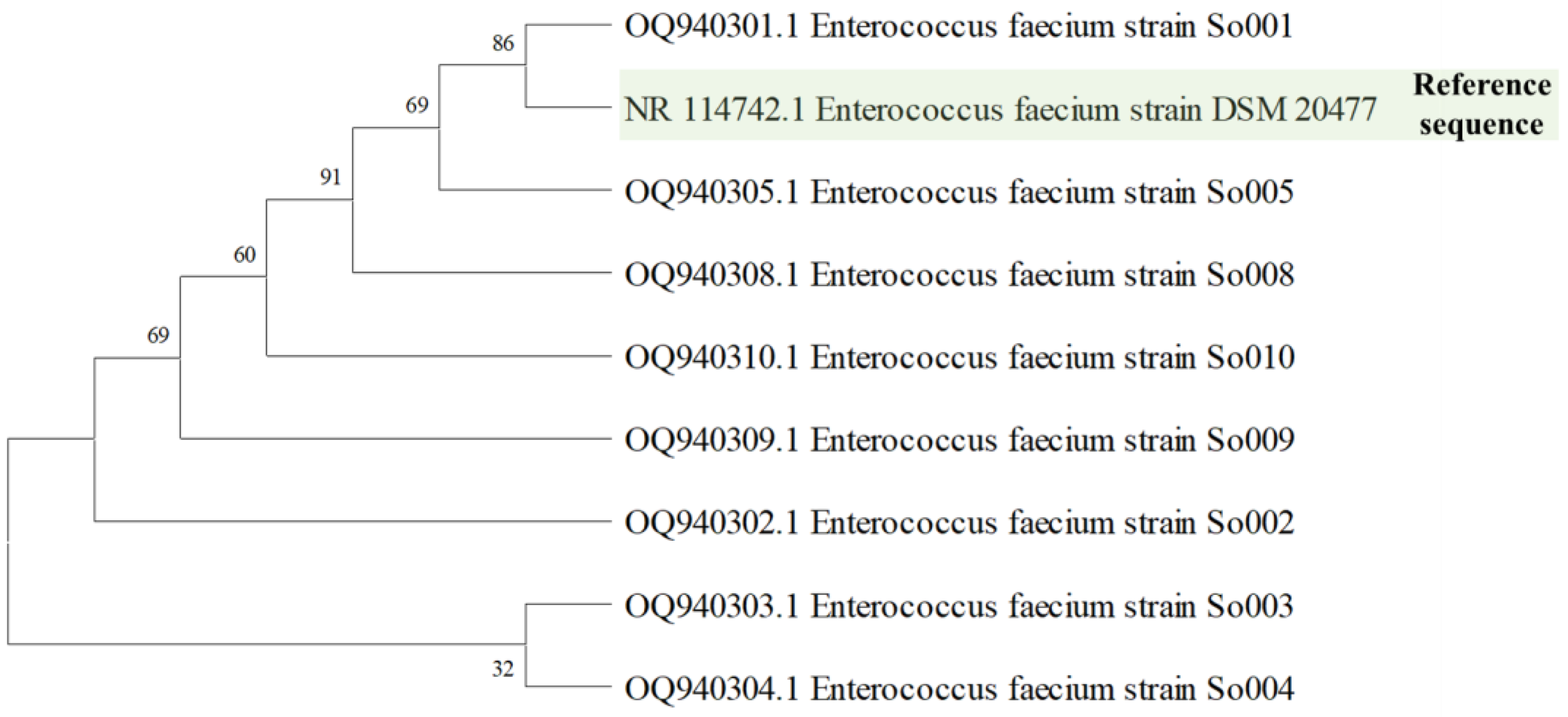
Figure 1. The evolutionary relationships of Enterococcus faecium strains isolated from fermented soybean paste. Phylogenetic trees are constructed by the neighbor-joining method.
Fermentation 09 00760 g001
Figure 2. The hemolytic properties of Enterococcus faecium isolates were compared with those of reference strains (S. aureus and L. rhamnosus).
Fermentation 09 00760 g002
Figure 3. Antibacterial activity of cell-free supernatant. a-OQ940301, b-OQ940302, c-OQ940303, d-OQ940304, f-OQ940305, g-OQ940308, h-OQ940309, i-OQ940310, and CK-TCH (50 μg/mL).
Fermentation 09 00760 g003
Figure 4. ABTS radical scavenging activity of isolated E. faecium strains.
Fermentation 09 00760 g004
Table 1. Genotypic characterization and GenBank accession number of LAB isolated from soybean paste.
Isolate
Code
Similarity-Based on 16S rRNASequence Length (bp)GenBank
Accession No.
So001E. faecium826OQ940301
So002E. faecium827OQ940302
So003E. faecium834OQ940303
So004E. faecium838OQ940304
So005E. faecium836OQ940305
So008E. faecium838OQ940308
So009E. faecium838OQ940309
So010E. faecium838OQ940310
Table 2. Survival of E. faecium strains exposed to a simulated gastric environment.
Cell Viability (log10 CFU/mL)
IsolatesTolerance to Bile SaltTolerance to Low pH
ControlBile Salt0 h3 h
OQ9403015.34 ± 0.205.19 ± 0.025.66 ± 0.055.49 ± 0.04
OQ9403025.41 ± 0.185.24 ± 0.085.68 ± 0.025.57 ± 0.09
OQ9403035.43 ± 0.125.32 ± 0.035.63 ± 0.015.54 ± 0.05
OQ9403045.44 ± 0.315.18 ± 0.055.62 ± 0.055.52 ± 0.1
OQ9403055.2 ± 0.285.06 ± 0.095.58 ± 0.045.58 ± 0.03
OQ9403085.34 ± 0.165.22 ± 0.025.50 ± 0.055.59 ± 0.03
OQ9403095.06 ± 0.394.81 ± 0.035.62 ± 0.065.51 ± 0.04
OQ9403104.7 ± 0.514.15 ± 0.275.51 ± 0.055.56 ± 0.05
Table 3. Hydrophobicity, auto-aggregation, and co-aggregation of E. faecium strains.
StrainAuto-Aggregation (%)Co-Aggregation (%)Co-Aggregation (%)Hydrophobicity (%)
Salmonella entericaStaphylococcus aureus
1 h3 h5 h1 h3 h5 h1 h3 h5 h
OQ9403015.07 ± 2.2315.72 ± 2.8716.16 ± 0.0614.68 ± 4.0623.62 ± 5.8928.10 ± 4.0210.39 ± 2.9623.29 ± 5.124.19 ± 0.8493.99 ± 0.09
OQ94030212.91 ± 4.6518.18 ± 7.8925.39 ± 3.589.32 ± 1.1712.77 ± 0.7924.84 ± 1.777.46 ± 2.8424.09 ± 5.5520.87 ± 5.3294.31 ± 0.04
OQ9403038.9 ± 4.0213.44 ± 3.8518.73 ± 1.446.07 ± 1.0414.73 ± 0.2521.29 ± 0.6610.57 ± 4.0716.51 ± 1.4924.93 ± 1.0894.49 ± 0.0
OQ9403049.84 ± 5.4116.14 ± 7.2218.28 ± 8.95.22 ± 1.239.22 ± 1.6620.38 ± 5.974.29 ± 3.012.71 ± 0.5922.16 ± 0.9194.83 ± 0.09
OQ9403056.32 ± 3.1917.08 ± 8.2320.78 ± 1.575.72 ± 1.699.22 ± 5.7117.93 ± 3.397.3 ± 1.1512.28 ± 0.423.23 ± 1.995.32 ± 0.03
OQ9403087.7 ± 2.5218.84 ± 2.9820.16 ± 2.954.15 ± 1.699.82 ± 4.6914.32 ± 4.0711.65 ± 1.966.95 ± 9.4812.66 ± 6.6394.99 ± 0.06
OQ94030913.67 ± 7.4425.74 ± 5.5424.52 ± 6.2118.01 ± 7.7918.33 ± 0.127.62 ± 3.4613.06 ± 5.6215.67 ± 0.8525.62 ± 1.6294.84 ± 0.04
OQ94031010.54 ± 4.6515.42 ± 5.4324.43 ± 9.99.78 ± 1.710.58 ± 1.1623.57 ± 3.417.68 ± 2.358.6 ± 0.4618.04 ± 2.4394.66 ± 0.04
Table 4. Evaluation of antibiotic susceptibility of E. faecium isolates.
Zone of Inhibition (mm)
OQ940301OQ940302OQ940303OQ940304OQ940305OQ940308OQ940309OQ940310
Tetracycline
(30 μg)
3.73 ± 0.252.9 ± 1.423.87 ± 1.153.7 ± 0.143.1 ± 0.263.87 ± 0.512.97 ± 0.552.83 ± 1.26
Vancomycin
(30 μg)
2.202.001.7 ± 0.42.201.8 ± 0.172.1 ± 0.141.83 ± 0.290.00
Erythromycin
(15 μg)
2.45 ± 1.480.001.502.27 ± 1.330.001.501.302.4 ± 1.56
Gentamycin
(10 μg)
0.000.000.000.000.000.000.000.00
Ampicillin
(10 μg)
3.17 ± 0.582.9 ± 0.953.2 ± 0.363.03 ± 0.062.97 ± 0.473.07 ± 0.312.87 ± 0.43.00
Disclaimer/Publisher’s Note: The statements, opinions and data contained in all publications are solely those of the individual author(s) and contributor(s) and not of MDPI and/or the editor(s). MDPI and/or the editor(s) disclaim responsibility for any injury to people or property resulting from any ideas, methods, instructions or products referred to in the content.