Article

Distribution of Plasmopara viticola Causing Downy Mildew in Russian Far East Grapevines

by
Nikolay N. Nityagovsky
1,2,
Alexey A. Ananev
1,
Andrey R. Suprun
1,
Zlata V. Ogneva
1,
Alina A. Dneprovskaya
1,2,
Alexey P. Tyunin
1,
Alexandra S. Dubrovina
1,
Konstantin V. Kiselev
1,
Nina M. Sanina
2 and
Olga A. Aleynova
1,*
1
Laboratory of Biotechnology, Federal Scientific Center of the East Asia Terrestrial Biodiversity, Far Eastern Branch of the Russian Academy of Sciences, 690022 Vladivostok, Russia
2
Institute of the World Ocean, Far Eastern Federal University, 690090 Vladivostok, Russia
*
Author to whom correspondence should be addressed.
Horticulturae 2024, 10(4), 326; https://doi.org/10.3390/horticulturae10040326
Submission received: 5 March 2024 / Revised: 22 March 2024 / Accepted: 26 March 2024 / Published: 27 March 2024

Abstract

:
Downy mildew is a severe disease that leads to significant losses in grape yields worldwide. It is caused by the oomycete Plasmopara viticola. The study of the distribution of this agent and the search for endophytic organisms that inhibit the growth of P. viticola are essential objectives to facilitate the transition to sustainable and high-yield agriculture, while respecting the environment. In this study, high-throughput sequencing of the ITS (ITS1f/ITS2 region) and 16S (V4 region) amplicons was employed to analyze 80 samples of leaves and stems from different grapevine species and cultivars grown in the Russian Far East (Vitis amurensis Rupr., Vitis coignetiae Pulliat, and several grapevine cultivars). The analysis revealed the presence of P. viticola in 53.75% of the grape samples. The pathogen P. viticola was not detected in V. amurensis samples collected near Vladivostok and Russky Island. Among the P. viticola-affected samples, only two (out of the eighty analyzed grape samples) from the Makarevich vineyard in Primorsky Krai exhibited disease symptoms, while the majority appeared visually healthy. We also found six distinct P. viticola ASVs in our metagenomic data. Based on phylogenetic analysis, we hypothesize that the P. viticola population in the Russian Far East may have originated from the invasive P. viticola clade aestivalis, which has spread around the world from North America. To identify putative microbial antagonists of P. viticola, a differential analysis of high-throughput sequencing data was conducted using the DESeq2 method to compare healthy and P. viticola-affected samples. The in silico analysis revealed an increased representation of certain taxa in healthy samples compared to P. viticola-affected ones: fungi—Kabatina sp., Aureobasidium sp., and Vishniacozyma sp.; bacteria—Hymenobacter spp., Sphingomonas spp., Massilia spp., Methylobacterium-Methylorubrum spp., and Chryseobacterium spp. This in-silico-obtained information on the potential microbial antagonists of P. viticola serves as a theoretical basis for the development of biocontrol agents for grapevine downy mildew.

1. Introduction

Grapevine downy mildew, caused by oomycetes Plasmopara viticola (Berk. and M.A. Curtis) Berl. and de Toni, is an extremely destructive affliction that poses a significant threat to vineyards [1]. This pathogen can infect all green parts of the vine during the warmer and wetter periods of the growing season, causing significant losses in a short period of time [2]. In controlling oomycete fungi, including P. viticola, it is crucial to consistently administer fungicides. This proactive approach serves to safeguard against potential harm and mitigate substantial financial repercussions (up to 75% in humid grapevine-producing areas worldwide) [2,3].
Grapevine downy mildew, which is a common disease in North America, was first identified in 1889. Certain grapevines, such as Muscadinia rotundifolia, have shown resistance to this pathogen [4]. On the other hand, all major Vitis vinifera cultivars are highly susceptible to downy mildew [5]. To address this problem, the use of cultivars with natural disease resistance is a cost-effective and environmentally friendly alternative to the use of fungicides [6]. Several American and Asian Vitis species, such as V. rupestris, V. rubra, V. candicans, V. amurensis, V. riparia, V. cinerea, and M. rotundifolia, show different levels of resistance to P. viticola. This ranges from moderate resistance in some species to high resistance in others [7,8,9].
V. amurensis Rupr., native to East Asia, is mainly found in the southern Far East of Russia to northern Korea. V. amurensis shows numerous advantageous characteristics, such as its resilience to downy mildew [10], anthracnose, and white rot [11], and the ability to withstand cold temperatures [12]. Furthermore, V. amurensis contains valuable medicinal compounds, such as stilbenes, which are known for their antioxidant, anticancer, antibacterial, and antiaging properties [13]. The unique characteristics of this species have led grapevine breeders to incorporate it into their selective breeding programs. Through a comprehensive analysis of quantitative trait loci (QTL) on linkage group 14, a significant QTL controlling resistance to downy mildew resistance in V. amurensis was identified. This specific QTL, known as “Resistance to Plasmopara viticola” (Rpv8, Rpv10, and Rpv12), represents the first set of QTLs conferring resistance to P. viticola to be derived from an Asian Vitis species [14,15,16]. However, it is likely that Rpv8, Rpv10, and Rpv12 are not present in every cultivar of this particular species. According to Wan et al. (2007), only one out of the nine wild V. amurensis accessions was found to be partially resistant in real cases of infection in China, while the rest were considered susceptible [17]. V. coignetiae Pulliat ex Planch., commonly known as crimson grapevine, is a deciduous climbing vine that is native to the temperate climates of East Asia. This includes regions such as Sakhalin Island in Russia, Japan, and Korea. This particular variety of grapevine is often used for its health juice and wine due to the abundance of polyphenols and anthocyanins found in its fruit [18,19]. However, according to a study conducted by Kim et al. in 2019, this grapevine species is susceptible to downy mildew [20].
As mentioned earlier, breeding resistant grape varieties is the most effective way to control downy mildew. However, the introduction of these varieties is a time-consuming and costly process. Given the favorable conditions for disease development, chemical control remains the most economically effective strategy for protecting crops from downy mildew. As an alternative to chemical fungicides, the use of biofungicides offers a biological approach to disease control [21]. Typically, endophytic microorganisms are used as biological control agents. Endophytes possess the ability to significantly impact host–pathogen dynamics, exerting their influence even prior to the emergence of disease. Notably, certain endophytes can instigate systemic resistance mechanisms within their host organisms, effectively stimulating the activation of defense genes targeting specific pathogens. [22]. For example, the Bacillus velezensis KOF112 showed biocontrol activities against downy mildew, inhibiting zoospore release from P. viticola zoosporangia [21]. Also, the endophytes of grape, such as the Bacillus, Variovorax, Pantoea, Staphylococcus, Herbaspirillum, and Sphingomonas bacterial genera, inhibited the mycelial growth of Phytophthora infestans used as a surrogate for P. viticola [23]. Moreover, dipeptides extracted from the grapevine endophyte Alternaria alternate showed efficacy in inhibiting P. viticola sporulation [24]. Culture filtrates obtained from the grape endophyte Acremonium spp. showed inhibitory activity against the P. viticola in vitro [22].
Therefore, the current study, using metagenome analysis, aimed to (I) detect the presence of ITS P. viticola sequences in wild V. amurensis, V. coignetiae grape, and cultivated grape of the Far East of Russia; and (II) perform a comparative analysis of the biodiversity of endophytic bacteria and fungi from healthy and mildew-infected grape samples in order to identify microorganisms that could theoretically be antagonists of P. viticola.

2. Materials and Methods

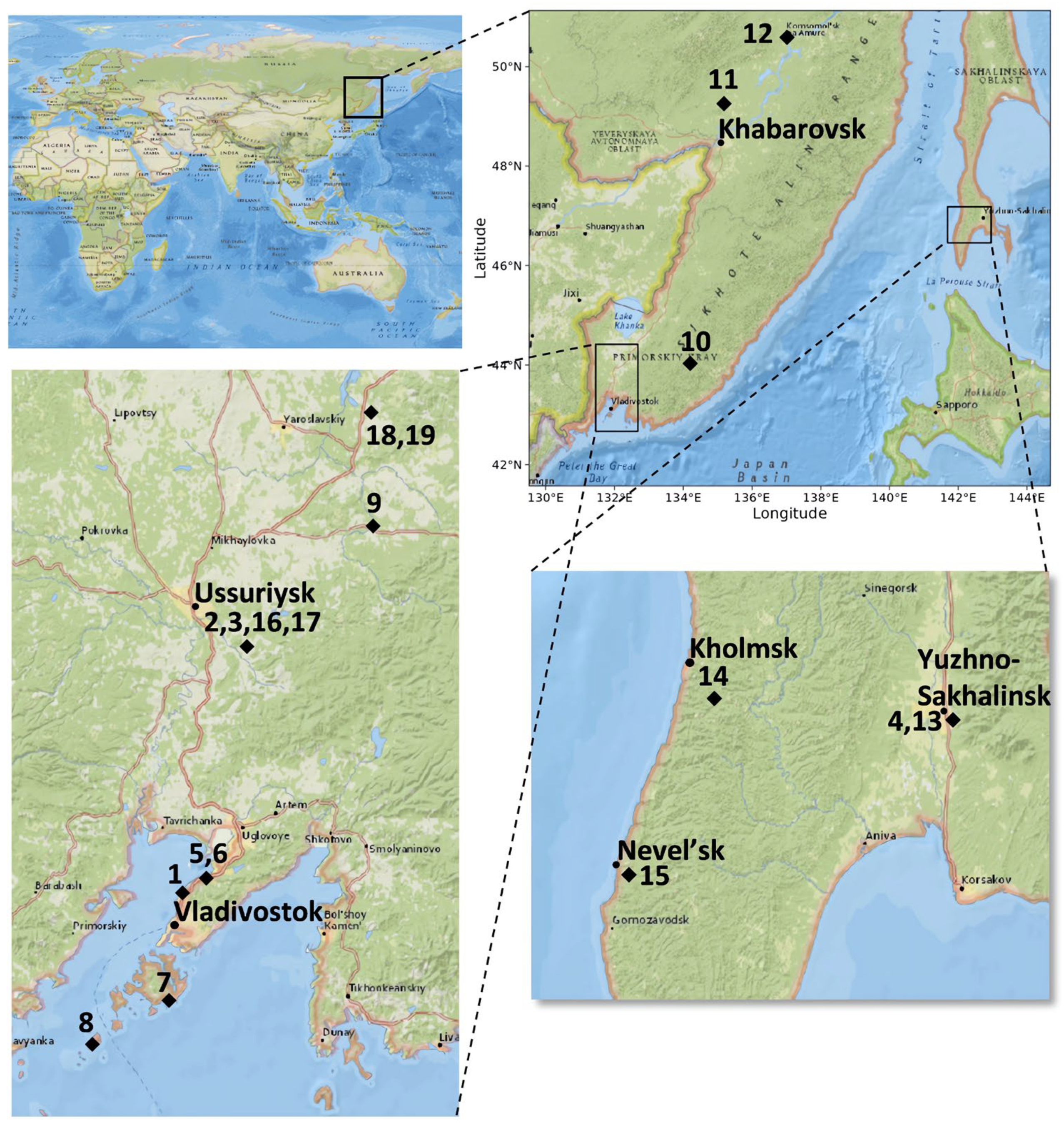
2.1. Plant Material and Surface Sterilization of Samples

To determine the presence of ITS1 sequences of P. viticola in grapevines from the Far East of Russia, a total of 11 asymptomatic tissue samples from V. amurensis, 3 samples from V. coignetiae, and 4 samples from cultivated grapevines were collected. V. amurensis (Gh) has been carefully cultivated under special conditions in the greenhouse at the Federal Scientific Center of the East Asia Terrestrial Biodiversity in Vladivostok, Russia. Additionally, a visually healthy sample of V. amurensis (M) and a sample showing downy mildew symptoms (M-dm) were collected from the commercial vineyard “Makarevich”. Finally, V. amurensis (S-Va) was sampled from the botanical garden on Sakhalin Island. The eight V. amurensis grapevines were collected from different non-protected natural populations. Two grapevines, P1 and P2, were found in close proximity to each other near Vladivostok, Russia, approximately 1 km apart. Another two grapevines, P3 and P4, were discovered on Russky and Rikord Islands in the southern Primorsky Territory of the Russian Far East. P5 and P6 were obtained from Ivanovka village and the Verkhne-Ussuriysky Research Station (SSA) of the Federal Scientific Center of the East Asia terrestrial. Lastly, two additional grapevines were collected from Litovko village (Kh-1) and Silinsky forest (Kh-2), situated in the southern Khabarovsk region of the Russian Far East. Additionally, one V. coignetiae grapevine was sampled from the botanical garden of Sakhalin Island (S-1). There were two additional V. coignetiae grapevines discovered within a natural population on Sakhalin Island, specifically near the cities Kholmsk (S-2) and Nevelsk (S-3). Furthermore, grapes from vineyards located in the Primorsky Territory of Russia were also gathered. Among the collected samples were (Ad) V. vinifera × V. amurensis cv. Adele (hybrid No. 82-41 F3) and (Muk) Vitis riparia × V. vinifera cv. Mukuzani (with an unknown pedigree), which were obtained from the Makarevich vineyard. The samples of Vitis labrusca × V. riparia cv. Alfa (https://www.vivc.de/index.php?r=passport%2Fview&id=346, accessed on 21 March 2024) (Alfa) and Vitis Elmer Swenson 2-7-13 cv. Prairie Star (https://www.vivc.de/index.php?r=passport%2Fview&id=23087, accessed on 21 March 2024) (Pr-St) were selected in PRIM ORGANICA vineyard (Figure 1). It is important to note that all samples, except M-dm, were looking healthy, i.e., without symptoms of downy mildew.
Plant samples were collected on days with little cloud cover and no precipitation, specifically between 11 to 12 in the morning. The air temperature at the time of collection was between 18 and 20 °C. Each sample was promptly transported to the laboratory within a timeframe of 3 h to 1 day. To ensure a comprehensive analysis, a minimum of four biological replicates, consisting of two stems and two leaves, were obtained for each grapevine sample. These replicates were subjected to next-generation sequencing (NGS) for further analysis. Finally, a total of 52 biological replicates of V. amurensis, 12 biological replicates of V. coignetiae, and 16 biological replicates of cultivated grapevines were collected and thoroughly analyzed.
To prepare the grapevine tissues for further analysis, each grapevine sample was thoroughly washed with soap and subjected to a sequential sterilization process. First, they were immersed in 75% ethanol for 2 min, followed by a 1-min treatment with 10% hydrogen peroxide. Finally, they were rinsed five times with sterile water [26,27]. In order to assess the effectiveness of this surface sterilization method, a 100 µL sample of the final rinse water was cultured on R2A (PanReac, AppliChem, Darmstadt, Germany) and potato dextrose agar (PDA, Neogene, Watford, UK) plates to ascertain the absence of any bacterial or fungal colony growth originating from the external sources.

2.2. DNA Isolation, Library Preparation, and Illumina MiSeq Sequencing

To carry out NGS analysis, DNA was isolated from 200 mg of surface-sterilized grape leaves and stems using the CTAB-spin method, as previously reported [28]. The DNA samples were then sent to a reputable commercial organization Syntol (Moscow, Russia) for high-throughput sequencing using Illumina technology. To ensure the quality and quantity of the DNA, it underwent evaluation through the Nanodrop-1000 (Nanodrop, Wilmington, DE, USA) and Quantus Fluorometer (Promega, Madison, WI, USA). The libraries were meticulously prepared for sequencing, adhering precisely to the protocol outlined in the manual “16S Metagenomic Sequencing Library Preparation” (Part # 15,044,223 Rev. B; Illumina). Bacterial 16S rRNA regions were amplified from all samples using the primers 515F (5′GGTAATACGKAGGKKGCDAGC) and 806R (5′RTGGACTACCAGGGTATCTAA), specifically designed to target Vitis sp. plants. The primers ITS1f (5′CTTGGTCATTTAGAGGAAGTAA) and ITS2 (5′GCTGCGTTCTTCATCGATGC) were utilized to amplify the fungal ITS1 rDNA regions in all of the samples. The Nextera® XT Index Kit reagents were used to index the amplicons. The library pool underwent sequencing on Illumina MiSeq platform, employing 2 × 250 paired-end reads, utilizing the MiSeq Reagent Kit v2, which allows for 500 cycles.
The accession numbers for 16S and ITS1 sequences have been successfully submitted and archived in the National Center for Biotechnology Information (NCBI) under the accession numbers PRJNA980748 and PRJNA998468 and in the database of laboratory Biotechnology, Federal Scientific Center of the East Asia Terrestrial Biodiversity, Far Eastern Branch of the Russian Academy of Sciences, Russia (https://biosoil.ru/downloads/biotech/Metagenoms/, accessed on 21 March 2024).

2.3. Data Processing
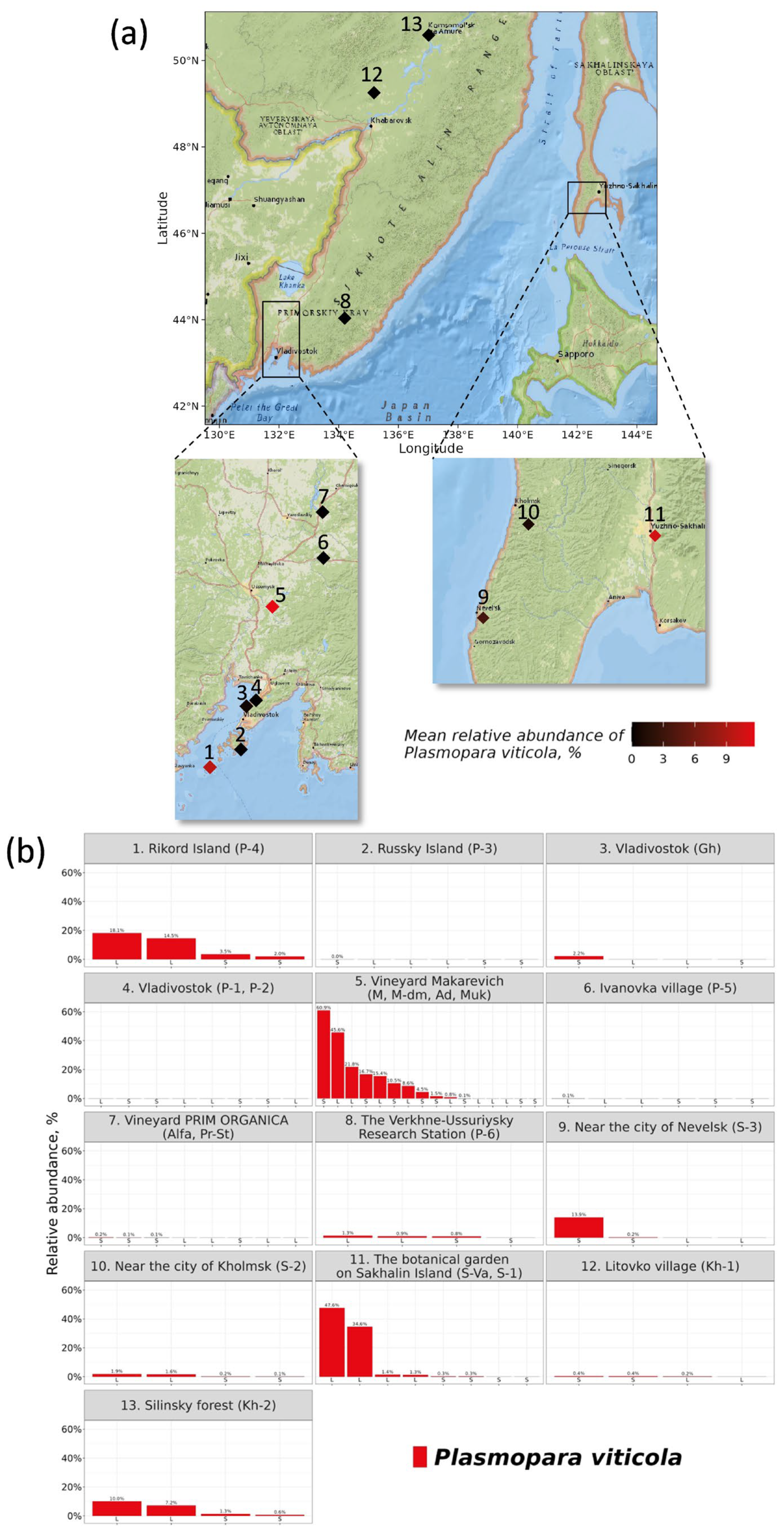
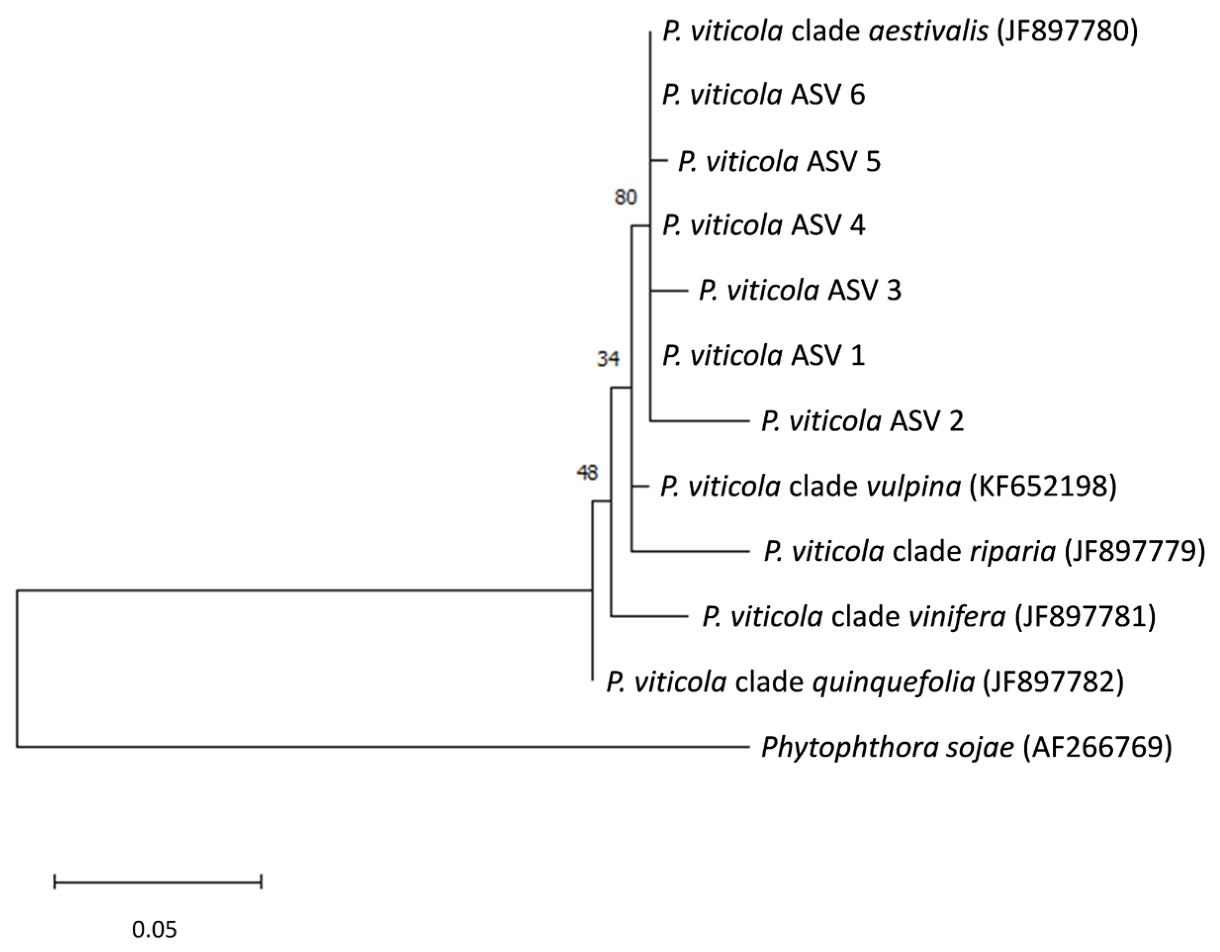
The Supporting Information Tables S2 and S3 provide an overview of the samples utilized in the bioinformatic analysis. Custom scripts based on the R and Bash languages were used to process the data obtained (https://github.com/niknit96/Nityagovsky_et.al.2024, accessed on 21 March 2024). The raw data were preprocessed using QIIME 2 [29] and DADA2 [30] programs. After eliminating the primers, PhiX reads and chimeric sequences, the paired-end reads were merged and arranged in a sorted order. Taxonomic identification of amplicon sequence variants (ASVs) was carried out utilizing the QIIME 2 Scikit-learn algorithm by employing the SILVA 138 pre-trained classifier for 16S sequences (99% OTUs from the V4 region of the sequences) [31]. Additionally, for the ITS sequences, the UNITE pre-trained classifier was utilized (99% OTUs from the ITS1F/ITS2 region of sequences) [32].
The qiime2R [33], phyloseq [34], and tidyverse [35] R libraries were used in pre-filtering and data preparation. Mitochondria, chloroplast, Viridiplantae, Metazoa, Rhizaria, Protista, Alveolata, and unidentified ASVs were deleted from the obtained data. Alpha diversity metrics in rarified samples to even sample depth were obtained using phyloseq R library [34]. The number of ASVs and Pielou’s evenness index were used to characterize richness and evenness in microbial communities, respectively. The Wilcoxon rank sum test was performed to analyze the alpha diversity data between groups. For beta diversity analysis, data were transformed to even sampling depth. The calculation of Bray–Curtis beta diversity data was conducted by employing the Vegan package, a widely recognized tool in the field [36], and converted to nonmetric multidimensional scaling (NMDS). Statistical validation of beta diversity data was performed using the PERMANOVA test with 999 permutations. Data on the differential abundance ASVs between samples, in which P. viticola was found and in which the pathogen was absent, were obtained using the DESeq2 statistical tool with false discovery rate correction [37]. Visualization was conducted using the ggplot2 [35], tidyterra [38], sf [39], maptiles [40], and ggmagnify [41] R libraries.
Evolutionary analyses of the P. viticola ASVs in our dataset with the cryptic species described in [42,43] were performed in MEGA X [44]. The sequences were aligned using the Muscle algorithm [45]. Evolutionary history was inferred using the maximum likelihood (ML) method and the general time reversible (GTR) model [46]. We used the GTR model for the selected region of the ITS because it was the best model for this region in P. viticola data, as described in [42]. To estimate the percentage of trees where the associated taxa cluster together, we used the bootstrap test with 1000 replicates [47]. Initial trees for the heuristic search were automatically obtained by applying the maximum parsimony (MP) method. The Phytophthora sojae ITS sequence was used as the outgroup for phylogenetic analyses. The original sequences, aligned sequences, and MEGA tree session file are presented in the Supplementary Materials.

3. Results

3.1. Distribution of the Plasmopara Viticola ITS1 Sequences in Grape Samples

The Illumina NGS technology was employed to generate a substantial amount of data, resulting in a total of 16,315,902 16S and 5,192,469 ITS1 paired-end reads. Extensive processing, including paired-end alignments, quality filtering, and the removal of unwanted sequences such as chimeric, mitochondria, chloroplast, Viridiplantae, Metazoa, Rhizaria, Protista, Alveolata, and unidentified sequences, led to the generation of 10,102,418 16S and 1,348,330 ITS1 sequences from 80 grape samples (2–6 samples from each plant) (Supporting Information Table S2). Analyzing the 16S data revealed that the average and median read numbers for the samples were 126,280 and 79,912, respectively. Similarly, for ITS1 data, the average and median read numbers were 16,854 and 14,373, respectively (Supporting Information Table S3).
The geographical range of P. viticola in the collected grape samples was analyzed. It should be noted that P. viticola only causes downy mildew in the family Vitaceae. Therefore, in this study, it is acceptable to determine the ITS1 P. viticola sequences before the genus level. Furthermore, using the NCBI nucleotide BLAST (nucleotide–nucleotide BLAST) algorithm, the P. viticola ITS1 sequences (Supporting Information Table S6) were determined to be P. viticola with high percentage identities (99–100%). It was shown that the greatest representation of P. viticola ITS1 sequences was in samples collected in the Makarevich vineyard. The highest representation of the P. viticola ITS1 sequence was 15.4–60.9% in grape sample M-dm, which had visible symptoms of downy mildew (Figure 2). In other samples without visible downy mildew symptoms, the percentage of P. viticola ITS1 sequences was 0–48%. During the analysis of metagenomic data, a large percentage of downy mildew was found on Sakhalin Island and Rikord Island. The greatest representation of P. viticola ITS1 sequences was in the botanical garden of Sakhalin Island, and the percentage ratio in V. coignetiae samples was higher (34–48%) compared to V. amurensis samples (0.3–1.4%). The representation of P. viticola sequences in samples collected on Rikord Island was 2–18%. Relatively small amounts of ITS of P. viticola sequences were detected in samples near the city of Nevelsk (0.2–14%) in the Silinsky forest of the Khabarovsky region (0.7–10%) (Figure 2). P. viticola sequences were present in trace amounts in the sample collected from a greenhouse at the Federal Scientific Center of the East Asia Terrestrial Biodiversity (0–2.2%), near the city of Kholmsk on Sakhalin Island (0.1–1.9%), the Verkhne-Ussuriysky Research Station (0–1.3%), Litovko village, the southern Khabarovsky region of the Russian Far East (0–0.4%), and PRIM ORGANICA vineyard (0–0.2%) (Figure 2). The grape samples collected near Vladivostok, on the Russky Island, and the village of Ivanovka did not have the ITS1 sequence of P. viticola in their metagenome (Figure 2).
We found six P. viticola ASVs in our metagenomic data. We performed a phylogenetic analysis of these ASVs with the cryptic species described in our colleagues’ works [42,43] (Figure 3). Based on the analysis, all of the ASVs clustered together in a well-supported branch, along with a representative ITS sequence of P. viticola clade aestivalis. Among the plants in which P. viticola ASVs were found are wild grapes V. amurensis (P-3, P-4, P-5, P-6, Kh-1, Kh-2, M, M-dm, and S-Va) and V. coignetiae (S-1, S-2, and S-3), as well as cultivated forms of grapes (Ad, Muk, Pr-St, and Alfa) (Table 1). According to our data, the most common P. viticola ASV is ASV 1, which was present in 37 out of 43 samples and had the highest mean relative abundance.

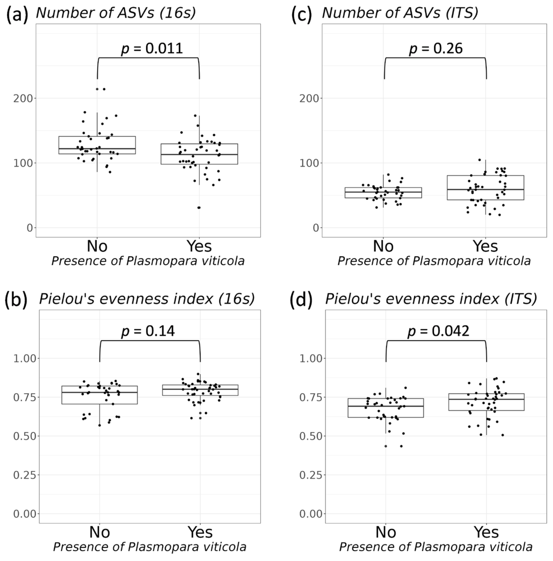
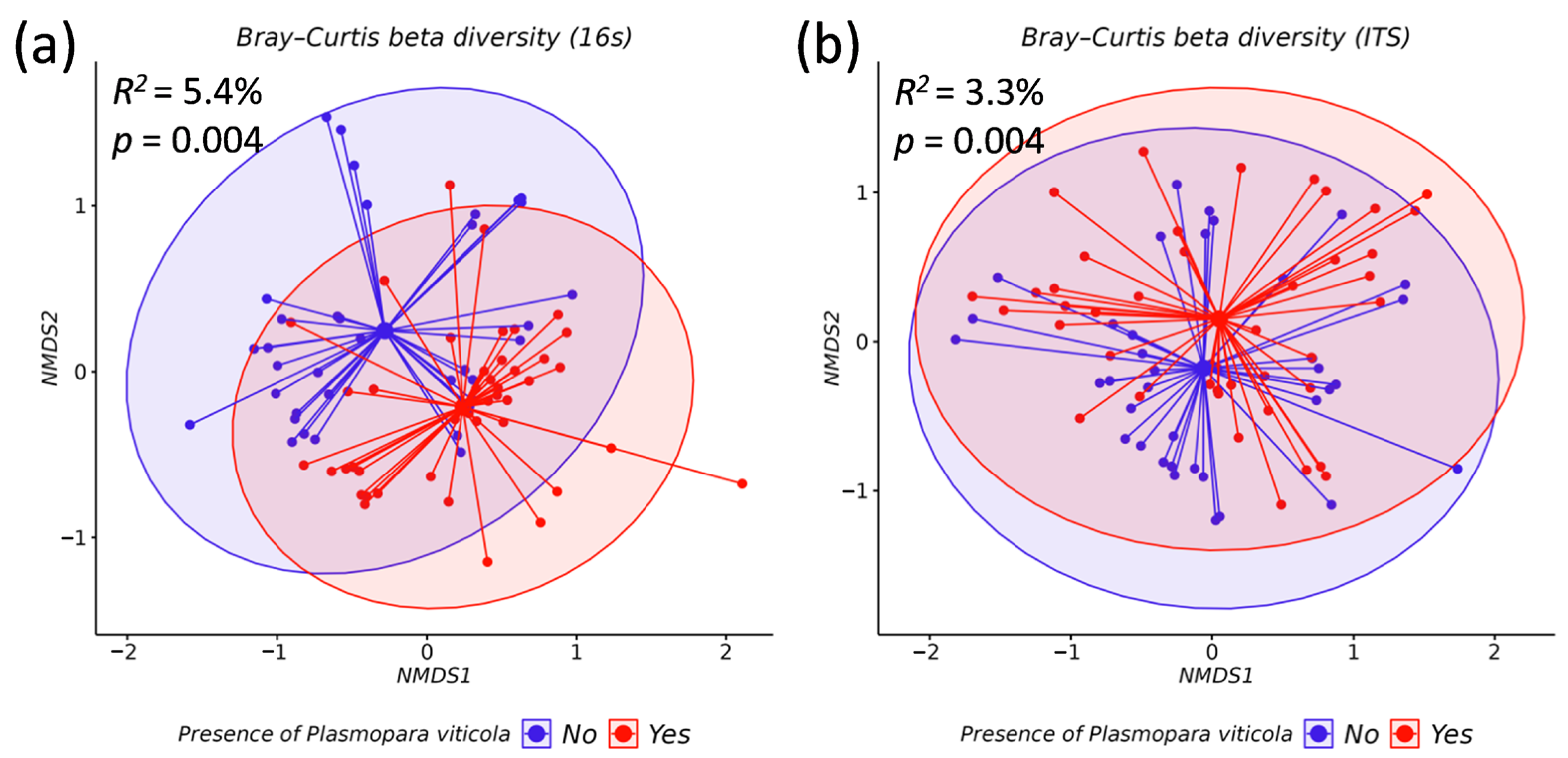
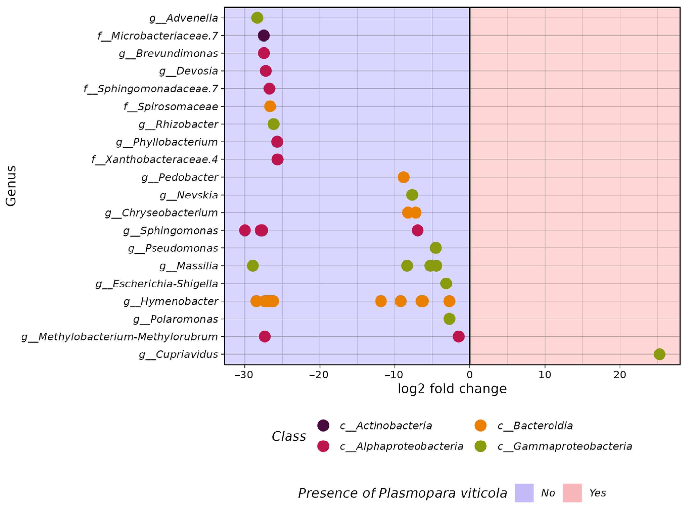
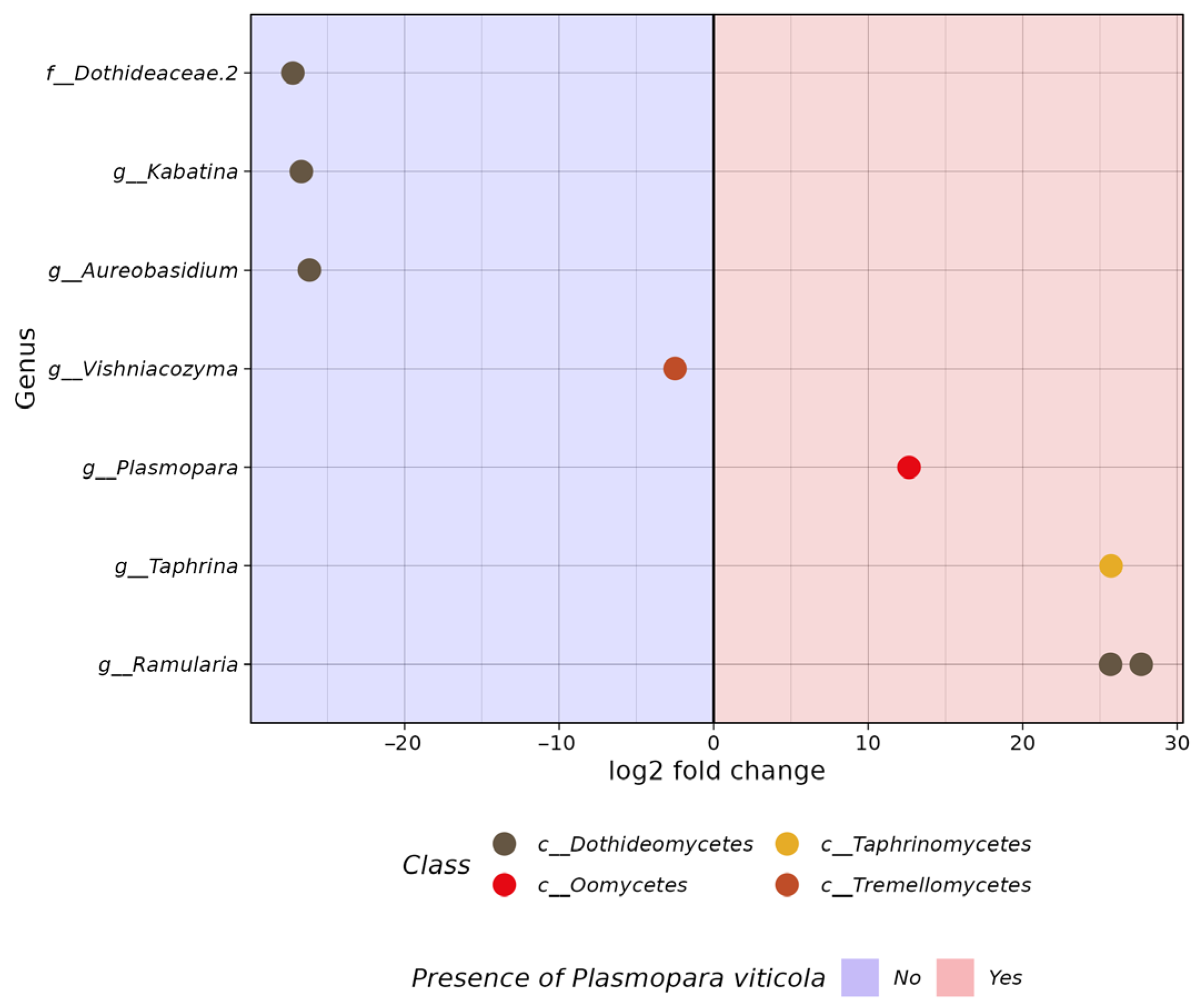
3.2. Comparative Analysis of the Biodiversity of Grape Endophytes in Grape Samples Affected by P. viticola

The outcomes of the alpha diversity analysis of the bacterial and fungal endophytic communities in the grape samples, categorized based on the occurrence of P. viticola, are visually presented in Figure 4a,b and Figure 4c,d, respectively. Based on the number of 16S ASVs, samples affected by P. viticola are characterized by a reduced richness of the bacterial community compared to healthy samples (p = 0.011, Figure 4a). According to the Pielou’s evenness index, the bacterial communities of P. viticola-affected and healthy samples have the same evenness (p = 0.14, Figure 4b). On the other hand, samples affected by P. viticola and healthy samples are not significantly different in terms of fungal community richness (p = 0.26, Figure 4c), but P. viticola-affected samples were characterized by a more even fungal community compared to healthy samples based on Pielou’s evenness index (p = 0.042, Figure 4d).
According to the NMDS ordination plots of beta diversity, P. viticola-affected and healthy endophytic bacterial or fungal communities overlap to a high degree, but fungal communities overlap more than bacterial communities (Figure 5a,b). The PERMANOVA test showed that the factor of the presence of P. viticola explained 4.4% of the differences between grape samples in the bacterial endophytic community (Figure 5a, Supporting Information Table S4), whereas this factor explained 3.2% of the differences between samples in the fungal endophytic community (Figure 5b, Supporting Information Table S4).

3.3. The In Silico Analysis of Potential Microorganisms Plasmopara viticola Antagonists

According to the DESeq2 results, healthy samples were characterized by an increased abundance of 40 bacterial ASVs compared to P. viticola-affected samples (Figure 6, Supporting Information Table S5). These ASVs belonged to 4 taxa of the class level or 18 taxa of the genus level. The largest number of ASVs belonged to class Bacteroidia (18), followed by Alphaproteobacteria (11), Gammaproteobacteria (10), and Actinobacteria (1). At the genus level, the largest number of ASVs belonged to Hymenobacter (14), Sphingomonas (4), Massilia (4), Methylobacterium-Methylorubrum (2), and Chryseobacterium (2), followed by Advenella, Microbacteriaceae (ASV 7), Brevundimonas, Devosia, Sphingomonadaceae (ASV 7), Spirosomaceae, Rhizobacter, Phyllobacterium, Xanthobacteraceae (ASV 4), Pedobacter, Nevskia, Pseudomonas, Escherichia-Shigella, and Polaromonas. ASV, belonging to the genus Cupriavidus, was characterized by an increased abundance in P. viticola-affected samples compared to healthy samples (Figure 6, Supporting Information Table S5).
In the fungal community, healthy samples were characterized by an increased abundance of four ASVs compared to P. viticola-affected samples (Figure 7, Supporting Information Table S6). These ASVs belonged to two taxa at the class level or four taxa at the genus level, namely: Dothideaceae (ASV 2), Kabatina, and Aureobasidium of the class Dothideomycetes, and Vishniacozyma of the class Tremellomycetes. However, samples affected by P. viticola are characterized by an increased abundance of 2 ASVs belonging to Ramularia and 1 ASV belonging to Taphrina (Figure 7, Supporting Information Table S6).

4. Discussion

Grapevine downy mildew, caused by the pathogen known as P. viticola, is widely recognized as one of the most significant diseases affecting grapes worldwide. Throughout the growing season, this particular pathogen has the ability to infect any green component of the vine whenever the weather conditions are warm and wet. In regions with temperate climates, where the grapevine experiences dormancy, the pathogen adapts by forming oospores to ensure its survival in the absence of a host. The extensive application of fungicides has the potential to result in the development of P. viticola isolates that possess resistance to these chemical agents [48,49].
This paper examines the distribution of P. viticola in both unprotected areas and vineyards of the Russian Far East through the analysis of NGS grape samples. According to NGS data, the highest representation of P. viticola ITS1 sequences was found in grape samples from the Makarevich vineyard. It is worth noting that only one of the collected grape samples showed visible symptoms of downy mildew. It is likely that P. viticola is circulating in the Magarach vineyard, and regular fungicide treatments only reduce the amount of the pathogen without destroying it. The fungicide treatment in the PRIM ORGANICA vineyard is the most effective in protecting against P. viticola, where the presence of this pathogen was minimal in the NGS samples. There was also a relatively high presence of P. viticola in V. amurensis samples collected on Rikord Island and in the Silinsky forest of the Khabarovsky region, as well as in V. coignetiae samples grown in the botanical garden on Sakhalin Island and near the town of Nevelsk. It is known that once a certain amount of time has passed, affected tissues of the host organism may display signs of downy mildew. The manifestation of these symptoms heavily relies on the specific environmental conditions of the region, as well as the susceptibility of the host to the disease [50]. P. viticola grows optimally under high relative humidity and mild temperatures [51]. It is likely that the climatic characteristics of northern regions and associated remote islands, namely, increased humidity, UV radiation, and low average temperatures, contribute to a more active distribution of P. viticola. The presence of P. viticola ITS1 in samples of wild grapes without visible symptoms of downy mildew may indicate the resistance of these species to this pathogen. It is known that susceptible grapevine species infected by P. viticola produce mainly trans-resveratrol (3,5,4′-trihydroxystilbene) and trans- and cis-piceid (3-O-β-D-glucoside of resveratrol) [52], whereas resistant species produce trans-resveratrol, trans-pterostilbene (3,5-dimethoxy-4′-hydroxystilbene), and cyclic dehydrodimers of resveratrol trans-ε-viniferin and trans-δ-viniferin [53,54,55]. The wild grape V. amurensis is an important source of stilbenes [11], containing six main stilbenes [56]. Previous research has shown that UV-C-induced biosynthesis of stilbenes and flavonoids in grape leaves, especially resveratrol biosynthesis in grape leaves, is greatly increased in response to UV-C irradiation [57]. In addition, genetic studies have identified 33 loci of resistance to P. viticola (Rpv) in American and Asian Vitis spp. and in some V. vinifera cultivars [58]. Thus, the resistance loci of an individual grapevine, namely, the presence of the loci “resistance to Plasmopara viticola” (Rpv8, Rpv10, and Rpv12), or the higher content of stilbenes, or the stimulation of stilbene biosynthesis by UV radiation or other external factors, can influence the level of P. viticola representation in wild grape samples.
A recent study has proposed a possible scenario for the spread of P. viticola around the world [59]. Through analyzing sequences of nuclear and mitochondrial genes from invasive grapevine downy mildew populations, it was determined that all of these populations belonged to a single clade aestivalis of P. viticola [42,43,59]. This clade is known to infect the wild summer grape V. aestivalis in North America. The study suggests that the pathogen first spread from North America to Europe and then to other parts of the world. Our data partially support the close relationship between the P. viticola ASVs present in the Russian Far East and the invasive clade. However, the data are not sufficient to determine whether the invasion originated directly from Europe or if it is a secondary introduction from another country. Unfortunately, we only sequenced the ITS amplicon, which provides a low resolution for population studies of P. viticola. To draw more accurate conclusions about P. viticola populations in the Russian Far East, further research with a larger sample size and using additional marker genes used in phylogenetic analysis is necessary.
We also analyzed the microbiome of grape samples affected to different degrees by the oomycete P. viticola. It is known that many plant pathogens can form a specific microbiome that can also be an indicator of the pathogen. A comparative analysis of the microbiomes of grape samples without P. viticola representation and samples with high P. viticola representation allowed us to identify microorganisms that could hypothetically be antagonists of the downy mildew pathogen or associated with this oomycete. For example, a grape virus associated with P. viticola has recently been analyzed. It is likely that some viruses could act as new biocontrol agents for P. viticola [60].
In this work, we found both positive and negative correlations in the number of some endophytic microorganisms depending on the representation of P. viticola ITS1 sequences in grape samples. For example, a high proportion of P. viticola ITS1 sequences correlated positively with a high proportion of 16S sequences from Cupriavidus endophytic bacteria in grape samples. There was early evidence that Cupriavidus species were associated with agricultural crops growing in alkaline soils [61]. It is possible that the association of these bacteria with P. viticola is related to the alkalinization of the internal tissues of grapes as a result of infection with P. viticola. The endophytic fungi genera Ramularia and Taphrina were also more abundant in grape samples with high levels of downy mildew. Ramularia, the white mold of plants, is a species-rich genus that harbors plant pathogens responsible for yield losses in many important crops, including barley, sugar beet, and strawberries [62]. It has been shown that barley plants with mlo resistance to downy mildew have an increased susceptibility to a new important disease—ramularia leaf spot [63]. In addition, Taphrina fungi are biotrophic plant pathogens that cause plant deformities [64]. Thus, infection with downy mildew leads to subsequent infection with other pathogenic fungi.
In addition to mildew-associated microorganisms, we found endophytes that are hypothetical antagonists of P. viticola. The presence of bacteria belonging to the genera Hymenobacter, Sphingomonas, Massilia, Methylobacterium-Methylorubrum, and Chryseobacterium was found to be significantly higher in grape samples with a low or absent content of P. viticola ITS1 sequences compared to samples highly infected with downy mildew. Some species of genera Hymenobacter are known to be UV-resistant [65], and the pathogen P. viticola is very sensitive to UV radiation [66]. Therefore, the inversely proportional number of endophytic bacteria Hymenobacter spp. and P. viticola is likely to be related to the amount of UV exposure of individual grape samples. It is known that several Methylobacterium and Sphingomonas strains work against the proliferation of plant pahogene Candidatus phytoplasma, which is the primary cause of grapevine yellows. Additionally, it is worth highlighting that the presence of genera Methylobacterium and Sphingomonas in significant numbers is directly linked to the production of characteristic sensory compounds found in well-rounded wines [67]. Moreover, the presence of Chryseobacterium species contribute to enhanced plant growth through biocontrol activity against plant pathogens, including Phytophthora capsici [68]. Also, a low number of ITS P. viticola sequences correlated with high-percentage representation of endophytic fungi of the taxa Dothideaceae.2, Kabatina, Aureobasidium, and Vishniacozyma in grape samples. According to the literature, some species of fungi of the genus Kabatina can synthesize enfumafungin, a novel antifungal compound [69]. In addition, several species of Aureobasidium fungi possess the remarkable capacity to produce volatile organic compounds (VOCs) that display inhibitory effects on grape pathogens, most notably Botrytis cinerea [70,71]. Vishniacozyma is a versatile yeast genus that has been discovered in various ecological settings. Research has revealed that the population of Vishniacozyma sp. thrives during the berry ripening phase, showcasing its ability to flourish in conditions characterized by high sugar content and low moisture levels. It is noteworthy that the pathogenic fungus P. viticola typically flourishes in moist environments. This discrepancy in preferred habitats could potentially explain the inverse correlation between the prevalence of P. viticola and the presence of Vishniacozyma sp. Additionally, Vishniacozyma sp. has exhibited promising biocontrol properties against both blue molds and gray molds, which commonly infect pears [72]. Recent investigations by Zhu et al. in 2021 [73] have unveiled a potential antagonistic effect of Vishniacozyma sp. on Erysisphe. Nevertheless, the biocontrol potential of the endophytic bacteria Methylobacterium spp., Sphingomonas spp., and Chryseobacterium spp. and the fungi Kabatina sp., Aureobasidium sp., and Vishniacozyma sp. for grape downy mildew requires further analysis. Together, these endophytic antagonists represent a valuable resource that will undoubtedly be used in the foreseeable future to develop biocontrol or integrated programs to reduce chemical use against downy mildew.

5. Conclusions

In this study, NGS was utilized for the first time to analyze the distribution of downy mildew in wild V. amurensis, V. coignetiae, and cultivated grapes. Our data suggest that the population of P. viticola in the Russian Far East may be related to an invasive clade aestivalis of P. viticola, which has spread from North America to other parts of the world. Bioinformatic methods were also used to identify endophytic microorganisms associated or antagonistic to the downy mildew. The in silico analysis showed that certain genera of endophytic bacteria, namely, Hymenobacter spp., Sphingomonas spp., Massilia spp., Methylobacterium-Methylorubrum spp., and Chryseobacterium spp., and fungi, namely, Kabatina sp., Aureobasidium sp., and Vishniacozyma sp., could be hypothetical antagonists of P. viticola. The results obtained provide an important basis for the development of downy mildew biocontrol tools based on natural endophytic microorganisms.

Supplementary Materials

The following supporting information can be downloaded at https://www.mdpi.com/article/10.3390/horticulturae10040326/s1: Supporting Information Table S1: The code, location and GPS coordinates samples of grapevines of Vitis amurensis, Vitis coignetiae and cultivated grapes collected at July 2022; Supporting Information Table S2: 16s data samples used in analysis; Supporting Information Table S3: ITS data samples used in analysis; Supporting Information Table S4: PERMANOVA results; Supporting Information Table S5: Identified by the DESeq2 tool significantly different abundant (adjusted p < 0.01) bacterial ASVs between grape samples which grouped based on presence of Plasmopara viticola; Supporting Information Table S6: Identified by the DESeq2 tool significantly different abundant (adjusted p < 0.01) fungal ASVs between grape samples which grouped based on presence of Plasmopara viticola; Original sequences.fasta: ITS sequences of P. viticola ASVs in our NGS dataset, ITS sequences of cryptic species of P. viticola and Phytophthora sojae ITS sequence; Aligned sequences.fasta: Aligned sequences used in phylogenetic analysis; MEGA tree session.mtsx: phylogenetic tree in MEGA tree session format.

Author Contributions

Conceptualization K.V.K. and O.A.A.; methodology and software N.N.N. and A.A.A.; validation N.M.S.; formal analysis Z.V.O., A.R.S. and A.A.D.; investigation N.N.N. and A.P.T.; resources, O.A.A.; data curation K.V.K.; writing—original draft preparation O.A.A.; writing—review and editing A.S.D.; visualization A.A.A.; supervision K.V.K.; project administration N.N.N.; funding acquisition O.A.A. All authors have read and agreed to the published version of the manuscript.

Funding

This work was supported by a grant from the Russian Science Foundation (grant number 22-74-10001, https://rscf.ru/project/22-74-10001) (accessed on 21 March 2024). The maintenance of the used microbial collection was carried out within the state assignment of the Ministry of Science and Higher Education of the Russian Federation (theme No. 124012200181-4).

Data Availability Statement

The data presented in this study are available within the article and Supplementary Materials.

Acknowledgments

We would like to thank the Sergei Makarevich, Alexander, and Anna Storozhenko for their primary contribution to the sample collection for the present study.

Conflicts of Interest

The authors declare no conflicts of interest.

References

  1. Lafon, R.; Clerjeau, M. Downy Mildew. In Compendium of Grape Diseases; APS Press: St. Paul, MN, USA, 1988; pp. 11–13. [Google Scholar]
  2. Koledenkova, K.; Esmaeel, Q.; Jacquard, C.; Nowak, J.; Clément, C.; Ait Barka, E. Plasmopara viticola the Causal Agent of Downy Mildew of Grapevine: From Its Taxonomy to Disease Management. Front. Microbiol. 2022, 13, 889472. [Google Scholar] [CrossRef]
  3. Massi, F.; Torriani, S.F.F.; Borghi, L.; Toffolatti, S.L. Fungicide Resistance Evolution and Detection in Plant Pathogens: Plasmopara viticola as a Case Study. Microorganisms 2021, 9, 119. [Google Scholar] [CrossRef]
  4. Feechan, A.; Anderson, C.; Torregrosa, L.; Jermakow, A.; Mestre, P.; Wiedemann-Merdinoglu, S.; Merdinoglu, D.; Walker, A.R.; Cadle-Davidson, L.; Reisch, B.; et al. Genetic Dissection of a TIR-NB-LRR Locus from the Wild North American Grapevine Species Muscadinia rotundifolia Identifies Paralogous Genes Conferring Resistance to Major Fungal and Oomycete Pathogens in Cultivated Grapevine. Plant J. 2013, 76, 661–674. [Google Scholar] [CrossRef]
  5. Yin, L.; An, Y.; Qu, J.; Li, X.; Zhang, Y.; Dry, I.; Wu, H.; Lu, J. Genome Sequence of Plasmopara viticola and Insight into the Pathogenic Mechanism. Sci. Rep. 2017, 7, 46553. [Google Scholar] [CrossRef]
  6. Delbac, L.; Delière, L.; Schneider, C.; Delmotte, F. Downy Mildew Is Able to Carry out Its Sexual Cycle on Resistant Grape Varieties: Sourced from the Research Article “Evidence for Sexual Reproduction and Fertile Oospore Production by Plasmopara viticola on the Leaves of Partially Resistant Grapevine Varieties” (ACTA Horticulturae, 1248. ISHS, 2019). Original Language of the Article: French. IVES Tech. Rev. Vine Wine 2020. [Google Scholar] [CrossRef]
  7. Olmo, H.P. Vinifera rotundifolia Hybrids as Wine Grapes. Am. J. Enol. Vitic. 1971, 22, 87–91. [Google Scholar] [CrossRef]
  8. Staudt, G.; Kassemeyer, H.H. Evaluation of Downy Mildew Resistance in Various Accessions of Wild Vitis Species. Vitis 1995, 34, 225–228. [Google Scholar]
  9. Díez-Navajas, A.M.; Wiedemann-Merdinoglu, S.; Greif, C.; Merdinoglu, D. Nonhost Versus Host Resistance to the Grapevine Downy Mildew, Plasmopara viticola, Studied at the Tissue Level. Phytopathology 2008, 98, 776–780. [Google Scholar] [CrossRef]
  10. Wu, J.; Zhang, Y.; Zhang, H.; Huang, H.; Folta, K.M.; Lu, J. Whole Genome Wide Expression Profiles of Vitis amurensis Grape Responding to Downy Mildew by Using Solexa Sequencing Technology. BMC Plant Biol. 2010, 10, 234. [Google Scholar] [CrossRef]
  11. Liu, L.; Li, H. Review: Research Progress in Amur Grape, Vitis amurensis Rupr. Can. J. Plant Sci. 2013, 93, 565–575. [Google Scholar] [CrossRef]
  12. Ma, Y.Y.; Zhang, Y.L.; Shao, H.; Lu, J. Differential Physio-Biochemical Responses to Cold Stress of Cold-Tolerant and Non-Tolerant Grapes (Vitis L.) from China. J. Agron. Crop Sci. 2010, 196, 212–219. [Google Scholar] [CrossRef]
  13. Chen, Q.; Diao, L.; Song, H.; Zhu, X. Vitis amurensis Rupr: A Review of Chemistry and Pharmacology. Phytomedicine 2018, 49, 111–122. [Google Scholar] [CrossRef]
  14. Blasi, P.; Blanc, S.; Wiedemann-Merdinoglu, S.; Prado, E.; Rühl, E.H.; Mestre, P.; Merdinoglu, D. Construction of a Reference Linkage Map of Vitis amurensis and Genetic Mapping of Rpv8, a Locus Conferring Resistance to Grapevine Downy Mildew. Theor. Appl. Genet. 2011, 123, 43–53. [Google Scholar] [CrossRef]
  15. Schwander, F.; Eibach, R.; Fechter, I.; Hausmann, L.; Zyprian, E.; Töpfer, R. Rpv10: A New Locus from the Asian Vitis Gene Pool for Pyramiding Downy Mildew Resistance Loci in Grapevine. Theor. Appl. Genet. 2012, 124, 163–176. [Google Scholar] [CrossRef]
  16. Venuti, S.; Copetti, D.; Foria, S.; Falginella, L.; Hoffmann, S.; Bellin, D.; Cindrić, P.; Kozma, P.; Scalabrin, S.; Morgante, M.; et al. Historical Introgression of the Downy Mildew Resistance Gene Rpv12 from the Asian Species Vitis amurensis into Grapevine Varieties. PLoS ONE 2013, 8, e61228. [Google Scholar] [CrossRef]
  17. Wan, Y.; Schwaninger, H.; He, P.; Wang, Y. Comparison of Resistance to Powdery Mildew and Downy Mildew in Chinese Wild Grapes. Vitis 2007, 46, 132–136. [Google Scholar]
  18. Takayama, F.; Nakamoto, K.; Kawasaki, H.; Mankura, M.; Egashira, T.; Ueki, K.; Hasegawa, A.; Okada, S.; Mori, A. Beneficial Effects of Vitis coignetiae Pulliat Leaves on Nonalcoholic Steatohepatitis in a Rat Model. Acta Medica Okayama 2009, 63, 105–111. [Google Scholar]
  19. Kim, M.J.; Paramanantham, A.; Lee, W.S.; Yun, J.W.; Chang, S.H.; Kim, D.C.; Park, H.S.; Choi, Y.H.; Kim, G.S.; Ryu, C.H.; et al. Anthocyanins Derived from Vitis coignetiae Pulliat Contributes Anti-Cancer Effects by Suppressing NF-κB Pathways in Hep3B Human Hepatocellular Carcinoma Cells and in vivo. Molecules 2020, 25, 5445. [Google Scholar] [CrossRef]
  20. Kim, B.R.; Kim, I.H.; Lee, J.S.; Choi, Y.J. First Report of Downy Mildew Caused by Plasmopara viticola on Vitis coignetiae in Korea. Plant Dis. 2019, 103, 1793. [Google Scholar] [CrossRef]
  21. Hamaoka, K.; Aoki, Y.; Suzuki, S. Isolation and Characterization of Endophyte Bacillus velezensis KOF112 from Grapevine Shoot Xylem as Biological Control Agent for Fungal Diseases. Plants 2021, 10, 1815. [Google Scholar] [CrossRef]
  22. Lo Piccolo, S.; Alfonzo, A.; Giambra, S.; Conigliaro, G.; Lopez-Llorca, L.V.; Burruano, S. Identification of Acremonium Isolates from Grapevines and Evaluation of Their Antagonism towards Plasmopara viticola. Ann. Microbiol. 2015, 65, 2393–2403. [Google Scholar] [CrossRef]
  23. Bruisson, S.; Zufferey, M.; L’Haridon, F.; Trutmann, E.; Anand, A.; Dutartre, A.; De Vrieze, M.; Weisskopf, L. Endophytes and Epiphytes from the Grapevine Leaf Microbiome as Potential Biocontrol Agents Against Phytopathogens. Front. Microbiol. 2019, 10, 491406. [Google Scholar] [CrossRef] [PubMed]
  24. Musetti, R.; Polizzotto, R.; Vecchione, A.; Borselli, S.; Zulini, L.; D’Ambrosio, M.; di Toppi, L.S.; Pertot, I. Antifungal Activity of Diketopiperazines Extracted from Alternaria alternata against Plasmopara viticola: An Ultrastructural Study. Micron 2007, 38, 643–650. [Google Scholar] [CrossRef]
  25. National Geographic Basemap. Available online: https://www.esri.com/news/arcuser/0312/national-geographic-basemap.html (accessed on 21 March 2024).
  26. Aleynova, O.A.; Nityagovsky, N.N.; Dubrovina, A.S.; Kiselev, K.V. The Biodiversity of Grapevine Bacterial Endophytes of Vitis amurensis Rupr. Plants 2022, 11, 1128. [Google Scholar] [CrossRef] [PubMed]
  27. Aleynova, O.A.; Nityagovsky, N.N.; Suprun, A.R.; Ananev, A.A.; Dubrovina, A.S.; Kiselev, K.V. The Diversity of Fungal Endophytes from Wild Grape Vitis amurensis Rupr. Plants 2022, 11, 2897. [Google Scholar] [CrossRef] [PubMed]
  28. Kiselev, K.V.; Nityagovsky, N.N.; Aleynova, O.A. A Method of DNA Extraction from Plants for Metagenomic Analysis Based on the Example of Grape Vitis amurensis Rupr. Appl. Biochem. Microbiol. 2023, 59, 361–367. [Google Scholar] [CrossRef]
  29. Bolyen, E.; Rideout, J.R.; Dillon, M.R.; Bokulich, N.A.; Abnet, C.C.; Al-Ghalith, G.A.; Alexander, H.; Alm, E.J.; Arumugam, M.; Asnicar, F.; et al. Reproducible, Interactive, Scalable and Extensible Microbiome Data Science Using QIIME 2. Nat. Biotechnol. 2019, 37, 852–857. [Google Scholar] [CrossRef] [PubMed]
  30. Callahan, B.J.; McMurdie, P.J.; Rosen, M.J.; Han, A.W.; Johnson, A.J.A.; Holmes, S.P. DADA2: High-Resolution Sample Inference from Illumina Amplicon Data. Nat. Methods 2016, 13, 581–583. [Google Scholar] [CrossRef] [PubMed]
  31. Quast, C.; Pruesse, E.; Yilmaz, P.; Gerken, J.; Schweer, T.; Yarza, P.; Peplies, J.; Glöckner, F.O. The SILVA Ribosomal RNA Gene Database Project: Improved Data Processing and Web-Based Tools. Nucleic Acids Res. 2013, 41, D590–D596. [Google Scholar] [CrossRef] [PubMed]
  32. Abarenkov, K.; Zirk, A.; Piirmann, T.; Pöhönen, R.; Ivanov, F.; Nilsson, R.H.; Kõljalg, U. UNITE QIIME Release for Eukaryotes; Version 29.11.2022; UNITE Community: London, UK, 2022. [Google Scholar] [CrossRef]
  33. Bisanz, J. qiime2R: Importing QIIME2 Artifacts and Associated Data into R Sessions. 2018. Available online: https://github.com/jbisanz/qiime2R (accessed on 21 March 2024).
  34. McMurdie, P.J.; Holmes, S. Phyloseq: An R Package for Reproducible Interactive Analysis and Graphics of Microbiome Census Data. PLoS ONE 2013, 8, e61217. [Google Scholar] [CrossRef]
  35. Wickham, H.; Averick, M.; Bryan, J.; Chang, W.; McGowan, L.D.; François, R.; Grolemund, G.; Hayes, A.; Henry, L.; Hester, J.; et al. Welcome to the Tidyverse. J. Open Source Softw. 2019, 4, 1686. [Google Scholar] [CrossRef]
  36. Oksanen, J.; Blanchet, F.G.; Friendly, M.; Kindt, R.; Legendre, P.; McGlinn, D.; Minchin, P.R.; O’Hara, R.B.; Simpson, G.L.; Solymos, P.; et al. Vegan: Community Ecology Package. 2020. Available online: https://CRAN.R-project.org/package=vegan (accessed on 21 March 2024).
  37. Love, M.I.; Huber, W.; Anders, S. Moderated Estimation of Fold Change and Dispersion for RNA-Seq Data with DESeq2. Genome Biol. 2014, 15, 550. [Google Scholar] [CrossRef] [PubMed]
  38. Hernangómez, D. Using the Tidyverse with Terra Objects: The Tidyterra Package. J. Open Source Softw. 2023, 8, 5751. [Google Scholar] [CrossRef]
  39. Pebesma, E. Simple Features for R: Standardized Support for Spatial Vector Data. R J. 2018, 10, 439–446. [Google Scholar] [CrossRef]
  40. Giraud, T. Maptiles: Download and Display Map Tiles. 2023. Available online: https://github.com/riatelab/maptiles (accessed on 21 March 2024).
  41. Hugh-Jones, D. Ggmagnify: Create a Magnified Inset of Part of a Ggplot Object. 2023. Available online: https://github.com/hughjonesd/ggmagnify (accessed on 21 March 2024).
  42. Rouxel, M.; Mestre, P.; Comont, G.; Lehman, B.L.; Schilder, A.; Delmotte, F. Phylogenetic and Experimental Evidence for Host-Specialized Cryptic Species in a Biotrophic Oomycete. New Phytol. 2013, 197, 251–263. [Google Scholar] [CrossRef]
  43. Rouxel, M.; Mestre, P.; Baudoin, A.; Carisse, O.; Delière, L.; Ellis, M.A.; Gadoury, D.; Lu, J.; Nita, M.; Richard-Cervera, S.; et al. Geographic Distribution of Cryptic Species of Plasmopara viticola Causing Downy Mildew on Wild and Cultivated Grape in Eastern North America. Phytopathology 2014, 104, 692–701. [Google Scholar] [CrossRef]
  44. Kumar, S.; Stecher, G.; Li, M.; Knyaz, C.; Tamura, K. MEGA X: Molecular Evolutionary Genetics Analysis across Computing Platforms. Mol. Biol. Evol. 2018, 35, 1547–1549. [Google Scholar] [CrossRef] [PubMed]
  45. Edgar, R.C. MUSCLE: Multiple Sequence Alignment with High Accuracy and High Throughput. Nucleic Acids Res. 2004, 32, 1792–1797. [Google Scholar] [CrossRef] [PubMed]
  46. Nei, M.; Kumar, S. Molecular Evolution and Phylogenetics; Oxford University Press: Oxford, UK, 2000; ISBN 978-0-19-513585-5. [Google Scholar]
  47. Felsenstein, J. Confidence limits on phylogenies: An approach using the bootstrap. Evolution 1985, 39, 783–791. [Google Scholar] [CrossRef]
  48. Toffolatti, S.L.; Serrati, L.; Sierotzki, H.; Gisi, U.; Vercesi, A. Assessment of QoI Resistance in Plasmopara viticola Oospores. Pest Manag. Sci. 2007, 63, 194–201. [Google Scholar] [CrossRef]
  49. Huang, X.; Wang, X.; Kong, F.; van der Lee, T.; Wang, Z.; Zhang, H. Detection and Characterization of Carboxylic Acid Amide-Resistant Plasmopara viticola in China Using a TaqMan-MGB Real-Time PCR. Plant Dis. 2020, 104, 2338–2345. [Google Scholar] [CrossRef]
  50. Yang, L.; Chu, B.; Deng, J.; Yuan, K.; Sun, Q.; Jiang, C.; Ma, Z. Use of a Real-Time PCR Method to Quantify the Primary Infection of Plasmopara viticola in Commercial Vineyards. Phytopathol. Res. 2023, 5, 19. [Google Scholar] [CrossRef]
  51. Mouafo-Tchinda, R.A.; Beaulieu, C.; Fall, M.L.; Carisse, O. Effect of Temperature on Aggressiveness of Plasmopara viticola f. sp. aestivalis and P. viticola f. sp. riparia from Eastern Canada. Can. J. Plant Pathol. 2021, 43, 73–87. [Google Scholar] [CrossRef]
  52. Pezet, R.; Pont, V.; Cuenat, P. Method to Determine Resveratrol and Pterostilbene in Grape Berries and Wines Using High-Performance Liquid Chromatography and Highly Sensitive Fluorimetric Detection. J. Chromatogr. A 1994, 663, 191–197. [Google Scholar] [CrossRef]
  53. Langcake, P. Disease Resistance of Vitis spp. and the Production of the Stress Metabolites Resveratrol, ε-Viniferin, α-Viniferin and Pterostilbene. Physiol. Plant Pathol. 1981, 18, 213–226. [Google Scholar] [CrossRef]
  54. Dercks, W.; Creasy, L.L. The Significance of Stilbene Phytoalexins in the Plasmopara viticola-Grapevine Interaction. Physiol. Mol. Plant Pathol. 1989, 34, 189–202. [Google Scholar] [CrossRef]
  55. Pezet, R.; Perret, C.; Jean-Denis, J.B.; Tabacchi, R.; Gindro, K.; Viret, O. δ-Viniferin, a Resveratrol Dehydrodimer: One of the Major Stilbenes Synthesized by Stressed Grapevine Leaves. J. Agric. Food Chem. 2003, 51, 5488–5492. [Google Scholar] [CrossRef] [PubMed]
  56. Kiselev, K.V.; Aleynova, O.A.; Grigorchuk, V.P.; Dubrovina, A.S. Stilbene Accumulation and Expression of Stilbene Biosynthesis Pathway Genes in Wild Grapevine Vitis amurensis Rupr. Planta 2017, 245, 151–159. [Google Scholar] [CrossRef] [PubMed]
  57. Xi, H.-F.; Ma, L.; Wang, L.-N.; Li, S.-H.; Wang, L.-J. Differential Response of the Biosynthesis of Resveratrols and Flavonoids to UV-C Irradiation in Grape Leaves. N. Z. J. Crop Hortic. Sci. 2015, 43, 163–172. [Google Scholar] [CrossRef]
  58. Possamai, T.; Wiedemann-Merdinoglu, S. Phenotyping for QTL Identification: A Case Study of Resistance to Plasmopara viticola and Erysiphe necator in Grapevine. Front. Plant Sci. 2022, 13, 930954. [Google Scholar] [CrossRef]
  59. Fontaine, M.C.; Labbé, F.; Dussert, Y.; Delière, L.; Richart-Cervera, S.; Giraud, T.; Delmotte, F. Europe as a Bridgehead in the Worldwide Invasion History of Grapevine Downy Mildew, Plasmopara viticola. Curr. Biol. 2021, 31, 2155–2166.e4. [Google Scholar] [CrossRef]
  60. Chiapello, M.; Rodríguez-Romero, J.; Nerva, L.; Forgia, M.; Chitarra, W.; Ayllón, M.A.; Turina, M. Putative New Plant Viruses Associated with Plasmopara viticola-Infected Grapevine Samples. Ann. Appl. Biol. 2020, 176, 180–191. [Google Scholar] [CrossRef]
  61. Estrada-de los Santos, P.; Vacaseydel-Aceves, N.B.; Martínez-Aguilar, L.; Cruz-Hernández, M.A.; Mendoza-Herrera, A.; Caballero-Mellado, J. Cupriavidus and Burkholderia Species Associated with Agricultural Plants That Grow in Alkaline Soils. J. Microbiol. 2011, 49, 867–876. [Google Scholar] [CrossRef] [PubMed]
  62. Videira, S.I.R.; Groenewald, J.Z.; Braun, U.; Shin, H.D.; Crous, P.W. All That Glitters Is Not Ramularia. Stud. Mycol. 2016, 83, 49–163. [Google Scholar] [CrossRef] [PubMed]
  63. McGrann, G.; Stavrinides, A.; Russell, J.; Corbitt, M.; Booth, A.; Chartrain, L.; Thomas, W.; Brown, J. A Trade off between mlo Resistance to Powdery Mildew and Increased Susceptibility of Barley to a Newly Important Disease, Ramularia Leaf Spot. J. Exp. Bot. 2014, 65, 1025–1037. [Google Scholar] [CrossRef] [PubMed]
  64. Tsai, I.J.; Tanaka, E.; Masuya, H.; Tanaka, R.; Hirooka, Y.; Endoh, R.; Sahashi, N.; Kikuchi, T. Comparative Genomics of Taphrina Fungi Causing Varying Degrees of Tumorous Deformity in Plants. Genome Biol. Evol. 2014, 6, 861–872. [Google Scholar] [CrossRef] [PubMed]
  65. Maeng, S.; Kim, M.K.; Subramani, G. Hymenobacter jejuensis sp. nov., a UV Radiation-Tolerant Bacterium Isolated from Jeju Island. Antonie Van Leeuwenhoek 2020, 113, 553–561. [Google Scholar] [CrossRef]
  66. Gadoury, D.M.; Sapkota, S.; Cadle-Davidson, L.; Underhill, A.; McCann, T.; Gold, K.M.; Gambhir, N.; Combs, D.B. Effects of Nighttime Applications of Germicidal Ultraviolet Light Upon Powdery Mildew (Erysiphe necator), Downy Mildew (Plasmopara viticola), and Sour Rot of Grapevine. Plant Dis. 2023, 107, 1452–1462. [Google Scholar] [CrossRef]
  67. Wassermann, B.; Korsten, L.; Berg, G. Plant Health and Sound Vibration: Analyzing Implications of the Microbiome in Grape Wine Leaves. Pathogens 2021, 10, 63. [Google Scholar] [CrossRef] [PubMed]
  68. Jung, H.; Lee, D.; Lee, S.; Kong, H.J.; Park, J.; Seo, Y.-S. Comparative Genomic Analysis of Chryseobacterium Species: Deep Insights into Plant-Growth-Promoting and Halotolerant Capacities. Microb. Genom. 2023, 9, 001108. [Google Scholar] [CrossRef]
  69. Peláez, F.; Cabello, A.; Platas, G.; Díez, M.T.; del Val, A.G.; Basilio, A.; Martán, I.; Vicente, F.; Bills, G.F.; Giacobbe, R.A.; et al. The Discovery of Enfumafungin, a Novel Antifungal Compound Produced by an Endophytic Hormonema Species Biological Activity and Taxonomy of the Producing Organisms. Syst. Appl. Microbiol. 2000, 23, 333–343. [Google Scholar] [CrossRef]
  70. Di Francesco, A.; Zajc, J.; Gunde-Cimerman, N.; Aprea, E.; Gasperi, F.; Placì, N.; Caruso, F.; Baraldi, E. Bioactivity of Volatile Organic Compounds by Aureobasidium Species against Gray Mold of Tomato and Table Grape. World J. Microbiol. Biotechnol. 2020, 36, 171. [Google Scholar] [CrossRef] [PubMed]
  71. Yalage Don, S.M.; Schmidtke, L.M.; Gambetta, J.M.; Steel, C.C. Aureobasidium pullulans Volatilome Identified by a Novel, Quantitative Approach Employing SPME-GC-MS, Suppressed Botrytis cinerea and Alternaria alternata In Vitro. Sci. Rep. 2020, 10, 4498. [Google Scholar] [CrossRef] [PubMed]
  72. Lutz, M.C.; Lopes, C.A.; Rodriguez, M.E.; Sosa, M.C.; Sangorrín, M.P. Efficacy and Putative Mode of Action of Native and Commercial Antagonistic Yeasts against Postharvest Pathogens of Pear. Int. J. Food Microbiol. 2013, 164, 166–172. [Google Scholar] [CrossRef] [PubMed]
  73. Zhu, L.; Li, T.; Xu, X.; Shi, X.; Wang, B. Succession of Fungal Communities at Different Developmental Stages of Cabernet Sauvignon Grapes from an Organic Vineyard in Xinjiang. Front. Microbiol. 2021, 12, 718261. [Google Scholar] [CrossRef]
Figure 1. Plant material collection sites. The numbers indicate the collection sites of the plant material, which are listed in Supporting Information Table S1. The geographical map used: National Geographic World Map (esri) [25].
Horticulturae 10 00326 g001
Figure 2. Relative representation of ITS1 sequences of Plasmopara viticola in grape samples: (a) geographic map with mean relative abundance of P. viticola in sample locations; (b) relative abundance of P. viticola in samples. The marks in the form of numbers on the map (a) correspond to the data in (b). The geographical map used: National Geographic World Map (esri) [25]. L—leaf; S—stem. Gh—V. amurensis in greenhouse at the Federal Scientific Center of the East Asia Terrestrial Biodiversity; M—V. amurensis in the commercial vineyard «Makarevich»; M-dm—V. amurensis with visible symptoms of P. viticola in «Makarevich» vineyard; S-Va—V. amurensis in the botanical garden on Sakhalin Island; P-1—V. amurensis in Vladivostok; P-2—V. amurensis in Vladivostok; P-3—V. amurensis in Russky Island; P-4—V. amurensis in Rikord Island; P-5—V. amurensis in Ivanovka village; P-6—V. amurensis in the Verkhne-Ussuriysky Research Station (SSA); Kh-1—V. amurensis in Litovko village, the southern Khabarovsky region of the Russian Far East; Kh-2—V. amurensis in the Silinsky forest; S-1—V. coignetiae in the botanical garden on Sakhalin Island; S-2—V. coignetiae near the city Kholmsk on Sakhalin Island; S-3—V. coignetiae near the city Nevelsk on Sakhalin Island; Pr-St—Vitis Elmer Swenson 2-7-13 cv. Prairie Star from commercial vineyard PRIM ORGANICA; Alfa- Vitis labrusca × Vitis riparia cv. Alfa from PRIM ORGANICA; Ad- Vitis vinifera × V. amurensis cv. Adele from commercial vineyard Makarevich; Muk—V. riparia × V. vinifera cv. Mukuzani.
Horticulturae 10 00326 g002
Figure 3. Evolutionary analysis of Plasmopara viticola ASVs in our NGS dataset with previously described cryptic species of P. viticola [42,43] using a maximum likelihood method. The ML method and the GTR model were utilized to deduce the evolutionary history. The tree with the highest log likelihood (−644,81) is shown. The branches display the percentage of trees in which the related taxa formed clusters, as determined by the bootstrap test (with 1000 replicates). Initial trees for the heuristic search were obtained automatically by applying the MP method. The tree is drawn to scale, with branch lengths measured in the number of substitutions per site. This analysis involved 12 nucleotide sequences. The final dataset consisted of a sum of 249 positions. The phylogenetic tree is rooted with the Phytophthora sojae ITS sequence. Evolutionary analyses were conducted in MEGA X. The original sequences, aligned sequences, and the MEGA tree session file are presented in the Supplementary Materials.
Horticulturae 10 00326 g003
Figure 4. The alpha diversity metrics between samples, which are grouped based on the presence of Plasmopara viticola. (a,b) Number of ASVs and Pielou’s evenness index for the endophytic bacterial community; (c,d) number of ASVs and Pielou’s evenness index for the endophytic fungal community.
Horticulturae 10 00326 g004
Figure 5. The comparison of endophytic bacterial and fungi communities of grapevines samples based on the presence of Plasmopara viticola: (a) Bray–Curtis beta diversity NMDS plot of grape endophytic bacteria; (b) Bray–Curtis beta diversity NMDS plot of grape endophytic fungi. The ellipses assume a multivariate normal distribution. The central points of ellipses are mean points.
Horticulturae 10 00326 g005
Figure 6. Significantly different abundant (adjusted p < 0.01) bacterial ASVs between grape samples, identified by the DESeq2 tool, which were grouped based on the presence of Plasmopara viticola. Dots mean ASVs, which were identified as genus-level taxa.
Horticulturae 10 00326 g006
Figure 7. Significantly different abundant (adjusted p < 0.01) fungal ASVs between grape samples, identified by the DESeq2 tool, which were grouped based on the presence of Plasmopara viticola. Dots mean ASVs, which were identified as genus-level taxa.
Horticulturae 10 00326 g007
Table 1. Representation of Plasmopara viticola ASVs in NGS samples.
Name of ASVOccurrence in the PlantsMean Relative Abundance, %Number of ASV-Affected SamplesTotal P. viticola-Affected Samples
P. viticola ASV 1P-4, P-5, P-6, Kh-1, Kh-2, M, M-dm, Ad, Muk, Pr-St, Alfa, S-Va, S-1, S-2, S-38.843743
P. viticola ASV 2P-3, P-4, P-6, M-dm, Ad, S-1, S-20.769
P. viticola ASV 3S-1, S-3, P-4, Kh-20.507
P. viticola ASV 4Gh, Kh-1, Muk, Ad, S-12.465
P. viticola ASV 5M-dm, S-11.752
P. viticola ASV 6S-10.281
Disclaimer/Publisher’s Note: The statements, opinions and data contained in all publications are solely those of the individual author(s) and contributor(s) and not of MDPI and/or the editor(s). MDPI and/or the editor(s) disclaim responsibility for any injury to people or property resulting from any ideas, methods, instructions or products referred to in the content.

