Article

Micropropagation Protocols for Three Elite Genotypes of Stevia rebaudiana Bertoni

by
Luis Alfonso Rodriguéz-Páez
1,*,
Yirlis Yadeth Pineda-Rodriguez
1,
Marcelo F. Pompelli
1,*,
Ana Melisa Jimenez-Ramirez
1,
Osmin José Genes-Avilez
2,
Juan de Dios Jaraba-Navas
1,
Alfredo Jarma-Orozco
1,
Enrique Combatt-Caballero
1,
Luis Eliécer Oviedo Zumaqué
2,
Isidro Elias Suarez-Padron
1,
Maria Ileana Oloriz-Ortega
3 and
Novisel Veitía Rodríguez
3
1
Facultad de Ciencias Agrícolas, Universidad de Córdoba, Montería 230002, Colombia
2
Facultad de Ciencias Básicas, Universidad de Córdoba, Montería 230002, Colombia
3
Instituto de Biotecnología de las Plantas, Universidad Central “Marta Abreu” de Las Villas, Santa Clara 54830, Cuba
*
Authors to whom correspondence should be addressed.
Horticulturae 2024, 10(4), 404; https://doi.org/10.3390/horticulturae10040404
Submission received: 1 March 2024 / Revised: 2 April 2024 / Accepted: 12 April 2024 / Published: 16 April 2024

Abstract

:
The Stevia rebaudiana Germplasm Bank at the University of Cordoba, Colombia, plays a pivotal role in conserving and efficiently utilizing the genetic variability of this species. Despite safeguarding promising genotypes with valuable traits, such as late flowering or a significant diterpenoid glycoside content, there is a need for an efficient mass propagation protocol for elite genotypes. This study aims to develop efficient in vitro micropropagation protocols for three elite S. rebaudiana genotypes (L020, L102, and Morita II). The methods employed various combinations of cytokinins and auxins following organogenesis protocols. The results showed that optimal shoot multiplication (17.3 shoots per explant) for L020 was achieved when cultures were grown on a basal medium MS supplemented with 1 μM 6-benzylaminopurine (BAP). For L102, optimal shoot multiplication (18.5 shoots per explant) was achieved in MS supplemented with 1 μM BAP and 0.5 μM naphthalene acetic acid (NAA), while for Morita II, the best treatment was an MS supplemented with 2 μM BAP and 0.5 μM NAA, producing 16.4 shoots per explant. This study successfully achieved micropropagation for promising S. rebaudiana genotypes, highlighting the significant impact of genotype on tissue culture, particularly in shoot multiplication. Developing a successful micropropagation system is crucial for the conservation and improvement of S. rebaudiana, with significant implications for its future use and performance.

1. Introduction

Stevia rebaudiana Bertoni is a semi-perennial plant belonging to the Asteraceae family and is originally from the Amambay region in Paraguay [1,2]. Its geographical reach has expanded significantly, spanning from Paraguay to Asia, including Japan, China, Malaysia, Singapore, South Korea, Thailand, as well as parts of Europe and North America [3,4,5,6]. Stevia species were used primarily as a sweetener and source of medicine by the Guarani tribes of Paraguay and Brazil. These days, however, its cultivation has spread to numerous nations worldwide. Actually, its presence is noted in regions such as Japan, China, Korea, Mexico, the United States, the United Kingdom, Indonesia, Canada, and throughout South America [7]. Stevia has been cultivated for over 200 years [8], and more than 150 countries have approved Stevia for a variety of uses [9]. In 2023, the Stevia market totaled USD 868.8 million and has been projected to grow annually at 7.9% to USD 1.72 billion in 2030 [8]. The S. rebaudiana leaves contain a significant percentage of steviol glycosides, with stevioside (4–20%, dry weight; DW), rebaudioside A (3%, DW), dulcoside A (0.5%, DW), and steviolbioside (trace) being the major components [10]. Furthermore, Stevia offers a range of medicinal benefits, including its role as an antihyperglycemic agent, its potential in combating cancer [11], its effectiveness in managing hypertension [12], its contraceptive properties [13], and its ability to prevent dental cavities [14]. Stevia also can act as an antimicrobial in pharmacological studies [15]. Thus, Stevia, along with other emerging alternative sweeteners [8], has the potential to play a significant role in reducing the use of sugar.
Stevia is self-incompatible and relies on cross-pollination, which is typically facilitated by bees, to ensure successful fruit production in production programs [16], usually producing sterile seeds [17,18] with very low vigor [19]. Seed propagation methods do not allow for the production of a homogenous population [4], leading to variabilities in sweetening levels and composition [20,21]. Furthermore, cutting propagation is constrained due to the limited quantity of individual plants that can be acquired simultaneously from a single mother plant [20,22,23]. Moreover, clonal propagation through micropropagation is considered viable for small- to large-scale production over a short period [3,4,5,21,22,23,24,25]. Plants produced through this method are genetically identical to the mother plant, ensuring a uniformity of desirable traits [3,18,22]. In general, many plant organs should be able to introduce the tissue culture in micropropagation protocols. Leaves [26], flower buds [26], petals [27,28], stamens [28], and anthers [29,30] have been used as mother tissue to introduce micropropagation. In S. rebaudiana, the vast majority of studies describe the use of stem tips to introduce micropropagation [3,5,18,20,22,25]. Cotyledonary leaves [6], shoot primordia [4], axillary shoots [17], and leaf explants [21] have also been documented in the literature. Tamura et al. [31] demonstrated successful in vitro clonal multiplication of S. rebaudiana through stem tip culture. The protocol involves culturing stem tips containing a few leaf primordia on Linsmaier and Skoog [32] agar medium supplemented with a high concentration (10 mg L−1) of kinetin (KIN). Within 80 days, a single shoot tip could yield between 50 and 100 shoots. Yang et al. [17] reported that 6-benzylaminopurine (BAP) has been demonstrated to have a far more significant generative effect on shoot formation than other treatments. However, many studies found that the capacity for multiple shoot formation depended on the size of the excised stem tip and/or the number of leaf primordia present [31,33]. In accordance with Tamura et al. [31], more than one million shoots could be obtained from an original stem tip within 12 months, which should be increased by 25% when cultivated in a temporary immersion bioreactor, as described by Vives et al. [18] using Morita II as the mother plant. However, since rebaudioside A has the most desirable flavor characteristics, improved genotypes with high levels of rebaudioside A need to be developed and propagated to support this [3,18,34].
Naranjo et al. [35] describe the first report that mass propagation through somatic embryogenesis in S. rebaudiana was genotype dependent. Moreover, genetic uniformity poses a significant risk by increasing disease susceptibility and restricting adaptability to diverse environmental conditions [36]. In response to this challenge, the University of Córdoba, Colombia, and the Institute of Plant Biotechnology (IBP), Cuba, initiated a Stevia genetic improvement program in 1999, supported by the “Gustavo Ballesteros Patrón” Germplasm Bank of Stevia segregating lines [37,38]. Two standout genotypes (L020 and L102), characterized by non-flowering, late flowering, upright plant architecture, and substantial levels of diterpene glycosides, hold great potential for the Stevia industry [39]. However, they also face the obstacle of lacking efficient seed propagation and multiplication systems. In light of these findings, this study has the main objective to contribute to the advancement of knowledge about Stevia tissue culture by establishing specific micropropagation protocols for three elite genotypes: L-020, L-102, and Morita II, originating from the Stevia segregating lines germplasm bank at the University of Córdoba [37,40]. This contribution aims to increase the availability of promising Stevia materials for tropical regions worldwide. Our main hypotheses are as follows: (i) it is possible to develop a valuable and effective protocol for the disinfection of the explants originally cultivated in greenhouse; (ii) it is possible to develop a cheaper protocol for mass propagation of Stevia rebaudiana from nodal segments; (iii) the transplantation of regenerated plants to a greenhouse should be facilitated by an in vitro rooting supplemented with a bi-directional, coordinated method before the full acclimatization of Stevia rebaudiana plants.

2. Materials and Methods

2.1. Selection and Collection of Plant Material

The selection of the elite genotypes (here referred to as L020, L102, and Morita II; [37]) for the mass multiplication program through tissue culture cultivation is grounded in strategically utilizing genetic variability to optimize crop yields. In a comparative analysis, genotypes L020, Morita II, and L102 stand out by demonstrating high efficiency in water use efficiency, excellent biomass production, late flowering, upright architecture, higher levels of steviol glycosides, and a more robust antioxidant system compared to other genotypes from the germplasm bank at the University of Córdoba [37,40], Montería, CO, Colombia (08°25′47″ N; 75°53′24″ W; 18 m.a.s.l.). Specifically, genotype L020 emerged as the frontrunner, displaying the highest content of stevioside and rebaudioside A, followed by genotype Morita II. Additionally, a strong correlation was identified between the electron transport rate and the mechanisms that enhance photosystem complexes. These previously reported characteristics of this genotype support our choice of L020, L102, and Morita II as elite genotypes.

2.2. Disinfection of Explants

This study was previously conducted using only the Morita II genotype, which is more abundant in our germplasm bank and more easily obtained. From data analysis with Morita II, the remaining genotypes were taken from mother plants and sent to the laboratory, as described below. The mother plants were established in 10 L polypropylene vessels (Figure 1) filled with sand river/clay/coconut fiber (1:1:1 w.w.) in a greenhouse under 50% shade provided by a neutral density black nylon net. To safeguard the mother plant from pathogen infection, every two weeks, applications of a biological fungicide including Trichoderma IT01 (Trichoderma harzianum INVEPAR T01; [41]) were made [38]. Fertilization was made as recommended by Combatt-Caballero et al. [42].
Nodal segments, approximately 2 to 3 cm in length, were taken from mother plants and packaged in transparent zip-lock poly bags for transport to the laboratory in thermal boxes filled with ice blocks to protect the explant from dehydration and to reduce its biological processes. In the laboratory, each explant was washed three times using distilled water plus Tween 80 (Sigma Aldrich, St. Louis, MO, USA) (3 drops 100 mL−1) under a shaker (300 rpm min−1) and then rinsed three times with distilled water also using a shaker. Following this, the explants were transferred to a LUMES LH laminar flow hood (Indair laminar flow hood, model LH12109, Indair Tech, Suzhou City, China) where they were immersed for 1 min in ethanol 70% (Sigma Aldrich, part number 65348-M) followed at 0, 5, 10, and 15 min in sodium hypochlorite (NaOCl) (0, 0.5, 1, and 1.5%) plus Tween 80 (2 drops 100 mL−1), in a total of 16 treatments. After that, the explants were rinsed twice with sterile distilled water and finally transferred in plant growth regulator (PGR) free medium composed of Murashige and Skoog [43] basal medium supplemented with Gamborg′s vitamins (Sigma Aldrish). Gelzan™ CM (Sigma-Aldrich) was used as the gelling agent at a concentration of 3 g L−1, and the sucrose (Sigma-Aldrish) at 30 g L−1 was used as a carbon source. The pH of the culture media was adjusted to 5.8 ± 0.02 and sterilized using 232 mg L−1 of Vitrofural® (l [G-1, 1-(5-bromofur-2-il)-2-bromo-nitroethene; in accordance of Rivero et al. [44]). All materials were transferred into 175 mL capacity plant tissue glass flasks plus Magenta™ B-caps (Sigma Aldrich) filled with 20 mL culture medium. Five nodal explants were transferred to each of the glass flasks, equidistantly arranged in each of the flasks. The cultivation conditions were in a 25 °C climatized room, under RGB-LED micro lamps (red, 660 nm wave length; green, 530 nm wave length; blue, 460 nm wave length; Wellmax Lighting Industry Co., Shanghai, China), providing 40 μmol photons m−2 s−1. The day/night illumination was 16/8 h, respectively. The glass flasks were placed at least 10 cm from each other to ensure good illumination of all glass flasks. Every 4 days, the glass flasks were randomly moved again to avoid the block effect. In this step of the study, the percentages of contamination, survival, and oxidation were evaluated for the presence of fungi or bacteria in the medium or explant. These conditions were evaluated at 0, 1, 2, 3, 4, 5, 6, and 7 days after explant transferred to the culture media. Each evaluation was reviewed twice by a trained laboratory technician and, in case of doubt, by a phytopathologist. To convert a visual observation (non-parametric evaluation) to parametrical data, a Likert [45] scale was used in which the laboratory technician assigns a value of 5 to 1, in descending order of efficiency. In this scale, a “5” denotes complete sterilization efficiency, without any point of infection, a “4” denotes good disinfection, a “3” denotes medium disinfection that needs some adjustments to sterilization concentrations or time, a “2” denotes partially contaminated, requiring substantial adjustments in the disinfection procedures, and “1” denotes completely infected without any possibility to recover the explants. The survival was evaluated after 30 days of in vitro using the methodology described above. In the Likert [45] scale, a “5” denotes that all five flasks contained a green and healthy plant, a “4” denotes that at least four flasks contained a green and healthy plant, a “3” denotes that at least three flasks contained a green and healthy plant, a “2” denotes that at least two flasks contained a green and healthy plant, and “1” denotes that one or none flasks contained a green and healthy plant. All treatments were repeated five times. After 7 days, the notes were inputted in a Microsoft® Excel 2023 worksheet and the median plus standard deviations were calculated.

2.3. Shoot Multiplication

All nodal explants that survived the disinfection process were used for organogenesis, where two types of cytokinins were tested: 6-benzylaminopurine (BAP; Sigma Aldrish) and Kinetin (KIN; Sigma Aldrish), supplemented with Naphthaleneacetic acid (NAA; Sigma Aldrish) under Murashige and Skoog [43] basal medium with Gamborg′s vitamins added. For all genotypes, both BAP or KIN in concentrations of 0 μM L−1, 0.5 μM L−1, 1 μM L−1, and 2 μM L−1 supplemented with four concentrations of NAA (0 μM L−1, 0.5 μM L−1, 1 μM L−1, and 2 μM L−1) were used, in a total of 96 treatments. Gelzan was used as the gelling agent at a concentration of 3 g L−1, and the sucrose at 30 g L−1 was used as a carbon source. All materials were transferred into 175 mL capacity plant tissue glass flasks plus Magenta caps filled with 20 mL of culture medium. The pH of the culture media was adjusted to 5.8 ± 0.02 and sterilized using 232 mg L−1 of Vitrofural® (l [G-1, 1-(5-bromofur-2-il)-2-bromo-nitroethene; in accordance of Rivero et al. [44]). One nodal explant was added to each glass flask. The cultivation conditions were in a 25 °C climatized room, under RGB-LED micro lamps, promoting 40 μmol photons m−2 s−1. The day/night illumination was 16/8 h, respectively. After transference, the results were computed after four weeks under aseptic conditions provided by LUMES LH laminar flow hood.

2.4. Root Induction

The best and most well-developed shoots (5–6 cm length and at least 8 leaves) were transferred to new flasks containing Murashige and Skoog [43] basal medium with Gamborg’s vitamins and supplemented with a combination of three auxins: indole-3-acetic acid (IAA; Sigma-Aldrish), indole-3-butyric acid (IBA; Sigma-Aldrish), and NAA, supplemented with BAP. The concentrations of 0 μM L−1, 0.5 μM L−1, 1 μM L−1, and 2 μM L−1 were evaluated for all PGR, totaling 144 treatments. Gelzan™ (3 g L−1) was used as the gelling agent, and sucrose (30 g L−1) was used as the carbon source. All materials were transferred into 175 mL capacity plant tissue glass flasks plus Magenta caps were filled with 20 mL of culture medium. The pH of the culture media was adjusted to 5.8 ± 0.02 and sterilized using 232 mg L−1 of Vitrofural® (l [G-1, 1-(5-bromofur-2-il)-2-bromo-nitroethene; in accordance of Rivero et al. [44]). One plantlet was added to each glass flask. The cultivation conditions were in a 25 °C climatized room, under RGB-LED micro lamps, promoting 40 μmol photons m−2 s−1. The day/night illumination was 16/8 h, respectively.
The in vitro-rooted S. rebaudiana shoots were maintained in a standard culture medium for 30 days, during which, the initially induced roots began to exhibit partial browning. The regenerated rooted S. rebaudiana plantlets were gently washed to remove any media residue with distilled water before transferring them to conic polyethylene tubes 55 cm3 (Arrud’Estufas Agrícolas, Olímpia, SP, Brazil) filled with coconut fibers/soil/sand river (1:1:1 w.w.) which were then kept under 50% shade for 30 days. The seedlings were placed in a greenhouse at ambient temperature and humidity. However, sprinklers have been adapted to moisten the air with water droplets every 2 h for the first two weeks and every 6 h for the next 3 weeks. Thus, 5-week-old acclimatized plants were then, the regenerated rooted S. rebaudiana plants were transferred permanently to Monteria’s soil [42], where their survival was evaluated weekly for 60 days, under ambient temperature and humidity without air conditions modification.

2.5. Experimental Design and Statistical Analyses

The experiments were conducted in a completely randomized block design with ten replicates. Two-way ANOVA analyzed all the data, and the means were compared using a Statistical Newman–Keuls (SNK) test (p < 0.05) by Statistic version 14.0 (StatSoft, Tulsa, OK, USA).

3. Results

3.1. Explant Disinfection

Table 1 shows that a combination of NaOCl at 0.5 to 1.5% resulted in an effective disinfection of the nodal explants. So, the treatment that emerges as the most effective in reducing contaminants aligned to the survival of the nodal explants was a combination of 0.5% of NaOCl shaken for 10 min, resulting in a 5 on the Likert [45] scale for disinfection aligned with a 4.9 on the Likert [45] scale for nodal shoot survival. Other treatments also reached a 5 or 4.9 on the Likert [45] scale, but phenolization increased as the NaOCl concentration and/or time of incubation increased. These results suggest that a lower NaOCl concentration combined with a moderate exposure time could be the most efficient strategy to minimize contamination and survival in S. rebaudiana micropropagation.

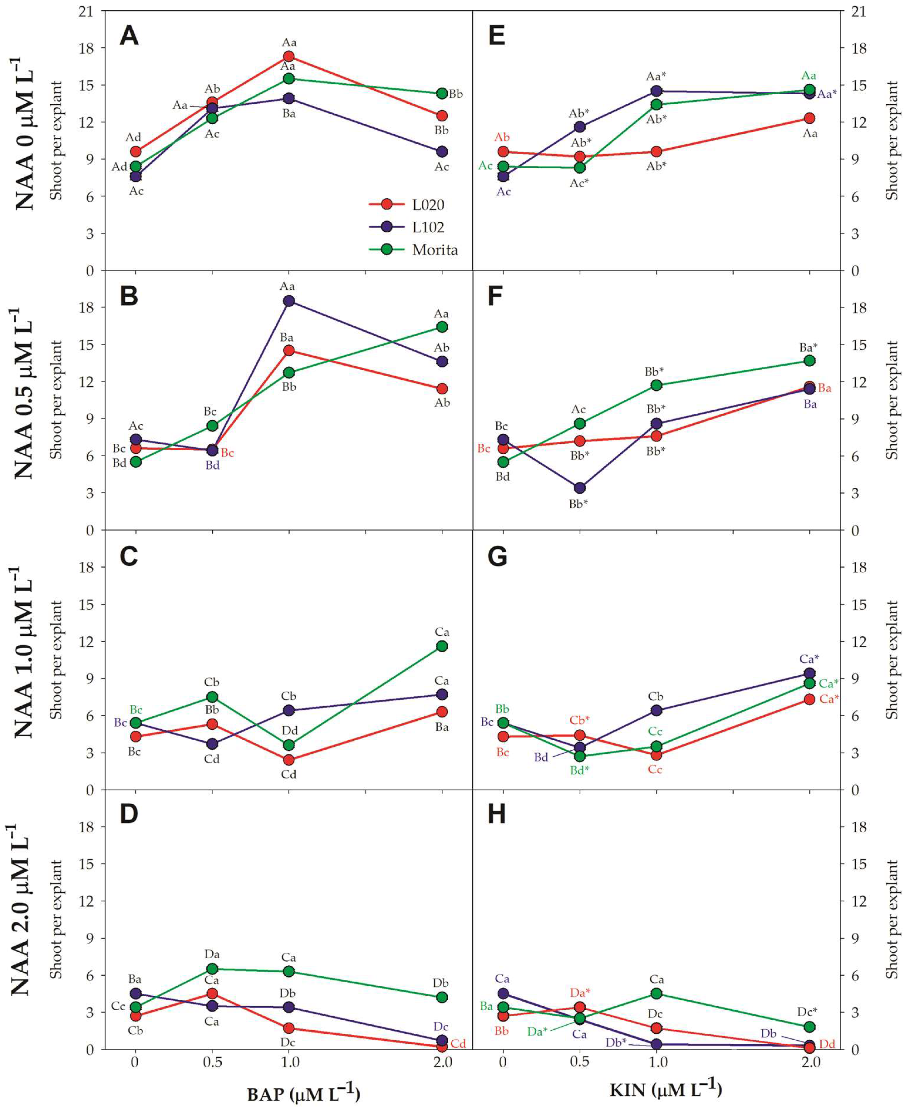
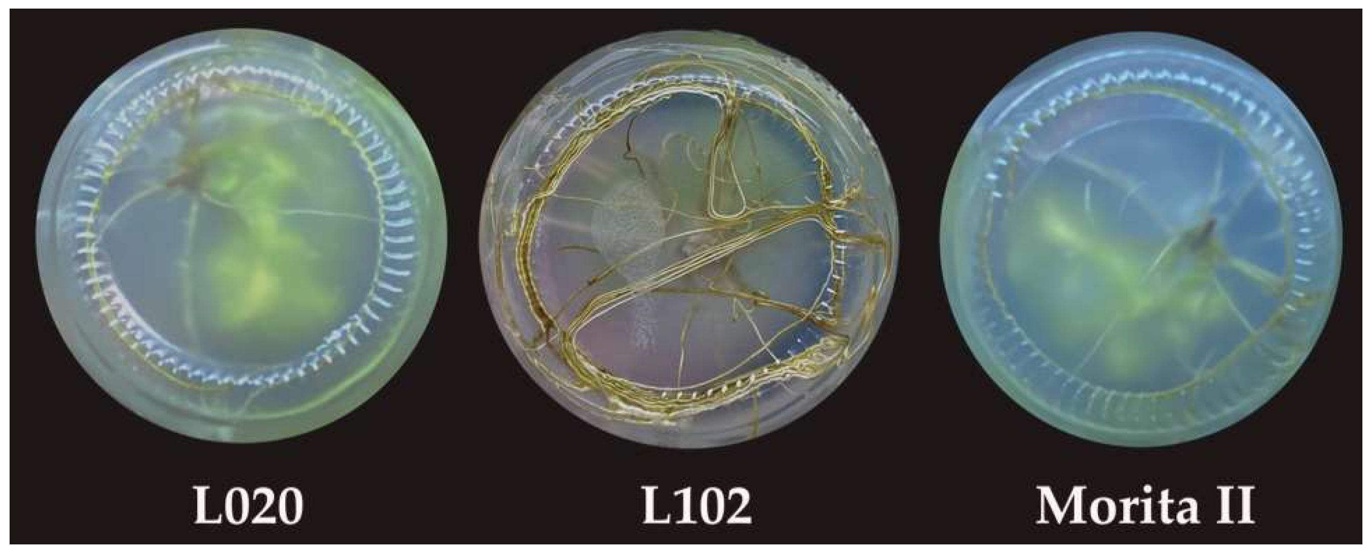
3.2. Shoot Multiplication
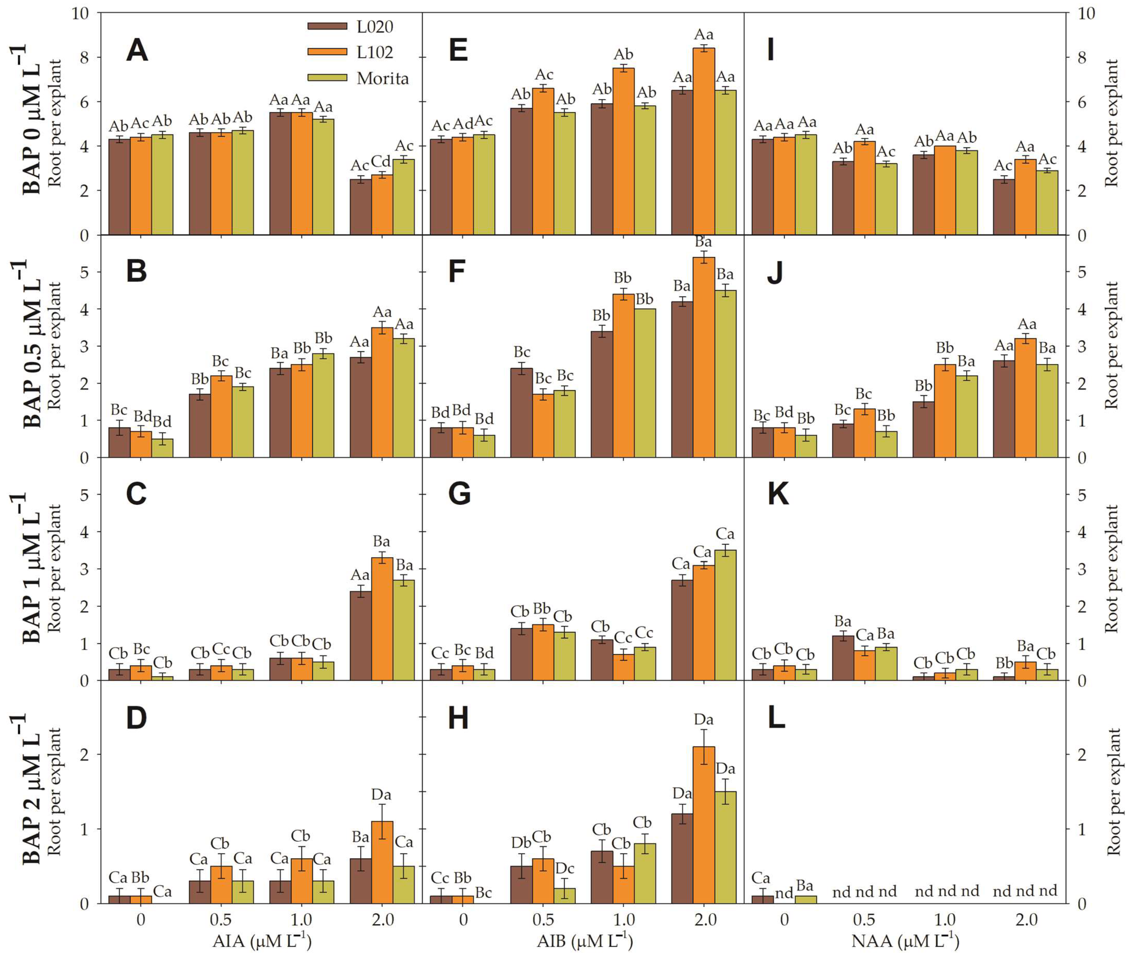
Seemingly, the description of our results seems to be genotype dependent. Moreover, Figure 2 shows that all genotypes had the best results under NAA 0 μM L−1 combined with BAP or KIN. The best concentration for BAP was 1 μM L−1 when used on genotypes L020 and L102. This concentration provides a better response (Figure 2A–D). Respectively, L020 and Morita II produce 17.30 ± 0.15 and 15.50 ± 0.17 shoots per explant. The genotype L102 obtains the best result in BAP (1 μM L−1) supplemented with NAA (0.5 μM L−1) with 18.50 ± 0.17 shoots per explant or 1.33-fold higher than BAP (1 μM L−1) with the absence of NAA. These conditions lead us to speculate that a concentration either lesser or higher than 1 μM L−1 of BAP produces significantly fewer shoots per explant. This pattern is well known in plant physiology, where it is identified as a response curve, where the shoot increases as the concentration of stimulant increases and then declines with higher concentrations. All treatments, for all genotypes, which combine an NAA higher than 1 μM L−1 result in fewer shoots per explant. Both L020 and L102 show the best results using a BAP 1 μM L−1. However, for Morita II, BAP 2 μM L−1 with an absence of NAA shows the best results with 1.29-fold higher shoot formation than those verified in BAP (1 μM L−1) supplemented with NAA (0.5 μM L−1). Just for a comparison, the mean of shoot per explant regenerated in NAA (0 μM L−1) was 5.82-, 3.65-, and 2.48-fold higher than those regenerated in NAA (2 μM L−1), respectively, for the L020, L102, and Morita II genotypes.
The shoot multiplication under KIN, irrespective of genotype, increased as the KIN concentration (2 μM L−1) increased. The best results were achieved when shoots were produced without NAA (Figure 2E–H). Respectively to L020, L102, and Morita II, KIN 2 μM L−1 promoted 1.28-, 1.88-, and 1.74-fold higher shoots than KIN 0 μM L−1. Similarly, all KIN treatments combined with NAA (0 μM L−1) promoted 5.15-, 6.32-, and 3.66-fold higher results than those treatments combined with NAA (2 μM L−1). For both BAP and KIN, an NAA greater than 0.5 μM L−1 reduces shoot production unless promoting calli formation under a higher concentration of NAA. The best results produced healthy plantlets (Figure 3) that were maintained in vitro until 5–7 cm tall when they were transferred to a rooting media.

3.3. Root Induction

Regardless of genotype, the IBA shows the best results to promote roots in the regenerated shoots. The rooting response increases in response to an increase in the IBA concentration. However, this is true only for BAP-free treatments. Respectively, for the genotypes L020, L102, and Morita II, the rooting response increases from 4.3 to 6.5, from 4.4 to 8.4, and from 4.5 to 6.5 when considering control with IBA (2 μM L−1; Figure 4 and Figure 5). Thus, the treatment composed of IBA (2 μM L−1) and BAP-free promoted 1.51-, 1.91-, and 1.44-fold more roots than the control plants treated by IBA and BAP-free. These treatments highlight a unique effectiveness in fostering robust root development. Similarly, BAP-free treatment promotes a median of 8.96-, 8.15-, and 8.92-fold more roots than with BAP (2 μM L−1).

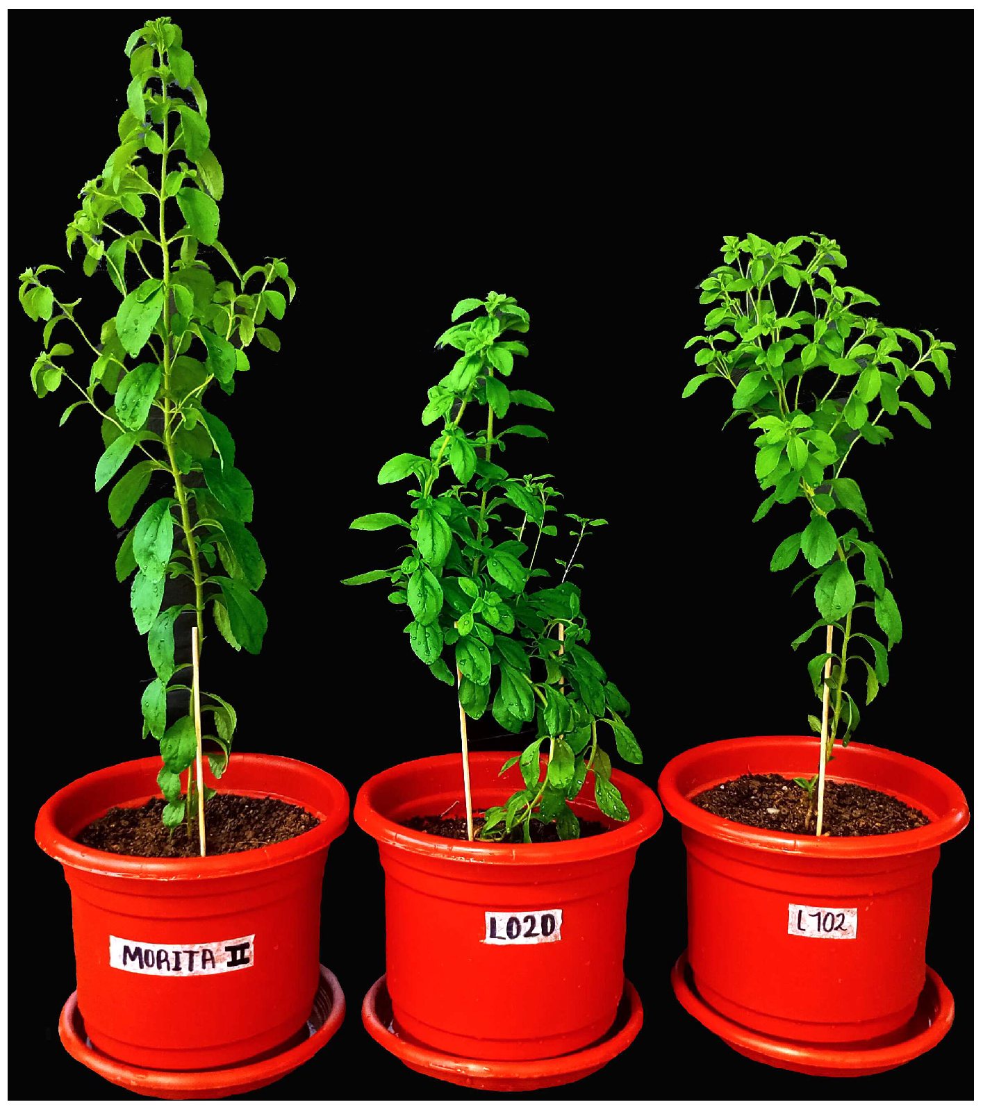
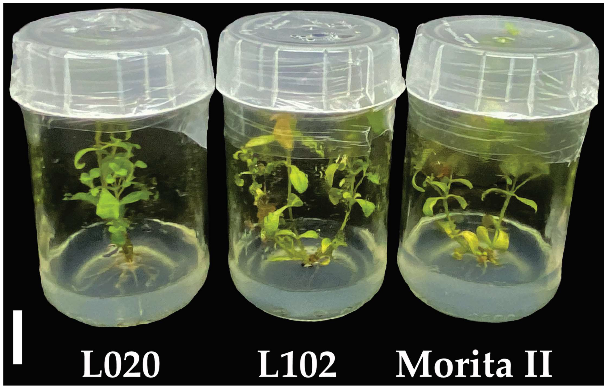
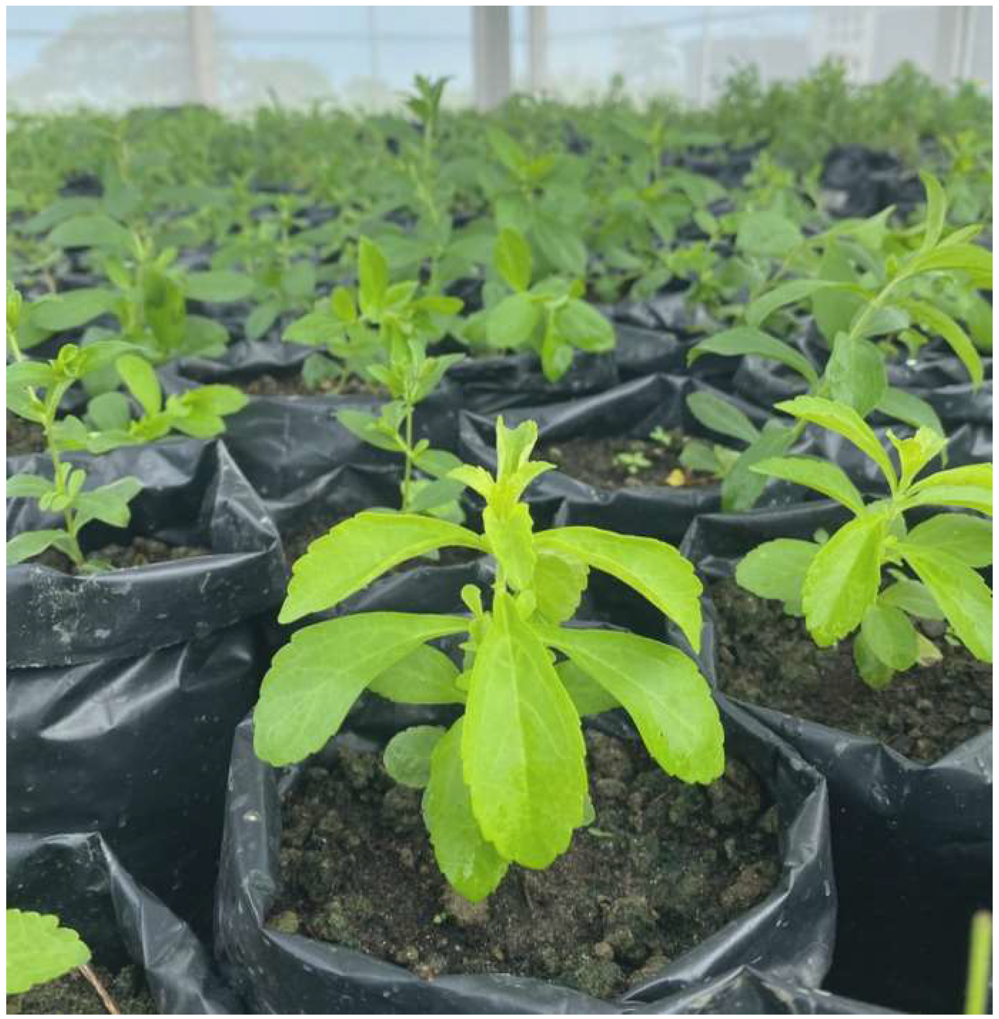
3.4. Acclimatization and Transference of Plantlets to Greenhouse

All healthy plants, without chlorosis or necrosis, were gently removed from the in vitro condition to the greenhouse. This procedure was completed in two steps: first, transplanting to conic polyethylene tubes and then directly into the soil in plastic bags (Figure 6). The survival rate of transferred plants was 97.6% (Figure 6).

4. Discussion

In this study, our focus was the optimization of the micropropagation of S. rebaudiana, addressing critical aspects such as explant disinfection, shoot multiplication, root induction, and acclimatization to ex vitro conditions. The results obtained provide valuable insights that significantly contribute to the mass propagation of S. rebaudiana elite genotype. Upon comparing these findings with the existing literature, both similarities and discrepancies emerge.
Concerning explant disinfection, the results using 0.5% sodium hypochlorite and 10 min of exposure proved highly effective, exhibiting insignificant contamination. This finding aligns with prior research suggesting the efficacy of sodium hypochlorite as a disinfectant for plant explants [46,47,48]. The combination of a moderate sodium hypochlorite concentration and reduced exposure time [49,50,51] emphasizes the importance of precision in disinfection to maximize cultivation efficiency. While other sources from the literature have explored various disinfection strategies, and some studies support the use of sodium hypochlorite [52,53,54], the variability in concentrations and exposure times underscores the need to adjust conditions based on species and micropropagation protocols [55,56]. Our results support the idea that optimizing these variables can lead to a more aseptic and efficient cultivation.
Our results identified specific treatments that promote shoot multiplication for each S. rebaudiana genotype, i.e., BAP 1 μM L−1 and BAP 1 μM L−1, supplemented with NAA 0.5 μM L−1, and BAP 2 μM L−1, supplemented with NAA 0.5 μM L−1, respectively, for the L020, L102, and Morita II genotypes. In accordance with previous research, specific combinations of auxins and cytokinins are crucial for inducing shoot multiplication in explants [54,57]. For example, Das et al. [58] observed that MS medium supplemented with 2 mg L−1 (~9.3 μM L−1) kinetin proved most effective for promoting multiple shoot multiplication, regenerating over 11 shoots from a single shoot tip explant within 35 days of culture medium while Yang et al. [59] described that the highest axillary shoot multiplication was reported in a medium containing BAP (2 mg/L~9 μM L−1), the same as reported by Rafiq et al. [60]. In another way, Hwang [61] described that the best performance (23.4 ± 2.1 shoots per explant) was obtained on MS medium supplemented with 2 mg L−1 IAA (~10 μM L−1) and 0.5 mg L−1 kinetin (KIN; ~2 μM L−1). Sivaram and Mukundan [23] describe that a combination of BAP (8.87 μM L−1) and IAA (5.71 μM L−1) resulted in a median of 11.2 shoots per explant. Ahmed et al. [22] described that the best combination for shoot multiplication was 1.5 mg L−1 BAP (~6.7 μM L−1) plus 0.5 mg L−1 BAP (~2.3 μM L−1), where 8.75 shoots per explant developed. Yang et al. [17] reported 13.8 shoots per explant under BAP 10 mg L−1 (~44 μM L−1). Rafiq et al. [5] describes that the best response to shoot formation was under 2.0 mg L−1 BAP (~9 μM L−1), which resulted in the formation of 8.33 shoots per explant. Vives et al. [18] describe the formation of four shoots per explant under BAP 0.25 mg L−1 (~2.2 μM L−1). Our best results, achieved on BAP 1 μM L−1 supplemented with NAA 0.5 μM L−1, were 1.65-fold higher than the best results described by Sivaram and Mukundan [23], 2.12-fold higher than the best results described by Ahmed et al. [22], 2.22-fold higher than those results reported by Rafiq et al. [5], 1.24-fold higher than those results reported by Yang et al. [17], and 4.63-fold higher than those results described by Vives et al. [18]. With respect to the results presented by Yang et al. [17], our results were 34% higher than those presented by the author. However, the results presented by Yang et al. [17] can lead to erroneous conclusions, because the best results presented by Yang et al. [17] were achieved with 44-fold higher BAP than concentration used in our study. Moreover, Jitendra et al. [20] describe that BAP (3.0 mg L−1) evoked the best response in shoot multiplication. With the incorporation of NAA or IAA, the shoot proliferation was improved; however, the shoots remained stunted. Incorporation of BAP or KIN into MS medium supported multiplication of shoots in culture. Rafiq et al. [5] and Jitendra et al. [20] reported that KIN was less effective than BAP to promote shoot multiplication on S. rebaudiana, results that are in accordance with our data. However, Espinal de Rueda et al. [62] describe KIN as the best PGR to achieve high shoot formation in S. rebaudiana. In any case, this study describes the formation of friable calli only in NAA treatments higher than 1 μM L−1 combined or not with BAP 1 and 2 μM L−1. These explants with calli were sub cultivated in lower concentrations of NAA and BAP 1 μM L−1 to try recuperating this shoot. In accordance with Dey et al. [6], Sivaram and Mukundan [23] friable calli are commonly found to develop from the base of microshoots when transferred to MS medium containing IBA or NAA, respectively, which, in turn, differentiated into microshoots [6]. This was also described by Yang et al. [59], a very bad tendency, suggesting that incorporation of NAA into the medium encourages challenges to the micropropagation of S. rebaudiana [17].
Rooting is a process that provides crucial insights for enhancing cultivation efficiency and root formation in in vitro plants [61,62]. Detailed attention to root averages per explant offers valuable cues on the most effective treatments, contributing to the optimization of propagation practices and boosting performance for in vitro plant production [63]. For root induction, Das et al. [58] found that the MS media without PGR worked dynamically. However, when supplemented with IAA and BAP, root induction had an adverse effect. In contrast, Rafiq et al. [5] describe that BAP 0.5 mg L−1 (~2.22 μM L−1) was the best treatment to promote root formation in nodular stem sections of S. rebaudiana. Alhady [64] described IBA as the best culture medium to promote root induction. Ahmed et al. [22] described the induction of 12.10 roots per explant under IAA 0.5 mg L−1 (~2.5 μM L−1) was 1.44-fold higher than our best results in MS supplemented with IBA 2.0 μM L−1. Yang et al. [17] describe the formation of eight roots per explant in NAA 10 mg L−1 (~54 μM L−1). Rafiq et al. [5] reported 6.2 roots per explant under NAA 0.5 mg L−1 2.7 μM L−1). Our best results were achieved under IBA (2 μM L−1) and formed 8.4 roots per explant. This result was 35.5% higher than the results reported by Rafiq et al. [5]. Our best results were aligned with those presented by Yang et al. [17]. However, the NAA concentration described in Yang et al. [17] was 27-fold higher than the concentration described in this study. Our results are in accordance with the finding of Jitendra et al. [20], which described that the full or half strength of the MS medium without any PGR failed to induce rooting of regenerated shoots. However, roots were observed on a medium containing IBA (1.0 mg L−1) where 3.6 roots per explant were reported. The number of roots decreased as the IBA concentration increased, as described in this study.
Additionally, in this study, we described a very high survival rate of 97.6%. These plants were hardened successfully and acclimatized to the greenhouse. These high survival rates agree with Ahmed et al. [22], who described a survival of 97.66% using IAA as a root inductor. However, our survival rates were 55% higher than those reported by Patel and Shah [65] using 0.1 mg L−1 IBA (~0.5 mM L−1), 24% higher than those reported by Rafiq et al. [5] or 39% higher than the survival rate reported by Ahmed et al. [22]. However, Alhady [64] describes IBA (1.0 or 2.0 mg/L; ~5 μM L−1) as the best culture medium to promote root induction (100%). These compact and sturdy plantlets are preferable to slender, fragile ones because they exhibit greater resilience against diverse environmental pressures, retain chlorophyll more effectively, and demonstrate superior viability [6].
In summary, this document outlines a procedure for the clonal micropropagation of S. rebaudiana elite genotypes. This study distinguishes itself from others for three key reasons: (1) the propagation of elite genotypes; (2) efficient shoot production; and (3) cost-effectiveness achieved through the use of lower concentrations of plant growth regulators than other studies described here. Rodriguez-Paez et al. [37] highlight in their study that the L020 genotype of S. rebaudiana shows high water use efficiency in converting energy into photoassimilates, making it an excellent choice for water-scarce regions. On the other hand, the L102 genotype of S. rebaudiana exhibits a high photosynthetic rate but relies on irrigation, resulting in lower water use efficiency. Furthermore, L020 boasts a total glycoside content 1.2-fold higher than Morita II, and 1.9-fold higher Rebaudioside A content than Morita II (considered the best genotype used in the majority of micropropagation protocols), showing a better taste in food and beverages production and consumption. The L102 genotype has only 70% of the total glycoside content compared to Morita. Additionally, L020 does not flower (at least in our study, encompassing 5 years and enabling multiple harvests), whereas L102 experiences late flowering and has an erect architecture, facilitating mechanized harvesting.

5. Conclusions

Our results make substantial contributions to the field of S. rebaudiana micropropagation by offering specific strategies to improve explant disinfection, shoot multiplication, and plantlets regeneration. This research successfully identified an optimized first step of micropropagation establishment, showcasing 0.5% sodium hypochlorite for 10 min as the most favorable disinfectant. Emphasis lies on the critical balance of concentration and exposure time for efficient contaminant reduction. In shoot and plantlet regeneration, the literature supports the pivotal role of hormonal regulation in caulogenesis and rhizogenesis, with various studies exploring specific combinations to optimize this process. In this study, we highlighted that the choice of cytokinin played a pivotal role in determining the efficacy of shoot multiplication. However, genotypic variability emphasizes the need to tailor protocols to the specific characteristics of each S. rebaudiana genotype. The combination of phytohormone responses in each of the genotypes underscores the need for adaptive protocols, allowing producers to maximize the micropropagation efficiency based on particular genetic traits. We have also outlined specific conditions fostering exceptional root development in each evaluated S. rebaudiana genotype. Choosing the most suitable treatment is crucial to maximize shoot production per explant in Stevia rebaudiana tissue culture. This study establishes a robust foundation for future research, enabling the continuous optimization of micropropagation protocols and contributing to the advancement of the Stevia industry. Moreover, the potential for cloning is immense, and therefore, axillary shoot proliferation may be used for the rapid multiplication of this sweetening plant on a large scale.

Author Contributions

Conceptualization, L.A.R.-P., M.I.O.-O. and N.V.R.; methodology, L.A.R.-P., M.F.P., Y.Y.P.-R., A.M.J.-R., O.J.G.-A. and J.d.D.J.-N.; formal analysis, L.A.R.-P. and M.F.P.; investigation, L.A.R.-P., M.F.P. and Y.Y.P.-R.; writing—original draft preparation, L.A.R.-P., J.d.D.J.-N., L.E.O.Z., I.E.S.-P. and M.F.P.; writing—review and editing, L.A.R.-P., M.F.P., A.M.J.-R., O.J.G.-A., L.E.O.Z., J.d.D.J.-N., I.E.S.-P. and N.V.R.; supervision, M.I.O.-O. and N.V.R.; funding acquisition, L.A.R.-P., A.J.-O., I.E.S.-P., L.E.O.Z., J.d.D.J.-N. and E.C.-C. All authors have read and agreed to the published version of the manuscript.

Funding

This research was financially supported by Facultad de Ciencias Agrícolas, Universidad de Córdoba, CO, Colombia, through the program: Production systems and food security. Research line: Agri-food production systems and hydrobiological resources. Grants: FCA-04-19.

Institutional Review Board Statement

Not applicable.

Informed Consent Statement

Not applicable.

Data Availability Statement

Data are contained within the article. Additional data can be obtained by contacting the corresponding author of the article.

Acknowledgments

The authors thank Facultad de Ciencias Agrícolas, Universidad de Córdoba, CO, Colombia, and Instituto de Biotecnología de las Plantas, Universidad Central “Marta Abreu” de Las Villas, Cuba, for assistance with the conditions necessary for this study.

Conflicts of Interest

The authors declare no conflicts of interest.

References

  1. Soejarto, D.D.; Compadre, C.M.; Kinghorn, D. Ethnobotanical notes in Stevia. Bot. Mus. Leafl. 1983, 29, 1–25. [Google Scholar] [CrossRef]
  2. Soejarto, D.D. Botany of Stevia and Stevia rebaudiana. In Stevia: The Genus Stevia; Kinghorn, A.D., Ed.; CRC Press: London, UK, 2001; pp. 22–28. [Google Scholar]
  3. Pande, S.S.; Gupta, P. Plant tissue culture of Stevia rebaudiana (Bertoni): A review. J. Pharmacogn. Phytother. 2013, 5, 26–33. [Google Scholar]
  4. Miyagawa, H.; Fujioka, N.; Kohda, H.; Yamasaki, K.; Taniguchi, K.; Tanaka, R. Studies on the tissue culture of Stevia rebaudiana and its components; (II). Induction of shoot primordia. Planta Med. 1986, 4, 321–323. [Google Scholar] [CrossRef] [PubMed]
  5. Rafiq, M.; Dahot, M.U.; Mangrio, S.M.; Naqvi, H.A.; Qarshi, I.A. In vitro clonal propagation and biochemical analysis of field established Stevia rebaudiana Bertoni. Pak. J. Bot. 2007, 39, 2467–2474. [Google Scholar]
  6. Dey, A.; Kundu, S.; Bandyopadhyay, A.; Bhattacharjee, A. Efficient micropropagation and chlorocholine chloride induced stevioside production of Stevia rebaudiana Bertoni. C. R. Biol. 2013, 336, 17–28. [Google Scholar] [CrossRef] [PubMed]
  7. Brandle, J.E.; Rosa, N. Heritability for yield, leaf: Stem ratio and stevioside content estimated from a landrace cultivar of Stevia rebaudiana. Can. J. Plant Sci. 1992, 72, 1263–1266. [Google Scholar] [CrossRef]
  8. Wibbens, K. Executive Review of the Stevia Food System: Plan B Project. Market Research Future. 2023. Available online: https://n9.cl/steviamarket (accessed on 11 April 2024).
  9. Martyn, D.; Darch, M.; Roberts, A.; Lee, H.Y.; Yaqiong Tian, T.; Kaburagi, N.; Belmar, P. Low-/No-Calorie Sweeteners: A Review of Global Intakes. Nutrients 2018, 10, 357. [Google Scholar] [CrossRef] [PubMed]
  10. Rivera-Avilez, J.A.; Jarma-Orozco, A.; Pompelli, M.F. Stevia rebaudiana Bertoni: The interaction of night interruption on gas exchange, flowering delay, and steviol glycosides synthesis. Horticulturae 2021, 7, 543. [Google Scholar] [CrossRef]
  11. Iatridis, N.; Kougioumtzi, A.; Vlataki, K.; Papadaki, S.; Magklara, A. Anti-cancer properties of Stevia rebaudiana; More than a sweetener. Molecules 2022, 27, 1362. [Google Scholar] [CrossRef]
  12. Liu, J.C.; Kao, P.K.; Chan, P.; Hsu, Y.H.; Hou, C.C.; Lien, G.S.; Hsieh, M.H.; Chen, Y.J.; Cheng, J.T. Mechanism of the antihypertensive effect of stevioside in anesthetized dogs. Pharmacology 2003, 67, 14–20. [Google Scholar] [CrossRef] [PubMed]
  13. Planas, G.M.; Kuć, J. Contraceptive properties of Stevia rebaudiana. Science 1968, 162, 1007. [Google Scholar] [CrossRef] [PubMed]
  14. Gamboa, F.; Margarita, C. Antimicrobial potential of extracts from Stevia rebaudiana leaves against bacteria of importance in dental caries. Acta Odontol. Latinoam. 2012, 25, 171–175. [Google Scholar] [PubMed]
  15. Debnath, M.; Ashwath, N.; Midmore, D.J. Physiological and morphological responses to abiotic stresses in two cultivars of Stevia rebaudiana (Bert.) Bertoni. S. Afr. J. Bot. 2019, 123, 124–132. [Google Scholar] [CrossRef]
  16. Gantait, S.; Das, A.; Banerjee, J. Geographical distribution, botanical discription and self-incompatibility mechanism of genus Stevia. Sugar Tech. 2018, 20, 1–10. [Google Scholar] [CrossRef]
  17. Yang, Y.-W.; Hsing, Y.-I.; Chang, W.-C. Clonal propagation of Stevia rebaudiana Bertoni through axillary shoot proliferation in vitro. Bot. Bull. Acad. Sin. 1981, 22, 57–62. [Google Scholar]
  18. Vives, K.; Andújar, I.; Lorenzo, J.C.; Concepción, O.; Hernández, M.; Escalona, M. Comparison of different in vitro micropropagation methods of Stevia rebaudiana B. including temporary immersion bioreactor (BIT®). Plant Cell Tiss. Organ. Cult. 2017, 131, 195–199. [Google Scholar] [CrossRef]
  19. Felippe, G.M.; Lucas, N.M.C.; Behar, l.; Oliveira, M.A.C. Observações a respeito de germinação de Stevia rebaudiana Bert. Hoehnea 1971, 1, 81–93. [Google Scholar]
  20. Jitendra, M.; Monika, S.; Ratan, S.D.; Priyanka, G.; Priyanka, S.; Kiran, D.J. Micropropagation of an Anti diabetic Plant—Stevia rebaudiana Bertoni, (Natural Sweetener) in Hadoti Region of South-East Rajasthan, India. J. Biol. Sci. 2012, 1, 37–42. [Google Scholar]
  21. Ferreira, C.M.; Handro, W. Micropropagation of Stevia rebaudiana through leaf explants from adult plants. Planta Med. 1988, 54, 157–160. [Google Scholar] [CrossRef]
  22. Ahmed, M.B.; Salahin, M.; Karim, R.; Razvy, M.A.; Hannan, M.M.; Sultana, R.; Hossain, M.; Islam, R. An efficient method for in vitro clonal propagation of a newly introduced sweetener plant (Stevia rebaudiana Bertoni.) in Bangladesh. Am. Eurasian J. Sci. Res. 2007, 2, 121–125. [Google Scholar]
  23. Sivaram, L.; Mukundan, U. In vitro culture studies on Stevia rebaudiana. In Vitro Cell. Dev. Biol.-Plant 2003, 39, 520–523. [Google Scholar] [CrossRef]
  24. Medorio-García, H.P.; Hernández-Domínguez, E.; Andueza-Noh, R.H.; López-Aguilar, D.R.; Ramírez-Mosqueda, M.A. Micropropagation of Stevia (Stevia rebaudiana Bert.) in RITA. In Micropropagation Methods in Temporary Immersion Systems. Methods in Molecular Biology; Ramírez-Mosqueda, M.A., Cruz-Cruz, C.A., Eds.; Humana: New York, NY, USA, 2024; Volume 2759. [Google Scholar]
  25. Razak, U.N.A.A.; Ong, C.B.; Yu, S.T.; Lau, L.K. In Vitro Micropropagation of Stevia rebaudiana Bertoni in Malaysia. Braz. Arch. Biol. Technol. 2014, 57, 23–28. [Google Scholar] [CrossRef]
  26. Chugh, S.; Guha, S.; Rao, I.U. Micropropagation of orchids: A review on the potential of different explants. Sci. Hort. Amst. 2009, 122, 507–520. [Google Scholar] [CrossRef]
  27. Beruto, M.; Lanteri, L.; Portogallo, C. Micropropagation of tree peony (Paeonia suffruticosa). Plant Cell Tiss. Org. Cult. 2004, 79, 249–255. [Google Scholar] [CrossRef]
  28. Wojciechowicz, M.K. Comparison of regenerative potential of petals, stamens and pistils of five Sedum species in vitro. Biodiv Res. Conserv. 2007, 5–8, 87–94. [Google Scholar]
  29. Khatun, R.; Islam, S.M.S.; Miah, M.A.B. Studies on plant regeneration efficiency through in vitro micropropagation and anther culture of twenty five rice cultivars in Bangladesh. J. Appl. Sci. Res. 2010, 6, 1705–1711. [Google Scholar]
  30. Sharma, V. Orchid micropropagation: Regeneration competence of anther culture. J. Biotechnol. Biomater. 2012, 2, 1000149. [Google Scholar] [CrossRef]
  31. Tamura, Y.; Nakamura, S.; Fukui, H.; Tabata, M. Comparison of Stevia plants grown from seeds, cuttings and stem tip cultures for growth and sweet diterpene glycosides. Plant Cell Rep. 1984, 3, 180–182. [Google Scholar] [CrossRef] [PubMed]
  32. Linsmaier, E.M.; Skoog, F. Organic growth requirements of tobacco tissue cultures. Physiol. Plant 1965, 18, 100–127. [Google Scholar] [CrossRef]
  33. Zou, Z.; Yang, L.; Gong, J.; Mo, Y.; Wang, J.; Cao, J.; An, F.; Xie, G. Genome-wide identification of Jatropha curcas aquaporin genes and the comparative analysis provides insights into the gene family expansion and evolution in Hevea brasiliensis. Front. Plant Sci. 2016, 7, 395. [Google Scholar] [CrossRef]
  34. Ramírez-Mosqueda, M.A.; Iglesias-Andreu, L.G.; Ramírez-Madero, G.; Hernández-Rincón, E.U. Micropropagation of Stevia rebaudiana Bert. in temporary immersion systems and evaluation of genetic fidelity. S. Afr. J. Bot. 2016, 106, 238–243. [Google Scholar] [CrossRef]
  35. Naranjo, E.J.; Fernandez Betin, O.; Urrea Trujillo, A.I.; Callejas Posada, R.; Atehortúa Garcés, L. Effect of genotype on the in vitro regeneration of Stevia rebaudiana via somatic embryogenesis. Acta Biol. Col. 2015, 21, 87–98. [Google Scholar] [CrossRef]
  36. Tavarini, S.; Passera, B.; Angelini, L.G. Crop and steviol glycoside improvement in Stevia by breeding. In Food Chemistry, Function and Analysis; Wölwer-Rieck, U., Ed.; Royal Society of Chemistry: Cambridge, UK, 2019; pp. 1–31. [Google Scholar]
  37. Rodriguez-Paez, L.A.; Jimenez-Ramirez, A.M.; Pompelli, M.F.; Pineda-Rodriguez, Y.Y.; Jarma-Orozco, A.; Jaraba-Navas, J.D.; Aramendiz-Tatis, H.; Combatt-Caballero, E.; Oloriz-Ortega, M.I.; Rodríguez, N.V. Physiological and enzymatic evaluation of selected genotypes of Stevia rebaudiana Bertoni. Agronomy 2023, 13, 403. [Google Scholar] [CrossRef]
  38. Rodriguez-Paez, L.A.; Jaraba-Navas, J.D.D.; Pineda-Rodriguez, Y.Y.; Begambre-Hernandez, M.; Pompelli, M.F.; Jimenez-Ramirez, A.M.; Gil-Rocha, A.; Jarma-Orozco, A.; Combatt-Caballero, E.; Aviña-Padilla, K.; et al. Natural biocontrol of Athelia rolfsii isolate INVEPAR-05 in Stevia rebaudiana Bertoni: Exploring the biocontrol potential of native Trichoderma spp. Strains. Preprints 2023, 2023, 2023081062. [Google Scholar] [CrossRef]
  39. Jimenez Ramirez, A. Caracterización Morfoagronómica de Clones de Estevia (Stevia rebaudiana Bert.) en la Región Caribe de Colombia. Master Thesis, Universidad de Córdoba, Córdoba, Spain, 2023. Available online: https://repositorio.unicordoba.edu.co/server/api/core/bitstreams/c2194056-8603-4da7-8b1d-e4c4723ebd2c/content (accessed on 22 February 2024).
  40. Ariza-González, A.R.; Jarma-Orozco, A.; Jaraba-Navas, J.D.; Pico-González, A.I.; Herazo-Cárdenas, D.S.; Vegliante Arrieta, D.; Vallejo-Isaza, A.; Pineda-Rodriguez, Y.Y.; Rodriguez-Paez, L.A.; Pompelli, M.F. Net photosynthesis and biomass production in stevia, eggplant, and cowpea can be improved by fertilization with cyanobacteria (Limnospira maxima). Horticulturae 2023, 9, 1309. [Google Scholar] [CrossRef]
  41. Jaraba-Navas, J.D.; Combatt-Caballero, E.M.; Jarma-Orozco, A.; Pineda-Rodriguez, Y.Y.; Rodriguez-Paez, L.A. Trichoderma harzianum isolate INVEPAR-T01 5.8S ribosomal RNA gene, partial sequence; internal transcribed spacer 2, complete sequence; and large subunit ribosomal RNA gene, partial sequence. GenBank 2021. Preprints. [Google Scholar]
  42. Combatt-Caballero, E.; Hernández-Burgos, J.; Jarma-Orozco, A.; Jaraba-Navas, J.; Rodríguez-Páez, L. Macroelements and mcroelements in the soil and their relationship with the content of steviol glucosides in Stevia rebaudiana Bert from five regions of Colombia. Horticulturae 2021, 7, 547. [Google Scholar] [CrossRef]
  43. Murashige, T.; Skoog, F. A revised medium for rapid growth and bio assays with tobacco tissue cultures. Physiol. Plant 1962, 15, 473–497. [Google Scholar] [CrossRef]
  44. Rivero, A.M.; Ramírez-Mosqueda, M.A.; Frómeta, O.M.; Morgado, M.M.E.; Paneca, M.R.; Escriba, R.C.R.; Gradaille, M.A.D.; Bello-Bello, J.J. Influence of Vitrofural® on sugarcane micropropagation using temporary immersion system. Plant Cell Tiss. Org. Cult. 2020, 141, 447–453. [Google Scholar] [CrossRef]
  45. Likert, R. A technique for the measurement of attitudes. Arch. Psychol. 1932, 140, 44–53. [Google Scholar]
  46. Pompelli, M.F.; Guerra, M.P. Ex situ conservation of Dyckia distachya: An endangered bromeliad from South Brazil. Crop Breed. Appl. Biotechnol. 2004, 4, 273–279. [Google Scholar] [CrossRef]
  47. Pompelli, M.F.; Guerra, M.P. Micropropagation enables the mass propagation and conservation of Dyckia distachya Hassler. Crop Breed. Appl. Biotechnol. 2005, 5, 117–126. [Google Scholar] [CrossRef]
  48. Smith, K.; Guo, S.; Zhu, Q.; Dong, X.; Liu, S. An evaluation of the environmental benefit and energy footprint of China’s stricter wastewater standards: Can benefit be increased? J. Clean. Prod. 2019, 219, 723–733. [Google Scholar] [CrossRef]
  49. Pais, A.K.; da Silva, A.P.; de Souza, J.C.; Teixeira, S.L.; Ribeiro, J.M.; Peixoto, A.R.; da Paz, C.D. Sodium hypochlorite sterilization of culture medium in micropropagation of Gerbera hybrida cv. Essandre. Afr. J. Biotechnol. 2016, 15, 1995–1998. [Google Scholar] [CrossRef]
  50. Tiwari, A.K.; Tripathi, S.; Lal, M.; Mishra, S. Screening of some chemical disinfectants for media sterilization during in vitro micropropagation of sugarcane. Sugar Tech. 2012, 14, 364–369. [Google Scholar] [CrossRef]
  51. Peiris, S.E.; De Silva, E.D.U.D.; Edussuriya, M.; Attanayake, A.M.U.R.K.; Peiris, B.C.N. CSUP technique: A low cost sterilization method using sodium hypochlorite to replace the use of expensive equipment in micropropagation. J. Natl. Sci. Found. Sri Lanka 2012, 40, 49–54. [Google Scholar] [CrossRef]
  52. Jawad, H.A.; Hussein, N.H.; Salef, F.F. Effect of sodium chloride on stevia plant growth in vitro. Int. J. Appl. Sci. Technol. 2022, 4, 171. [Google Scholar] [CrossRef]
  53. Shaafi, B.; Mosavi, S.; Abdollahi, M.R.; Sarikhani, H. The optimized protocols for production, adaptation and keeping of the produced artificial seeds from encapsulated lateral buds in Stevia Rebaudiana (Bertoni). Agrotech. Ind. Crops 2021, 1, 24–35. [Google Scholar] [CrossRef]
  54. Jadid, N.; Anggraeni, S.; Ramadani, M.R.N.; Arieny, M.; Mas’ud, F. In Vitro propagation of Indonesian stevia (Stevia rebaudiana) genotype using axenic nodal segments. BMC Res. Notes 2024, 17, 45. [Google Scholar] [CrossRef]
  55. Ghose, A.K.; Abdullah, S.N.A.; Md Hatta, M.A.; Megat Wahab, P.E. In Vitro regeneration of stevia (Stevia rebaudiana Bertoni) and evaluation of the impacts of growth media nutrients on the biosynthesis of steviol glycosides (SGs). Agronomy 2022, 12, 1957. [Google Scholar] [CrossRef]
  56. Ahmed, K.Z.; Osman, S.A.-M.; Saber, S.M.; Abdel-Hamed, A.A. An efficient in vitro culture protocol of stevia plants (Stevia rebaudiana Bertoni var. chaine.2) with cytogenetical studies. J. Agric. Res. Dev. 2018, 38, 93–113. [Google Scholar] [CrossRef]
  57. Vilariño, S.; Florido, M.D.C.; García, J.L.; Cantos, M. Effects of culture system and substrate composition on micropropagated plantlets of two varieties of Stevia rebaudiana Bert. Physiologia 2023, 3, 74–85. [Google Scholar] [CrossRef]
  58. Das, A.; Gantait, S.; Mandal, N. Micropropagation of an elite medicinal plant: Stevia rebaudiana Bert. Int. J. Agric. Res. 2011, 6, 40–48. [Google Scholar] [CrossRef]
  59. Hwang, S.J. Rapid in vitro propagation and enhanced stevioside accumulation in Stevia rebaudiana Ber. J. Plant Biol. 2006, 49, 267–270. [Google Scholar] [CrossRef]
  60. Espinal de Rueda, D.; Delvalle, W.; Cifuentes, E.; Ramia, N. Propagación in vitro de Stevia rebaudiana B: A partir de segmentos nodales. Ceiba 2006, 47, 11–18. [Google Scholar] [CrossRef]
  61. Kumar, N.; Reddy, M.P. In Vitro plant propagation: A review. J. Envirom Sci. 2011, 27, 61–72. [Google Scholar] [CrossRef]
  62. Jangid, V.K.; Senthil-Kumar, M.; Chandran, D.; Sinharoy, S. Callus induction and efficient in vitro plant regeneration protocol for Chickpea. Plant Cell Tiss. Organ. Cult. 2024, 156, 21. [Google Scholar] [CrossRef]
  63. Raha, P.; Saha, G.; Khatua, I.; Bandyopadhyay, T.K. Direct shoot from root and true-to-type micropropagation of Limonium “Misty Blue” in partially immersed culture on an aluminum mesh raft. In Micropropagation Methods in Temporary Immersion Systems. Methods in Molecular Biology; Ramírez-Mosqueda, M.A., Cruz-Cruz, C.A., Eds.; Humana: New York, NY, USA, 2024; Volume 2759. [Google Scholar]
  64. Alhady, M.R.A.A. Micropropagation of Stevia rebaudiana Bertoni.: A new sweetening crop in Egypt. Glob. J. Biotechnol. Biochem. 2011, 6, 178–182. [Google Scholar]
  65. Patel, R.M.; Shah, R.R. Regeneration of Stevia plant through callus culture. Indian J. Pharm. Sci. 2009, 71, 46–50. [Google Scholar] [CrossRef]
Figure 1. Three-months-old Morita II, L020, and L102, Stevia rebaudiana mother plants established in a greenhouse.
Horticulturae 10 00404 g001
Figure 2. Effect of 6-benzylaminopurine (BAP; AD) or kinetin (KIN; EH) supplemented with Naphthaleneacetic acid (NAA) on shoot multiplication in three genotypes (L020, L102, and Morita II) of Stevia rebaudiana. Means followed by different lowercase letters denote statistical differences between BAP or KIN concentration in the same NAA concentration and genotype. Means followed by different uppercase letters denote statistical differences between NAA concentration in the same BAP or KIN concentration and genotype (SNK; p ≤ 0.05), and * denotes a statistical difference between BAP and KIN in the same NAA concentration and genotype. All values denote means ± SE. n = 10.
Horticulturae 10 00404 g002
Figure 3. Plantlets regenerated from the best treatment for each Stevia rebaudiana genotype. For L020, L102, and Morita II, the best treatment was, respectively, BAP 1 μM L−1, BAP 1 μM L−1, supplemented with NAA 0.5 μM L−1, and BAP 2 μM L−1, supplemented with NAA 0.5 μM L−1. This picture shows how healthy the plants were slightly before the process and rooting.
Horticulturae 10 00404 g003
Figure 4. Effect of indole-3-acetic acid (IAA; AD), indole-3-butyric acid (IBA; EH), and Naphthaleneacetic acid (NAA; IL) supplemented with 6-benzylaminopurine (BAP) on root induction in three genotypes (L020, L102, and Morita II) of Stevia rebaudiana. Means followed by different lowercase letters denote statistical differences between IAA, IBA, or NAA concentration in the same BAP concentration and genotype. Means followed by different uppercase letters denote statistical differences between BAP concentration in the same IAA, IBA, or NAA concentration and genotype (SNK; p ≤ 0.05). All values denote means ± SE. n = 10.
Horticulturae 10 00404 g004
Figure 5. Rooted L020, L102, and Morita II S. rebaudiana plantlets. The rooting was promoted with indole-3-butyric acid (IBA; 2 μM L−1) after 30 days on seedling rooting medium.
Horticulturae 10 00404 g005
Figure 6. Full healthy Stevia rebaudiana acclimatized plants after 45 days. In this picture, all genotypes were merged to show the uniformity of acclimatized plants.
Horticulturae 10 00404 g006
Table 1. Contamination and survival rate of nodal explants excised from Morita II Stevia rebaudiana genotype and transferred to a basal medium.
NaOCl (%)Time (min)ContaminationSurvival Rate
002.6 ± 0.0 Aa1.0 ± 0.0 Ab
52.7 ± 0.1 Ca1.2 ± 0.1 Cab
102.8 ± 0.0 Ba1.5 ± 0.1 Da
152.9 ± 0.1 Ba1.8 ± 0.2 Da
0.502.6 ± 0.0 Ab1.0 ± 0.0 Ad
52.9 ± 0.2 Bb2.1 ± 0.1 Bc
105.0 ± 0.0 Aa4.9 ± 0.1 Aa
155.0 ± 0.0 Aa2.5 ± 0.2 Ab
102.6 ± 0.0 Ac1.0 ± 0.0 Ac
53.0 ± 0.1 Bb2.3 ± 0.1 Bb
104.9 ± 0.1 Aa4.0 ± 0.1 Ba
155.0 ± 0.1 Aa2.1 ± 0.1 Bb
1.502.6 ± 0.0 Ac1.0 ± 0.0 Ad
53.9 ± 0.3 Ab3.8 ± 0.1 Aa
105.0 ± 0.0 Aa2.6 ± 0.2 Cb
155.0 ± 0.0 Aa2.1 ± 0.2 Bc
Values followed by lowercase letters denote statistical differences between NaOCl exposure time in the same sodium hypochlorite (NaOCl) concentration, and uppercase letters denote statistical differences between NaOCl concentration at the same exposure time. The values denote a mean (±SE; n = 5; SNK, p ≤ 0.05) of the notes attributed by the Likert [45] scale in descending order of efficiency. In this scale, “5”, complete sterilization efficiency, without any point of infection; “4”, good disinfection; “3” denotes medium disinfection that needs some adjustments to sterilization concentrations or time; “2” partially contaminated, requiring substantial adjustments in the disinfection procedures; and “1” completely infected without any possibility to recover the explants. For survival, “5” denotes that all five flasks contained a green and healthy plant; “4”, at least four flasks contained a green and healthy plant; “3”, at least three flasks contained a green and healthy plant; “2” at least two flasks contained a green and healthy plant; and “1” one or no flasks contained a green and healthy plant.
Disclaimer/Publisher’s Note: The statements, opinions and data contained in all publications are solely those of the individual author(s) and contributor(s) and not of MDPI and/or the editor(s). MDPI and/or the editor(s) disclaim responsibility for any injury to people or property resulting from any ideas, methods, instructions or products referred to in the content.

