Article

Lasiodiplodia iraniensis and Diaporthe spp. Are Associated with Twig Dieback and Fruit Stem-End Rot of Sweet Orange, Citrus sinensis, in Florida

1
Department of Agricultural, Forest and Food Sciences (DISAFA), University of Torino, 10095 Grugliasco, Italy
2
Interdepartmental Centre for the Innovation in the Agro-Environmental Sector, AGROINNOVA, University of Torino, 10095 Grugliasco, Italy
3
Department of Agriculture, Food and Environment (Di3A), University of Catania, 95123 Catania, Italy
4
Keyplex, 400 N. New York Ave Suite 200, Winter Park, FL 32789, USA
5
Plant Pathology Laboratory, School of Plant and Environmental Sciences, Alson H. Smith Jr. Agricultural Research and Extension Center, Virginia Polytechnic Institute and State University, Winchester, VA 24061, USA
*
Author to whom correspondence should be addressed.
Horticulturae 2024, 10(4), 406; https://doi.org/10.3390/horticulturae10040406
Submission received: 29 March 2024 / Revised: 11 April 2024 / Accepted: 12 April 2024 / Published: 17 April 2024
(This article belongs to the Section Plant Pathology and Disease Management (PPDM))

Abstract

:
Florida ranks among the most important citrus growing regions in the USA. The present study investigates the occurrence, diversity, and pathogenicity of fungal species associated with symptomatic sweet orange (Citrus sinensis) cv. Valencia plants and fruit. The survey was conducted on twigs and fruit collected in Southwest Florida during 2022. Based on morphological and molecular characteristics, the identified isolates belonged to the species Lasiodiplodia iraniensis, Diaporthe pseudomangiferae, and Diaporthe ueckerae. The pathogenicity of representative isolates was evaluated on citrus fruit and plants. Lasiodiplodia iraniensis was the most virulent on fruit and plants, followed by Diaporthe pseudomangiferae. Diaporthe ueckerae had the lowest virulence on fruit, and it was not pathogenic to plants. In vitro tests were performed to assess the effect of temperature on mycelial radial growth. The optimum temperature of growth ranged from 26.0 to 28.4 °C for all the evaluated species, and L. iraniensis showed the fastest mycelial growth. This study represents the first report of L. iraniensis as a causal agent of tree dieback and fruit stem-end rot on C. sinensis worldwide. Moreover, D. pseudomangiferae and D. ueckerae are reported here for the first time in association with citrus diseases worldwide.

1. Introduction

Citrus is one of the world’s most economically important fruit crops, appreciated by consumers for its nutritional value, characterized by a high dose of vitamin C, sugar, organic acids, amino acids, and minerals [1]. Citrus cultivation is distributed in 160 countries of tropical and subtropical regions, spanning six continents on a total area of 12.7 million hectares [2,3]. From 2022 to 2023, 158 million tons of citrus were harvested worldwide [3]. The USA is one of the largest world citrus producers, after China and Brazil, with an economic value of USD 2.91 billion, with California, Florida, Arizona, and Texas representing the main areas [4]. Florida citrus orchards cover about 230,266 hectares, counting more than 74 million citrus trees, which produce 36% of the American citrus. In 2022–2023, 609 tons, including 372 tons of Valencia oranges and 237 tons of non-Valencia oranges, were produced [4].
Several biotic factors affect citrus production and marketing value [5]. Dieback and stem-end rot diseases represent a major threat to citrus cultivation and production in different citrus-producing countries [6,7,8,9,10].
Dieback and twig blight of citrus were reported since the 1900s [11,12] with symptoms such as sunken dark-colored cankers, twig and branch dieback, gummosis, decline, and, in severe cases, plant death [9,13,14]. A wide range of fungal species, which infect wood entering through natural openings or pruning wounds and colonizing vascular tissues, were associated with dieback and trunk diseases [6,15]. Particularly, species belonging to the Botryosphaeriaceae family were reported as causal agents of canker and dieback on citrus, along with Colletotrichum spp. and Diaporthe spp. [6,13,16,17,18,19,20,21].
Stem-end rot is one of the most common and economically important decays on all types of citrus fruit in Florida and other hot, humid tropical and subtropical regions of the world [22,23,24]. The symptoms of stem-end rot appear as small dark brown to black spots, which progress into fruit decay, discoloration, and softening [8,25] and may occur in the field on attached fruit, with subsequent fruit drop. The causal agents of this disease infect fruit at the stem-end before harvest [26]. Several species of Botryosphaeriaceae and Diaporthe spp. were identified as causal agents of stem-end rot on citrus [7,27,28,29].
Botryosphaeriaceae is a cosmopolitan fungal family affecting a broad range of plant hosts [30,31,32]. Particularly, species of the genera Diplodia, Dothiorella, Lasiodiplodia, Neofusicoccum, and Neoscytalidium were reported as citrus pathogens [6,9,13,14,16,24,33,34,35,36,37]. In California, several Botryosphaeriaceae species were isolated from necrotic tissues of citrus branch canker and rootstock by Adesemoye et al. [13], including D. mutila, D. seriata, D. viticola, Doth. iberica, L. parva, N. australe, N. luteum, N. mediterraneum, N. Parvum, and Ne. dimidiatum.
Diaporthe genus includes pathogens infecting leaves, stems, roots and seeds, endophytes, or saprobes on decaying tissue [38,39]. Pathogenic species cause fruit rot, leaf spot, blight, melanose, canker, dieback, and wilt on a wide range of economically important crops, such as avocado, blueberry, citrus, grapevine, mango, and soybeans [40,41,42,43,44,45,46]. Several species of Diaporthe were reported in association with citrus diseases [19,47]. In Europe, Guarnaccia et al. [18] reported Dia. limonicola and Dia. melitensis as causal agents of severe woody cankers on C. limon and isolated Dia. foeniculina from eight Citrus species (Bergamot orange, C. bergamia; round kumquat, C. japonica; lemon, C. limon; pomelo, C. maxima; Calamansi C. mitis; grapefruit, C. paradisi; mandarin orange, C. reticulata, and sweet orange, C. sinensis). Moreover, Udayanga et al. [47] recovered Dia. cytosporella from symptomatic citrus in Spain, Italy, and the USA. Dia. citri is considered one of the most important pathogens of citrus that is widespread on several hosts, including C. limon, C. maxima, C. paradisi, C. reticulata, and C. sinensis [47,48,49,50,51,52].
During 2022, dieback and stem-end rot symptoms were observed in several citrus orchards in different areas in Florida. Considering the high economic importance of citrus industry in Florida and the serious threat posed by Botryosphaeriaceae and Diaporthe species, an extensive survey was conducted with the aim to (1) assess the presence of fungal species associated with the dieback and stem-end rot, (2) provide accurate identification of the species through molecular analysis and phylogenies, (3) assess the morphological characteristics of the species found and temperature effect on their mycelial growth rate, and (4) evaluate pathogenicity on citrus fruit and plants.

2. Materials and Methods

2.1. Field Sampling and Fungal Isolation

Field surveys were performed from April to October 2022 in thirty-five citrus orchards in South-central Florida (Figure 1; Table 1). Twigs and fruit samples were collected from symptomatic trees of Citrus sinensis cv. Valencia, aged between 3 and 12 years, and showed dieback and stem-end rot symptoms (Figure 2). The symptomatic samples were surface disinfected with 1.5% sodium hypochlorite for 60 s, washed twice in sterile distilled water (SDW), and dried on sterile absorbent paper. Small pieces (5 × 5 mm) were cut from the margin of necrotic lesions and placed on potato dextrose agar (PDA, VWR Chemicals, Leuven, Belgium) Petri dishes with 25 mg L−1 of streptomycin sulphate (Sigma-Aldrich, St. Louis, MO, USA) (PDA-S). Plates were incubated at 25 ± 1 °C under 12 h photoperiod for 2–4 days, and pure cultures were obtained by transferring mycelial plugs from the margins of the growing colony on new PDA-S plates.

2.2. Molecular Characterization

A total of 0.1 g of mycelium grown on PDA-S at 25 ± 1 °C for 7 days was scraped to extract DNA using E.Z.N.A fungal DNA Mini-Kit (Omega Bio-Tek, Darmstadt, Germany) following the provided instructions. The identification of isolates (Table S1) was achieved by DNA amplification and sequencing of a combined data set of 3 loci: the nuclear ribosomal internal transcribed spacer (ITS) region, the translation elongation factor 1-α (tef1), and β-tubulin genomic regions (tub2). Amplification of ITS was performed using the primers ITS1/ITS4 [53], while the primers EF1-728F/EF1-986R [54] were used to amplify the partial tef1 gene. The tub2 locus was partially amplified with primers T1/Bt2b [55,56]. The PCR amplification mixtures and thermal conditions adopted for all the considered loci were performed as described by Pavlic et al. [57] and Slippers et al. [31]. For each PCR reaction, 5 µL of PCR product were used to assess amplification by electrophoresis at 100 V on 1% agarose gels (VWR Life Science AMRESCO® biochemicals) stained with GelRedTM. PCR amplicons were sequenced by Eurofins Genomics Service (Cologne, Germany). The obtained DNA sequences were analyzed using Geneious v. 11.1.5 (Auckland, New Zealand).

2.3. Phylogenetic Analyses

DNA sequences were compared with NCBI’s GenBank nucleotide database through the standard nucleotide Basic Local Alignment Search Tool (BLAST) [58] to determine the closest taxonomic species of the studied isolates. The three genomic regions, which included both the newly obtained sequences and the reference sequences downloaded from GenBank, were aligned using MAFFT v. 7 online server (http://mafft.cbrc.jp/alignment/server/index.html accessed on 1 November 2023) [59] and then manually adjusted in MEGA v. 7 when necessary [60]. Phylogenetic analyses were performed individually for each locus and then as multi-locus analyses of three concatenated loci. The reference sequences were selected based on recent studies on the family Botryosphaeriaceae and the genus Diaporthe [6,19,46,61,62] (Table 2). Dothiorella viticola (CBS 117009) was selected as outgroup for species belonging to the Botryosphaeriaceae [6]. Diaporthella corylina (CBS 121124) was used as outgroup for Diaporthe spp. [19]. Multi-locus phylogenetic analyses were performed based on Bayesian Inference (BI) and Maximum Parsimony (MP) criteria. For BI analyses, the best evolutionary model was estimated using MrModeltest v. 2.3 [63] for each partition and included in the analyses. MrBayes v. 3.2.5 [64] was used to generate the best phylogenetic tree based on optimal setting criteria for each locus through Markov Chain Monte Carlo (MCMC) method. The MCMC analyses, which started from a random tree topology, used four chains. Pre-burn and heating parameters were set to 0.25 and 0.2, respectively. The trees were sampled every 1000 generations, and the analyses ended when the average standard deviation of split frequencies was lower than 0.01. Multi-locus analyses based on the MP criterion were performed with Phylogenetic Analyses Using Parsimony (PAUP) v. 4.0b10 [65]. Phylogenetic relationships were established by heuristic searches with 100 random additional sequences. Tree bisection reconnection (TBR) was used with branch swapping option on “best trees” with all characters weighted equally and alignment gaps considered as fifth base. Tree length (TL), consistency index (CI), retention index (RI), and rescaled consistency index (RC) were calculated to estimate parsimony. Bootstrap analyses were based on 1000 replications and the obtained trees were visualized with FigTree version 1.6.6 [66]. Sequences generated in this study were deposited in GenBank (Table 2).

2.4. Morphological Characterization

Based on the results obtained from molecular characterization, five representative isolates of Botryosphaeriaceae (CVG 1930, CVG 1945, CVG 1980, CVG 2155, CVG 2160) and four of Diaporthe spp. (CVG 1937, CVG 1938, CVG 2045, CVG 2046) were selected to evaluate their morphology. Agar plugs (6 mm diam) were transferred from actively growing cultures to the center of fresh Petri dishes containing PDA-S. Isolates of Lasiodiplodia were placed onto the center of Petri dishes containing 2% water agar supplemented with sterile pine needles (Pine Needles agar or PNA) [67], while isolates of Diaporthe were transferred onto malt extract agar (MEA; Oxoid, Fisher Scientific, Pittsburg, PA, USA) to induce sporulation. Plates were incubated at 25 ± 1 °C under a 12 h photoperiod. Colony characteristics of Lasiodiplodia and Diaporthe were observed after 10 days, and colors were determined according to Rayner [68]. Cultures were examined daily for conidiomata development. Conidia characteristics were observed by mounting fungal structures in SDW. The length and width of 100 conidia were measured for each isolate using an optic microscope (40× magnification). Average length and width, as well as standard deviations were calculated.

2.5. Effect of Temperature on Mycelial Growth

Six representative isolates (CVG 1929 and CVG 1930 for Lasiodiplodia spp., CVG 1937, CVG 1938, CVG 2045, and CVG 2046 for Diaporthe spp.) were selected and grown on PDA-S at 25 ± 1 °C for 7 days in the dark. Mycelial plugs (5 mm diameter) were taken from actively growing colonies, placed onto new PDA-S plates, and incubated at 5, 10, 15, 20, 25, 30, and 35 °C in the dark. The two perpendicular diameters of the same colonies were measured using a scale ruler from 3 to 5 days after inoculation, depending on the mycelial growth of each isolate. The radial growth rate (mm day−1) was calculated from the obtained mean data. Ten replicate plates per isolate and temperature combination were considered in a completely randomized design. A nonlinear adjustment of the data was applied for each isolate through the generalized Analytis Beta model [69] to assess the variation in mycelial growth rate over temperature [70]. The average growth rates for each isolate and temperature were adjusted to a regression curve to estimate the minimum, maximum, and optimum growth temperature, along with the maximum growth rate (MGR) [70].

2.6. Pathogenicity Tests

2.6.1. Pathogenicity on Fruit

Five representative isolates of Botryosphaeriaceae (CVG 1930, CVG 1945, CVG 1980, CVG 2155, CVG 2160) and four representative isolates of Diaporthe spp. (CVG 2045, CVG 2046, CVG 1937, CVG 1938) were used for pathogenicity test on fruit. The selected isolates were the only ones able to produce conidia on PNA or MEA among all the others. The isolates were inoculated on wounded fruit of C. sinensis cv. Valencia. The trial was conducted using three replicates of 15 fruit for each tested isolate. Fruit were washed, surface disinfected by immersion in 2% sodium hypochlorite for 5 min and rinsed twice in sterile distilled water for 5 min, then dried on absorbent paper. The pedicel was removed, and each fruit was wounded with a sterile needle at the stem-end, as conducted by Aiello et al. [71] and Huang et al. [28]. A single inoculation was performed at the stem-end for each fruit with 20 µL of conidial suspension (105 conidia mL−1). Conidial suspensions were prepared for each isolate by adding 10 mL of SDW to 7-year-old cultures growth on PDA-S, scraping the mycelia then filtering through muslin cloth. Control fruit were inoculated with 20 µL of SDW. After inoculation, fruit were placed in plastic boxes containing filter paper with SDW and covered with plastic bags, which were removed after 2 days. Fruit were incubated at 25 ± 1 °C with 12 h photoperiod. After 10 days, symptoms development was evaluated measuring two perpendicular diameters of the necrotic lesions. To fulfill Koch’s postulates, re-isolation was performed using the same procedure described above. The obtained colonies were identified through the assessment of morphological and molecular characteristics.

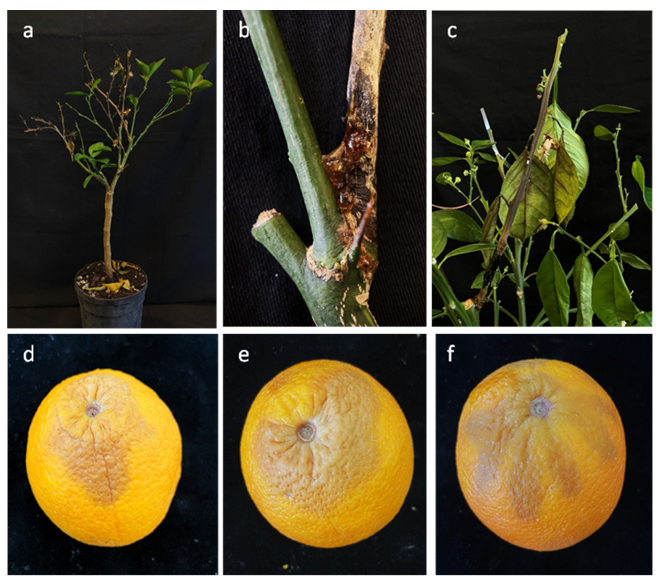
2.6.2. Pathogenicity on Plants

Two representative isolates of Botryosphaeriaceae (CVG 1929 and CVG 1985) and two isolates of Diaporthe spp. (CVG 1938, CVG 2046) were selected among the isolates found in association with citrus twigs for pathogenicity tests on plants. Their capacity to infect wood and induce twig blight was evaluated on two-year-old potted plants of C. sinensis cv. Valencia. Each fungal isolate was inoculated on six plants. For each plant, four twigs were inoculated as replicates. The inoculum consisted of a small piece (~5 mm) of mycelial plug from 5-day-old and 28-day-old cultures of isolates on PDA for Lasiodiplodia and for Diaporthe suspected isolates, respectively. The bark was first gently scraped using a sterile blade, and then the mycelial plug was inserted upside down onto the wound. Wounds were sealed with Parafilm (Bemis Co, Neenah, WI, USA) to prevent desiccation. Control consisted of sterile PDA mycelial plugs placed on bark wounds. All the inoculated plants were incubated in the growth chamber with a 12 h photoperiod and maintained at 25 ± 1 °C and regularly watered and monitored daily for development of symptoms. Disease incidence (%) and disease severity (lesion length cm) were evaluated 7 and 14 days post inoculations. Re-isolations were performed as mentioned above to fulfill Koch’s postulates.

2.7. Statistical Analysis

The data obtained from the experiment conducted to evaluate the temperature effect on mycelial growth rate were subjected to statistical analysis as follows. Data of optimum growth temperature and MGR were evaluated for normality and homogeneity of residual variances. One-way ANOVA was performed when both ANOVA assumptions were satisfied for optimum growth temperature and MGR data. The optimum growth temperature or MGR was considered as a dependent variable, and isolates were considered as independent variables. For each variable, isolate means were compared according to Tukey’s honestly significant difference (HSD) test at α = 0.05 [72]. Data were analyzed using Statistix 10 software [73]. Data obtained from pathogenicity tests on fruit and plants were subjected to statistical analysis to assess the aggressiveness of the tested isolates. Considering fruit, as the two obtained perpendicular diameters of necrotic lesions on fruit have different lengths, the mathematical formula for elliptic surface was used to calculate the necrotic lesion areas induced by inoculated isolates [74]. Necrotic areas were compared and analyzed using RStudio (https://www.R-project.org/ accessed on 1 November 2023). Normality and homogeneity of residual variances were evaluated with Shapiro–Wilk and Levene’s tests, respectively. One-way analysis of variance (ANOVA) was carried out to compare the average of necrotic areas among the different species and the control. Bonferroni post hoc test (at p < 0.05) was used to evaluate statistically significance differences in means of necrotic area surface. For plants, the frequency of branch dissection was calculated based on the numbers of dissected branches recorded. Data were analyzed using RStudio (https://www.R-project.org/ accessed on 1 November 2023). Shapiro–Wilk and Levene’s tests were used to evaluate normality and homogeneity of residual variances, respectively. To compare the frequency of branches dissection among isolates, ANOVA was carried out. Bonferroni post hoc test (at p < 0.05) was used to determine statistically significant differences.

3. Results

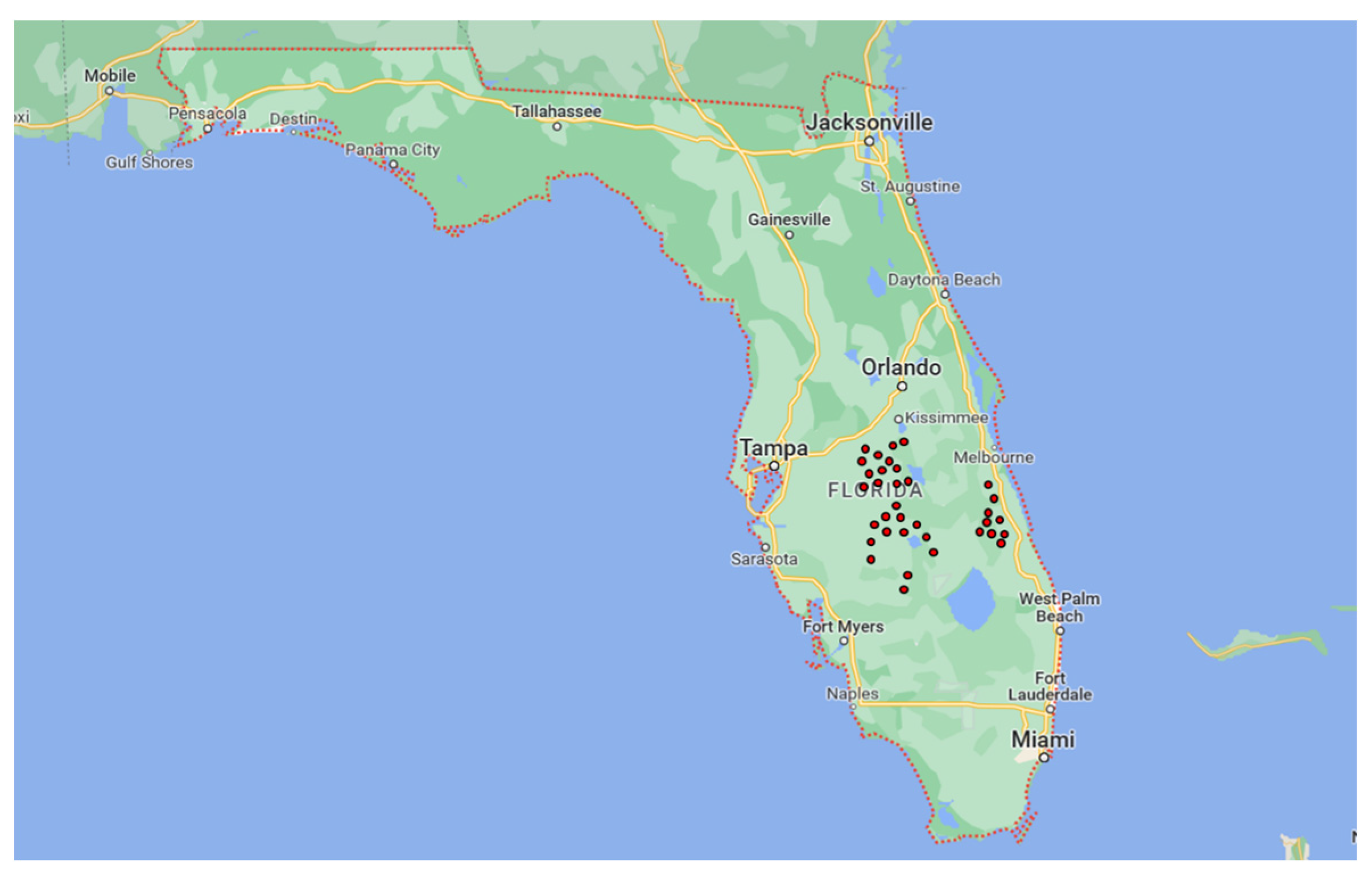
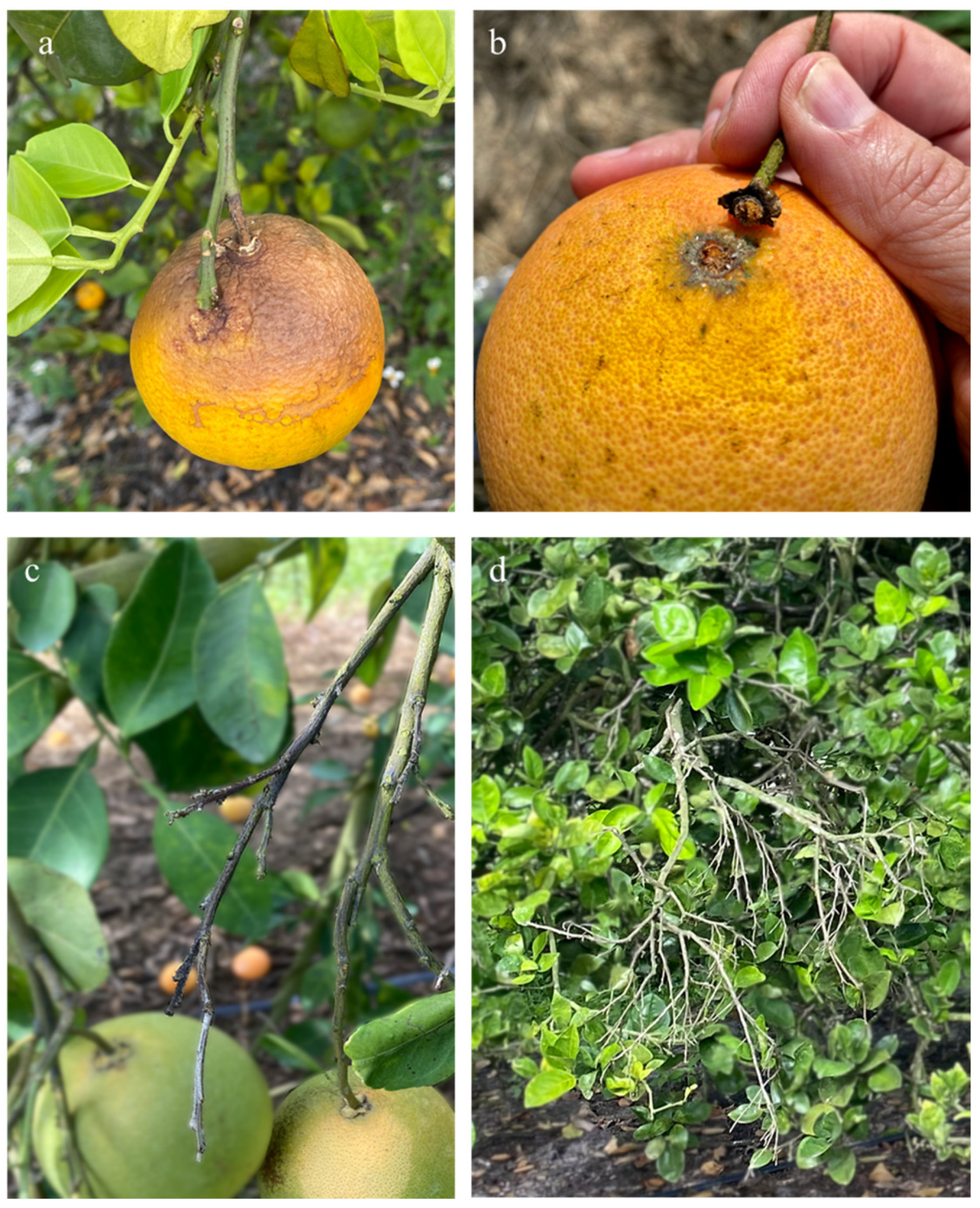
3.1. Field Sampling and Isolation

In the 35 sampled Florida orchards, dieback and stem-end rot seriously reduced the plant health and fruit yield, respectively. Citrus trees showed a wide variety of symptoms, including twig and branch dieback often associated with gummosis exudate (Figure 2c,d). Branch and twig longitudinal sections revealed necrotic brown discoloration. Necrotic lesions at the stem-end were observed on green and ripe fruit. Green fruit showed yellowing at the stem-end, while ripe fruit exhibited brown necrotic tissue, and in both cases, a brown rot occurred at the calyx end (Figure 2a,b). A total of 70 fungal isolates were obtained from sampled orchards. The preliminary identification of collected isolates was based on morphology. Fifty-five isolates were identified as Botryosphaeriaceae-like and 15 as Diaporthe spp. Twenty-six representative isolates were selected for molecular analysis.

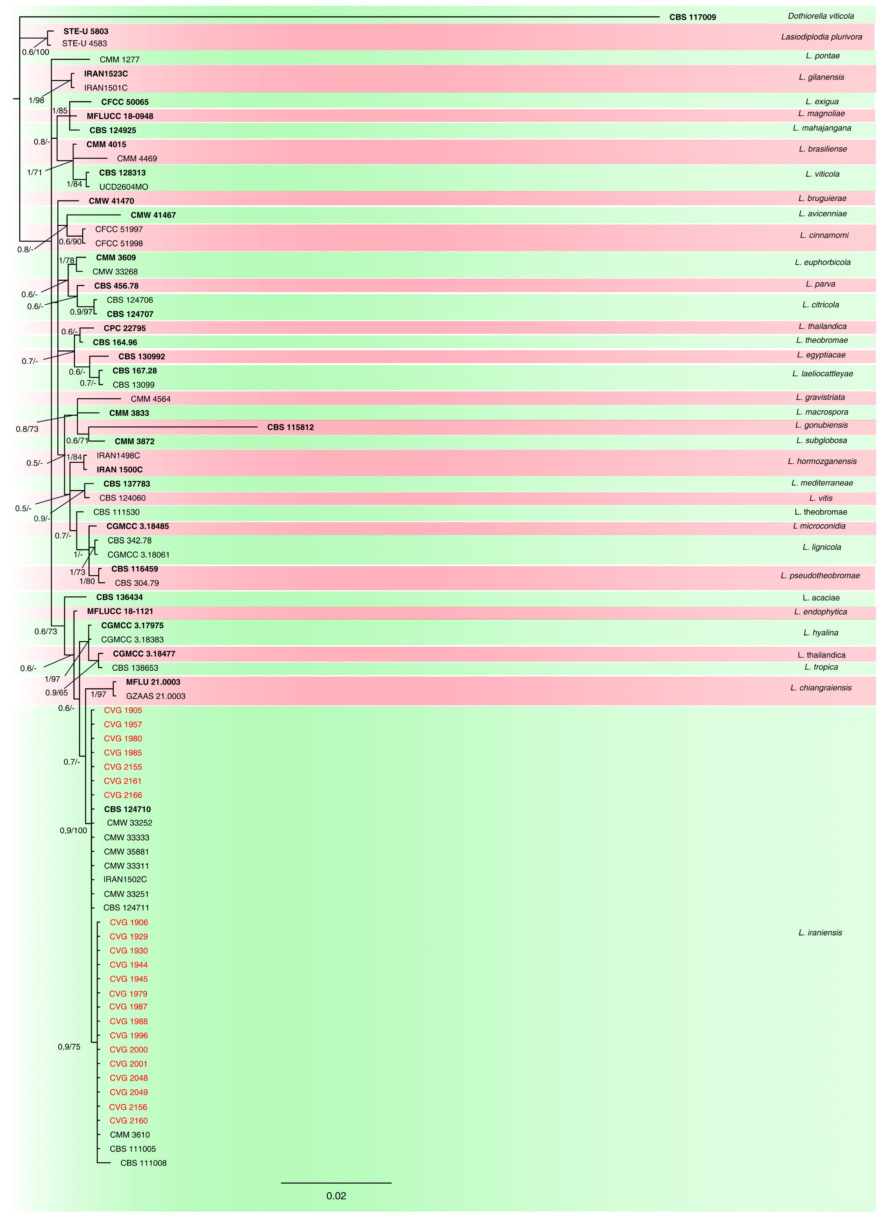
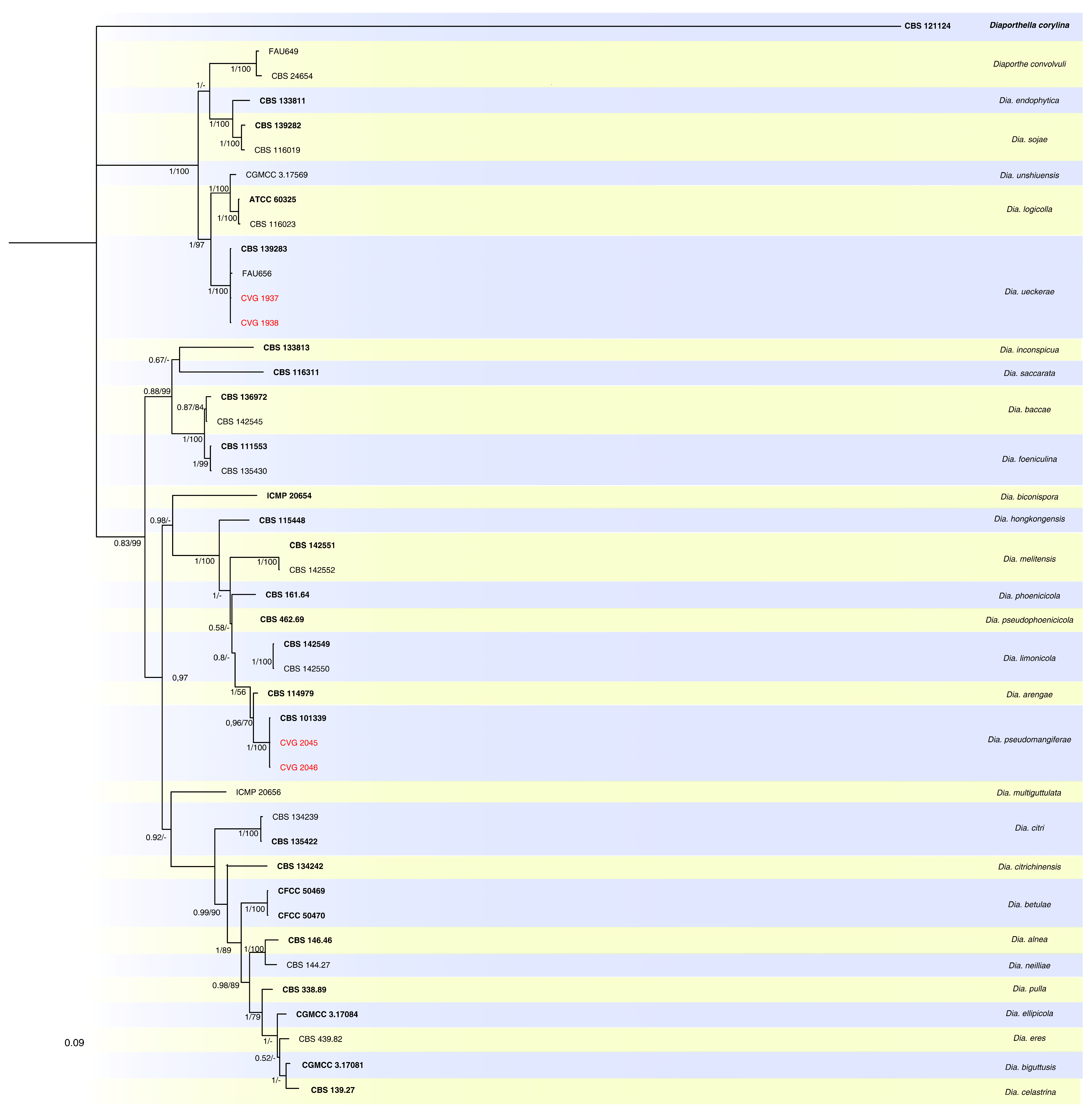
3.2. Phylogenetic Analyses
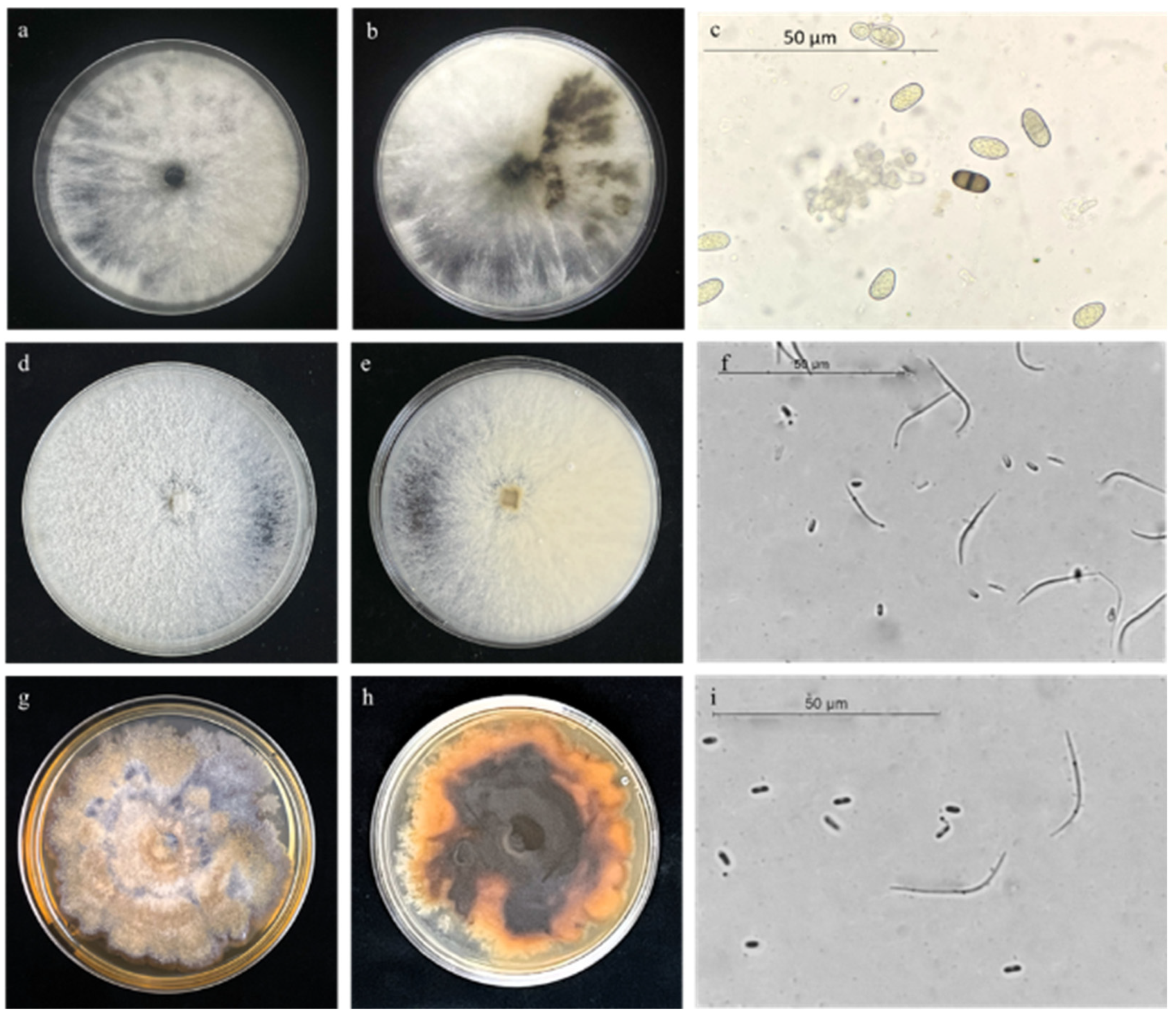
Three alignments representing single locus analyses of ITS, tef1, and tub2 and one combined alignment of all three loci were analyzed for Botryosphaeriaceae and Diaporthe isolates. The three single loci alignments produced topologically similar trees. The combined locus phylogeny of Botryosphaeriaceae consisted of 82 sequences, including the outgroup Dothiorella viticola (CBS 117009). The analyses included a total of 1356 characters (ITS:1-493, tef1:498-899, tub2:904-1,356). For the Bayesian analyses, MrModeltest suggested the fixed state frequency for analyzing ITS, Dirichlet state frequency for tef1 and Dirichlet, and fixed state frequencies for tub2. Based on the results of MrModeltest, the following models were adopted: K80 + G and K80 + I for ITS, K80 + G and HKY + I + G for tef1, and GTR + I and GTR + G for tub2. In the Bayesian analyses, the ITS had 98 unique site patterns, while the partial tef1 gene had 113, and the partial tub2 gene had 99. The analyses ran for 37,535,000 generations, resulting in 75,072 trees, of which 56,304 trees were used to calculate the posterior probabilities. Considering the concatenated phylogenetic analyses, twenty-two isolates clustered with 11 reference strains and the ex-type of Lasiodiplodia iraniensis, forming a highly supported clade (0.9/100). Regarding the MP analysis, 216 characters resulted as parsimony-informative, 219 were variables, and 1219 were constant. A maximum of 1000 equally most parsimonious trees were saved (Tree length = 427, CI = 0.806, RI = 0.845, and RC = 0.681). Bootstrap support values obtained with the parsimony analyses are reported on the Bayesian phylogenetic tree (Figure 3).
The Diaporthe multi-locus phylogenetic analyses consisted of 44 sequences, including the outgroup D. corylina (CBS 121124). The phylogenetic analyses included a total of 1.726 characters (ITS:1-552, tef1:557-880, tub2:885-1726). For the Bayesian analyses, MrModeltest proposed the fixed state frequency distributions for analyzing ITS and Dirichlet state frequency for tef1 and tub2. In line with MrModeltest’s recommendations, the following models were used: SYM + I + G for ITS, GTR + I + G, and HKY + I + G for tef1 and K80 + G and HKY + G for tub2. In the Bayesian analyses, the ITS had 156 unique site patterns, the partial tef1 gene had 245, and the tub2 locus had 415. The analyses ran for 720,000 generations, resulting in 1442 trees, of which 817 trees were used to calculate the posterior probabilities. For the MP analysis, 590 characters resulted as parsimony-informative, 415 were variable, and 713 were constant. A maximum of 1000 equally most parsimonious trees were saved (Tree length = 2.271, CI = 0.661, RI = 0.854, and RC = 0.564). Bootstrap support values obtained with the parsimony analyses are reported on the Bayesian phylogenetic tree (Figure 4). Considering the combined analyses, two isolates clustered with one reference strain and the ex-type of Dia. ueckerae, while two isolates clustered with the ex-type of Dia. pseudomangiferae forming a highly supported clade (1/100).

3.3. Morphology

Morphological characteristics, supported by phylogenetic analysis, were used to describe the three identified species.
L. iraniensis was characterized by a cottony, fast-growing colony with abundant aerial mycelium, which covered the entire PDA-S Petri dishes after 7 days (Figure 5a,b). Initially, the colony was white or light gray and then became smoke-grey to olivaceous-grey (Figure 5a). The reverse colony was white or pale grey which turned later to dark grey or greenish grey (Figure 5b). On PNA, L. iraniensis produced pycnidial, dark brown to black conidiomata covered with dense mycelium. Initially, conidia were hyaline, subglobose to ovoid, unicellular with granular content, becoming dark brown, ovoid to ellipsoid, and 1-septate with longitudinal striations (Figure 5c). Mature conidia of the strains CVG 1930, CVG 1945, CVG 1980, CVG 2155, and CVG 2160 had dimensions of (20.1-) 24.2 (-29.2) × (13.8-) 14.6 (-16.3) µm (mean ± SD = 24.2 ± 1.5 × 14.6 ± 1.0 µm).
Dia. ueckerae showed a dense and felt-like colony that covered the PDA-S plates within 10 days. Front colony was white, becoming cream to pale grey (Figure 5d). The reverse colony was white, turning to grey with brownish spots (Figure 5e). On MEA, the strains CVG 1937 and CVG 1938 of Dia. ueckerae produced pycnidial, subglobose dark brown to black conidiomata. Alpha conidia were aseptate, hyaline, smooth, fusiform, and apex rounded (Figure 5f), with size of (5.4-) 8.3 (-9.2) × (2.3) 3.5 (-4.5) µm (mean ± SD = 8.3 ± 1.0 × 3.5 ± 0.8 µm). Beta conidia were aseptate, hyaline, smooth, filiform, curved, and eguttulate (Figure 5f) with dimensions of (17.3-) 21.4 (-24.3) × (1.0-) 1.5 (2.5) µm (mean ± SD = 21.4 ± 1.5 × 1.5 ± 0.4 µm).
Dia. pseudomangiferae had white moderate aerial mycelia with patches from pale luteous to luteous (yellowish or yellow-brown) in reverse (Figure 5g,h). Superficial, pycnidial, and black conidiomata were produced on MEA by the strains CVG 2045 and CVG 2046. Alpha conidia were aseptate, hyaline, smooth, and fusiform (Figure 5i) with a size of (4.2-) 7.3 (-11.0) × (1.3) 2.5 (-3.5) µm (mean ± SD = 7.3 ± 1.2 × 2.5 ± 1.1 µm). Beta conidia were aseptate, hyaline, smooth, and curved (Figure 5i) with dimensions of (14.5-) 25.4 (-30.4) × (1.3-) 1.3 (2.3) µm (mean ± SD = 25.4 ± 1.3 × 1.3 ± 1.0 µm).

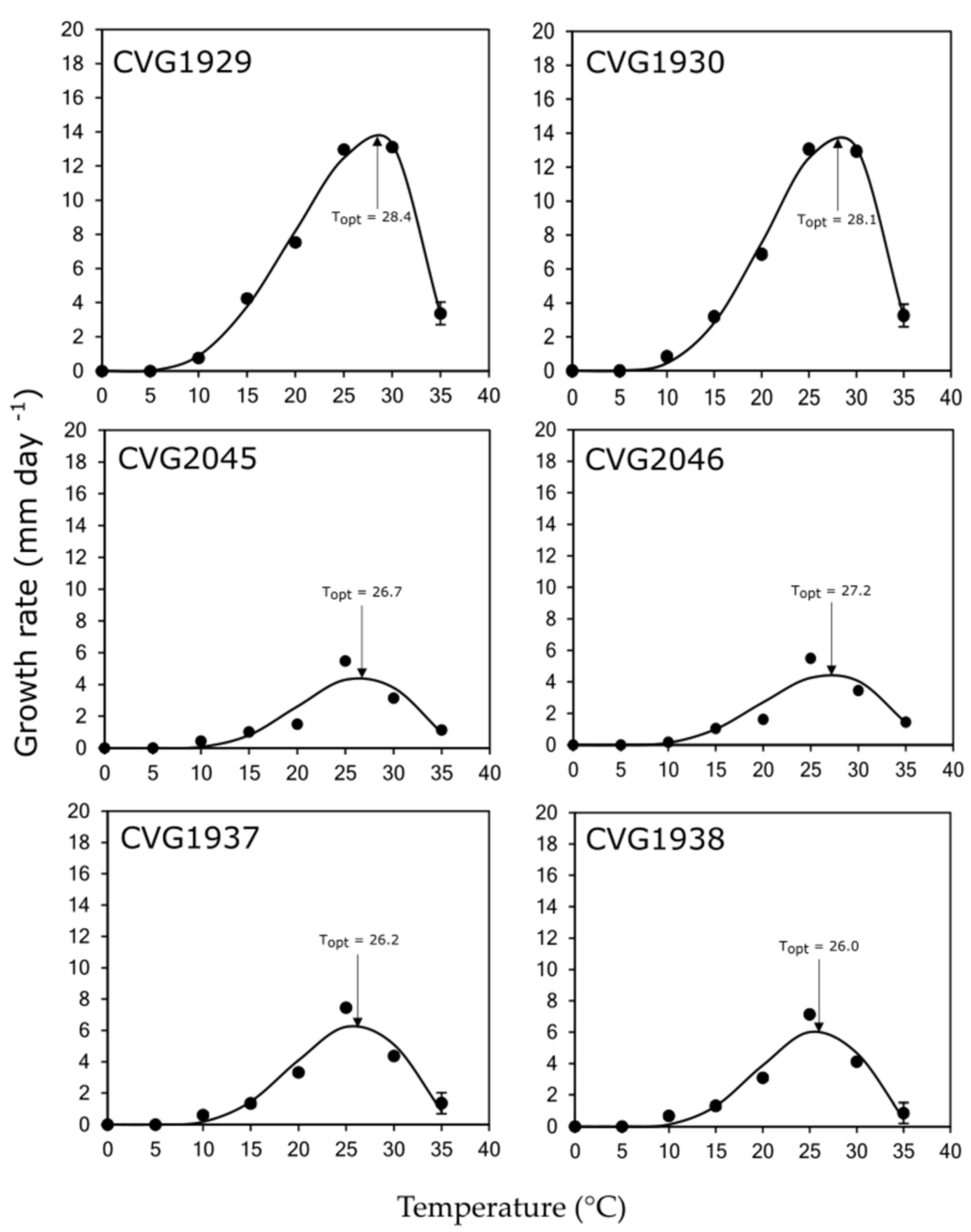
3.4. Effect of Temperature on Mycelial Growth

All the selected isolates were able to grow from 10 to 35 °C, while no mycelial growth was recorded at 5 °C in all cases. No significant differences were found in optimum growth temperature and MGR (p > 0.05) among isolates. However, there was a significant interaction among isolates and species in optimum growth temperature (p < 0.05); thus, the data were not combined, and isolates were kept separate for both the analyses on optimum growth temperature and MGR (Figure 6, Table 3). The optimum growth temperature ranged from 26.0 to 28.4 °C for Dia. ueckerae isolate CVG 1938, and L. iraniensis isolate CVG 1929, respectively.
Concerning the MGR, isolates belonging to L. iraniensis had the highest mycelial growth, with MGR being 13.7–13.8 mm day−1, followed by isolates of Dia. ueckerae (MGR = 6.1–6.3 mm day−1). Isolates belonging to Dia. pseudomangiferae showed the lowest MGR with respect to the other isolates, with MGR being 4.5 mm day−1.

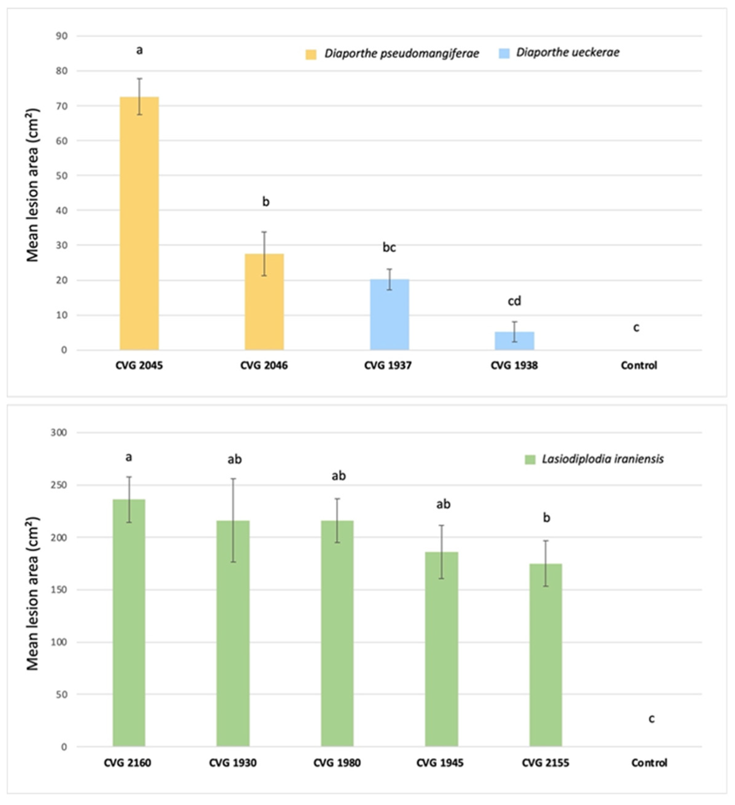
3.5. Pathogenicity on Fruit

Dia. pseudomangiferae, Dia. ueckerae and L. iraniensis produced soft, watery rot at the stem-end of inoculated fruit of C. sinensis cv. Valencia. Discoloration started from the button where the fruit were wounded, and the conidia suspension was added. Ten days after inoculation, lesions extended to the whole fruit, producing a rot (Figure 7d–f).
The isolates of L. iraniensis produced the largest necrotic area compared to both species of Diaporthe spp. and the water control (Figure 8). The isolate CVG 2160 was the most virulent, producing a necrotic area of 233 cm2 (Figure 8). The other L. iraniensis isolates showed different virulence levels, ranging from 175 cm2 to 216 cm2 for the strains CVG 2155 and CVG 1930, respectively (Figure 8). Isolates of Dia. pseudomangiferae showed a higher virulence than those of Dia. ueckerae. In particular, isolates CVG 2045 and CVG 2046 of Dia. pseudomangiferae produced a mean necrotic area of 72 cm2 and 27 cm2, respectively (Figure 8). While isolates of Dia. ueckerae, CVG 1937, and CVG 1938 were able to produce necrotic lesions, with mean areas of 20 cm2 and 7 cm2, respectively (Figure 8). No symptoms were observed on water control fruit. All the fungal species were successfully re-isolated from the outer margin of necrotic tissues of inoculated fruit, fulfilling Koch’s postulates. The recovery of inoculated isolates of species ranged between 80% and 90%. The identity of the re-isolated isolates was confirmed through morphological features and molecular analyses of the tub2 locus.

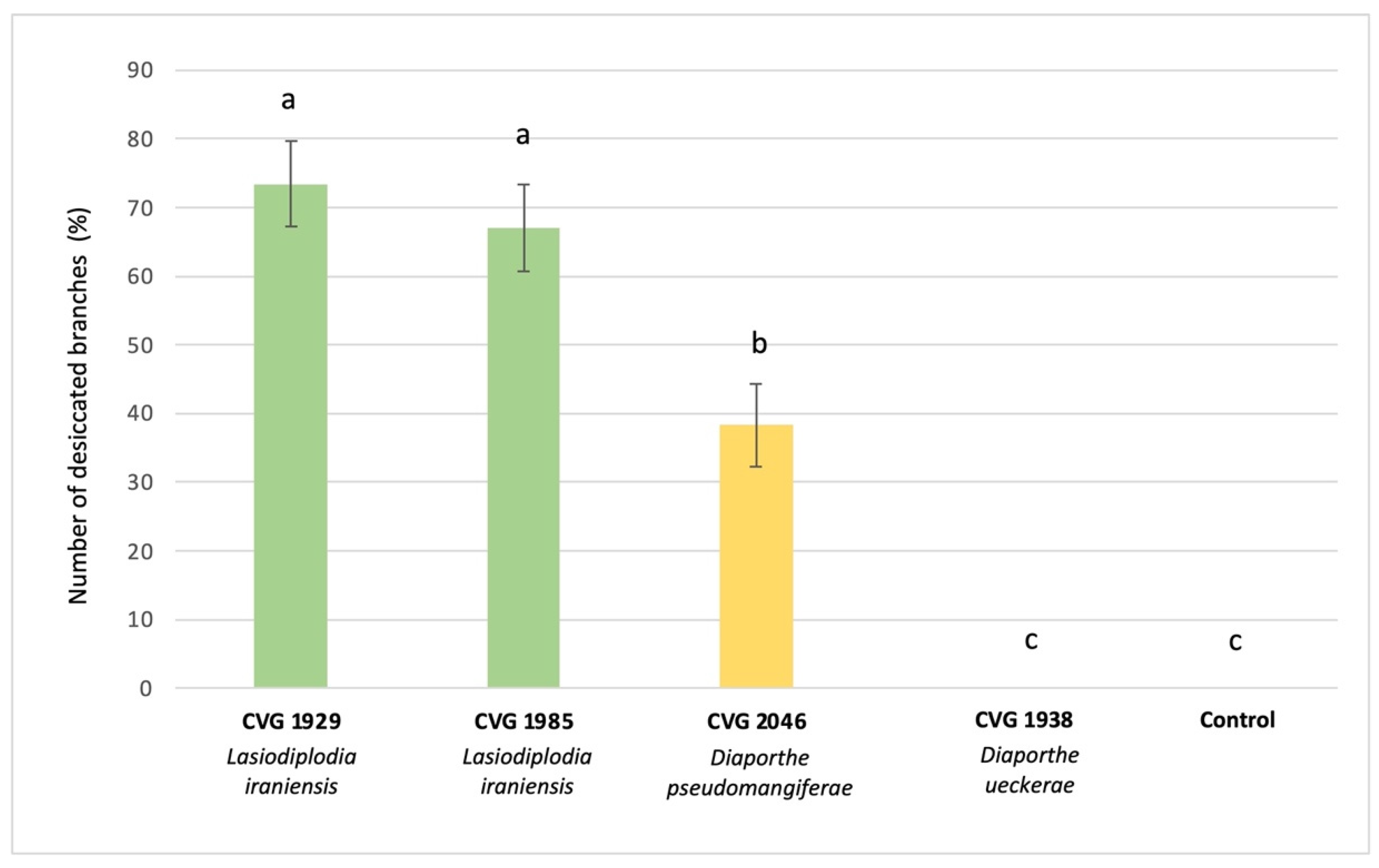
3.6. Pathogenicity on Plants

Two tested isolates of L. iraniensis (CVG 1929 and CVG 1985) and the isolate CVG 2046 of Dia. pseudomangiferae were able to cause necrotic lesions and gummosis at the inoculation point after 7 days (Figure 7b), while a complete twig decay occurred after 14 days (Figure 7a,c). Plants inoculated with the isolate CVG 1938 of Dia. ueckerae showed no symptoms, as control plants. Tested isolates of L. iraniensis showed a high level of virulence: CVG 1929 produced 73% of decayed twigs on a total number of 24 plants inoculated, followed by CVG 1985 with 67% (Figure 9). The tested isolate of Dia. pseudomangiferae (CVG 2046) caused 38% of decayed twigs, showing lower virulence compared to L. iraniensis isolates (Figure 9).

4. Discussion

The present study is the first that aimed at revealing the occurrence, diversity, and pathogenicity of fungal species in association with twig blight, branch dieback, fruit rot, and decline of C. sinensis cv. Valencia in Florida. Severe symptoms of dieback and stem-end rot were observed on branches, twigs, and fruit in several orchards across South-central Florida. Different fungal isolates were recovered from plant material samples collected in 35 orchards and they were preliminary identified as Botryosphaeriaceae-like and Diaporthe-like according to their colony morphology [30,39,75]. We identified for the first time three different species as the main causal agents of this disease: L. iraniensis, Dia. pseudomangiferae, and Dia. ueckerae.
Lasiodiplodia iraniensis was described in 2010 as a new species in association with Citrus sp., Juglans sp., and Mangifera indica in Iran [76]. Later, this pathogen was reported on cashews, Anacardium occidentale [77], great bougainvillea, Bougainvillea spectabilis [78], gum trees, Eucalyptus spp. [79], mango, Mangifera indica [80,81], and toothbrush tree, Salvadora persica [76]. Concerning citrus, it was reported in association with dieback disease on key lime, C. aurantiifolia in Oman [80], Persian lime, C. latifolia in Mexico [33] and mandarin, C. reticulata in Pakistan, where it was found in association with Colletotrichum siamense [82]. Recently, L. mitidjana was reported on C. sinensis in Algeria, causing branch canker and dieback [16]. To our knowledge, this study is the first report of L. iraniensis as a causal agent of dieback and stem-end rot diseases on C. sinensis worldwide. Other Lasiodiplodia spp. reported to cause stem-end rot on Citrus spp. are L. theobromae [24] and L. pseudotheobromae [37,83].
Diaporthe ueckerae is a ubiquitous pathogen that opportunistically infects humans and plants [46]. It was described for the first time by Udayanga et al. [46] on Cucumis sp. and then reported on peanut, Arachis hypogaea [84], tea plant, Camellia sinensis [85], lemon-scented gum, Eucalyptus citriodora [86], soybean, Glycine max [87], white-fleshed pitahaya, Selenicereus undatus [88], and mango, M. indica [25]. Diaporthe pseudomangiferae is a plant pathogen first described by Gomes et al. [39] and isolated from mango [89,90], cacao, Theobroma cacao [89,91], and kiwifruit Actinidia deliciosa [92]. It was reported in Puerto Rico, California, the Dominican Republic, Mexico, China, and South Korea [39,89,90,91,92]. To our knowledge, Dia. pseudomangiferae and Dia. ueckerae are reported in this study for the first time in association with citrus dieback diseases and stem-end rot worldwide. Other Diaporthe species are reported as causal agents of pre- or post-harvest diseases on citrus: Dia. citri, Dia. foeniculina, Dia. limonicola, and Dia. melitensis [7,19,28].
We show that all the tested isolates have an optimum growth temperature in a range between 26.0 and 28.4 °C with different MGR based on the species, in agreement with other reports [32,93]. Climate conditions in the majority of Florida are reported as humid subtropical (Cfa Köppen climate type) with higher temperature and humidity in summer and warm, occasionally cold, dry winters, thus being suitable for the development and spread of these pathogens [94]. Moreover, isolates belonging to L. iraniensis had both the highest optimum growth temperature and MGR with respect to the other tested species. In general, species of Lasiodiplodia are well adapted to places with higher annual mean temperatures, and Botryosphaeriaceae are known as fast-growing fungi [32,95,96]. The present in vitro findings suggest that L. iraniensis could have a greater colonization ability on the host plant tissues. Thus, further investigations will aim to assess the in planta development of these pathogens on mature trees depending on temperature conditions and the possible effect of temperature on disease severity to predict and model the disease progress.
Concerning the pathogenicity, the tested isolates of L. iraniensis were able to cause soft, watery stem-end rot when inoculated on citrus fruit, and they were more virulent in comparison to the other tested fungal species. This outcome is consistent with the results reported by Li et al. [97] on mango, where L. iraniensis was the most virulent species along with Botryosphaeria scharifii. The different virulence levels found among L. iraniensis isolates suggest a possible intraspecific variability that could be addressed in future studies. We suspect that opportunistic strains of this fungal species requiring plant stresses, like drought, as the preconditioning factor to allow infection establishment could exist, potentially explaining the variable virulence levels we detected. For example, a relatively closely related species, Botryosphaeria dothidea, is a well-known opportunistic pathogen of various tree species, including apple [98] and coast redwood, requiring drought stress to infect wood [99,100,101]. A pathogenicity test conducted on potted C. sinensis plants cv. Valencia confirmed L. iraniensis as the most virulent species, able to cause more than 70% of completely decayed twigs of young plants. The obtained reslts agree with previous studies conducted on this species affecting citrus trees: Xiao et al. [36] reported Lasiodiplodia as the most virulent genus on C. reticulata shoots and on C. paradisi × C. reticulata plants in China, while Bautista-Cruz et al. [33] described L. iraniensis as one of the most virulent species on Persian lime, C. latifolia, plants in Mexico. Similar results were also reported on dragon fruit, S. undatus, cashews, Anacardium, and macadamia nut, Macadamia integrifolia [77,102,103]. Considering Dia. pseudomangiferae, the tested isolates were pathogenic on both fruit and plants, while for Dia. ueckerae, the isolates were pathogenic only on fruit. The symptoms caused by the isolate CVG 1938 of Dia. ueckerae on fruit were statistically similar to the water control; thus, it is possible that it was not able to cause symptoms on wood because it is either a more complex matrix or an unsuitable ecological niche overall. In this case, it is also possible that Dia. ueckerae might require some form of plant stress as the prerequisite to infect C. sinensis wood. Further inoculation studies of Dia. ueckerae on well-watered plants vs. drought-stressed plants will provide clearer information on its pathogenicity.
Several studies demonstrated a positive correlation between different citrus diseases and the early drop of fruit [104,105,106,107]. Thus, considering L. iraniensis, Dia. ueckerae, and Dia. pseudomangiferae as causal agents of dieback disease and stem-end rot, a possible role of these species as biotic factors contributing to the early drop of citrus fruit in the sampled area is postulated, but further studies are needed to investigate this aspect. Furthermore, the physiological status of the sampled trees indicating the presence or absence of plant stress, with a special focus on drought, might be an important aspect in these investigations. Additional assays on wood and fruit co-infection with the identified fungal species will provide useful information about their role in the development and progress of the mentioned disease. Moreover, Botryosphaeriaceae and Diaporthe spp. are reported to infect plants mainly through natural openings or wounds [108,109]. Different cultural practices, such as pruning and fruit thinning, could cause wounds on plants, thus favoring the entry of these pathogens and subsequent development of dieback and stem-end rot. Optimal management of orchards and the protection of wound cuts is recommended to avoid the spread of these pathogens. Future investigations on the ecology, epidemiology, and sensitivity of fungicides labeled for use in Florida will improve the knowledge about these pathogens to develop effective management strategies. In this context, the role of irrigation and nutrient balance in plant health should be explored as a possible way to prevent stress conditions which could make the plants less vulnerable to the pathogens identified and characterized in this study.

Supplementary Materials

The following supporting information can be downloaded at: https://www.mdpi.com/article/10.3390/horticulturae10040406/s1, Table S1: Collection details and of isolates of this study.

Author Contributions

Conceptualization, M.F. and V.G.; methodology, V.P., M.F. and D.A.; software, I.M.; validation, S.G.A., D.S. and V.G.; formal analysis, G.D. and I.M.; investigation, V.P., M.F. and V.G.; resources, M.F.; data curation, V.P. and V.G.; writing—original draft preparation, V.P.; writing—review and editing, D.S, G.P. and V.G.; visualization, V.G.; supervision, V.G.; project administration, V.G.; funding acquisition, V.G. All authors have read and agreed to the published version of the manuscript.

Funding

This research was funded by KeyPlex. Part of this work was granted by the European Commission—NextGenerationEU, Project “Strengthening the MIRRI Italian Research Infrastructure for Sustainable Bioscience and Bioeconomy”, code n. IR0000005.

Data Availability Statement

The raw data supporting the conclusions of this article will be made available by the authors on request.

Conflicts of Interest

Author Mauricio Flores was employed by the company KeyPlex, 400 N. New York Ave Suite 200, Winter Park, FL 32789, USA. The remaining authors wish to disclose that, at the time of conducting this research and submitting the manuscript, they were not subject to any commercial or financial relationships that could be perceived as a potential conflict of interest.

References

  1. Rehman, S.U.; Abbasi, K.S.; Qayyum, A.; Jahangir, M.; Sohail, A.; Nisa, S.; Tareen, M.N.; Tareen, M.J.; Sopade, P. Comparative analysis of citrus fruits for Nutraceutical properties. Food Sci. Technol. 2020, 40, 153–157. [Google Scholar] [CrossRef]
  2. ISTAT. Istituto Nazionale di Statistica. 2023. Available online: https://www.istat.it/ (accessed on 1 November 2023).
  3. FAOSTAT. Food and Agriculture Organization Corporate Statistical Database. 2023. Available online: https://www.fao.org (accessed on 20 December 2023).
  4. USDA. United States Department of Agriculture. 2023. Available online: https://www.usda.gov (accessed on 20 December 2023).
  5. Timmer, L.W.; Garnsey, S.M.; Graham, J.H. Compendium of Citrus Diseases; American Phytopathology Society: Saint Paul, MN, USA, 2000. [Google Scholar]
  6. Bezerra, J.D.P.; Crous, P.W.; Aiello, D.; Gullino, M.L.; Polizzi, G.; Guarnaccia, V. Genetic Diversity and Pathogenicity of. Plants 2021, 10, 492. [Google Scholar] [CrossRef] [PubMed]
  7. Chaisiri, C.; Liu, X.Y.; Lin, Y.; Luo, C.X. Diaporthe citri: A Fungal Pathogen Causing Melanose Disease. Plants 2022, 11, 1600. [Google Scholar] [CrossRef] [PubMed]
  8. Galsurker, O.; Diskin, S.; Maurer, D.; Feygenberg, O.; Alkan, N. Fruit Stem-End Rot. Horticulturae 2018, 4, 50. [Google Scholar] [CrossRef]
  9. Mayorquin, J.S.; Wang, D.H.; Twizeyimana, M.; Eskalen, A. Identification, Distribution, and Pathogenicity of Diatrypaceae and Botryosphaeriaceae Associated with Citrus Branch Canker in the Southern California Desert. Plant Dis. 2016, 100, 2402–2413. [Google Scholar] [CrossRef] [PubMed]
  10. Vitale, A.; Aiello, D.; Azzaro, A.; Guarnaccia, V.; Polizzi, G. An Eleven-Year Survey on Field Disease Susceptibility of Citrus Accessions to Colletotrichum and Alternaria Species. Agriculture 2021, 11, 536. [Google Scholar] [CrossRef]
  11. Fawcett, H.S.; Burger, O.F. A gum-inducing Diplodia of peach and orange. Mycologia 1911, 3, 151–153. [Google Scholar] [CrossRef]
  12. Punithalingam, E. Plant Diseases Attributed to Botryodiplodia Theobromae Pat; J. Cramer.: Englewood Cliffs, NJ, USA, 1980. [Google Scholar]
  13. Adesemoye, A.O.; Mayorquin, J.S.; Wang, D.H.; Twizeyimana, M.; Lynch, S.C.; Eskalen, A. Identification of Species of Botryosphaeriaceae Causing Bot Gummosis in Citrus in California. Plant Dis. 2014, 98, 55–61. [Google Scholar] [CrossRef] [PubMed]
  14. Espargham, N.; Mohammadi, H.; Gramaje, D. A Survey of Trunk Disease Pathogens within Citrus Trees in Iran. Plants 2020, 9, 754. [Google Scholar] [CrossRef]
  15. Crous, P.W.; Wingfield, M.J. Fungi infecting woody plants: Emerging frontiers. Persoonia 2018, 40, I–III. [Google Scholar] [CrossRef]
  16. Berraf-Tebbal, A.; Mahamedi, A.E.; Aigoun-Mouhous, W.; Špetík, M.; Čechová, J.; Pokluda, R.; Baránek, M.; Eichmeier, A.; Alves, A. Lasiodiplodia mitidjana sp. nov. and other Botryosphaeriaceae species causing branch canker and dieback of Citrus sinensis in Algeria. PLoS ONE 2020, 15, e0232448. [Google Scholar] [CrossRef]
  17. Camiletti, B.X.; Lichtemberg, P.S.F.; Paredes, J.A.; Carraro, T.A.; Velascos, J.; Michailides, T.J. Characterization of. Phytopathology 2022, 112, 1454–1466. [Google Scholar] [CrossRef]
  18. Guarnaccia, V.; Groenewald, J.Z.; Polizzi, G.; Crous, P.W. High species diversity in Colletotrichum associated with citrus diseases in Europe. Persoonia 2017, 39, 32–50. [Google Scholar] [CrossRef] [PubMed]
  19. Guarnaccia, V.; Crous, P.W. Emerging citrus diseases in Europe caused by species of. IMA Fungus 2017, 8, 317–334. [Google Scholar] [CrossRef] [PubMed]
  20. Riolo, M.; Aloi, F.; Pane, A.; Cara, M.; Cacciola, S. Twig and Shoot Dieback of Citrus, a New Disease Caused by Colletotrichum Species. Cells 2021, 10, 449. [Google Scholar] [CrossRef]
  21. Leonardi, G.R.; Aiello, D.; Camilleri, G.; Piattino, V.; Polizzi, G.; Guarnaccia, V. A new disease of kumquat (Fortunella margarita) caused by Colletotrichum karsti: Twig and branch dieback. Phytopathol. Mediterr. 2023, 62, 333–348. [Google Scholar] [CrossRef]
  22. Ismail, M.; Zhang, J. Post-harvest Citrus Diseases and their control. Outlooks Pest Manag. 2004, 15, 29–35. [Google Scholar] [CrossRef]
  23. Brown, G.E.; Eckert, J.W. Diplodia stem-end rot. Compend. Citrus Dis. 2000, 2, 43–44. [Google Scholar]
  24. Zhang, J. Lasiodiplodia theobromae in Citrus Fruit (Diplodia Stem-End Rot). In Postharvest Decay: Control Strategies; Academic Press: Cambridge, MA, USA, 2014; pp. 309–335. [Google Scholar]
  25. Lim, L.; Mohd, M.H.; Zakaria, L. Identification and pathogenicity of Diaporthe species associated with stem-end rot of mango (Mangifera indica L.). Eur. J. Plant Pathol. 2019, 155, 687–696. [Google Scholar] [CrossRef]
  26. Guimarães, J.E.R.; de la Fuente, B.; Pérez-Gago, M.B.; Andradas, C.; Carbó, R.; Mattiuz, B.H.; Palou, L. Antifungal activity of GRAS salts against Lasiodiplodia theobromae in vitro and as ingredients of hydroxypropyl methylcellulose-lipid composite edible coatings to control Diplodia stem-end rot and maintain postharvest quality of citrus fruit. Int. J. Food Microbiol. 2019, 301, 9–18. [Google Scholar] [CrossRef] [PubMed]
  27. Awan, Q.N.; Akgül, D.S.; Unal, G. First Report of Lasiodiplodia pseudotheobromae Causing Postharvest Fruit Rot of Lemon in Turkey. Plant Dis. 2016, 100, 2327. [Google Scholar] [CrossRef]
  28. Huang, F.; Hou, X.; Dewdney, M.M.; Fu, Y.; Chen, G.Q.; Hyde, K.D.; Li, H.Y. Diaporthe species occurring on citrus in China. Fungal Divers. 2013, 61, 237–250. [Google Scholar] [CrossRef]
  29. Kucharek, T.; Whiteside, J.O.; Brown, E. Melanose and Phomopsis Stem-End Rot of Citrus. In Plant Pathology Fact Sheet; Florida Cooperative Extension Service; Institute of Food and Agricultural Sciences, University of Florida: Gainesville, FL, USA, 2000; pp. 26–30. [Google Scholar]
  30. Phillips, A.J.L.; Alves, A.; Abdollahzadeh, J.; Slippers, B.; Wingfield, M.J.; Groenewald, J.Z.; Crous, P.W. The Botryosphaeriaceae: Genera and species known from culture. Stud. Mycol. 2013, 76, 51–167. [Google Scholar] [CrossRef]
  31. Slippers, B.; Boissin, E.; Phillips, A.J.L.; Groenewald, J.Z.; Lombard, L.; Wingfield, M.J.; Postma, A.; Burgess, T.; Crous, P.W. Phylogenetic lineages in the Botryosphaeriales: A systematic and evolutionary framework. Stud. Mycol. 2013, 76, 31–49. [Google Scholar] [CrossRef]
  32. Batista, E.; Lopes, A.; Alves, A. What do we know about Botryosphaeriaceae? An overview of a worldwide cured dataset. Forests 2021, 12, 313. [Google Scholar] [CrossRef]
  33. Bautista-Cruz, M.A.; Almaguer-Vargas, G.; Leyva-Mir, S.G.; Colinas-León, M.T.; Correia, K.C.; Camacho-Tapia, M.; Robles-Yerena, L.; Michereff, S.J.; Tovar-Pedraza, J.M. Phylogeny, Distribution, and Pathogenicity of Lasiodiplodia Species Associated with Cankers and Dieback Symptoms of Persian Lime in Mexico. Plant Dis. 2019, 103, 1156–1165. [Google Scholar] [CrossRef]
  34. El-Ganainy, S.M.; Ismail, A.M.; Iqbal, Z.; Elshewy, E.S.; Alhudaib, K.A.; Almaghasla, M.I.; Magistà, D. Diversity among Lasiodiplodia Species Causing Dieback, Root Rot and Leaf Spot on Fruit Trees in Egypt, and a Description of Lasiodiplodia newvalleyensis sp. nov. J. Fungi 2022, 8, 1203. [Google Scholar] [CrossRef]
  35. Polizzi, G.; Aiello, D.; Vitale, A.; Giuffrida, F.; Groenewald, J.Z.; Crous, P.W. First Report of Shoot Blight, Canker, and Gummosis Caused by Neoscytalidium dimidiatum on Citrus in Italy. Plant Dis. 2009, 93, 1215. [Google Scholar] [CrossRef]
  36. Xiao, X.E.; Wang, W.; Crous, P.W.; Wang, H.K.; Jiao, C.; Huang, F.; Pu, Z.X.; Zhu, Z.R.; Li, H.Y. Species of Botryosphaeriaceae associated with citrus branch diseases in China. Persoonia 2021, 47, 106–135. [Google Scholar] [CrossRef]
  37. Sultana, R.; Islam, M.S.; Rahman, H.; Alam, M.S.; Islam, M.A.; Sikdar, B. Characterization of Lasiodiplodia pseudotheobromae associated with citrus stem-end rot disease in Bangladesh. Int. J. Biosci. 2018, 13, 252–262. [Google Scholar]
  38. Guarnaccia, V.; Groenewald, J.Z.; Woodhall, J.; Armengol, J.; Cinelli, T.; Eichmeier, A.; Ezra, D.; Fontaine, F.; Gramaje, D.; Gutierrez-Aguirregabiria, A.; et al. diversity and pathogenicity revealed from a broad survey of grapevine diseases in Europe. Persoonia 2018, 40, 135–153. [Google Scholar] [CrossRef] [PubMed]
  39. Gomes, R.R.; Glienke, C.; Videira, S.I.; Lombard, L.; Groenewald, J.Z.; Crous, P.W. Diaporthe: A genus of endophytic, saprobic and plant pathogenic fungi. Persoonia 2013, 31, 1–41. [Google Scholar] [CrossRef] [PubMed]
  40. Aiello, D.; Guarnaccia, V.; Costanzo, M.B.; Leonardi, G.R.; Epifani, F.; Perrone, G.; Polizzi, G. Woody Canker and Shoot Blight Caused by Botryosphaeriaceae and Diaporthaceae on Mango and Litchi in Italy. Horticulturae 2022, 8, 330. [Google Scholar] [CrossRef]
  41. Guarnaccia, V.; Vitale, A.; Cirvilleri, G.; Aiello, D.; Susca, A.; Epifani, F.; Perrone, G.; Polizzi, G. Characterisation and pathogenicity of fungal species associated with branch cankers and stem-end rot of avocado in Italy. Eur. J. Plant Pathol. 2016, 146, 963–976. [Google Scholar] [CrossRef]
  42. Hilario, S.; Santos, L.; Alves, A. Diversity and Pathogenicity of Diaporthe Species Revealed from a Survey of Blueberry Orchards in Portugal. Agriculture 2021, 11, 1271. [Google Scholar] [CrossRef]
  43. Lombard, L.; van Leeuwen, G.C.M.; Guarnaccia, V.; Polizzi, G.; van Rijswick, P.C.J.; Rosendahl, K.; Gabler, J.; Crous, P.W. Diaporthe species associated with Vaccinium, with specific reference to Europe. Phytopathol. Mediterr. 2014, 53, 287–299. [Google Scholar]
  44. Manawasinghe, I.S.; Dissanayake, A.J.; Li, X.H.; Liu, M.; Wanasinghe, D.N.; Xu, J.P.; Zhao, W.S.; Zhang, W.; Zhou, Y.Y.; Hyde, K.D.; et al. High Genetic Diversity and Species Complexity of Diaporthe Associated with Grapevine Dieback in China. Front. Microbiol. 2019, 10, 473533. [Google Scholar] [CrossRef] [PubMed]
  45. Martino, I.; Tabone, G.; Giordano, R.; Gullino, M.L.; Guarnaccia, V. First Report of Diaporthe eres Causing Stem Blight and Dieback on Highbush Blueberry (Vaccinium corymbosum) in Italy. Plant Dis. 2023, 107, 1236. [Google Scholar] [CrossRef] [PubMed]
  46. Udayanga, D.; Castlebury, L.A.; Rossman, A.Y.; Chukeatirote, E.; Hyde, K.D. The Diaporthe sojae species complex: Phylogenetic re-assessment of pathogens associated with soybean, cucurbits and other field crops. Fungal Biol. 2015, 119, 383–407. [Google Scholar] [CrossRef]
  47. Udayanga, D.; Castlebury, L.A.; Rossman, A.Y.; Hyde, K.D. Species limits in Diaporthe: Molecular re-assessment of D-citri, D-cytosporella, D-foeniculina and D-rudis. Persoonia 2014, 32, 83–101. [Google Scholar] [CrossRef]
  48. Chaisiri, C.; Liu, X.Y.; Lin, Y.; Li, J.B.; Xiong, B.; Luo, C.X. Phylogenetic Analysis and Development of Molecular Tool for Detection of Diaporthe citri Causing Melanose Disease of Citrus. Plants 2020, 9, 329. [Google Scholar] [CrossRef] [PubMed]
  49. Mondal, S.N.; Vicent, A.; Reis, R.F.; Timmer, L.W. Saprophytic colonization of citrus twigs by Diaporthe citri and factors affecting pycnidial production and conidial survival. Plant Dis. 2007, 91, 387–392. [Google Scholar] [CrossRef]
  50. Chen, G.Q.; Jiang, L.Y.; Xu, F.S.; Xu, F.; Li, H.Y. In vitro and in vivo screening of fungicides for controlling citrus melanose caused by Diaporthe citri. J. Zhejiang Univ. Agric. Life Sci. 2010, 36, 440–444. [Google Scholar]
  51. Guarnaccia, V.; Crous, P.W. Species of Diaporthe on Camellia and Citrus in the Azores Islands. Phytopathol. Mediterr. 2018, 57, 307–319. [Google Scholar] [CrossRef]
  52. Jiang, L.; Xu, F.; Huang, Z.; Huang, F.; Chen, G.; Li, H. Occurrence and control of citrus melanose caused by Diaporthe citri. Acta Agric. Zhejiangensis 2012, 24, 647–653. [Google Scholar]
  53. White, T.J.; Bruns, T.; Lee, S.; Taylor, J. Amplification and direct sequencing of fungal ribosomal RNA genes for phylogenetics. PCR Protoc. Guide Methods Appl. 1990, 18, 315–322. [Google Scholar]
  54. Carbone, I.; Kohn, L.M. A method for designing primer sets for speciation studies in filamentous ascomycetes. Mycologia 1999, 91, 553–556. [Google Scholar] [CrossRef]
  55. Glass, N.L.; Donaldson, G.C. Development of primer sets designed for use with the PCR to amplify conserved genes from filamentous ascomycetes. Appl. Environ. Microbiol. 1995, 61, 1323–1330. [Google Scholar] [CrossRef] [PubMed]
  56. O’Donnell, K.; Cigelnik, E. Two divergent intragenomic rDNA ITS2 types within a monophyletic lineage of the fungusfusariumare nonorthologous. Mol. Phylogenet. Evol. 1997, 7, 103–116. [Google Scholar] [CrossRef]
  57. Pavlic, D.; Slippers, B.; Coutinho, T.A.; Wingfield, M.J. Multiple gene genealogies and phenotypic data reveal cryptic species of the Botryosphaeriaceae: A case study on the Neofusicoccum parvum/N. ribis complex. Mol. Phylogenet. Evol. 2009, 51, 259–268. [Google Scholar] [CrossRef]
  58. Boratyn, G.M.; Camacho, C.; Cooper, P.S.; Coulouris, G.; Fong, A.; Ma, N.; Madden, T.L.; Matten, W.T.; McGinnis, S.D.; Merezhuk, Y.; et al. BLAST: A more efficient report with usability improvements. Nucleic Acids Res. 2013, 41, W29–W33. [Google Scholar] [CrossRef] [PubMed]
  59. Katoh, K.; Standley, D.M. MAFFT Multiple Sequence Alignment Software Version 7: Improvements in Performance and Usability. Mol. Biol. Evol. 2013, 30, 772–780. [Google Scholar] [CrossRef]
  60. Kumar, V.; Reinartz, W. Creating Enduring Customer Value. J. Mark. 2016, 80, 36–68. [Google Scholar] [CrossRef]
  61. Zhang, W.; Groenewald, J.Z.; Lombard, L.; Schumacher, R.K.; Phillips, A.J.L.; Crous, P.W. Evaluating species in Botryosphaeriales. Persoonia 2021, 46, 63–115. [Google Scholar] [CrossRef] [PubMed]
  62. Du, Z.; Fan, X.L.; Hyde, K.D.; Yang, Q.; Liang, Y.M.; Tian, C.M. Phylogeny and morphology reveal two new species of Diaporthe from Betula spp. in China. Phytotaxa 2016, 269, 90–102. [Google Scholar] [CrossRef]
  63. Nylander, J. MrModeltest v. 2 Program Distributed by the Author; Evolutionary Biology Centre, Uppsala University: Uppsala, Sweden, 2004. [Google Scholar]
  64. Ronquist, F.; Huelsenbeck, J.P. MrBayes 3: Bayesian phylogenetic inference under mixed models. Bioinformatics 2003, 19, 1572–1574. [Google Scholar] [CrossRef]
  65. Cummings, M.P. PAUP* Phylogenetic Analysis Using Parsimony (* and Other Methods). In Dictionary of Bioinformatics Computational Biology; John Wiley & Sons Inc.: Hoboken, NJ, USA, 2004. [Google Scholar]
  66. Page, R.D.M. Tree View: An application to display phylogenetic trees on personal computers. Bioinformatics 1996, 12, 357–358. [Google Scholar] [CrossRef]
  67. Smith, H.; Wingfield, M.J.; Crous, P.W.; Coutinho, T.A. Sphaeropsis sapinea and Botryosphaeria dothidea endophytic in Pinus spp and Eucalyptus spp in South Africa. S. Afr. J. Bot. 1996, 62, 86–88. [Google Scholar] [CrossRef]
  68. Rayner, R.W. A Mycological Colour Chart; Commonwealth Mycological Institute: Kew, Australia, 1970. [Google Scholar]
  69. Hau, B.; Kranz, J. Mathematics and statistics for analyses in epidemiology. In Epidemics of Plant Diseases: Mathematical Analysis and Modeling; Springer: Berlin/Heidelberg, Germany, 1990; pp. 12–52. [Google Scholar]
  70. López-Moral, A.; Raya-Ortega, M.C.; Agustí-Brisach, C.; Roca, L.F.; Lovera, M.; Luque, F.; Arquero, O.; Trapero, A. Morphological, pathogenic, and molecular characterization of Colletotrichum acutatum isolates causing almond anthracnose in Spain. Plant Dis. 2017, 101, 2034–2045. [Google Scholar] [CrossRef]
  71. Aiello, D.; Carrieri, R.; Guarnaccia, V.; Vitale, A.; Lahoz, E.; Polizzi, G. Characterization and pathogenicity of colletotrichum gloeosporioides and C. karstii causing preharvest disease on citrus sinensis in Italy. J. Phytopathol. 2015, 163, 168–177. [Google Scholar] [CrossRef]
  72. Steel, R.G.; Torrie, J.H. Bioestadística: Principios y Procedimientos; McGraw-Hill: New York, NY, USA, 1985. [Google Scholar]
  73. Software, A. Statistix 10 User’s Manual; Analytical Software: Tallahassee, FL, USA, 2013. [Google Scholar]
  74. Karadžić, D.; Stanivuković, Z.; Milanović, S.; Sikora, K.; Radulović, Z.; Račko, V.; Kardošová, M.; Ďurkovič, J.; Milenković, I. Development of Neonectria punicea pathogenic symptoms in juvenile Fraxinus excelsior trees. Front. Plant Sci. 2020, 11, 592260. [Google Scholar] [CrossRef] [PubMed]
  75. Crous, P.W.; Wingfield, M.J.; Richardson, D.M.; Le Roux, J.J.; Strasberg, D.; Edwards, J.; Roets, F.; Hubka, V.; Taylor, P.W.; Heykoop, M.; et al. Fungal Planet description sheets: 400–468. Persoonia 2016, 36, 316–458. [Google Scholar] [CrossRef] [PubMed]
  76. Abdollahzadeh, J.; Javadi, A.; Mohammadi Goltapeh, E.; Zare, R.; Phillips, A.J. Phylogeny and morphology of four new species of Lasiodiplodia from Iran. Persoonia 2010, 25, 1–10. [Google Scholar] [CrossRef] [PubMed]
  77. Netto, M.S.B.; Lima, W.G.; Correia, K.C.; Da Silva, C.F.B.; Thon, M.; Martins, R.B.; Miller, R.N.G.; Michereff, S.J.; Câmara, M.P.S. Analysis of phylogeny, distribution, and pathogenicity of Botryosphaeriaceae species associated with gummosis of Anacardium in Brazil, with a new species of Lasiodiplodia. Fungal Biol. 2017, 121, 437–451. [Google Scholar] [CrossRef] [PubMed]
  78. Li GuoQing Li, G.; Arnold, R.J.; Liu FeiFei Liu, F.; Li JieQiong Li, J.; Chen ShuaiFei Chen, S. Identification and pathogenicity of Lasiodiplodia species from Eucalyptus urophylla × grandis, Polyscias balfouriana and Bougainvillea spectabilis in southern China. J. Phytopathol. 2015, 163, 956–967. [Google Scholar]
  79. Negi, N.; Krishna, R.; Meena, R.K.; Pandey, A.; Bhandari, M.S.; Pandey, S. First report of Lasiodiplodia iraniensis causing leaf spot disease of Eucalyptus in India. Physiol. Mol. Plant Pathol. 2023, 127, 102113. [Google Scholar] [CrossRef]
  80. Al-Sadi, A.M.; Al-Wehaibi, A.N.; Al-Shariqi, R.M.; Al-Hammadi, M.S.; Al-Hosni, I.A.; Al-Mahmooli, I.H.; Al-Ghaithi, A.G. Population Genetic Analysis Reveals Diversity in Lasiodiplodia Species Infecting Date Palm, Citrus, and Mango in Oman and the UAE. Plant Dis. 2013, 97, 1363–1369. [Google Scholar] [CrossRef] [PubMed]
  81. Marques, M.W.; Lima, N.B.; de Morais, M.A.; Barbosa, M.A.G.; Souza, B.O.; Michereff, S.J.; Phillips, A.J.L.; Câmara, M.P.S. Species of Lasiodiplodia associated with mango in Brazil. Fungal Divers. 2013, 61, 181–193. [Google Scholar] [CrossRef]
  82. Fayyaz, A.; Bonello, P.; Tufail, M.R.; Amrao, L.; Habib, A.; Gai, Y.; Sahi, S.T. First report of citrus withertip (tip dieback), a disease complex caused by Colletotrichum siamense and Lasiodiplodia iraniensis, on Citrus reticulata cv. Kinnow in Punjab, Pakistan. Plant Dis. 2018, 102, 2659. [Google Scholar] [CrossRef]
  83. Chen, J.; Zhu, Z.; Fu, Y.; Cheng, J.; Xie, J.; Lin, Y. Identification of Lasiodiplodia pseudotheobromae Causing Fruit Rot of Citrus in China. Plants 2021, 10, 202. [Google Scholar] [CrossRef]
  84. Thompson, S.M.; Grams, R.A.; Neate, S.M.; Shivas, R.G.; Ryley, M.J.; Tan, Y.P.; Aitken, E.A.B.; Wright, G.C.; O’Connor, D.J. First Reports of Diaporthe kongii, D. masirevicii, and D. ueckerae Associated with Stem and Peg Dieback on Peanut in Australia. Plant Dis. 2018, 102, 1459. [Google Scholar] [CrossRef]
  85. Gao, Y.H.; Liu, F.; Cai, L. Unravelling Diaporthe species associated with Camellia. Syst. Biodivers. 2016, 14, 102–117. [Google Scholar] [CrossRef]
  86. Liao, W.J.; Luo, L.F.; Zou, D.X.; Wu, Y.J.; Chen, S.Y.; Luo, J. First Report of Branch Blight Caused by Diaporthe ueckerae on Eucalyptus citriodora in China. Plant Dis. 2023, 107, 563. [Google Scholar] [CrossRef]
  87. López-Cardona, N.; Guevara-Castro, A.; Gañán-Betancur, L.; Amaya-Gomez, C.V. First Report of Diaporthe ueckerae Causing Stem Canker on Soybean (Glycine max) in Colombia. Plant Dis. 2021, 105, 4162. [Google Scholar] [CrossRef]
  88. Wang, Y.C.; Liu, J.H.; Huang, C.C.; Hong, C.F. First Report of Dragon Fruit (Hylocereus undatus) Stem Rot Caused by Diaporthe ueckerae in Taiwan. Plant Dis. 2022, 106, 1527. [Google Scholar] [CrossRef]
  89. Serrato-Diaz, L.M.; Rivera-Vargas, L.I.; French-Monar, R.D. First Report of Diaporthe pseudomangiferae Causing Inflorescence Rot, Rachis Canker, and Flower Abortion of Mango. Plant Dis. 2014, 98, 1004–1005. [Google Scholar] [CrossRef]
  90. Li, S.; Xu, L.; Zhang, W. First Report of Postharvest Stem-End Rot of Mango Fruit (Mangifera indica) Caused by Diaporthe pseudomangiferae in China. Plant Dis. 2023, 107, 582. [Google Scholar] [CrossRef]
  91. Serrato-Diaz, L.M.; Ayala-Silva, T.; Goenaga, R. First Report of Diaporthe tulliensis and D. pseudomangiferae Causing Cacao Pod Rot in Puerto Rico. Plant Dis. 2022, 106, 2530. [Google Scholar] [CrossRef]
  92. Seungeun, G.; Wonyong, K.; Kwang-Yeol, Y. Emergence of multiple Diaporthe species causing kiwifruit rot and occurrence of resistance to a methyl benzimidazole carbamate fungicide in South Korea. Crop Prot. 2022, 158, 106016. [Google Scholar] [CrossRef]
  93. Yi, R.; Chen, Y.; Han, J.; Hu, Q.; Li, H.; Wu, H. Identification and biological characteristics of Diaporthe ueckerae causing dieback disease on Michelia shiluensis. Sci. Silvae Sin. 2018, 54, 80–88. [Google Scholar]
  94. PRISM Climate Group. PRISM Spatial Climate Datasets for the Coterminous United States. Oregon State University. 2022. Available online: https://prism.oregonstate.edu/ (accessed on 1 November 2023).
  95. Batista, E.; Lopes, A.; Miranda, P.; Alves, A. Can species distribution models be used for risk assessment analyses of fungal plant pathogens? A case study with three Botryosphaeriaceae species. Eur. J. Plant Pathol. 2023, 165, 41–56. [Google Scholar] [CrossRef]
  96. Slippers, B.; Wingfield, M.J. Botryosphaeriaceae as endophytes and latent pathogens of woody plants: Diversity, ecology and impact. Fungal Biol. Rev. 2007, 21, 90–106. [Google Scholar] [CrossRef]
  97. Li, L.; Mohd, M.; Mohamed Nor, N.; Subramaniam, S.; Latiffah, Z. Identification of Botryosphaeriaceae associated with stem-end rot of mango (Mangifera indica L.) in Malaysia. J. Appl. Microbiol. 2021, 130, 1273–1284. [Google Scholar] [CrossRef]
  98. Sutton, T.B.; Aldwinckle, H.S.; Agnello, A.M.; Walgenbach, J.F. Compendium of Apple and Pear Diseases and Pests, 2nd ed.; American Phytopathological Society: St. Paul, MN, USA, 2014. [Google Scholar]
  99. Aćimović, S.G.; Rooney-Latham, S.; Albu, S.; Grosman, D.M.; Doccola, J.J. Characterization and pathogenicity of Botryosphaeriaceae fungi associated with declining urban stands of coast redwood in California. Plant Dis. 2018, 102, 1950–1957. [Google Scholar] [CrossRef]
  100. Schoeneweiss, D.F. Role of environmental stress in diseases of woody plants. Plant Dis. 1981, 65, 308–314. [Google Scholar] [CrossRef]
  101. Wene, E.; Schoeneweiss, D. Localized freezing predisposition to Botryosphaeria canker in differentially frozen woody stems. Can. J. Bot. 1980, 58, 1455–1458. [Google Scholar] [CrossRef]
  102. Ganesan, S.; Kumari, N.; Sahu, S.; Pattanaik, M.; Kishore, K. Identification of Lasiodiplodia species inciting stem rot of dragon fruit in India through polyphasic approach. 3 Biotech 2023, 13, 333. [Google Scholar]
  103. Mohankumar, V.; Dann, E.K.; Akinsanmi, O.A. Diversity and Pathogenicity of Botryosphaeriaceae Associated with Macadamia Branch Dieback in Australia. Plant Dis. 2022, 106, 2576–2582. [Google Scholar] [CrossRef]
  104. Dutta, S.K.; Gurung, G.; Yadav, A.; Laha, R.; Mishra, V.K. Factors associated with citrus fruit abscission and management strategies developed so far: A review. N. Z. J. Crop Hortic. Sci. 2022, 51, 467–488. [Google Scholar] [CrossRef]
  105. Moreira, R.R.; Machado, F.J.; Lanza, F.E.; Trombin, V.G.; Bassanezi, R.B.; De Miranda, M.P.; Barbosa, J.C.; Da Silva, G.J., Jr.; Behlau, F. Impact of diseases and pests on premature fruit drop in sweet orange orchards in Sao Paulo state citrus belt, Brazil. Pest Manag. Sci. 2022, 78, 2643–2656. [Google Scholar] [CrossRef]
  106. Tang, L.; Chhajed, S.; Vashisth, T. Preharvest Fruit Drop in Huanglongbing-affected ‘Valencia’ Sweet Orange. J. Am. Soc. Hortic. Sci. 2019, 144, 107–117. [Google Scholar] [CrossRef]
  107. Zhao, W.; Gottwald, T.; Bai, J.H.; McCollum, G.; Irey, M.; Plotto, A.; Baldwin, E. Correlation of Diplodia (Lasiodiplodia theobromae) infection, huanglongbing, ethylene production, fruit removal force and pre-harvest fruit drop. Sci. Hortic. 2016, 212, 162–170. [Google Scholar] [CrossRef]
  108. Moral, J.; Morgan, D.; Trapero, A.; Michailides, T.J. Ecology and epidemiology of diseases of nut crops and olives caused by Botryosphaeriaceae fungi in California and Spain. Plant Dis. 2019, 103, 1809–1827. [Google Scholar] [CrossRef]
  109. Lawrence, D.P.; Travadon, R.; Baumgartner, K. Diversity of Diaporthe species associated with wood cankers of fruit and nut crops in northern California. Mycologia 2015, 107, 926–940. [Google Scholar] [CrossRef]
Figure 1. Geographical distribution of the 35 sampled citrus orchards in Florida. Red dots represent sampled areas.
Horticulturae 10 00406 g001
Figure 2. Symptoms caused by Diaporthe spp. and Lasiodiplodia spp. on Citrus sinensis cv. Valencia in Florida. (a,b) Stem-end rot on ripe fruit; (c,d) twig and branch dieback.
Horticulturae 10 00406 g002
Figure 3. Phylogenetic tree of Lasiodiplodia spp., resulting from a Bayesian analysis of the combined ITS, tef1, and tub2 sequence alignment. Bayesian posterior probabilities (PP) and Maximum likelihood bootstrap support values (ML-BS) are reported at the nodes (PP/MLBS). Ex-type strains are indicated in bold, and species are delimited with colored blocks. The isolates collected in the present study are indicated in red. The tree was rooted to Dothiorella viticola (CBS 117009).
Horticulturae 10 00406 g003
Figure 4. Phylogenetic tree of Diaporthe spp., resulting from a Bayesian analysis of the combined ITS, tef1, and tub2 sequence alignment. Bayesian posterior probabilities (PP) and Maximum likelihood bootstrap support values (ML-BS) are reported at the nodes (PP/MLBS). Ex-type strains are indicated in bold, and species are delimited with colored blocks. The isolates collected in the present study are indicated in red. The tree was rooted to Diaporthella corylina (CBS 121124).
Horticulturae 10 00406 g004
Figure 5. Morphological characteristics of the front and reverse sides of colonies and the conidia of the different fungal species grown on PDA-S. (ac) Lasiodiplodia iraniensis; (df) Diaporthe ueckerae; (gi) Diaporthe pseudomangiferae.
Horticulturae 10 00406 g005
Figure 6. Effect of temperature on mycelial growth rate (mm day−1) of the 6 representative isolates selected in this study grown on PDA-S at 5, 10, 15, 20, 25, 30, and 35 °C in the dark for 3 to 5 days. Average growth rates over temperature were adjusted for each isolate to a nonlinear regression curve through the Analytis Beta model. Data points represent the means of ten replicated plates each. Vertical bars represent the standard error of the means. CVG 1929, CVG 1930—Lasiodiplodia iraniensis. CVG 2045, CVG 2046—Diaporthe pseudomangiferae. CVG 1937, CVG 1938—Diaporthe ueckerae.
Horticulturae 10 00406 g006
Figure 7. Pathogenicity tests of selected L. iraniensis, Dia. pseudomangiferae and Dia. ueckerae isolates on Citrus sinensis cv. Valencia two-year-old potted plants and fruit. (a) ‘Valencia’ plant inoculated with Dia. pseudomangiferae: (b) inoculation point with abundant gummosis of ‘Valencia’ plant caused by L. iraniensis; (c) twig dieback of ‘Valencia’ plant caused by L. iraniensis; (d) stem-end rot on ‘Valencia’ fruit inoculated by Dia. pseudomangiferae; (e) stem-end rot on ‘Valencia’ fruit inoculated by Dia. ueckerae; (f) stem-end rot on ‘Valencia’ fruit inoculated by L. iraniensis.
Horticulturae 10 00406 g007
Figure 8. Mean lesion area (in cm2) on inoculated fruit of C. sinensis cv. Valencia, obtained from diameters measured 10 days after inoculation with conidia suspension at 1 × 10⁵ cfu/mL. Means in each histogram indicated by different letters are significantly different (p < 0.05) according to the Bonferroni post hoc test. Vertical bars indicate standard error of the mean. Data from each pathogenicity test were analyzed separately.
Horticulturae 10 00406 g008
Figure 9. Numbers of desiccated branches expressed as percentage on a total of 24 branches inoculated with mycelial plug (~5 mm) from two-year-old potted plants of C. sinensis cv. Valencia. Means in each histogram indicated by different letters are significantly different (p < 0.05) according to Bonferroni post hoc test. Vertical bars indicate standard error of the mean.
Horticulturae 10 00406 g009
Table 1. Location and GPS coordinates of the sampled citrus orchards in Florida.
LocalityOrchardGPS Coordinates
Groves127.5471351 N–81.6733289 W
Sebring227.1606129 N–81.3105547 W
327.1597714 N–81.3176948 W
427.50406 N–81.33028 W
527.50373 N–81.34373 W
627.50437 N–81.22116 W
727.28401 N–81.13406 W
827.28227 N–81.17358 W
DeSoto County927.3130647 N–81.6936935 W
1027.2813967 N–81.7091179 W
Polk County1127.976646 N–81.48899 W
1227.95506 N–81.471910 W
St. Lucie County School District1327.4730 N–80.62818 W
1427.48017 N–80.62691 W
1527.46471 N–80.62985 W
1627.49084 N–80.62699 W
1727.45678 N–80.49815 W
1827.45860 N–80.51303 W
1927.45855 N–80.50092 W
Lakeland2027.84340 N–81.57374 W
2127.88448 N–81.54064 W
2227.55239 N–81.34291 W
2327.92471 N–81.47095 W
2427.92141 N–81.6386 W
2527.87577 N–81.55236 W
Lake Wales2627.55475 N–81.33492 W
2727.55481 N–81.33383 W
2827.55232 N–81.34283 W
2927.56271 N–81.33534 W
3027.553237 N–81.34307 W
Osceola County3128.15235 N–81.41578 W
3228.09085 N–81.24568 W
Hillcrest Heights3327.82674 N–81.53213 W
Indian River County School District3427.81489 N–80.62050 W
3527.62212 N–80.6298 W
Table 2. Collection details and GenBank accession numbers of isolates included in this study.
SpeciesIsolate Code (1)HostCountryGenBank Accession Number (2)
ITStef1tub2
Diaporthe alneaCBS 146.46BetulaceaeNetherlandsKC343008KC343734KC343976
Diaporthe arengaeCBS 114979Arenga engleriHong KongKC343034KC343760KC344002
Diaporthe baccaeCBS 136972Vaccinium corymbosumItalyKJ160565KJ160597MF418509
CBS 142545Citrus sinensis cv. TaroccoItalyMF418351MF418430MF418519
Diaporthe betulaeCFCC 50469Betula platyphyllaChinaKT732950KT733016KT733020
CFCC 50470Betula platyphyllaChinaKT732951KT733017KT733021
Diaporthe biconisporaICMP20654Citrus grandisChinaKJ490597KJ490476KJ490418
Diaporthe biguttusisCGMCC 3.17081Lithocarpus glabraChinaKF576282KF576257KF576306
Diaporthe celastrinaCBS 139.27Celastrus sp.USAKC343047KC343773KC344015
Diaporthe citriCBS 134239Citrus sinensisUSA, FloridaKC357553KC357522KC357456
CBS 135422Citrus sp.USAKC843311KC843071KC843187
Diaporthe citrichinensisCBS 134242Citrus sp.ChinaJQ954648JQ954666MF418524
Diaporthe convolvuliFAU649Convolvulus arvensisCanadaKJ590721KJ590765-
CBS 124654Convolvulus arvensisTurkeyKC343054KC343780KC344022
Diaporthe ellipicolaCGMCC 3.17084Lithocarpus glabraChinaKF576270KF576245KF576294
Diaporthe endophyticaCBS 133811Schinus terebinthifoliusBrazilKC343065KC343791KC344033
Diaporthe eresCBS 439.82Cotoneaster sp.ScotlandKC343090KC343816KC344058
Diaporthe foeniculinaCBS 111553Foeniculum vulgareSpainKC343101KC343827KC344069
CBS 135430Citrus limonUSAKC843301KC843110KC843215
Diaporthe hongkongensisCBS 115448Dichroa febrifugaChinaKC343119kc343845KC344087
Diaporthe inconspicuaCBS 133813Maytenus ilicifoliaBrazilKC343123KC343849KC344091
Diaporthe limonicolaCBS 142549Citrus limonMalta, GozoMF418422MF418501MF418582
CBS 142550Citrus limonMalta, ZurrieqMF418423MF418502MF418583
Diaporthe logicollaATCC 60325Glycine maxUSAKJ590728KJ590767KJ610883
CBS 116023Glycine maxUSAKC343198KC343924KC344166
Diaporthe melitensisCBS 142551Citrus limonMalta, GozoMF418424MF418503MF418584
CBS 142552Citrus limonMalta, GozoMF418425MF418504MF418585
Diaporthe multiguttulataICMP20656Citrus grandisChinaKJ490633KJ490512KJ490454
Diaporthe neilliaeCBS 144. 27Spiraea sp.USAKC343144KC343870KC344112
Diaporthe phoenicicolaCBS 161.64Areca catechuIndiaKC343032.1KC343758.1KC344000.1
Diaporthe pseudomangiferaeCBS 101339Mangifera indicaDominican RepublicKC343181KC343907KC344149
CVG 2045Citrus sinensis cv. ValenciaUSA, FloridaPP312931PP329599PP329603
CVG 2046Citrus sinensis cv. ValenciaUSA, FloridaPP312932PP329600PP329604
Diaporthe pseudophoenicicolaCBS 462.69Phoenix dactyliferaSpainKC343184KC343910KC344152
Diaporthe pullaCBS 338.89Hedera helixYugoslaviaKC343152KC343878KC344120
Diaporthe saccarataCBS 116311Protea repensSouth AfricaKC343190KC343916KC344158
Diaporthe sojaeCBS 139282Glycine maxUSAKJ590719KJ590762KJ610875
CBS 116019Caperonia palustrisUSAKC343175KC343901KC344143
Diaporthe uekeriCBS 139283Cucumis meloUSA, OklahomaNR 147543OM370952OM370953
FAU656Cucumis meloUSAKJ590726KJ590747KJ610881
CVG 1937Citrus sinensis cv. ValenciaUSA, FloridaPP312929PP329597PP329601
CVG 1938Citrus sinensis cv. ValenciaUSA, FloridaPP312930PP329598PP329602
Diaporthe unshiuensisCGMCC 3.17569Citrus unshiuChinaKJ490587KJ490466KJ490408
Diaporthella corylinaCBS 121124Corylus sp.ChinaKC343004KC343488KC343972
Dothiorella viticolaCBS 117009Citrus sinensis, twigItalyAY905554AY905559EU673104
Lasiodiplodia acaciaeCBS 136434Acacia sp., leaf spotIndonesiaMT587421MT592133MT592613
Lasiodiplodia avicenniaeCMW 41467Avicennia marinaSouth AfricaKP860835KP860680KP860758
Lasiodiplodia brasiliensisCMM4015Mangifera indicaBrazilJX464063JX464049-
CMM4469Anacardium occidentaleBrazilKT325574.1KT325580.1-
Lasiodiplodia bruguieraeCMW 41470Bruguiera gymnorrhizaSouth AfricaNR_147358KP860678KP860756
Lasiodiplodia chiangraiensisMFLUCC 21- 0003-ThailandMW760854MW815630MW815628
GZCC 21- 0003-ThailandMW760853MW815629MW815627
Lasiodiplodia cinnamomiCFCC 51997Cinnamomum camphoraChinaMG866028MH236799MH236797
CFCC 51998Cinnamomum camphoraChinaMG866029MH236800MH236798
Lasiodiplodia citricolaCBS 124707Citrus sp.IranGU945354GU945340KP872405
CBS 124706Citrus sp.IranGU945353GU945339KU887504
Lasiodiplodia endophyticaMFLUCC 18-1121Magnolia candoliiChinaMK501838MK584572MK550606
Lasiodiplodia egyptiacaeCBS 130992Mangifera indicaEgyptJN814397JN814424
Lasiodiplodia euphorbiaceicolaCMM 3609Jatropha curcasBrazilKF234543KF226689KF254926
CMW 33268Adansonia sp.SenegalKU887131KU887008KU887430
Lasiodiplodia exiguaCERC 1961Pistacia vera cv. Kerman, twigsUSA, ArizonaKP217059KP217067KP217075
Lasiodiplodia gravistriataCMM 4564Anacardium humileBrazilKT250949.1KT250950
Lasiodiplodia gilanensisIRAN 1523CCitrus sp., fallen twigsIranGU945351GU945342KP872411
IRAN1501CCitrus sp.IranGU945352GU945341KU887510
Lasiodiplodia gonubiensisCBS 115812Syzygium cordatumSouth AfricaAY639595DQ103566DQ458860
Lasiodiplodia hyalinaCGMCC 3.17975Acacia confusaChinaKX499879KX499917KX499992
CGMCC 3.18383woody plantChinaKY767661KY751302KY751299
Lasiodiplodia hormozganensisIRAN 1500COlea sp.IranGU945355GU945343KP872413
IRAN1498CMangifera indicaIranGU945356GU945344KU887514
Lasiodiplodia iraniensisCBS 124710; IRAN 1520CSalvadora persica, twigsIranGU945346GU945334KP872415
CMW 33252Adansonia sp.-KU887065KU886947KU887422
CMW 33333Adansonia sp.-KU887085KU886963KU887448
CMW 35881Adansonia sp.-KU887092KU886970KU887464
CMW 33311Adansonia sp.-KU887084KU886962KU887442
IRAN1502CJuglans sp.IranGU945347GU945335KU887517
CMM 3610Jatropha curcasBrazilKF234544KF226690KF254927
CBS 111005; STE-U 1136;
CPC 1136
--MT587430MT592142MT592624
CBS 111008; STE-U 1135;
CPC 1135
--MT587431MT592143MT592625
CBS 124711; IRAN 1502CJuglans sp., twigsIranGU945347GU945335KU887517
CVG 1905Citrus sinensis cv. ValenciaUSA, FloridaPP309948PP389256PP319979
CVG 1906Citrus sinensis cv. ValenciaUSA, FloridaPP309949PP389257PP319980
CVG 1929Citrus sinensis cv. ValenciaUSA, FloridaPP309950PP389258PP319981
CVG 1930Citrus sinensis cv. ValenciaUSA, FloridaPP309951PP389259PP319982
CVG 1944Citrus sinensis cv. ValenciaUSA, FloridaPP309952PP389260PP319983
CVG 1945Citrus sinensis cv. ValenciaUSA, FloridaPP309953PP389261PP319984
CVG 1957Citrus sinensis cv. ValenciaUSA, FloridaPP309954PP389262PP319985
CVG 1979Citrus sinensis cv. ValenciaUSA, FloridaPP309955PP389263PP319986
CVG 1980Citrus sinensis cv. ValenciaUSA, FloridaPP309956PP389264PP319987
CVG 1985Citrus sinensis cv. ValenciaUSA, FloridaPP309957PP389265PP319988
CVG 1987Citrus sinensis cv. ValenciaUSA, FloridaPP309958PP389266PP319989
CVG 1988Citrus sinensis cv. ValenciaUSA, FloridaPP309959PP389267PP319990
CVG 1996Citrus sinensis cv. ValenciaUSA, FloridaPP309960PP389268PP319991
CVG 2000Citrus sinensis cv. ValenciaUSA, FloridaPP309961PP389269PP319992
CVG 2001Citrus sinensis cv. ValenciaUSA, FloridaPP309962PP389270PP319993
CVG 2048Citrus sinensis cv. ValenciaUSA, FloridaPP309963PP389271PP319994
CVG 2049Citrus sinensis cv. ValenciaUSA, FloridaPP309964PP389272PP319995
CVG 2155Citrus sinensis cv. ValenciaUSA, FloridaPP309965PP389273PP319996
CVG 2156Citrus sinensis cv. ValenciaUSA, FloridaPP309966PP389274PP319997
CVG 2160Citrus sinensis cv. ValenciaUSA, FloridaPP309967PP389275PP319998
CVG 2161Citrus sinensis cv. ValenciaUSA, FloridaPP309968PP389276PP319999
CVG 2166Citrus sinensis cv. ValenciaUSA, FloridaPP309969PP389277PP320000
Lasiodiplodia lignicolaCBS 342.78Sterculia oblongaGermanyKX464140KX464634KX464908
CGMCC 3.18061Woody branchChinaKX499889KX499927KX500002
Lasiodiplodia laeliocattleyaeCBS 167.28LaeliocattleyaItalyKU507487KU507454-
CBS 130992Mangifera indicaEgyptNR_120002KU507454KU887508
Lasiodiplodia macrosporaCMM 3833Jatropha curcasBrazilKF234557KF226718KF254941
Lasiodiplodia magnoliaeMFLUCC 18-0948Magnolia candolii, dead leavesChinaMK499387MK568537MK521587
Lasiodiplodia mahajanganaCBS 124925; CMW 27801Terminalia catappaMadagascarFJ900595FJ900641FJ900630
Lasiodiplodia mediterraneaCBS 137783Quercus ilex, branch cankerItalyKJ638312KJ638331-
Lasiodiplodia microconidiaCGMCC 3.18485Aquilaria crassnaLaosKY783441KY848614-
Lasiodiplodia parvaCBS 456.78Cassava fieldColombiaEF622083EF622063KP872419
Lasiodiplodia plurivoraSTE-U 5803Prunus salicinaSouth AfricaEF445362EF445395KP872421
STE-U 4583Vitis viniferaSouth AfricaAY343482EF445396KU887525
Lasiodiplodia pontaeCMM 1277Spondias purpureBrazilKT151794KT151791KT151797
Lasiodiplodia pseudotheobromaeCBS 116459Gmelina arboreaCosta RicaEF622077EF622057EU673111
CBS 304.79Rosa cv. Ilona, branchesNetherlandsEF622079EF622061MT592630
Lasiodiplodia subglobosaCMM 3872Jatropha curcasBrazilKF234558KF226721KF254942
Lasiodiplodia thailandicaCPC 22795Mangifera indicaThailandKJ193637KJ193681-
Lasiodiplodia theobromaeCBS 164.96Fruit along coral reef coastPapua New GuineaAY640255AY640258EU673110
Lasiodiplodia tropicaCGMCC 3.18477Aquilaria crassnaLaosKY783454KY848616KY848540
Lasiodiplodia viticolaCBS 128313Vitis viniferaUSAHQ288227HQ288269HQ288306
UCD 2604MOVitis viniferaUSAHQ288228HQ288270HQ288307
Lasiodiplodia vitisCBS 124060Vitis viniferaItalyKX464148KX464642KX464917
(1) ATCC: American Type Culture Collection, Virginia, USA; BL: Personal number of B.T. Linaldeddu; Bot: Personal number of S. Denman; CBS: CBS-KNAW Fungal Biodiversity Centre, Utrecht, The Netherlands; CFCC: China Forestry Culture Collection Center, Beijing, China; CGMCC: China General Microbiological Culture Collection Center; CMM: Culture Collection of Phytopathogenic Fungi “Prof. Maria Menezes”, Universidade Federal Rural de Pernambuco, Recife, Brazil; CMW: Tree Pathology Co-operative Program, Forestry and Agricultural Biotechnology Institute, University of Pretoria, South Africa; CPC: Working collection of P.W. Crous, housed at CBS; DAR: Plant Pathology Herbarium, Orange Agricultural Institute, Forest Road, Orange. NSW 2800, Australia; FAU: culture collection of Systematic Mycology and Microbiology Laboratory, USDA-ARS, Beltsville, Maryland, USA; GZCC: Guizhou Academy of Agricultural Sciences Culture Collection, GuiZhou, China; ICMP: International Collection of Microorganisms from Plants, Landcare Research, Aukland, New Zealand; IRAN: Iranian Fungal Culture Collection, Iranian Research Institute of Plant Protection, Iran; MFLUCC: Mae Fah Luang University Culture Collection, Chiang Rai, Thailand; STE-U: Culture collection of the Department of Plant Pathology, University of Stellenbosch, South Africa; UCD: University of California, Davis, Plant Pathology Department Culture Collection; UCR: University of California, Riverside. Sequences generated in this study indicated in italics. Ex-type isolates are indicated in bold font. (2) ITS: internal transcribed spacers 1 and 2 together with 5.8S nrDNA; tef1: translation elongation factor 1-α gene; tub2: beta-tubulin gene.
Table 3. Effect of temperature on mycelial growth of representative fungal isolates selected in this study, grown on PDA at 5, 10, 15, 20, 25, 30, and 35 °C in the dark from 4 to 7 days (1).
Family/Fungal SpeciesIsolateAnalytis Beta Model (2)Temperature (°C) (3,5)MGR
(mm day−1) (4,5)
R2abOptimumMinimumMaximum
Lasiodiplodia iraniensisCVG 19290.99272.650.7828.4 a4.035.513.7 a
CVG 19300.99243.421.1328.1 a4.036.013.8 a
Diaporthe pseudomangiferaeCVG 20450.84744.202.1926.7 b5.038.04.5 c
CVG 20460.86753.641.7427.2 bc4.538.04.5 c
Diaporthe ueckeraeCVG 19370.92574.112.0026.2 cd4.037.06.3 b
CVG 19380.97744.202.0526.0 d4.536.56.1 b
(1) Data represent the average of five replicated Petri dishes per isolate and temperature combination; (2) Analytis Beta model, where R2 = coefficient of determination, and a, b = coefficients of regression; (3) For each isolate, temperature average growth rates were adjusted to a regression curve to estimate the minimum, maximum and optimum growth temperature; (4) MGR: Maximum growth rate (mm per day) obtained by the Analytis Beta model at the optimum growth temperature; (5) Means in a column followed by the same letter do not differ significantly according to Tukey’s HSD test at p = 0.05 applied to untransformed optimum growth temperature data and log-transformed MGR data [70].
Disclaimer/Publisher’s Note: The statements, opinions and data contained in all publications are solely those of the individual author(s) and contributor(s) and not of MDPI and/or the editor(s). MDPI and/or the editor(s) disclaim responsibility for any injury to people or property resulting from any ideas, methods, instructions or products referred to in the content.

