Abstract
A novel approach known as seed priming has been developed to improve seed germination and, ultimately, increase growth and yield. For high-value crops like pomegranates (Punica granatum) in high-altitude regions like the Taif area, affordable, sustainable, and efficient seed treatments are yet to be discovered. In this study, we employed a green synthesis method using pomegranate peel and coffee ground extracts to synthesize silver nanoparticles (AgNPs) at a concentration of 80 mg/mL. These AgNPs were then utilized to prime pomegranate seeds for 24 h. Furthermore, a chemical reduction method using trisodium citrate was used for comparison. The adsorption of NPs was verified using scanning electron microscopy (SEM) and energy dispersive X-ray (EDX), while their incorporation was confirmed by transmission electron microscopy (TEM). We further validated our EM results with X-ray fluorescence (XRF) and inductively coupled plasma–optical emission spectroscopy (ICP-OES). According to the findings of this study, AgNPs were observed to be present within seeds even after undergoing storage during priming. There is a possibility that the results obtained could potentially contribute to maintaining the quality of crops in a sustainable and eco-friendly manner.
1. Introduction
The escalating global population is exerting substantial pressure on food security, posing a major challenge to meeting the food needs of the growing population. Fertilizers play a pivotal role in augmenting crop yields, but conventional options have certain constraints. Soluble fertilizers present a substantial environmental problem, as they can dissolve in water and easily seep into groundwater and bodies of water, leading to pollution. On the contrary, insoluble fertilizers offer a sustainable solution. Nevertheless, their restricted bioavailability hampers their effectiveness. The inability of plants to access the essential micronutrients that are locked in insoluble particles hinders their capacity to foster robust plant growth. Such challenges underscore the urgent need for cutting-edge and eco-friendly fertilization methods to support agricultural productivity while minimizing environmental impacts [1,2]. Nanotechnology offers significant promise for agricultural applications. Nanoparticles, due to their distinct qualities, possess the potential to distribute nutrients among plants more effectively. Recently, nanotechnology has garnered considerable attention, especially in the context of agriculture, as it can function as a growth stimulant, plant protection agent, additive, and nano-fertilizer [3]. Many studies have demonstrated that various nanostructures produce favorable effects on numerous crops, especially during the embryonic and seedling stages. Seeds constitute the fundamental foundation for natural crop development, as they represent the initial stage in the agricultural production process. Seed production is a vital component of the food security system [4].
Many crop seeds, including pomegranates (Punica granatum L.), possess a tough outer coat that regulates gas exchange, water uptake, and nutrient flow. This rigid coat, while beneficial for seed protection, can hinder water permeability and ultimately reduce germination rates [4]. This challenge is particularly pertinent for high-value crops like pomegranates, which are widely cultivated and commercially important in Saudi Arabia [4]. The fruit boasts a wealth of polyphenolic phytochemicals with potent antioxidant properties [5,6]. These properties have contributed to the use of pomegranates in traditional medicine for treating various diseases [5,6]. Pomegranate peel, which constitutes a significant portion (around 60%) of the fruit’s weight, is often considered agricultural waste [7]. However, it is rich in valuable nutrients like minerals, vitamins, phenolics, and antioxidants with potential health benefits [7,8,9]. Finding ways to utilize this byproduct would be a sustainable practice. By overcoming the seed germination challenge with techniques like seed priming, we can ensure the continued cultivation and economic viability of this valuable crop in Saudi Arabia.
Seed priming is a technology that may provide an effective solution to overcome the challenge posed by the hard seed coat in pomegranates, which presents a significant obstacle to successful germination. This approach involves applying pre-sowing treatments with various amendments to improve the physiological state of seeds prior to germination [4,10]. Seed priming offers numerous advantages that directly address the limitations of the hard seed coat. It ensures quick and consistent germination, thereby enabling better resource management, including water, fertilizer, and labor [11,12]. Furthermore, it notably shortens the imbibition period, activates germination inducers, initiates DNA repair processes, promotes absorption, and triggers antioxidant responses linked to pre-germination metabolism [13,14,15,16,17]. For that, the technique of priming is applied to enhance seed germination in both optimal and unfavorable conditions [18]. By reducing the need for re-seeding and supplemental resources, seed priming contributes to sustainable pomegranate cultivation, particularly in water-scarce regions like Saudi Arabia [19].
As previously mentioned, conventional seed priming has proven to be an effective solution for addressing the difficulty posed by the hardiness of pomegranate seeds. However, recent progress in nanotechnology has opened new avenues for even more precise methods. Nanoparticles, due to their unique properties, such as a large surface area, exhibit great potential for seed priming, a technique also known as nano-priming [19]. The utilization of nanoparticles as carriers for delivering beneficial substances to seeds has the potential to improve processes such as germination and seedling development. Various studies indicate that nano-priming can stimulate the breakage of seed dormancy by activating enzymes that initiate embryonic differentiation. The outcomes of these studies suggest that nanoparticles have the potential to enhance the efficiency of seed treatment by nano-priming, thus leading to improved crop productivity [6]. This targeted delivery of specific molecules could be particularly advantageous for pomegranates, as their rigid seed coat impedes water uptake and enzyme activity. Furthermore, metal nanoparticles hold great potential as intelligent nano-fertilizers and nano-pesticides [20]. This one-step seed priming method enhances germination, supplies essential nutrients, and provides potential protection for young pomegranate seedlings. By merging the established benefits of seed priming with the precise delivery capabilities of nanoparticles, nano-priming offers a promising avenue for enhancing pomegranate cultivation.
Silver nanoparticles (AgNPs) possess a high surface area, allowing them to act as efficient carriers for delivering specific molecules directly to seeds [19]. This targeted delivery has the potential to address the limitations imposed by the rigid seed coat in pomegranates. By delivering molecules that promote water uptake, enzyme activity, or other germination processes, AgNPs could significantly enhance and synchronize germination rates. Many studies have examined the effects of AgNPs on seed germination, root elongation, and plant reactions such as cellular oxidative stress or cytotoxicity [21,22,23,24,25]. The biochemical and physiochemical properties of AgNPs are significant compared to bulk materials. Various applications have been developed using AgNPs, owing to their fungicidal and bactericidal properties. Moreover, these nanoparticles may affect oxidative stress in algae, bacteria, and higher plants [26]. Depending on the dosage, different sizes of AgNPs showed varying effects on the growth and inhibition of plant species [27]. Research on the influence of silver nanoparticles (AgNPs) on plant growth has produced inconsistent outcomes. While some studies have shown positive effects on seed germination and growth, others have reported negative effects. These studies encompassed a range of plant crops, including Brassica juncea [28], Phaseolus vulgaris [29], Zea mays [30], Eruca sativa [31], and Oryza sativa [32],
Silver nanoparticles (AgNPs) can be created for agricultural purposes through multiple methods [33,34,35,36,37]. Conventional approaches involve chemical reactions that employ severe chemicals. In this process, nanoparticles are generated in a liquid medium that incorporates a variety of reactants, including reducing agents such as sodium borohydride [38] and potassium bitartrate [39]. Green synthesis, which involves using plant extracts to produce AgNPs, presents a promising alternative to traditional methods that raise concerns over environmental and plant health risks [40]. This eco-friendly method has several benefits. Plant extracts function as both reducing agents, converting silver ions into metallic silver, and capping agents for stabilizing the nanoparticles [41,42,43]. This natural approach potentially reduces cytotoxicity compared to chemically synthesized AgNPs. The effectiveness of silver nanoparticles (AgNPs) as a seed priming agent might depend on the method used to synthesize them. AgNPs synthesized via the green synthesis method, due to their biocompatible properties, could be more effective and safer in promoting seed germination and seedling development in crops such as pomegranates.
It is crucial to verify the uptake of nano-primed AgNPs before conducting germination tests. Confirming AgNP uptake helps determine whether the seed priming process introduces nanoparticles into the seeds, which is essential for understanding how AgNPs might affect germination and subsequent growth. Without uptake verification, the results of germination tests might be inconclusive, making it unclear whether any observed effects on germination are due to the AgNPs themselves or some other factors. Demonstrating AgNP uptake strengthens the credibility of our research by showing a clear link between the priming process and the potential impact on seed germination. Techniques like SEM, EDX, TEM, XRF, and ICP-OES can be used to verify uptake, providing valuable evidence that the AgNPs are indeed entering the seeds, which improves result interpretation.
The primary objective of this study is to explore the interactions between silver nanoparticles (AgNPs) and Taify pomegranate seeds during the seed priming process. This entails examining two key aspects: adsorption, which involves analyzing the extent to which AgNPs adhere to the seed surface, and incorporation, which focuses on determining whether AgNPs can enter the seeds after priming treatment. To achieve this objective, the study employs a variety of analytical techniques. Energy-dispersive X-ray spectroscopy (EDX) and electron microscopy (EM) were used to visualize and qualitatively characterize nanomaterials on the seed surface. Additionally, XRF (X-ray fluorescence) and ICP-OES (inductively coupled plasma–optical emission spectroscopy) techniques were employed to detect nanoparticles incorporated into Punica granatum L. seed samples. A secondary objective of this study is to compare the effectiveness of AgNPs synthesized using green (e.g., pomegranate peel extract, ground coffee bean extract) and chemical methods in seed priming as a method for delivering AgNPs to Taify pomegranate seeds. Ultimately, by gaining a deeper understanding of the interaction between AgNPs and seeds during priming, this research aims to lay the groundwork for future studies on the potential benefits of this approach for Taify pomegranate cultivation.
2. Materials and Methods
2.1. Chemicals
Silver nitrate (AgNO3) and trisodium citrate were purchased for biological and chemical synthesis. Silver nitrate (100%, w/w) was obtained from Alfa Aesar (Ward Hill, MA, USA), and trisodium citrate dihydrate (ACS reagent, ≥99.0%) was acquired from Sigma-Aldrich (Milan, Italy). Deionized (DI) water was used to prepare all aqueous solutions. The reagents used were all analytical grades. Glassware was washed with aqua regia (a mixture of concentrated hydrochloric acid (HCl) and nitric acid (HNO3), typically in a 3:1 ratio), followed by three rinses with deionized (DI) water.
2.2. Plant Materials
Fifty seeds of Punica granatum L. (pomegranate) were acquired from commercially available, mature pomegranates purchased from a local retailer in Taif, Saudi Arabia. The pomegranates were carefully inspected visually to ensure they were free of any spots or damage. The seeds were then removed from the fleshy tissue and were washed repeatedly with deionized (DI) water to remove any surface dirt or debris that may have adhered to them.
Green synthesis materials comprised air-dried fruit peels from pomegranates, which were utilized for seed collection as a reducing agent in the synthesis of silver nanoparticles (AgNPs). Furthermore, roasted ground coffee beans purchased from commercial sources were employed as another reducing agent in the green synthesis process.
2.3. Preparation of Plant Extracts
To prepare plant extracts to reduce and stabilize AgNPs, we utilized the same procedure previously described by Abdelmigid et al. [44]. Freshly picked pomegranates (Punica granatum L.) and Arabic coffee beans (Coffea arabica) were purchased from a local shop in Taif governorate, Saudi Arabia. It was necessary to wash the pomegranate fruits thoroughly with distilled water and to separate the seeds before obtaining the peels. After being dehydrated at low temperature overnight to full dryness, the peels were ground into a uniform coarse powder utilizing a household blender. A room-temperature air-drying process was used to dry coffee beans after they were ground and used to prepare coffee drinks. Figure 1 illustrates the steps involved in collecting coffee ground waste and pomegranate peel (PP). Using 250 mL Milli-Q ultra-pure distilled water in a 500 mL Erlenmeyer flask, 50 g of finely powdered peels were added and boiled for 10–15 min for each waste product separately. Afterward, the aqueous plant extracts were prepared by filtering them through Whatman No. 1 filter paper.
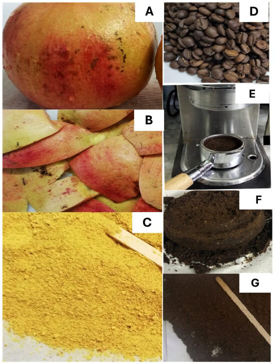
Figure 1.
Waste collection steps for pomegranate peels (A–C) and coffee grounds (D–G) (adapted from Abdelmigid et al. [44]).
2.4. Green Synthesis of Ag Nanoparticles
As a metal precursor, AgNO3 was used to generate AgNPs. Silver nitrate (AgNO3) solution was prepared by dissolving 0.017 g in 100 mL of deionized double-distilled water and stirring at room temperature for about 10 min. The solution was filtered with filter paper No. 4 to obtain a clear solution. Then, 15 mL of plant extract was added separately to 20 mL of aqueous 0.001 M AgNO3 solution in 100 mL Erlenmeyer flasks. Gradually, the extract was poured into the solution while heating it at 70 °C for 1 h, with continuous stirring. As a result, the color of the solution changed from a pale light green to a light brown and then to a dark brown. This color change indicated the presence of AgNPs in the solution. Repeated centrifugation at 10,000 rpm for 15 min, followed by dispersion of the pellet in sterile DI water four times, was used to purify AgNPs from the solution [44]. Following 48 h of lyophilization, the nanoparticles suspended in water were analyzed for their composition and structure. The AgNPs, which were synthesized using pomegranate peel extract, have been referred to as AgNPs_PPE, while those synthesized using coffee ground beans have been distinguished as AgNPs_CE throughout the manuscript.
2.5. Chemical Synthesis of Ag Nanoparticles
The chemical synthesis of silver nanoparticles was carried out in this study, according to Fang et al. [45]. Distilled water (15 mL) was used to dissolve AgNO3 (2 g), while trisodium citrate (1 g) was dissolved in 100 mL of distilled water. After boiling 50 mL of a 10−3 M AgNO3 solution, it was gently added to trisodium citrate (1%, 5 mL, Sigma-Aldrich, Milan, Italy) for five minutes at 90 °C with vigorous stirring. Once the reduction began, 2.5 mL of trisodium citrate was added dropwise until a pale yellow appeared in the solution. To distinguish these particles throughout the manuscript, we refer to them as AgNPs_Chem. Similar to the biologically prepared AgNPs, AgNPs_Chem particles were obtained through centrifugation and subsequent drying.
2.6. Characterization of Silver Nanoparticles
2.6.1. UV-Vis Spectroscopy
To characterize the prepared silver nanoparticles (AgNPs_PPE, AgNPs_CE, and AgNPs_Chem), UV-1601 spectroscopy (Shimadzu, Kyoto, Japan) was used for optical absorption measurements at wavelengths ranging from 200 to 800 nm. Autoclaved distilled water was used as a reference.
2.6.2. Size Distribution and Zeta Potential
The average size and surface charge of the AgNPs synthesized at 25 °C with a detection angle of 90° were measured using a Zetasizer Nano ZS particle size analyzer (Malvern Instruments Ltd., Malvern, Worcestershire, UK). To prevent nanoparticle aggregation, all powder samples were freshly resuspended in 0.9% saline solution and sonicated at high speed for 30 min before assessment.
2.6.3. Surface Morphology
To determine the surface morphology and size of AgNPs, scanning electron microscopy (SEM, JEOL JSM-639OLA Analytical Scanning Electron Microscope, Tokyo, Japan, which is located in Taif University’s Electron Microscope Unit) was performed at 20 kV (2 × 10⁴) V with magnifications of 500, 2000, and 3000 with scale bars of 50 μm, 10 μm, and 5 μm, respectively. Before scanning, the silver nanoparticle powders (AgNPs_PPE, AgNPs_CE, and AgNPs_Chem) were coated with gold using a Cressington Sputter Coater (108 Auto, thickness controller MTM-10, UK). X-ray diffraction (XRD) was used to assess the crystallinity of the synthesized nanoparticles (Pan Analytical, X-pert pro, The Netherlands). At 30 kV (3 × 10⁴ V) and 100 mA (1 × 10⁻¹ A), XRD was used to document the spectrum at a wavelength of 1.5406 Å in the 2θ (from the range of 20–80°). The XRD patterns were plotted using OriginLab software® (2018) and compared with the JCPDS card number 040783.
2.6.4. Fourier Transforms Infrared Spectroscopy (FTIR)
A small amount of the synthesized AgNP powder was mixed with the KBr powder to create a homogeneous mixture. The mixture was subjected to high pressure to form a thin disc-shaped pellet. The resulting KBr pellet containing AgNPs was placed in an FTIR spectrometer for analysis. KBr is advantageous in this process because it is transparent in the infrared region and does not interfere with the analysis of the AgNPs. For the detection of possible functional groups in synthesized AgNPs, FTIR spectroscopy (Agilent Technologies, Santa Clara, CA, USA) at a wavenumber between 450 and 4000 cm−1 was used.
2.7. Preparations of Nanopriming Solutions and Seed Priming
AgNPs (AgNPs_PPE, AgNPs_CE, and AgNPs_Chem) synthesized by biological and chemical methods were freshly prepared as 80 mg/L nanopriming solutions by ultrasonic vibration (100 W (1 × 10² W), 40 kHz (4 × 10⁴ Hz)) for 30 min [46]. The concentrations of the nanopriming solutions were selected based on the preliminary experiments. Milli-Q deionized water was used as the hydropriming solution (HP) [46]. The seeds were surface-sterilized in 1% sodium hypochlorite for 3 min and rinsed three times with deionized water. The seeds were then soaked in a AgNP-priming solution for 24 h (rotatory shaker). The ratio of seed weight to solution volume was approximately 1:4 g/mL. After applying deionized water for 3 min, the seeds were dried on Whatman filter paper. The sterile pouches were then sealed and refrigerated at a temperature of 4 °C until needed.
2.8. Assaying AgNPs Adsorption on a Seed Surface by SEM Analysis
Scanning electron microscopy (SEM) was used to evaluate the distribution and structural features of nanoparticles adsorbed on the nanoprimed seeds after washing with distilled water and air-drying. The seeds were coated with a layer of gold before scanning using a Cressington 108 Sputter Coater (auto, thickness controller MTM-10; Watford, UK).
2.9. Energy Dispersive X-ray (EDX)
A JEOL–JSM-1400 PLUS system with an EDX detector was used for identifying the chemical composition of the Ag nanoparticles and for detecting the elemental composition of the Punica granatum L. seeds. All samples were coated with a thin layer of gold for analysis. In the EDX spectrum, the detected signal was plotted as a function of energy. The following are the measurement conditions for the EDX detector: an acceleration voltage of 20.00 kV, a magnification of ×270, and a process time of T2. The measurement detector possesses a live time of 30.00 s, a real-time of 30.22 s, and a dead time of 1.00%. The count rate is 2528.00 CPS. The JEOL system's specific spectra processing system was operated according to the manufacturer's recommended protocols. Similarly, the calibration of the EDX detector was carried out according to the manual instructions provided.
2.10. Assaying AgNPs Internalization
Randomly selected nanoprimed seeds were dissected and prefixed in 4F1G in phosphate buffer solution (pH = 7.2) at 4 °C for 3 h. After that, specimens were postfixed in 2% OsO4 in the same buffer at 4 °C for 2 h. Various concentrations of acetone were then used to dehydrate samples. They were then embedded in resin for polymerization and cut into sections approximately 90 Å thick. The sections were placed on grid copper and stained with uranyl acetate for 5 min. They were then stained with 1% lead citrate for 2 min, rinsed again in pure water, and stored in grid boxes. Ultrathin sections were prepared using a Leica ultracut UCT ultramicrotome. A JEOL–JSM-1400 PLUS TEM (JEOL, Tokyo, Japan) was used to obtain TEM images.
2.11. Inductively Coupled Plasma Spectrometer (ICP–OES) Analysis of NP Uptake
ICP-OES was used to determine the total AgNP uptake or bioaccumulation in the nanoprimed pomegranate seeds. The following steps were followed: First, 0.1 g of finely ground pomegranate seeds were dried at 100 °C for 24 h. To dissolve all residues, 10 mL aqua regia (HNO3/HCL) was added and slowly heated to 150 °C. Two replications of this procedure were conducted, each with 10 mL aqua regia added to achieve complete digestion (~2 mL). Subsequently, a Whatman filter paper was used to purify the solution, and then a 100 mL volumetric flask was used to dilute it with Millipore water. The AgNP content was quantified in triplicate using an inductively coupled plasma spectrometer (ICPE-9000, Shimadzu, Kyoto, Japan). The instrument was calibrated using a standard blank and mixed-metal calibration standard before analysis. Following the calibration plot, the most accurate linear regression correlation coefficient was calculated (R2 ≥ 0.9999) from the calibration plot. Multi-element standards and analytical references were purchased from AccuStandard (New Haven, CT, USA).
2.12. Energy Dispersive X-ray Fluorescence (EDXRF) Analysis of NP Uptake
According to Tezotto et al. [47], the examination was performed following their protocol. EDXRF analysis was conducted on the same pomegranate seeds prepared as a loose powder for ICP-OES analysis. Polypropylene film (Mylar®) was used to cover a 20 mm diameter polyethylene cup filled with one gram of ground seeds. With a Shimadzu EDX-720 energy dispersive X-ray fluorescence spectrometer, the samples were radiated in triplicate for 300 s under vacuum. X-ray tubes were used for irradiating the samples (15 kV (15 × 103 V) for Na to Sc and 50 kV (5 × 10⁴ V)). There was an automatic adjustment of the current to a maximum of 1A, and a 10 mm collimator was selected. The detection was carried out with a liquid nitrogen-cooled Si (Li) detector. The same method was used to verify the precision and trueness of certified reference materials (CRMs). The intensity of element Kα counts per second (cps/µA) was brought from the sample X-ray spectrum deconvolution using the EDX Shimadzu software package (EDX-7000/8000/8100).
3. Results
3.1. Silver Nanoparticle (AgNP) Characterization
3.1.1. UV-Vis Spectroscopy
The creation of silver nanoparticles is demonstrated by the change in color from pale yellow, brown, and colorless, respectively, to dark brown in PPE, CE, and trisodium citrate (Figure 2A–C). Using UV-visible spectroscopy, the ability of the aqueous solution to produce nanoparticles was further demonstrated. Figure 2D shows the UV-vis absorption spectra of AgNPs_PPE (350 nm), AgNPs_ CE (with two distinct peaks at 320 nm and 440 nm), and AgNPs_ Chem (430 nm).
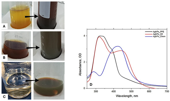
Figure 2.
The successful creation of silver nanoparticles shown by the change in the color of (A) pomegranate peel extract (PPE), (B) coffee bean extract (CE), (C) trisodium citrate, and UV-vis sharp absorption spectra of AgNPs (D), respectively. Arrows indicate the color change.
3.1.2. Size Distribution and Zeta Potential
Zeta potential was used to estimate the stability of the produced AgNPs, where higher zeta potential values indicate more stable nanoparticles. A chemically produced AgNPs_Chem (46.7 mV) was found to be more stable than AgNP_CE (12.6 mV) and AgNPs_PPE (7.98 mV), which had the least stability. Figure 3A shows how a reduction in AgNPs stability causes particle aggregation, which leads to a size increase for AgNPs_PPE = 591.9 nm, AgNPs_CE = 273.7 nm, and AgNPs_Chem = 62.75 nm. This suggests that chemically synthesized AgNPs (AgNPs_Chem) were the most stable, followed by those from coffee extracts (AgNPs_CE), and lastly, those from pomegranate peel extracts (AgNPs_PPE).
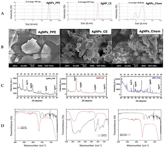
Figure 3.
Characterization of the investigated AgNPs in this study: (A) zeta potential, (B) scanning electron microscopy, (C) XRD patterns, and (D) FTIR spectra (Adapted from Abdelmigid et al. [44]).
3.1.3. Scanning Electron Microscopy (SEM) and XRD Analysis
Scanning electron microscopy (SEM) analysis, as shown in Figure 3B, provides visual evidence of the crystalline structure for both chemically and biologically synthesized AgNPs. X-ray diffraction (XRD) further confirms this crystallinity (Figure 3C). The four characteristic peaks observed in the XRD pattern correspond to the face-centered cubic (FCC) crystal structure of silver nanoparticles according to Bragg’s reflection principle. These peaks appear at 2θ values of 38.3°, 44.49°, 64.6°, and 77.5°, which can be assigned to Ag crystal planes (111), (200), (220), and (311), respectively. Interestingly, the XRD pattern of AgNPs_PPE reveals additional diffraction peaks at 2θ of 27.90°, 32.38°, 46.43°, 55.00°, 57.00°, and 67.00°. These additional peaks warrant further investigation to identify the potential presence of other crystalline phases or impurities associated with the biological synthesis process using pomegranate peel extract.
3.1.4. FTIR Evaluation of AgNPs
Biomolecules affecting silver nanoparticle capping, reduction, and stability were identified using FTIR analysis of PPE, CE, and trisodium citrate. In Figure 3D, the differences between synthesized AgNPs and their reference are highlighted. Any change in the sharpness or position of a peak is considered a “group contribution” in the synthesis of silver nanoparticles. In comparison with PPE, the various AgNPs_PPE peaks were sharper. The sharp peak that was detected at 2927 cm−1 was attributed to C-H stretching, 1641 cm−1 to C-O stretching vibration in quinine, 1351 cm−1 to the C=C aromatic ring, 1228 cm−1 to C-O stretching in ether, ester, or phenol, and 1055 cm−1 to C-N stretching of the aliphatic primary amine. The phenolic groups are located between 1315 and 1037 cm−1 and 1456 and 1600 cm−1. In comparison with AgNP extract CE, FTIR analysis revealed distinct sharp peaks. FTIR analysis of the AgNP_CE identified functional groups potentially involved in nanoparticle capping and stabilization. A broad band between 3600 and 3000 cm−1 indicates N-H, O-H, and asymmetric C-H stretching vibrations. Additional peaks at 2922 and 2852 cm−1 further support these functional groups. Aromatic and aliphatic C-H, C-O, and C-N vibrations were observed in the 1400–900 cm−1 region. Peaks between 1700 and 1500 cm−1 suggest C-C and C-N stretching vibrations. The AgNP_Chem synthesis was found to yield different peaks related to C-H stretching deformation (1282 cm−1), nitro compound (NO) as NO2 (1402 cm−1), and C=O stretching asymmetry in COO− (1582 cm−1), while at 3312 cm−1, amine stretching was observed.
3.2. AgNP Adsorption and Localization on Seed Coats
SEM/EDX Analysis
Scanning electron microscopy (SEM) analysis was employed to investigate the size, shape, and distribution of AgNPs adsorbed onto the surface of Punica granatum seeds following nanopriming. Additionally, SEM was used to assess the overall morphology and integrity of pomegranate seeds (Figure 4A–D). Compared to the smooth surface texture observed in control seeds (Figure 4A), nanopriming with AgNPs resulted in significant alterations in seed coat morphology (Figure 4B–D). Notably, seeds treated with AgNPs exhibited numerous large pores with a distinctive honeycomb-like structure. The SEM images revealed that the majority of the AgNPs, regardless of their source (AgNPs_PPE, AgNPs_CE, or AgNPs_Chem), appeared to be spherical. Interestingly, the synthesized nanoparticles displayed a tendency to form both small and larger aggregates, which became entrapped within the seed coat fibers (Figure 4B–D). Differences were observed in the clustering behavior of the AgNPs depending on the synthesis method. Seeds treated with AgNPs_PPE and AgNPs_CE displayed larger clusters of nanoparticles scattered across the seed surface (Figure 4B,C). In contrast, seeds treated with AgNPs_Chem exhibited a more even distribution of individual nanoparticles, with fewer evident clusters (Figure 4D). Overall, SEM analysis provides valuable insights into the impact of AgNPs on the surface morphology of Punica granatum seeds. These findings suggest that nanopriming with AgNPs alters the seed coat structure and influences the distribution and clustering behavior of the nanoparticles.
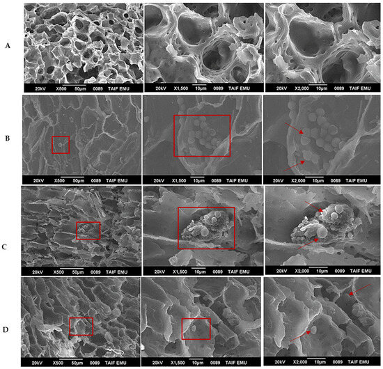
Figure 4.
SEM images of nanoprimed Punica granatum seeds at various magnifications to visualize the adsorption of AgNPs. (A) Hydroprimed seeds (control) show no nanoparticles. (B–D) Seeds primed with different AgNPs: (B) AgNPs _PPE, (C) AgNPs_CE, and (D) AgNPs_ Chem. Red boxes and arrows highlight representative examples of adsorbed AgNPs on the seed coat surface at different magnifications (×500, ×1500, and ×2000).
Energy-dispersive X-ray spectroscopy (EDX) confirmed the presence of AgNPs on the seed coat and provided information regarding their elemental composition. EDX spectra (Figure 5) revealed a distinct Ag peak in the nanoprimed samples (AgNPs_PPE, AgNPs_CE, AgNPs_Chem), which was absent in the hydroprimed (HP) control seeds. This confirms the successful deposition of AgNPs onto the seed coat surface following the nanopriming treatments. Table 1 summarizes the quantitative elemental composition data obtained from the EDX analysis. Comparatively, the control seeds had a low mass percentage of C (47.78%) and a high mass percentage of O (52.22%) (Table 1). In the nanoprimed samples, there was a variation in carbon and oxygen contents; all nanoprimed samples displayed a decrease in oxygen content and an increase in carbon content. This suggests the adsorption of organic compounds onto the surface of the AgNPs. These organic compounds likely originated from the plant extracts (Punica granatum (PPE) and Coffea arabica (CE)) used in the synthesis process, which are known to play a role in reducing and stabilizing AgNPs. The presence of trisodium citrate, another component of the synthesis solution, also contributed to the observed carbon signal.
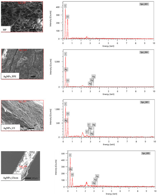
Figure 5.
Analysis of hydroprimed (HP) and nanoprimed Punica granatum seeds using SEM-EDX technology. AgNPs_PPE, AgNPs_CE, and AgNPs_Chem adsorption on the surface of the seed coat is shown in panels. The element peaks (C, O, and Ag) are displayed in EDX spectra. The region of interest on the seed surface is revealed by the SEM photos (ROI).

Table 1.
Elemental composition of control and AgNP-primed Punica granatum seeds determined by EDX. The amount of the adsorbed C, O, and Ag elements are expressed as mean (%) ± SE values of mass% derived from sample replications used for the study.
Interestingly, the nanoprimed samples exhibited variations in carbon and oxygen contents. AgNP_PPE-primed seeds had the highest carbon content (74.94%) and the lowest oxygen content (24.95%), suggesting a greater abundance of organic material on their surface than other treatments. Conversely, AgNP_CE-primed seeds displayed lower carbon content (49.69%) and higher oxygen content (49.0%), potentially indicating a different interaction with the plant extract. AgNP_Chem-primed seeds showed intermediate values (C: 60.4%, O: 39.16%). Overall, EDX analysis provided strong evidence for the successful deposition of AgNPs onto the seed coat surface and revealed differences in the surface composition of AgNPs derived from various synthesis methods.
3.3. AgNP Uptake in Seeds
3.3.1. TEM Analysis
Transmission electron microscopy (TEM) was employed to investigate the potential translocation of AgNPs from the seed coat into the Punica granatum embryo. Figure 6A–D presents the TEM micrographs at various magnifications. In contrast to the absence of nanoparticles observed in hydroprimed control seeds (Figure 6A), TEM micrographs of nanoprimed seeds (Figure 6B–D) revealed the presence of electron-dense deposits within the embryo. These deposits appeared as individual dark particles and larger aggregates, suggesting the potential internalization of AgNPs, and higher magnification images (Figure 6C,D) provided further insight into the morphology of the internalized nanoparticles. The dark deposits exhibited a predominantly spherical or ellipsoidal shape, consistent with the expected morphology of AgNPs. These observations suggest the potential translocation of AgNPs from the seed coat to the Punica granatum embryo. The presence of individual particles and aggregates indicates that internalization may occur through various mechanisms.
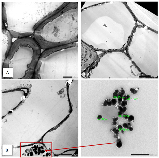
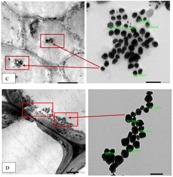
Figure 6.
TEM images of Punica granatum seed cross-sections after being primed with (A) distilled water (hydroprimed), (B) AgNPs_ PPE, (C) AgNPs_CE, and (D) AgNPs_Chem. The TEM image of the manufactured NPs shows a similar pattern to the details of the Ag nanoparticle clusters found inside seed tissue. (The AgNPs are indicated by red boxes and arrows in the image).
3.3.2. X-ray Fluorescence (XRF) and ICP–OES Analyses
Although TEM provides valuable information about the morphology and size of nanoparticles within a sample, it has limitations in quantifying their total uptake. To address this, X-ray fluorescence (XRF) and inductively coupled plasma–optical emission spectroscopy (ICP-OES) techniques were employed to measure the total Ag content within Punica granatum seeds following nanopriming with AgNPs at a concentration of 80 mg/L. Both XRF and ICP-OES analyses successfully confirmed the presence of Ag in nanoprimed seed samples compared to hydroprimed controls (Figure 7 and Figure 8). However, the techniques yielded different quantitative results for different types of AgNPs (AgNPs_PPE, AgNPs_CE, and AgNPs_Chem). ICP-OES analysis revealed a significantly higher Ag content in the AgNPs_PPE primed seeds than that in the XRF analysis. Conversely, AgNPs_CE and AgNPs_Chem displayed higher Ag values, as measured by XRF. Notably, the AgNP_ Chem-primed seeds exhibited the highest XRF value (0.083 mg/g) but the lowest ICP-OES value. XRF spectrum analysis further confirmed the presence of Ag in all nanoprimed seed samples (AgNPs_PPE, AgNPs_CE, and AgNPs_Chem), as evidenced by the distinct Ag peaks in the spectra (Figure 8). No such peaks were observed in the XRF spectra of the hydroprimed seeds.
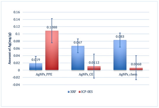
Figure 7.
Measurements of the amount of AgNPs (mg/g) in P. granatum seeds subjected to AgNPs_PPE, AgNPs_ CE, and AgNPs_Chem using XRF and ICP-OES. The amount of Ag element is expressed as mean values derived from sample replications used for the investigation or mean ± SE.
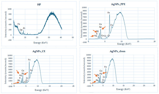
Figure 8.
Analysis of P. granatum L. seeds treated with AgNPs_PPE and AgNPs_CE using X-ray fluorescence spectroscopy (XRF) compared with hydroprimed seeds (HP). Arrows indicate Ag signal in nanoprimed seeds.
4. Discussion
This study leverages the green synthesis of silver nanoparticles (AgNPs) using readily available plant extracts, as previously reported by our research group (Abdelmigid et al. [44]). This study leverages an eco-friendly approach for AgNP synthesis by utilizing ground coffee and aqueous pomegranate peel extracts as both reducing and stabilizing agents. This method offers advantages in terms of cost-effectiveness and reduced risk compared to traditional techniques [48]. Here, we build upon characterization data from previous studies [44] to explore the potential relationship between the size and morphology of the synthesized AgNPs and their ability to penetrate or incorporate into Punica granatum seeds. This analysis provides further evidence for the successful synthesis of functional AgNPs. For comparison, we also included chemically synthesized AgNPs_Chem using trisodium citrate as a reducing agent. Characterization techniques confirmed that all synthesized NPs were in their pure crystalline form. The observed color change during AgNP formation using plant extracts aligns with findings from similar studies. For instance, Jabir et al. [49] reported a color shift from yellow to brown in Annona muricata peel extract upon green synthesis of silver nanoparticles, further supporting the reduction process.
As a result of their strong surface plasmon resonances (SPRs), UV absorbance was used to verify the synthesis of AgNP_PPE, AgNP_CE, and AgNP_Chem colloidal metal nanoparticles [50,51,52]. Maximum absorbance was observed between 320 and 430 nanometers in the recorded UV spectrum, which indicates that silver ions have been reduced to colloidal silver. There have been reports of colloidal silver nanoparticles ranging in size from 350 to 600 nanometers [52,53,54,55]. Because of their different physical properties, including size and shape, which affect their SPRs, the AgNPs formed in this study showed different UV spectra. UV-vis spectroscopy at the SPRs 421 nm, 405–430 nm, and 420 nm showed that AgNPs were formed by reducing Ag+ to Ag0 using PPE, spent coffee ground extracts, and trisodium citrate, respectively [44,56,57,58]. Increasing wavelengths are associated with a decrease in the size of AgNPs, as observed in the current study [59].
Studies have demonstrated that nanoparticles with a more negative potential, such as −46.7 mV, exhibit improved repulsion forces, resulting in enhanced stability, dispersion, and colloidal properties [60]. Based on our findings, chemically synthesized AgNPs displayed smaller diameters and higher zeta potentials than PPE- and CE-synthesized AgNPs. Researchers previously found that AgNPs made from PPE (31.6 nm; 118.6–231.7 nm), CE (21–255 nm), and trisodium citrate (5–10 nm) have a smaller diameter [44,56,58,59,60,61,62]. The size of AgNPs can be adjusted by modifying the pH and concentration of AgNO3. At alkaline pH levels, where the pH is inversely proportional to the size of the AgNPs, the reaction rate increases with successive crystallization into smaller particles. Furthermore, it is important to note that the repulsion between nanoparticles tends to increase as the pH level increases, whereas the aggregate ability tends to decrease because of the complete surface charge. However, it is worth mentioning that previous research [59] indicated that the size of silver nanoparticles (AgNPs) tends to decrease as the nitrate concentration increases from 1 M to 5 M.
X-ray diffraction (XRD) analysis confirmed the crystalline nature of the synthesized AgNPs, with characteristic peaks observed at 38.3°, 44.49°, 64.6°, and 77.5°. These peaks correspond to the face-centered cubic (FCC) structure of metallic silver, aligning with findings from previous studies utilizing trisodium citrate and other plant extracts for AgNP synthesis [4,63,64,65,66]. The presence of additional diffraction peaks in the AgNP_PPE spectrum at 27.90°, 32.38°, 46.43°, 55.00°, 57.00°, and 67.00° may be attributed to the complex phytochemical composition of pomegranate peel extract (PPE). Similar unassigned peaks have been reported in other studies [67,68].
Fourier transform infrared (FTIR) spectroscopy provided valuable insights into the functional groups potentially involved in the bioreduction and stabilization of the synthesized AgNPs. The spectra of AgNPs_PPE, AgNPs_CE, and AgNPs_Chem were compared with the reference spectra to identify the key functional groups. The presence of phenolic groups in all AgNP types is evident from the peaks observed between 1315–1037 cm−1 and 1456–1600 cm−1. These functional groups, particularly flavanones present in plant extracts, are well established for their role in reducing silver ions (Ag+) to metallic silver (Ag0) [69]. Additionally, they can act as capping agents, stabilizing newly formed AgNPs [70]. Studies by Shankar et al. [71] support this mechanism, demonstrating the adsorption of flavanones from plant extracts onto metal nanoparticles with characteristic bands around 1074 cm−1, potentially present in our spectra. Plant extracts, especially those rich in flavonoids, such as pomegranate peel extract (PPE), offer a multitude of bioactive compounds that contribute to AgNP synthesis and stabilization. Besides flavanols, PPE contains gallotannins, quercetin, ellagic acid derivatives, catechins, procyanidins, and kaempferol [72]. These compounds might contribute to the observed effects; the presence of free caffeine in coffee ground extract (CE) might explain the additional sharp peaks and broad bands observed in the AgNP_CE spectrum, along with the shared flavonoids with AgNPs_PPE. These findings are consistent with previous reports of caffeine vibrations in coffee extracts [4,73,74]. Finally, the FTIR spectrum of AgNPs_Chem, synthesized using trisodium citrate, displayed peaks corresponding to the functional groups associated with the reducing agent. These include C-H stretching deformation, nitro compound (NO2), C=O stretching asymmetry in COO−, and a possible amine stretching vibration, as reported elsewhere [75,76,77,78].
Scanning electron microscopy (SEM) revealed the presence of a porous structure on the surface of Punica granatum L. seeds [4,79]. This porous structure, inherent to lignocellulosic materials, likely arises due to the evaporation of volatile substances during seed coat formation [4,79]. These pores offer potential adsorption sites for mineral compounds, including AgNPs. Although SEM cannot visualize internal structures, it can be valuable for assessing particle purity and aggregation on the seed surface [80]. Previous studies have suggested that smaller nanoparticles exhibit higher surface energy, leading to increased interaction with cell walls [4,81,82]. This interaction could potentially lead to the development or expansion of existing pores, thereby enhancing the penetration of nanoparticles into the seed coat. The results of our energy-dispersive X-ray spectroscopy (EDX) analysis align with this hypothesis. Smaller AgNPs_Chem (62.68 nm) were detected in higher abundance within pomegranate seeds than larger AgNPs_CE (273.7 nm) and AgNPs_PPE (591 nm).
The higher prevalence of AgNPs_Chem within seeds aligns with observations from X-ray fluorescence (XRF) analysis. This suggests that smaller nanoparticles can readily pass through cellular membranes via existing pores or potentially promote the formation of new ones [83,84,85]. Additionally, their smaller size and potentially higher zeta potential might contribute to a more efficient interaction with the seed coat, facilitating pore formation, and the presence of a 3 eV optical absorbance band identified through EDX analysis confirmed the presence of pure metallic silver nanoparticles within the seeds [86,87]. Furthermore, EDX and FTIR analyses revealed the presence of organic molecules (C and O) on the AgNP surface, indicating their role as reducing and capping agents [88,89]. The broad and sharp peaks observed in the FTIR spectra suggest the presence of various functional groups associated with these capping agents [90,91,92]. The increased carbon (C) signal observed in the EDX analysis for AgNP-primed seeds compared to hydroprimed seeds further supports the presence of organic capping agents on the AgNP surface.
Transmission electron microscopy (TEM) analysis provided compelling evidence for the successful internalization of AgNPs into the seed embryos of Punica granatum L. This observation aligns with the findings of a previous study on watermelon seeds treated with AgNPs [93]. TEM images revealed the presence of AgNPs within the seed embryo, suggesting the potential for Ag+ uptake and nanoparticle localization.
Interestingly, our TEM analysis did not reveal a specific or uniform distribution pattern for internalized AgNPs. This finding is consistent with observations reported by Song et al. [94], who described clusters of AgNPs surrounding plasma membranes of watermelon seed embryos. Such clustering may be attributed to the dynamic nature of the cytoplasm, where individual AgNPs can be internalized and form aggregates. Similar observations of nanoparticle agglomeration within plant cells have been reported previously. Nicolás-Álvarez et al. [95] demonstrated the presence of titanium dioxide (TiO2) nanoparticles in agglomerated forms within tomato root cells, suggesting a potential shared mechanism for internalized nanoparticle behavior across plant species.
Inductively coupled plasma–optical emission spectrometry (ICP-OES) analysis confirmed the penetration of Ag ions released from AgNPs into Punica granatum L. seed tissues, as evidenced by the presence of detectable silver compared to hydroprimed controls. This finding aligns with the observations reported by Zhou et al. [96], who demonstrated similar Ag ion uptake in plant tissues exposed to AgNPs. While X-ray fluorescence (XRF) analysis offers a valuable tool for quantifying the elemental composition on surfaces, its penetration depth is limited compared to ICP-OES [97]. This explains the observed discrepancies between the XRF and ICP-OES results of our study. XRF analysis likely detected primarily surface-bound Ag, potentially leading to higher Ag concentrations in the AgNPs_CE and AgNPs_Chem treatments because of their larger size and potentially greater surface area. In contrast, ICP-OES requires complete sample mineralization, solubilizing both surface and internalized Ag, which may explain the lower XRF-measured Ag concentrations in AgNPs_PPE treatments, possibly due to deeper tissue penetration of these smaller nanoparticles. These observations highlight the importance of considering the limitations of the analytical technique when interpreting Ag quantification data, particularly for heterogeneous samples, such as plant tissues.
The integrated analytical approach employed in this study, utilizing SEM, EDX, TEM, XRF, and ICP-OES, successfully confirmed the absorption and incorporation of AgNPs into Punica granatum seeds. This approach provides a robust method for verifying the effectiveness of nanopriming before investigating its potential positive or negative effects on target crops. It is important to acknowledge that the identification and quantification of nanomaterials within complex biological matrices, such as plant tissues, requires specialized techniques, such as transmission electron microscopy (TEM) and inductively coupled plasma research [98]. Our findings regarding AgNP penetration into pomegranate seeds using TEM, XRF, and ICP-OES are consistent with the observations reported in other studies on various crops. Similar results have been documented for AgNP uptake in tomatoes [95], maize [99], and mung bean seeds [100]. Furthermore, the internalization of metal-based nanoparticles by plant tissues extends beyond AgNPs, as studies have shown comparable behavior for Fe3O4, Au, and Cu nanoparticles [101,102,103]. Collectively, these findings suggest a potentially widespread mechanism for nanoparticle uptake in plants.
5. Conclusions
This research evaluated the efficacy of synthesized silver nanoparticles (AgNPs) as seed-priming agents to minimize nanomaterial release into the environment. Our study utilized green and chemical synthesis techniques to produce AgNPs. The green synthesis technique successfully produced purified crystalline Ag nanoparticles from aqueous extracts of pomegranate fruit peel (PPE) and coffee (CE). Additionally, trisodium citrate was employed in the chemical reduction process to synthesize AgNPs. Based on the results of this work, the following key findings can be emphasized:
- The seeds of pomegranate demonstrated successful internalization and the uptake of nanoparticles, which were validated through various analytical methods, such as UV-visible spectroscopy, zeta potential analysis, FTIR spectroscopy, XRD, SEM, and TEM.
- The significance of the electron microscopy (EM) analysis has been emphasized in verifying the incorporation of nanoparticles in determining AgNP distribution within plants, verifying the incorporation of nanoparticles, and determining the distribution of silver nanoparticles (AgNPs) within plant tissues.
- Elemental analysis techniques (EDX, XRF, ICP-OES) were successful in producing comprehensive outcomes that corroborated the findings obtained from the use of scanning electron microscopy (SEM) and transmission electron microscopy (TEM).
Although this research demonstrates the potential of AgNP nanopriming as a targeted approach for delivering nanoparticles to seeds, additional investigation into the optimal dosage and long-term effects of AgNPs on different plant species is necessary to ensure safe and sustainable agricultural practices.
6. Future Directions
Understanding the molecular mechanisms underlying plant responses to nanopriming (germination, growth, physiology, metabolism, genetics) will be essential for optimizing nanopriming protocols for improved agricultural outcomes.
Author Contributions
Conceptualization, H.M.A., A.A.A. and M.M.M.; methodology, H.M.A., A.A.A. and M.M.M.; software, H.M.A.; validation, H.M.A., A.A.A. and M.M.M.; formal analysis, A.A.A.; investigation, H.M.A.; resources, A.A.A.; data curation, M.M.M.; writing—original draft preparation, H.M.A.; writing, A.A.A. and M.M.M.; visualization, A.A.A.; supervision, H.M.A.; project administration, H.M.A.; funding acquisition, A.A.A. All authors have read and agreed to the published version of the manuscript.
Funding
The research was funded by Taif University, Saudi Arabia, Project No. (TU-DSPP-2024-213).
Data Availability Statement
The original contributions presented in the study are included in the article, further inquiries can be directed to the corresponding author.
Acknowledgments
The authors extend their appreciation to Taif University, Saudi Arabia, for supporting this work through project number (TU-DSPP-2024-213).
Conflicts of Interest
The authors declare no conflicts of interest.
References
- Singh, A.; Singh, N.Á.; Afzal, S.; Singh, T.; Hussain, I. Zinc oxide nanoparticles: A review of their biological synthesis, antimicrobial activity, uptake, translocation and biotransformation in plants. J. Mater. Sci. 2018, 53, 185–201. [Google Scholar] [CrossRef]
- Liu, R.; Lal, R. Synthetic apatite nanoparticles as a phosphorus fertilizer for soybean (Glycine max). Sci. Rep. 2014, 14, 5686. [Google Scholar] [CrossRef] [PubMed]
- Janmohammadi, M.; Sabaghnia, N. Effect of pre-sowing seed treatments with silicon nanoparticles on germinability of sunflower (Helianthus annuus). Bot Lith. 2015, 21, 13–21. [Google Scholar] [CrossRef] [PubMed]
- Abdelmigid, H.M.; Alyamani, A.A.; Hussien, N.A.; Morsi, M.M.; Alhumaidi, A. Integrated Approaches for Adsorption and Incorporation Testing of Green-Synthesized TiO2NPs Mediated by Seed-Priming Technology in Punica granatum L. Agronomy 2022, 12, 1601. [Google Scholar] [CrossRef]
- Endo, E.H.; Ueda-Nakamura, T.; Nakamura, C.V.; Filho, B.P.D. Activity of Spray-dried Microparticles Containing Pomegranate Peel Extract against Candida albicans. Molecules 2012, 17, 10094–10107. [Google Scholar] [CrossRef] [PubMed]
- Ismail, T.; Sestili, P.; Akhtar, S. Pomegranate peel and fruit extracts: A review of potential anti-inflammatory and anti-infective effects. J. Ethnopharmacol. 2012, 143, 397–405. [Google Scholar] [CrossRef] [PubMed]
- Habibipour, R.; Moradi-Haghgou, L.; Farmany, A. Green synthesis of AgNPs@ PPE and its Pseudomonas aeruginosa biofilm formation activity compared to pomegranate peel extract. Int. J. Nanomed. 2019, 14, 6891. [Google Scholar] [CrossRef] [PubMed]
- Leesombun, A.; Sariya, L.; Taowan, J.; Nakthong, C.; Thongjuy, O.; Boonmasawai, S. Natural Antioxidant, Antibacterial, and Antiproliferative Activities of Ethanolic Extracts from Punica granatum L. Tree Barks Mediated by Extracellular Signal-Regulated Kinase. Plants 2022, 11, 2258. [Google Scholar] [CrossRef]
- Elkady, A.I. Crude alkaloid extract of Rhazya stricta inhibits cell growth and sensitizes human lung cancer cells to cisplatin through induction of apoptosis. Genet. Mol. Biol. 2013, 36, 12–21. [Google Scholar] [CrossRef]
- Jisha, K.C.; Vijayakumari, K.; Puthur, J.T. Seed priming for abiotic stress tolerance: An overview. Acta Physiol. Plant. 2013, 35, 1381–1396. [Google Scholar] [CrossRef]
- Bayat, M.; Zargar, M.; Murtazova, K.M.-S.; Nakhaev, M.R.; Shkurkin, S.I. Ameliorating Seed Germination and Seedling Growth of Nano-Primed Wheat and Flax Seeds Using Seven Biogenic Metal-Based Nanoparticles. Agronomy 2022, 12, 811. [Google Scholar] [CrossRef]
- Chen, K.; Arora, R. Priming memory invokes seed stress-tolerance. Environ. Exp. Bot. 2013, 94, 33–45. [Google Scholar] [CrossRef]
- Devika, O.S.; Singh, S.; Sarkar, D.; Barnwal, P.; Suman, J.; Rakshit, A. Seed Priming: A Potential Supplement in Integrated Resource Management Under Fragile Intensive Ecosystems. Front. Sustain. Food Syst. 2021, 5, 654001. [Google Scholar] [CrossRef]
- Forti, C.; Ottobrino, V.; Bassolino, L.; Toppino, L.; Rotino, G.L.; Pagano, A.; Macovei, A.; Balestrazzi, A. Molecular dynamics of pre-germinative metabolism in primed eggplant (Solanum melongena L.) seeds. Hortic. Res. 2020, 1, 7. [Google Scholar] [CrossRef]
- Du, B.; Luo, H.; He, L.; Zhang, L.; Liu, Y.; Mo, Z.; Pan, S.; Tian, H.; Duan, M.; Tang, X. Rice seed priming with sodium selenate: Effects on germination, seedling growth, and biochemical attributes. Sci. Rep. 2019, 9, 4311. [Google Scholar] [CrossRef] [PubMed]
- Marthandan, V.; Geetha, R.; Kumutha, K.; Renganathan, V.G.; Karthikeyan, A.; Ramalingam, J. Seed Priming: A Feasible Strategy to Enhance Drought Tolerance in Crop Plants. Int. J. Mol. Sci. 2020, 21, 8258. [Google Scholar] [CrossRef]
- Yan, H.; Mao, P. Comparative Time-Course Physiological Responses and Proteomic Analysis of Melatonin Priming on Promoting Germination in Aged Oat (Avena sativa L.) Seeds. Int. J. Mol. Sci. 2021, 22, 811. [Google Scholar] [CrossRef]
- Ibrahim, E.A. Seed priming to alleviate salinity stress in germinating seeds. J. Plant Physiol. 2016, 192, 38–46. [Google Scholar] [CrossRef]
- Ko, K.-S.; Koh, D.-C.; Kong, I.C. Evaluation of the Effects of Nanoparticle Mixtures on Brassica Seed Germination and Bacterial Bioluminescence Activity Based on the Theory of Probability. Nanomaterials 2017, 7, 344. [Google Scholar] [CrossRef]
- Vijayaraghavan, K.; Ashokkumar, T. Plant-mediated biosynthesis of metallic nanoparticles: A review of literature, factors affecting synthesis, characterization techniques and applications. J. Environ. Chem. Eng. 2017, 5, 4866–4883. [Google Scholar] [CrossRef]
- Cox, A.; Venkatachalam, P.; Sahi, S.; Sharma, N. Silver and titanium dioxide nanoparticle toxicity in plants: A review of current research. Plant Physiol. Biochem. 2016, 107, 147–163. [Google Scholar] [CrossRef] [PubMed]
- Dasgupta, N.; Ranjan, S.; Mundekkad, D.; Ramalingam, C.; Shanker, R.; Kumar, A. Nanotechnology in agro-food: From field to plate. Food Res. Int. 2015, 69, 381–400. [Google Scholar] [CrossRef]
- Takeuchi, M.T.; Kojima, M.; Luetzow, M. State of the art on the initiatives and activities relevant to risk assessment and risk management of nanotechnologies in the food and agriculture sectors. Food Res. Int. 2014, 64, 976–981. [Google Scholar] [CrossRef] [PubMed]
- Marchiol, L.; Mattiello, A.; Pošćić, F.; Giordano, C.; Musetti, R. In vivo synthesis of nanomaterials in plants: Location of silver nanoparticles and plant metabolism. Nanoscale Res. Lett. 2014, 9, 101. [Google Scholar] [CrossRef] [PubMed]
- Ribeiro, C.A.; Albuquerque, L.J.; de Castro, C.E.; Batista, B.L.; de Souza, A.L.; Albuquerque, B.L.; Zilse, M.S.; Bellettini, I.C.; Giacomelli, F.C. One-pot synthesis of sugar-decorated gold nanoparticles with reduced cytotoxicity and enhanced cellular uptake. Colloids Surf. A Physicochem. Eng. Asp. 2019, 580, 123690. [Google Scholar] [CrossRef]
- Jiang, H.S.; Li, M.; Chang, F.Y.; Li, W.; Yin, L.Y. Physiological analysis of silver nanoparticles and AgNO3 toxicity to Spirodela polyrhiza. Environ. Toxicol. Chem. 2012, 31, 1880–1886. [Google Scholar] [CrossRef] [PubMed]
- Hojjat, S.S.; Hojjat, H. Effect of nano silver on seed germination and seedling growth in fenugreek seed. Int. J. Food Eng. 2015, 1, 106–110. [Google Scholar] [CrossRef]
- Sharma, P.; Bhatt, D.; Zaidi, M.G.; Saradhi, P.P.; Khanna, P.K.; Arora, S. Silver nanoparticle-mediated enhancement in growth and antioxidant status of Brassica juncea. Appl. Biochem. Biotechnol. 2012, 167, 2225–2233. [Google Scholar] [CrossRef] [PubMed]
- El-Naggar, N.E.; Hussein, M.H.; Shaaban-Dessuuki, S.A.; Dalal, S.R. Production, extraction and characterization of Chlorella vulgaris soluble polysaccharides and their applications in AgNPs biosynthesis and biostimulation of plant growth. Sci. Rep. 2020, 10, 3011. [Google Scholar] [CrossRef]
- Mustapha, T.; Misni, N.; Ithnin, N.R.; Daskum, A.M.; Unyah, N.Z. A Review on Plants and Microorganisms Mediated Synthesis of Silver Nanoparticles, Role of Plants Metabolites and Applications. Int. J. Environ. Res. Public Health. 2022, 19, 674. [Google Scholar] [CrossRef]
- Vannini, C.; Domingo, G.; Onelli, E.; Prinsi, B.; Marsoni, M.; Espen, L.; Bracale, M. Morphological and proteomic responses of Eruca sativa exposed to silver nanoparticles or silver nitrate. PLoS ONE 2013, 8, e68752. [Google Scholar] [CrossRef] [PubMed]
- Mahakham, W.; Sarmah, A.K.; Maensiri, S.; Theerakulpisut, P. Nanopriming technology for enhancing germination and starch metabolism of aged rice seeds using phytosynthesized silver nanoparticles. Sci. Rep. 2017, 7, 8263. [Google Scholar] [CrossRef] [PubMed]
- Li, M.; Yu, H.; Cheng, Y.; Guo, Y.; Yao, W.; Xie, Y. Simultaneous and rapid determination of polycyclic aromatic hydrocarbons by facile and green synthesis of silver nanoparticles as effective SERS substrate. Ecotoxicol. Environ. Saf. 2020, 200, 110780. [Google Scholar] [CrossRef] [PubMed]
- Trotsiuk, L.; Antanovich, A.; Lizunova, A.; Kulakovich, O. Direct synthesis of amphiphilic polyvinylpyrrolidone-capped gold nanoparticles in chloroform. Colloid Interface Sci. Commun. 2020, 37, 100289. [Google Scholar] [CrossRef]
- Adamo, C.B.; Junger, A.S.; Bressan, L.P.; da Silva, J.A.; Poppi, R.J.; de Jesus, D.P. Fast and straightforward in-situ synthesis of gold nanoparticles on a thread-based microfluidic device for application in surface-enhanced Raman scattering detection. Microchem. J. 2020, 156, 104985. [Google Scholar] [CrossRef]
- Bandeira, M.; Giovanela, M.; Roesch-Ely, M.; Devine, D.M.; da Silva Crespo, J. Green synthesis of zinc oxide nanoparticles: A review of the synthesis methodology and mechanism of formation. Sustain. Chem. Pharm. 2020, 15, 100223. [Google Scholar] [CrossRef]
- Ohara, Y.; Akazawa, K.; Shibata, K.; Hirota, T.; Kodama, Y.; Amemiya, T.; Wang, J.; Yamaguchi, T. Seed-mediated gold nanoparticle synthesis via photochemical reaction of benzoquinone. Colloids Surf. A Physicochem. Eng. Asp. 2020, 586, 124209. [Google Scholar] [CrossRef]
- Kim, J.S.; Kuk, E.; Yu, K.N.; Kim, J.H.; Park, S.J.; Lee, H.J.; Kim, S.H.; Park, Y.K.; Park, Y.H.; Hwang, C.Y.; et al. Antimicrobial effects of silver nanoparticles. Nanomed. Nanotechnol. Biol. Med. 2007, 3, 95–101. [Google Scholar] [CrossRef] [PubMed]
- Tan, Y.; Dai, X.; Li, Y.; Zhu, D. Preparation of gold, platinum, palladium, and silver nanoparticles by the reduction of their salts with a weak reductant–potassium bitartrate. J. Mater. Chem. 2003, 13, 1069–1075. [Google Scholar] [CrossRef]
- Hassaan, M.A.; Hosny, S. Green synthesis of Ag and Au nanoparticles from micro and macro algae-review. Int. J. Atmos. Ocean. Sci. 2018, 2, 10–22. [Google Scholar] [CrossRef]
- Singh, P.; Kim, Y.J.; Zhang, D.; Yang, D.C. Biological synthesis of nanoparticles from plants and microorganisms. Trends Biotechnol. 2016, 34, 588–599. [Google Scholar] [CrossRef] [PubMed]
- Rajan, R.; Chandran, K.; Harper, S.L.; Yun, S.I.; Kalaichelvan, P.T. Plant extract synthesized silver nanoparticles: An ongoing source of novel biocompatible materials. Ind. Crops Prod. 2015, 70, 356–373. [Google Scholar] [CrossRef]
- Khan, M.; Shaik, M.R.; Adil, S.F.; Khan, S.T.; Al-Warthan, A.; Siddiqui, M.R.; Tahir, M.N.; Tremel, W. Plant extracts as green reductants for the synthesis of silver nanoparticles: Lessons from chemical synthesis. Dalton Trans. 2018, 47, 11988–12010. [Google Scholar] [CrossRef]
- Abdelmigid, H.M.; Morsi, M.M.; Hussien, N.A.; Alyamani, A.A.; Sufyani, A.; Moslah, N. Comparative Analysis of nanosilver Particles synthesized by different approaches and their antimicrobial efficacy. J. Nanomater. 2021, 15, 2021. [Google Scholar] [CrossRef]
- Fang, J.; Zhong, C.; Mu, R. The study of deposited silver particulate films by simple method for efficient SERS. Chem. Phys. Lett. 2005, 401, 271–275. [Google Scholar] [CrossRef]
- Basavegowda, N.; Lee, Y.R. Synthesis of silver nanoparticles using Satsuma mandarin (Citrus unshiu) peel extract: A novel approach towards waste utilization. Mater. Lett. 2013, 109, 31–33. [Google Scholar] [CrossRef]
- Tezotto, T.; Favarin, J.L.; Paula Neto, A.; Gratão, P.L.; Azevedo, R.A.; Mazzafera, P. Simple procedure for nutrient analysis of coffee plant with energy dispersive X-ray fluorescence spectrometry (EDXRF). Sci. Agric. 2013, 70, 263–267. [Google Scholar] [CrossRef]
- Mussatto, S.I.; Machado, E.; Martins, S.; Teixeira, J.A. Production, composition, and application of coffee and its industrial residues. Food Bioprocess Technol. 2011, 5, 661–672. [Google Scholar] [CrossRef]
- Jabir, M.S.; Hussien, A.A.; Sulaiman, G.M. Green synthesis of silver nanoparticles from Eriobotrya japonica extract: A promising approach against cancer cells proliferation, inflammation, allergic disorders and phagocytosis induction. Artif. Cells Nanomed. Biotechnol. 2021, 49, 48–60. [Google Scholar] [CrossRef]
- Mulvaney, P. Surface plasmon spectroscopy of nanosized metal particles. Langmuir 1996, 12, 788–800. [Google Scholar] [CrossRef]
- Anandalakshmi, K.; Venugobal, J.; Ramasamy, V. Characterization of silver nanoparticles by green synthesis method using Pedalium murex leaf extract and their antibacterial activity. Appl. Nanosci. 2016, 6, 399–408. [Google Scholar] [CrossRef]
- Henglein, A. Physicochemical properties of small metal particles in solution: “microelectrode” reactions, chemisorption, composite metal particles, and the atom-to-metal transition. J. Phys. Chem. 1993, 97, 5457–5471. [Google Scholar] [CrossRef]
- Sastry, M.; Patil, V.; Sainkar, S.R. Electrostatically controlled diffusion of carboxylic acid derivatized silver colloidal particles in thermally evaporated fatty amine films. J. Phys. Chem. B. 1998, 102, 1404–1410. [Google Scholar] [CrossRef]
- Jabir, M.S.; Saleh, Y.M.; Sulaiman, G.M. Green synthesis of silver nanoparticles using Annona muricata extract as an inducer of apoptosis in cancer cells and inhibitor for NLRP3 inflammasome via enhanced autophagy. Nanomaterials 2021, 11, 384. [Google Scholar] [CrossRef]
- Saeb, A.; Alshammari, A.S.; Al-Brahim, H.; Al-Rubeaan, K.A. Production of silver nanoparticles with strong and stable antimicrobial activity against highly pathogenic and multidrug-resistant bacteria. Sci. World J. 2014, 2014, 704708. [Google Scholar] [CrossRef] [PubMed]
- Saad, P.G.; Castelino, R.D.; Ravi, V.; Al-Amri, I.S.; Khan, S.A. Green synthesis of silver nanoparticles using Omani pomegranate peel extract and two polyphenolic natural products: Characterization and comparison of their antioxidant, antibacterial, and cytotoxic activities. Beni-Suef Univ. J. Basic Appl. Sci. 2021, 10, 29. [Google Scholar] [CrossRef]
- Zuorro, A.; Iannone, A.; Miglietta, S.; Lavecchia, R. Green Synthesis of Silver Nanoparticles Using Spent Coffee Ground Extracts: Process Modelling and Optimization. Nanomaterials 2022, 12, 2597. [Google Scholar] [CrossRef] [PubMed]
- Quintero-Quiroz, C.; Acevedo, N.; Zapata-Giraldo, J.; Botero, L.E.; Quintero, J.; Zárate-Triviño, D.; Saldarriaga, J.; Pérez, V.Z. Optimization of silver nanoparticle synthesis by chemical reduction and evaluation of its antimicrobial and toxic activity. Biomater. Res. 2019, 23, 27. [Google Scholar] [CrossRef] [PubMed]
- Anigol, L.B.; Charantimath, J.S.; Gurubasavaraj, P.M. Effect of concentration and pH on the size of silver nanoparticles synthesized by green chemistry. Org. Med. Chem. Int. J. 2017, 3, 1–5. [Google Scholar] [CrossRef]
- Mukherjee, S.; Chowdhury, D.; Kotcherlakota, R. Potential theranostics application of bio-synthesized silver nanoparticles (4-in-1 system). Theranostics 2014, 4, 316–335. [Google Scholar] [CrossRef]
- Vinay, C.H.; Goudanavar, P.; Acharya, A. Development and characterization of pomegranate and orange fruit peel extract based silver nanoparticles. J. Manmohan Meml. Inst. Health Sci. 2018, 22, 72–85. [Google Scholar] [CrossRef]
- Panzella, L.; Cerruti, P.; Aprea, P.; Paolillo, R.; Pellegrino, G.; Moccia, F.; Condorelli, G.G.; Vollaro, A.; Ambrogi, V.; Catania, M.R.; et al. Silver nanoparticles on hydrolyzed spent coffee grounds (HSCG) for green antibacterial devices. J. Clean. Prod. 2020, 268, 122352. [Google Scholar] [CrossRef]
- Vanlalveni, C.; Lallianrawna, S.; Biswas, A.; Selvaraj, M.; Changmai, B.; Rokhum, S.L. Green synthesis of silver nanoparticles using plant extracts and their antimicrobial activities: A review of recent literature. RSC Adv. 2021, 11, 2804–2837. [Google Scholar] [CrossRef]
- Sufyani, A.; Moslah, N.; Hussien, N.A.; Hawsawi, Y.M. Characterization and anticancer potential of silver nanoparticles biosynthesized from Olea chrysophylla and Lavandula dentata leaf extracts on HCT116 colon cancer cells. J. Nanomater. 2019, 2019, 7361695. [Google Scholar] [CrossRef]
- Yerragopu, P.S.; Hiregoudar, S.; Nidoni, U.; Ramappa, K.T.; Sreenivas, A.G.; Doddagoudar, S.R. Chemical synthesis of silver nanoparticles using tri-sodium citrate, stability study and their characterization. Int. Res. J. Pure Appl. Chem. 2020, 21, 37–50. [Google Scholar] [CrossRef]
- Liu, X.; Shan, K.; Shao, X. Nanotoxic effects of silver nanoparticles on normal HEK-293 cells in comparison to cancerous HeLa cell line. Int. J. Nanomed. 2021, 16, 753–761. [Google Scholar] [CrossRef] [PubMed]
- Kumar, V.; Yadav, S.K. Plant-mediated synthesis of silver and gold nanoparticles and their applications. J. Chem. Technol. Biotechnol. 2009, 84, 151–157. [Google Scholar] [CrossRef]
- Suvith, V.S.; Philip, D. Catalytic degradation of methylene blue using biosynthesized gold and silver nanoparticles. Spectrochim. Acta. Part A Mol. Biomol. Spectrosc. 2014, 118, 526–532. [Google Scholar] [CrossRef]
- Jeeva, K.; Thiyagarajan, M.; Elangovan, V.; Geetha, N.; Venkatachalam, P. Caesalpinia coriaria leaf extracts mediated biosynthesis of metallic silver nanoparticles an their antibacterial activity against clinically isolated pathogens. Ind. Crops Prod. 2014, 52, 714–720. [Google Scholar] [CrossRef]
- Dogiparthi, L.K.; Sana, S.S.; Shaik, S.Z.; Kalvapalli, M.R.; Kurupati, G.; Kumar, G.S.; Gangadhar, L. Phytochemical mediated synthesis of silver nanoparticles and their antibacterial activity. SN Appl. Sci. 2021, 3, 631. [Google Scholar] [CrossRef]
- Shankar, S.S.; Rai, A.; Ahmad, A.; Sastry, M. Rapid synthesis of Au, Ag, and bimetallic Au core–Ag shell nanoparticles using Neem (Azadirachta indica) leaf broth. J. Colloid Interface Sci. 2004, 275, 496–502. [Google Scholar] [CrossRef]
- Nasiriboroumand, M.; Montazer, M.; Barani, H. Preparation and characterization of biocompatible silver nanoparticles using pomegranate peel extract. J. Photochem. Photobiol. 2018, 179, 98–104. [Google Scholar] [CrossRef] [PubMed]
- Al Qarni, F.; Alomair, N.A.; Mohamed, H.H. Environment-Friendly Nanoporous Titanium Dioxide with Enhanced Photocatalytic Activity. Catalysts 2019, 9, 799. [Google Scholar] [CrossRef]
- Srivastava, S.K.; Singh, V.B. Ab initio and DFT studies of the structure and vibrational spectra of anhydrous caffeine. Spectrochim. Acta Part A Mol. Biomol. Spectrosc. 2013, 115, 45–50. [Google Scholar] [CrossRef]
- Barbin, D.F.; Felicio, A.L.D.M.; Sun, D.W.; Nixdorf, S.L.; Hirooka, E.Y. Application of infrared spectral techniques on quality and compositional attributes of coffee: An overview. Food Res. Int. 2014, 61, 23–32. [Google Scholar] [CrossRef]
- Chien, H.W.; Kuo, C.J.; Kao, L.H.; Lin, G.Y.; Chen, P.Y. Polysaccharidic spent coffee grounds for silver nanoparticle immobilization as a green and highly efficient biocide. Int. J. Biol. Macromol. 2019, 140, 168–176. [Google Scholar] [CrossRef]
- Singh, J.; Bajaj, R.; Kaur, H.; Kaur, H.; Kaur, N. Chemo-bio synthesis of silver nanoparticles. J. Nano Res. 2016, 4, 00092. [Google Scholar] [CrossRef]
- Ranoszek-Soliwoda, K.; Tomaszewska, E.; Socha, E. The role of tannic acid and sodium citrate in the synthesis of silvernanoparticles. J. Nanopart. Res. 2017, 19, 273. [Google Scholar] [CrossRef]
- Bello, O.S.; Adegoke, K.A.; Akinyunni, O.O. Preparation and characterization of a novel adsorbent from Moringa oleifera leaf. Appl. Water Sci. 2017, 7, 1295–1305. [Google Scholar] [CrossRef]
- Zhang, X.F.; Liu, Z.G.; Shen, W.; Gurunathan, S. Silver Nanoparticles: Synthesis, Characterization, Properties, Applications, and Therapeutic Approaches. Int. J. Mol. Sci. 2016, 17, 1534. [Google Scholar] [CrossRef]
- Rawashdeh, R.Y.; Harb, A.M.; AlHasan, A.M. Biological interaction levels of zinc oxide nanoparticles; lettuce seeds as case study. Heliyon 2020, 6, e03983. [Google Scholar] [CrossRef]
- Tang, Y.J.; Wu, S.G.; Huang, L.; Head, J.; Chen, D.R.; Kong, I.C. Phytotoxicity of Metal Oxide Nanoparticles is Related to Both Dissolved Metals Ions and Adsorption of Particles on Seed Surfaces. J. Pet. Environ. Biotechnol. 2012, 3, 126. [Google Scholar] [CrossRef]
- Xiong, C.; Zheng, Y.; Feng, Y.; Yao, C.; Ma, C.; Zheng, X.; Jiangd, J. Preparation of a novel chloromethylated polystyrene-2-amino- 1,3,4-thiadiazole chelating resin and its adsorption properties and mechanism for separation and recovery of Pt (IV) from aqueous solutions. J. Mater. Chem. A 2014, 2, 5379–5386. [Google Scholar] [CrossRef] [PubMed]
- Kurczyńska, E.; Godel-Jędrychowska, K.; Sala, K.; Milewska-Hendel, A. Nanoparticles—Plant Interaction: WhatWe Know, Where We Are? Appl. Sci. 2021, 11, 5473. [Google Scholar] [CrossRef]
- Xu, L.; Zhu, Z.; Sun, D.W. Bioinspired Nanomodification Strategies: Moving from Chemical-Based Agrosystems to Sustainable Agriculture. ACS Nano 2021, 15, 12655–12686. [Google Scholar] [CrossRef] [PubMed]
- Magudapathy, P.; Gangopadhyay, P.; Panigrahi, B.K.; Nair, K.G.M.; Dhara, S. Ion beam sputtering and nanostructures of noble metals. Phys. B. 2001, 299, 142–146. [Google Scholar] [CrossRef]
- Gowramma, B.; Keerthi, U.; Rafi, M.; Rao, D.M. Biogenic silver nanoparticles production and characterization from native stain of Corynebacterium species and its antimicrobial activity. Biotech 2015, 5, 195–201. [Google Scholar] [CrossRef] [PubMed]
- Dada, A.O.; Adekola, F.A.; Odebunmi, E.O. A novel zerovalent manganese for removal of copper ions: Synthesis, characterization and adsorption studies. Appl. Water Sci. 2017, 7, 1409–1427. [Google Scholar] [CrossRef]
- Femi-Adepoju, A.G.; Dada, A.O.; Otun, K.O.; Adepoju, A.O.; Fatoba, O.P. Green synthesis of silver nanoparticles using terrestrial fern (Gleichenia Pectinata (Willd.) C. Presl.): Characterization and antimicrobial studies. Heliyon 2019, 5, e01543. [Google Scholar] [CrossRef]
- Corral-Bobadilla, M.; Lostado-Lorza, R.; Somovilla-Gomez, F.; Escribano-García, R. Effective use of activated carbon from olive stone waste in the biosorption removal of Fe (III) ions from aqueous solutions. J. Clean. Prod. 2021, 294, 126332. [Google Scholar] [CrossRef]
- Rosson, E.; Garbo, F.; Marangoni, G.; Bertani, R.; Lavagnolo, M.C.; Moretti, E.; Talon, A.; Mozzon, M.; Sgarbossa, P. Activated Carbon from Spent Coffee Grounds: A Good Competitor of Commercial Carbons for Water Decontamination. Appl. Sci. 2020, 10, 5598. [Google Scholar] [CrossRef]
- El-Azazy, M.; El-Shafie, A.S.; Morsy, H. Biochar of Spent Coffee Grounds as Per Se and Impregnated with TiO2: Promising Waste-Derived Adsorbents for Balofloxacin. Molecules 2021, 26, 2295. [Google Scholar] [CrossRef] [PubMed]
- Acharya, P.; Jayaprakasha, G.K.; Kevin, M.; Crosby, K.M.; Jifon, J.L.; Patil, B.S. Nanoparticle-Mediated Seed Priming Improves Germination, Growth, Yield, and Quality of Watermelons (Citrullus lanatus) at multi-locations in Texas. Sci. Rep. 2020, 10, 5037. [Google Scholar] [CrossRef] [PubMed]
- Cullity, B.D. Elements of X-ray Diffraction; Addison-Wesley Publishing: Boston, MA, USA, 1956. [Google Scholar]
- Nicolás-Álvarez, D.E.; Andraca-Adame, J.A.; Chanona-Pérez, J.J.; Méndez-Méndez, J.V.; Borja-Urby, R.; Cayetano-Castro, N.; Martínez-Gutiérrez, H.; López-Salazar, P. Effects of TiO2 Nanoparticles Incorporation into Cells of Tomato Roots. Nanomaterials 2021, 11, 1127. [Google Scholar] [CrossRef] [PubMed]
- Zhou, X.; Jia, X.; Zhang, Z.; Chen, K.; Wang, L.; Chen, H.; Yang, Z.; Li, C.; Zhao, L. AgNPs seed priming accelerated germination speed and altered nutritional profile of Chinese cabbage. Sci. Total Environ. 2022, 808, 151896. [Google Scholar] [CrossRef] [PubMed]
- Yashveer, S.; Redhu, N.S.; Singh, V.; Sangwan, S.; Laxman, H.V.; Tokas, J.; Malhotra, S.; Khurana, S.; Sindhu, A. Nanoparticles in Agriculture: Characterization, Uptake and Role in Mitigating Heat Stress. Nat. Resourses Hum. Health 2022, 2, 160–181. [Google Scholar] [CrossRef]
- Kolbert, Z.; Szőllősi, R.; Rónavári, A. Nanoforms of essential metals: From hormetic phyto effects to agricultural potential and Árpád. J. Exp. Bot. 2022, 73, 1825–1840. [Google Scholar] [CrossRef] [PubMed]
- Tilahun Bekele, E.; Gonfa, B.A.; Sabir, F.K. Use of different natural products to control growth of titanium oxide nanoparticles in green solvent emulsion, characterization, and their photocatalytic application. Bioinorg. Chem. Appl. 2021, 13, 2021. [Google Scholar] [CrossRef] [PubMed]
- Shah, T.; Latif, S.; Saeed, F.; Ali, I.; Ullah, S.; Alsahli, A.; Jan, S.; Ahmad, P. Seed priming with titanium dioxide nanoparticles enhances seed vigor, leaf water status, and antioxidant enzyme activities in maize (Zea mays L.) under salinity stress. J. King Saud Univ. Sci. 2021, 33, 101207. [Google Scholar] [CrossRef]
- Judy, J.D.; Unrine, J.M.; Rao, W.; Wirick, S.; Bertsch, P.M. Bioavailability of Gold Nanomaterials to Plants: Importance of Particle Size and Surface Coating. Environ. Sci. Technol. 2012, 46, 8467–8474. [Google Scholar] [CrossRef]
- Tombuloglu, H.; Slimani, Y.; Tombuloglu, G.; Korkmaz, A.D.; Baykal, A.; Almessiere, M.; Ercan, I. Impact of superparamagnetic iron oxide nanoparticles (SPIONs) and ionic iron on the physiology of summer squash (Cucurbita pepo): A comparative study. Plant Physiol. Biochem. 2019, 139, 56–65. [Google Scholar] [CrossRef]
- Ibrahim, A.S.; Ali, G.A.; Hassanein, A.; Attia, A.M.; Marzouk, E.R. Toxicity and Uptake of CuO Nanoparticles: Evaluation of an Emerging Nanofertilizer on Wheat (Triticum aestivum L.) Plant. Sustainability 2022, 9, 4914. [Google Scholar] [CrossRef]
Disclaimer/Publisher’s Note: The statements, opinions and data contained in all publications are solely those of the individual author(s) and contributor(s) and not of MDPI and/or the editor(s). MDPI and/or the editor(s) disclaim responsibility for any injury to people or property resulting from any ideas, methods, instructions or products referred to in the content. |
© 2024 by the authors. Licensee MDPI, Basel, Switzerland. This article is an open access article distributed under the terms and conditions of the Creative Commons Attribution (CC BY) license (https://creativecommons.org/licenses/by/4.0/).